Mind-Mirror: EEG-Guided Image Evolution
9
Presented at Human-Computer Interface International 2009, San Diego CA Mind-Mirror: EEG-Guided Image Evolution Nima Bigdely Shamlo, Scott Makeig 1 Swartz Center for Computational Neuroscience, Institute for Neural Computation, University of California, San Diego, USA {nima, scott}@sccn.ucsd.edu Abstract. We report the design and performance of a brain-computer interface (BCI) system for real-time single-trial binary classification of viewed images based on participant-specific dynamic brain response signatures in high-density (128-channel) electroencephalographic (EEG) data acquired during a rapid serial visual presentation (RSVP) task. We propose a brain- computer interface (BCI) system for evolving images in real-time based on subject feedback produced by EEG. The goal of this system is to produce a picture best resembling a subject’s ‘imagined’ image. This system evolves images using Compositional Pattern Producing Networks (CPPNs) via the NeuroEvolution of Augmenting Topologies (NEAT) genetic algorithm. Fitness values for NEAT-based evolution are derived from a real-time EEG classifier as images are presented using a rapid serial visual presentation (RSVP) paradigm. Here we report the design, and first performance in a training session, of the proposed system. Training session image clips created from the output of the image evolution algorithm using equal fitness values were presented in rapid succession (8/s) in 2-s bursts. Participants indicated by subsequent button press whether or not each burst of images included an image resembling the target image (two eyes). Approximately half of the bursts included such a (‘two-eyes’) target image. Independent component analysis (ICA) was used to extract a set of independent source time-courses and their minimally-redundant low-dimensional informative features in the time and time-frequency amplitude domains from 128-channel EEG data recorded during clip burst presentations in a training session. The naive Bayes fusion of two Fisher discriminant classifiers, computed from the 100 most discriminative time and time-frequency features respectively, was used to estimate the likelihood that each clip contained a target feature. For the pilot subject, the area under the receiver operator characteristic (ROC) curve, by tenfold cross-validation on bursts with correct manual response, was 0.96 using time-domain feature, 0.97 for time-frequency domain features and 0.98 for Bayes fusion of posterior probabilities calculated using time and time-frequency domain features. Key words: human-computer interface (HCI), brain-computer interface (BCI), evolutionary algorithms, genetic algorithms (GA), electroencephalography (EEG), independent component analysis (ICA), rapid serial visual presentation (RSVP), genetic art, evolutionary art. 1 Introduction Recent advances in Evolutionary Algorithms have lead to the introduction of methods for evolving images based on human feedback [6]. These methods present a
-
Upload
independent -
Category
Documents
-
view
2 -
download
0
Transcript of Mind-Mirror: EEG-Guided Image Evolution
Presented at Human-Computer Interface International 2009, San Diego CA
MindMirror:EEGGuidedImageEvolution
Nima Bigdely Shamlo, Scott Makeig
1Swartz Center for Computational Neuroscience, Institute for Neural Computation, University of California, San Diego, USA
{nima, scott}@sccn.ucsd.edu
Abstract. We report the design and performance of a brain-computer interface (BCI) system for real-time single-trial binary classification of viewed images based on participant-specific dynamic brain response signatures in high-density (128-channel) electroencephalographic (EEG) data acquired during a rapid serial visual presentation (RSVP) task. We propose a brain-computer interface (BCI) system for evolving images in real-time based on subject feedback produced by EEG. The goal of this system is to produce a picture best resembling a subject’s ‘imagined’ image. This system evolves images using Compositional Pattern Producing Networks (CPPNs) via the NeuroEvolution of Augmenting Topologies (NEAT) genetic algorithm. Fitness values for NEAT-based evolution are derived from a real-time EEG classifier as images are presented using a rapid serial visual presentation (RSVP) paradigm. Here we report the design, and first performance in a training session, of the proposed system. Training session image clips created from the output of the image evolution algorithm using equal fitness values were presented in rapid succession (8/s) in 2-s bursts. Participants indicated by subsequent button press whether or not each burst of images included an image resembling the target image (two eyes). Approximately half of the bursts included such a (‘two-eyes’) target image. Independent component analysis (ICA) was used to extract a set of independent source time-courses and their minimally-redundant low-dimensional informative features in the time and time-frequency amplitude domains from 128-channel EEG data recorded during clip burst presentations in a training session. The naive Bayes fusion of two Fisher discriminant classifiers, computed from the 100 most discriminative time and time-frequency features respectively, was used to estimate the likelihood that each clip contained a target feature. For the pilot subject, the area under the receiver operator characteristic (ROC) curve, by tenfold cross-validation on bursts with correct manual response, was 0.96 using time-domain feature, 0.97 for time-frequency domain features and 0.98 for Bayes fusion of posterior probabilities calculated using time and time-frequency domain features.
Key words: human-computer interface (HCI), brain-computer interface (BCI), evolutionary algorithms, genetic algorithms (GA), electroencephalography (EEG), independent component analysis (ICA), rapid serial visual presentation (RSVP), genetic art, evolutionary art.
1 Introduction
Recent advances in Evolutionary Algorithms have lead to the introduction of methods for evolving images based on human feedback [6]. These methods present a

2
succession of images to a user who rates each image on how closely it resembles an imagined target picture. These user fitness ratings are exploited to evolve the sequence of pictures towards the imagined target. An example of such a system is Picbreeder (picbreeder.com), a collaborative web application for evolving pictures. Although these evolutionary methods can be quite successful in maximizing fitness, they are limited by the speed at which humans can provide fitness-value feedback and the amount of feedback they can generate before becoming fatigued.
Several brain-computer interface (BCI) systems for broadening user feedback
performance in image processing tasks have been developed in last few years (Bigdely-Shamlo et al. [2] and Sajda et al. [7], [8], [9]). These interfaces use a rapid serial visual presentation (RSVP) paradigm to present a stream of image ‘clips’ drawn from a larger search image at high presentation rates (about 12/s). Subjects look for clips containing a set of predefined target types (e.g., airplanes or helipads) among the clips presented in the stream while EEG signals are recorded and analyzed in near-real time to detect dynamic brain signatures of target recognition. These signatures are used to find the image clip in the sequence in which the subject spotted a target feature.
Basa et al. [5] proposed an EEG-based genetic art evolution algorithm based on
evaluation of ‘interestingness’ of each image. They trained SVM classifiers on EEG and an array of other physiological measures recorded during exposure to emotion-inducing images. In test sessions, their classifiers were able to distinguish positive and negative emotions elicited by genetic art images, produced without subject online feedback, with 61% accuracy.
Here we propose a real-time EEG-based system for evolving images based on
subject feedback produced by EEG classifier output during an RSVP stream of generated images. Bursts of images generated by an image evolution algorithm are presented to the subject. Images resembling the targeted or ‘imagined’ image elicit some level of target response which may be recognized by the EEG classification algorithm, generating feedback to the image evolution algorithm guiding the generation of the next RSVP images. This process is repeated continuously until the images in the RSVP stream wholly or mostly resemble the ‘imagined’ image with adequate detail and accuracy. At that point, the presented images would ‘mirror’ the original target image in the mind of the viewer.
2 Methods
2.1 Training Experiment
In previous work [2], we demonstrated online EEG classification to detect 'flickers of recognition' associated with spotting a target feature in a sequence of images displayed using rapid serial visual presentation (RSVP) at 12 Hz. Here, we report initial training session results of individualized EEG-based classification of images in
3
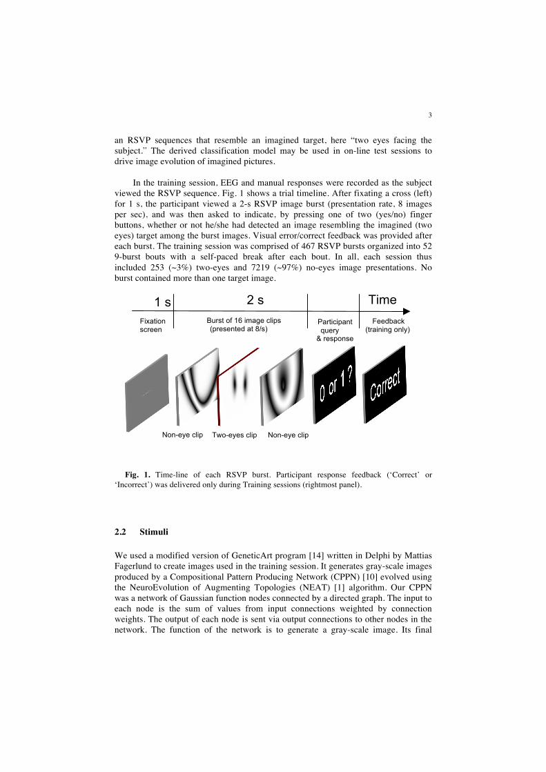
an RSVP sequences that resemble an imagined target, here “two eyes facing the subject.” The derived classification model may be used in on-line test sessions to drive image evolution of imagined pictures. In the training session, EEG and manual responses were recorded as the subject viewed the RSVP sequence. Fig. 1 shows a trial timeline. After fixating a cross (left) for 1 s, the participant viewed a 2-s RSVP image burst (presentation rate, 8 images per sec), and was then asked to indicate, by pressing one of two (yes/no) finger buttons, whether or not he/she had detected an image resembling the imagined (two eyes) target among the burst images. Visual error/correct feedback was provided after each burst. The training session was comprised of 467 RSVP bursts organized into 52 9-burst bouts with a self-paced break after each bout. In all, each session thus included 253 (~3%) two-eyes and 7219 (~97%) no-eyes image presentations. No burst contained more than one target image.
Fig. 1. Time-line of each RSVP burst. Participant response feedback (‘Correct’ or
‘Incorrect’) was delivered only during Training sessions (rightmost panel).
2.2 Stimuli
We used a modified version of GeneticArt program [14] written in Delphi by Mattias Fagerlund to create images used in the training session. It generates gray-scale images produced by a Compositional Pattern Producing Network (CPPN) [10] evolved using the NeuroEvolution of Augmenting Topologies (NEAT) [1] algorithm. Our CPPN was a network of Gaussian function nodes connected by a directed graph. The input to each node is the sum of values from input connections weighted by connection weights. The output of each node is sent via output connections to other nodes in the network. The function of the network is to generate a gray-scale image. Its final
Feedback (training only)
Time Participant query
& response
2 s Burst of 16 image clips (presented at 8/s)
Fixation screen
Non-eye clip Two-eyes clip Non-eye clip
1 s

4
output is a matrix of light intensities at each pixel of the output image. To generate an image, the light intensity at each pixel is calculated by providing values indicating the location of the pixel in the image (x, y values between 0 and 1, and also the distance to the center of the image).
The topology of the CPPN network (its connections, connection weights, and node
types) is evolved by the NEAT algorithm. Each new generation of images is created by applying genetic operators (mating, mutation…) to the individual images (or image pairs) in the previous generation based on their received fitness values. NEAT begins by evolving simple (low node / connection-count) networks, creating more complex networks that result in higher fitness values. By starting the search from a low-dimensional space, NEAT increases the chance of finding a desirable solution by preventing the problems associated with searching in a high dimensional space of many nodes and connection weights. In our case, network starts with simple shapes and can increase the amount of detail in later image generations.
We used two sets of two-eyes and no-eyes images generated by the network in previous behavioral pilot sessions to prepare the stimuli for the training session. To create non-eye images, we ran the GeneticArt program for many generations giving equal fitness values to all images in each generation. This produced thousands of ‘random’ images not shaped by a specific fitness function. We then went through these images and manually deleted any that had a resemblance to the target image. Two-eyes images were evolved by providing appropriate manual feedback to the GeneticArt program. After generating many of these images, we picked a variety of two-eyes images with different shapes for use as targets in the training session. We hypothesized that sudden changes in image size and contrast might induce EEG responses to visual surprise [11]. Therefore, to minimize variations in feature size and contrast we normalized center of mass location and angular momentum magnitude of the images.
Two-second RSVP bursts, each containing 16 (547 by 343 pixel) images, were created using two-eyes and non-eye images. In half of these bursts, an two-eyes image was displayed in the burst at a randomly selected latency between 250 ms and 1750 ms. The 250-ms edge buffers prevented interference in classification from EEG responses to burst edges.
2.3 Experimental Setup
RSVP bursts were presented at 8 Hz using our custom stimulus presentation program. Events corresponding to image display, image type (eye or non-eye), and subject manual responses were recorded synchronously with EEG in a single file. EEG was collected using a BIOSEMI Active View 2 system with 128 electrodes mounted in a whole-head elastic electrode cap (E-Cap, Inc) with a custom near-uniform montage across the scalp, and bony parts of the upper face. Computer data acquisition was performed via USB using a customized acquisition driver at a 512-Hz sampling rate.

5
In the pilot experiment, we recorded one hour of data from one subject (28 years old male).
2.4 Data Preprocessing
Preprocessing of training session data was performed in the Matlab environment (Mathworks, Inc.) using EEGLAB [12] functions and data structures. The scalp channel data were first high-passed using an FIR filter above 2 Hz. Two channels containing no EEG signals were then removed. The rest of the EEG data were then re-referenced from the Biosemi active reference to average reference. EEG data epochs from 1.5 s before burst presentation until 2 s after the end of each 2-s burst were extracted from the continuous EEG and concatenated. Data epochs were then decomposed using extended-infomax ICA [4-6] to obtain 126 maximally independent components (ICs), spatial filters separating EEG sensor data into maximally temporally independent processes, most appearing to predominantly represent the contribution to the scalp data of one brain EEG or non-brain artifact source, respectively. Only epochs with correct subject response (438 out of 467, ~94%) were selected in classifier training and cross-validation.
Fig. 2. Flowchart of the classifier system. Both (left) temporal activity patterns (phase-locking following stimulus onsets) and (right) event-related changes in spectral amplitude are used by the classifier (center). [From N. Bigdely Shamlo et al., IEEE Transactions on Neural Systems and Rehabilitation Engineering, 2008].
6
2.5 Image Classification
We used the methods described previously [2] for classifying images into two-eyes (target) and non-eye like (non-targets). Fig. 2 summarizes the classification process. Information from both time-domain and frequency-domain features of EEG signals following image presentations were exploited. Time-course templates were selected by a novel method that involved, for every IC, performing a second-level deconvolutive ICA decomposition on the time-domain IC activations following image presentations. To do this, for each IC we constructed matrices of size 410 lags by 6570 images by concatenating columns consisting of lagged IC activation time series in the 800 ms following each image presentation. Applying principal component analysis (PCA), we then reduced the rows of this matrix to 50. A second-level ICA was then performed on the dimension-reduced matrix to find 50 independent time course templates (ITCs) for each IC. These features form a natural spatiotemporal decomposition of the EEG signal with low interdependency, making them natural candidate features for an EEG classifier. Time-frequency amplitude ITs (ITAs) were found similarly by decomposing the single-trial spectrograms of the IC activations following image onsets, which were vectorized and concatenated into a large (frequencies × latencies, images) matrix that was reduced to its first 50 principal dimensions by PCA before ICA decomposition. Observing that, as expected, the weights for a number of these time-domain and time-frequency domain dimensions contained robust information for target/non-target image classification, we used the 100 most informative ITs of each domain over all ICs (as determined by computing individual area under the ROC curves for the target/non-target discrimination) as input to a Fisher discriminant linear classifier. For time-frequency domain features, we used the frequency-domain independent template weights for each image as input. For time-domain features, we used columns of the transpose of the second-level ICA mixing matrix as matched filters (Fig. 3) since in preliminary testing this gave slightly better results than using the decomposed IC activations themselves.
3 Results
After preprocessing the data and extracting classification features as described above, we performed a ten-fold cross-validation on them. Using only the most informative 100 time-domain features in each fold, an area under the ROC curve of 0.96 was achieved for this subject. Ten-fold cross-validation results for frequency-domain features alone (the top 100 in each validation fold) produced an area under the ROC curve of 0.97. Maximum performance was achieved when posterior probabilities of being a target obtained from the time-domain and frequency-domain classifiers in each validation fold were combined using naive Bayesian fusion (i.e., the two probabilities were multiplied). In this case, the area under the ROC curve was 0.98.

7
4 Discussion
4.1 The Proposed Mind-Mirror System
A training session is the first step in creating the proposed Mind-Mirror system. The second half of the system development involves using classifier output online for image evolution. Based on our initial RSVP studies [2], we hypothesized that the subject-specific classifier trained using a certain image target can likely be applied to online evolution of other image targets. Thus, although the Mind-Mirror classifier reported here was trained on an ‘two-eyes’ target, we predict during test sessions it may correctly detect an EEG ‘target recognition’ activity pattern in sessions using different target images, for example a car, house, or butterfly.
Fig. 3. Schematic of the proposed Mind-Mirror system in intended operation. Here, the subject is imagining an image of a sports car. EEG target-recognition signals fed to the image evolution algorithm from the real-time EEG classifier continually shape the evolution of the image sequence to resemble more and more closely the subject’s imagined car image. In the test session, the subject observes RSVP image bursts produced by the image evolution algorithm. If subject detects an image with some resemblance to imagined picture, it will be reflected in a high target-probability value output of the EEG classification algorithm. These values are then sent to the image evolution program to evolve a new generation of images, which are then displayed in the next RSVP burst. This establishes a feedback loop, shown in Fig. 3 (right), that amplifies image features resembling the imagined target picture. After the subject views many such bursts, the system may consistently generate a ‘mirror image’ of the image the subject has been mentally imagining. By eliminating the need for subject manual feedback in the Mind-Mirror system, its speed and user convenience might be increased.
4.2 EEG ‘flickers’ of target recognition
Both time and time/frequency domains contain information regarding EEG signatures of target detection [2]. To investigate the information content of sources used in the classifier, we calculated the tenfold cross-validation area under the ROC
Image Evolution Real-time Classifier EEG Acquisition Stimulus Presentation
Disk
Evolved clips
Test session

8
curve for individual ICs. All the independent templates (ITs) for each IC were used as described in Fig. 2. Equivalent dipole sources for ICs were then localized using a spherical four-shell (BESA) head model and components with a residual variance of the scalp projection from the equivalent dipole model below 20% were selected. Fig. 3 shows these dipoles in the MNI head space. The size and color of each dipole reflect its amount of information relevant to target detection, measured by area under the ROC of the single-IC classifiers. Brodmann areas associated with the IC dipole locations were found [13] and are displayed below for the most informative ICs.
Fig. 3. Equivalent dipoles of subject ICs with low (< 0.2) dipole model residual variance. The color and size of each dipole sphere indicates the amount of information about target classification in the IC activations. Numbers on the most informative (red-yellow) dipole markers indicate the Brodmann area (BA) number most likely associated with this equivalent dipole location.
The most posterior IC (with a dual dipole model) was located in or near BA19 (bilateral occipital cortex). Activity in these areas has been associated in functional brain imaging experiments with higher order visual processing involving motion sensing and object shape recognition [16]. Two other most informative ICs were located in/near right precuneus (BA31) associated with visual attentive processes, including shifts of attention between object features [15], and in the right superior parietal lobule (BA7) associated with matching rotated objects to some visual template [17].

9
References
1. Stanley, K. O.: Evolving Neural Networks through Augmenting Topologies. Evolutionary Computation, vol. 10(2), pp. 99-. (2002)
2. Bigdely-Shamlo, N., Vankov, A., Ramirez, R., Makeig, S.: Brain Activity-Based Image Classification From Rapid Serial visual Presentation. IEEE Transactions on Neural Systems and Rehabilitation Engineering,, vol. 16, no 4 (2008)
3. Lee, T., Girolami, M., Sejnowski, T. J.: Independent Component Analysis Using an Extended Infomax Algorithm for Mixed Subgaussian and Supergaussian Sources. Neural Computation, vol. 11, no. 2, pp. 417--441 (Feb. 1999)
4. Bell, A., Sejnowski, T.: An Information Maximization Approach to Blind Separation and Blind Deconvolution. Neural Computation, vol. 7, pp. 1129--1159 (July 1995)
5. Basa, T., Go, C.,Yoo, K., Lee, W.: Using Physiological Signals to Evolve Art., LNCS, vol. 3907, pp. 633--641 (2006)
6. Secretan, J., Beato, N., D'Ambrosio, D. B., Rodriguez, A., Campbell, A., Stanley, K. O.: Picbreeder: Evolving Pictures Collaboratively Online. Proc. Computer Human Interaction Conf. (CHI), New York, NY, ACM, 10 pages (2008)
7. Sajda, P., Gerson, A., Parra, L.: High-throughput image search via single-trial event detection in a rapid serial visual presentation task. In: Proc. 1st Inter. IEEE EMBS Conf. on Neural Engineering, Capri Island, Italy (2003)
8. Gerson A., Parra L., Sajda P.: Cortically-coupled computer vision for rapid image search. IEEE Transactions on Neural Systems and Rehabilitation Engineering, vol. 14 (2), pp. 174--179 (2006)
9. Parra, L.C., Christoforou, C., Gerson, A. D., Dyrholm, M., Luo, A., Wagner, M., Philiastides, M. G., Sajda, P.: Spatio-temporal linear decoding of brain state: Application to performance augmentation in high-throughput tasks. IEEE Signal Processing Magazine, vol. 25, no. 1, pp. 95--115 (Jan. 2008)
10. Stanley, K. O.: Exploiting Regularity Without Development. Proc. AAAI Fall Symposium on Developmental Systems, Menlo Park, CA, AAAI Press, 8 pages (2006)
11. Einhuser, W., Mundhenk, T. N., Baldi, P., Koch, C., Itti, L.: A bottom-up model of spatial attention predicts human error patterns in rapid scene recognition. J. of Vision, vol.7, no.10, pp.1--13 (2007)
12. Delorme, A., Makeig, S.: EEGLAB: an open source toolbox for analysis of single-trial EEG dynamics including independent component analysis. J. of Neuroscience Methods, vol. 134 (1), p. 9, http://sccn.ucsd.edu/eeglab
13. Talairach Client, http://www.talairach.org/client.html 14. DelphiNEAT GeneticArt program, http://www.mattiasfagerlund.com/DelphiNEAT/ 15. Cavanna, A.E., Trimble, M.R.: The precuneus: a review of its functional anatomy and
behavioural correlates. Brain 129 (2006) 564-583 16. Worden, M.S., Foxe, J.J., Wang, N., Simpson, G.V.: Anticipatory biasing of visuospatial
attention indexed by retinotopically specific alpha-band electroencephalography increases over occipital cortex. J Neurosci 20 (2000) RC63
17. Harris, I. M., Egan, G. F., Sonkkila, C., Tochon-Danguy, H. J., Paxinos, G. Watson, J. D. G., Selective right parietal lobe activation during mental rotation: A parametric PET study. Brain, Vol. 123, (2000) 65-73.