Tian Xian Liquid (TXL) induces apoptosis in HT29 colon cancer cell in vitro and inhibits tumor...
7
RESEARCH Open Access Tian Xian Liquid (TXL) induces apoptosis in HT-29 colon cancer cell in vitro and inhibits tumor growth in vivo Qing Liu 1 , Yao Tong 1* , Stephen Cho Wing Sze 1 , Wing Keung Liu 2 , Lam Lam 1 , Ellie Shihng Meir Chu 1 , Christine Miu Ngan Yow 3 Abstract Background: Tian Xian Liquid (TXL) is a Chinese medicine decoction and has been used as an anticancer dietary supplement. The present study aims to investigate the effects of TXL on the apoptosis of HT-29 cells and tumor growth in vivo. Method: HT-29 colon cancer cells were treated with gradient dilution of TXL. The mitochondrial membrane potential was measured by JC-1 assay. The release of cytochrome c from mitochondrial and apoptosis-related proteins Bax, Bcl-2, cleaved caspase-3, 9 were examined by Western blot analysis. HT-29 cells were implanted in nude mice to examine the effects of TXL on tumor growth. Result: TXL inhibited HT-29 xenografted model and showed a strong and dose-dependent inhibitory effect on the proliferation of HT-29 cells. Mitochondrial membrane potential was reduced by TXL at the concentration of 0.5% above. For Western blot analysis, an increase in Bax expression and a decrease in Bcl-2 expression were observed in TXL-treated cells. TXL treatment increased the protein level of cleaved casepase-3 and caspase-9, and the release of cytochrome c in cytoplasm was up-regulated as well. Conclusion: TXL significantly inhibits cell proliferation in the HT-29 cells and HT-29 xenografted model via the mitochondrial cell death pathway. Background Colorectal carcinoma increased up to four folds in the past decade and the mortality is rising [1]. Much pro- gress has been achieved in alternative medicine [2] such as Chinese medicine. Most methods of chemotherapy for cancer induce cancer cell apoptosis. Excessive apoptosis causes hypo- trophy such as ischemic damage whereas insufficient apoptosis leads to uncontrolled cell proliferation such as cancer [3]. Chemotherapeutic agents may cause mito- chondrial dysfunction leading to depolarization of the inner mitochondrial membrane potential ( Δψm) [4], triggering the caspases cascade by releasing several cas- pase activators. Among them, cytochrome c activates caspases by forming a complex with Apaf-1 and procaspase-9, thereby triggering caspase-9 activation which subsequently cleaves the effector caspase-3[5,6]. Tian Xian Liquid (TXL), an aqueous extraction of Chi- nese medicinal herbs including Radix Ginseng, Cordyceps, Radix Astragali, Radix Glycyrrhizae, Rhizoma Dioscorea, Margarita, Fructus Lycii, Ganoderma, Fructus Ligustri Lucidi, Herba Scutellariae Barbatae, has been used as an anticancer dietary supplement for more than a decade [7]. Previous experiments reported that TXL had inhibitory effects on human cervical carcinoma C-33A cells and human lung carcinoma H1299 cells[7]. The present study aims to investigate the effects of TXL on the apoptosis of HT-29 cells and tumor growth in vivo. Methods Cell culture Human colon cancer cell HT-29 (ATCC® Number:HTB- 38™) was obtained from the American Type Culture * Correspondence: [email protected] 1 School of Chinese Medicine, Li Ka Shing Faculty of Medicine, University of Hong Kong, Pokfulam, Hong Kong SAR, China Liu et al. Chinese Medicine 2010, 5:25 http://www.cmjournal.org/content/5/1/25 © 2010 Liu et al; licensee BioMed Central Ltd. This is an Open Access article distributed under the terms of the Creative Commons Attribution License (http://creativecommons.org/licenses/by/2.0), which permits unrestricted use, distribution, and reproduction in any medium, provided the original work is properly cited.
-
Upload
independent -
Category
Documents
-
view
3 -
download
0
Transcript of Tian Xian Liquid (TXL) induces apoptosis in HT29 colon cancer cell in vitro and inhibits tumor...

RESEARCH Open Access
Tian Xian Liquid (TXL) induces apoptosis in HT-29colon cancer cell in vitro and inhibits tumorgrowth in vivoQing Liu1, Yao Tong1*, Stephen Cho Wing Sze1, Wing Keung Liu2, Lam Lam1, Ellie Shihng Meir Chu1,Christine Miu Ngan Yow3
Abstract
Background: Tian Xian Liquid (TXL) is a Chinese medicine decoction and has been used as an anticancer dietarysupplement. The present study aims to investigate the effects of TXL on the apoptosis of HT-29 cells and tumorgrowth in vivo.
Method: HT-29 colon cancer cells were treated with gradient dilution of TXL. The mitochondrial membranepotential was measured by JC-1 assay. The release of cytochrome c from mitochondrial and apoptosis-relatedproteins Bax, Bcl-2, cleaved caspase-3, 9 were examined by Western blot analysis. HT-29 cells were implanted innude mice to examine the effects of TXL on tumor growth.
Result: TXL inhibited HT-29 xenografted model and showed a strong and dose-dependent inhibitory effect on theproliferation of HT-29 cells. Mitochondrial membrane potential was reduced by TXL at the concentration of 0.5%above. For Western blot analysis, an increase in Bax expression and a decrease in Bcl-2 expression were observed inTXL-treated cells. TXL treatment increased the protein level of cleaved casepase-3 and caspase-9, and the release ofcytochrome c in cytoplasm was up-regulated as well.
Conclusion: TXL significantly inhibits cell proliferation in the HT-29 cells and HT-29 xenografted model via themitochondrial cell death pathway.
BackgroundColorectal carcinoma increased up to four folds in thepast decade and the mortality is rising [1]. Much pro-gress has been achieved in alternative medicine [2] suchas Chinese medicine.Most methods of chemotherapy for cancer induce
cancer cell apoptosis. Excessive apoptosis causes hypo-trophy such as ischemic damage whereas insufficientapoptosis leads to uncontrolled cell proliferation such ascancer [3]. Chemotherapeutic agents may cause mito-chondrial dysfunction leading to depolarization of theinner mitochondrial membrane potential (Δψm) [4],triggering the caspases cascade by releasing several cas-pase activators. Among them, cytochrome c activatescaspases by forming a complex with Apaf-1 and
procaspase-9, thereby triggering caspase-9 activationwhich subsequently cleaves the effector caspase-3[5,6].Tian Xian Liquid (TXL), an aqueous extraction of Chi-
nese medicinal herbs including Radix Ginseng, Cordyceps,Radix Astragali, Radix Glycyrrhizae, Rhizoma Dioscorea,Margarita, Fructus Lycii, Ganoderma, Fructus LigustriLucidi, Herba Scutellariae Barbatae, has been used as ananticancer dietary supplement for more than a decade [7].Previous experiments reported that TXL had inhibitoryeffects on human cervical carcinoma C-33A cells andhuman lung carcinoma H1299 cells[7]. The present studyaims to investigate the effects of TXL on the apoptosis ofHT-29 cells and tumor growth in vivo.
MethodsCell cultureHuman colon cancer cell HT-29 (ATCC® Number:HTB-38™) was obtained from the American Type Culture
* Correspondence: [email protected] of Chinese Medicine, Li Ka Shing Faculty of Medicine, University ofHong Kong, Pokfulam, Hong Kong SAR, China
Liu et al. Chinese Medicine 2010, 5:25http://www.cmjournal.org/content/5/1/25
© 2010 Liu et al; licensee BioMed Central Ltd. This is an Open Access article distributed under the terms of the Creative CommonsAttribution License (http://creativecommons.org/licenses/by/2.0), which permits unrestricted use, distribution, and reproduction inany medium, provided the original work is properly cited.
Collection (ATCC, USA) and cultured in RPMI 1640(Hyclone, USA) supplemented with fetal bovine serum(10%), penicillin (100 units/ml) and streptomycin(100 mg/ml) (Hyclone, USA) in a humidified incubator(37°C) containing 95% air and 5% CO2. Trypsin(Hyclone, USA) was used for trypsination.
Preparation of TXLTian Xian Liquid (TXL) (Batch number: L2-171040) wasprovided by China-Japan Feida Union Company Ltd.and stored away from light at 4°C. TXL was diluted andincorporated into the cell culture medium RPMI 1640.Residues were removed by filtration.
Cell proliferationCell proliferation was assessed in vitro with 3-(4,5-Dimethylthiazol-2-yl)-2,5-Diphenyltetrazolium Bromide(MTT) according to the manufacturer’s protocol (Roche,USA). HT-29 cells (10000 per well) were incubated intriplicates in a 96-well plate. TXL was serially dilutedwith RPMI1640 and the final concentrations were 0.25,0.5, 1, 2 and 5%. The plates were incubated with orwithout TXL for 24 and 48 hours. At the end of theincubation, cells were exposed to MTT (10 μL, 5 mg/mL in phosphate-buffered saline) in culture medium forfour hours at 37°C. The supernatant was removed and150 μL DMSO (Sigma, USA) was added to dissolve theformazan crystals. The absorbance was measured at595 nm with an ELISA plate reader (Bio-Rad, USA).
DAPI stainingDAPI (Sigma, USA) (4’ 6-diamidino 2-phenylindole)-stained nuclei were observed with fluorescence micro-scopy. HT-29 cells (70-80% confluent) in 24-welluncoated plates were exposed to 0.5% and 1% TXL for24 hours respectively. Cells were fixed with 4% parafor-maldehyde for 30 minutes and incubated with 1 μg/mLDAPI solution for 30 minutes in the dark. Stained cellswere imaged under a fluorescence microscope (CarlZeiss, Germany).
Assessment of apoptosis by determination ofmitochondrial membrane potentialMitochondrial membrane potential was assessed by 5, 5’,6, 6’-tetrachloro-1, 1’, 3, 3’tetraethylbenzimidazolylcarbo-cyanine iodide (JC-1) according to the manufacturer’sprotocol (Biotium, USA). After trypsinization and centri-fugation (500× g)(Eppendorf, Germany) for ten minutesat room temperature, the pellets of cell culture with orwithout TXL were re-suspended in RPMI 1640 medium(1 ml), stained with 5 mg/ml JC-1 for 30 minutes at37°C in the dark, washed twice in phosphate bufferedsaline (PBS) and re-suspended in 0.5 ml PBS. Δψmdepletion was observed under a fluorescence
microscope. A green filter was used for green-fluores-cent monomer at depolarized membrane potentials anda red filter for orange-fluorescent J-aggregate at hyper-polarized membrane potentials.To measure the quantitative change of mitochondrial
potential, we applied JC-1 with fluorescence platereader. Briefly, cells (1 × 10⁵) in 100 μl culture medium/well were seeded in black 96-well plate (Nunc, Den-mark) and treated with TXL (0.15, 0.3, 0.6, 1.25 and2.5%). After 24 and 48 hours incubation, JC-1 (5 μg/ml)was added for the last 30 minutes of treatment. Cellswere washed twice with PBS to remove unbound dye.The concentration of retained JC-1 dye was measured(490 nm excitation/600 nm emission) with a lumines-cence spectrometer (PerkinElmer, USA).
Western blotThe HT29 cells were incubated with increasing concen-trations of TXL (0, 0.5%, 0.75%, 1%) for 48 hours. Forthe time-course experiment, HT29 were treated with 1%TXL for 12, 24, or 48 hours. Cellular levels of cleavedcaspase-3, 9 (Cell Signaling Technology, USA) Bax/Bcl-2cytochrome C and glyceraldehyde 3-phosphate dehydro-genase (GAPDH) (Santa Cruz Biotechnology, USA) weredetermined by Western blot. Lysates were preparedfrom 1 × 10⁷ cells by dissolving cell pellets in 100 μl oflysis buffer. Lysates were centrifuged (Eppendorf, Ger-many) at 18000× g for 15 minutes and the supernatantwas collected. The protein concentration was estimatedwith the Bio-Rad protein assay kit (Bio-Rad, USA) usingbovine serum albumin as a standard. Sample proteinswere resolved by 10% sodium dodecylsulfate polyacryla-mide gel (Bio-Rad, USA) electrophoresis and then elec-trophoretically transferred to PVDF membrane(Millipore, USA) and blocked with 5% BSA (Sigma,USA). Subsequently the primary antibodies caspase-9,cleaved caspase3, Bax, Bcl-2, cytochrome C andGAPDH were added. After overnight incubation at 4°Cthe blots were washed, exposed to HRP-conjugated cor-responding secondary antibodies for one hour andfinally were visualized by ECL Advanced Solution (GEHealthcare Life Sciences, USA). Digital images were cap-tured by Gel Doc™ gel documentation system (Bio-Rad,USA) and intensity was quantified using Quantity-Onesoftware version 4.62(Bio-Rad, USA).
In vivo tumor-growth inhibition studiesThe experiment was approved by the Department ofHealth, Hong Kong SAR, China and the Committee onthe Use of Live Animals in Teaching and Research(CULATR) of Li Ka Shing Faculty of Medicine, Univer-sity of Hong Kong. Six-week-old female nude mice werepurchased from the Laboratory Animal Unit, Universityof Hong Kong and kept under sterile conditions in
Liu et al. Chinese Medicine 2010, 5:25http://www.cmjournal.org/content/5/1/25
Page 2 of 7
accordance with the institutional guidelines of animalcare. The HT-29 carcinoma was established in nudemice by injecting the suspensions of HT-29 (1 × 10⁶
cells per animal) [8] cells subcutaneously into the rightflank of each animal. When the tumors became palpable(size: 18 mm3) after xenografting, mice were dividedinto three groups (n = 8) by a random numbered table:(1) Control group orally administered with 200 μl PBS);(2) 5-fluorouracil (5-FU) (Choongwae, Korea) group(injected intraperitoneally with 5-FU, 30 mg per kg ofbody weight) three times a week [9,10]; (3) TXL group(orally administered with 200 μl TXL daily for 14 days.To evaluate the antitumor activity of TXL, we measuredthe tumor volume with a digital caliper six times everyweek (from day1 to day 6 and from day 8 to day 14)and calculated using the formula: (longest diameter) ×(shortest diameter)2 × 0.5. The body weights of all ani-mals were recorded throughout the experiment to assessdrug toxicity.
Statistical analysisData were presented as mean and standard deviation(SD). When one-way ANOVA showed significant differ-ences among groups, Tukey’s post hoc test was used todetermine the specific pairs of groups that were statisti-cally different. A level of P < 0.05 was considered statis-tically significant. Analysis was performed with thesoftware SPSS version 16.0 (SPSS Inc, USA).
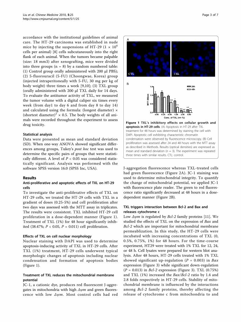
ResultsAnti-proliferative and apoptotic effects of TXL on HT-29cellsTo investigate the anti-proliferative effects of TXL onHT-29 cells, we treated the HT-29 cells with TXL in agradient of doses (0.25-5%) and cell proliferation aftertwo days was assessed with the MTT assay in triplicates.The results were consistent. TXL inhibited HT-29 cellproliferation in a dose-dependent manner (Figure 1).Treatment of TXL (1%) for 48 hour significantly inhib-ited (38.47%; P < 0.05, P = 0.011) cell proliferation.
Effects of TXL on cell nuclear morphologyNuclear staining with DAPI was used to determineapoptosis-inducing activity of TXL in HT-29 cells. AfterTXL (1%) treatment, HT-29 cells underwent typicalmorphologic changes of apoptosis including nuclearcondensation and formation of apoptosis bodies(Figure 1).
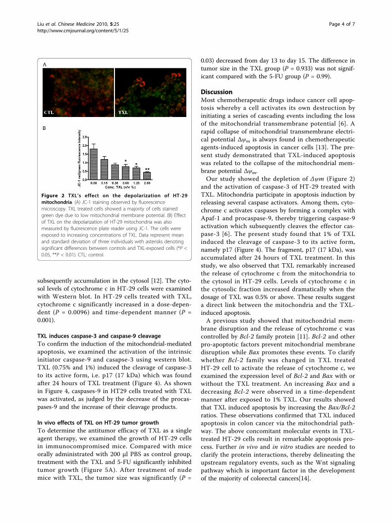
Treatment of TXL reduces the mitochondrial membranepotentialJC-1, a cationic dye, produces red fluorescent J-aggre-gates in mitochondria with high Δψm and green fluores-cence with low Δψm. Most control cells had red
J-aggregation fluorescence whereas TXL-treated cellshad green fluorescence (Figure 2A). JC-1 staining wasused to determine mitochondrial integrity. To quantifythe change of mitochondrial potential, we applied JC-1with fluorescence plate reader. The green to red fluores-cence ratio significantly decreased at 48 hours in a dose-dependent manner (Figure 2B).
TXL triggers interaction between Bcl-2 and Bax andreleases cytochrome cLow Δψm is regulated by Bcl-2 family proteins [11]. Westudied the effects of TXL on the expression of Bax andBcl-2 which are important for mitochondrial membranepermeablization. In this study, the HT-29 cells wereincubated with increasing concentrations of TXL (0,0.5%, 0.75%, 1%) for 48 hours. For the time-courseexperiment, HT29 were treated with 1% TXL for 12, 24,or 48 h. Cell lysates were prepared for western blot ana-lysis. After 48 hours, HT-29 cells treated with 1% TXLshowed significant up-regulation (P = 0.003) in Baxexpression (Figure 3) while significant down-regulation(P = 0.013) in Bcl-2 expression (Figure 3). TXL (0.75%)and TXL (1%) increased the Bax/Bcl-2 ratio by 1.4 and2.8 folds respectively in HT-29 cells. Stability of mito-chondrial membrane is influenced by the interactionsamong Bcl-2 family proteins, thereby affecting therelease of cytochrome c from mitochondria to and
Figure 1 TXL’s inhibitory effects on cellular growth andapoptosis in HT-29 cells. (A) Apoptosis in HT-29 after TXLtreatment for 48 hours was determined by staining the cell withDAPI. Apoptotic cell exhibiting characteristic chromatincondensation were observed by fluorescence microscopy. (B) Cellproliferation was assessed after 24 and 48 hours with the MTT assayas described in Methods. Results (optical densities) are expressed asmean and standard deviation (n = 3). The experiment was repeatedthree times with similar results. CTL: control.
Liu et al. Chinese Medicine 2010, 5:25http://www.cmjournal.org/content/5/1/25
Page 3 of 7
subsequently accumulation in the cytosol [12]. The cyto-sol levels of cytochrome c in HT-29 cells were examinedwith Western blot. In HT-29 cells treated with TXL,cytochrome c significantly increased in a dose-depen-dent (P = 0.0096) and time-dependent manner (P =0.001).
TXL induces caspase-3 and caspase-9 cleavageTo confirm the induction of the mitochondrial-mediatedapoptosis, we examined the activation of the intrinsicinitiator caspase-9 and casapse-3 using western blot.TXL (0.75% and 1%) induced the cleavage of caspase-3to its active form, i.e. p17 (17 kDa) which was foundafter 24 hours of TXL treatment (Figure 4). As shownin Figure 4, caspases-9 in HT29 cells treated with TXLwas activated, as judged by the decrease of the procas-pases-9 and the increase of their cleavage products.
In vivo effects of TXL on HT-29 tumor growthTo determine the antitumor efficacy of TXL as a singleagent therapy, we examined the growth of HT-29 cellsin immunocompromised mice. Compared with miceorally administrated with 200 μl PBS as control group,treatment with the TXL and 5-FU significantly inhibitedtumor growth (Figure 5A). After treatment of nudemice with TXL, the tumor size was significantly (P =
0.03) decreased from day 13 to day 15. The difference intumor size in the TXL group (P = 0.933) was not signif-icant compared with the 5-FU group (P = 0.99).
DiscussionMost chemotherapeutic drugs induce cancer cell apop-tosis whereby a cell activates its own destruction byinitiating a series of cascading events including the lossof the mitochondrial transmembrane potential [6]. Arapid collapse of mitochondrial transmembrane electri-cal potential Δψm is always found in chemotherapeuticagents-induced apoptosis in cancer cells [13]. The pre-sent study demonstrated that TXL-induced apoptosiswas related to the collapse of the mitochondrial mem-brane potential Δψm.Our study showed the depletion of Δψm (Figure 2)
and the activation of caspase-3 of HT-29 treated withTXL. Mitochondria participate in apoptosis induction byreleasing several caspase activators. Among them, cyto-chrome c activates caspases by forming a complex withApaf-1 and procaspase-9, thereby triggering caspase-9activation which subsequently cleaves the effector cas-pase-3 [6]. The present study found that 1% of TXLinduced the cleavage of caspase-3 to its active form,namely p17 (Figure 4). The fragment, p17 (17 kDa), wasaccumulated after 24 hours of TXL treatment. In thisstudy, we also observed that TXL remarkably increasedthe release of cytochrome c from the mitochondria tothe cytosol in HT-29 cells. Levels of cytochrome c inthe cytosolic fraction increased dramatically when thedosage of TXL was 0.5% or above. These results suggesta direct link between the mitochondria and the TXL-induced apoptosis.A previous study showed that mitochondrial mem-
brane disruption and the release of cytochrome c wascontrolled by Bcl-2 family protein [11]. Bcl-2 and otherpro-apoptotic factors prevent mitochondrial membranedisruption while Bax promotes these events. To clarifywhether Bcl-2 family was changed in TXL treatedHT-29 cell to activate the release of cytochrome c, weexamined the expression level of Bcl-2 and Bax with orwithout the TXL treatment. An increasing Bax and adecreasing Bcl-2 were observed in a time-dependentmanner after exposed to 1% TXL. Our results showedthat TXL induced apoptosis by increasing the Bax/Bcl-2ratios. These observations confirmed that TXL inducedapoptosis in colon cancer via the mitochondrial path-way. The above concomitant molecular events in TXL-treated HT-29 cells result in remarkable apoptosis pro-cess. Further in vivo and in vitro studies are needed toclarify the protein interactions, thereby delineating theupstream regulatory events, such as the Wnt signalingpathway which is important factor in the developmentof the majority of colorectal cancers[14].
Figure 2 TXL’s effect on the depolarization of HT-29mitochondria. (A) JC-1 staining observed by fluorescencemicroscopy. TXL treated cells showed a majority of cells stainedgreen dye due to low mitochondrial membrane potential. (B) Effectof TXL on the depolarization of HT-29 mitochondria was alsomeasured by fluorescence plate reader using JC-1. The cells wereexposed to increasing concentrations of TXL. Data represent meanand standard deviation of three individuals with asterisks denotingsignificant differences between controls and TXL-exposed cells (*P <0.05, **P < 0.01). CTL: control.
Liu et al. Chinese Medicine 2010, 5:25http://www.cmjournal.org/content/5/1/25
Page 4 of 7

Figure 3 Effect of TXL on cytochrome c. Total protein from HT-29 treated with 1%TXL for 12, 24 and 48 hours (A) or indicated concentrationof TXL for 48 hours (B) were analyzed by Western blot with specific antibodies against Bax and Bcl-2. Protein from cytosolic fraction of HT-29which has been treated with TXL (1%) for 12, 24 and 48 hours (C) or indicated concentration of TXL for 48 hours (D) were analyzed by Westernblot with specific antibodies against cytochrome c. GAPDH antibody was used as control for equal loading. The relative expressions of proteinswere quantified using Bio-Rad Quatity-One software. Results are expressed as mean and standard deviation (n = 3),*P < 0.05 **P <0.01compared with control group.
Figure 4 TXL Induces caspase-3 and caspase-9 cleavage. Protein from HT-29 which has been treated with 1%TXL for 12, 24 and 48 hours (A)and protein from HT-29 which has been treated with indicated concentration of TXL for 48 hours (B) were analyzed by Western blot withspecific antibodies against caspase-9 and cleaved caspase-3. GAPDH antibody was used as control for equal loading. The relative expressions ofproteins were quantified using Bio-Rad Quatity-One software. Results are expressed as means and standard deviation (n = 3),*P < 0.05 **P < 0.01compared with control group.
Liu et al. Chinese Medicine 2010, 5:25http://www.cmjournal.org/content/5/1/25
Page 5 of 7

We studied TXL’s effects on the growth of HT-29 celllines grown in vitro and compared those results with itseffect on tumor growth in vivo. We found that TXLattenuated the growth of xenografted HT-29 tumors invivo. The injection of 5-fluorouracil (5-FU), a commonchoice for single-agent chemotherapy of advanced coloncancer [15,16] on nude mice significantly attenuatedtumor growth (Figure 5A). However, during the 5-FUtreatment, the body weight of nude mice was signifi-cantly decreased on day 10 (P < 0.05, P = 0.038) (Figure5B). The components of TXL such as Radix Ginseng,Cordyceps, Radix Astragali, Fructus Lycii, Ganodermaare commonly used in China as immune-stimulatingagents. Whether TXL’s immunomodulation effect andprotection effect on stomach and digestive system redu-cing toxic side effects of 5-FU will be investigated in thefuture.Because 5-fluorouracil (5-FU) as common choice for
single-agent chemotherapy of advanced colon cancer isalso recognized for its toxicity including fatigue, diarrheaand sometimes myelosuppression, much attention hasbeen focused on exploring complementary and alterna-tive medicine. This study may provide a platform forevaluation the function of Chinese medicine decoctionson treatment of cancer, which has no significant sideeffect. To further evaluate the potential of TXL as anadjuvant agent in colon cancer chemotherapy, we arestudying TXL’s effect on attenuation of 5-FU-inducedside effect and the synergistic anti-tumor effect of the5-FU/TXL.
ConclusionTXL significantly inhibits cell proliferation in the HT-29cells and HT-29-bearing mouse model. TXL-inducedapoptosis is likely achieved through the mitochondrialcell death pathway as indicated by a reduction in mito-chondrial membrane potential, and the decrease of Bcl-2/Bax ratio and the release of cytochrome c followed bythe activation of caspase-3 and caspase-9.
AbbreviationsTXL: Tian Xian Liquid; 5-FU: 5-fluorouracil; Δψm: mitochondrial membranepotential; MTT: 3-(4,5-Dimethylthiazol-2-yl)-2,5-Diphenyltetrazolium Bromide;DAPI: 4’ 6-diamidino 2-phenylindole; JC-1: 5, 5’, 6, 6’-tetrachloro-1, 1’, 3,3’tetraethylbenzimidazolylcarbocyanine iodide; PBS: phosphate bufferedsaline; SD: standard deviation; GAPDH: glyceraldehyde 3-phosphatedehydrogenase.
AcknowledgementsThis research was supported by a grant from Seed Funding Programme forApplied Research (200807160015), Small Project Funding (200807176239),the University of Hong Kong and the contract research funding from China-Japan Feida Union Company Ltd.
Author details1School of Chinese Medicine, Li Ka Shing Faculty of Medicine, University ofHong Kong, Pokfulam, Hong Kong SAR, China. 2School of BiomedicalSciences, Faculty of Medicine, Chinese University of Hong Kong, Shatin, NT,Hong Kong SAR, China. 3Medical Laboratory Science Section, Department ofHealth Technology and Informatics, Hong Kong Polytechnic University, HungHom, Hong Kong SAR, China.
Authors’ contributionsQL performed the experiments, analyzed data and drafted the manuscript.YT and SCWS designed the study and revised the manuscript. ESMC andLL conducted the in vivo experiments. WKL designed the in vivoexperiment and prepared the human colon cancer cells. CMNY designedthe in vitro experiment. All authors read and approved the finalmanuscript.
Competing interestsThis research has received a grant from China-Japan Feida UnionCompany Ltd.
Received: 13 February 2010 Accepted: 21 July 2010Published: 21 July 2010
References1. Sung JJ, Lau JY, Goh KL, Leung WK: Increasing incidence of colorectal
cancer in Asia: implications for screening. Lancet Oncol 2005, 6:871-876.2. Nelson PS, Montgomery B: Unconventional therapy for prostate cancer:
good, bad or questionable? Nature Rev 2003, 3:845-858.3. Thompson CB: Apoptosis in the pathogenesis and treatment of disease.
Science 1995, 267:1456-1462.4. Zamzami N, Marchetti P, Castedo M, Decaudin D, Macho A, Hirsch T,
Susin SA, Petit PX, Mignotte B, Kroemer G: Sequential reduction ofmitochondrial transmembrane potential and generation of reactiveoxygen species in early programmed cell death. J Exp Med 1995,182:367-377.
5. Gamen S, Anel A, Pineiro A, Naval J: Caspases are the main executionersof Fas-mediated apoptosis, irrespective of the ceramide signallingpathway. Cell Death Differ 1998, 5:241-249.
Figure 5 Effect of TXL on tumor volume (A) and body weight (B) in HT-29-bearing mouse model. HT-29 cells were injected in nude miceas described in Methods. A total of 24 mice were divided into three groups. Data are expressed as mean and standard deviation (n = 8).Significantly difference compared with control group:*+ P < 0.05, **++ P < 0.01.
Liu et al. Chinese Medicine 2010, 5:25http://www.cmjournal.org/content/5/1/25
Page 6 of 7
6. Elmore S: Apoptosis: a review of programmed cell death. Toxicol Pathol2007, 35:495-516.
7. Sun A, Chia JS, Chiang CP, Hsuen SP, Du JL, Wu CW, Wang WB: Thechinese herbal medicine Tien-Hsien liquid inhibits cell growth andinduces apoptosis in a wide variety of human cancer cells. J AlternComplement Med 2005, 11:245-256.
8. Ninomiya I, Terada I, Yoshizumi T, Takino T, Nagai N, Morita A, Fushida S,Nishimura G, Fujimura T, Ohta T, Miwa K: Anti-metastatic effect ofcapecitabine on human colon cancer xenografts in nude mouse rectum.Int J Cancer 2004, 112:135-142.
9. Wada H, Nagano H, Yamamoto H, Arai I, Ota H, Nakamura M,Damdinsuren B, Noda T, Marubashi S, Miyamoto A, et al: Combinationtherapy of interferon-alpha and 5-fluorouracil inhibits tumorangiogenesis in human hepatocellular carcinoma cells by regulatingvascular endothelial growth factor and angiopoietins. Oncol Rep 2007,18:801-809.
10. Tai IT, Dai M, Owen DA, Chen LB: Genome-wide expression analysis oftherapy-resistant tumors reveals SPARC as a novel target for cancertherapy. J Clin Invest 2005, 115:1492-1502.
11. Cory S, Adams JM: The Bcl2 family: regulators of the cellular life-or-deathswitch. Nature Rev 2002, 2:647-656.
12. Gross A, McDonnell JM, Korsmeyer SJ: BCL-2 family members and themitochondria in apoptosis. Genes Dev 1999, 13:1899-1911.
13. Sen S, D’Incalci M: Apoptosis. Biochemical events and relevance tocancer chemotherapy. FEBS Lett 1992, 307:122-127.
14. Morin P, Sparks A, Korinek V, Barker N, Clevers H, Vogelstein B, Kinzler K:Activation of beta-catenin-Tcf signaling in colon cancer by mutations inbeta-catenin or APC. Science 1997, 275:1787-1790.
15. Wilke HJ, Van Cutsem E: Current treatments and future perspectives incolorectal and gastric cancer. Ann Oncol 2003, 14(Suppl 2):ii49-55.
16. Louvet C, Andre T, Tigaud JM, Gamelin E, Douillard JY, Brunet R, Francois E,Jacob JH, Levoir D, Taamma A, et al: Phase II study of oxaliplatin,fluorouracil, and folinic acid in locally advanced or metastatic gastriccancer patients. J Clin Oncol 2002, 20:4543-4548.
doi:10.1186/1749-8546-5-25Cite this article as: Liu et al.: Tian Xian Liquid (TXL) induces apoptosis inHT-29 colon cancer cell in vitro and inhibits tumor growth in vivo.Chinese Medicine 2010 5:25.
Submit your next manuscript to BioMed Centraland take full advantage of:
• Convenient online submission
• Thorough peer review
• No space constraints or color figure charges
• Immediate publication on acceptance
• Inclusion in PubMed, CAS, Scopus and Google Scholar
• Research which is freely available for redistribution
Submit your manuscript at www.biomedcentral.com/submit
Liu et al. Chinese Medicine 2010, 5:25http://www.cmjournal.org/content/5/1/25
Page 7 of 7