High-Dose Radioimmunotherapy with 90Y-Ibritumomab Tiuxetan: Comparative Dosimetric Study for...
10
High-Dose Radioimmunotherapy with 90 Y- Ibritumomab Tiuxetan: Comparative Dosimetric Study for Tailored Treatment Marta Cremonesi 1 , Mahila Ferrari 1 , Chiara Maria Grana 2 , Anna Vanazzi 3 , Mike Stabin 4 , Mirco Bartolomei 2 , Stefano Papi 2 , Gennaro Prisco 2 , Giovanni Martinelli 3 , Giovanni Paganelli 2 , and Pier Francesco Ferrucci 5 1 Division of Medical Physics, European Institute of Oncology, Milan, Italy; 2 Division of Nuclear Medicine, European Institute of Oncology, Milan, Italy; 3 Division of Hematoncology, European Institute of Oncology, Milan, Italy; 4 Department of Radiology and Radiological Sciences, Vanderbilt University, Nashville, Tennessee; and 5 Melanomas and Sarcomas Division, European Institute of Oncology, Milan, Italy High-dose 90 Y-ibritumomab tiuxetan therapy and associated autologous stem cell transplantation (ASCT) were applied after dosimetry. This paper reports dosimetric findings for 3 different methods, including image corrections and actual organ mass corrections. Our first goal was to identify the most reliable and feasible dosimetric method to be adopted in high-dose therapy with 90 Y-ibritumomab tiuxetan. The second goal was to verify the safety of the prescribed activity and the best timing of stem cell reinfusion. Methods: Twenty-two patients with refractory non-Hodgkin’s lymphoma were enrolled into 3 activity groups escalating to 55.5 MBq/kg. A somewhat arbitrary cutoff of 20 Gy to organs (except red marrow) was defined as a safe limit for patient recruitment. ASCT was considered of low risk when the dose to reinfused stem cells was less than 50 mGy. 111 In- Ibritumomab tiuxetan (185 MBq) was administered for dosime- try. Blood samples were collected up to 130 h after injection to derive individual blood clearance rates and red marrow doses. Five whole-body images were acquired up to 7 d after injection. A transmission scan and a low-dose CT scan were also acquired. The conjugate-view technique was used, and images were cor- rected for background, scatter, and attenuation. Absorbed doses were calculated using the OLINDA/EXM software, adjust- ing doses for individual organ masses. The biodistribution data were analyzed for dosimetry by the conjugate-view technique using 3 methods. Method A was a patient-specific method ap- plying background, scatter, and attenuation correction, with absorbed doses calculated using the OLINDA/EXM software and doses adjusted for individual organ masses and individually estimated blood volumes. Method B was a reference method us- ing the organ masses of the reference man and woman phan- toms. Method C was a simplified method using standard blood and red marrow volumes and no corrections. Results: The me- dians and ranges (in parentheses) for dose estimates (mGy/ MBq) according to method A were 1.7 (0.3–3.5) for lungs, 2.8 (1.8–10.6) for liver, 1.7 (0.6–3.8) for kidneys, 1.9 (0.8–5.0) for spleen, 0.8 (0.4–1.0) for red marrow, and 2.8 (1.3–4.7) for testes. None of patients had to postpone ASCT. Absorbed doses from method B differed from method A by up to 100% for liver, 80% for kidneys, 335% for spleen, and 80% for blood because of dif- ferences between standard and actual masses. Compared with method A, method C led to dose overestimates of up to 4-fold for lungs, 2-fold for liver, 5-fold for kidneys, 7-fold for spleen, 2-fold for red marrow, and 2-fold for testes. Conclusion: Patient- specific dosimetry with image correction and mass adjustment is recommended in high-dose 90 Y-ibritumomab tiuxetan ther- apy, for which liver is the dose-limiting organ. Overly simplified dosimetry may provide inaccurate information on the dose to critical organs, the recommended values of administered activ- ity, and the timing of ASCT. Key Words: high-dose Zevalin; patient-specific dosimetry; au- tologous stem cell transplantation; NHL; radioimmunotherapy J Nucl Med 2007; 48:1871–1879 DOI: 10.2967/jnumed.107.044016 Radioimmunotherapy with 90 Y-ibritumomab tiuxetan (Zevalin) has shown promising results in phase I–III studies for the treatment of low-grade follicular and high-grade non-Hodgkin’s lymphoma, providing statistically and clin- ically significant higher overall response rates and complete responses than those obtained with rituximab alone, with only transient hematologic toxicity. The potential of this therapy could be further improved if the major limitation of possible severe bone marrow toxicity could be overcome (1– 3). Red marrow transplantation with associated high-dose 90 Y-ibritumomab tiuxetan therapy may be applied, similar to the strategy used in high-dose chemotherapy. In our center, a phase I–II trial based on high-dose 90 Y-ibritumomab tiuxetan followed by autologous stem cell transplantation (ASCT) was designed and addressed to patients with refractory or nonresponsive non-Hodgkin’s lymphoma. In this study, red marrow toxicity could be surmounted, but critical organs other than red marrow emerged because of higher injected activities. Along with the improved complexity of the Received Jun. 8, 2007; revision accepted Aug. 18, 2007. For correspondence or reprints contact: Giovanni Paganelli, MD, Division of Nuclear Medicine, European Institute of Oncology, via Ripamonti, 435, 20141 Milan, Italy. E-mail: [email protected] COPYRIGHT ª 2007 by the Society of Nuclear Medicine, Inc. 90 Y-IBRITUMOMAB TIUXETAN DOSIMETRY • Cremonesi et al. 1871 by on March 20, 2016. For personal use only. jnm.snmjournals.org Downloaded from
Transcript of High-Dose Radioimmunotherapy with 90Y-Ibritumomab Tiuxetan: Comparative Dosimetric Study for...

High-Dose Radioimmunotherapy with 90Y-Ibritumomab Tiuxetan: Comparative DosimetricStudy for Tailored Treatment
Marta Cremonesi1, Mahila Ferrari1, Chiara Maria Grana2, Anna Vanazzi3, Mike Stabin4, Mirco Bartolomei2,Stefano Papi2, Gennaro Prisco2, Giovanni Martinelli3, Giovanni Paganelli2, and Pier Francesco Ferrucci5
1Division of Medical Physics, European Institute of Oncology, Milan, Italy; 2Division of Nuclear Medicine, European Institute ofOncology, Milan, Italy; 3Division of Hematoncology, European Institute of Oncology, Milan, Italy; 4Department of Radiology andRadiological Sciences, Vanderbilt University, Nashville, Tennessee; and 5Melanomas and Sarcomas Division, European Institute ofOncology, Milan, Italy
High-dose 90Y-ibritumomab tiuxetan therapy and associatedautologous stem cell transplantation (ASCT) were applied afterdosimetry. This paper reports dosimetric findings for 3 differentmethods, including image corrections and actual organ masscorrections. Our first goal was to identify the most reliable andfeasible dosimetric method to be adopted in high-dose therapywith 90Y-ibritumomab tiuxetan. The second goal was to verifythe safety of the prescribed activity and the best timing of stemcell reinfusion. Methods: Twenty-two patients with refractorynon-Hodgkin’s lymphoma were enrolled into 3 activity groupsescalating to 55.5 MBq/kg. A somewhat arbitrary cutoff of 20Gy to organs (except red marrow) was defined as a safe limitfor patient recruitment. ASCT was considered of low risk whenthe dose to reinfused stem cells was less than 50 mGy. 111In-Ibritumomab tiuxetan (185 MBq) was administered for dosime-try. Blood samples were collected up to 130 h after injection toderive individual blood clearance rates and red marrow doses.Five whole-body images were acquired up to 7 d after injection.A transmission scan and a low-dose CT scan were also acquired.The conjugate-view technique was used, and images were cor-rected for background, scatter, and attenuation. Absorbeddoses were calculated using the OLINDA/EXM software, adjust-ing doses for individual organ masses. The biodistribution datawere analyzed for dosimetry by the conjugate-view techniqueusing 3 methods. Method A was a patient-specific method ap-plying background, scatter, and attenuation correction, withabsorbed doses calculated using the OLINDA/EXM softwareand doses adjusted for individual organ masses and individuallyestimated blood volumes. Method B was a reference method us-ing the organ masses of the reference man and woman phan-toms. Method C was a simplified method using standard bloodand red marrow volumes and no corrections. Results: The me-dians and ranges (in parentheses) for dose estimates (mGy/MBq) according to method A were 1.7 (0.3–3.5) for lungs, 2.8(1.8–10.6) for liver, 1.7 (0.6–3.8) for kidneys, 1.9 (0.8–5.0) forspleen, 0.8 (0.4–1.0) for red marrow, and 2.8 (1.3–4.7) for testes.
None of patients had to postpone ASCT. Absorbed doses frommethod B differed from method A by up to 100% for liver, 80%for kidneys, 335% for spleen, and 80% for blood because of dif-ferences between standard and actual masses. Compared withmethod A, method C led to dose overestimates of up to 4-fold forlungs, 2-fold for liver, 5-fold for kidneys, 7-fold for spleen, 2-foldfor red marrow, and 2-fold for testes. Conclusion: Patient-specific dosimetry with image correction and mass adjustmentis recommended in high-dose 90Y-ibritumomab tiuxetan ther-apy, for which liver is the dose-limiting organ. Overly simplifieddosimetry may provide inaccurate information on the dose tocritical organs, the recommended values of administered activ-ity, and the timing of ASCT.
Key Words: high-dose Zevalin; patient-specific dosimetry; au-tologous stem cell transplantation; NHL; radioimmunotherapy
J Nucl Med 2007; 48:1871–1879DOI: 10.2967/jnumed.107.044016
Radioimmunotherapy with 90Y-ibritumomab tiuxetan(Zevalin) has shown promising results in phase I–III studies
for the treatment of low-grade follicular and high-grade
non-Hodgkin’s lymphoma, providing statistically and clin-
ically significant higher overall response rates and complete
responses than those obtained with rituximab alone, with
only transient hematologic toxicity. The potential of this
therapy could be further improved if the major limitation of
possible severe bone marrow toxicity could be overcome (1–
3). Red marrow transplantation with associated high-dose90Y-ibritumomab tiuxetan therapy may be applied, similar to
the strategy used in high-dose chemotherapy. In our center, a
phase I–II trial based on high-dose 90Y-ibritumomab tiuxetan
followed by autologous stem cell transplantation (ASCT)
was designed and addressed to patients with refractory or
nonresponsive non-Hodgkin’s lymphoma. In this study, red
marrow toxicity could be surmounted, but critical organs
other than red marrow emerged because of higher injected
activities. Along with the improved complexity of the
Received Jun. 8, 2007; revision accepted Aug. 18, 2007.For correspondence or reprints contact: Giovanni Paganelli, MD, Division
of Nuclear Medicine, European Institute of Oncology, via Ripamonti, 435,20141 Milan, Italy.
E-mail: [email protected] ª 2007 by the Society of Nuclear Medicine, Inc.
90Y-IBRITUMOMAB TIUXETAN DOSIMETRY • Cremonesi et al. 1871
by on March 20, 2016. For personal use only. jnm.snmjournals.org Downloaded from
therapy, accurate dosimetry information was consideredessential in designing the clinical protocol.
An overview of the literature clearly shows that dosimetricresults in different centers may not be uniform or comparable(4). Dose estimates are often obtained by different methods,with and without various important image corrections, andsometimes using unspecified procedures. The patient-to-patient variability in absorbed doses is frequently difficultto evaluate.
The first dosimetric data on patients treated with 90Y-ibritumomab tiuxetan were obtained from multicenter trialsperformed to obtain approval from the Food and DrugAdministration (5–7). These trials found that spleen andliver are the main source organs, red marrow is the criticalorgan for dosimetry, and activity retention in the body ispersistent. Ensuing studies (8) focused on obtaining moreaccurate radiation dose estimates. Additional source organswere identified, and some dose estimates were revised (e.g.,the dose to testes was substantially reduced, as it waspreviously overestimated because of inadequate attenuationcorrection). In any case, no correlation was found betweenred marrow dose and toxicity effects (9), although theadministration of 14.8 MBq/kg has been proved to limithematologic toxicity. Because red marrow is thought to bethe only limiting organ, dosimetry is not required by the U.S.Food and Drug Administration for the standard-protocol useof 90Y-ibritumomab tiuxetan.
However, for high-dose 90Y-ibritumomab tiuxetan asso-ciated with ASCT, it was not possible to identify either a‘‘safe’’ maximal activity to be administered or a uniquecritical organ for all patients (10–12). It was decided thatbetter patient-specific dosimetric information was needed.
This paper reports our dosimetric findings for 3 differentmethods: method A, patient-specific dosimetry includingimage corrections for background, attenuation, scatter, andactual organ masses; method B, dosimetry including imagecorrections but standard organ masses; and method C, sim-plified dosimetry with standard organ masses and no imagecorrections.
The principal aim was to evaluate differences betweenthe dosimetric results of the 3 methods and to verify if moresimplified methods could to be adopted in the planning ofhigh-dose 90Y-ibritumomab tiuxetan therapy. Identificationof the second critical organ with bone marrow rescue was asecond endpoint, and finally, the best timing for ASCT afterhigh-dose 90Y-ibritumomab tiuxetan was also investigated.
MATERIALS AND METHODS
Patient Inclusion CriteriaAll patients had histologically confirmed refractory or trans-
formed CD20-positive B-cell non-Hodgkin’s lymphoma, not suit-able for high-dose chemotherapy with ASCT. All patients wereolder than 18 y; had adequate cardiac, pulmonary, renal, and liverfunctions; and were negative for bone marrow involvement at themoment of therapy (ascertained by biopsy). Platelets needed to beat least 100 · 109/L, but no limit was fixed for white blood cells
and hemoglobin because of the subsequent autologous stem cellreinfusion.
Patient Exclusion CriteriaPregnancy and lactation were considered exclusion criteria.
Patients were also excluded if they had received cytotoxic chemo-therapy, external-beam radiotherapy, or cytokine therapy within theprevious 4 wk; if they had received prior radioimmunotherapy; or ifthey had a history of receiving human antimurine antibodies orhuman antichimeric antibodies. Previous high-dose chemotherapywith ASCT, bulky disease, and prior exposure to rituximab were notconsidered exclusion criteria.
Patient Study DesignTwenty-two patients (17 men, 5 women; aged 28–75 y) were
enrolled into 3 groups of escalating activity. Group I comprised 4patients who received an activity of 29.6 MBq/kg. The eligibilityand allocation of patients to a group receiving a higher activity(group II, 44.4 MBq/kg; or group III, 55.5 MBq/kg) wasestablished on the basis of the dosimetric results. Except for redmarrow, the cutoff of 20 Gy to normal organs was considered safe(13), and possible higher doses to the spleen were not consideredan exclusion criterion (14).
Autologous stem cells were collected via apheresis aftermobilization with granulocyte colony-stimulating factor alone ortogether with chemotherapy (cyclophosphamide, 2 g/m2), havinga target of at least 2.0 · 109 CD341/kg.
The study was approved by the institutional review board of thecenter, and written informed consent was obtained from all patients.
Radiolabeling111In-Ibritumomab tiuxetan was radiolabeled according to the
manufacturer instructions. An optimized procedure was used toradiolabel the high-activity 90Y-conjugate, as described elsewhere(15). At the end of incubation, the reaction was stopped with theprovided formulation buffer, containing diethylenetriaminepenta-acetic acid to bind any possible free 90Y31. The radiochemicalpurity of 111In-/90Y-ibritumomab tiuxetan was evaluated in tripli-cate, spotting a small drop on instant thin-layer chromatographysilica gel paper, which was subsequently run in saline. The stripswere exposed and analyzed in a phosphor-storage-screen radio-chromatography scanner (Cyclone; Perkin-Elmer). The radiophar-maceutical was administered only when average radiochemicalpurity was greater than 98%. The absence of unbound 90Y31 and ofany additional doses due to its tropism was therefore guaranteed.
DosimetryPatients were injected with approximately 185 MBq of 111In-
ibritumomab tiuxetan after the infusion of rituximab (250 mg/m2)for pretreatment dosimetry.
Pharmacokinetics and BiodistributionSerial blood samples (1 mL) were collected at 15 min; at 1, 3, 5,
16, 24, 48, 90, and 130 h; and on day 7 after injection to determinethe individual clearance of antibody from the blood. Before radio-pharmaceutical administration, patients underwent a transmissionscan using a 57Co-flood source, for possible attenuation correction(16), and a low-dose CT scan (80 mA), for evaluation of individualorgan masses. Serial scintigraphic whole-body images (anterior andposterior views, 10 cm/min) were acquired at 1, 16, 24, and 48 h andon days 4 and 6 after injection. Energy windows were set at thetwo 111In peaks (173 and 247 keV, 20%) and at a third interval(140 keV, 9%) for possible scatter correction (17). Figure 1 shows
1872 THE JOURNAL OF NUCLEAR MEDICINE • Vol. 48 • No. 11 • November 2007
by on March 20, 2016. For personal use only. jnm.snmjournals.org Downloaded from

the transmission scan, the 111In and scatter window images, and thefinal scatter-corrected image obtained by image subtraction.
Data AnalysisThree different methods were used for dosimetry evaluation,
and the results were analyzed separately.Method A. The conjugate-view technique was applied to ante-
rior and posterior images after background, scatter, attenuation,and physical decay corrections (18). Counts in whole-body imageswere normalized at the first image, scanning the patient with 100%of the injected activity. The number of decays (NDs) per unitinjected activity (19)—mathematically equivalent to the previ-ously used quantity of residence time (t) (20)—were calculatedfrom multiexponential fits to the time–activity curves for liver,spleen, lungs, heart wall, kidneys, testes, and remainder of body.The SAAM software (SAAM Institute, University of Washington(21)) was used to fit the data using 2 equations:
A%ðtÞ 5 +iAi � e2ai�t
for organs with only elimination phases (typically, blood, re-mainder of body, and lungs) and
A%ðtÞ 5 A1 � ð1 2 e2a1�tÞ1 A2 � ðe2a2�tÞ
for organs with an observable uptake phase in addition to awashout phase (typically, liver, spleen, and testes).
The time–activity curve for blood was evaluated with rescalingfor the individual blood mass based on patient sex, weight, andheight. The ND in the red marrow (NDRM) was derived from theblood-based method of Sgouros (22):
NDRM 5 NDblood ·mRM
mblood·
0:19
1 2 hematocrit
� �
5 NDblood ·mRM
mblood· fo;
where mRM and mblood are the individual red marrow and bloodmasses, hematocrit is the measured patient hematocrit before injec-tion, NDblood is the ND in the blood, and f0 is 0.19/(1 2 hematocrit).
The red marrow mass was derived assuming a fixed ratio of redmarrow to blood mass (male, 1,120/5,000; female, 1,300/3,500).The total absorbed dose to red marrow and the dose from 13 dafter therapy (due to the ASCT) were extrapolated from the bloodcurve (Fig. 2). Absorbed doses to target organs were calculated byentering the ND values for all the source organs in the OLINDA/EXM software (23,24) and adjusting the doses reported by thesoftware for individual weight and organ masses for kidneys, liver,spleen (determined from CT), and red marrow.
Method B. The influence of mass adjustment on the absorbeddose evaluations was assessed by repeating the calculations forevery patient, inserting in OLINDA/EXM the same NDs as inmethod A but maintaining the organ masses of the reference manand woman phantoms (19).
Method C. The time–activity curve for the blood was used toextrapolate NDRM by the blood-derived method, but standardblood and red marrow volumes for male and female subjects wereused. The blood clearance curve was fitted by a monoexponentialequation. The conjugate view technique was applied to analyze theimages, with physical decay correction. The NDs in kidneys, liver,lungs, red marrow, spleen, testes, and remainder of body werecalculated by monoexponential fits of the experimental time–activity curves. Reference man and woman organ masses wereused for all organs and tissues. Absorbed doses were evaluatedwith the ZevMIRD spreadsheet (Ryan Belanger Associates (25,26)).
For comparison of the results from methods A, B, and C, theratios of dose for each method were calculated for the targetorgans of each patient.
TreatmentTwenty of the 22 patients received therapy 1 wk after dosim-
etry, with the administration of rituximab (250 mg/m2) followedby the prescribed activity of 90Y-ibritumomab tiuxetan. Althoughthe dosimetry of normal organs was favorable, the clinical eval-uation and rapid progression of the disease led to the medicaldecision to avoid therapy in 2 patients.
FIGURE 1. Whole-body acquisitions.(A) Transmission scan acquired beforeinjection of radiocompound for possibleattenuation correction. Energy window iscentered on 57Co peak (122 keV, 20%).Flood source of 57Co was set over 1 of 2detectors of double-head g-camera fac-ing patient. (B) From left to right, serialanterior views acquired with energy win-dows centered on the two 111In peaks(173 and 247 keV, 20%), with energywindow centered on 140 keV (9%) forpossible scatter correction, and withscatter correction.
90Y-IBRITUMOMAB TIUXETAN DOSIMETRY • Cremonesi et al. 1873
by on March 20, 2016. For personal use only. jnm.snmjournals.org Downloaded from
On the basis of 90Y-ibritumomab tiuxetan blood clearance,ASCT was done 13 d after therapy, unless the estimated dose tothe reinfused stem cells suggested that the procedure be delayedfor a suitable time (1 or 2 d), to give less than 50 mGy (14).
During treatment, adverse events were assessed according tothe Common Toxicities Criteria (version 3.0) of the NationalCancer Institute.
RESULTS
Masses
For each patient, the actual masses of kidneys, liver, andspleen, assessed by CT, were 306–524 g in men and 281–422 g in women, 1,340–2,330 g in men and 1,060–2,830 gin women, and 183–803 g in men and 90–653 g in women,respectively. The blood volume range was 4,000–6,300 mLin men and 3,260–5,000 mL in women, leading to a redmarrow range of 895–1,410 g in men and 1,210–1,855 g inwomen. The total body weight range was 55–108 kg in menand 58–130 kg in women.
Pharmacokinetics
Clearance from the blood was slow, as expected, with atypical mono- or biexponential trend and a median effectiveblood half-life of 31.6 h (range, 18.8–66.6 h) with a mono-exponential function. Using method A, the median value ofNDblood was 38.5 h (range, 18.6–49.8 h), whereas NDblood wasapproximately 42.8 h (range, 20.0–98.4 h) using method C.
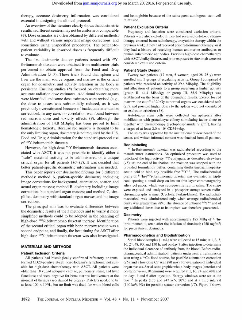
Figure 2 shows 2 different biologic time–activity curvesfor blood in a female patient: one for the patient-specificblood volume (4,950 mL) and another for the referencewoman blood volume (3,500 mL). The experimental dataof percentage injected activity in the blood derived withthe 2 different hypotheses were fitted by a biexponentialfunction (method A) and a monoexponential function(method C). In this patient, the ND and red marrow ab-sorbed dose were, respectively, 1.89 h and 0.38 mGy/MBq
with method A and 2.38 h and 0.84 mGy/MBq with methodC, leading to a 120% difference in red marrow dose.
Biodistribution
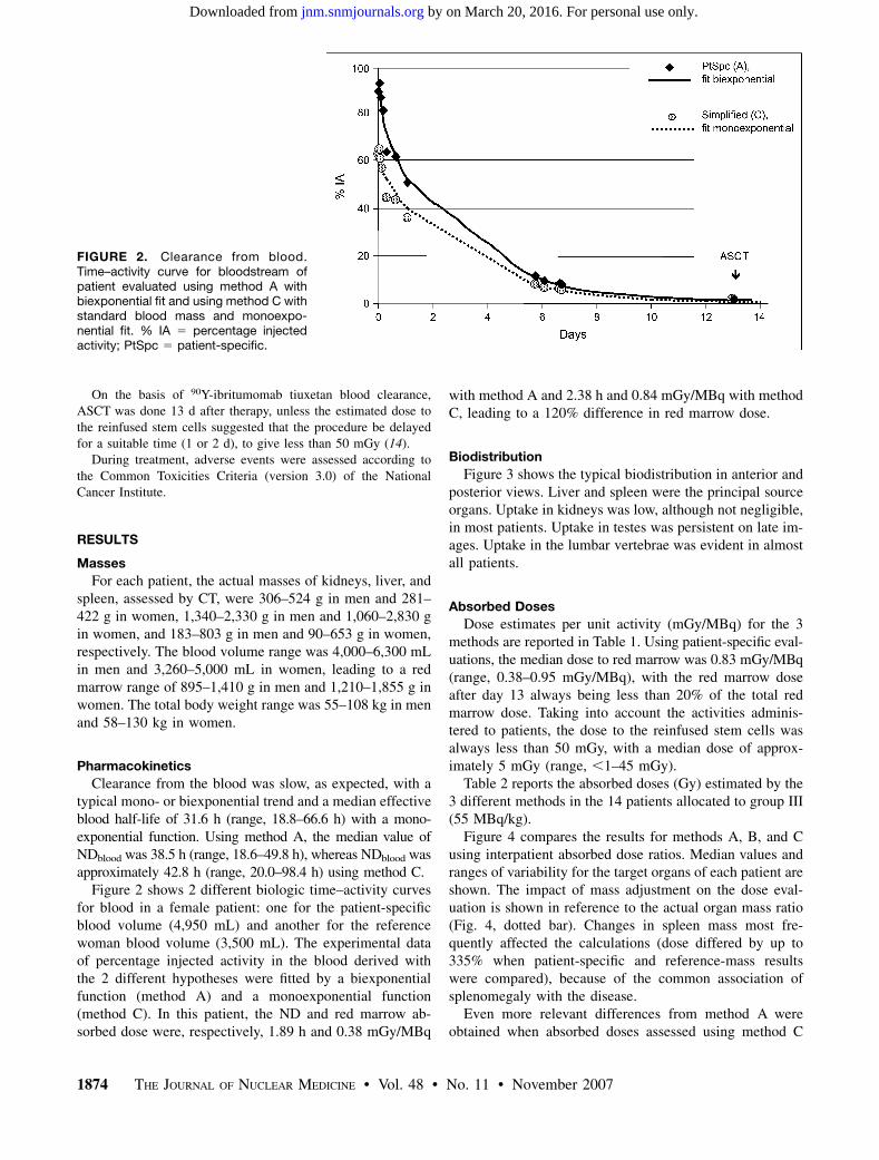
Figure 3 shows the typical biodistribution in anterior andposterior views. Liver and spleen were the principal sourceorgans. Uptake in kidneys was low, although not negligible,in most patients. Uptake in testes was persistent on late im-ages. Uptake in the lumbar vertebrae was evident in almostall patients.
Absorbed Doses
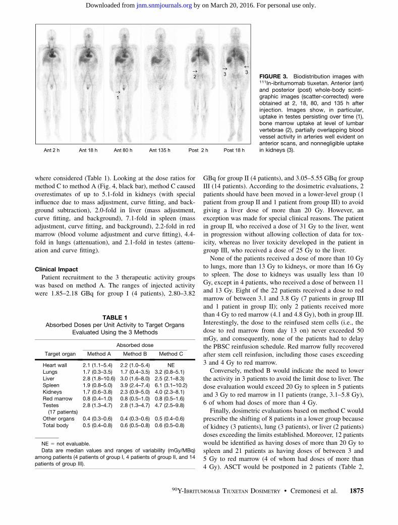
Dose estimates per unit activity (mGy/MBq) for the 3methods are reported in Table 1. Using patient-specific eval-uations, the median dose to red marrow was 0.83 mGy/MBq(range, 0.38–0.95 mGy/MBq), with the red marrow doseafter day 13 always being less than 20% of the total redmarrow dose. Taking into account the activities adminis-tered to patients, the dose to the reinfused stem cells wasalways less than 50 mGy, with a median dose of approx-imately 5 mGy (range, ,1–45 mGy).
Table 2 reports the absorbed doses (Gy) estimated by the3 different methods in the 14 patients allocated to group III(55 MBq/kg).
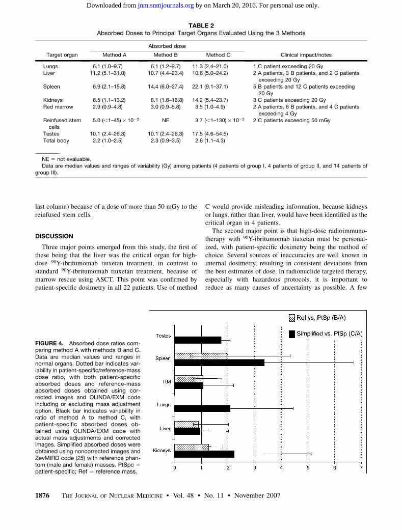
Figure 4 compares the results for methods A, B, and Cusing interpatient absorbed dose ratios. Median values andranges of variability for the target organs of each patient areshown. The impact of mass adjustment on the dose eval-uation is shown in reference to the actual organ mass ratio(Fig. 4, dotted bar). Changes in spleen mass most fre-quently affected the calculations (dose differed by up to335% when patient-specific and reference-mass resultswere compared), because of the common association ofsplenomegaly with the disease.
Even more relevant differences from method A wereobtained when absorbed doses assessed using method C
FIGURE 2. Clearance from blood.Time–activity curve for bloodstream ofpatient evaluated using method A withbiexponential fit and using method C withstandard blood mass and monoexpo-nential fit. % IA 5 percentage injectedactivity; PtSpc 5 patient-specific.
1874 THE JOURNAL OF NUCLEAR MEDICINE • Vol. 48 • No. 11 • November 2007
by on March 20, 2016. For personal use only. jnm.snmjournals.org Downloaded from
where considered (Table 1). Looking at the dose ratios formethod C to method A (Fig. 4, black bar), method C causedoverestimates of up to 5.1-fold in kidneys (with specialinfluence due to mass adjustment, curve fitting, and back-ground subtraction), 2.0-fold in liver (mass adjustment,curve fitting, and background), 7.1-fold in spleen (massadjustment, curve fitting, and background), 2.2-fold in redmarrow (blood volume adjustment and curve fitting), 4.4-fold in lungs (attenuation), and 2.1-fold in testes (attenu-ation and curve fitting).
Clinical Impact
Patient recruitment to the 3 therapeutic activity groupswas based on method A. The ranges of injected activitywere 1.85–2.18 GBq for group I (4 patients), 2.80–3.82
GBq for group II (4 patients), and 3.05–5.55 GBq for groupIII (14 patients). According to the dosimetric evaluations, 2patients should have been moved in a lower-level group (1patient from group II and 1 patient from group III) to avoidgiving a liver dose of more than 20 Gy. However, anexception was made for special clinical reasons. The patientin group II, who received a dose of 31 Gy to the liver, wentin progression without allowing collection of data for tox-icity, whereas no liver toxicity developed in the patient ingroup III, who received a dose of 25 Gy to the liver.
None of the patients received a dose of more than 10 Gyto lungs, more than 13 Gy to kidneys, or more than 16 Gyto spleen. The dose to kidneys was usually less than 10Gy, except in 4 patients, who received a dose of between 11and 13 Gy. Eight of the 22 patients received a dose to redmarrow of between 3.1 and 3.8 Gy (7 patients in group IIIand 1 patient in group II); only 2 patients received morethan 4 Gy to red marrow (4.1 and 4.8 Gy), both in group III.Interestingly, the dose to the reinfused stem cells (i.e., thedose to red marrow from day 13 on) never exceeded 50mGy, and consequently, none of the patients had to delaythe PBSC reinfusion schedule. Red marrow fully recoveredafter stem cell reinfusion, including those cases exceeding3 and 4 Gy to red marrow.
Conversely, method B would indicate the need to lowerthe activity in 3 patients to avoid the limit dose to liver. Thedose evaluation would exceed 20 Gy to spleen in 5 patientsand 3 Gy to red marrow in 11 patients (range, 3.1–5.8 Gy),6 of whom had doses of more than 4 Gy.
Finally, dosimetric evaluations based on method C wouldprescribe the shifting of 8 patients in a lower group becauseof kidney (3 patients), lung (3 patients), or liver (2 patients)doses exceeding the limits established. Moreover, 12 patientswould be identified as having doses of more than 20 Gy tospleen and 21 patients as having doses of between 3 and5 Gy to red marrow (4 of whom had doses of more than4 Gy). ASCT would be postponed in 2 patients (Table 2,
FIGURE 3. Biodistribution images with111In-ibritumomab tiuxetan. Anterior (ant)and posterior (post) whole-body scinti-graphic images (scatter-corrected) wereobtained at 2, 18, 80, and 135 h afterinjection. Images show, in particular,uptake in testes persisting over time (1),bone marrow uptake at level of lumbarvertebrae (2), partially overlapping bloodvessel activity in arteries well evident onanterior scans, and nonnegligible uptakein kidneys (3).
TABLE 1Absorbed Doses per Unit Activity to Target Organs
Evaluated Using the 3 Methods
Absorbed dose
Target organ Method A Method B Method C
Heart wall 2.1 (1.1–5.4) 2.2 (1.0–5.4) NE
Lungs 1.7 (0.3–3.5) 1.7 (0.4–3.5) 3.2 (0.8–5.1)
Liver 2.8 (1.8–10.6) 3.0 (1.6–8.0) 2.5 (2.1–8.3)
Spleen 1.9 (0.8–5.0) 3.9 (2.4–7.4) 6.1 (3.1–10.2)Kidneys 1.7 (0.6–3.8) 2.3 (0.9–5.0) 4.0 (2.3–8.1)
Red marrow 0.8 (0.4–1.0) 0.8 (0.5–1.0) 0.8 (0.5–1.6)
Testes(17 patients)
2.8 (1.3–4.7) 2.8 (1.3–4.7) 4.7 (2.5–9.8)
Other organs 0.4 (0.3–0.6) 0.4 (0.3–0.6) 0.5 (0.4–0.6)
Total body 0.5 (0.4–0.8) 0.6 (0.5–0.8) 0.6 (0.5–0.8)
NE 5 not evaluable.
Data are median values and ranges of variability (mGy/MBq)
among patients (4 patients of group I, 4 patients of group II, and 14patients of group III).
90Y-IBRITUMOMAB TIUXETAN DOSIMETRY • Cremonesi et al. 1875
by on March 20, 2016. For personal use only. jnm.snmjournals.org Downloaded from
last column) because of a dose of more than 50 mGy to thereinfused stem cells.
DISCUSSION
Three major points emerged from this study, the first ofthese being that the liver was the critical organ for high-dose 90Y-ibritumomab tiuxetan treatment, in contrast tostandard 90Y-ibritumomab tiuxetan treatment, because ofmarrow rescue using ASCT. This point was confirmed bypatient-specific dosimetry in all 22 patients. Use of method
C would provide misleading information, because kidneysor lungs, rather than liver, would have been identified as thecritical organ in 4 patients.
The second major point is that high-dose radioimmuno-therapy with 90Y-ibritumomab tiuxetan must be personal-ized, with patient-specific dosimetry being the method ofchoice. Several sources of inaccuracies are well known ininternal dosimetry, resulting in consistent deviations fromthe best estimates of dose. In radionuclide targeted therapy,especially with hazardous protocols, it is important toreduce as many causes of uncertainty as possible. A few
TABLE 2Absorbed Doses to Principal Target Organs Evaluated Using the 3 Methods
Absorbed dose
Target organ Method A Method B Method C Clinical impact/notes
Lungs 6.1 (1.0–9.7) 6.1 (1.2–9.7) 11.3 (2.4–21.0) 1 C patient exceeding 20 GyLiver 11.2 (5.1–31.0) 10.7 (4.4–23.4) 10.6 (5.0–24.2) 2 A patients, 3 B patients, and 2 C patients
exceeding 20 Gy
Spleen 6.9 (2.1–15.8) 14.4 (6.0–27.4) 22.1 (9.1–37.1) 5 B patients and 12 C patients exceeding
20 GyKidneys 6.5 (1.1–13.2) 8.1 (1.6–16.8) 14.2 (5.4–23.7) 3 C patients exceeding 20 Gy
Red marrow 2.9 (0.9–4.8) 3.0 (0.9–5.8) 3.5 (1.0–4.9) 2 A patients, 6 B patients, and 4 C patients
exceeding 4 Gy
Reinfused stemcells
5.0 (,1–45) · 1023 NE 3.7 (,1–130) · 1023 2 C patients exceeding 50 mGy
Testes 10.1 (2.4–26.3) 10.1 (2.4–26.3) 17.5 (4.6–54.5)
Total body 2.2 (1.0–2.5) 2.3 (0.9–3.5) 2.6 (1.1–4.3)
NE 5 not evaluable.
Data are median values and ranges of variability (Gy) among patients (4 patients of group I, 4 patients of group II, and 14 patients ofgroup III).
FIGURE 4. Absorbed dose ratios com-paring method A with methods B and C.Data are median values and ranges innormal organs. Dotted bar indicates var-iability in patient-specific/reference-massdose ratio, with both patient-specificabsorbed doses and reference-massabsorbed doses obtained using cor-rected images and OLINDA/EXM codeincluding or excluding mass adjustmentoption. Black bar indicates variability inratio of method A to method C, withpatient-specific absorbed doses ob-tained using OLINDA/EXM code withactual mass adjustments and correctedimages. Simplified absorbed doses wereobtained using noncorrected images andZevMIRD code (25) with reference phan-tom (male and female) masses. PtSpc 5
patient-specific; Ref 5 reference mass.
1876 THE JOURNAL OF NUCLEAR MEDICINE • Vol. 48 • No. 11 • November 2007
by on March 20, 2016. For personal use only. jnm.snmjournals.org Downloaded from
comparisons showed that standard organ masses often arenot correct for patients affected by serious diseases; actualorgan masses should be measured and used wheneverpossible (11,12). Moreover, image correction, especiallyof attenuation, is necessary to reduce the drawbacks ofplanar quantification methods; background and scatter cor-rections may further improve the activity estimates.
The third major point emerging from this study is thataccurate evaluation of the time–activity blood curve of eachpatient allows determination of the timing for ASCT.Because ASCT was planned, the limit of 3 Gy to redmarrow for standard 90Y-ibritumomab tiuxetan was notapplied. The major focus instead was on delivering minimalirradiation to reinfused stem cells. Again, mass adjustment,but also better regression analyses, avoided nonnegligibleoverestimates of red marrow doses and the consequentdelay of ASCT.
We are aware that transferring an experimental protocol tothe clinical setting would require a reasonably easy method,striking a balance between the accuracy and the difficulty ofprocedures. In routine practice, a simplified procedure isoften applied to evaluate dosimetry and delineate the essen-tial dosimetric characteristics of new radiocompounds(7,25,27), including standard 90Y-ibritumomab tiuxetan(25). However, as the patient-specific vs. simplified doseratios show (Fig. 4, black bar), an overly simplified methodshould be avoided because of the clinical impact on tailoredhigh-dose 90Y-ibritumomab tiuxetan therapy (10–12). Thereported dose estimate results are expected not to be abso-lutely accurate but certainly more accurate than estimatesobtained by the other methods. The assumptions made in themethod chosen are more realistic and amply demonstrated inthe literature. Certainly, more sophisticated corrections couldfurther refine the dosimetric evaluations, including suchcorrections as partial background subtraction and compart-mental modeling (18), but unacceptable discrepancies be-tween more reliable and more simplified evaluations havebeen shown. The differences imputable to actual mass scal-ing are considerable but not exhaustive (Fig. 4, dotted bar).Attenuation plays a crucial role, especially for source organsattenuated differently from abdomen (e.g., lungs, testes).Background and scatter also contribute to data uncertainty,and corrections for these effects should be included. It wasdifficult to summarize the effect of each correction, becausethe importance and magnitude of each correction variedstrongly among patients. This variation was evident in massadjustment but applied also to attenuation, scatter, andbackground corrections. However, the importance of anyone correction was found to depend on the source organsinvolved. To summarize, mass adjustment is crucial to avoidover- or underestimation of dose (Fig. 2), especially forspleen, liver, and kidneys; the contribution ranges fromnegligible to strong and is not always linearly related to thepatient’s weight. Attenuation correction is of major impactfor all organs, especially lungs and testes (e.g., a factor of ;2in lungs and a factor of in ;3 in testes), and the use of a mean
attenuation factor is not appropriate for all organs. Usually,background and scatter corrections are next in importance. Ifno scatter windows can be set to acquire images, backgroundmight include taking into account scatter subtraction, al-though a specific scatter subtraction is preferable. Finally, theeffect of appropriate fitting is lower but still significant:Multiexponential functions typically improved the fits for thetime–activity curve of liver and, in some cases, of blood andred marrow. Overall, significant overestimates were observed,up to about 2-fold in liver and red marrow, 2-fold in testes (28),4-fold in lungs, 5-fold in kidneys, and 7-fold in spleen.
The results from method A led to the observation that,following red marrow, liver is the limiting organ. Theuptake in kidneys is persistent and nonnegligible in mostpatients (Fig. 3), particularly in those with compromisedrenal function. The dose to lungs is usually well below 20Gy, considered an approximate tolerance dose for lungs inexternal-beam radiotherapy (the tolerance dose that willproduce 5% complications in 5 y [TD5/5] is 18 Gy (29)).Despite the typically observed high uptake, the dose tospleen was lower than expected, because of the spleno-megaly that was present in almost all patients and is often acharacteristic of the disease. The dose to ovaries is usuallynot of concern, but the dose to testes can exceed thethreshold of 6 Gy for permanent sterility (30), possiblybecause of the high tissue vascularization. However, spermcells were recruited from all young patients before theystarted chemotherapy.
Our median absorbed doses agree with results in theliterature except for liver, spleen, and kidneys (heart wall,2.80 mGy/MBq; lungs, 2.05 mGy/MBq; red marrow,0.59 mGy/MBq; testes, 2.80 mGy/MBq; total body, 0.54mGy/MBq; liver, 4.32 mGy/MBq; spleen, 7.35 mGy/MBq;kidneys, 0.22 mGy/MBq) (8,13,18,28,31,32). These excep-tions can be explained by taking into account the fact thatin the previous studies, a whole-body–averaged attenuationcorrection factor was used instead of a transmission scanfor attenuation correction, and kidneys were not identifiedas major source organs. The number of patients included inthe studies (179 vs. 22) was also quite different (13), and acomparison of the median values may be of poor value.Moreover, the clinical history of our patients was consistentwith a major influence of mass scaling, possibly even morethan for the patients usually recruited for standard 90Y-ibritumomab tiuxetan therapy, and especially for spleen,liver, and kidneys (Fig. 4, dotted bar). Finally, the selectionof curve fitting can further contribute to differences in thebiokinetics of liver and spleen, which typically show aprolonged uptake phase not easily interpretable by mono-exponential equations.
In the clinical protocol considered in this study, the limitof 20 Gy for irradiation of the liver, as well as of other vitalorgans, was a conservative choice (13). Actually, highertolerance doses were indicated from experience withexternal-beam radiotherapy (TD5/5 values of 23 Gy forkidneys and 30 Gy for liver) (29). Evidence also exists
90Y-IBRITUMOMAB TIUXETAN DOSIMETRY • Cremonesi et al. 1877
by on March 20, 2016. For personal use only. jnm.snmjournals.org Downloaded from
that tissue tolerance may be higher during radionuclidetherapy than during external-beam radiotherapy (33,34)because of the difference in dose rates. Nevertheless, theirradiation of vital organs in patients undergoing high-dose90Y-ibritumomab tiuxetan deserves special caution becauseof the clinical history of the patients. Previous aggressivetherapies may have affected the liver, red marrow, andkidneys. Irradiation after chemotherapy can generate seri-ous and unpredictable effects. This is a further reason thatliver, not lungs, may often be the organ of concern. Thedose limit must be applied conservatively because of thepossible influence of such prior chemotherapy. Other po-tential strategies are suggested from this point. If high-dose90Y-ibritumomab tiuxetan could be proposed as a second-linetreatment, higher liver thresholds and, consequently, higheractivities of 90Y-ibritumomab tiuxetan could be acceptable.
A final comment on the method used for red marrowdosimetry is worthwhile. The recruited patients had nega-tive bone marrow biopsy findings at the moment of therapy.This fact supported the assessment of red marrow dose bythe blood method for monoclonal antibodies (35,36), whichwas considered the reference in therapy-planning evalua-tions. However, in many patients scintigraphic imagessuggested some bone marrow uptake, especially in thevertebrae, with intensities that did not necessarily vary inrelation to the disease. This finding raised the possibilitythat a contribution from specific marrow uptake could bemissed (37,38). If so, the absence of a correlation betweendose and toxicity, stated in the literature, might be partiallyexplained, and a more adequate model for the red marrowdose should be applied to minimize the chance of toxicity.This topic should be further explored and possibly com-pared with the toxicity of the ASCT.
CONCLUSION
In high-dose 90Y-ibritumomab tiuxetan therapy, overlysimplified dosimetry methods may produce misleading in-dications, lowering allowable values of administered activityand shifting the time of autologous stem cell reinfusion. Wesuggest that tailored therapy be performed after patient-individualized dosimetry, with adjustment of standard modelorgan masses to patient-specific values and with applicationof all possible image corrections.
With marrow rescue, the liver is the second limiting organin these cases. Calculation of liver dose deserves specialattention because of the large number of chemotherapy linesthat the patient population typically receives.
An accurate dosimetric evaluation is mandatory in newclinical trials designed to increase the chances of cure notonly in nonresponding patients but also, possibly, in theearly stages of therapeutic planning.
ACKNOWLEDGMENTS
We thank Deborah Console for typing the manuscript,and we thank Annalisa Rossi, Dr. Silvia Melania Baio,
Dr. Demetrio Arico, Dr. Liliana Calabrese, and AngelaCocquio for assisting the patients. We are especially gratefulto Dr. Jeff Siegel for useful discussions and to the ItalianAssociation for Cancer Research (AIRC) for financialsupport.
REFERENCES
1. Witzig TE, Gordon LI, Cabanillas F, et al. Randomized controlled trial of
yttrium-90–labeled ibritumomab tiuxetan radioimmunotherapy versus rituximab
immunotherapy for patients with relapsed or refractory low-grade, follicular, or
transformed B-cell non-Hodgkin’s lymphoma. J Clin Oncol. 2002;20:2453–
2463.
2. Witzig TE, Flinn IW, Gordon LI, et al. Treatment with ibritumomab tiuxetan
radioimmunotherapy in patients with rituximab-refractory follicular non-
Hodgkin’s lymphoma. J Clin Oncol. 2002;20:3262–3269.
3. Morschhauser F, Illidge T, Huglo D, et al. Efficacy and safety of yttrium 90
ibritumomab tiuxetan in patients with relapsed or refractory diffuse large B-cell
lymphoma not appropriate for autologous stem cell transplantation. Blood.
2007;110:54–58.
4. Wessels BW, Bolch WE, Bouchet LG, et al. Bone marrow dosimetry using
blood-based models for radiolabeled antibody therapy: a multiinstitutional
comparison. J Nucl Med. 2004;45:1725–1733.
5. Wiseman GA, White CA, Stabin M, et al. Phase I/II 90Y-Zevalin (yttrium-90
ibritumomab tiuxetan, IDEC-Y2B8) radioimmunotherapy dosimetry results in
relapsed or refractory non-Hodgkin’s lymphoma. Eur J Nucl Med. 2000;27:766–777.
6. Zevalin Web site. Zevalin support services page. Available at: http://www.
zevalin.com/SupportServices/supportService.htm. Accessed August 30, 2007.
7. U.S. Food and Drug Administration Web site. Review of biologics license
application for IDEC pharmaceutical ZEVALIN� kit, BLA no. 125019.
Available at: http://www.fda.gov/ohrms/dockets/ac/01/briefing/3782b2_03_
FDA%20Backgrounder.pdf. Accessed August 30, 2007.
8. Wiseman GA, Leigh BR, Dunn WL, Stabin MG, White CA. Additional radiation
absorbed dose estimates for Zevalin� radioimmunotherapy. Cancer Biother
Radiopharm. 2003;18:253–258.
9. Wiseman GA, Leigh BR, Erwin WD, et al. Radiation dosimetry results from a
phase II trial of ibritumomab tiuxetan (Zevalin) radioimmunotherapy for patients
with non-Hodgkin’s lymphoma and mild thrombocytopenia. Cancer Biother
Radiopharm. 2003;18:165–178.
10. Siegel JA, Stabin MG, Brill AB. The importance of patient-specific radiation
dose calculations for the administration of radionuclides in therapy. Cell Mol
Biol (Noisy-le-grand). 2002;48:451–459.
11. Cremonesi M, Ferrari M, Stabin M, et al. Comparison of dosimetry methods for
high dose Zevalin therapy in refractory NHL patients [abstract]. J Nucl Med.
2005;46(suppl):87P.
12. Rajendran JG, Fisher DR, Gopal AK, Durack LD, Press OW, Eary JF. High-dose131I-tositumomab (anti-CD20) radioimmunotherapy for non-Hodgkin’s lym-
phoma: adjusting radiation absorbed dose to actual organ volumes. J Nucl Med.
2004;45:1059–1064.
13. Wiseman GA, Kornmehl E, Leigh B, et al. Radiation dosimetry results and safety
correlations from 90Y-ibritumomab tiuxetan radioimmunotherapy for relapsed or
refractory non-Hodgkin’ lymphoma: combined data from 4 clinical trials. J Nucl
Med. 2003;44:465–474.
14. Nademanee A, Forman S, Molina A, et al. A phase 1/2 trial of high-dose yttrium-
90-ibritumomab tiuxetan in combination with high-dose etoposide and cyclo-
phosphamide followed by autologous stem cell transplantation in patients with
poor-risk or relapsed non-Hodgkin lymphoma. Blood. 2005;106:2896–2902.
15. Papi S, Urbano N, Tosi G, Ferrari M, Paganelli G, Chinol M. Radiolabeling of
Zevalin� with high 90Y activity: optimization of the procedure and reduction of
the radiopharmacist radiation exposure [abstract]. Eur J Nucl Med Mol Imaging.
2006;33(suppl):S90.
16. Savi A, Lecchi M, Albertini F, et al. Evaluation of attenuation correction in
planar In-111 biodistribution studies [abstract]. Eur J Nucl Med Mol Imaging.
2004;31(suppl):S229.
17. Cremonesi M, Migliazza M, Bodei L, et al. Reconstruction algorithm validation
for In-111-labelled radiopharmaceuticals in hybrid SPECT-CT imaging system
[abstract]. Eur J Nucl Med. 2002;29(suppl):S93.
18. Siegel JA, Thomas SR, Stubbs JB, et al. Techniques for quantitative radiophar-
maceutical biodistribution data acquisition and analysis for use in human radiation
dose estimates: MIRD pamphlet no.16. J Nucl Med. 1999;40(suppl):S37–S61.
19. Stabin MG, Siegel JA. Physical models and dose factors for use in internal dose
assessment. Health Phys. 2003;85:294–310.
1878 THE JOURNAL OF NUCLEAR MEDICINE • Vol. 48 • No. 11 • November 2007
by on March 20, 2016. For personal use only. jnm.snmjournals.org Downloaded from
20. Loevinger R, Budinger TF, Watson EE. MIRD Primer for Absorbed Dose
Calculations. Reston, VA: Society of Nuclear Medicine; 1988.
21. Foster D, Barret P. Developing and testing integrated multicompartment models
to describe a single-input multiple-output study using the SAAM II software
system. In: Proceedings of the Sixth International Radiopharmaceutical
Dosimetry Symposium. Oak Ridge, Tennessee: Oak Ridge Institute for Science
and Education;1998.
22. Sgouros G. Bone marrow dosimetry for radioimmunotherapy: theoretical
considerations. J Nucl Med. 1993;34:689–694.
23. Stabin MG, Sparks RB, Crowe E. OLINDA/EXM: the second-generation per-
sonal computer software for internal dose assessment in nuclear medicine. J Nucl
Med. 2005;46:1023–1027.
24. RADAR Web site. Available at: www.doseinfo-radar.com. Accessed August 30,
2007.
25. Belanger R. ZevMIRD: A Workbook for Zevalin� Dosimetry. San Diego, CA:
Ryan–Belanger Associates; 1997.
26. Stabin MG. MIRDOSE: Personal computer software for internal dose assess-
ment in nuclear medicine. J Nucl Med. 1996;37:538–546.
27. Forster GJ, Engelbach MJ, Brockmann JJ, et al. Preliminary data on
biodistribution and dosimetry for therapy planning of somatostatin receptor
positive tumours: comparison of 86Y-DOTATOC and 111In-DTPA-octreotide. Eur
J Nucl Med. 2001;28:1743–1750.
28. Wiseman GA, Leigh BR, Dunn WL, Stabin MG, White CA. Additional radiation
absorbed dose estimates for Zevalin� radioimmunotherapy. Cancer Biother
Radiopharm. 2003;18:253–258.
29. Emami B, Lyman J, Brown A, et al. Tolerance of normal tissue to therapeutic
irradiation. Int J Radiat Oncol Biol Phys. 1991;21:109–122.
30. International Commission on Radiological Protection. Radiological Protection
on Biomedical Research: ICRP Publication 62. New York, NY: Pergamon Press;
1992. ICRP62.
31. Wu RK, Siegel JA. Absolute quantitation of radioactivity using the buildup
factor. Med Phys. 1984;11:189–192.
32. Tennvall J, Fischer M, Bischof Delaloye A, et al. EANM procedure guideline for
radio-immunotherapy for B-cell lymphoma with 90Y-radiolabelled ibritumomab
tiuxetan (Zevalin). Eur J Nucl Med Mol Imaging. 2007;34:616–622.
33. Barone R, Borson-Chazot F, Valkema R, et al. Patient-specific dosimetry in predicting
renal toxicity with 90Y-DOTATOC: relevance of kidney volume and dose rate in
finding a dose–effect relationship. J Nucl Med. 2005;46(suppl):99S–106S.
34. Dale R, Carabe-Fernandez A. The radiobiology of conventional radiotherapy and its
application to radionuclide therapy. Cancer Biother Radiopharm. 2005;20:47–51.
35. Sgouros G, Stabin M, Erdi Y, et al. Red marrow dosimetry for radiolabeled anti-
bodies that bind to marrow, bone, or blood components. Med Phys. 2000;27:
2150–2164.
36. Sgouros G. Dosimetry of internal emitters. J Nucl Med. 2005;46(suppl):18S–27S.
37. Shen S, Meredith RF, Duan J, et al. Improved prediction of myelotoxicity using a
patient-specific imaging dose estimate for non-marrow-targeting 90Y-antibody
therapy. J Nucl Med. 2002;43:1245–1253.
38. Siegel J, Lee R, Pawlyk D, Horowitz J, Sharkey R, Goldenberg D. Sacral
scintigraphy for bone marrow dosimetry in radioimmunotherapy. Int J Appl
Radiat Instr 1989;16:553–559.
90Y-IBRITUMOMAB TIUXETAN DOSIMETRY • Cremonesi et al. 1879
by on March 20, 2016. For personal use only. jnm.snmjournals.org Downloaded from

Doi: 10.2967/jnumed.107.0440162007;48:1871-1879.J Nucl Med.
Gennaro Prisco, Giovanni Martinelli, Giovanni Paganelli and Pier Francesco FerrucciMarta Cremonesi, Mahila Ferrari, Chiara Maria Grana, Anna Vanazzi, Mike Stabin, Mirco Bartolomei, Stefano Papi, Dosimetric Study for Tailored Treatment
Y-Ibritumomab Tiuxetan: Comparative90High-Dose Radioimmunotherapy with
http://jnm.snmjournals.org/content/48/11/1871This article and updated information are available at:
http://jnm.snmjournals.org/site/subscriptions/online.xhtml
Information about subscriptions to JNM can be found at:
http://jnm.snmjournals.org/site/misc/permission.xhtmlInformation about reproducing figures, tables, or other portions of this article can be found online at:
(Print ISSN: 0161-5505, Online ISSN: 2159-662X)1850 Samuel Morse Drive, Reston, VA 20190.SNMMI | Society of Nuclear Medicine and Molecular Imaging
is published monthly.The Journal of Nuclear Medicine
© Copyright 2007 SNMMI; all rights reserved.
by on March 20, 2016. For personal use only. jnm.snmjournals.org Downloaded from