BINOL−Amino Acid Conjugates as Triggerable Carriers of DNA-Targeted Potent Photocytotoxic Agents
10
BINOL-Amino Acid Conjugates as Triggerable Carriers of DNA-Targeted Potent Photocytotoxic Agents Filippo Doria, †,‡ Sara N. Richter, §,‡ Matteo Nadai, § Stefano Colloredo-Mels, † Mariella Mella, † Manlio Palumbo, | and Mauro Freccero* ,† Dipartimento di Chimica Organica, UniVersità di PaVia, V. le Taramelli 10, 27100 PaVia, Italy, Dipartimento di Istologia, Microbiologia e Biotecnologie Mediche, UniVersità di PadoVa, Via Gabelli, 63, 35121, PadoVa, Italy, and Dipartimento di Scienze Farmaceutiche, UniVersità di PadoVa, Via Marzolo, 5, 35131 PadoVa, Italy ReceiVed July 9, 2007 Mild photoactivation of new BINOL-amino acid and -amino ester conjugates (BINOLAMs) yielded alkylating and DNA cross-linking agents with high photoefficiency and superior cytotoxicity. Detection of the transient electrophile, by laser flash photolysis (LFP), suggests that BINOL-quinone methides (QMs) are key intermediates in the process. QMs trapping by water, monitored in a time-dependent product distribution analysis, demonstrated that the phototriggered reactivity of BINOLAMs as bis-alkylating agents is the result of a two-step process involving sequential photogeneration of monoalkylating QMs. Light activation of the BINOL-L-amino esters produced cytotoxic QMs very effective against human tumor LoVo cells with EC 50 in the 130–230 nM range. Trimethylpsoralen (PS) is about 4 times less potent than our newly tested compounds. BINOL-L-proline methyl ester showed notable photoselectivity because it displayed cytotoxic effects upon irradiation only and was able to efficiently reach the target DNA inside the cells, where it forms both alkylated and cross-linked species. Introduction Several strategies have been devised and developed for the selective and mild activation of DNA-modifying agents. The generation of reactive intermediates, starting from stable precur- sors, with DNA-binding properties, has great potential for the development of chemotherapeutic prodrugs 1 and for the inves- tigation and understanding of DNA–protein interactions. 2 Therefore, it is not surprising that advances in areas of oxidative, reductive, hydrolytic, and photochemical activation protocols continue to generate promising and clinically relevant agents. 3 Among them, the phototriggerable cleaving 1b,4 and alkylat- ing/cross-linking 1a,b,4 agents have attracted a great deal of attention in the past decade, mainly because, contrary to most nonphotochemically activable DNA-modifying agents, they do not require a coreagent under conditions not always compatible with in ViVo applications. 4 Although in the recent past a wide variety of photochemically activated DNA reactive agents have been discovered and investigated, they mainly act as cleavers involving radical reactive intermediates. Fairly limited examples of photochemically activated DNA-alkylating agents have been reported. 5 In fact, to date, psoralens are the only well-established class of drugs known to induce DNA or RNA cross-linking upon photoactivation. 6 Recently, our research has been focused on another class of reagents that similarly express a triggerable ability to alkylate nucleic acids through the generation of highly reactive quinone methides (QMs) a (Scheme 1). 7 These electrophilic intermediates have been generated by photochemical 5,7–10 and thermal 7a,11–13 (fluoride and pH- induced) mild activation procedures. In both cases, the genera- tion of a phenolate, by excited-state proton transfer 14 or by fluoride-induced desylilation, 11–13 is the key mechanistic step leading to the electrophilic QMs. QMs, particularly those with an ortho geometry (o-QMs), have been successfully used to accomplish amino acid, 7a oligopeptide, 7a nucleoside mono- and bis-alkylations, 11,12 and DNA-cross-linking 5,7b,15 in water (ac- cording the sequence reported in Scheme 2). In more detail, Wan, 8,14 Kresge, 9 Saito, 10 Zhou, 15b and our group 7 described the photogeneration of QMs starting from benzyl alcohols (1, Scheme 1), 8,9 Mannich bases (2, Scheme 1), 7,10 and their quaternary ammonium salts (3, Scheme 1) in water. 7,15b These reactive intermediates, with tunable electrophilicity by substitu- ent effects, 13 have also been trapped by biological nucleophiles and detected by nanosecond laser flash photolysis (LFP). 7a - e,8,9 Recently, we have also shown that BINOL derivatives, such as the bis-quaternary ammonium salt 3b, are capable of undergoing bis-alkylation in water and DNA cross-linking with promising potency by UV–vis photoactivation (Scheme 3). 7b Furthermore, unlike precursors 1-3, the quaternary am- monium salts 3a and 3b exhibit high absorptivity at a wave- length longer than 360 nm and excellent solubility under * To whom correspondence should be addressed: Università di Pavia, Dipartimento di Chimica Organica V. le Taramelli 10, 27100 Pavia, Italy. Telephone: +39-0382-987668. Fax: +39-0382-987323. E-mail: [email protected]. † Università di Pavia. ‡ These authors contributed equally to this work. § Dipartimento di Istologia, Microbiologia e Biotecnologie Mediche, Università di Padova. | Dipartimento di Scienze Farmaceutiche, Università di Padova. a Abbreviations: QMs, quinone methides; BINOLAM, BINOL-amino acid and -amino ester conjugate; PS, 4,5′,8-trimethylpsoralen; LFP, laser flash photolysis; XL, cross-linking. Scheme 1. Photochemical and Thermal (Fluoride and pH-Induced) Generation of the Prototype QM J. Med. Chem. 2007, 50, 6570–6579 6570 10.1021/jm070828x CCC: $37.00 2007 American Chemical Society Published on Web 11/30/2007
Transcript of BINOL−Amino Acid Conjugates as Triggerable Carriers of DNA-Targeted Potent Photocytotoxic Agents

BINOL-Amino Acid Conjugates as Triggerable Carriers of DNA-Targeted PotentPhotocytotoxic Agents
Filippo Doria,†,‡ Sara N. Richter,§,‡ Matteo Nadai,§ Stefano Colloredo-Mels,† Mariella Mella,† Manlio Palumbo,| andMauro Freccero*,†
Dipartimento di Chimica Organica, UniVersità di PaVia, V. le Taramelli 10, 27100 PaVia, Italy, Dipartimento di Istologia, Microbiologia eBiotecnologie Mediche, UniVersità di PadoVa, Via Gabelli, 63, 35121, PadoVa, Italy, and Dipartimento di Scienze Farmaceutiche,UniVersità di PadoVa, Via Marzolo, 5, 35131 PadoVa, Italy
ReceiVed July 9, 2007
Mild photoactivation of new BINOL-amino acid and -amino ester conjugates (BINOLAMs) yieldedalkylating and DNA cross-linking agents with high photoefficiency and superior cytotoxicity. Detection ofthe transient electrophile, by laser flash photolysis (LFP), suggests that BINOL-quinone methides (QMs)are key intermediates in the process. QMs trapping by water, monitored in a time-dependent productdistribution analysis, demonstrated that the phototriggered reactivity of BINOLAMs as bis-alkylating agentsis the result of a two-step process involving sequential photogeneration of monoalkylating QMs. Lightactivation of the BINOL-L-amino esters produced cytotoxic QMs very effective against human tumor LoVocells with EC50 in the 130–230 nM range. Trimethylpsoralen (PS) is about 4 times less potent than ournewly tested compounds. BINOL-L-proline methyl ester showed notable photoselectivity because it displayedcytotoxic effects upon irradiation only and was able to efficiently reach the target DNA inside the cells,where it forms both alkylated and cross-linked species.
Introduction
Several strategies have been devised and developed for theselective and mild activation of DNA-modifying agents. Thegeneration of reactive intermediates, starting from stable precur-sors, with DNA-binding properties, has great potential for thedevelopment of chemotherapeutic prodrugs1 and for the inves-tigation and understanding of DNA–protein interactions.2
Therefore, it is not surprising that advances in areas of oxidative,reductive, hydrolytic, and photochemical activation protocolscontinue to generate promising and clinically relevant agents.3
Among them, the phototriggerable cleaving1b,4 and alkylat-ing/cross-linking1a,b,4 agents have attracted a great deal ofattention in the past decade, mainly because, contrary to mostnonphotochemically activable DNA-modifying agents, they donot require a coreagent under conditions not always compatiblewith in ViVo applications.4 Although in the recent past a widevariety of photochemically activated DNA reactive agents havebeen discovered and investigated, they mainly act as cleaversinvolving radical reactive intermediates. Fairly limited examplesof photochemically activated DNA-alkylating agents have beenreported.5 In fact, to date, psoralens are the only well-establishedclass of drugs known to induce DNA or RNA cross-linking uponphotoactivation.6 Recently, our research has been focused onanother class of reagents that similarly express a triggerableability to alkylate nucleic acids through the generation of highlyreactive quinone methides (QMs)a (Scheme 1).7
These electrophilic intermediates have been generated byphotochemical5,7–10 and thermal7a,11–13 (fluoride and pH-induced) mild activation procedures. In both cases, the genera-tion of a phenolate, by excited-state proton transfer14 or byfluoride-induced desylilation,11–13 is the key mechanistic stepleading to the electrophilic QMs. QMs, particularly those withan ortho geometry (o-QMs), have been successfully used toaccomplish amino acid,7a oligopeptide,7a nucleoside mono- andbis-alkylations,11,12 and DNA-cross-linking5,7b,15 in water (ac-cording the sequence reported in Scheme 2). In more detail,Wan,8,14 Kresge,9 Saito,10 Zhou,15b and our group7 describedthe photogeneration of QMs starting from benzyl alcohols (1,Scheme 1),8,9 Mannich bases (2, Scheme 1),7,10 and theirquaternary ammonium salts (3, Scheme 1) in water.7,15b Thesereactive intermediates, with tunable electrophilicity by substitu-ent effects,13 have also been trapped by biological nucleophilesand detected by nanosecond laser flash photolysis (LFP).7a-e,8,9
Recently, we have also shown that BINOL derivatives, suchas the bis-quaternary ammonium salt 3b, are capable ofundergoing bis-alkylation in water and DNA cross-linking withpromising potency by UV–vis photoactivation (Scheme 3).7b
Furthermore, unlike precursors 1-3, the quaternary am-monium salts 3a and 3b exhibit high absorptivity at a wave-length longer than 360 nm and excellent solubility under
* To whom correspondence should be addressed: Università di Pavia,Dipartimento di Chimica Organica V. le Taramelli 10, 27100 Pavia, Italy.Telephone: +39-0382-987668. Fax: +39-0382-987323. E-mail:[email protected].
† Università di Pavia.‡ These authors contributed equally to this work.§ Dipartimento di Istologia, Microbiologia e Biotecnologie Mediche,
Università di Padova.| Dipartimento di Scienze Farmaceutiche, Università di Padova.aAbbreviations: QMs, quinone methides; BINOLAM, BINOL-amino
acid and -amino ester conjugate; PS, 4,5′,8-trimethylpsoralen; LFP, laserflash photolysis; XL, cross-linking.
Scheme 1. Photochemical and Thermal (Fluoride andpH-Induced) Generation of the Prototype QM
J. Med. Chem. 2007, 50, 6570–65796570
10.1021/jm070828x CCC: $37.00 2007 American Chemical SocietyPublished on Web 11/30/2007
physiological conditions, which are considered the basic re-quirements for useful DNA-alkylating agents. Unfortunately,the binol derivatives 1a(b)-3a(b) exhibit severe limitations forin ViVo applications because of the presence of the permanentlycharged quaternary ammonium moieties, which prevent binolsfrom crossing biological membranes. Contrary, the phototrig-gerable BINOL unit tethered to amino acid derivatives andoligonucleotides could be characterized by adequate cell perme-ability16 and biological target recognition properties,17 respec-tively.Here,wedescribe thefirststep towardBINOL-oligopeptideconjugates as selective and triggerable carriers of bifunctionalBINOL-QMs targeting DNA. We report the synthesis, pho-toreactivity, DNA cross-linking properties, and photocytotoxicityof rational BINOL-amino acid derivatives (BINOLAMs).Indeed, the novel derivatives represent valuable lead compoundsfor selective cellular DNA-damaging activity.
Results and Discussion
The target compounds investigated in the current study are3,3′-bis-CH2Y-substituted BINOLs represented by the generalstructure, highlighted in red in Chart 1.
They all contain two CH2Y arms on the BINOL moiety,which act as absorbing chromophores for the activation of theBINOLAMs by photoinduced HX elimination. Compounds4–10 (Chart 1) differ from each other by the Y group structure,which was chosen among N-substituted L-amino acids andrelated amino esters, with or without basic side-chain groups:L-proline (4, 5, and 6), L-alanine (7), L-valine (8), and L-lysine(9 and 10). The Y moiety structures were selected for thefollowing reasons: (a) organic molecules exhibiting net positivecharges under physiological conditions should exhibit enhancedDNA binding through electrostatic interactions with the polya-nionic backbone of the nucleic acid; (b) the spacer length couldmodulate the binding properties of the precursors; (c) theprotonation-deprotonation equilibria in the amino acid deriva-tives should remarkably improve cell permeability of the newBINOL conjugates in comparison to that of the fully chargedquaternary ammonium salts (3a and 3b), previously investigatedas DNA cross-linking agents.7b The role as chiral ligands inthe modern enantioselective catalysis of BINOL derivatives18
propelled us to exploit the robust chemistry of the BINOLs to
prepare new diastereomerically pure BINOLAMs 4–6 andenantiopure BINOLs 3a-3c. These allowed us to explorepossible diastereo- and enantioselectivity effects on DNA cross-linking potency and photocytotoxicity/cytotoxicity properties ofthe BINOLAMs.
Synthesis of BINOL-Amino Acid Conjugates. The BINO-LAMs 4–10 investigated as a phototriggerable source of bis-alkylating species were synthesized mainly by a reductiveamination reaction,19 using the dialdehyde 1120 in a stepwiseapproach with the preformation of the intermediate imine, andfurther reduction in a separate step (Scheme 4).
After surveying many of the commercially available hydridereagents, we selected sodium triacetoxyborohydride [NaBH-(OAc)3] based on the resulting overall reductive aminationreaction yields (always higher than 85%). Imine preformationwas achieved adding the dialdehyde 11 to L-alanine-OMe,L-valine-OMe, L-proline-OMe, L-proline-OtBu, and N-ε-t-bu-tyloxycarbonyl-L-lysin-t-butyl ester in anhydrous CH2Cl2 and4 Å molecular sieves. The diastereomeric L-diprolino esters(S,S,S)- and (R,S,S)-4 and -5 and their related amino acid 6 werealso synthesized starting from the BINOL-3,3′-bis-CH2Br (12)in higher yields (according to Scheme 5) or following an originalphotochemical protocol optimized by our group, starting fromracemic BINOLAM 2b.7c
In both cases, a 1:1 diastereomeric mixture was obtained.The (S,S,S)-4 and (S,S,S)-5 adducts were the first, among thetwo diastereoisomers, to be eluted from preparative columnchromatography (7:3 cyclohexane/ethyl acetate), with very gooddiastereomeric excess (de) g 99%. Crystallization of thediastereomeric mixture from benzene and ethyl acetate affordedthe (S,S,S) and (R,S,S) pure isomers, respectively, with a slightlylower purity (dee 95%). The absolute configurations of (R,S,S)-4, (S,S,S)-4, (R,S,S)-5, and (S,S,S)-5 were assigned by acomparison to authentic samples synthesized quantitativelystarting from chiral (R)- and (S)-dibromide 12 and proline methyland t-butyl esters, respectively (Scheme 5). The BINOL-aminoacid conjugates (6 and 10) were quantitatively prepared startingfrom the t-butyl esters, following the standard deprotectionprotocol (TFA, Et3SiH, and CH2Cl2 at room temperature for25 min).21
BINOL-Amino Acid and Ester Conjugates as Photot-riggerable Bis-alkylating Agents. To test 4–10 as phototrig-gerable precursors of bis-alkylating QMs, we first explored thephotoreactivity of 4–6 as pure diastereoisomers and 7, 8, and10 as 1:1 diastereomeric mixtures. The BINOL-QMs werephotogenerated both at 310 and 360 nm wavelengths and trappedby water, morpholine, and 2-mercaptoethanol (Scheme 6), inbuffered (pH 7.5) 1:1 H2O/CH3CN. In more detail, photolysisof diastereomerically pure (S,S,S)- and (R,S,S)-4–6, in theabsence of an added nucleophile, gives the alcohol (R)- and(S)-1b, respectively, in quantitative yields, with completeretention of the BINOL-moiety configuration (after 10 min ofirradiation). Photolysis of 4–6 in a water solution containingmorpholine and mercaptoethanol similarly afforded the bis-morpholino adduct 13 and the bifunctional thioether 14, in goodyields, accompanied by small amounts (<15%) of alcohol 1b.Fairly similar conversion rates were achieved with the BINOL-L-valine-OMe ester (8) and the BINOL-L-lysine (10). The onlyexception to this trend was due to BINOL-alanine-OMe ester(7), which turned to be much less reactive, requiring a longerirradiation time (10 min) and yielding a lower conversion (lowerthan 35%, see the Supporting Information for detailed informa-tion). Monoalkylated adducts were not detected by high-performance liquid chromatography (HPLC) under the reaction
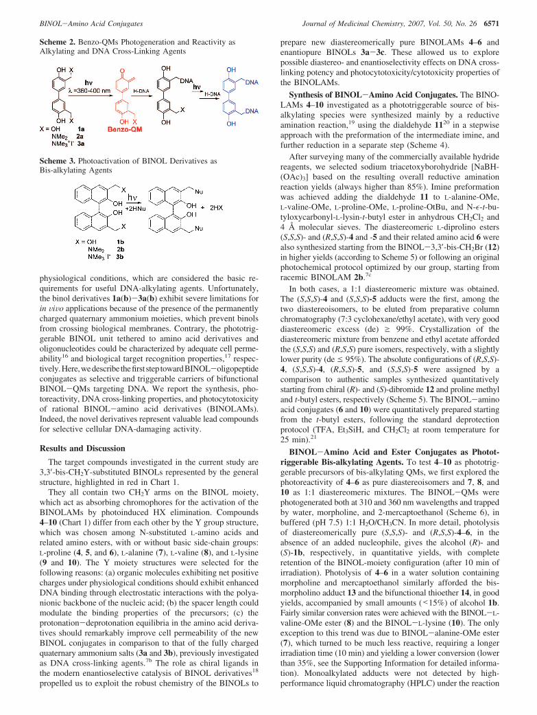
Scheme 2. Benzo-QMs Photogeneration and Reactivity asAlkylating and DNA Cross-Linking Agents
Scheme 3. Photoactivation of BINOL Derivatives asBis-alkylating Agents
BINOL-Amino Acid Conjugates Journal of Medicinal Chemistry, 2007, Vol. 50, No. 26 6571

conditions described above, and if formed, their yields werelower than 5%.
Mechanistic Insights from Time-Dependent ProductDistribution Analysis and Quantum Yield Measurements.We have shown that the photoactivation of adducts 4-10 asbifunctional alkylating agents is a general key feature of theBINOLAMs. Therefore, considering worthwhile the investiga-tion of its mechanism, we run three sets of clarifying experi-ments: (i) monitoring the photohydration process by time-dependent product distribution analysis, (ii) evaluating thephotochemical efficiency by quantum yield measurements, and(iii) detecting the electrophilic transient by LFP. Running the
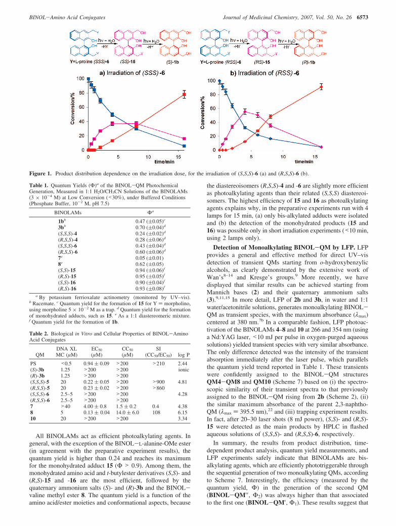
photochemical reaction of (S,S,S)- and (R,S,S)-6 under lowconversion (<40%) conditions, achievable for short irradiationtimes (0.5–5 min, in a merry-go-round photoreactor, with 2lamps at 15 W, 310 nm; ca. 25 °C, and N2 purged), it was clearthat the monohydrated intermediates [(S,S)- and (R,S)-15; seeFigure 1] were formed first and subsequently reacted to givealcohols (S)-1b and (R)-1b, respectively. The profile of theconverted precursor (S,S,S)-6 into the intermediate monohy-drated adduct (S,S)-15, toward the final product (S)-1b, as afunction of the irradiation time is reported in Figure 1a. A similartime-dependent product distribution analysis was run for thediastereoisomer (R,S,S)-6 (Figure 1b).
The monohydrated adducts (S,S)-15 and (R,S)-15 were formedrapidly, reaching a maximum yield after 4–5 min of irradiation(Figure 1). Photoinduced decomposition of the resulting adductswas also rapid, with 80–90% conversion of the starting materialinto alcohols (S)-1b and (R)-1b within 15 min (Figure 1). Acloser comparison of the two product profiles in Figure 1a and1b reveals that the diastereoisomer (R,S,S)-6 is slightly morephotoreactive than (S,S,S)-6. Such a difference is confirmed byquantum yield measurements (Table 2). In fact, irradiation of(R,S,S)-6 generates the monohydrated adduct (R,S)-15, with a56% conversion after 4 min of irradiation, and the irradiationof (S,S,S)-6 generates the monohydrated adduct (S,S)-15, witha 37% conversion after the same irradiation time. In acomparable fashion, we have monitored the product distributionas a function of the irradiation time also for the L-proline t-butylesters [(R,S,S)-4 and (S,S,S)-4] in a 1:1 acetonitrile/watersolution. In this case, both (R,S)-16 and (S,S)-16 resulting fromthe monohydration process reached a maximum after 7 min andno measurable difference in the photoreactivity between thediastereoisomers was recorded.
These time-dependent profiles, displaying the trend of reactant(6) consumption, intermediates (15), and final product formation(1b) of the hydration process, demonstrate that the BINOL-aminoester and BINOL-amino acid conjugates (4 and 6, respectively)both act as phototriggerable bis-alkylating agents through thesequential generation of two monoalkylating quinone methides.In addition, adduct (R,S,S)-6 exhibits a slightly higher efficiencyin the photogeneration of the monohydration adduct in com-parison to its diastereoisomer (S,S,S)-6. To evaluate the pho-toefficiency of the BINOL-amino acids and BINOL-aminoesters (4–8) in the generation of alkylating QMs, we alsomeasured their quantum yields, comparing them to otherphotoreactive BINOLs, including the monohydrated adducts 15and 16. The measured quantum yields are listed in Table 1.
Chart 1. BINOL-Amino Acid Derivatives (4-10) Synthesized and Tested as Photocytotoxic Bis-alkylating Agents
Scheme 4. Synthetic Protocol for the BINOLAMs 4, 5, and7-9a
a (i) Molecular sieves at 4 Å in CH2Cl2; (ii) NaBH(OAc)3, CH2Cl2, andN2.
Scheme 5. Synthesis of BINOL-Diprolino Derivatives 4-6a
a (i) K2CO3, CH2Cl2, ∆, 5 h. (ii) TFA, Et3SiH, CH2Cl2, room temperature,25 min.
Scheme 6. Photoreactivity of BINOLAMs 4-10 in Water, 10-3
M, in a Merry-go-round Photoreactor, with 4 Lamps at 15 W,310 and 360 nm; ca. 25 °C, 15 and 40 min, Respectively, andN2 Purged
6572 Journal of Medicinal Chemistry, 2007, Vol. 50, No. 26 Doria et al.
All BINOLAMs act as efficient photoalkylating agents. Ingeneral, with the exception of the BINOL-L-alanine-OMe ester(in agreement with the preparative experiment results), thequantum yield is higher than 0.24 and reaches its maximumfor the monohydrated adduct 15 (Φ > 0.9). Among them, themonohydrated amino acid and t-butylester derivatives (S,S)- and(R,S)-15 and -16 are the most efficient, followed by thequaternary ammonium salts (S)- and (R)-3b and the BINOL-valine methyl ester 8. The quantum yield is a function of theamino acid/ester moieties and conformational aspects, because
the diastereoisomers (R,S,S)-4 and -6 are slightly more efficientas photoalkylating agents than their related (S,S,S) diastereoi-somers. The highest efficiency of 15 and 16 as photoalkylatingagents explains why, in the preparative experiments run with 4lamps for 15 min, (a) only bis-alkylated adducts were isolatedand (b) the detection of the monohydrated products (15 and16) was possible only in short irradiation experiments (<10 min,using 2 lamps only).
Detection of Monoalkylating BINOL-QM by LFP. LFPprovides a general and effective method for direct UV–visdetection of transient QMs starting from o-hydroxybenzylicalcohols, as clearly demonstrated by the extensive work ofWan’s8–14 and Kresge’s groups.9 More recently, we havedisplayed that similar results can be achieved starting fromMannich bases (2) and their quaternary ammonium salts(3).9,11,15 In more detail, LFP of 2b and 3b, in water and 1:1water/acetonitrile solutions, generates monoalkylating BINOL-QM as transient species, with the maximum absorbance (λmax)centered at 380 nm.7b In a comparable fashion, LFP photoac-tivation of the BINOLAMs 4–8 and 10 at 266 and 354 nm (usinga Nd:YAG laser, <10 mJ per pulse in oxygen-purged aqueoussolutions) yielded transient species with very similar absorbance.The only difference detected was the intensity of the transientabsorption immediately after the laser pulse, which parallelsthe quantum yield trend reported in Table 1. These transientswere confidently assigned to the BINOL-QM structuresQM4-QM8 and QM10 (Scheme 7) based on (i) the spectro-scopic similarity of their transient spectra to that previouslyassigned to the BINOL-QM rising from 2b (Scheme 2), (ii)the similar maximum absorbance of the parent 2,3-naphtho-QM (λmax ) 395.5 nm),22 and (iii) trapping experiment results.In fact, after 20–30 laser shots (8 mJ power), (S,S)- and (R,S)-15 were detected as the main products by HPLC in flashedaqueous solutions of (S,S,S)- and (R,S,S)-6, respectively.
In summary, the results from product distribution, time-dependent product analysis, quantum yield measurements, andLFP experiments safely indicate that BINOLAMs are bis-alkylating agents, which are efficiently phototriggerable throughthe sequential generation of two monoalkylating QMs, accordingto Scheme 7. Interestingly, the efficiency (measured by thequantum yield, Φ) in the generation of the second QM(BINOL-QM′′ , Φ2) was always higher than that associatedto the first one (BINOL-QM′, Φ1). These results suggest that
Figure 1. Product distribution dependence on the irradiation dose, for the irradiation of (S,S,S)-6 (a) and (R,S,S)-6 (b).
Table 1. Quantum Yields (Φ)a of the BINOL-QM PhotochemicalGeneration, Measured in 1:1 H2O/CH3CN Solutions of the BINOLAMs(3 × 10⁻⁴ M) at Low Conversion (<30%), under Buffered Conditions(Phosphate Buffer, 10-2 M, pH 7.5)
BINOLAMs Φa
1bb 0.47 ((0.05)c
3bb 0.70 ((0.04)d
(S,S,S)-4 0.24 ((0.02)d
(R,S,S)-4 0.28 ((0.06)d
(S,S,S)-6 0.43 ((0.04)d
(R,S,S)-6 0.60 ((0.06)d
7e 0.05 ((0.01)8e 0.62 ((0.05)(S,S)-15 0.94 ((0.06)f
(R,S)-15 0.95 ((0.05)f
(S,S)-16 0.90 ((0.04)f
(R,S)-16 0.93 ((0.08)f
a By potassium ferrioxalate actinometry (monitored by UV–vis).b Racemate. c Quantum yield for the formation of 15 for Y ) morpholino,using morpholine 5 × 10⁻² M as a trap. d Quantum yield for the formationof monohydrated adducts, such as 15. e As a 1:1 diastereomeric mixture.f Quantum yield for the formation of 1b.
Table 2. Biological in Vitro and Cellular Properties of BINOL-AminoAcid Conjugates
QMDNA XLMC (µM)
EC50
(µM)CC50
(µM)SI
(CC50/EC50) log P
PS <0.5 0.94 ( 0.09 >200 >210 2.44(S)-3b 1.25 >200 >200 ionic(R)-3b 1.25 >200 >200(S,S,S)-5 20 0.22 ( 0.05 >200 >900 4.81(R,S,S)-5 20 0.23 ( 0.02 >200 >860(S,S,S)-6 2.5–5 >200 >200 4.28(R,S,S)-6 2.5–5 >200 >2007 >40 4.00 ( 0.8 1.5 ( 0.2 0.4 4.388 5 0.13 ( 0.04 14.0 ( 6.0 108 6.1510 20 >200 >200 3.34
BINOL-Amino Acid Conjugates Journal of Medicinal Chemistry, 2007, Vol. 50, No. 26 6573

the BINOLAMs may act as photoactivatable mono- and bis-alkylating agents.
DNA Cross-Linking. The DNA-DNA cross-linking abilityof compounds (S)-3b, (R)-3b, (S,S,S)-5, (R,S,S)-5, (S,S,S)-6,(R,S,S)-6, 7, 8, and 10 was investigated using a negativelysupercoiled plasmid DNA (pBR322) in an alkaline agarose gelassay (Figure 2).7b Compounds (S,S,S)-4, (R,S,S)-4, and 9 werenot included because of their low solubility in aqueous solution.DNA cross-linking (XL) experiments were carried out in 50mM phosphate-buffered solutions at pH 7.5. Samples wereirradiated at 360 nm for 20 min. Each compound was used atincreasing concentrations (0.62–80 µM, lanes 1–8 in Figure 2).Irradiated DNA without substrates (lanes D) or with PS (lanesPS) were used as controls.
The nonreacted plasmid was present in its supercoiled (S),linear (L), and nicked (N) forms. Interstrand cross-linkingactivities of tested compounds were evident as a XL band (XL),which run slightly slower than the linear form (Figure 2).Activities were measured as the minimal compound concentra-tion (DNA XL MC) required to obtain XL effects (Table 2).
XL activities were measured for the two enantiomeres S andR of compound 3b and diastereoisomers S,S,S and R,S,S ofcompounds 5 and 6. Compounds (S)-3b and (R)-3b induceddetectable XL at concentrations as low as 1.25 µM [lanes 2–8
in Figure 2, QM (S)-3b and (R)-3b]. These data are in totalagreement with previous results obtained with the racemicmixture of BINOL-QM 3b.7b Diastereoisomers 6 were onlyslightly less active than 3b; XL effects were evident at 2.5–5µM [lanes 3–8 in Figure 2, (S,S,S)-6 and (R,S,S)-6]. BINOLderivatives 5 were around 5–8-fold less potent than 6, becausethey displayed detectable XL only at a 20 µM concentration[lanes 6–8 in Figure 2, QM (S,S,S)-5 and (R,S,S)-5]. Nosignificant differences in XL potency were found between thetwo enantiomers (S)-3b and (R)-3b or diastereoisomers (S,S,S)-6and (R,S,S)-6 of each active compound (compare lanes 3–8 inFigure 2), indicating the absence of detectable enantio- ordiastereoselectivity in the DNA photo-cross-linking activity bythese BINOL derivatives.
On the basis of these results, compounds 7, 8, and 10 weretested as racemic mixtures. Of these, BINOL derivative 8 wasthe most active, with a DNA XL MC of 5 µM (lanes 4–8 inFigure 2). Compounds 7 and 10 resulted less potent, being activeat concentrations of 40 and 20 µM, respectively. In addition, inthe presence of high concentrations of these two latter com-pounds (80 µM), unreacted DNA amounts decreased, withoutcorresponding XL formation. This likely indicates the unspecificreaction of BINOL derivatives 7 and 10 with material used forhandling after photoirradiation.
Scheme 7. Sequential Photogeneration of Two Monoalkylating BINOL-QMs
Figure 2. DNA cross-linking (XL) concentration-dependent activity of BINOL-amino acid conjugates. Plasmid DNA was mixed with increasingamounts (0.62, 1.25, 2.5, 5, 10, 20, 40, and 80 µM, lanes 1–8) of each compound (shown above each gel image), in phosphate buffer (50 mM, pH7.5). Reaction mixtures were irradiated at 360 nm for 20 min and loaded on a 1% alkaline agarose gel. Gels were stained with ethidium bromide.Lane D is nontreated, irradiated DNA. Lane PS is a control for XL species induced by 4,5′,8-trimethylpsoralen at 360 nm. Nonreacted supercoiled,nicked, and linear forms of plasmid are indicated as S, N, and L. XL species are indicated as XL, on the right of each gel image.
6574 Journal of Medicinal Chemistry, 2007, Vol. 50, No. 26 Doria et al.

It is noteworthy that none of the tested compounds modifiedDNA in the absence of photoactivation (data not shown).
Our results clearly indicate that cross-linking activity dependsupon three counter-balancing properties of BINOL-QM precur-sors: (1) efficiency of the primary photoactivation step, (2)percentage of BINOL derivative in the bis-cationic form, (3)presence of bulky substituents. The effect of the photoactivationefficiency is obviously very important, as highlighted by theless potent XL BINOL derivative 7, which is mainly the resultof its lowest photochemical quantum yield. Concerning the effectof the net charge on the precursors, we have already reportedthat bis-cationic forms of QMs induce electrostatic pre-associa-tion of the QM precursors to the ionic DNA phosphatebackbone, hence enhancing a subsequent covalent reaction.7b
In fact, XL efficiency increased when performing reactions inacid solutions, where the percentage of bis-cationic forms areenhanced. As shown in Figure 3, XL bands increased steadilyfrom pH 8.5 to 5.5 in the presence of 20 µM of compounds(S,S,S)-6, (R,S,S)-6, and 10. All other BINOL-amino acidconjugates displayed a similar behavior in this range of pH (datanot shown). On the other hand, bulky nonionic moieties mayboth mask positive charges or hinder an effective interactionbetween reactive groups, hence modulating cross-linkingefficiency.
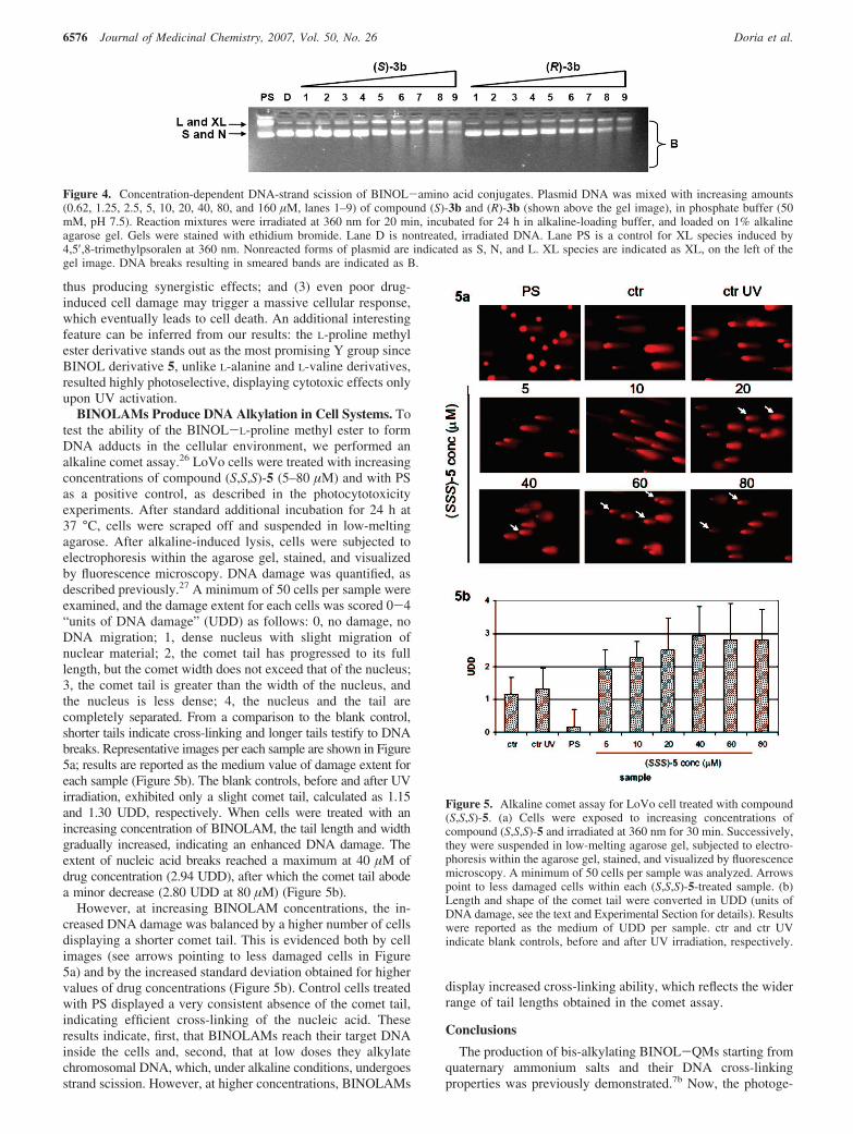
As demonstrated by the quantum yield measurements andLFP experiments, BINOLAMs are able to form both mono- aswell as bis-alkylated and XL species. To visualize alkylation,reaction mixtures were treated for 24 h at room temperature inalkaline agarose gel buffer, prior to loading onto alkaline agarosegels. Alkaline denaturing conditions destabilize alkylated bases,thus inducing DNA scissions.23 As shown in Figure 4, atincreasing concentrations of compounds (S)-3b and (R)-3b, theband corresponding to XL species augmented up to a drugconcentration of 10 µM (lanes 1–5 in Figure 4). However, athigher drug concentrations (20–160 µM), the XL band decreasedand converted to a smeared band, comprising species runningfaster than the XL band itself and SC DNA as well (lanes 6–9in Figure 4). A similar behavior was obtained with all testedBINOLAMs (data not shown). Importantly, the control PS inthese conditions did not induce DNA-strand scission. Theseresults indicate that the XL species formed by BINOLAMs butnot by PS are alkali-sensitive.
Evaluation of BINOL-Amino Acid Conjugates Photocy-totoxicity versus Cytotoxicity. Photocytotoxic and cytotoxiceffects of BINOL-amino acid conjugates (S)-3b, (R)-3b,(S,S,S)-5, (R,S,S)-5, (S,S,S)-6, (R,S,S)-6, 7, 8, and 10 wereinvestigated on the epithelial-like human colorectal adenocar-cinoma LoVo cell line. This is an established cell line widelyemployed as a human model in carcinogenesis studies. Inaddition, colon cancer is a representative malignancy efficientlytreated with Food and Drug Administration (FDA)-approved
and investigational drugs for phototherapy.24 Cells were exposedto increasing concentrations of tested compounds (12 nM-200µM), and after 24 h, they were either irradiated at 360 nm for30 min at 37 °C or kept in the dark. After an additional 24 h,cell damage was assessed by a MTT assay. Results werereported as the effective drug concentration able to kill 50% ofthe cell population after photoirradiation (EC50) and the cytotoxicconcentration that killed 50% of cell population withoutphotoirradiation (CC50). Selectivity indexes (SI) were measuredas the ratio of CC50 over EC50 values. As shown in Table 2,three of nine compounds showed EC50 in the nanomolar range.In particular, the most active compound was BINOL derivative8, with an EC50 of 130 ( 40 nM. However, it also showedcytotoxicity prior to UV irradiation at doses of 14.00 ( 6.00µM (SI ) 108). Compounds (S,S,S)-5 and R,S,S)-5 were slightlyless active, displaying EC50 of 220 ( 50 and 230 ( 20 nM,respectively, although they did not show cytotoxicity withoutirradiation up to 200 µM (SI > 860). Once again, chiralproperties did not confer appreciably different biological activi-ties. The BINOLAM 7 exhibited EC50 in the low micromolarrange (4.00 ( 0.08 µM), yet it resulted even more cytotoxicwithout UV treatment (CC50 of 1.50 ( 0.02 µM, SI ) 0.4).Finally, BINOL derivatives (S)-3b, (R)-3b, (S,S,S)-6, (R,S,S)-6, and 10 did not show either photocytotoxic or cytotoxic effectsat doses as high as 200 µM. PS, used as a control, in theseconditions displayed EC50 of 940 ( 90 nM and CC50 > 200µM.
Cellular drug effects depend upon not only selective targetrecognition but also the ability of the compound to enter thecell compartment through the plasmatic membrane. Thisproperty is usually theoretically and experimentally defined aslog D (D ) distribution), which is largely related to thecompound hydrophobic/hydrophilic characteristics, expressedas log P (P stands for partitioning in n-octanol/water). Theoreti-cal log P values for the neutral forms of tested BINOL-QMswere calculated according to the Crippen’s fragmentationmethod (Table 2).25 BINOL-amino acid conjugates 6 and 10showed the lowest log P calculated on their neutral structure.In addition, the pI for compound 6, roughly evaluated from thepI of their conjugated amino acids (pI 6.30, for proline), suggestthat it should be mainly negatively charged, at pH 7.5. Similarconsideration holds for compound 10, suggesting that it shouldbe mainly positively charged, at the working pH. Obviously,compound 3b is permanently charged. Therefore, the access of3b, 6, and 10 to the intracellular environment results severelyhindered, as proven by the inability to induce either photocy-totoxicity or cytotoxicity (Table 2). In contrast, BINOLAMs 5,7, and 8 are only partially charged at physiological pH, and theneutral forms can diversely permeate according to their hydro-phobic properties. This statement is supported by the fact thata hydroxyl group in ortho to a dimethylaminomethyl moietyreduces the basicity of the amino group (i.e., 2-dimethylami-nomethyl-phenols pKa1, 8.1; benzyl-dimethyl-amine pKa, 9.1).For the BINOLAMs 5, 7, and 8, we found a correlation betweenlog P values and photocytotoxicity (8 > 5 > 7) (Table 2).
Hence, amino acid methyl esters appear to be most suitablefor increasing cell-killing activity. In addition, the scale ofphotocytotoxicity soundly correlates with the ability to induceDNA XL (8 > 5 > 7), even though with different potency(nanomolar versus micromolar range, respectively). This biasedactive concentration range of results in Vitro and, in the cells,may rest on a number of reasons: (1) cellular enzymes maymetabolize the original compound enhancing its potency; (2)additional drug target(s) may be present in the cell environment,
Figure 3. pH dependence activity of compounds (S,S,S)-3b, (R,S,S)-3b, and 10. Plasmid DNA with 20 mM of indicated QMs was irradiatedat 360 nm in phosphate buffer at pH 5.5, 6.5, 7.5, and 8.5. Lane D isnontreated, irradiated DNA. Nonreacted forms of plasmid are indicatedas S and N. XL species are indicated as XL, on the right of each gelimage.
BINOL-Amino Acid Conjugates Journal of Medicinal Chemistry, 2007, Vol. 50, No. 26 6575
thus producing synergistic effects; and (3) even poor drug-induced cell damage may trigger a massive cellular response,which eventually leads to cell death. An additional interestingfeature can be inferred from our results: the L-proline methylester derivative stands out as the most promising Y group sinceBINOL derivative 5, unlike L-alanine and L-valine derivatives,resulted highly photoselective, displaying cytotoxic effects onlyupon UV activation.
BINOLAMs Produce DNA Alkylation in Cell Systems. Totest the ability of the BINOL-L-proline methyl ester to formDNA adducts in the cellular environment, we performed analkaline comet assay.26 LoVo cells were treated with increasingconcentrations of compound (S,S,S)-5 (5–80 µM) and with PSas a positive control, as described in the photocytotoxicityexperiments. After standard additional incubation for 24 h at37 °C, cells were scraped off and suspended in low-meltingagarose. After alkaline-induced lysis, cells were subjected toelectrophoresis within the agarose gel, stained, and visualizedby fluorescence microscopy. DNA damage was quantified, asdescribed previously.27 A minimum of 50 cells per sample wereexamined, and the damage extent for each cells was scored 0-4“units of DNA damage” (UDD) as follows: 0, no damage, noDNA migration; 1, dense nucleus with slight migration ofnuclear material; 2, the comet tail has progressed to its fulllength, but the comet width does not exceed that of the nucleus;3, the comet tail is greater than the width of the nucleus, andthe nucleus is less dense; 4, the nucleus and the tail arecompletely separated. From a comparison to the blank control,shorter tails indicate cross-linking and longer tails testify to DNAbreaks. Representative images per each sample are shown in Figure5a; results are reported as the medium value of damage extent foreach sample (Figure 5b). The blank controls, before and after UVirradiation, exhibited only a slight comet tail, calculated as 1.15and 1.30 UDD, respectively. When cells were treated with anincreasing concentration of BINOLAM, the tail length and widthgradually increased, indicating an enhanced DNA damage. Theextent of nucleic acid breaks reached a maximum at 40 µM ofdrug concentration (2.94 UDD), after which the comet tail abodea minor decrease (2.80 UDD at 80 µM) (Figure 5b).
However, at increasing BINOLAM concentrations, the in-creased DNA damage was balanced by a higher number of cellsdisplaying a shorter comet tail. This is evidenced both by cellimages (see arrows pointing to less damaged cells in Figure5a) and by the increased standard deviation obtained for highervalues of drug concentrations (Figure 5b). Control cells treatedwith PS displayed a very consistent absence of the comet tail,indicating efficient cross-linking of the nucleic acid. Theseresults indicate, first, that BINOLAMs reach their target DNAinside the cells and, second, that at low doses they alkylatechromosomal DNA, which, under alkaline conditions, undergoesstrand scission. However, at higher concentrations, BINOLAMs
display increased cross-linking ability, which reflects the widerrange of tail lengths obtained in the comet assay.
Conclusions
The production of bis-alkylating BINOL-QMs starting fromquaternary ammonium salts and their DNA cross-linkingproperties was previously demonstrated.7b Now, the photoge-
Figure 4. Concentration-dependent DNA-strand scission of BINOL-amino acid conjugates. Plasmid DNA was mixed with increasing amounts(0.62, 1.25, 2.5, 5, 10, 20, 40, 80, and 160 µM, lanes 1–9) of compound (S)-3b and (R)-3b (shown above the gel image), in phosphate buffer (50mM, pH 7.5). Reaction mixtures were irradiated at 360 nm for 20 min, incubated for 24 h in alkaline-loading buffer, and loaded on 1% alkalineagarose gel. Gels were stained with ethidium bromide. Lane D is nontreated, irradiated DNA. Lane PS is a control for XL species induced by4,5′,8-trimethylpsoralen at 360 nm. Nonreacted forms of plasmid are indicated as S, N, and L. XL species are indicated as XL, on the left of thegel image. DNA breaks resulting in smeared bands are indicated as B.
Figure 5. Alkaline comet assay for LoVo cell treated with compound(S,S,S)-5. (a) Cells were exposed to increasing concentrations ofcompound (S,S,S)-5 and irradiated at 360 nm for 30 min. Successively,they were suspended in low-melting agarose gel, subjected to electro-phoresis within the agarose gel, stained, and visualized by fluorescencemicroscopy. A minimum of 50 cells per sample was analyzed. Arrowspoint to less damaged cells within each (S,S,S)-5-treated sample. (b)Length and shape of the comet tail were converted in UDD (units ofDNA damage, see the text and Experimental Section for details). Resultswere reported as the medium of UDD per sample. ctr and ctr UVindicate blank controls, before and after UV irradiation, respectively.
6576 Journal of Medicinal Chemistry, 2007, Vol. 50, No. 26 Doria et al.

neration of related transient electrophiles has been achieved inexcellent quantum yields using BINOL-amino acid and -aminoester derivatives as the precursors. QM-trapping experimentsshow that bis-alkylation is the result of the sequential generationof two monoalkylating BINOL-QMs. The majority of theBINOL-amino acid and -amino ester precursors, when pho-toactivated at 360 nm in the presence of supercoiled plasmidDNA (pBR322), displayed DNA cross-linking potency com-parable to that of the quaternary ammonium salts (range of 1–5µM). Unlike the latter, the amino acid methyl esters exhibitpotent photocytotoxicity against the human colorectal adeno-carcinoma LoVo cell line in the nanomolar concentration range(130–230 nM). Among the 3,3′-CH2Y-disubstituted BINOLstested, the L-proline methyl ester congener stands out as themost promising lead because, contrary to other derivatives, itshowed remarkably high photoselectivity, displaying prominentcell killing effects only after UV irradiation. In this connection,it is worth recalling that PS, one of the most effective phototoxicagents thus far reported, is about 4 times less potent than ournewly tested compounds. In addition, we have shown thatBINOLAMs efficiently reach their DNA target inside the cells.However, their mechanism of action differs to some extent fromthat of a photoactivable reference drug, such as PS. In fact, wehave displayed that the photogenerated QMs inside the cellsexhibit mono- and bis-alkylating properties, resulting in DNAscission and cross-linking. Analysis of DNA sequence specificityand alkylation product characterization represent the next steptoward the development of potent photocytotoxic-selectivecompounds.
Experimental Section
General Procedures. The BINOLs 2b and 3b,7b,26 the dialde-hyde 11,20,28 and the dibromide 1229 have been synthesizedaccording to published procedures. The diastereomerically pure(S,S,S)- and (R,S,S) BINOL-esters 4 and 5 have been previouslysynthesized and characterized.7c The alkylation adducts 13 and 14were purified and characterized by a comparison to authenticsamples.7b
General Reductive Amination for the Synthesis of 4, 5,and 7–9. The followed procedure is a modified version that isalready published.19 A suspension of L-amino-ester HCl (5 mmol)in CH2Cl2 (25 mL) was added to a CH2Cl2 solution of Et3N (1mL). The solution was stirred at room temperature for 1 h, andthen a solution of dialdehyde 11 (700 mg, 2 mmol) in CH2Cl2 wasadded together with molecular sieves at 4 Å. This solution wasstirred under nitrogen atmosphere, at room temperature. After 16 h,NaBH(OAc)3 (880 mg, 4 mmol) was added. The resulting suspen-sion was further stirred for another 3 h at room temperature, underan inert atmosphere. At this time, a solution of Na2CO3 was addedto the mixture, then the organic layer was separated, and the residualsolution was extract twice with CH2Cl2.
The mixture of the reaction has been separated by silica-gelcolumn chromatography, eluting with 7:3 cyclohexane/ethylacetate.
Synthesis of 6 and 10 by Deprotection of 4 and 9: GeneralProcedure.21 The t-butyl amino ester (1 mmol) was deprotectedby dissolving it in a solution of trifluoroacetic acid (1.94 mL, 13mmol) and dichloromethane (10 mL) in the presence of triethyl-silane (0.8 mL, 2.5 mmol), at room temperature. After stirring for25 min, 1 M HCl (1 mL) was added and the solvent was removedin vacuo. The residue was suspended in diethyl ether, and theproduct was isolated by filtration. The yields were almost quantita-tive (93%), only using triethylsilane as the carbocation scavenger.
3,3′-Bis-(2-carboxypyrrolidin-1-ylmetil)-1,1′-dinaphtyl-2,2′-diolo ·2HCl (6). (S,S,S)-6 2HCl. White crystals. mp > 180 °C(dec.). 1H NMR (D2O) δ: 1.85 (broad s, 2 H), 2.01 (broad s, 4 H),2.41 (broad s, 2 H), 3.25 (broad s, 2 H), 3.59 (broad s, 2 H), 4.18(broad s, 2 H), 4.40 (d, 2 H, J ) 11.3 Hz), 4.55 (d, 2 H, J )11.3),
6.56 (broad s, 2 H), 6.68 (broad s, 2 H), 7.10 (broad s, 2 H), 7.77(broad s, 2 H), 7.96 (broad s, 2 H). 1H NMR (DMSO-d6) δ:1.95–2.11 (m, 6 H), 2.43–2.51 (m, 2 H), 3.38–3.41 (m, 2 H),3.62–3.75 (m, 2 H), 4.47–4.52 (m, 2 H), 4.56–4.79 (m, 4 H),6.93–7.01 (m, 2 H), 7.21–7.30 (m, 2 H), 7.32–7.38 (m, 2 H), 7.92(dd, 2 H, J ) 4.0, J ) 7.8 Hz), 8.14 (d, 2 H, J ) 7.5 Hz). 13CNMR (D2O) δ: 22.54 (CH2), 28.42 (CH2), 54.80 (CH2), 55.07(CH2), 67.06 (CH), 113.23 (C), 119.33 (C), 123.47 (CH), 124.34(CH), 128.08 (C), 128.12 (CH), 128.66 (CH), 134.03 (CH), 134.34(C), 151.13 (C), 171.85 (C). Anal. Calcd (C32H34Cl2N2O6): C, H,Cl, N.
(R,S,S)-6 ·2HCl. White crystals. mp > 177 °C (dec.). 1H NMR(D2O) δ: 1.82 (broad s, 2 H), 2.05 (broad s, 4 H), 2.44 (broad s, 2H), 3.25 (broad s, 2 H), 3.62 (broad s, 2 H), 4.17 (broad s, 2 H),4.42 (d, 2 H, J ) 11.3 Hz), 4.58 (d, 2 H, J )11.3), 6.59 (broad s,2 H), 6.70 (broad s, 2 H), 7.12 (broad s, 2 H), 7.80 (broad s, 2 H),8.01 (broad s, 2 H). 13C NMR (D2O) δ: 22.60 (CH2), 28.55 (CH2),54.81 (CH2), 55.17 (CH2), 67.12 (CH), 113.33 (C), 119.37 (C),123.57 (CH), 124.37 (CH), 128.00 (C), 128.22 (CH), 128.69 (CH),134.06 (CH), 134.39 (C), 151.17 (C), 171.89 (C). Anal. Calcd(C32H34Cl2N2O6): C, H, Cl, N.
7. Equimolar diastereomeric mixture. Yellow oil. 1H NMR(CDCl3) δ: 1.25–1.45 (s, 6 H), 3.25–3.75 (m, 2 H), 3.8 (s, 6 H),4.00–4.15 (m, 2 H), 4.25–4.45 (m, 2 H), 7.10–7.35 (m, 6 H),7.70–7.90 (m, 4 H). 13C NMR (CDCl3) δ: 18.84 (CH3), 51.31(CH2), 52.10 (CH), 55.40 (CH3), 123.06 (CH), 124.7(CH), 126.1(CH), 127.7 (CH), 127.9 (CH), 133.65 (C), 153.18 (C), 174.77 (C).Anal. Calcd (C30H32N2O6): C, H, N.
8. Equimolar diastereomeric mixture. Yellow oil. 1H NMR(CDCl3) δ: 1.25–1.45 (s, 12 H), 3.10–3.25 (m, 2 H), 3.65–3.80(m, 2 H), 3.85 (s, 6 H), 3.90–4.35 (m, 4 H), 7.10–7.35 (m, 6 H),7.70–7.90 (m, 4 H). 13C NMR (CDCl3) δ: 18.97 (CH3), 51.81(CH2), 51.93 (CH), 60.31 (CH3), 123.06 (CH), 124.7(CH), 126.1(CH), 127.7 (CH), 127.9 (CH), 133.45 (C), 154.08 (C), 174.13 (C).Anal. Calcd (C34H40N2O6): C, H, N.
9. The compound was not characterized by NMR and used forthe further step in the synthesis of 10.
10. Equimolar diastereomeric mixture. Yellow oil. 1H NMR(D2O) δ: 1.30–2.10 (m, 12 H), 2.60–3.10 (m, 4 H), 3.75–3.95 (m,2 H), 4.25–4.65 (m, 4 H), 6.90–7.00 (m, 2 H), 7.20–7.40 (m, 4 H),7.80–8.15 (m, 4 H). 13C NMR (D2O) δ: 21.49 (CH2), 24.80 (CH2),26.25 (CH2), 28.73 (CH2), 28.89 (CH2), 29.19 (CH2), 38.92 (CH2),46.52 (CH2), 47.02 (CH2), 47.47 (CH2), 48.81 (CH), 52.74 (CH),113.62 (C), 113.69 (C), 123.75 (CH), 123.81 (CH), 124.59 (CH),128.3 (CH), 128.45 (C), 128.66 (C), 128.71 (CH), 133.0 (CH), 133.5(CH), 133.6 (CH), 134.35 (C), 134.5 (C), 151.16 (C), 151.30 (C),171.79 (C), 171.89 (C). Anal. Calcd (C34H44Cl2N4O6): C, H,Cl, N.
(S,S)-15 ·HCl. Yellow oil. 1H NMR (D2O) δ: 1.55–1.75 (m, 2H), 2.15–2.22 (m, 1 H), 2.28–2.45 (m, 1 H), 2.48–2.60 (m, 1 H),3.15–3.25 (m, 1 H), 3.65–3.80 (m, 1 H), 3.85 (d, 1 H, J ) 13.2),4.60 (d, 1 H, J ) 13.2), 5.60 (m, 2 H), 5.70 (m, 1 H, acid),7.20–7.30 (m, 4 H), 7.30–7.40 (m, 2 H), 7.55–7.70 (m, 4 H), 8.50(s, 2 H, acid). 13C NMR (C5D5N) δ: 23.39 (CH2), 29.93 (CH2),53.14 (CH2), 57.27 (CH2), 61.39 (CH2), 66.25 (CH), 116.09 (C),116.34 (C), 125.44 (CH), 125.84 (CH), 126.27 (CH), 126.43 (CH),126.48 (CH), 126.61 (CH), 128.32 (CH), 128.89 (CH), 128.96 (CH),129.26 (CH), 133.05 (C), 134.41 (C), 153.07 (C), 155.23 (C),176.15 (C). Anal. Calcd (C27H26ClNO5): C, H, Cl, N.
(R,S)-15 ·HCl. Yellow oil. 1H NMR (C5D5N) δ: 1.60–1.80 (m,2 H), 2.05–2.20 (m, 1 H), 2.25–2.40 (m, 1 H), 2.48–2.60 (m, 1 H),3.00–3.15 (m, 1 H), 3.65–3.80 (m, 1 H), 3.90 (d, 1 H, J ) 13.2),4.82 (d, 1 H, J ) 13.2), 5.62 (m, 2 H), 5.70 (m, 1 H, acid),7.20–7.30 (m, 4 H), 7.30–7.40 (m, 2 H), 7.55–7.70 (m, 4 H), 8.50(s, 2 H, acid). 13C NMR (C5D5N) δ: 23.39 (CH2), 29.93 (CH2),53.14 (CH2), 57.27 (CH2), 61.39 (CH2), 66.25 (CH), 116.09 (C),116.34 (C), 125.44 (CH), 125.84 (CH), 126.27 (CH), 126.43 (CH),126.48 (CH), 126.61 (CH), 128.32 (CH), 128.89 (CH), 128.96 (CH),129.26 (CH), 133.05 (C), 134.41 (C), 153.07 (C), 155.23 (C),176.15 (C). Anal. Calcd (C27H26ClNO5): C, H, Cl, N.
BINOL-Amino Acid Conjugates Journal of Medicinal Chemistry, 2007, Vol. 50, No. 26 6577

(S,S)-16. Yellow oil. 1H NMR (CD3OD) δ: 1.25–1.35 (m, 9 H),1.75–2.00 (m, 3 H), 2.25–2.40 (m, 1 H), 2.60–2.80 (m, 1 H),3.20–3.35 (m, 1 H), 3.45–3.55 (m, 1 H), 4.18 (d, 2 H, J ) 13.2Hz), 4.30 (d, 2 H, J ) 13.2 Hz), 4.95 (s, 2 H), 6.80–7.00 (m, 2 H),7.00–7.30 (m, 4 H), 7.75–7.85 (m, 3 H), 7.95 (s, 1 H). 13C NMR(MeOD) δ: 24.39 (CH2), 28.41 (CH3), 32.13 (CH2), 54.89 (CH2),58.63 (CH2), 62.24 (CH2), 68.02 (CH), 83.85 (C), 115.95 (C),116.34 (C), 124.38 (CH), 124.64 (CH), 125.89 (CH), 126.05 (CH),127.09 (CH), 127.84 (CH), 128.05 (CH), 130.63 (CH), 130.91 (CH),131.71 (CH), 135.21 (C), 136.17 (C), 152.59 (C), 155.16 (C),173.91 (C). Anal. Calcd (C31H33NO5): C, H, N.
(R,S)-16. Yellow oil. 1H NMR (CD3OD) δ: 1.15–1.20 (m, 9H), 1.80–2.05 (m, 2 H), 2.20–2.30 (m, 2 H), 2.72–2.87 (m, 2 H),3.40–3.55 (m, 1 H), 4.15 (d, 2 H, J ) 13.2 Hz), 4.50 (d, 2 H, J )13.2 Hz), 4.95 (s, 2 H), 6.80–7.00 (m, 2 H), 7.00–7.30 (m, 4 H),7.75–7.85 (m, 3 H), 7.95 (s, 1 H). 13C NMR (MeOD) δ: 24.39(CH2), 28.41 (CH3), 32.13 (CH2), 54.89 (CH2), 58.63 (CH2), 62.24(CH2), 68.02 (CH), 83.85 (C), 115.95 (C), 116.34 (C), 124.38 (CH),124.64 (CH), 125.89 (CH), 126.05 (CH), 127.09 (CH), 127.84 (CH),128.05 (CH), 130.63 (CH), 130.91 (CH), 131.71 (CH), 135.21 (C),136.17 (C), 152.59 (C), 155.16 (C), 173.91 (C). Anal. Calcd(C31H33NO5): C, H, N.
Cross-Linking of Plasmid DNA. Compounds (S)-3b, (R)-3b,(S,S,S)-5, (R,S,S)-5, (S,S,S)-6, (R,S,S)-6, 7, 8, and 10 were dissolvedin DMSO to a final concentration of 20 mM. These stock solutionswere diluted in mQ-grade H2O and used for reactions. PlasmidpBR322 (0.5 µg/sample) was mixed with increasing amounts (0.62,1.25, 2.5, 5, 10, 20, 40, and 80 µΜ, as indicated in each figure) ofeach compound, in phosphate buffer (50 mM, pH 7.5). For pH-dependent experiments, QMs at 20 µM doses were reacted withpBR322 in 50 mM phosphate buffer at pH 5.5, 6.5, 7.5, and 8.5. Asaturated water solution of PS was used to get a 0.5 µMconcentration of drug in the control samples. Reaction mixtureswere irradiated on ice for 20 min at 360 nm at 120 W in aphotochemical multirays reactor (Helios Italquartz, Italy). Irradiatedsolutions were added to alkaline agarose gel loading buffer [50mM NaOH, 1 mM ethylenediaminetetraacetic acid (EDTA), 3%Ficoll, and 0.02% bromophenol blue] and loaded on a 1% alkalineagarose gel containing 50 mM NaOH and 1 mM EDTA. Gels wererun in 50 mM NaOH and 1 mM EDTA at 12 V for 15–16 h, stainedwith ethidium bromide (0.5 µg/mL) for 10 min, and subsequentlywashed in water for 10 min. Stained gels were visualized in Gel-Doc 1000 (Bio-Rad, Italy), and DNA bands were quantified byQuantity One software (Bio-Rad).
Photocytotoxicity and Cytotoxicity Assays. Human colorectaladenocarcinoma cells (LoVo) were a kind gift of Dr. C. Marzano,Department of Pharmaceutical Sciences, University of Padova, Italy.LoVo cells were grown as monolayers in Dulbecco’s ModifiedEagle Medium/F-12 nutrient mixture (D-MEM/F-12) 1:1 mixture(Invitrogen, Italy) with 10% fetal bovine serum (FBS) supplementedwith penicillin (100 units/mL) and streptomycin (100 µg/mL) in ahumidified atmosphere with 5% CO2 at 37 °C.
Cytotoxic effects on tumor cell growth were determined by aMTT assay. QM compounds and TMP were dissolved and dilutedinto working concentrations with DMSO. Cells (1.75 × 10⁴ cells/well) were plated onto 96-microwell plates to a final volume of100 µL and allowed an overnight period for attachment. At day 1,1 µL of each dilution of tested compounds was added per well toget a 1% final concentration of drug solvent per well; at day 2,medium was removed, cells washed with phosphate-buffered saline(PBS), and fresh medium was added. Immediately after, QM- andTMP-treated cells were irradiated for 30 min at 360 nm in aphotochemical multirays reactor and incubated at 37 °C for anadditional 24 h. Control cells (without any compound but with 1%drug solvent) were treated in the exact same conditions. Cellsurvival was evaluated by a MTT assay: 10 µL of freshly dissolvedsolution of MTT (5 mg/mL in PBS) were added to each well, andafter 4 h of incubation, 100 µL of solubilization solution [10%sodium dodecyl sulfate (SDS) and 0.01 M HCl] were added. Afterovernight incubation at 37 °C, absorbance was read at 540 nm.Data were expressed as mean values of three individual experiments
conducted in triplicate. The percentage of cell survival wascalculated as follows: cell survival ) (Awell - Ablank)/(Acontrol - Ablank)× 100, where blank denotes the medium without cells.
Alkaline Comet Assay. Cross-link formation and DNA damagewere determined by the alkaline comet assay.26 Human colorectaladenocarcinoma cells (LoVo) were plated (4 × 10⁵ cells/well) onto6-well plates to a final volume of 2 mL and allowed an overnightperiod for attachment. At day 1, medium was removed and replacedwith medium containing compound (S,S,S)-5 at working dilutionsto get a 1% final DMSO concentration per well; at day 2, mediumwas removed, cells washed with PBS, and fresh medium was added.Immediately after, cells were irradiated for 30 min at 360 nm in aphotochemical multirays reactor and incubated at 37 °C for anadditional 24 h. Control cells (without compound but with 1%DMSO) were treated in the exact same conditions. After 24 h ofincubation, medium was removed and replaced with mincingsolution (HBSS Ca2+, Mg2+ free, 20 mM EDTA, 10% DMSO)and cells were scraped off and resuspended to get approximately 2× 10⁶ cells/mL. A total of 10 µL of cells were suspended in 65 µLof 0.5% low-melting point agarose and dispensed onto a clearmicroscope slide precoated with 1% normal melting point agarose(air dried). Cells were lysed overnight at 4 °C in cold lysis buffer(2.5 M NaCl, 100 mM EDTA, and 10 mM Tris-HCl at pH 10.0)containing freshly added 1% Triton X-100. Slides were then washedin Tris and incubated in alkaline electrophoresis buffer (300 mMNaOH and 1 mM EDTA at pH > 13) for 30 min, followed byelectrophoresis in the same buffer at 12 V and 300 mA for 40 min.Slides were then rinsed with neutralization buffer (0.4 M Tris atpH 7.5) and left to air dry. All procedures were carried out underdim yellow light. After drying, slides were stained with 75 µL ofa 10 µg/mL stock solution of propidium iodide and incubated for20 min. Images were analyzed using a Leica fluorescence micro-scope DM4500B, with an excitation filter of 515–560 nm and abarrier filter of 590 nm, at 20× magnification. Images were capturedby an online charge-coupled device (CCD) camera Leica DFC480.
DNA damage was quantitated after electrophoresis of slides asdescribed previously.27 A minimum of 50 cells per sample wereexamined, and the extent of damage to each cell was assigned ascore of 0-4 as follows: 0, no damage, no DNA migration; 1, densenucleus with slight migration of nuclear material; 2, the comet tailhas progressed to its full length, but the comet width does notexceed that of the nucleus; 3, the comet tail is greater than thewidth of the nucleus, and the nucleus is less dense; 4, the nucleusand the tail are completely separated.
Acknowledgment. This research was supported in part byPavia University (Grant FAR 2005) and “Consorzio CINMPIS”.We also thank Prof. Remo Gandolfi for helpful discussions.
Supporting Information Available: 1H and 13C NMR for 4,5, and 11, experimental general procedures, and preparativephotolysis of BINOLAMs in the presence of water, morpholine,and 2-mercaptoethanol. This material is available free of chargevia the Internet at http://pubs.acs.org.
References(1) (a) Noll, D. M.; Mason, T. M.; Miller, P. S. Formation and Repair of
Interstrand Cross-Links in DNA. Chem. ReV. 2006, 106, 277–301.(b) Rajski, S. R.; Williams, R. M. DNA Cross-Linking Agents asAntitumor Drugs. Chem. ReV. 1998, 98, 2723–2795. (c) Dervan, P. B.Design of Sequence-Specific DNA-Binding Molecules. Science 1986,232, 464–471. (d) Sigman, D. S.; Mazumder, A.; Perrin, D. M.Chemical Nucleases. Chem. ReV. 1993, 93, 2295–2316.
(2) (a) Gniazdowski, M.; Cera, C. The Effects of DNA Covalent Adductson in Vitro Transcription. Chem. ReV. 1996, 96, 619–634. (b) Kohn,K. W. In Topics in Structural and Molecular Biology: 3. MolecularAspects of Anti-cancer Drug Action; Neidle, S., Waring, M., Eds.;Verlag Chemie: Weinheim, Germany, 1994; Vol. 315, p 330. (c)Lippard, S. J.; Kane, S. A. Photoreactivity of Platinum(II) in Cisplatin-Modified DNA Affords Specific Cross-Links to HMG DomainProteins. Biochemistry 1996, 35, 2180–2188.
(3) Kolkenberg, S. E.; Boger, D. L. Mechanisms of in Situ Activationfor DNA-Targeting Antitumor Agents. Chem. ReV. 2002, 102, 2477–2495.
6578 Journal of Medicinal Chemistry, 2007, Vol. 50, No. 26 Doria et al.

(4) Saito, I.; Nakatani, K. Design of DNA-Cleaving Agents. Bull. Chem.Soc. Jpn. 1996, 69, 3007–3019.
(5) Chatterjee, M.; Rokita, S. E. The Role of a Quinone Methide in theSequence Specific Alkylation of DNA. J. Am. Chem. Soc. 1994, 116,1690–1697.
(6) (a) Woo, J.; Hopkins, P. B. Template-Directed Modification of Single-Stranded DNA by Psoralen-Tethered Oligonucleotides: Sites ofPhotoadduct Formation Analyzed by Sequence-Specific and Sequence-Random Cleavage. J. Am. Chem. Soc. 1991, 113, 5457–5459. (b)Straub, K.; Kanne, D.; Hearst, J. E.; Rapoport, H. Isolation andCharacterization of Pyrimidine-Psoralen Photoadducts from DNA.J. Am. Chem. Soc. 1981, 103, 2347–2355. (c) Sastry, S. S.; Spielmann,P. H.; Hoang, Q. S.; Phillips, A. M.; Sancar, A.; Hearst, J. H. Laser-Induced Protein-DNA Cross-Links via Psoralen Furanside Monoad-ducts. Biochemistry 1993, 32, 5526–5538.
(7) (a) Modica, E.; Zanaletti, R.; Freccero, M.; Mella, M. Alkylation ofAmino Acids and Glutathione in Water by o-Quinone Methide.Reactivity and Selectivity. J. Org. Chem. 2001, 66, 41–52. (b) Richter,S.; Maggi, S.; Colloredo-Mels, S.; Palumbo, M.; Freccero, M. BinolQuinone Methides as Bisalkylating and DNA Cross-Linking Agents.J. Am. Chem. Soc. 2004, 126, 13973–13979. (c) Colloredo-Mels, S.;Doria, F.; Verga, D.; Freccero, M. Photogenerated Quinone Methidesas Useful Intermediates in the Synthesis of Chiral BINOL Ligands. J.Org. Chem. 2006, 71, 3889–3895. (d) Verga, D.; Richter, S. N.;Palumbo, M.; Gandolfi, R.; Freccero, M. Bipyridyl Ligands asPhotoactivatable Mono- and Bis-alkylating Agents Capable of DNACross-Linking. Org. Biomol. Chem. 2007, 5 (2), 233–235. (e) Freccero,M. Quinone Methides as Alkylating and Cross-Linking Agents. Mini-ReV. Org. Chem. 2004, 1, 403–415.
(8) (a) Diao, L.; Cheng, Y.; Wan, P. Quinone Methide Intermediates fromthe Photolysis of Hydroxybenzyl Alcohols in Aqueous Solution. J. Am.Chem. Soc. 1995, 117, 5369–5370. (b) Brousmiche, D.; Wan, P.Photogeneration of an o-Quinone Methide from Pyridoxine (VitaminB6) in Aqueous Solution. Chem. Commun. 1998, 491–492.
(9) (a) Chiang, Y.; Kresge, A. J.; Zhu, Y. Flash Photolytic Generationand Study of p-Quinone Methide in Aqueous Solution. An EstimateofRateandEquilibriumConstantsforHeterolysisof theCarbon-BromineBond in p-Hydroxybenzyl Bromide. J. Am. Chem. Soc. 2002, 123,6349–6356. (b) Chiang, Y.; Kresge, A. J.; Zhu, Y. Flash PhotolyticGeneration of o-Quinone-phenylmethide and o-Quinone-(p-anisyl)methide in Aqueous Solution and Investigation of Their Reactions inThat Medium. Saturation of Acid-Catalyzed Hydration. J. Am. Chem.Soc. 2002, 123, 717–722. (c) Chiang, Y.; Kresge, A. J.; Zhu, Y. FlashPhotolytic Generation of ortho-Quinone Methide in Aqueous Solutionand Study of Its Chemistry in That Medium. J. Am. Chem. Soc. 2001,123, 8089–8094.
(10) Nakatani, K.; Higashida, N.; Saito, I. Highly Efficient PhotochemicalGeneration of o-Quinone Methide from Mannich Bases of PhenolDerivatives. Tetrahedron Lett. 1997, 38, 5005–5008.
(11) Yang, J.; Pande, P.; Shearer, J.; Greenberg, W. A.; Zeng, Q.; Rokita,S. E. Quinone Methide Alkylation of Deoxycytidine. J. Org. Chem.1997, 62, 3010–3012.
(12) (a) Veldhuyzen, W. F.; Shallop, A. J.; Jones, R. A.; Rokita, S. E.Thermodynamic versus Kinetic Products of DNA Alkylation asModeled by Reaction of Deoxyadenosine. J. Am. Chem. Soc. 2001,123, 11126–11132. (b) Pande, P.; Shearer, J.; Yang, J.; Greenberg,W. A.; Rokita, S. E. Alkylation of Nucleic Acids by a Model QuinoneMethide. J. Am. Chem. Soc. 1999, 121, 6773–6779.
(13) Weinert, E. E.; Dondi, R.; Colloredo-Mels, S.; Frankenfield, K. N.;Mitchell, C. H.; Freccero, M.; Rokita, S. E. Substituents on QuinoneMethides Strongly Modulate Formation and Stability of Their Nu-cleophilic Adducts. J. Am. Chem. Soc. 2006, 128, 11940–11947.
(14) (a) Flegel, M.; Lukeman, M.; Huck, L.; Wan, P. Photoaddition of Waterand Alcohols to the Anthracene Moiety of 9-(2′-Hydroxyphenyl)an-thracene via Formal Excited State Intramolecular Proton Transfer.J. Am. Chem. Soc. 2004, 126, 7890–7897. (b) Brousmiche, D. W.;Xu, M.; Lukeman, M.; Wan, P. Photohydration and Photosolvolysisof Biphenyl Alkenes and Alcohols via Biphenyl Quinone Methide-Type Intermediates and Diarylmethyl Carbocations. J. Am. Chem. Soc.2003, 125, 12961–12970. (c) Lukeman, M.; Wan, P. Excited-StateIntramolecular Proton Transfer in o-Hydroxybiaryls: A New Routeto Dihydroaromatic Compounds. J. Am. Chem. Soc. 2003, 125, 1164–1165. (d) Lukeman, M.; Wan, P. A New Type of Excited-StateIntramolecular Proton Transfer: Proton Transfer from Phenol OH toa Carbon Atom of an Aromatic Ring Observed for 2-Phenylphenol.J. Am. Chem. Soc. 2002, 124, 9458–9464.
(15) (a) Veldhuyzen, W. F.; Praveen, P.; Rokita, S. E. A Transient Productof DNA Alkylation Can Be Stabilized by Binding Localization. J. Am.Chem. Soc. 2003, 125, 14005–14013. (b) Wang, P.; Liu, R.; Wu, X.;Ma, H.; Cao, X.; Zhou, P.; Zhang, J.; Weng, X.; Zhang, X.-L.; Qi, J.;Zhou, X.; Weng, L. A. Potent, Water-Soluble and Photoinducible DNACross-Linking Agent. J. Am. Chem. Soc. 2003, 125, 1116–1117.
(16) (a) Jarver, P.; Langel, U. The use of cell-penetrating peptides as atool for gene regulation. Drug DiscoVery Today 2004, 9, 395–402.(b) Wagstaff, K. M.; Jans, D. A. Protein transduction: cell penetratingpeptides and their therapeutic applications. Curr. Med. Chem. 2006,13, 1371–1387. (c) Lundberg, P.; Langel, U. A brief introduction tocell-penetrating peptides. J. Mol. Recognit. 2003, 16, 227–233.
(17) Kaihatsu, K.; Janowski, B. A.; Corey, D. R. Recognition of chromo-somal DNA by PNAs. Chem. Biol. 2004, 11, 749–758.
(18) (a) Shibasaki, M.; Matsunaga, S. Design and Application of Linked-BINOL Chiral Ligands in Bifunctional Asymmetric Catalysis. Chem.Soc. ReV. 2006, 35, 269–279. (b) Brunel, J. M. BINOL: A VersatileChiral Reagent. Chem. ReV. 2005, 105, 857–897. (c) Chen, Y.; Yekta,S.; Yudin, A. K. Modified BINOL Ligands in Asymmetric Catalysis.Chem. ReV. 2003, 103, 3155–3211.
(19) Abdel-Magid, A. F.; Kenneth, G.; Carson, B. D.; Cynthia, A. M.; Shah,R. D. Reductive Amination of Aldehydes and Ketones with SodiumTriacetoxyborohydride. Studies on Direct and Indirect ReductiveAmination Procedures. J. Org. Chem. 1996, 61, 3849–3862.
(20) Brunner, H.; Goldbrunner, J. Asymmetrische Katalysen, IL: OptischAktive Binaphthylderivate—Synthese und Einsatz in Ubergangsmet-allkatalysatoren. Chem. Ber. 1989, 122, 2005–2009.
(21) Mehta, A.; Jaouhari, R.; Benson, T. J.; Douglas, K. T. ImprovedEfficiency and Selectivity in Peptide Synthesis: Use of Triethylsilaneas a Carbocation Scavenger in Deprotection of t-Butyl Esters andt-Butoxycarbonyl-Protected Sites. Tetrahedron Lett. 1992, 33, 5441–5444.
(22) Musil, L.; Koutek, B.; Pisova, M.; Soucek, M. Delocalization andStability of o- and p-Quinone Methides: A HMO Study. Collect. Czech.Chem. Commun. 1981, 46, 1148–1159.
(23) Maxam, A. M.; Gilbert, W. Sequencing End-Labeled DNA with Base-Specific Chemical Cleavages. Methods Enzymol. 1980, 65, 499–560.
(24) (a) Wang, J. B.; Liu, L. X. Use of Photodynamic Therapy in MalignantLesions of Stomach, Bile Duct, Pancreas, Colon and Rectum.Hepatogastroenterology 2007, 75, 718–724. (b) Busch, T. M.; Hahn,S. M.; Wileyto, E. P.; Koch, C. J.; Fraker, D. L.; Zhang, P.; Putt, M.;Gleason, K.; Shin, D. B.; Emanuele, M. J.; Jenkins, K.; Glatstein, E.;Evans, S. M. Hypoxia and Photofrin Uptake in the IntraperitonealCarcinomatosis and Sarcomatosis of Photodynamic Therapy Patients.Clin. Cancer Res. 2004, 14, 4630–4638. (c) Woodhams, J. H.;MacRobert, A. J.; Novelli, M.; Bown, S. G. Photodynamic Therapywith WST09 (Tookad): Quantitative Studies in Normal Colon andTransplanted Tumours. Int. J. Cancer 2006, 2, 477–482. (d) Hahn,S. M.; Putt, M. E.; Metz, J.; Shin, D. B.; Rickter, E.; Menon, C.;Smith, D.; Glatstein, E.; Fraker, D. L.; Busch, T. M. Photofrin Uptakein the Tumor and Normal Tissues of Patients Receiving IntraperitonealPhotodynamic Therapy. Clin. Cancer Res. 2006, 18, 5464–5470. (e)Zawacka-Pankau, J.; Issaeva, N.; Hossain, S.; Pramanik, A.; Selivanova,G.; Podhajska, A. J. Protoporphyrin IX Interacts with Wild-Type p53Protein in Vitro and Induces Cell Death of Human Colon Cancer Cellsin a p53-Dependent and -Independent Manner. J. Biol. Chem. 2007,4, 2466–2472.
(25) Ghose, A. K.; Crippen, J. M. Atomic Physicochemical Parameters forThree-Dimensional Structure-Directed Quantitative Structure-ActivityRelationships. 2. Modeling Dispersive and Hydrophobic Interactions.J. Chem. Inf. Comput. Sci. 1987, 27, 21–35.
(26) Singh, N. P.; McCoy, M. T.; Tice, R. R.; Schneider, E. L. A SimpleTechnique for Quantitation of Low Levels of DNA Damage inIndividual Cells. Exp. Cell Res. 1988, 175, 184–191.
(27) Collins, A. R.; Ma, A. G.; Duthie, S. J. The Kinetics of Repair ofOxidative DNA Damage (Strand Breaks and Oxidised Pyrimidines)in Human Cells. Mutat. Res. 1995, 336, 69–77.
(28) Moneta, W.; Baret, P.; Pierre, J.-L. Synthèses de récepteurs Macro-cycliques Contenant des Groupments Hydroxyls ou Métoxyles Con-vergents, dans une Géométrie Définie, Effet de Matrice du Bore. Bull.Soc. Chim. Fr. 1988, 6, 995–1004.
(29) Cram, D. J.; Helgeson, C.; Peacock, C.; Kaplan, L.; Domeier, L.;Moreau, P.; Koga, K.; Mayer, J.; Chao, Y.; Siegel, M.; Hoffmann,D.; Sogah, G. D. Y. Host-Guest Complexation. 8. MacrocyclicPolyethers Shaped by Two Rigid Substituted Dinaphthyl or DitetralylUnits. J. Org. Chem. 1978, 43, 1930–1946.
JM070828X
BINOL-Amino Acid Conjugates Journal of Medicinal Chemistry, 2007, Vol. 50, No. 26 6579