Mutations within the Autographa californica Nucleopolyhedrovirus FP25K Gene Decrease the...
12
JOURNAL OF VIROLOGY, 0022-538X/99/$04.0010 Oct. 1999, p. 8559–8570 Vol. 73, No. 10 Copyright © 1999, American Society for Microbiology. All Rights Reserved. Mutations within the Autographa californica Nucleopolyhedrovirus FP25K Gene Decrease the Accumulation of ODV-E66 and Alter Its Intranuclear Transport SHARON C. BRAUNAGEL, 1 JARED K. BURKS, 2 GERMAN ROSAS-ACOSTA, 2 ROBERT L. HARRISON, 2,3 H. MA, 4 AND M. D. SUMMERS 1,2,4 * Texas Agricultural Experiment Station, 1 Department of Entomology, 2 and Department of Biochemistry and Biophysics, 4 Texas A&M University, College Station, Texas 77843-2475, and Department of Entomology, Iowa State University, Ames, Iowa 50011 3 Received 27 January 1999/Accepted 23 June 1999 Previous reports indicate that mutations within the Autographa californica nucleopolyhedrosis virus FP25K gene (open reading frame 61) significantly reduce incorporation of enveloped nucleocapsids into viral occlu- sions. We report that FP25K is a nucleocapsid protein of both the budded virus (BV) and occluded virus (ODV), and we describe the effects of two FP25K mutations (480-1 [N-terminal truncation] and FP-bgal [C-terminal fu- sion]) on the expression and cellular localization of ODV-E66 and ODV-E25. Significantly decreased amounts of ODV-E66 are detected in cells infected with 480-1 or FP-bgal viral mutants, even though during FP-bgal infection, steady-state levels of ODV-E66 transcripts remain unchanged. While ODV-E66 is normally detected in intranuclear microvesicles and ODV envelopes by 24 h postinfection (p.i.), ODV-E66 remains cytosolic throughout infection in cells infected with 480-1 virus (up to 96 h p.i.), and its intranuclear localization is not detected until 96 h p.i. in cells infected with the FP-bgal mutant virus. The nuclear localization of ODV-E25 is not affected during infection by the FP-bgal mutant; however, its trafficking is significantly delayed during infection by the 480-1 mutant. Temporal Western blot analyses of cell lysates show that both 480-1 and FP-bgal mutant virus infections result in altered accumulation patterns of several structural proteins, including gp67, BV/ODV-E26, and the major capsid protein p39. In addition to BV/ODV-E26, ODV-E66 and gp67 may interact with FP25K, and ODV-E25 and p39 may also be components of a protein complex containing ODV-E66 and FP25K. Together, these data suggest that FP25K and its associated protein complex(es) may play an important role in the targeting and intracellular transport of viral proteins during infection. Autographa californica nucleopolyhedrovirus (AcMNPV) few polyhedra (FP) mutants were first observed by Hink and Vail in infected Trichoplusia ni cultures (16). By definition, FP mutants are viral isolates which produce few (,10) polyhedra compared to wild-type (WT) viral isolates, which produce 50 or more polyhedra per cell. Mutations in several regions of the AcMNPV genome can result in an FP phenotype. Examples include insertion of the copia-like transposable element (TE-D) in the HindIII-K region of the baculovirus genome (map units [m.u.] 85.1 to 87.2 [24]), deletions within the PstI-G fragment (m.u. 8.6 to 10.2 [21]), deletions within the PstI-I fragment (m.u. 14.3 to 17.9 [21]), and mutations within the FP25K gene. Using nine FP mutants and marker rescue, Fraser et al. (12) identified a region of the genome that was consis- tently altered in an FP phenotype (HindIII-I region, m.u. 33.8 to 37.7) and noted that the FP virus-infected cells were missing a 25-kDa protein. The coding region for this 25-kDa protein, called the FP25K gene, was sequenced by Beames and Sum- mers (2), and FP25K gene sequences are now available for five baculovirus species (Fig. 1). This study addresses only features of an AcMNPV FP phenotype due to mutations within the FP25K gene. The plaque phenotype caused by mutations within the FP25K gene is a decreased number of nuclear occlusions; how- ever, upon more careful examination, the effects of FP25K gene mutations are more complex. The number of occlusions produced by an FP mutant can vary according to cell type. In vitro, FP mutant infections produce fewer occlusions in the T. ni cell line (TN-368) than in Spodoptera frugiperda cells lines (Sf21 or Sf9) (reference 11 and unpublished observations), and when larvae are infected by hemocoelic injection with an FP mutant, the number of occlusions produced per cell varies according to the type of infected tissue (27). In addition, FP mutant occlusions contain fewer virions than WT occlusions, predominantly of the enveloped single nucleocapsid type, and the envelopment of the occluded baculovirus form (occluded derived virus [ODV]) is impaired (10, 23, 25, 27). The altered and decreased ODV envelopment may correlate with the ob- served altered morphology and electron density of virus-in- duced intranuclear membranes (13). Cells infected with FP mutants release more budded virus (BV) into the medium (10, 13, 26), and virus progeny are still observed budding at the plasma membrane at 72 h post infection (p.i.) (26), whereas in cells infected with WT virus, BV production has decreased to barely detectable levels by 72 h p.i. (33). The prolonged period of BV cell surface maturation is reflected in increased BV titers. Potter et al. (26) showed that FP mutants of T. ni nucleopolyhedrovirus increased in frequency in vitro from un- detectable levels up to 93% of total plaques in 10 serial pas- sages, while Fraser and Hink (10) demonstrated that the FP phenotype in Galleria melonella NPV increased from undetect- able levels up to nearly 100% of the cells showing an FP phenotype in five serial passages. The Lymantria dispar NPV produces an FP phenotype even more rapidly, with several isolates showing a 92% plaque FP phenotype in only one passage (30). Thus, the FP phenotype provides a selection * Corresponding author. Mailing address: Texas A&M University, Department of Entomology, College Station, TX 77843-2475. Phone: (409) 847-9036. Fax: (409) 845-8934. E-mail: [email protected]. 8559
Transcript of Mutations within the Autographa californica Nucleopolyhedrovirus FP25K Gene Decrease the...
JOURNAL OF VIROLOGY,0022-538X/99/$04.0010
Oct. 1999, p. 8559–8570 Vol. 73, No. 10
Copyright © 1999, American Society for Microbiology. All Rights Reserved.
Mutations within the Autographa californica NucleopolyhedrovirusFP25K Gene Decrease the Accumulation of ODV-E66
and Alter Its Intranuclear TransportSHARON C. BRAUNAGEL,1 JARED K. BURKS,2 GERMAN ROSAS-ACOSTA,2
ROBERT L. HARRISON,2,3 H. MA,4 AND M. D. SUMMERS1,2,4*
Texas Agricultural Experiment Station,1 Department of Entomology,2 and Department of Biochemistry and Biophysics,4
Texas A&M University, College Station, Texas 77843-2475, and Department of Entomology,Iowa State University, Ames, Iowa 500113
Received 27 January 1999/Accepted 23 June 1999
Previous reports indicate that mutations within the Autographa californica nucleopolyhedrosis virus FP25Kgene (open reading frame 61) significantly reduce incorporation of enveloped nucleocapsids into viral occlu-sions. We report that FP25K is a nucleocapsid protein of both the budded virus (BV) and occluded virus (ODV),and we describe the effects of two FP25K mutations (480-1 [N-terminal truncation] and FP-bgal [C-terminal fu-sion]) on the expression and cellular localization of ODV-E66 and ODV-E25. Significantly decreased amountsof ODV-E66 are detected in cells infected with 480-1 or FP-bgal viral mutants, even though during FP-bgalinfection, steady-state levels of ODV-E66 transcripts remain unchanged. While ODV-E66 is normally detectedin intranuclear microvesicles and ODV envelopes by 24 h postinfection (p.i.), ODV-E66 remains cytosolicthroughout infection in cells infected with 480-1 virus (up to 96 h p.i.), and its intranuclear localization is notdetected until 96 h p.i. in cells infected with the FP-bgal mutant virus. The nuclear localization of ODV-E25is not affected during infection by the FP-bgal mutant; however, its trafficking is significantly delayed duringinfection by the 480-1 mutant. Temporal Western blot analyses of cell lysates show that both 480-1 and FP-bgalmutant virus infections result in altered accumulation patterns of several structural proteins, including gp67,BV/ODV-E26, and the major capsid protein p39. In addition to BV/ODV-E26, ODV-E66 and gp67 may interactwith FP25K, and ODV-E25 and p39 may also be components of a protein complex containing ODV-E66 andFP25K. Together, these data suggest that FP25K and its associated protein complex(es) may play an importantrole in the targeting and intracellular transport of viral proteins during infection.
Autographa californica nucleopolyhedrovirus (AcMNPV)few polyhedra (FP) mutants were first observed by Hink andVail in infected Trichoplusia ni cultures (16). By definition, FPmutants are viral isolates which produce few (,10) polyhedracompared to wild-type (WT) viral isolates, which produce 50 ormore polyhedra per cell. Mutations in several regions of theAcMNPV genome can result in an FP phenotype. Examplesinclude insertion of the copia-like transposable element(TE-D) in the HindIII-K region of the baculovirus genome(map units [m.u.] 85.1 to 87.2 [24]), deletions within the PstI-Gfragment (m.u. 8.6 to 10.2 [21]), deletions within the PstI-Ifragment (m.u. 14.3 to 17.9 [21]), and mutations within theFP25K gene. Using nine FP mutants and marker rescue, Fraseret al. (12) identified a region of the genome that was consis-tently altered in an FP phenotype (HindIII-I region, m.u. 33.8to 37.7) and noted that the FP virus-infected cells were missinga 25-kDa protein. The coding region for this 25-kDa protein,called the FP25K gene, was sequenced by Beames and Sum-mers (2), and FP25K gene sequences are now available for fivebaculovirus species (Fig. 1). This study addresses only featuresof an AcMNPV FP phenotype due to mutations within theFP25K gene.
The plaque phenotype caused by mutations within theFP25K gene is a decreased number of nuclear occlusions; how-ever, upon more careful examination, the effects of FP25Kgene mutations are more complex. The number of occlusions
produced by an FP mutant can vary according to cell type. Invitro, FP mutant infections produce fewer occlusions in the T.ni cell line (TN-368) than in Spodoptera frugiperda cells lines(Sf21 or Sf9) (reference 11 and unpublished observations), andwhen larvae are infected by hemocoelic injection with an FPmutant, the number of occlusions produced per cell variesaccording to the type of infected tissue (27). In addition, FPmutant occlusions contain fewer virions than WT occlusions,predominantly of the enveloped single nucleocapsid type, andthe envelopment of the occluded baculovirus form (occludedderived virus [ODV]) is impaired (10, 23, 25, 27). The alteredand decreased ODV envelopment may correlate with the ob-served altered morphology and electron density of virus-in-duced intranuclear membranes (13). Cells infected with FPmutants release more budded virus (BV) into the medium (10,13, 26), and virus progeny are still observed budding at theplasma membrane at 72 h post infection (p.i.) (26), whereas incells infected with WT virus, BV production has decreased tobarely detectable levels by 72 h p.i. (33). The prolonged periodof BV cell surface maturation is reflected in increased BVtiters. Potter et al. (26) showed that FP mutants of T. ninucleopolyhedrovirus increased in frequency in vitro from un-detectable levels up to 93% of total plaques in 10 serial pas-sages, while Fraser and Hink (10) demonstrated that the FPphenotype in Galleria melonella NPV increased from undetect-able levels up to nearly 100% of the cells showing an FPphenotype in five serial passages. The Lymantria dispar NPVproduces an FP phenotype even more rapidly, with severalisolates showing a 92% plaque FP phenotype in only onepassage (30). Thus, the FP phenotype provides a selection
* Corresponding author. Mailing address: Texas A&M University,Department of Entomology, College Station, TX 77843-2475. Phone:(409) 847-9036. Fax: (409) 845-8934. E-mail: [email protected].
8559
advantage for in vitro virus maturation. However, there is noobserved selection for the FP phenotype when virus is pas-saged by per os feeding of insect larvae (10). Hence, the FPphenotype resulting in reduced ODV and enhanced BV mat-uration is not an advantage for horizontal transmission frominsect to insect in the natural infection of insect populations.
In an FP mutant-infected cell the rate of polyhedrin mRNAexpression is significantly reduced compared to that of WTinfection (15), and polyhedrin localizes less efficiently to thenucleus during the early occlusion phase of infection (24 h p.i.)(20), yet polyhedrin mRNA stability is similar in WT- and FPmutant-infected cells (15). While polyhedrin gene expression issignificantly altered, the steady-state mRNA levels of anothervery late gene, the p10 gene, remain unaltered (15).
In summary, while the FP plaque phenotype is the mosteasily identified effect of mutations within the FP25K gene,such mutations also result in (i) increased production of BV,(ii) decreased amounts of viral occlusions, (iii) decreasedamounts of ODV production, (iv) altered morphology of in-tranuclear membranes, (v) decreased amounts of polyhedrinmRNA, and (vi) altered transport of polyhedrin protein intothe nucleus. However, little is known about the function(s) of
the FP25K protein, even though immunoelectron microscopydetects FP25K protein in electron-dense regions in both thecytoplasm and nucleus (13). Considering the varied effects ofmutations within the FP25K gene, it is possible that FP25K hasmore than one function during the invasion and infection pro-cess.
In this study we show that FP25K is a structural protein ofthe nucleocapsid of AcMNPV and that it may interact with orinfluence the levels or rate of protein accumulation of severalstructural proteins of the virus. Infection by two FP mutants,480-1 and FP-bgal, result in significantly decreased amounts ofODV-E66 protein; however, steady-state levels of ODV-E66transcripts remain unchanged compared to the WT. Transportof ODV-E66 into the nucleus is impaired in both of thesemutants, with the 480-1 mutant having the most significanteffect. Transport of ODV-E25 (28) into the nucleus is differentfor the two mutants: ODV-E25 transport is delayed duringinfection by 480-1 but is unaffected when cells are infected withFP-bgal. Studies designed to identify proteins interacting withFP25K suggest that ODV-E26 and ODV-E66 may interactdirectly with FP25K and that ODV-E25 and p39 may be com-ponents of a complex containing FP25K.
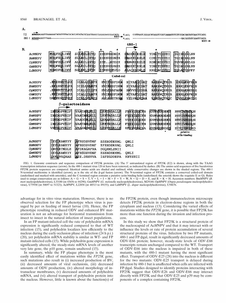
FIG. 1. Genomic constructs and sequence comparison of FP25K proteins. (A) The 59 untranslated region of FP25K (E2) is shown, along with the TAAGtranscription initiation sequence (arrow). In the 480-1 mutant virus 120 nt have been removed, as indicated by dashes. (B) The amino acid sequences of five baculovirusFP25K protein sequences are compared. Identical amino acids are shaded and outlined, while conservative changes are shown in the shaded regions. The 480-1N-terminal methionine is identified (arrow), as is the site of the b-gal fusion (arrow). The N-terminal region of FP25K contains a conserved coiled-coil domain(underlined and marked with asterisks), and the C-terminal region contains a putative actin binding helix (underlined; the asterisk shows the requisite E or Q). Rulesused to assign conservation are as follows: A 5 G 5 S 5 T, V 5 L 5 I 5 M 5 F 5 Y 5 W, N 5 Q 5 D 5 E, and R 5 K 5 H. Accession numbers: BmMNPV (B.mori nucleopolyhedrovirus), L33180 (nt 43656 to 44298); GmMNPV (G. melonella nucleopolyhedrovirus), M29140; OpMNPV (Orgyia pseudosugata nucleopolyhedro-virus), U75930 (nt 50697 to 51323); AcMNPV, L22858 (nt 48513 to 49155); and LdMNPV (L. dispar nucleopolyhedrovirus), U58676.
8560 BRAUNAGEL ET AL. J. VIROL.

MATERIALS AND METHODS
Insect cell lines and virus. S. frugiperda IPLB-Sf21-AE clonal isolate 9 (Sf9)cells were cultured in suspension at 27°C in TNMFH medium (31) supplementedwith 10% fetal bovine serum (complete medium). AcMNPV (strain E2) was usedto infect cells at a known multiplicity of infection (MOI), with time zero set at thetime of virus addition. After 1 h of adsorption, cells were washed and resus-pended in fresh complete medium. The FP mutant viruses 480-1 and FP-bgalwere described by Beames and Summers (1, 2) and are summarized in Fig. 1.
Western blot analysis of infected cells, virus, and virus fractionation. Sf9 cellswere infected with either AcMNPV (WT) or 480-1 or FP-bgal virus (MOI, 20),and at an appropriate time p.i., the cells were collected and washed once withphosphate-buffered saline (PBS). Cell pellets were resuspended in PBS contain-ing protease inhibitors (20 mg of leupeptin per ml, 20 mg of aprotinin per ml, 20mg of pepstatin A per ml, 0.5 mM phenylmethylsulfonyl fluoride, 1 mM E64).Cells were broken by sonication, and protein concentrations were determined bythe method of Bradford (4).
BV was purified from the cell culture supernatant of infected cells (36 h p.i.),and ODV was purified from infected-cell lysates (72 h p.i.), by the techniquedescribed by Braunagel and Summers (7). Purified virus was further fractionatedinto envelope and nucleocapsid fractions (7). The purified virus and respectiveenvelope and nucleocapsid fractions were analyzed by using the ODV envelopemarker proteins ODV-E66, ODV-E56, ODV-E18, ODV-E25, BV/ODV-E26,and ODV-EC27, and the BV was characterized by using the envelope markersgp64 and BV/ODV-E26. ODV, BV, and viral fractions were analyzed by usingthe nucleocapsid marker p39. An example of such a characterization is shown inFig. 3F.
Sodium dodecyl sulfate-polyacrylamide gel electrophoresis (SDS-PAGE) wasperformed as described by Laemmli (22) (4% stacking gel, 12.5% separating gel).Samples were incubated in 1.5% SDS–0.5% b-mercaptoethanol–25 mM Tris-HCl (pH 6.8)–7% glycerol for 15 min at 65°C. Test gels were run and stained withCoomassie blue to visually confirm that protein was loaded at equal concentra-tions per sample. Following electrophoresis, the gels were transferred ontoImmobilon-P membranes (Millipore, Bedford, Mass.). The membranes wereblocked with TTBS-BLOTTO (150 mM NaCl, 10 mM Tris, and 0.1% Tween 20[pH 8.0] supplemented with 1% nonfat dry milk). Antibody was bound overnight(4°C), the blots were washed twice with TBS, and horseradish peroxidase-linkedimmunoglobulin G (1:5,000) was bound for 1 h at room temperature. The blotswere washed three times with TTBS, reacted for 1 min with ECL (Amersham,Arlington Heights, Ill.) chemiluminescence reagent, and exposed to X-ray film.For each antibody determination the entire experiment was performed as amatched set; thus, a direct comparison of signal intensities reflects differingamounts of bound antibody.
The following antibodies and dilutions were used for Fig. 3: anti-FP25K, no.2804 (1:1,000); anti-E66, no. 5297 (1:1,000); anti-E25 (provided by G. Rohr-mann, Oregon State University, Corvallis) (1:2,000); anti-E56, no. 6543 (1:1,000);anti-E18, no. 7350 (1:1,000); anti-EC27, no. 7351 (1:1,000); anti-gp67 B12D5(provided by L. Volkman, University of California, Berkeley) (1:1,000); anti-E26,no. 7554 (1:1,000); anti-p78/83 (provided by C. Richardson, Amgen Institute,Toronto, Ontario, Canada) (1:2,000); anti-p39, p10C6 (provided by L. Volkman)(1:1,000); and anti-gp41, monoclonal antibody 3.10, 6.31 (provided by P. Faulk-ner, Queens University, Kingston, Ontario, Canada) (1:1,000).
Immunofluorescence microscopy. Cells were processed for light microscopy byusing a modification of previously described procedures (8). Sf9 cells were in-fected (MOI, 20), and at the appropriate time p.i. cells were rinsed with Grace’smedium and fixed with 3.7% paraformaldehyde in PBS (20 mM phosphate, 140mM NaCl, pH 7.2) for 10 min at room temperature. The fixative was removed,and cells were washed and permeabilized with methanol (10 min) and subse-quently treated with 0.5% Triton-X 100 (10 min), followed by two rinses withPBS. The cells were blocked for 1 h in 1% normal goat serum–3% bovine serumalbumin and then incubated with primary antibody (anti-FP25K, no. 2804,1:1,000; anti-E66, no. 5297, 1:1,000; or anti-E25, 1:2,000 in 1% normal goatserum in PBS) overnight at 4°C. Cells were rinsed three times, and secondaryantibody (fluorescein isothiocyanate [FITC] (Sigma, St. Louis, Mo.; 1:100 inPBS) was added and incubated for 1 h. The cells were washed, and the nucleuswas visualized by staining with DAPI (49,6-diamidino-2-phenylindole) (0.1 mg/mlin PBS). Cells were viewed and photographed with a Zeiss Axiovert 135 pho-tomicroscope. Each experiment was performed three to five times, and obser-vations were made by using both FITC- and tetramethyl rhodamine isothiocya-nate-labeled secondary antibodies. Thus, several thousand cells were viewed perexperiment, and representative cells were chosen for data presentation.
Quantitative primer extension. Sf9 cells were infected with either AcMNPV orFP-bgal (MOI, 10), and infected-cell RNA was isolated by the method of Chir-gwin et al. (9). Primer extensions were performed with 30 mg of RNA hybridizedto specific oligonucleotide probes labeled with [g-32P]ATP (29). The oligonucleo-tide sequence of the ODV-E66 probe was 59-GATAGGTACAAAAAACATATTAAAAATATTA CAACTATGAC-39, and that of the vp39 probe was 59-CGCGAAAATGCAGCGATTAACTCTCATTTGTCGCGGCGCC-39. RNA-prim-er hybrids were precipitated with ethanol, washed with 70% ethanol, and resus-pended in 30 ml of reverse transcription reaction mix (50 mM Tris [pH 7.6], 60mM KCl, 10 mM MgCl2, 0.66 mM deoxynucleoside triphosphates, 1 mM dithio-threitol, 40 ml of RNasin, 50 ml of actinomycin D per ml, and 150 U of Moloney
murine leukemia virus reverse transcriptase [U.S. Biochemicals, Cleveland,Ohio]). Extension of the annealed primers was performed at 42°C for 2 h. Thereaction products were ethanol precipitated and resuspended in 2 ml of 0.1 NNaOH. After a 30-min incubation to eliminate the RNA template, 4 ml of se-quencing stop buffer was added, and the samples were boiled for 3 min andanalyzed by electrophoresis on a urea–6% polyacrylamide gel together with asequencing ladder generated by using the same oligonucleotides. The gels weredried and subjected to autoradiography, and the primer extension products werequantitated with the FUJIX BAS2000 bioimaging analyzer system (Fuji PhotoFilm Co., Tokyo, Japan).
Immunoprecipitation. A total of 1.5 3 106 infected cells (MOI, 20) were usedfor each immunoprecipitation. At the appropriate time, cells were collected andresuspended in 500 ml of TBN buffer (50 mM Tris [pH 8.0], 150 mM NaCl, 10 mgof leupeptin per ml, 10 mg of aprotinin per ml, 1 mg of pepstatin A per ml, 1 mMphenylmethylsulfonyl fluoride, 1 mM E64) supplemented with either 0.2%Tween 20 or 1% Nonidet P-40 (NP-40). Cells were incubated for 20 min at 4°Cand then lysed by passage through a 25-gauge needle four times. The lysedextract was centrifuged (3,000 rpm, 10 min, 4°C, Microfuge), and the supernatantwas preabsorbed for 1 h with 25 ml of preimmune serum at 4°C. Protein A-aga-rose (Sigma) (40 ml of a 50% slurry) was added to the extract and incubated for1 h at 4°C. The immune complexes formed during preadsorption were pelletedat 3,000 rpm for 15 min in a Microfuge. The preadsorbed extract was thenimmunoprecipitated with the appropriate antibody overnight (25 ml, 4°C) (ODV-E66, no. 5297; FP25K, no. 2804; gp67, AcV1). Protein A-agarose (40 ml, 50%slurry) was added and incubated for 2 h at 4°C. The agarose beads were washedthree times in TBN and then once in TBS. The washed beads (20 ml) wereprepared for SDS-PAGE (4% stacking gel, 12.5% separating gel).
Yeast two-hybrid library construction and screen. Sf9 cells were infected(MOI, 20), and after 1 h of adsorption, the virus inoculum was removed and thecells were resuspended in complete medium. At 18 or 24 h p.i., cells werecollected and mRNA was isolated by using either the Poly A Tract System 1000(Promega, Madison, Wis.) or the Poly(A) Pure mRNA Isolation Kit (Ambion,Austin, Tex.). The cDNA library was then constructed by using the Two HybridcDNA Construction Kit (Clontech, Palo Alto, Calif.). The libraries were ampli-fied, and the resulting titers of the amplified libraries were as follows: 18 h p.i.,1.31 3 1012; 24 h p.i., 3.25 3 1013. To harvest large quantities of DNA from eachlibrary, a 1-ml aliquot of amplified library was diluted and grown on 200 Luriabroth-ampicillin supplemented plates (150-mm diameter), bacteria were har-vested, and plasmid DNA was purified by using the Plasmid Giga Kit (Qiagen,Valencia, Calif.).
The appropriate genes were cloned into the yeast binding domain vector.D23-E66 was constructed in the yeast vector pAS2-1 such that the N-terminalMet was followed by the FLAG epitope (D-Y-K-D-D-D-D-K) (Kodak, NewHaven, Conn.) followed by amino acids 24 to 704 of ODV-E66 (16). Libraryscreens were performed with the Matchmaker Two Hybrid System 2 (Clontech)and the complementation assay with blue selection for b-galactosidase (b-gal)activity. The colonies that showed a positive blue interacting color reactionwithin an 8-h period were confirmed by using a secondary reaction. The yeastactivation domain plasmid was then purified and transformed into Escherichiacoli DH5a, and DNA was amplified and sequenced.
RESULTS
FP25K homology comparison and selection of mutant vi-ruses. One goal of this study was to examine the effects ofFP25K deletions and potential interactions between FP25Kand structural proteins of AcMNPV. To consider a design forFP25K gene mutations, we examined the predicted structuralfeatures of the FP25K protein. Computer-assisted analysis re-vealed a highly conserved coiled-coil domain (a structural mo-tif often involved in protein-protein interactions) at the Nterminus and a putative actin binding helix (Fig. 1). Two pre-viously reported viral mutants (1, 2), each lacking one of theseregions, were selected for these studies. The 480-1 mutant viruscontains a 120-nucleotide (nt) deletion extending from posi-tion 245 to 177 (relative to the FP25K initiation codon),resulting in translation initiation at an internal Met (Fig. 1A).Thus, in this mutant the putative coiled-coil domain was de-leted, resulting in an FP25K protein with a 31-amino-acidN-terminal truncation (13) (Fig. 1). The FP-bgal mutant lacksthe C-terminal half of FP25K, including the putative actingbinding helix, and instead contains the amino acids b-gal. Notethat although the FP25K protein is highly conserved through-out the putative actin binding region, the last 20 to 25 aminoacid residues show little conservation (Fig. 1B).
VOL. 73, 1999 FP25K MUTATIONS ALTER ODV-E66 INTRANUCLEAR TRAFFICKING 8561
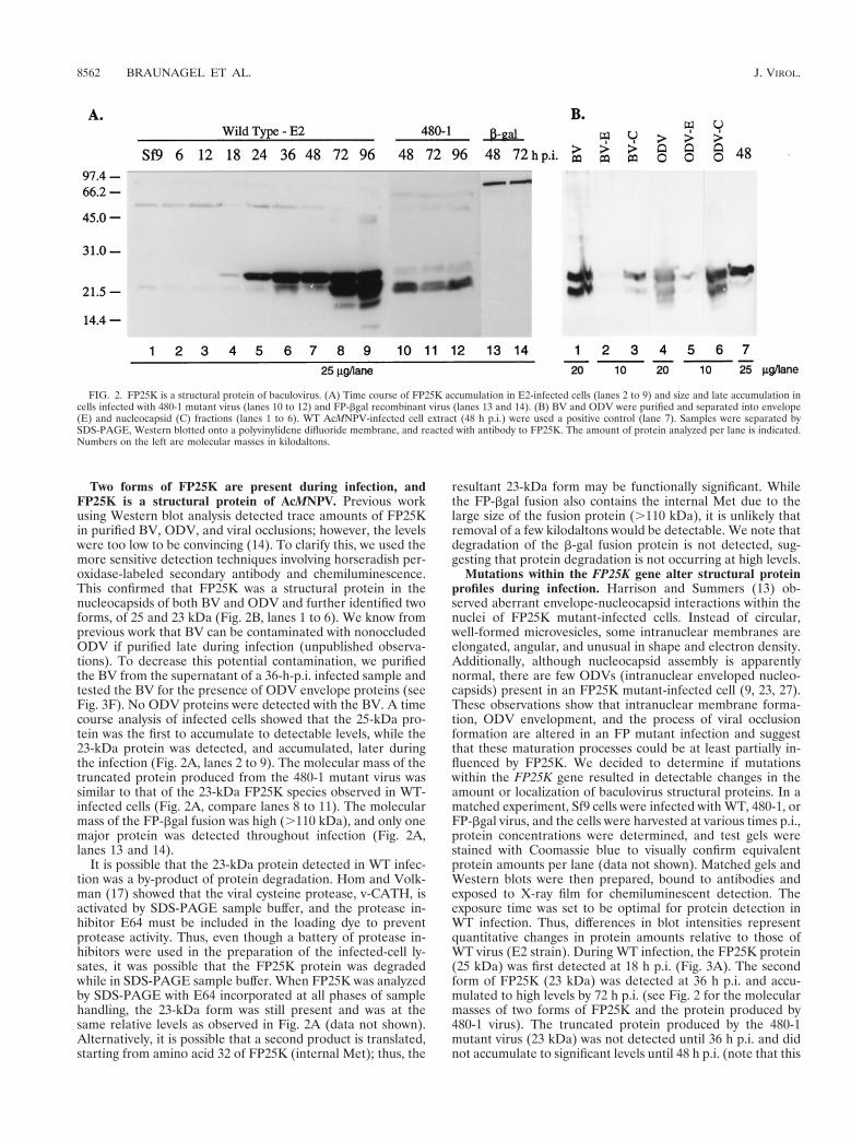
Two forms of FP25K are present during infection, andFP25K is a structural protein of AcMNPV. Previous workusing Western blot analysis detected trace amounts of FP25Kin purified BV, ODV, and viral occlusions; however, the levelswere too low to be convincing (14). To clarify this, we used themore sensitive detection techniques involving horseradish per-oxidase-labeled secondary antibody and chemiluminescence.This confirmed that FP25K was a structural protein in thenucleocapsids of both BV and ODV and further identified twoforms, of 25 and 23 kDa (Fig. 2B, lanes 1 to 6). We know fromprevious work that BV can be contaminated with nonoccludedODV if purified late during infection (unpublished observa-tions). To decrease this potential contamination, we purifiedthe BV from the supernatant of a 36-h-p.i. infected sample andtested the BV for the presence of ODV envelope proteins (seeFig. 3F). No ODV proteins were detected with the BV. A timecourse analysis of infected cells showed that the 25-kDa pro-tein was the first to accumulate to detectable levels, while the23-kDa protein was detected, and accumulated, later duringthe infection (Fig. 2A, lanes 2 to 9). The molecular mass of thetruncated protein produced from the 480-1 mutant virus wassimilar to that of the 23-kDa FP25K species observed in WT-infected cells (Fig. 2A, compare lanes 8 to 11). The molecularmass of the FP-bgal fusion was high (.110 kDa), and only onemajor protein was detected throughout infection (Fig. 2A,lanes 13 and 14).
It is possible that the 23-kDa protein detected in WT infec-tion was a by-product of protein degradation. Hom and Volk-man (17) showed that the viral cysteine protease, v-CATH, isactivated by SDS-PAGE sample buffer, and the protease in-hibitor E64 must be included in the loading dye to preventprotease activity. Thus, even though a battery of protease in-hibitors were used in the preparation of the infected-cell ly-sates, it was possible that the FP25K protein was degradedwhile in SDS-PAGE sample buffer. When FP25K was analyzedby SDS-PAGE with E64 incorporated at all phases of samplehandling, the 23-kDa form was still present and was at thesame relative levels as observed in Fig. 2A (data not shown).Alternatively, it is possible that a second product is translated,starting from amino acid 32 of FP25K (internal Met); thus, the
resultant 23-kDa form may be functionally significant. Whilethe FP-bgal fusion also contains the internal Met due to thelarge size of the fusion protein (.110 kDa), it is unlikely thatremoval of a few kilodaltons would be detectable. We note thatdegradation of the b-gal fusion protein is not detected, sug-gesting that protein degradation is not occurring at high levels.
Mutations within the FP25K gene alter structural proteinprofiles during infection. Harrison and Summers (13) ob-served aberrant envelope-nucleocapsid interactions within thenuclei of FP25K mutant-infected cells. Instead of circular,well-formed microvesicles, some intranuclear membranes areelongated, angular, and unusual in shape and electron density.Additionally, although nucleocapsid assembly is apparentlynormal, there are few ODVs (intranuclear enveloped nucleo-capsids) present in an FP25K mutant-infected cell (9, 23, 27).These observations show that intranuclear membrane forma-tion, ODV envelopment, and the process of viral occlusionformation are altered in an FP mutant infection and suggestthat these maturation processes could be at least partially in-fluenced by FP25K. We decided to determine if mutationswithin the FP25K gene resulted in detectable changes in theamount or localization of baculovirus structural proteins. In amatched experiment, Sf9 cells were infected with WT, 480-1, orFP-bgal virus, and the cells were harvested at various times p.i.,protein concentrations were determined, and test gels werestained with Coomassie blue to visually confirm equivalentprotein amounts per lane (data not shown). Matched gels andWestern blots were then prepared, bound to antibodies andexposed to X-ray film for chemiluminescent detection. Theexposure time was set to be optimal for protein detection inWT infection. Thus, differences in blot intensities representquantitative changes in protein amounts relative to those ofWT virus (E2 strain). During WT infection, the FP25K protein(25 kDa) was first detected at 18 h p.i. (Fig. 3A). The secondform of FP25K (23 kDa) was detected at 36 h p.i. and accu-mulated to high levels by 72 h p.i. (see Fig. 2 for the molecularmasses of two forms of FP25K and the protein produced by480-1 virus). The truncated protein produced by the 480-1mutant virus (23 kDa) was not detected until 36 h p.i. and didnot accumulate to significant levels until 48 h p.i. (note that this
FIG. 2. FP25K is a structural protein of baculovirus. (A) Time course of FP25K accumulation in E2-infected cells (lanes 2 to 9) and size and late accumulation incells infected with 480-1 mutant virus (lanes 10 to 12) and FP-bgal recombinant virus (lanes 13 and 14). (B) BV and ODV were purified and separated into envelope(E) and nucleocapsid (C) fractions (lanes 1 to 6). WT AcMNPV-infected cell extract (48 h p.i.) were used a positive control (lane 7). Samples were separated bySDS-PAGE, Western blotted onto a polyvinylidene difluoride membrane, and reacted with antibody to FP25K. The amount of protein analyzed per lane is indicated.Numbers on the left are molecular masses in kilodaltons.
8562 BRAUNAGEL ET AL. J. VIROL.
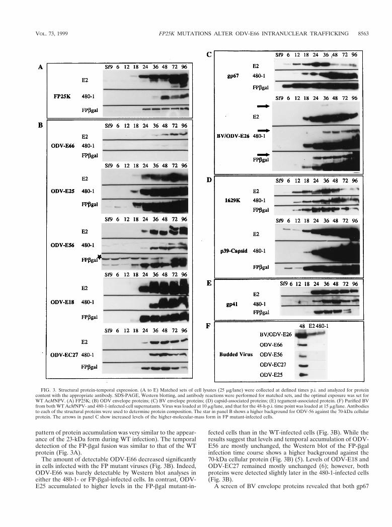
pattern of protein accumulation was very similar to the appear-ance of the 23-kDa form during WT infection). The temporaldetection of the FP-bgal fusion was similar to that of the WTprotein (Fig. 3A).
The amount of detectable ODV-E66 decreased significantlyin cells infected with the FP mutant viruses (Fig. 3B). Indeed,ODV-E66 was barely detectable by Western blot analyses ineither the 480-1- or FP-bgal-infected cells. In contrast, ODV-E25 accumulated to higher levels in the FP-bgal mutant-in-
fected cells than in the WT-infected cells (Fig. 3B). While theresults suggest that levels and temporal accumulation of ODV-E56 are mostly unchanged, the Western blot of the FP-bgalinfection time course shows a higher background against the70-kDa cellular protein (Fig. 3B) (5). Levels of ODV-E18 andODV-EC27 remained mostly unchanged (6); however, bothproteins were detected slightly later in the 480-1-infected cells(Fig. 3B).
A screen of BV envelope proteins revealed that both gp67
FIG. 3. Structural protein-temporal expression. (A to E) Matched sets of cell lysates (25 mg/lane) were collected at defined times p.i. and analyzed for proteincontent with the appropriate antibody. SDS-PAGE, Western blotting, and antibody reactions were performed for matched sets, and the optimal exposure was set forWT AcMNPV. (A) FP25K; (B) ODV envelope proteins; (C) BV envelope proteins; (D) capsid-associated proteins; (E) tegument-associated protein. (F) Purified BVfrom both WT AcMNPV- and 480-1-infected-cell supernatants. Virus was loaded at 10 mg/lane, and that for the 48-h-p.i. time point was loaded at 15 mg/lane. Antibodiesto each of the structural proteins were used to determine protein composition. The star in panel B shows a higher background for ODV-56 against the 70-kDa cellularprotein. The arrows in panel C show increased levels of the higher-molecular-mass form in FP mutant-infected cells.
VOL. 73, 1999 FP25K MUTATIONS ALTER ODV-E66 INTRANUCLEAR TRAFFICKING 8563
and BV/ODV-E26 accumulated to higher levels during FPmutant infection than during WT infection (Fig. 3C). Addi-tionally, the higher-molecular-mass, immunoreactive form ofBV/ODV-E26 (3) was also detected at increased levels in theFP mutant-infected cells than in the WT-infected cells (Fig.3C). The levels of the capsid protein p78/83 (1629K) was notaltered compared those in WT infection. However, anothernucleocapsid protein, p39, was detected in significantly higherquantities in 480-1 mutant-infected cells but not during FP-bgal infection (Fig. 3D). We also observed that in both WT-and FP-bgal-infected cells, p39 was detected at 6 h p.i. Earlierstudies of p39 suggest that transcription of the p39 gene occurslate (32); however, an early transcription consensus motif(CAGT) is present at nt 2317, just 4 nt away from a utilizedlate transcription initiation motif. Thus, it is possible that p39is transcribed at low levels early in infection. p39 was notdetected at 2 or 4 h p.i. (data not shown). Effects on the onlyknown tegument protein, gp41, were minimal; however, a de-creased amount of gp41 was observed very late in FP-bgalinfection (72 to 96 h p.i.) (Fig. 3E).
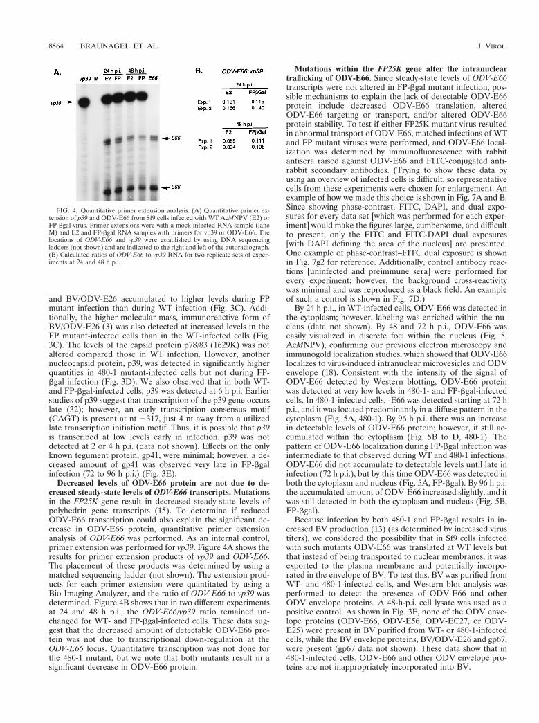
Decreased levels of ODV-E66 protein are not due to de-creased steady-state levels of ODV-E66 transcripts. Mutationsin the FP25K gene result in decreased steady-state levels ofpolyhedrin gene transcripts (15). To determine if reducedODV-E66 transcription could also explain the significant de-crease in ODV-E66 protein, quantitative primer extensionanalysis of ODV-E66 was performed. As an internal control,primer extension was performed for vp39. Figure 4A shows theresults for primer extension products of vp39 and ODV-E66.The placement of these products was determined by using amatched sequencing ladder (not shown). The extension prod-ucts for each primer extension were quantitated by using aBio-Imaging Analyzer, and the ratio of ODV-E66 to vp39 wasdetermined. Figure 4B shows that in two different experimentsat 24 and 48 h p.i., the ODV-E66/vp39 ratio remained un-changed for WT- and FP-bgal-infected cells. These data sug-gest that the decreased amount of detectable ODV-E66 pro-tein was not due to transcriptional down-regulation at theODV-E66 locus. Quantitative transcription was not done forthe 480-1 mutant, but we note that both mutants result in asignificant decrease in ODV-E66 protein.
Mutations within the FP25K gene alter the intranucleartrafficking of ODV-E66. Since steady-state levels of ODV-E66transcripts were not altered in FP-bgal mutant infection, pos-sible mechanisms to explain the lack of detectable ODV-E66protein include decreased ODV-E66 translation, alteredODV-E66 targeting or transport, and/or altered ODV-E66protein stability. To test if either FP25K mutant virus resultedin abnormal transport of ODV-E66, matched infections of WTand FP mutant viruses were performed, and ODV-E66 local-ization was determined by immunofluorescence with rabbitantisera raised against ODV-E66 and FITC-conjugated anti-rabbit secondary antibodies. (Trying to show these data byusing an overview of infected cells is difficult, so representativecells from these experiments were chosen for enlargement. Anexample of how we made this choice is shown in Fig. 7A and B.Since showing phase-contrast, FITC, DAPI, and dual expo-sures for every data set [which was performed for each exper-iment] would make the figures large, cumbersome, and difficultto present, only the FITC and FITC-DAPI dual exposures[with DAPI defining the area of the nucleus] are presented.One example of phase-contrast–FITC dual exposure is shownin Fig. 7g2 for reference. Additionally, control antibody reac-tions [uninfected and preimmune sera] were performed forevery experiment; however, the background cross-reactivitywas minimal and was reproduced as a black field. An exampleof such a control is shown in Fig. 7D.)
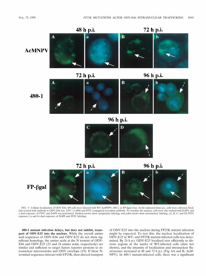
By 24 h p.i., in WT-infected cells, ODV-E66 was detected inthe cytoplasm; however, labeling was enriched within the nu-cleus (data not shown). By 48 and 72 h p.i., ODV-E66 waseasily visualized in discrete foci within the nucleus (Fig. 5,AcMNPV), confirming our previous electron microscopy andimmunogold localization studies, which showed that ODV-E66localizes to virus-induced intranuclear microvesicles and ODVenvelope (18). Consistent with the intensity of the signal ofODV-E66 detected by Western blotting, ODV-E66 proteinwas detected at very low levels in 480-1- and FP-bgal-infectedcells. In 480-1-infected cells, -E66 was detected starting at 72 hp.i., and it was located predominantly in a diffuse pattern in thecytoplasm (Fig. 5A, 480-1). By 96 h p.i. there was an increasein detectable levels of ODV-E66 protein; however, it still ac-cumulated within the cytoplasm (Fig. 5B to D, 480-1). Thepattern of ODV-E66 localization during FP-bgal infection wasintermediate to that observed during WT and 480-1 infections.ODV-E66 did not accumulate to detectable levels until late ininfection (72 h p.i.), but by this time ODV-E66 was detected inboth the cytoplasm and nucleus (Fig. 5A, FP-bgal). By 96 h p.i.the accumulated amount of ODV-E66 increased slightly, and itwas still detected in both the cytoplasm and nucleus (Fig. 5B,FP-bgal).
Because infection by both 480-1 and FP-bgal results in in-creased BV production (13) (as determined by increased virustiters), we considered the possibility that in Sf9 cells infectedwith such mutants ODV-E66 was translated at WT levels butthat instead of being transported to nuclear membranes, it wasexported to the plasma membrane and potentially incorpo-rated in the envelope of BV. To test this, BV was purified fromWT- and 480-1-infected cells, and Western blot analysis wasperformed to detect the presence of ODV-E66 and otherODV envelope proteins. A 48-h-p.i. cell lysate was used as apositive control. As shown in Fig. 3F, none of the ODV enve-lope proteins (ODV-E66, ODV-E56, ODV-EC27, or ODV-E25) were present in BV purified from WT- or 480-1-infectedcells, while the BV envelope proteins, BV/ODV-E26 and gp67,were present (gp67 data not shown). These data show that in480-1-infected cells, ODV-E66 and other ODV envelope pro-teins are not inappropriately incorporated into BV.
FIG. 4. Quantitative primer extension analysis. (A) Quantitative primer ex-tension of p39 and ODV-E66 from Sf9 cells infected with WT AcMNPV (E2) orFP-bgal virus. Primer extensions were with a mock-infected RNA sample (laneM) and E2 and FP-bgal RNA samples with primers for vp39 or ODV-E66. Thelocations of ODV-E66 and vp39 were established by using DNA sequencingladders (not shown) and are indicated to the right and left of the autoradiograph.(B) Calculated ratios of ODV-E66 to vp39 RNA for two replicate sets of exper-iments at 24 and 48 h p.i.
8564 BRAUNAGEL ET AL. J. VIROL.
480-1 mutant infection delays, but does not inhibit, trans-port of ODV-E25 into the nucleus. While the overall aminoacid sequences of ODV-E66 and ODV-E25 do not show sig-nificant homology, the amino acids at the N termini of ODV-E66 and ODV-E25 (23 and 24 amino acids, respectively) aresimilar and sufficient to target fusion reporter proteins to in-tranuclear microvesicles and ODV envelope (19). If these N-terminal sequences interact with FP25K, then altered transport
of ODV-E25 into the nucleus during FP25K mutant infectionmight be expected. To test this, the nuclear localization ofODV-E25 in WT- and FP25K mutant-infected cells was deter-mined. By 24 h p.i. ODV-E25 localized very efficiently to dis-crete regions in the nuclei of WT-infected cells (data notshown), and the intensity of localization and intranuclear flu-orescence increased at 48 and 72 h p.i. (Fig. 6A and B, AcM-NPV). In 480-1 mutant-infected cells, there was a significant
FIG. 5. Cellular localization of ODV-E66. Sf9 cells were infected with WT AcMNPV, 480-1, or FP-bgal virus. At the indicated times p.i., cells were collected, fixed,and reacted with antibody to ODV-E66 (no. 5297; 1:1,000) and FITC-conjugated secondary antibody. To visualize the nucleus, cells were also stained with DAPI, anda dual exposure of FITC and DAPI was performed. Dashed arrows show cytoplasmic labeling, and solid arrows show intranuclear labeling. (A, B, C, and D) FITCexposure; (a and b) dual exposure of DAPI and FITC labeling.
VOL. 73, 1999 FP25K MUTATIONS ALTER ODV-E66 INTRANUCLEAR TRAFFICKING 8565

delay of localization of ODV-E25 in the nucleus. By 48 h p.i.ODV-E25 was located predominantly in the cytoplasm (Fig.6A, 480-1); however, by 72 h p.i., ODV-E25 was detected inthe nucleus in a pattern similar to that for WT infection (Fig.6B, 480-1). Like for WT infection, immunoelectron microscopyshowed that at 72 h p.i. ODV-E25 was present in intranuclearmicrovesicles and the ODV envelope in the 480-1 mutant-infected cells (data not shown). The nuclear localization ofODV-E25 in the FP-bgal mutant-infected cells was indistin-guishable from that in WT infection: by 24 h p.i. ODV-E25 wasdetected predominantly within the nucleus (data not shown),and it remained intranuclear throughout infection (Fig. 6A andB, FP-bgal).
Localization of FP25K in WT- and FP mutant-infected cells.To provide an overview of FP25K localization during infection,we used immunofluorescence microscopy of WT-, 480-1-, orFP-bgal-infected cells (Fig. 7). Figure 7 also exemplifies howthe representative cells shown throughout this work were se-lected. An example of these is shown in Fig. 7a1, a2, b1, and b2and then enlarged in Fig. 7A and B. This analysis showed thatsignificant amounts of FP25K were present in the cytoplasmthroughout infection (Fig. 7A to C). It is known from previouswork that FP25K protein is present in both cytoplasmic andnuclear fraction, and it accumulates in electron-dense regions(14). These discrete regions enriched in FP25K could be de-tected as the microscope operator “focused through” the cell;however, they were not easily discernible by using a single
focus, as shown in Fig. 7. The mutant 480-1 FP25K protein wasnot detected until 48 h p.i. (Fig. 7E), and even then the levelswere too low to convincingly localize this protein; however,most of the truncated protein appeared to be cytoplasmic. By72 and 96 h p.i., the localization of the truncated protein moreclosely resembled that seen in WT infection (Fig. 7F and datanot shown). The FP-bgal fusion protein showed a pattern oflocalization similar to that in WT infection (Fig. 7G, g1, andg2). Since the results for FP-bgal were so similar to those forthe WT, only one time point is shown (48 h p.i.), and anexample of the matched phase-contrast–FITC dual exposure isalso shown (Fig. 7g2). Like the results seen with antibodies toODV-E66 and ODV-E25, background FITC levels of FP25Kantibody tested against uninfected cells were reproduced asblack field (Fig. 7D).
FP25K potentially interacts with other baculovirus struc-tural proteins. The Western blot analysis showed that com-pared to WT infection, infections by the FP25K mutant virusesresulted in increased protein levels of both BV envelope pro-teins, BV/ODV-E26 and gp67 (Fig. 3C). Previous data showthat FP25K is capable of interacting with BV/ODV-E26 (3)(summarized in Table 1). Antibody to gp67 (AcV1) coprecipi-tated FP25K, and the total amount of precipitated FP25Kdecreased as the amount of gp67 decreased (Fig. 8A, lanes 2 to5). When the reciprocal experiment was performed with anti-body to FP25K, gp67 was not coprecipitated; however, the
FIG. 6. Cellular localization of ODV-E25. Sf9 cells were infected with WT AcMNPV, 480-1, or FP-bgal virus. At the indicated times p.i., cells were collected, fixed,and reacted with antibody to ODV-E25 (1:1,000) and FITC-conjugated secondary antibody. To visualize the nucleus, cells were also stained with DAPI, and a dualexposure of FITC and DAPI was performed. Dashed arrows show cytoplasmic labeling, and solid arrows show intranuclear labeling. (A and B) FITC exposure alone;(a and b) dual exposure of DAPI and FITC labeling.
8566 BRAUNAGEL ET AL. J. VIROL.
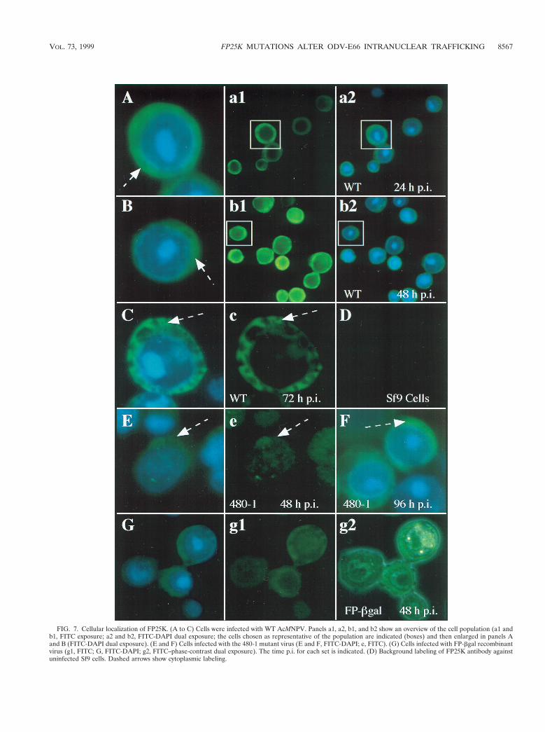
FIG. 7. Cellular localization of FP25K. (A to C) Cells were infected with WT AcMNPV. Panels a1, a2, b1, and b2 show an overview of the cell population (a1 andb1, FITC exposure; a2 and b2, FITC-DAPI dual exposure; the cells chosen as representative of the population are indicated (boxes) and then enlarged in panels Aand B (FITC-DAPI dual exposure). (E and F) Cells infected with the 480-1 mutant virus (E and F, FITC-DAPI; e, FITC). (G) Cells infected with FP-bgal recombinantvirus (g1, FITC; G, FITC-DAPI; g2, FITC–phase-contrast dual exposure). The time p.i. for each set is indicated. (D) Background labeling of FP25K antibody againstuninfected Sf9 cells. Dashed arrows show cytoplasmic labeling.
VOL. 73, 1999 FP25K MUTATIONS ALTER ODV-E66 INTRANUCLEAR TRAFFICKING 8567
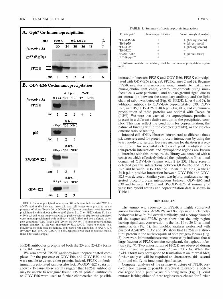
FP25K antibodies precipitated both the 23- and 25-kDa forms(Fig. 8A, lane 1).
We also tested FP25K antibody-immunoprecipitated com-plexes for the presence of ODV-E66 and ODV-E25, and wewere unable to detect either protein. Indeed, FP25K antibody-immunoprecipitated samples also lack BV/ODV-E26 (data notshown). Because these results suggest that FP25K antibodiesmay be unable to recognize bound FP25K protein, antibodiesto ODV-E66 were used to further characterize the possible
interaction between FP25K and ODV-E66. FP25K coprecipi-tated with ODV-E66 (Fig. 8B; FP25K, lanes 2 and 3). BecauseFP25K migrates at a molecular weight similar to that of im-munoglobulin light chain, control experiments using unin-fected cells were performed, and no background signal due toan interaction between the secondary antibody and the lightchain of rabbit was detected (Fig. 8B, FP25K, lanes 4 and 5). Inaddition, antibody to ODV-E66 coprecipitated p39, ODV-E25, and BV/ODV-E26 at 48 h p.i. (Fig. 8B), and coimmuno-precipitation of these proteins was optimal with Tween 20(0.2%). We note that each of the coprecipitated proteins ispresent in a different relative amount in the precipitated com-plex. This may reflect the conditions for coprecipitation, thenature of binding within the complex (affinity), or the stoichi-ometric ratio of binding.
Infected-cell cDNA libraries constructed at different timesp.i. were screened for protein-protein interactions by using theyeast two-hybrid system. Because nuclear localization is a req-uisite event for successful detection of yeast two-hybrid pro-tein-protein interactions and hydrophobic regions are knownto interfere with this transport, the library was screened with aconstruct which effectively deleted the hydrophobic N-terminaldomain of ODV-E66 (amino acids 2 to 23). These screensdetected positive interactions between ODV-E66 and ODV-E25 and between ODV-E66 and FP25K at 18 h p.i., while at24 h p.i. a positive interaction between ODV-E66 and ODV-E25 was detected. Similar yeast two-hybrid analyses also sug-gested protein-protein interactions between ODV-E66 andp39 and between FP25K and BV/ODV-E26. A summary ofyeast two-hybrid results and coprecipitation data is shown inTable 1.
DISCUSSION
The amino acid sequence of FP25K is highly conservedamong baculoviruses. AcMNPV and Bombyx mori nucleopoly-hedrovirus have 96.7% overall similarity, and a comparison ofall the sequenced FP25K genes show that the only regionlacking significant conservation is the last 19 to 26 C-terminalamino acids (Fig. 1). Immunoblot analyses performed withpurified AcMNPV ODV and BV show that FP25K is a struc-tural protein in the nucleocapsids of both progeny viruses (Fig.2); however, immunofluorescence microscopy indicates that alarge fraction of FP25K remains cytoplasmic throughout infec-tion (Fig. 7). Two major forms of FP25K are observed duringinfection and in purified virus: 25 and 23 kDa. While the23-kDa form may correspond to translation at an internal Met,further analyses will be required to characterize this secondform and clarify its functional significance.
Computer analyses of the primary structure of FP25K pre-dicted two regions of possible structural relevance: a coiled-coil region and a putative actin binding helix (Fig. 1). Viralmutants lacking either of these regions were chosen for further
FIG. 8. Immunoprecipitation analyses. Sf9 cells were infected with WT Ac-MNPV and at the indicated times p.i., and cell lysates were prepared in thepresence of either Tween 20 or NP-40. (A) Protein complexes were immuno-precipitated with antibody (Ab) to gp67 (lanes 2 to 5) or FP25K (lane 1). Lane6, 30-h-p.i. cell lysate sample analyzed as positive control. (B) Protein complexeswere immunoprecipitated with antibody to ODV-E66 and two different deter-gent conditions (0.2% Tween 20 [T20] or 1% NP-40). The immunoprecipitatedprotein complex (20 ml) was analyzed by SDS-PAGE, Western blotted to apolyvinylidene difluoride membrane, and reacted with antibodies to FP25K, p39,BV/ODV-E26, or ODV-E25. A 48-h-p.i. cell lysate was used as positive control(lane 1 for each sample).
TABLE 1. Summary of protein-protein interactions
Protein paira Immunoprecipitation Yeast two-hybrid analysis
*E66-FP25K 1 1 (library screen)*E66-p39 1 1 (direct cross)*E66-E25 1 1 (library screen)*E66-E26 1 2FP25K-E26* 1 1 (direct cross)FP25K-gp67* 1 2
a Asterisks indicate the antibody used for the immunoprecipitation experi-ment.
8568 BRAUNAGEL ET AL. J. VIROL.

study, and infection by either FP25K mutant resulted in adecreased amount of detectable ODV-E66 (Fig. 3). Immuno-fluorescence microscopy showed that during infection by bothmutants, ODV-E66 displayed an altered pattern of intranu-clear localization. This effect was more dramatic during infec-tion by the 480-1 mutant, with infection resulting in a cytoplas-mic location of ODV-E66. In FP-bgal-infected cells, someODV-E66 was detected within the nucleus; however, this traf-ficking was delayed and diminished compared to that with WTinfection (Fig. 5). Since the two mutants produced equivalentamounts of ODV-E66, the effect of the 480-1 mutation onnuclear transport of ODV-E66 is likely not related to a mini-mal protein requirement for initiation of nuclear transport.Additionally, while the amounts of ODV-E25 which accumu-late during WT and 480-1 infection are similar, nuclear trans-port of ODV-E25 was delayed by approximately 48 h, whileintranuclear transport was not affected, in cells infected withFP-bgal. These results suggest that the N-terminal region ofFP25K may be important for normal trafficking of ODV-E66and ODV-E25. Lack of the N-terminal region does not resultin detectable relocalization of ODV envelope proteins to theplasma membrane (data not shown) or incorporation into BV(ODV-E66, ODV-E56, ODV-EC27, and ODV-E25) (Fig. 3F).While the mutant FP-bgal virus was generated from the pa-rental E2 virus, 480-1 is a spontaneous, naturally occurringFP25K mutant virus, and it is possible that additional muta-tions are present (1).
Western blot analysis of a time course of infected cellsshowed that in addition to that of ODV-E66, accumulationlevels of several baculovirus structural proteins were altered.Compared to those in WT-infected cells, the amounts of BVenvelope proteins gp67 and BV/ODV-E26 and the major cap-sid protein p39 increased in mutant-infected cells. We notethat the proteins which exhibited an altered temporal accumu-lation pattern during FP mutant infection (Fig. 3) were thesame proteins that protein-protein interaction studies sug-gested interact with FP25K (Fig. 8 and Table 1). Results fromimmunoprecipitation studies suggest that FP25K may be amember of protein complexes which include gp67, ODV-E66,and BV/ODV-E26. While FP25K coprecipitates with each ofthese proteins, these proteins are not precipitated in the re-ciprocal experiment (with antibody to FP25K). This suggeststhat FP25K may be an integral component of these complexes,and when bound, FP25K is masked from antibody recognition.The interactions of FP25K and ODV-E66 and of FP25K andBV/ODV-E26 have been confirmed by using the complemen-tary yeast two-hybrid technique (Table 1) (3). However, a yeasttwo-hybrid cross between FP25K and gp67 was negative, sug-gesting that this interaction may be mediated by other proteins.Only the 25-kDa form is detected coprecipitating with gp67,ODV-E66, or BV/ODV-E26 (Fig. 8) (3), while antibody toFP25K precipitates both the 25- and 23-kDa forms. These datasuggest that these two forms may be functionally distinct.
The data presented here indicate that in addition to theother, better-characterized effects of mutations within theFP25K gene, the accumulation patterns of several structuralproteins are altered, and intranuclear transport of both ODV-E66 and ODV-E25 is compromised. We note that in the ab-sence of a fully functional FP25K gene, intranuclear local-ization of polyhedrin is also compromised during the earlyocclusion phase of infection (20). FP25K could be affectingprotein accumulation and trafficking by regulating transcrip-tion, mRNA stability, translation, or altered protein stability ofone or many viral proteins. While transcription of ODV-E66was not altered during an infection with the FP-bgal virus (Fig.4), FP mutant virus infection can alter steady-state levels of
polyhedrin (15). The phenotypic hallmarks of the FP mutant,including apparently normal numbers of nucleocapsids butlack of ODV envelopment and a decreased amount of viralocclusions, may not be due to the mutant FP25K protein per sebut may result from improper stoichiometry, localization, orprotein complex formation of other viral proteins. Studiesaimed to further characterize the composition of protein com-plexes containing FP25K and their role in the trafficking path-way(s) of ODV envelope proteins are under way.
ACKNOWLEDGMENTS
We thank Gabriella Marcano and Shawn Williamson for the devel-opment of infected-cell cDNA yeast two-hybrid libraries. We thankTao Hong, Gabriella Marcano, and Shawn Williamson for cloninggenes into yeast two-hybrid vectors and Erin Pinkerton and Bill Keyesfor their excellent technical support. We thank the following people forproviding antisera: Christopher Richardson (Amgen Institute, To-ronto, Ontario, Canada) (p78/83), George Rohrmann (Oregon StateUniversity, Corvallis) (ODV-E25, p78/83), Loy Volkman (Universityof California, Berkeley) (capsid, gp64), and Peter Faulkner (Queen’sUniversity, Kingston, Ontario, Canada) (gp41).
This work was supported in part by National Institutes of Healthgrant 2RO1GM47552 (to M.D.S. and S.C.B.) and Texas AgriculturalExperiment Station Project TEXO8078 (to M.D.S.).
REFERENCES
1. Beames, B., and M. D. Summers. 1988. Comparisons of host cell DNAinsertions and altered transcription at the site of insertions in few polyhedrabaculovirus mutants. Virology 162:206–220.
2. Beames, B., and M. D. Summers. 1989. Location and nucleotide sequence ofthe 25K protein missing from baculovirus few polyhedra (FP) mutants.Virology 168:344–353.
3. Beniya, H., S. C. Braunagel, and M. D. Summers. 1998. Autographa califor-nica nuclear polyhedrosis virus: subcellular localization and protein traffick-ing of BV/ODV-E26 to intranuclear membranes and viral envelopes. Virol-ogy 240:64–75.
4. Bradford, M. M. 1976. A rapid and sensitive method for the quantitation ofmicrogram quantities of protein utilizing the principle of protein-dye bind-ing. Anal. Biochem. 72:248–254.
5. Braunagel, S. C., D. M. Elton, H. Ma, and M. D. Summers. 1996. Identifi-cation and analysis of an Autographa californica nuclear polyhedrosis virusstructural protein of the occlusion-derived virus envelope: ODV-E56. Virol-ogy 217:97–110.
6. Braunagel, S. C., H. He, P. Ramamurthy, and M. D. Summers. 1996. Tran-scription, translation, and cellular localization of three Autographa califor-nica nuclear polyhedrosis virus structural proteins: ODV-E18, ODV-E35and ODV-EC27. Virology 222:100–114.
7. Braunagel, S. C., and M. D. Summers. 1994. Autographa californica nuclearpolyhedrosis virus, PDV, and ECV viral envelopes and nucleocapsids: struc-tural proteins, antigens, lipid and fatty acid profiles. Virology 202:315–328.
8. Charlton, C. A., and L. E. Volkman. 1991. Sequential rearrangement andnuclear polymerization of actin in baculovirus-infected Spodoptera frugiperdacells. J. Virol. 65:1219–1227.
9. Chirgwin, J. M., A. E. Przbyla, R. J. MacDonald, and W. J. Rutter. 1979.Isolation of biologically active ribonucleic acid from sources enriched inribonuclease. Biochemistry 18:5294–5299.
10. Fraser, M. J., and W. F. Hink. 1982. The isolation and characterization of theMP and FP plaque variants of Galleria mellonella nuclear polyhedrosis virus.Virology 117:366–378.
11. Fraser, M. J., and W. F. Hink. 1982. Comparative sensitivity of severalplaque assay techniques employing TN-368 and IPLB-SF 21AE insect celllines for plaque variants of Galleria mellonella nuclear polyhedrosis virus.J. Invert. Pathol. 40:89–97.
12. Fraser, M. J., G. E. Smith, and M. D. Summers. 1983. Acquisition of hostcell DNA sequences by baculoviruses: relationship between host DNA in-sertions and FP mutants of Autographa californica and Galleria mellonellanuclear polyhedrosis virus. J. Virol. 47:287–300.
13. Harrison, R. L., and M. D. Summers. 1995. Mutations in the Autographacalifornica multinucleocapsid nuclear polyhedrosis virus 25 kDa protein generesult in reduced virion occlusion, altered intranuclear envelopment andenhanced virus production. J. Gen. Virol. 76:1451–1459.
14. Harrison, R. L., and M. D. Summers. 1995. Biosynthesis and localization ofthe Autographa californica nuclear polyhedrosis virus 25K gene product.Virology 208:279–288.
15. Harrison, R. L., D. L. Jarvis, and M. D. Summers. 1996. The role of theAcMNPV 25K gene, “FP25,” in baculovirus polh and p10 expression. Virol-ogy 226:34–46.
VOL. 73, 1999 FP25K MUTATIONS ALTER ODV-E66 INTRANUCLEAR TRAFFICKING 8569

16. Hink, W. F., and P. V. Vail. 1973. A plaque assay for titration of alfalfalooper nuclear polyhedrosis virus in a cabbage looper (TN-368) cell line.J. Invert. Pathol. 22:168–174.
17. Hom, L. G., and L. E. Volkman. 1998. Preventing proteolytic artifacts in thebaculovirus expression system. BioTechniques 25:18–20.
18. Hong, T., S. C. Braunagel, and M. D. Summers. 1994. Transcription, trans-lation, and cellular localization of PDV-E66: a structural protein of the PDVenvelope of Autographa californica nuclear polyhedrosis virus. Virology 204:210–222.
19. Hong, T., M. D. Summers, and S. C. Braunagel. 1997. N-terminal sequencesfrom Autographa californica nuclear polyhedrosis virus envelope proteinsODV-E66 and ODV-E25 are sufficient to direct reporter proteins to thenuclear envelope, intranuclear microvesicles and the envelope of the occlu-sion derived virus. Proc. Natl. Acad. Sci. USA 94:4050–4055.
20. Jarvis, D. L., D. A. Bohlmeyer, and A. Garcia. 1992. Enhancement of poly-hedrin nuclear localization during baculovirus infection. J. Virol. 66:6903–6911.
21. Kumar, S., and L. K. Miller. 1987. Effects of serial passage of Autographacalifornica nuclear polyhedrosis virus in cell culture. Virus Res. 7:335–349.
22. Laemmli, U. K. 1970. Cleavage of structural proteins during assembly of thehead of bacteriophage T4. Nature 227:680–695.
23. MacKinnon, E. A., J. F. Henderson, D. B. Stoltz, and P. Faulkner. 1974.Morphogenesis of nuclear polyhedrosis virus under conditions of prolongedpassage in vitro. J. Ultrastruct. Res. 49:419–435.
24. Miller, D. W., and L. K. Miller. 1982. A virus mutant with an insertion of acopia-like transposable element. Nature 299:562–564.
25. Potter, K. N., P. Faulkner, and E. A. MacKinnon. 1976. Strain selection
during serial passage of Trichoplusia ni nuclear polyhedrosis virus. J. Virol.18:1040–1050.
26. Potter, K. N., R. P. Jaques, and P. Faulkner. 1978. Modification of Trichop-lusia ni nuclear polyhedrosis virus passaged in vivo. Intervirology 9:76–85.
27. Ramoska, W. A., and W. F. Hink. 1974. Electron microscope examination oftwo plaque variants from a nuclear polyhedrosis virus of the alfalfa looper,Autographa californica. J. Invert. Pathol. 23:197–201.
28. Russell, L. Q., and G. F. Rohrmann. 1993. A 25-kDa protein is associatedwith the envelopes of occluded baculovirus virions. Virology 195:532–540.
29. Sambrook, J., E. F. Fritsch, and T. Maniatis. 1989. Molecular cloning: alaboratory manual. Cold Spring Harbor Laboratory, Cold Spring Harbor,N.Y.
30. Slavicek, J. M., N. Hayes-Plazolles, and M. E. Kelly. 1995. Rapid formationof few polyhedra mutants of Lymantria dispar multinucleocapsid nuclearpolyhedrosis virus during serial passage in cell culture. Biol. Control 5:251–261.
31. Summers, M. D., and G. E. Smith. 1987. A manual of methods for baculo-virus vectors and insect cell culture procedures. Texas Agric. Exp. StationBull. 1555:10–18; 38–48.
32. Thiem, S. M., and L. K. Miller. 1989. Identification, sequence, and tran-scriptional mapping of the major capsid protein gene of the baculovirusAutographa californica nuclear polyhedrosis virus. J. Virol. 63:2008–2018.
33. Volkman, L. E., M. D. Summers, and C.-H. Hsieh. 1976. Occluded andnonoccluded nuclear polyhedrosis virus grown in Trichoplusia ni: compara-tive neutralization, comparative infectivity, and in vitro growth studies. J. Vi-rol. 19:820–832.
8570 BRAUNAGEL ET AL. J. VIROL.


![2,4,6,8-Tetrakis(4-ethylphenyl)-3,7-diazabicyclo[3.3.1]nonan-9-one K. Rajesh, V. Vijayakumar, A. P. Safwan, Kong Wai Tan and Edward R. T. Tiekink, Acta Cryst. (2010). E66, o1316](https://static.fdokumen.com/doc/165x107/631b2fe5d5372c006e03d45b/2468-tetrakis4-ethylphenyl-37-diazabicyclo331nonan-9-one-k-rajesh-v.jpg)