Effects of Deletion and Overexpression of the Autographa californica Nuclear Polyhedrosis Virus...
14
JOURNAL OF VIROLOGY, 0022-538X/01/$04.000 DOI: 10.1128/JVI.75.22.10829–10842.2001 Nov. 2001, p. 10829–10842 Vol. 75, No. 22 Copyright © 2001, American Society for Microbiology. All Rights Reserved. Effects of Deletion and Overexpression of the Autographa californica Nuclear Polyhedrosis Virus FP25K Gene on Synthesis of Two Occlusion-Derived Virus Envelope Proteins and Their Transport into Virus-Induced Intranuclear Membranes GERMA ´ N ROSAS-ACOSTA, 1 SHARON C. BRAUNAGEL, 2 AND MAX D. SUMMERS 1,2,3 * Department of Entomology 1 and Department of Biochemistry and Biophysics, 3 Texas A&M University, and Texas Agriculture Experimental Station, 2 College Station, Texas 77843-2475 Received 12 April 2001/Accepted 11 August 2001 Partial deletions within Autographa californica open reading frame 61 (FP25K) alter the expression and accumulation profile of several viral proteins and the transport of occlusion-derived virus (ODV)-E66 to intranuclear membranes during infection (S. C. Braunagel et al., J. Virol. 73:8559–8570, 1999). Here we show the effects of a full deletion and overexpression of FP25K on the transport and expression of two ODV envelope proteins, ODV-E66 (E66) and ODV-E25 (E25). Deletion and overexpression of FP25K substantially altered the levels of expression of E66 during infection. Compared with cells infected with wild-type (wt) virus, the levels of E66 were reduced fivefold in cells infected with a viral mutant lacking FP25K (FP25K) and were slightly increased in cells infected with a viral mutant overexpressing FP25K (FP25K polh ). In contrast, no significant changes were observed in the levels of E25 among wt-, FP25K-, and FP25K polh -infected cells. The changes observed in the levels of E66 among the different viral mutants were not accompanied by changes in either the time of synthesis, membrane association, protein turnover, or steady-state transcript abundance. Deletion of FP25K also substantially altered the transport and localization of E66 during infection. In cells infected with the FP25K mutant virus, E66 accumulated in localized regions at the nuclear periphery and the outer nuclear membrane and did not traffic to intranuclear membranes. In contrast, in cells infected with the FP25K polh mutant virus E66 trafficked to intranuclear membranes. For comparison, E25 was normally transported to intranuclear membranes in both FP25K- and FP25K polh -infected cells. Altogether these studies suggest that FP25K affects the synthesis of E66 at a posttranscriptional level, probably by altering the translation of E66; additionally, the block in transport of E66 at the nuclear envelope in FP25K-infected cells suggests that the pathway of E66 trafficking to the inner nuclear membrane and intranuclear microvesicles is specifically regulated and must be influenced by factors that do not control the traffic of E25. Nuclear polyhedrosis viruses produce two types of viral progeny with distinctly different structural and biological prop- erties. The first type of viral progeny made during infection, the budded virus (BV), is produced when nucleocapsids as- sembled in the nucleus are translocated into the cytoplasm and bud through the plasma membrane, obtaining an envelope in the process. The second form of viral progeny made, the oc- clusion-derived virus (ODV), is produced when nucleocapsids retained in the nucleus are enveloped using virus-induced in- tranuclear membranes which appear to be derived from the nuclear membrane (9, 17). The ODV is then occluded within a proteinaceous crystal (polyhedron) composed of the viral pro- tein polyhedrin. Consistent with their different origins, studies on the composition of the viral envelopes of the Autographa californica baculovirus (AcMNPV) show that BV and ODV envelopes exhibit substantial differences in protein and lipid composition and in total protein per lipid content (6). Such differences parallel the functional roles played by ODVs and BVs during the life cycle of the virus: ODVs, responsible for host-to-host dispersal of the virus, are highly infectious for gut cells of the larval host (30) but not for cells maintained in culture; BVs, responsible for cell-to-cell dispersion of the virus within the infected larval host, are highly infectious for cul- tured cells but not for gut cells. The remarkable differences in protein composition observed between ODV and BV enve- lopes indicate that the traffic of their protein components must be tightly regulated so that they may efficiently reach their intended destination with the proper stoichiometry for assem- bly. Mutations affecting the viral protein FP25K alter the traffic of some ODV envelope proteins during infection. In cells infected with the viral mutant FP-gal, in which the first 125 N-terminal residues of FP25K were fused to -galactosidase, the traffic of ODV-E66 (E66) to the nucleus is delayed, while that of ODV-E25 (E25) is normal (4). Furthermore, in cells infected with the viral mutant 480-1, which lacks the first 32 N-terminal residues of FP25K, E66 remains cytoplasmic throughout infection, while E25 trafficking to the nucleus is delayed (4). FP25K was initially identified as a gene located in a region of the baculovirus genome associated with mutations leading to a decrease in the number of occlusions produced within the nucleus of infected cells (the few polyhedra, or FP, phenotype) (1, 11). The FP25K gene is one of the 65 open reading frames (ORFs) present in every one of the seven members of the Baculoviridae family sequenced to date (7) and codes for a structural protein of the viral nucleocapsid highly * Corresponding author. Mailing address: Department of Entomol- ogy, Texas A&M University, College Station, TX 77843-2475. Phone: (979) 847-9036. Fax: (979) 845-8934. E-mail: [email protected]. 10829 on December 9, 2014 by guest http://jvi.asm.org/ Downloaded from
Transcript of Effects of Deletion and Overexpression of the Autographa californica Nuclear Polyhedrosis Virus...
JOURNAL OF VIROLOGY,0022-538X/01/$04.00�0 DOI: 10.1128/JVI.75.22.10829–10842.2001
Nov. 2001, p. 10829–10842 Vol. 75, No. 22
Copyright © 2001, American Society for Microbiology. All Rights Reserved.
Effects of Deletion and Overexpression of the Autographa californicaNuclear Polyhedrosis Virus FP25K Gene on Synthesis of Two
Occlusion-Derived Virus Envelope Proteins and TheirTransport into Virus-Induced Intranuclear MembranesGERMAN ROSAS-ACOSTA,1 SHARON C. BRAUNAGEL,2 AND MAX D. SUMMERS1,2,3*
Department of Entomology1 and Department of Biochemistry and Biophysics,3 Texas A&M University, andTexas Agriculture Experimental Station,2 College Station, Texas 77843-2475
Received 12 April 2001/Accepted 11 August 2001
Partial deletions within Autographa californica open reading frame 61 (FP25K) alter the expression andaccumulation profile of several viral proteins and the transport of occlusion-derived virus (ODV)-E66 tointranuclear membranes during infection (S. C. Braunagel et al., J. Virol. 73:8559–8570, 1999). Here we showthe effects of a full deletion and overexpression of FP25K on the transport and expression of two ODV envelopeproteins, ODV-E66 (E66) and ODV-E25 (E25). Deletion and overexpression of FP25K substantially altered thelevels of expression of E66 during infection. Compared with cells infected with wild-type (wt) virus, the levelsof E66 were reduced fivefold in cells infected with a viral mutant lacking FP25K (�FP25K) and were slightlyincreased in cells infected with a viral mutant overexpressing FP25K (FP25Kpolh). In contrast, no significantchanges were observed in the levels of E25 among wt-, �FP25K-, and FP25Kpolh-infected cells. The changesobserved in the levels of E66 among the different viral mutants were not accompanied by changes in either thetime of synthesis, membrane association, protein turnover, or steady-state transcript abundance. Deletion ofFP25K also substantially altered the transport and localization of E66 during infection. In cells infected withthe �FP25K mutant virus, E66 accumulated in localized regions at the nuclear periphery and the outer nuclearmembrane and did not traffic to intranuclear membranes. In contrast, in cells infected with the FP25Kpolhmutant virus E66 trafficked to intranuclear membranes. For comparison, E25 was normally transported tointranuclear membranes in both �FP25K- and FP25Kpolh-infected cells. Altogether these studies suggest thatFP25K affects the synthesis of E66 at a posttranscriptional level, probably by altering the translation of E66;additionally, the block in transport of E66 at the nuclear envelope in �FP25K-infected cells suggests that thepathway of E66 trafficking to the inner nuclear membrane and intranuclear microvesicles is specificallyregulated and must be influenced by factors that do not control the traffic of E25.
Nuclear polyhedrosis viruses produce two types of viralprogeny with distinctly different structural and biological prop-erties. The first type of viral progeny made during infection,the budded virus (BV), is produced when nucleocapsids as-sembled in the nucleus are translocated into the cytoplasm andbud through the plasma membrane, obtaining an envelope inthe process. The second form of viral progeny made, the oc-clusion-derived virus (ODV), is produced when nucleocapsidsretained in the nucleus are enveloped using virus-induced in-tranuclear membranes which appear to be derived from thenuclear membrane (9, 17). The ODV is then occluded within aproteinaceous crystal (polyhedron) composed of the viral pro-tein polyhedrin. Consistent with their different origins, studieson the composition of the viral envelopes of the Autographacalifornica baculovirus (AcMNPV) show that BV and ODVenvelopes exhibit substantial differences in protein and lipidcomposition and in total protein per lipid content (6). Suchdifferences parallel the functional roles played by ODVs andBVs during the life cycle of the virus: ODVs, responsible forhost-to-host dispersal of the virus, are highly infectious for gutcells of the larval host (30) but not for cells maintained in
culture; BVs, responsible for cell-to-cell dispersion of the viruswithin the infected larval host, are highly infectious for cul-tured cells but not for gut cells. The remarkable differences inprotein composition observed between ODV and BV enve-lopes indicate that the traffic of their protein components mustbe tightly regulated so that they may efficiently reach theirintended destination with the proper stoichiometry for assem-bly.
Mutations affecting the viral protein FP25K alter the trafficof some ODV envelope proteins during infection. In cellsinfected with the viral mutant FP-�gal, in which the first 125N-terminal residues of FP25K were fused to �-galactosidase,the traffic of ODV-E66 (E66) to the nucleus is delayed, whilethat of ODV-E25 (E25) is normal (4). Furthermore, in cellsinfected with the viral mutant 480-1, which lacks the first 32N-terminal residues of FP25K, E66 remains cytoplasmicthroughout infection, while E25 trafficking to the nucleus isdelayed (4). FP25K was initially identified as a gene located ina region of the baculovirus genome associated with mutationsleading to a decrease in the number of occlusions producedwithin the nucleus of infected cells (the few polyhedra, or FP,phenotype) (1, 11). The FP25K gene is one of the 65 openreading frames (ORFs) present in every one of the sevenmembers of the Baculoviridae family sequenced to date (7) andcodes for a structural protein of the viral nucleocapsid highly
* Corresponding author. Mailing address: Department of Entomol-ogy, Texas A&M University, College Station, TX 77843-2475. Phone:(979) 847-9036. Fax: (979) 845-8934. E-mail: [email protected].
10829
on Decem
ber 9, 2014 by guesthttp://jvi.asm
.org/D
ownloaded from
conserved among members of the Nucleopolyhedrovirus genus(4). FP25K is dispensable in vitro (22) but is essential fornatural infections, as it is required for normal ODV envelop-ment and morphology (10, 15, 21). In addition to producing theFP phenotype, mutations affecting FP25K produce a wide va-riety of effects, including enhanced production of BVs (10, 15,23) and a block in the postmortem liquefaction of the larvalhost (19). Mutations in the FP25K gene also result in signifi-cant alterations in the apparent expression and/or accumula-tion of several viral proteins, including a significant decrease inpolyhedrin synthesis (accompanied by a decrease in its steady-state transcript levels) (14) and significant increases in thesynthesis of some structural viral proteins of BVs, such as gp67,BV/ODV-E26, and p39 (4). In spite of knowledge on theeffects produced by mutations in FP25K, the function of thisprotein is still unknown.
In this study the effects of deleting and overexpressingFP25K on the synthesis and transport of E25 and E66 weredetermined. Deletion of FP25K dramatically decreased theamount of E66 protein without affecting E66 steady-state tran-script levels, association to cellular membranes, or turnover.Deletion of FP25K also resulted in a block on the traffic of E66from the endoplasmic reticulum (ER) and outer nuclear mem-brane to intranuclear membranes (i.e., inner nuclear mem-brane, virus-induced microvesicles, and viral envelopes). Over-expression of FP25K increased the amount of E66 whichaccumulated during infection and allowed its normal traffic to
intranuclear membranes. In contrast, both deletion and over-expression of FP25K exerted no effect in protein steady-statelevels and nuclear trafficking of E25, indicating that the effectsobserved on E66 were protein specific. These results support arole for FP25K on the posttranscriptional regulation of theexpression of E66 during infection and suggest that FP25Kitself, or another protein regulated by FP25K, plays a role inthe transport of E66 to virus-induced intranuclear membranes.
MATERIALS AND METHODS
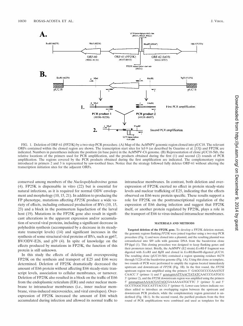
Targeted deletion of the FP25K gene. To develop a FP25K deletion mutant,the genomic regions flanking FP25K were joined together using a two-step PCRprocedure (Fig. 1) and were cloned into a plasmid, and the resulting plasmid wascotransfected into Sf9 cells with genomic DNA from the baculovirus cloneFP-�gal (1). This cloning procedure was designed to keep flanking genes andtheir promoters intact. Briefly, the AcMNPV (E2 strain) EcoRI-F fragment wasdigested with EcoRI and BglII and cloned in EcoRI/BamHI-digested pUC18.The resulting clone (pUC18-5kb) contained a region spanning residues 46278through 51226 of the baculovirus genome (Fig. 1A). Using this clone as template,two rounds of PCR were performed to amplify the regions located immediatelyupstream and downstream of FP25K (Fig. 1B). In the first round, the FP25Kupstream region was amplified using the primers 5� GAGCGCCCGAAATGTCAACC 3� (primer 1) and 5� gctatcgttttATTAACTATATCAACCCGATGCG3� (primer 2), and the FP25K downstream region was amplified using the primers5� gatatagttaatAAAACGATAGCGAAAAAATGCTTC 3� (primer 3) and 5�GCCTTGGCTGCCATTTACCG 3� (primer 4). Lower-case letters indicate res-idues added to introduce an overlapping region between the upstream anddownstream PCR products, while the complementary region generated is un-derlined (Fig. 1B-1). In the second round, the purified products from the firstround of PCR amplifications were combined and used as templates for the
FIG. 1. Deletion of ORF-61 (FP25K) by a two-step PCR procedure. (A) Map of the AcMNPV genomic region cloned into pUC18. The relevantORFs contained within the cloned region are shown. The transcription start sites for lef-9 (as described by Guarino et al. [13]) and FP25K areindicated. Numbers in parentheses indicate the position (in base pairs) in the AcMNPV-C6 genome. (B) Representation of clone pUC18-5kb, therelative locations of the primers used for PCR amplification, and the products obtained during the first (1) and second (2) rounds of PCRamplification. The regions covered by the PCR products obtained during the first amplification are indicated. The complementary regionintroduced in primers 2 and 3 is represented by saw-toothed lines. Notice that the strategy followed fully deletes ORF-61 without altering thetranscription initiation sites for the adjacent ORFs.
10830 ROSAS-ACOSTA ET AL. J. VIROL.
on Decem
ber 9, 2014 by guesthttp://jvi.asm
.org/D
ownloaded from
reaction. No additional primers were added for the initial 4 cycles of amplifica-tion. Thereafter, primers 1 and 4 were added and the PCR was allowed tocontinue for 28 additional cycles to give a PCR product in which the upstreamand downstream regions of FP25K were directly connected, thus deleting FP25K(residues 48550 to 49158) (Fig. 1B-2). The PCR product obtained wasNheI/HindIII digested and cloned into the 4,631-bp fragment produced by theNheI/HindIII digestion of pUC18-5kb. Then a 1,613-bp HindIII fragment excisedout of pUC18-5kb during the NheI/HindIII digestion was added back. Theresulting clone, named pUC18-FP25Kdel#3.3, was fully sequenced by usinginternal primers. Cotransfection was performed according to Summers andSmith (28), and the recombinant viruses generated were screened by colorselection in plaque assays overlaid with 5-bromo-4-chloro-3-indolyl-�-D-galacto-pyranoside (X-Gal). White plaques were picked and further purified by threeadditional rounds of plaque purification. The purified FP25K deletion mutantobtained (referred to as �FP25K) was amplified, and the absence of FP25K wasconfirmed by PCR analyses and Southern blotting of �FP25K genomic DNA.
Overexpression of FP25K. To develop a viral mutant containing a second copyof FP25K in the polyhedrin locus, FP25K was cloned in the pBACgus-1 vector(Novagen, Inc., Madison, Wis.). Sf9 cells were transfected using Bsu361-digestedBacPak AcMNPV genomic DNA and the recombinant pBACgus-1 plasmidaccording to the methods of Summers and Smith (28). Recombinant viruses wereselected for glucoronidase activity in plaque assays overlaid with 5-bromo-4-chloro-3-indolyl-�-D-glucoronide cyclohexylammonium salt (X-Gluc). Positiveplaques were purified by two additional rounds of plaque purification. Thepurified recombinant virus obtained (referred to as FP25Kpolh) was amplified,and the presence of a second copy of FP25K in the polyhedrin locus was con-firmed by PCR analysis and Southern blotting of FP25Kpolh genomic DNA.
Cell culture, virus infections, and metabolic labeling. Spodoptera frugiperdaIPLB-Sf21 clonal isolate 9 (Sf9) cells were cultured in suspension at 27°C inTNM-FH medium (28) supplemented with 10% fetal bovine serum (completemedium). AcMNPV strain E2 was used as a wild-type (wt) virus control. Allinfections were performed at a multiplicity of infection of 20, and time zero wasset at the time of virus addition. For metabolic labeling, cells were seeded andinfected at a density of 3 � 106 cells/flask in 5 ml of complete medium in 25-mltissue culture flasks. One hour before adding the label the culture medium wascollected and replaced with 1 ml of methionine-deficient Grace’s medium sup-plemented with 0.5% fetal bovine serum. To recover dislodged cells and main-tain a constant cell number, the collected culture medium was centrifuged(1,000 � g, 5 min, room temperature) and the cell pellet was resuspended in 1 mlof methionine-deficient Grace’s medium, added back to the flask, and incubatedat 27°C. One hour later, 200 �Ci of Tran35S-Label (ICN Pharmaceuticals Inc.,Costa Mesa, Calif.) was added and the cells were incubated at 27°C for thedesired amount of time. For pulse-chase experiments, after the labeling periodthe cells were washed twice with complete medium and incubated at 27°C for thedesired amount of time. Upon collection the cells were centrifuged (1,000 � g,5 min, room temperature), washed with 1� phosphate-buffered saline (PBS),centrifuged again, and frozen as wet pellets at �80°C in aliquots of 1.5 � 106 cellsper tube. Upon thawing the pellets were processed immediately.
Immunoprecipitation and immunoblotting. For time course analyses of pro-tein expression, cell extracts were prepared from frozen cell pellets containing1.5 � 106 metabolically labeled cells collected at different times postinfection. Todissolve virus occlusions, each cell pellet was incubated in 0.04 N NaOH for 10min at 37°C. The resulting extracts were mixed with 525 �l of 1� radioimmu-noprecipitation assay (RIPA) buffer (100 mM NaCl, 50 mM Tris [pH 8.0], 1%NP-40, 1% deoxycholate, 0.1% sodium dodecyl sulfate [SDS]) and passedthrough a 25-gauge needle 10 times to shear the DNA. At this stage a 20-�laliquot was taken, mixed with an equal volume of 4� sample buffer (100 mMTris-HCl [pH 6.8], 8% SDS, 4% �-mercaptoethanol, 0.04% bromophenol blue,20% glycerol), incubated for 15 min at 65°C, and used for SDS-polyacrylamidegel electrophoresis (PAGE) and immunoblot analyses as described below. Inpreparation for immunoprecipitation analysis, the residual sample was clarifiedby centrifugation at 13,000 � g for 10 min at 4°C and the supernatant waspreabsorbed with 25 �l of preimmune rabbit serum for 1 h at 4°C, incubated with40 �l of a 50% slurry of Protein A agarose for 1 h at 4°C, and pelleted at 1,000 �g for 15 min at 4°C. The preabsorbed extract was split into three aliquots of 175�l (equivalent to 4.4 � 105 cells), each of which was immunoprecipitated with 10�l of the appropriate rabbit serum overnight at 4°C, followed by incubation with20 �l of a 50% slurry of Protein A agarose for 1 h at 4°C. The agarose beads werewashed three times in 1� RIPA buffer and once in 1� Tris-buffered saline (TBS)(140 mM NaCl, 25 mM Tris [pH 8.0]), mixed with 20 �l of 4� sample buffer, andincubated for 15 min at 65°C and the immunoprecipitated proteins were ana-lyzed by SDS-PAGE.
SDS-PAGE analyses were performed using 4 and 12.5% stacking and resolv-
ing gels, respectively, as described by Laemmli (20). Following electrophoresisthe proteins were transferred onto Immobilon-P membranes (Millipore, Bed-ford, Mass.) and the membranes were either processed for immunoblotting orwere directly exposed to film or a phosphorscreen for detection of labeledblotted proteins. For immunoblotting the membranes were blocked with 1�TTBS (1� TBS � 0.05% Tween 20) supplemented with 3% nonfat dry milk (1�blocking solution) for 1 h at room temperature. Rabbit serum directed againstthe appropriate protein (E66, serum 5297; E25, serum 10234; FP25K, serum2804) was added and incubated with the membrane overnight at 4°C. For timecourse analyses of protein expression, rabbit antisera were used at a final dilutionof 1:10,000 in 50 ml of 1� blocking solution. Upon incubation the membraneswere washed three times in 1� TTBS and incubated for 1 h at room temperaturewith horseradish peroxidase-conjugated anti-rabbit immunoglobulin G (IgG)(Santa Cruz Biotechnology, Inc., Santa Cruz, Calif.) at a final dilution of 1:12,500in 50 ml of 1� blocking solution. The membranes were washed three times in 1�TTBS and once in 1� TBS and were developed using the NEN RenaissanceWestern blot chemiluminescence reagent kit (NEN Life Science Products, Bos-ton, Mass.). To strip the membranes for subsequent rounds of immunoblotting,the membranes were washed three times in 1� TTBS, incubated for 30 min at55°C in stripping buffer (62.5 mM Tris–HCl [pH 6.8], 2% SDS, 100 mM �-mer-captoethanol), and washed five additional times in 1� TTBS. To test for residualenzymatic activity, the membranes were developed as above. Before reuse themembranes were washed four additional times in 1� TTBS. Subsequent roundsof immunoblotting were started at the blocking step.
To estimate the relative abundance of specific viral proteins, cell extracts wereprepared from frozen cell pellets containing 3 � 106 cells collected at 28 hpostinfection (hpi). Each cell pellet was resuspended in 500 �l of sterile MilliQwater, mixed with 2 �l of 10 N NaOH, and incubated for 10 min at 37°C. The cellextract was mixed with 500 �l of 4� sample buffer and incubated for 3 min at100°C. Aliquots (50 �l per lane) of the resulting sample were used for SDS-PAGE analysis. Following electrophoresis the proteins were transferred ontoImmobilon-P membranes. Upon transfer the membranes were blocked as aboveand incubated with the appropriate rabbit antisera at a final dilution of 1:2,000in 15 ml of 1� blocking solution. Upon incubation the membranes were washedthree times in 1� TTBS and incubated for 1 h at room temperature with 2 �Ciof 125I-labeled anti-rabbit IgG (ICN Pharmaceuticals, Inc.) in 15 ml of 1�blocking solution. The membranes were washed four times in 1� TTBS, driedout, and exposed to a phosphorscreen at �80°C. All quantitative analyses wereperformed using ImageQuant v. 0.5.0 software in a Storm FluorImager (Molec-ular Dynamics, Sunnyvale, Calif.).
Fractionation of integral membrane proteins with Triton X-114. Detergentfractionations were performed according to the methods of Bordier (3). Briefly,frozen cell pellets containing 3 � 106 cells collected at 24 and 48 hpi wereresuspended in 666 �l of ice-cold 1� PBS–1% Triton X-114. The cell extract wasincubated for 1 h at 4°C and clarified by centrifugation at 14,000 � g for 10 minat 4°C. The supernatant was loaded onto a 66-�l sucrose cushion (6% sucrose–0.06% Triton X-114 in 1� PBS) and incubated for 3 min at 37°C, and theresulting detergent and aqueous phases were resolved by centrifugation at 500 �g for 5 min at 37°C. The detergent phase was resuspended with 333 �l of ice-cold1� PBS and incubated for 3 min at 37°C, and the detergent and aqueous phaseswere resolved again as above. The new aqueous phase was added to the firstaqueous phase, and the detergent phase was resuspended in 1 ml of ice-cold 1�PBS and mixed with an equal volume of 4� sample buffer. The pooled aqueousphase was mixed with 33 �l of 11.4% Triton X-114, incubated for 3 min at 37°C,and centrifuged at 500 � g for 5 min at 37°C. The resulting aqueous phase wasmixed with an equal volume of 4� sample buffer. The samples were incubatedfor 15 min at 65°C and analyzed by SDS-PAGE using 35 �l per lane.
Primer extension. Sf9 cells grown in suspension (2 � 106 cells/ml) wereinfected with either AcMNPV-E2 (wt), �FP25K, or FP25Kpolh. At 28 hpi theinfected cells were collected and polyadenylated mRNA was purified using thePoly(A) Pure mRNA isolation kit (Ambion, Inc., Austin, Tex.). Primer exten-sions were performed with 5 �g of mRNA hybridized to specific probes labeledwith [�-32P]ATP. The oligonucleotide sequence of the probes used were thefollowing: E66 probe, 5� CGGCAAGGGATTGAGATCAATAAAAGC 3�; p39(capsid) probe, 5� TTTGTCGCGGCGCCATACCCACGGGCACTAGCGCCATATTG 3�; E25 probe 1, 5� GCAAAACGATAAGTAACACGATTCCCCAC3�; E25 probe 2, 5� GAGATTTAGGTTGTGCAAATGTTTCAAAAGTACAC3�. To provide internal controls, the primer extension reaction mixtures for E66and E25 contained a mix of the gene-specific probe and the p39 probe. For E25primer extensions, only E25 probe 1 was used; E25 probe 2 was used for pre-liminary primer extension analyses performed to determine transcription startsites for E25. mRNA-primer hybrids were precipitated with 100% ethanol,washed with 70% ethanol, and resuspended in 30 �l of reverse transcription mix
VOL. 75, 2001 NUCLEAR TRANSPORT OF VIRAL ENVELOPE PROTEINS 10831
on Decem
ber 9, 2014 by guesthttp://jvi.asm
.org/D
ownloaded from
(50 mM Tris [pH 8.3], 75 mM KCl, 3 mM MgCl2, 0.666 mM deoxynucleosidetriphosphates, 1 mM dithiothreitol, 40 U of RNasin, 50 �g of actinomycin D, and10 U of SuperScript II RNase H-negative reverse transcriptase [Life Technolo-gies, Inc., Gaithersburg, Md.]). Reverse transcription was performed for 1 h at42°C. The reaction products were ethanol precipitated and resuspended in 3 �lof 100 mM NaOH and 6 �l of sequencing stop buffer. The samples were boiledfor 3 min and analyzed by electrophoresis on a urea–6% polyacrylamide geltogether with a sequencing ladder generated with the same oligonucleotides. Thegels were dried and the primer extension products were quantified using a StormFluorImager.
Immunofluorescence confocal microscopy. Infected Sf9 cells were collected at36 and 48 hpi, washed, and resuspended in Grace’s media, and 2.1 � 105 cellswere transferred to a 1-well cytofuge concentrator (StatSpin Technologies, Nor-wood, Mass.). The cells were allowed to attach for 5 min at room temperatureand were fixed with 3.7% paraformaldehyde in PBS (20 mM phosphate, 140 mMNaCl, pH 7.2) for 10 min at room temperature. The cells were washed threetimes with PBS, permeabilized with methanol (10 min) and 0.5% Triton X-100in PBS (10 min), and washed twice again with PBS. The cells were blocked for1 h in blocking solution (1% chicken serum, 3% bovine serum albumin in PBS)and incubated overnight at 4°C with the appropriate antibodies diluted in block-ing solution (lamin, monoclonal antibody ADL67 [27], 1:500, provided by P. A.Fisher, Department of Pharmacological Sciences, University of New York atStony Brook, Stony Brook; E66, serum 5297, 1:1,000; E25, serum [1:2,500]provided by G. Rohrmann, Oregon State University, Corvallis; FP25K, serum2804, 1:2,500). The cells were rinsed three times with PBS and incubated withAlexa Fluor 488-conjugated anti-rabbit IgG or Alexa Fluor 594-conjugated anti-mouse IgG (both from Molecular Probes, Inc., Eugene, Oreg.) diluted 1:2,000 inblocking solution. After three washes with PBS the cells were stained with DAPI(4�,6�-diamidino-2-phenylindole) at 0.1 �g/ml in PBS for 5 s, washed threeadditional times with PBS, and viewed with a Zeiss CARV confocal microscope.In each experiment at least 15 large fields of view were observed, each fieldcontaining an average of 50 to 60 cells. Then, cells representing the pattern seenin 80% or more of the cells observed were selected to collect confocal Z-stacksections at 0.75-�m intervals. Confocal images of at least five different represen-tative cells were collected per experiment. Each experiment was performedseveral times. Three-dimensional reconstructions and image deconvolution wereperformed using the Zeiss KS 400 Imaging System, release 3.0.
Immunoelectron microscopy. Immunoelectron microscopy was performed aspreviously reported (5, 16) by using Sf9 cell cultures collected at 48 hpi. Rabbitantisera were used at a final dilution of 1:1,000. Bound rabbit antibodies weredetected using anti-rabbit IgG gold-conjugated goat antibodies (25 nm; ElectronMicroscopy Sciences, Fort Washington, Pa.) at a 1:15 dilution.
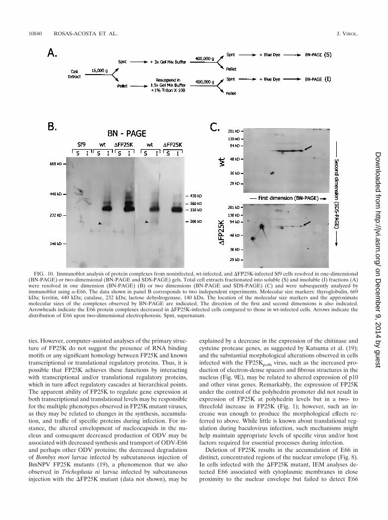
BN-PAGE and two-dimensional BN-PAGE and SDS-PAGE analyses. Bluenative (BN)-PAGE was performed according to the guidelines of Schagger andvon Jagow (25) by using linear 6 to 13% acrylamide gradient gels. In preparationfor BN-PAGE, frozen pellets of infected (30 hpi) or noninfected Sf9 cells con-taining 2 � 107 cells were resuspended in sterile MilliQ water supplemented with200 U of DNase I/ml up to a final volume of 600 �l. The cell suspension waspassed through a 27.5-gauge needle several times, sonicated in a water bathsonicator for 45 s, and incubated on ice for 2 h. The cell extract produced wasfractionated by differential centrifugation as illustrated in Fig. 10A. Briefly, theextract was centrifuged at 15,000 � g for 10 min at 4°C. The supernatant obtainedwas mixed with an equal volume of 3� Gel Mix Buffer (1.5 M Aminocaproicacid, 150 mM Bis-Tris, pH 7.0) and centrifuged at 400,000 � g for 30 min at 15°Cin a TLA-100 rotor (Beckman Instruments Inc., Palo Alto, Calif.). The resultingsupernatant (soluble fraction supernatant) was mixed with blue dye (5% ServaBlue G in 500 mM Aminocaproic acid) at a ratio of 200 �l of supernatant per 10�l of blue dye. The pellet from the 15,000 � g spin was washed once with MilliQwater, and the resulting pellet was resuspended in 540 �l of a 1:1 dilution of 3�Gel Mix Buffer and MilliQ water and was mixed with 60 �l of a 10% TritonX-100 solution. The solubilized pellet was then centrifuged at 400,000 � g, andthe resulting supernatant (insoluble fraction supernatant) was mixed with bluedye, as described above. Sixty microliters of the blue-dye sample mix were loadedper lane. The gels were run overnight at 200 V and 4°C, equilibrated in 1�Transfer Buffer (25 mM Tris, 192 mM glycine, 0.04% SDS, 20% methanol) for15 min, blotted to Immobilon P membranes, and processed for immunoblottingas described above. For two dimensional analyses, after completion of the firstdimension (BN-PAGE) the gel strip corresponding to the lane of interest was cutaway, soaked in 50 ml of denaturing solution (1% [wt/vol] SDS and 1% [vol/vol]�-mercaptoethanol) for 2 h, and soaked in 50 ml of 1� SDS-PAGE runningbuffer for 5 min. Thereafter the gel strip was fixed between two glass plates andexcess running buffer was removed, and a discontinuous 10% resolving SDS-
PAGE gel was poured so that the immobilized gel strip was surrounded by thestacking gel. The gel was run and blotted into Immobilon P membranes.
RESULTS
Deletion of FP25K decreases the levels of ODV-E66 withoutaltering its time of synthesis. Previous studies indicated thatpartial deletions of FP25K resulted in altered synthesis andtransport of several late and very late baculovirus proteins (4,14, 15). To further study the role of FP25K, two new virusmutants were developed, one containing a precise deletionof the FP25K gene (�FP25K) and another containing anadditional copy of FP25K under the polyhedrin promoter(FP25Kpolh). Sf9 cells infected with the �FP25K virus exhib-ited the typical FP phenotype associated with mutations withinthe FP25K gene (data not shown), as described by Beames andSummers (1). Cells infected with the viral mutant containingan additional copy of FP25K under the polyhedrin promoter(FP25Kpolh) exhibited no significant characteristics by lightmicroscopy other than the occlusion-negative phenotype.
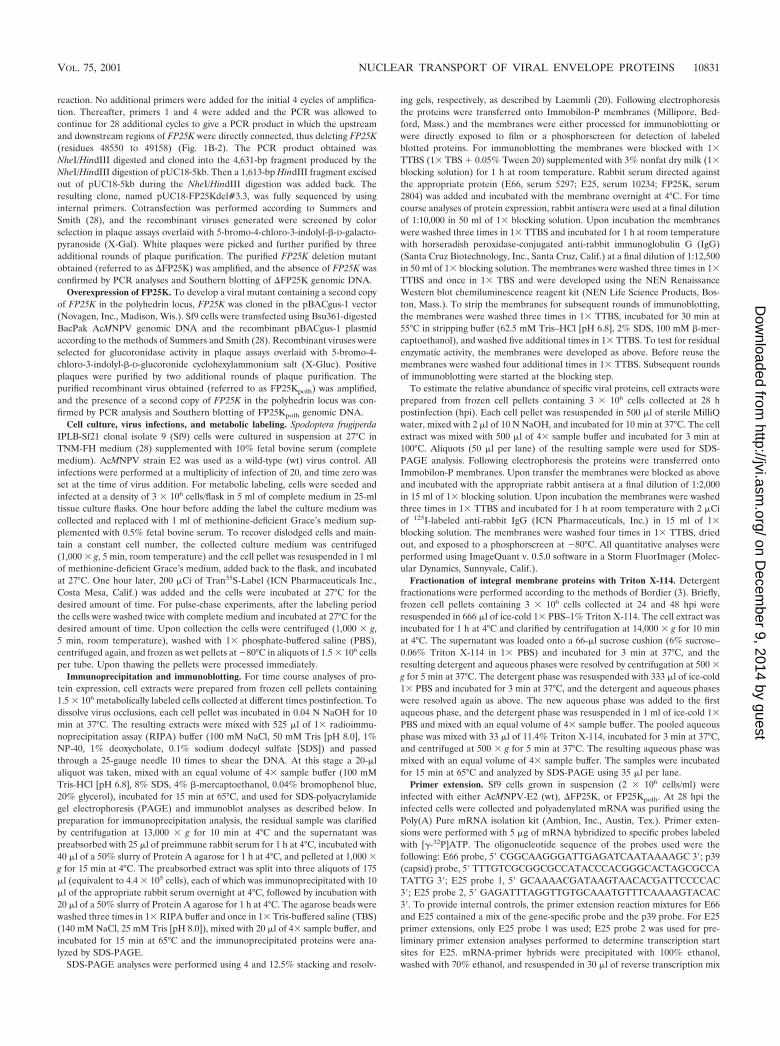
Partial deletions of FP25K decrease the accumulation ofE66 but not of E25 (4). To evaluate the effect that the fulldeletion and the overexpression of FP25K exert on the expres-sion of E66 and E25, immunoblot analyses were performedusing metabolically labeled cells infected with wt, �FP25K, andFP25Kpolh viruses and antibodies raised against E66 (-E66),E25 (-E25), and FP25K (-FP25K). As expected, there was anoticeable increase in the level of FP25K during FP25Kpolh
infection, and FP25K was not detected in �FP25K-infectedcells (Fig. 2A). E25 accumulated to similar levels in wt-,�FP25K-, and FP25Kpolh-infected cells (data not shown).In contrast, significantly lower levels of E66 were detectedin �FP25K-infected cells compared to those with wt- andFP25Kpolh-infected cells: whereas in wt- and FP25Kpolh-in-fected cells E66 was easily detected starting at 24 hpi, in�FP25K-infected cells E66 was barely detectable throughoutinfection (Fig. 2B).
To generate a quantitative estimate of the differences in theaccumulation of E66 and E25 synthesized in wt-, �FP25K-, andFP25Kpolh-infected Sf9 cells, three independent immunoblot-ting experiments were performed using total cell extracts fromequal numbers of infected cells obtained in three independentinfections collected at 28 hpi, treated with NaOH, and solubi-lized in 4� sample buffer. The relative amounts of E66 andE25 were determined using -E66 or -E25, 125I-labeled anti-rabbit IgG, and phosphodensitometry. Large differences wereobserved in the total accumulation of E66 at 28 hpi among thedifferent viruses. Compared to that in wt-infected cells, E66accumulation decreased 4.2-fold in �FP25K-infected cells andwas slightly increased in FP25Kpolh-infected cells (Fig. 3A andC). In contrast, the accumulation of E25 was not significantlyaltered in cells infected with the different viruses (Fig. 3B andD).
To determine if the decreased accumulation of E66 ob-served in �FP25K-infected cells was due to a delay in thesynthesis of E66, the time course of E66 and FP25K synthesisin cells infected with the different viruses was determined byusing immunoprecipitation analyses and the same set of la-beled total cell extracts used in the first set of immunoblotanalyses described above. To make these immunoprecipita-
10832 ROSAS-ACOSTA ET AL. J. VIROL.
on Decem
ber 9, 2014 by guesthttp://jvi.asm
.org/D
ownloaded from

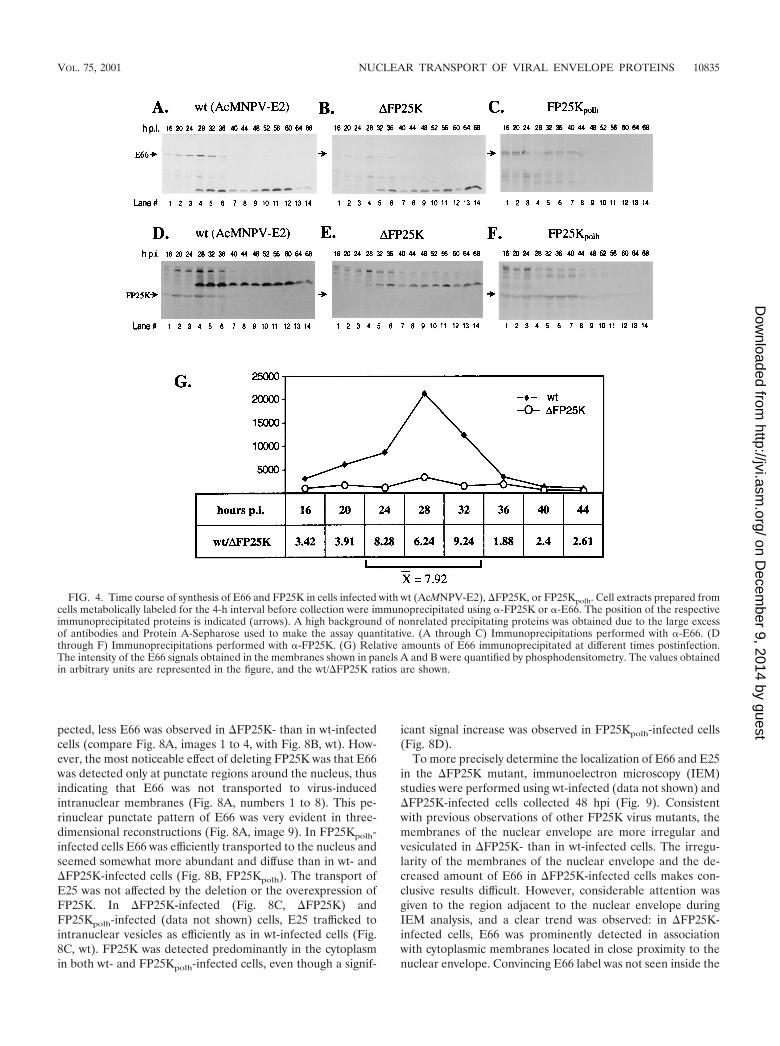
tions quantitative, control experiments were performed whichtitrated the amount of antibody required to assure saturation.These experiments showed that as the amount of antibody andProtein A were increased to achieve saturating conditions, thebackground increased consistently (data not shown); this back-ground is seen throughout Fig. 4. In wt-infected cells, E66synthesis was detected as early as 16 hpi (Fig. 4A, lane 1),reached maximum levels between 28 and 32 hpi (Fig. 4A, lane4), and decreased thereafter (Fig. 4A, lanes 5 to 14). The timecourse of E66 synthesis observed in �FP25K-infected cells wasidentical to that observed in wt-infected cells, with the excep-tion that the amount of E66 detected at each time point wassignificantly lower (Fig. 4B). To quantify the relative differ-ences in synthesis of E66 between wt- and �FP25K-infectedcells, the band intensities of the immunoprecipitated E66 weredetermined by phosphodensitometry. The largest differencesin synthesis of E66 between wt- and �FP25K-infected cellsoccurred between 24 and 36 hpi, the interval during which E66synthesis is maximum. During this period, there was 7.92-foldless E66 produced in �FP25K-infected than in wt-infected cells(Fig. 4G). In FP25Kpolh-infected cells, E66 synthesis remainedrelatively constant between 16 and 44 hpi (Fig. 4C, lanes 1 to7) and decreased thereafter (Fig. 4C, lanes 8 to 14). Thepattern of FP25K synthesis observed in wt- and FP25Kpolh-infected cells was similar to that of E66: in wt-infected cells,FP25K synthesis was detectable at 16 hpi (Fig. 4D, lane 1),reached maximum levels between 28 and 32 hpi (Fig. 4D, lane4), and decreased thereafter (Fig. 4D, lanes 5 to 14). InFP25Kpolh-infected cells, FP25K synthesis remained relativelyconstant between 16 and 44 hpi (Fig. 4F, lanes 1 to 7) anddecreased thereafter (Fig. 4F, lanes 8 to 14). Synthesis ofFP25K was not detected in the �FP25K mutant at any time(Fig. 4E).
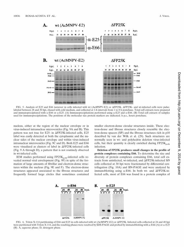
E66 and E25 exhibit minimal turnover in wt- and �FP25K-infected cells. To determine if the decreased levels of E66 in�FP25K-infected cells were due to an increased turnover rateof E66, immunoprecipitation analyses were performed usingpulse-labeled wt- and �FP25K-infected cell extracts collected
at different times postchase. In both wt- and �FP25K-infectedcells no significant differences were observed in the amount ofE66 and E25 precipitated throughout the 8-h period analyzed(Fig. 5A). Aliquots from the samples used for immunoprecipi-tation were also run on SDS-PAGE gels to determine theoverall profile of protein synthesis and degradation. Indeed,the overall protein profile of the pulse-labeled cell extractsindicates very little turnover for those proteins synthesizedbetween 28 and 30 hpi in both wt- and �FP25K-infected cellsduring the 8-h period analyzed (Fig. 5B). The relative amountsof E66 and E25 precipitated from each virus and the overallprotein profiles obtained resembled those observed when thesamples were collected immediately after the labeling period,suggesting that minimal protein turnover occurred between 0and 1 h postchase.
E66 and E25 expressed in �FP25K-infected cells are inte-gral membrane proteins. E66 and E25 are predicted to beintegral membrane proteins of the ODV envelope, and in vitrotranslation assays performed in the presence of microsomalmembranes support such a prediction for E66 (17). To evalu-ate if the absence of FP25K affected the association of E66 andE25 with membranes, wt- and �FP25K-infected cells collectedat 24 and 48 hpi were fractionated into aqueous and detergentfractions by using Triton X-114 and were examined by immu-noblot analysis. In both wt- and �FP25K-infected cells E66 andE25 partitioned with the detergent phase (Fig. 6A and B). Incomparison, p39 (capsid), a major structural protein of thevirus capsid that lacks transmembrane domains, partitionedexclusively with the aqueous phase in wt- and �FP25K-infectedcells (data not shown).
Steady-state levels of E66 and E25 transcripts are not sig-nificantly altered by deleting or overexpressing FP25K. Todetermine if deletion of FP25K alters the steady-state levels ofE66 and/or E25 transcripts, primer extension analyses wereperformed. Since no data were available on the transcriptionstart sites used for E25, a temporal series of primer extensionanalyses of E25 were performed using two different probes andmRNA from wt-infected cells collected at different times
FIG. 2. Accumulation profiles of E66 and FP25K in wt-infected (AcMNPV), �FP25K-infected, and FP25Kpolh-infected cells. Cells infected withwt, �FP25K, or FP25Kpolh were metabolically labeled for the 4-h interval before collection and were lysed and clarified. Aliquots correspondingto equal cell numbers were run on SDS–12.5% PAGE gels and transferred to Immobilon membranes. The membranes were sequentially probedwith -FP25K, -E25 (data not shown), and -E66. (A) Immunoblots performed using -FP25K. (B) Immunoblots performed using -E66. Hourspostinfection (hpi) indicate the time postinfection at which the samples were labeled.
VOL. 75, 2001 NUCLEAR TRANSPORT OF VIRAL ENVELOPE PROTEINS 10833
on Decem
ber 9, 2014 by guesthttp://jvi.asm
.org/D
ownloaded from
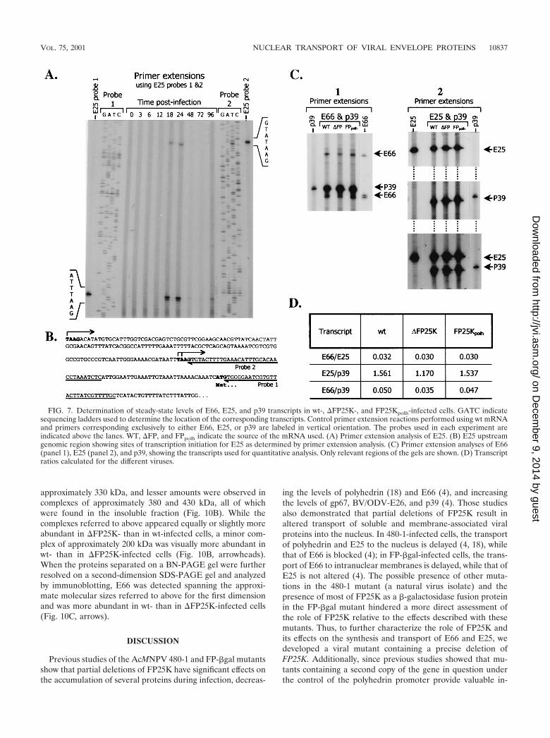
postinfection. These analyses found that E25 transcripts initi-ated at two TAAG motifs at positions �70 and �219 and weremade throughout the late and very late phases of infection,being first detected at 18 hpi (Fig. 7A and B). Two additionaltranscription initiation sites located at least 350 bp furtherupstream from the �219 start site were detected (data notshown). Transcripts initiated at those positions clearly exhib-ited the same temporal pattern but could not be resolved onthe gels and therefore were not used in the quantitative anal-yses. For quantitative comparisons of steady-state levels of E66and E25 transcripts, mRNA was isolated from wt-, �FP25K-,and FP25Kpolh-infected cells collected at 28 hpi, and primerextension analyses were performed using equal amounts ofpurified mRNA. p39 primer extensions were performed asinternal controls. The relative intensity of the signals producedby the different E66, E25, and p39 transcripts were determinedby phosphodensitometry, and the values obtained were aver-aged for each gene. No significant differences were observed inthe usage of specific transcriptional start sites for E66, E25, orp39 among the different viruses (Fig. 7C). Two ratios werecalculated and used to measure differences in E66 steady-statetranscript levels, E66/E25 and E66/p39. E25 was chosen be-cause its protein levels did not exhibit significant variations
among the different viral mutants (Fig. 3B and D), while p39was chosen because it has been used as a control for othersimilar studies (4). The E66/E25 transcript ratios obtainedindicated no differences in relative E66 transcript abundanceamong wt-, �FP25K-, and FP25Kpolh-infected cells (Fig. 7D).Similarly, the E66/p39 ratios indicated only minor differencesin the steady-state levels of E66 transcripts among the differentviruses. A direct comparison of the values obtained for the E66and E25 primer extension products in wt-, �FP25K-, andFP25Kpolh-infected cells also revealed very small differences inthe steady state of such transcripts among the different virusmutants.
Intranuclear transport of E66 in �FP25K-infected cells isblocked at the level of the nuclear envelope. Mutations affect-ing FP25K block the transport of E66 to intranuclear vesiclesbut only delay the transport of E25 to such structures (4). Toassess the effects of deleting and overexpressing FP25K on theintracellular transport of E66 and E25, immunofluorescenceconfocal microscopy analyses of wt-, �FP25K-, and FP25Kpolh-infected cells collected at 36 and 48 hpi were performed. Inthese experiments hundreds of cells were viewed, multipleconfocal images were obtained, and representative Z-sectionimages obtained with each viral mutant are shown. As ex-
FIG. 3. Relative amounts of E66 and E25 produced in wt-, �FP25K-, and FP25Kpolh-infected cells as determined by quantitative immuno-blotting. Equal numbers of infected cells were collected at 28 hpi and processed as indicated in Materials and Methods. The samples were resolvedon SDS–12.5% PAGE gels, transferred onto Immobilon membranes, and immunoblotted using -E66 or -E25 and 125I-labeled goat anti-rabbitIgG. (A and B) Representative immunoblots performed using -E66 and -E25, respectively. Sf9, uninfected Sf9 cells; wt, �FP25K, and FP25Kpolh,cells infected with the respective virus. (C and D) Relative amount in arbitrary units of E66 and E25, respectively. Rectangles represent the averagevalues obtained from three independent experiments performed using samples from independent infections. Vertical bars represent the standarderror obtained. Numbers under the bars indicate the average value obtained for each set of samples.
10834 ROSAS-ACOSTA ET AL. J. VIROL.
on Decem
ber 9, 2014 by guesthttp://jvi.asm
.org/D
ownloaded from
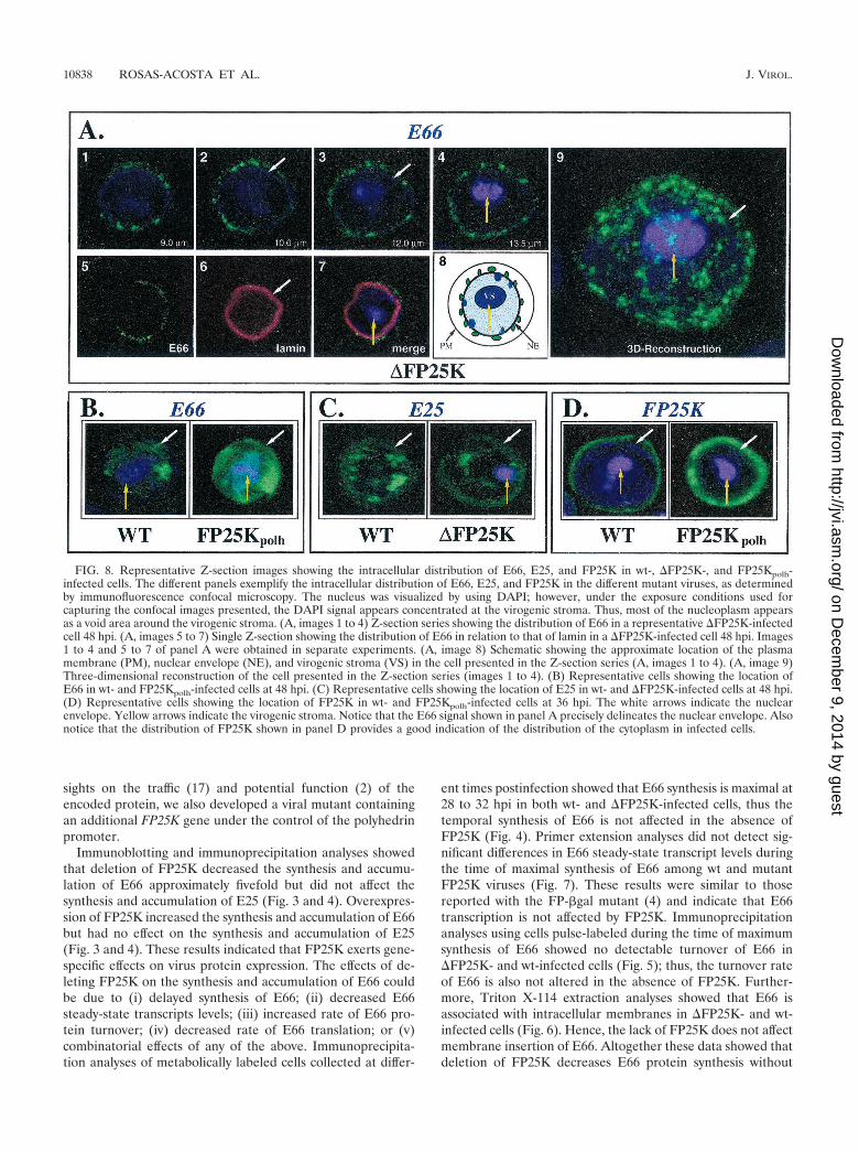
pected, less E66 was observed in �FP25K- than in wt-infectedcells (compare Fig. 8A, images 1 to 4, with Fig. 8B, wt). How-ever, the most noticeable effect of deleting FP25K was that E66was detected only at punctate regions around the nucleus, thusindicating that E66 was not transported to virus-inducedintranuclear membranes (Fig. 8A, numbers 1 to 8). This pe-rinuclear punctate pattern of E66 was very evident in three-dimensional reconstructions (Fig. 8A, image 9). In FP25Kpolh-infected cells E66 was efficiently transported to the nucleus andseemed somewhat more abundant and diffuse than in wt- and�FP25K-infected cells (Fig. 8B, FP25Kpolh). The transport ofE25 was not affected by the deletion or the overexpression ofFP25K. In �FP25K-infected (Fig. 8C, �FP25K) andFP25Kpolh-infected (data not shown) cells, E25 trafficked tointranuclear vesicles as efficiently as in wt-infected cells (Fig.8C, wt). FP25K was detected predominantly in the cytoplasmin both wt- and FP25Kpolh-infected cells, even though a signif-
icant signal increase was observed in FP25Kpolh-infected cells(Fig. 8D).
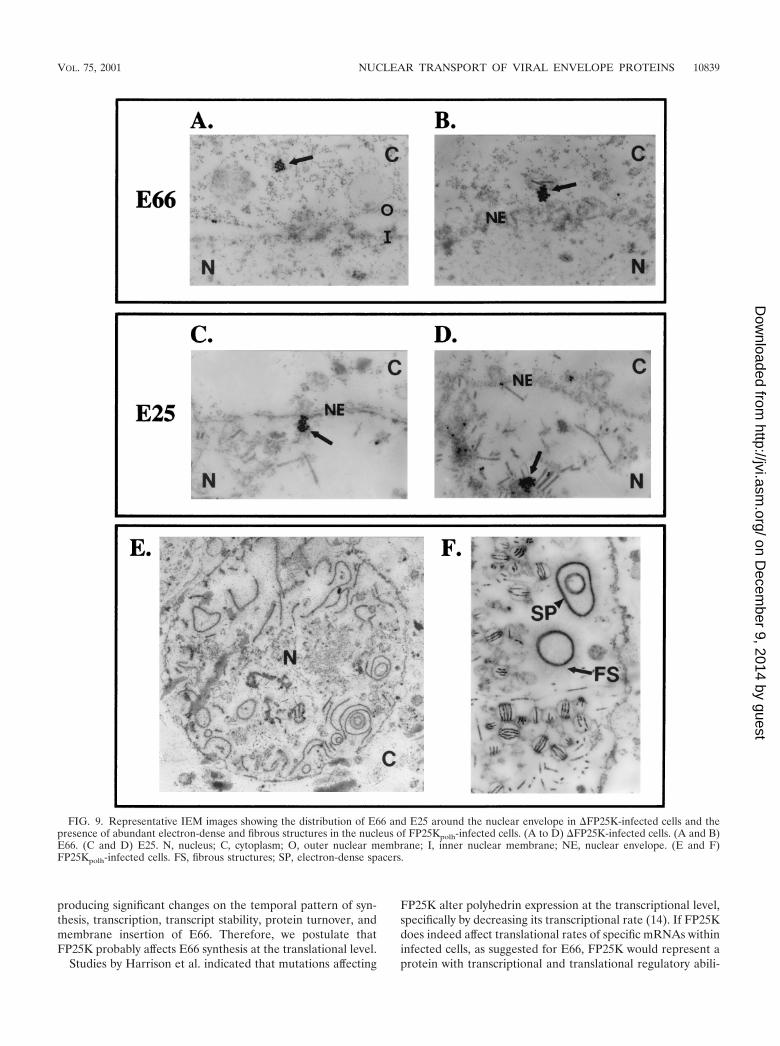
To more precisely determine the localization of E66 and E25in the �FP25K mutant, immunoelectron microscopy (IEM)studies were performed using wt-infected (data not shown) and�FP25K-infected cells collected 48 hpi (Fig. 9). Consistentwith previous observations of other FP25K virus mutants, themembranes of the nuclear envelope are more irregular andvesiculated in �FP25K- than in wt-infected cells. The irregu-larity of the membranes of the nuclear envelope and the de-creased amount of E66 in �FP25K-infected cells makes con-clusive results difficult. However, considerable attention wasgiven to the region adjacent to the nuclear envelope duringIEM analysis, and a clear trend was observed: in �FP25K-infected cells, E66 was prominently detected in associationwith cytoplasmic membranes located in close proximity to thenuclear envelope. Convincing E66 label was not seen inside the
FIG. 4. Time course of synthesis of E66 and FP25K in cells infected with wt (AcMNPV-E2), �FP25K, or FP25Kpolh. Cell extracts prepared fromcells metabolically labeled for the 4-h interval before collection were immunoprecipitated using -FP25K or -E66. The position of the respectiveimmunoprecipitated proteins is indicated (arrows). A high background of nonrelated precipitating proteins was obtained due to the large excessof antibodies and Protein A-Sepharose used to make the assay quantitative. (A through C) Immunoprecipitations performed with -E66. (Dthrough F) Immunoprecipitations performed with -FP25K. (G) Relative amounts of E66 immunoprecipitated at different times postinfection.The intensity of the E66 signals obtained in the membranes shown in panels A and B were quantified by phosphodensitometry. The values obtainedin arbitrary units are represented in the figure, and the wt/�FP25K ratios are shown.
VOL. 75, 2001 NUCLEAR TRANSPORT OF VIRAL ENVELOPE PROTEINS 10835
on Decem
ber 9, 2014 by guesthttp://jvi.asm
.org/D
ownloaded from
nucleus, either at the region of the nuclear envelope or invirus-induced intranuclear microvesicles (Fig. 9A and B). Thispattern was not true for E25: in �FP25K-infected cells, E25label was easily detected at both the cytoplasmic and the nu-clear sides of the nuclear envelope and within virus-inducedintranuclear microvesicles (Fig. 9C and D). Both E25 and E66were visualized as clusters of label in �FP25K-infected cells(Fig. 9 A through D), a pattern that is not routinely observedin wt-infected cells.
IEM studies performed using FP25Kpolh-infected cells re-vealed normal viral envelopment (Fig. 9F) in spite of the for-mation of large amounts of fibrillar and electron-dense struc-tures within the nucleus (Fig. 9E and F). The electron-densestructures appeared associated to the fibrous structures andfrequently formed large circles that sometimes contained
smaller electron-dense circular structures inside. These elec-tron-dense and fibrous structures closely resemble the elec-tron-dense spacers (SP) and the fibrous structures rich in p10described by van der Wilk et al. (29). Such structures arenormally seen in wt- and polyhedrin deletion virus-infectedcells, but their quantity is clearly enriched during FP25Kpolh
infection.Deletion of FP25K produces small changes in the profile of
protein complexes containing E66. To determine the size anddiversity of protein complexes containing E66, total cell ex-tracts from uninfected, wt-infected, and �FP25K-infected Sf9cells collected at 30 hpi were fractionated by differential cen-trifugation (Fig. 10A) and BN-PAGE and were analyzed byimmunoblotting using -E66. In both wt- and �FP25K-in-fected cells, most of E66 was found in a protein complex of
FIG. 5. Analysis of E25 and E66 turnover in cells infected with wt (AcMNPV-E2) or �FP25K. �FP25K- and wt-infected cells were pulse-labeled between 28 and 30 hpi, chased with cold medium, and collected at 1-h intervals from 1 to 8 h postchase. Total cell extracts were preparedand immunoprecipitated with -E66 or -E25. (A) Immunoprecipitations performed using -E25 and -E66. (B) Total cell extracts of samplesused for immunoprecipitations. The positions of the molecular size protein markers are indicated. h p.c., hours postchase.
FIG. 6. Triton X-114 partitioning of E66 and E25 in cells infected with wt (AcMNPV-E2) or �FP25K. Infected cells collected at 24 and 48 hpiwere partitioned with Triton X-114, and the resulting phases were resolved by SDS-PAGE and probed by immunoblotting with -E66 (A) or -E25(B). A, aqueous phase; D, detergent phase.
10836 ROSAS-ACOSTA ET AL. J. VIROL.
on Decem
ber 9, 2014 by guesthttp://jvi.asm
.org/D
ownloaded from
approximately 330 kDa, and lesser amounts were observed incomplexes of approximately 380 and 430 kDa, all of whichwere found in the insoluble fraction (Fig. 10B). While thecomplexes referred to above appeared equally or slightly moreabundant in �FP25K- than in wt-infected cells, a minor com-plex of approximately 200 kDa was visually more abundant inwt- than in �FP25K-infected cells (Fig. 10B, arrowheads).When the proteins separated on a BN-PAGE gel were furtherresolved on a second-dimension SDS-PAGE gel and analyzedby immunoblotting, E66 was detected spanning the approxi-mate molecular sizes referred to above for the first dimensionand was more abundant in wt- than in �FP25K-infected cells(Fig. 10C, arrows).
DISCUSSION
Previous studies of the AcMNPV 480-1 and FP-�gal mutantsshow that partial deletions of FP25K have significant effects onthe accumulation of several proteins during infection, decreas-
ing the levels of polyhedrin (18) and E66 (4), and increasingthe levels of gp67, BV/ODV-E26, and p39 (4). Those studiesalso demonstrated that partial deletions of FP25K result inaltered transport of soluble and membrane-associated viralproteins into the nucleus. In 480-1-infected cells, the transportof polyhedrin and E25 to the nucleus is delayed (4, 18), whilethat of E66 is blocked (4); in FP-�gal-infected cells, the trans-port of E66 to intranuclear membranes is delayed, while that ofE25 is not altered (4). The possible presence of other muta-tions in the 480-1 mutant (a natural virus isolate) and thepresence of most of FP25K as a �-galactosidase fusion proteinin the FP-�gal mutant hindered a more direct assessment ofthe role of FP25K relative to the effects described with thesemutants. Thus, to further characterize the role of FP25K andits effects on the synthesis and transport of E66 and E25, wedeveloped a viral mutant containing a precise deletion ofFP25K. Additionally, since previous studies showed that mu-tants containing a second copy of the gene in question underthe control of the polyhedrin promoter provide valuable in-
FIG. 7. Determination of steady-state levels of E66, E25, and p39 transcripts in wt-, �FP25K-, and FP25Kpolh-infected cells. GATC indicatesequencing ladders used to determine the location of the corresponding transcripts. Control primer extension reactions performed using wt mRNAand primers corresponding exclusively to either E66, E25, or p39 are labeled in vertical orientation. The probes used in each experiment areindicated above the lanes. WT, �FP, and FPpolh indicate the source of the mRNA used. (A) Primer extension analysis of E25. (B) E25 upstreamgenomic region showing sites of transcription initiation for E25 as determined by primer extension analysis. (C) Primer extension analyses of E66(panel 1), E25 (panel 2), and p39, showing the transcripts used for quantitative analysis. Only relevant regions of the gels are shown. (D) Transcriptratios calculated for the different viruses.
VOL. 75, 2001 NUCLEAR TRANSPORT OF VIRAL ENVELOPE PROTEINS 10837
on Decem
ber 9, 2014 by guesthttp://jvi.asm
.org/D
ownloaded from
sights on the traffic (17) and potential function (2) of theencoded protein, we also developed a viral mutant containingan additional FP25K gene under the control of the polyhedrinpromoter.
Immunoblotting and immunoprecipitation analyses showedthat deletion of FP25K decreased the synthesis and accumu-lation of E66 approximately fivefold but did not affect thesynthesis and accumulation of E25 (Fig. 3 and 4). Overexpres-sion of FP25K increased the synthesis and accumulation of E66but had no effect on the synthesis and accumulation of E25(Fig. 3 and 4). These results indicated that FP25K exerts gene-specific effects on virus protein expression. The effects of de-leting FP25K on the synthesis and accumulation of E66 couldbe due to (i) delayed synthesis of E66; (ii) decreased E66steady-state transcripts levels; (iii) increased rate of E66 pro-tein turnover; (iv) decreased rate of E66 translation; or (v)combinatorial effects of any of the above. Immunoprecipita-tion analyses of metabolically labeled cells collected at differ-
ent times postinfection showed that E66 synthesis is maximal at28 to 32 hpi in both wt- and �FP25K-infected cells, thus thetemporal synthesis of E66 is not affected in the absence ofFP25K (Fig. 4). Primer extension analyses did not detect sig-nificant differences in E66 steady-state transcript levels duringthe time of maximal synthesis of E66 among wt and mutantFP25K viruses (Fig. 7). These results were similar to thosereported with the FP-�gal mutant (4) and indicate that E66transcription is not affected by FP25K. Immunoprecipitationanalyses using cells pulse-labeled during the time of maximumsynthesis of E66 showed no detectable turnover of E66 in�FP25K- and wt-infected cells (Fig. 5); thus, the turnover rateof E66 is also not altered in the absence of FP25K. Further-more, Triton X-114 extraction analyses showed that E66 isassociated with intracellular membranes in �FP25K- and wt-infected cells (Fig. 6). Hence, the lack of FP25K does not affectmembrane insertion of E66. Altogether these data showed thatdeletion of FP25K decreases E66 protein synthesis without
FIG. 8. Representative Z-section images showing the intracellular distribution of E66, E25, and FP25K in wt-, �FP25K-, and FP25Kpolh-infected cells. The different panels exemplify the intracellular distribution of E66, E25, and FP25K in the different mutant viruses, as determinedby immunofluorescence confocal microscopy. The nucleus was visualized by using DAPI; however, under the exposure conditions used forcapturing the confocal images presented, the DAPI signal appears concentrated at the virogenic stroma. Thus, most of the nucleoplasm appearsas a void area around the virogenic stroma. (A, images 1 to 4) Z-section series showing the distribution of E66 in a representative �FP25K-infectedcell 48 hpi. (A, images 5 to 7) Single Z-section showing the distribution of E66 in relation to that of lamin in a �FP25K-infected cell 48 hpi. Images1 to 4 and 5 to 7 of panel A were obtained in separate experiments. (A, image 8) Schematic showing the approximate location of the plasmamembrane (PM), nuclear envelope (NE), and virogenic stroma (VS) in the cell presented in the Z-section series (A, images 1 to 4). (A, image 9)Three-dimensional reconstruction of the cell presented in the Z-section series (images 1 to 4). (B) Representative cells showing the location ofE66 in wt- and FP25Kpolh-infected cells at 48 hpi. (C) Representative cells showing the location of E25 in wt- and �FP25K-infected cells at 48 hpi.(D) Representative cells showing the location of FP25K in wt- and FP25Kpolh-infected cells at 36 hpi. The white arrows indicate the nuclearenvelope. Yellow arrows indicate the virogenic stroma. Notice that the E66 signal shown in panel A precisely delineates the nuclear envelope. Alsonotice that the distribution of FP25K shown in panel D provides a good indication of the distribution of the cytoplasm in infected cells.
10838 ROSAS-ACOSTA ET AL. J. VIROL.
on Decem
ber 9, 2014 by guesthttp://jvi.asm
.org/D
ownloaded from
producing significant changes on the temporal pattern of syn-thesis, transcription, transcript stability, protein turnover, andmembrane insertion of E66. Therefore, we postulate thatFP25K probably affects E66 synthesis at the translational level.
Studies by Harrison et al. indicated that mutations affecting
FP25K alter polyhedrin expression at the transcriptional level,specifically by decreasing its transcriptional rate (14). If FP25Kdoes indeed affect translational rates of specific mRNAs withininfected cells, as suggested for E66, FP25K would represent aprotein with transcriptional and translational regulatory abili-
FIG. 9. Representative IEM images showing the distribution of E66 and E25 around the nuclear envelope in �FP25K-infected cells and thepresence of abundant electron-dense and fibrous structures in the nucleus of FP25Kpolh-infected cells. (A to D) �FP25K-infected cells. (A and B)E66. (C and D) E25. N, nucleus; C, cytoplasm; O, outer nuclear membrane; I, inner nuclear membrane; NE, nuclear envelope. (E and F)FP25Kpolh-infected cells. FS, fibrous structures; SP, electron-dense spacers.
VOL. 75, 2001 NUCLEAR TRANSPORT OF VIRAL ENVELOPE PROTEINS 10839
on Decem
ber 9, 2014 by guesthttp://jvi.asm
.org/D
ownloaded from
ties. However, computer-assisted analyses of the primary struc-ture of FP25K do not suggest the presence of RNA bindingmotifs or any significant homology between FP25K and knowntranscriptional or translational regulatory proteins. Thus, it ispossible that FP25K achieves these functions by interactingwith transcriptional and/or translational regulatory proteins,which in turn affect regulatory cascades at hierarchical points.The apparent ability of FP25K to regulate gene expression atboth transcriptional and translational levels may be responsiblefor the multiple phenotypes observed in FP25K mutant viruses,as they may be related to changes in the synthesis, accumula-tion, and traffic of specific proteins during infection. For in-stance, the altered envelopment of nucleocapsids in the nu-cleus and consequent decreased production of ODV may beassociated with decreased synthesis and transport of ODV-E66and perhaps other ODV proteins; the decreased degradationof Bombyx mori larvae infected by subcutaneous injection ofBmNPV FP25K mutants (19), a phenomenon that we alsoobserved in Trichoplusia ni larvae infected by subcutaneousinjection with the �FP25K mutant (data not shown), may be
explained by a decrease in the expression of the chitinase andcysteine protease genes, as suggested by Katsuma et al. (19);and the substantial morphological alterations observed in cellsinfected with the FP25Kpolh virus, such as the increased pro-duction of electron-dense spacers and fibrous structures in thenucleus (Fig. 9E), may be related to altered expression of p10and other virus genes. Remarkably, the expression of FP25Kunder the control of the polyhedrin promoter did not result inexpression of FP25K at polyhedrin levels but in a two- tothreefold increase in FP25K (Fig. 1); however, such an in-crease was enough to produce the morphological effects re-ferred to above. While little is known about translational reg-ulation during baculovirus infection, such mechanisms mighthelp maintain appropriate levels of specific virus and/or hostfactors required for essential processes during infection.
Deletion of FP25K results in the accumulation of E66 indistinct, concentrated regions of the nuclear envelope (Fig. 8).In cells infected with the �FP25K mutant, IEM analyses de-tected E66 associated with cytoplasmic membranes in closeproximity to the nuclear envelope but failed to detect E66
FIG. 10. Immunoblot analysis of protein complexes from noninfected, wt-infected, and �FP25K-infected Sf9 cells resolved in one-dimensional(BN-PAGE) or two-dimensional (BN-PAGE and SDS-PAGE) gels. Total cell extracts fractionated into soluble (S) and insoluble (I) fractions (A)were resolved in one dimension (BN-PAGE) (B) or two dimensions (BN-PAGE and SDS-PAGE) (C) and were subsequently analyzed byimmunoblot using -E66. The data shown in panel B corresponds to two independent experiments. Molecular size markers: thyroglobulin, 669kDa; ferritin, 440 kDa; catalase, 232 kDa; lactose dehydrogenase, 140 kDa. The location of the molecular size markers and the approximatemolecular sizes of the complexes observed by BN-PAGE are indicated. The direction of the first and second dimensions is also indicated.Arrowheads indicate the E66 protein complexes decreased in �FP25K-infected cells compared to those in wt-infected cells. Arrows indicate thedistribution of E66 upon two-dimensional electrophoresis. Spnt, supernatant.
10840 ROSAS-ACOSTA ET AL. J. VIROL.
on Decem
ber 9, 2014 by guesthttp://jvi.asm
.org/D
ownloaded from
associated with membranes inside the nucleus (i.e., inner nu-clear membrane, virus-induced microvesicles, and viral enve-lopes) (Fig. 9A and B). In contrast, trafficking of E25 in�FP25K-infected cells appeared normal, as E25 was detectedin membranes associated with the nuclear envelope as well asin intranuclear microvesicles (Fig. 9C and D). Our previousstudy showed that E25 localization to intranuclear mi-crovesicles was delayed in 480-1- but not in FP-�gal-infectedcells. Since both mutant viruses contain significant portions ofthe FP25K gene yet only 480-1 infection resulted in a delay ofE25 localization to intranuclear microvesicles, this result maybe revealing functions that still exist within the truncatedFP25K protein expressed during 480-1 infection. It is alsopossible that since 480-1 is a naturally occurring viral isolate,other spontaneous, as-yet uncharacterized mutations existwithin the 480-1 genome. The use of the precise deletion of theFP25K gene shows that E66 localization is specifically affected,and this effect cannot be generalized to other ODV envelopeproteins.
Due to the differences observed in E66 trafficking between�FP25K- and wt-infected cells, we considered it important tocompare the size of E66 protein complexes in cells infectedwith these viruses. To this end we developed a fractionationprocedure that removes nucleocapsids and virions and solubi-lizes membrane-associated protein complexes by using non-ionic detergents, thus selecting for complexes representingthose interactions involved in protein traffic prior to assembly(Fig. 10A). The solubilized complexes were analyzed by BN-PAGE and immunoblotting. Such analyses showed that in bothwt- and �FP25K-infected cells E66 is present in at least fourmajor protein complexes, ranging in size from 200 to 430 kDa.Except for the 200-kDa complex, all the E66 protein com-plexes observed seemed equally or slightly more abundant in�FP25K- than in wt-infected cells (Fig. 10B). Such a result wasunexpected, as E66 is produced in lesser quantities in �FP25K-than in wt-infected cells (Fig. 3 and 4). When proteins sepa-rated by BN-PAGE were further resolved by an SDS-PAGEsecond dimension, significantly stronger E66 bands were de-tected in wt- than in �FP25K-infected cells for all the com-plexes observed. The similarity in the profiles obtained in thetwo-dimensional analyses confirmed that E66 forms proteincomplexes of equal size in wt- and �FP25K-infected cells (Fig.10C). The differences observed between the one- and two-dimensional analyses may relate to changes in the predominantepitopes exposed by E66 under native and denatured condi-tions and the type of protein complexes formed by E66 in wt-and �FP25K-infected cells. The 200-kDa complex appears tobe an E66-protein complex down-regulated in the absence ofFP25K and therefore may constitute an important target forfuture studies.
Based on the above data, we considered two possible mech-anisms of how FP25K may function to regulate the traffic ofE66. First, it is possible that, in the absence of FP25K, de-creased quantity or improper folding of E66 results in alteredtransport. It is possible that the decreased amount of E66produced in cells infected with the �FP25K mutant mightaffect its trafficking. However, in our previous study (4) weshowed that, during infection with the FP-�gal mutant, thequantity of E66 decreased to levels equivalent to those ob-served in �FP25K-infected cells, but still some E66 trafficked
normally to intranuclear microvesicles. Thus, the block in theintranuclear trafficking of E66 should not be entirely due to thedecreased amount of protein. Alternatively, in the absence ofFP25K, E66 may not be folded properly, thus affecting itstrafficking. Indeed, IEM analyses showed that, in the absenceof FP25K, E66 is detected in clusters (Fig. 9A and B) seldomfound in wt-infected cells, which might represent misfolded oraggregated E66. However, like E66, E25 was also detected insimilar clusters, yet its traffic was unaffected (Fig. 9C and D).Additionally, E66 was found in protein complexes of similarsize in �FP25K- and wt-infected cells (Fig. 10). Furthermore,while molecular size may limit the traffic of large proteins fromthe outer to the inner nuclear membrane (26), a large integralmembrane fusion protein (125-�-galactosidase, molecular sizeof 110 kDa) traffics to the nuclear envelope and to virus-induced intranuclear microvesicles as efficiently as ODV enve-lope proteins during baculovirus infection (17). These obser-vations suggest that although size limitations and foldingrequirements cannot be discounted, the differences seen in thetraffic of E66 and E25 in �FP25K-infected cells do not repre-sent an intrinsic limitation of transport due to molecular size.While protein misfolding is usually accompanied by an in-creased turnover, the turnover rate of E66 appeared to beidentical in wt- and �FP25K-infected cells.
Second, the transport of E66 may be mediated by specificfactors whose synthesis or activity is regulated by FP25K. It ispossible that after E66 is inserted into membranes (a featurethat is not disrupted in cells infected with the �FP25K mutant[Fig. 6]), a combination of factors may help it to progresstoward the nuclear envelope. Transport models for residentproteins of the inner nuclear membrane suggest that theseproteins diffuse laterally along the continuous membranes ofthe ER, outer nuclear membrane, and inner nuclear mem-brane, where they are retained via interactions with chromatin,lamins, or other resident proteins (8, 12, 24, 26). Althoughnone of the ODV envelope proteins contain inner nuclearmembrane retention signals, the continued movement of ODVenvelope proteins to membranes inside the nucleus duringinfection may serve to remove proteins from the diffusionalpool. However, altered retention does not explain why E66would locate to discrete membranes at the exterior of thenucleus in the absence of FP25K. It is possible that a lipidgradient from the ER to the nuclear envelope and intranuclearvesicles could provide directionality to the lateral diffusion ofproteins along the membrane. However, if such a gradientwere the only factor regulating protein movement along thecontinuous membranes of the ER and nuclear envelope, thenmutations affecting the trafficking of one envelope protein(E66) potentially could alter the trafficking of others (E25).The specific effects observed on the trafficking of E66 suggestthat, in the absence of FP25K, proteins or protein activitiesthat facilitate movement of E66 to intranuclear membranes arealtered. This could include factors that generate a facilitateddiffusion pathway or a more complex, active transport pathway.
Our data clearly argue against an unregulated lateral diffu-sion model for the transport of E66 to intranuclear mi-crovesicles. It suggests that during AcMNPV infection thetransport of E66 to intranuclear microvesicles is specificallyregulated and that the factors controlling this trafficking in-clude FP25K or a protein(s) regulated by FP25K. Further
VOL. 75, 2001 NUCLEAR TRANSPORT OF VIRAL ENVELOPE PROTEINS 10841
on Decem
ber 9, 2014 by guesthttp://jvi.asm
.org/D
ownloaded from
characterization of the components of complexes containingE66 may identify transport factors and help discern the mech-anism involved in the trafficking of E66 from its site of inser-tion at the ER to the nuclear envelope and to virus-inducedintranuclear microvesicles. Considering that viruses are veryefficient manipulators of cellular pathways but rarely invententirely new pathways not used by their host, we expect thatstudies on the integral membrane proteins of the baculovirusODV envelope may reveal insights into the mechanism gov-erning the transport of membrane proteins to the nuclear en-velope in uninfected cells.
ACKNOWLEDGMENTS
We thank Paul A. Fisher (Department of Pharmacological Sciences,University of New York at Stony Brook, Stony Brook) for providingthe monoclonal antibody ADL67 and George Rohrmann (OregonState University, Corvallis) for providing the -E25 rabbit serum. Wethank Jared Burks for his expert assistance with confocal microscopy.Electron microscopy was performed using the facilities of the ElectronMicroscopy Center at Texas A&M University.
This work was supported in part by National Institutes of HealthGrant 2RO1GM47552 (M.D.S., S.C.B.) and the Texas AgriculturalExperimental Station Project TEXO8078 (M.D.S.).
REFERENCES
1. Beames, B., and M. D. Summers. 1989. Location and nucleotide sequence ofthe 25K protein missing from baculovirus few polyhedra (FP) mutants.Virology 168:344–353.
2. Beniya, H., S. C. Braunagel, and M. D. Summers. 1998. Autographa cali-fornica nuclear polyhedrosis virus: subcellular localization and protein traf-ficking of BV/ODV-E26 to intranuclear membranes and viral envelopes.Virology 240:64–75.
3. Bordier, C. 1981. Phase separation of integral membrane proteins in TritonX-114 solution. J. Biol. Chem. 256:1604–1607.
4. Braunagel, S. C., J. K. Burks, G. Rosas-Acosta, R. L. Harrison, H. Ma, andM. D. Summers. 1999. Mutations within the Autographa californica nucle-opolyhedrovirus FP25K gene decrease the accumulation of ODV-E66 andalter its intranuclear transport. J. Virol. 73:8559–8570.
5. Braunagel, S. C., R. Parr, M. Belyavskyi, and M. D. Summers. 1998. Au-tographa californica nucleopolyhedrovirus infection results in Sf9 cell cyclearrest at G2/M phase. Virology 244:195–211.
6. Braunagel, S. C., and M. D. Summers. 1994. Autographa californica nuclearpolyhedrosis virus, PDV, and ECV viral envelopes and nucleocapsids: struc-tural proteins, antigens, lipid and fatty acid profiles. Virology 202:315–328.
7. Chen, X., W. F. Ijkel, R. Tarchini, X. Sun, H. Sandbrink, H. Wang, S. Peters,D. Zuidema, R. K. Lankhorst, J. M. Vlak, and Z. Hu. 2001. The sequence ofthe Helicoverpa armigera single nucleocapsid nucleopolyhedrovirus ge-nome. J. Gen. Virol. 82:241–257.
8. Ellenberg, J., E. D. Siggia, J. E. Moreira, C. L. Smith, J. F. Presley, H. J.Worman, and J. Lippincott-Schwartz. 1997. Nuclear membrane dynamicsand reassembly in living cells: targeting of an inner nuclear membraneprotein in interphase and mitosis. J. Cell Biol. 138:1193–1206.
9. Fraser, M. J. 1986. Ultrastructural observations of virion maturation inAutographa californica nuclear polyhedrosis virus infected Spodoptera frugi-perda cell cultures. J. Ultrastruct. Mol. Struct. Res. 95:189–195.
10. Fraser, M. J., and W. F. Hink. 1982. Comparative sensitivity of severalplaque assay techniques employing TN-368 and IPLB-SF 21AE insect celllines for plaque variants of Galleria mellonella nuclear polyhedrosis virus.J. Invertebr. Pathol. 40:89–97.
11. Fraser, M. J., G. E. Smith, and M. D. Summers. 1983. Acquisition of hostcell DNA sequences by baculoviruses: relationship between host DNA in-sertions and FP mutants of Autographa californica and Galleria mellonellanuclear polyhedrosis viruses. J. Virol. 47:287–300.
12. Furukawa, K., C. E. Fritze, and L. Gerace. 1998. The major nuclear envelopetargeting domain of LAP2 coincides with its lamin binding region but isdistinct from its chromatin interaction domain. J. Biol. Chem. 273:4213–4219.
13. Guarino, L. A., B. Xu, J. Jin, and W. Dong. 1998. A virus-encoded RNApolymerase purified from baculovirus-infected cells. J. Virol. 72:7985–7991.
14. Harrison, R. L., D. L. Jarvis, and M. D. Summers. 1996. The role of theAcMNPV 25K gene, “FP25” in baculovirus polh and p10 expression. Virol-ogy 226:34–46.
15. Harrison, R. L., and M. D. Summers. 1995. Mutations in the Autographacalifornica multinucleocapsid nuclear polyhedrosis virus 25 kDa proteingene result in reduced virion occlusion, altered intranuclear envelopmentand enhanced virus production. J. Gen. Virol. 76:1451–1459.
16. Hong, T., S. C. Braunagel, and M. D. Summers. 1994. Transcription, trans-lation, and cellular localization of PDV-E66: a structural protein of the PDVenvelope of Autographa californica nuclear polyhedrosis virus. Virology204:210–222.
17. Hong, T., M. D. Summers, and S. C. Braunagel. 1997. N-terminal sequencesfrom Autographa californica nuclear polyhedrosis virus envelope proteinsODV-E66 and ODV-E25 are sufficient to direct reporter proteins to thenuclear envelope, intranuclear microvesicles and the envelope of occlusionderived virus. Proc. Natl. Acad. Sci. USA 94:4050–4055.
18. Jarvis, D. L., D. A. Bohlmeyer, and A. Garcia, Jr. 1992. Enhancement ofpolyhedrin nuclear localization during baculovirus infection. J. Virol. 66:6903–6911.
19. Katsuma, S., Y. Noguchi, C. L. Zhou, M. Kobayashi, and S. Maeda. 1999.Characterization of the 25K FP gene of the baculovirus Bombyx mori nucle-opolyhedrovirus: implications for postmortem host degradation. J. Gen. Vi-rol. 80:783–791.
20. Laemmli, U. K. 1970. Cleavage of structural proteins during assembly of thehead of bacteriophage T4. Nature 227:680–685.
21. MacKinnon, E. A., J. F. Henderson, D. B. Stoltz, and P. Faulkner. 1974.Morphogenesis of nuclear polyhedrosis virus under conditions of prolongedpassage in vitro. J. Ultrastruct. Res. 49:419–435.
22. Potter, J. N., P. Faulkner, and E. A. MacKinnon. 1976. Strain selectionduring serial passage of Trichoplusia ni nuclear polyhedrosis virus. J. Virol.18:1040–1050.
23. Potter, K. N., R. P. Jaques, and P. Faulkner. 1978. Modification of Trichop-lusia ni nuclear polyhedrosis virus passaged in vivo. Intervirology 9:76–85.
24. Rolls, M. M., P. A. Stein, S. S. Taylor, E. Ha, F. McKeon, and T. A.Rapoport. 1999. A visual screen of a GFP-fusion library identifies a new typeof nuclear envelope membrane protein. J. Cell Biol. 146:29–44.
25. Schagger, H., and G. von Jagow. 1991. Blue native electrophoresis for iso-lation of membrane protein complexes in enzymatically active form. Anal.Biochem. 199:223–231.
26. Soullam, B., and H. J. Worman. 1995. Signals and structural features in-volved in integral membrane protein targeting to the inner nuclear mem-brane. J. Cell Biol. 130:15–27.
27. Stuurman, N., N. Maus, and P. A. Fisher. 1995. Interphase phosphorylationof the Drosophila nuclear lamin: site-mapping using a monoclonal antibody.J. Cell Sci. 108:3137–3144.
28. Summers, M. D., and G. E. Smith. 1987. A manual of methods for baculo-virus vectors and insect cell culture procedures. Texas Agric. Exp. StationBull. 1555:10–48.
29. van F. der Wilk, J. W., M. van Lent, and J. M. Vlak. 1987. Immunogolddetection of polyhedrin, p10, and virion antigens in Autographa californicanuclear polyhedrosis virus-infected Spodoptera frugiperda cells. J. Gen. Vi-rol 68:2615–2623.
30. Volkman, L. E., and M. D. Summers. 1977. Autographa californica nuclearpolyhedrosis virus: comparative infectivity of the occluded, alkali-liberated,and nonoccluded forms. J. Invertebr. Pathol. 30:102–103.
10842 ROSAS-ACOSTA ET AL. J. VIROL.
on Decem
ber 9, 2014 by guesthttp://jvi.asm
.org/D
ownloaded from