The Sgs1 Helicase of Saccharomyces cerevisiae Inhibits Retrotransposition of Ty1 Multimeric Arrays
Upload
independentCategory
view
1download
0
SURE: Shizuoka University REpository
http://ir.lib.shizuoka.ac.jp/
This document is downloaded at: 2016-07-26T16:00:12Z
Title Construction of hybrib Autographa californica nuclear
polyhedrosis bacmid by modification of p143 helicase
Author(s) Deo, Vipin Kumar; Hiyoshi, Masato; Park, Enoch Y.
Citation Journal of Virological Methods. 134(1-2), p. 212-216
Issue Date 2006-06
URL http://hdl.handle.net/10297/3737
Version author
Rights
Construction of hybrid Autographa californica nuclear
polyhedrosis bacmid by modification of p143 helicase
Vipin Kumar Deo, Masato Hiyoshi, Enoch Y Park*
Laboratory of Biotechnology, Department of Applied Biological Chemistry, Faculty of
Agriculture, Shizuoka University, 836 Ohya, Shizuoka 422-8529, Japan
* Corresponding author. Tel. & Fax.: +81-54-238-4887. E-mail address: [email protected] (Enoch Y. Park)
2
Abstract
We developed a new hybrid nuclear polyhedrosis virus (NPV) bacmid capable of
infecting Spodoptera frugiperda, Tricoplusia ni, and Bombyx mori, and B. mori cell
lines for producing hybrid recombinant baculovirus that can carry a gene of interest and
express it in a broad range of hosts. A GFPuv-β1,3-N-acetylglucosaminyltransferase 2
fusion gene was expressed successfully in silkworm larvae using this hybrid bacmid.
The hybrid NPV bacmid provides an altogether simple and realistically feasible method
for large-scale applications using silkworm larvae. It can be easily managed in E. coli,
which has no biohazard safety concerns, in addition to the baculovirus-based expression
system.
Keywords: Hybrid bacmid; insect cell; Bombyx mori; silkworm;
β1,3-N-acetylglucosaminyltransferase

3
1. Introduction
Baculovirus expression systems (BESs) are widely used to express heterologous
genes in cultured insect cells and insect larvae hosts; gene expression is driven by the
strong polyhedrin promoter. These systems also have great advantages for the
expression of eukaryotic proteins because insect cells can add post-translational
modifications to proteins so that they are similar to their native forms and can be
handled easily (Croizier et al., 1994; Luckow et al, 1991 and 1993; O’Relly), which is
not the case for bacterial expression systems.
The expression of foreign genes using conventional preparations of recombinant
baculoviruses requires the cotransfection of plasmid transfer vectors and circular
wild-type genomic viral DNA into insect cells. This approach needs at least 40 days, as
it requires multiple plaque assays, purification from nonrecombinant parental virus and
amplification of the recombinant baculovirus. A more rapid and efficient method to
generate recombinant baculoviruses was developed using the site-specific transposition
of an expression cassette into a baculovirus shuttle vector (AcNPV bacmid) that can be
propagated in E. coli (Luckow et al, 1993). This bacmid contains a mini-F replicon, a
kanamycin resistance marker and a segment of DNA carrying the lacZα gene with an
attachment site for the bacterial transposon Tn7 at its N terminus. Recombinant bacmid
DNA can be rapidly isolated from small-scale cultures and then used to transfect insect
cells. Viral stocks harvested from the transfected cells can be used to infect insect cells
for subsequent protein expression. The time required for the introduction of a foreign
gene and isolation of the recombinant baculovirus was reduced significantly from 40

4
days to 7-10 days by the use of this bacmid. However, this AcNPV bacmid can only be
used in cell lines from Spodoptera frugiperda and Tricoplusia ni, but neither in
silkworms nor their cell lines. Recently, Motohashi et al. (2005) have developed the
BmNPV bacmid system, which can directly infect silkworms and B. mori cell lines.
This makes the large-scale production of eukaryotic proteins in silkworms possible; the
protein expression level using silkworms is 10- to 100-fold higher than that using insect
cells.
The AcNPV bacmid does not replicate in B. mori cell lines and the BmNPV
bacmid does not replicate in S. frugiperda or T. ni cell lines. Although AcNPV and
BmNPV are closely related to each other, having collinear genome organizations, their
host ranges are different. Much research (Arguad et al., 1998; Kamita and Maeda, 1997;
Kondo and Maeda, 1991; Maeda et al., 1993; Mori et al., 1992; Muneta et al., 2003) has
expanded the host range of AcNPV vectors by producing hybrid-AcNPV (Hy-AcNPV).
The p143 gene is the host-range genetic determinant for AcNPV infectivity in B. mori
cell lines (Croizier et al., 1994; Maeda et al., 1993). This gene is essential to AcNPV
DNA replication and has been suggested to encode a helicase (Lu and Carstens, 1991).
Maeda et al. (1993) isolated a hybrid baculovirus of BmNPV and AcNPV that is capable
of replicating in both S. frugiperda and T. ni cell lines. Their nucleotide sequencing
analyses revealed that a 572-bp Sac I-Hind III fragment, BmScH, is responsible for the
expanded host range of AcNPV and that it is localized within BmNPV dnahel (Kamita
and Maeda, 1997). Two adjacent nucleotides (A and T) appeared to be the minimal
essential sequence necessary to expand the host range of AcNPV, encoded by a single
5
amino acid difference between BmNPV (Asp) and AcNPV (Ser). Muneta et al. (2003)
applied this hybrid AcNPV to the production of interleukin-18 in silkworms and
obtained 20-fold more interleukin-18 than when using Sf 9 cells. However, although
this hybrid baculovirus is able to expand the host range, the method still involved the
isolation, purification, and amplification of the recombinant virus, which is tedious and
time consuming. This inability of the recombinant virus to infect different host cells
requires the same tedious preparations, which causes inexplicable loss for respective
hosts each time. Therefore, a bacmid with an expanded host range is desirable for the
improvement of qualitative and quantitative efficiency of protein expression.
In this paper, we report a new hybrid nuclear polyhedrosis virus (hybrid NPV)
bacmid that is infectious for S. frugiperda and T. ni cell lines as well as silkworms. As
this method utilizes the advantages of a bacmid it can be easily prepared and screened in
E. coli to produce sufficient DNA for recombinant virus preparation. This method
allows recombinant virus production in S. frugiperda and T. ni cell lines, and large-scale
protein expression in silkworms.
2. Materials and Methods
2.1. Cell lines, bacterial cells, plasmids, bacmid and media
The Sf 9 cell line and AcNPV bacmid were purchased from Invitrogen (Carlsbad,
CA, USA). The Bm5 cell line was kindly provided by Professor K. S. Boo of the Insect
Pathology Laboratory of Seoul National University. E. coli DH5α, E. coli TOP10, E.
6
coli DH10Bac and Electro Max DH10B were obtained from Invitrogen. Wild-type viral
BmNPV DNA was obtained from Funakoshi Co., (Tokyo, Japan), and the transfer
vector for the AcNPV bacmid, pUC18, was kindly provided by National Institute of
Genetics (Mishima, Japan). Sf 900 II serum-free medium (Invitrogen) supplemented
with 1% antibiotic-antimycotic (Invitrogen) and 1% FBS, and TNM-FH medium
(Invitrogen) containing 10% FBS were used for Sf 9 and Bm5 cell cultures. Insect cells
were grown in 25 cm2 tissue culture flasks (Falcon). A plasmid vector harboring a
GFPuv-β1,3-N-acetylglucosaminyltransferase 2 fusion gene fused with the melittin
signal sequence (me-β-3GnT2) (Kato et al., 2003 and 2004) was used for evaluation of
hybrid bacmid in silkworm larvae. Supplements, if necessary, were added into medium
at the following concentrations: ampicillin, 100 µg/ml; kanamycin, 50 µg/ml;
5-bromo-4-chloro-3-indolyl-β-galactopyranoside (X-Gal), 100 µg/ml; gentamycin, 7
µg/ml; tetracycline, 10 µg/ml and isopropyl-β-D-thiogalactopyranoside (IPTG), 40
µg/ml.
2.2. Construction of plasmid carrying DNA helicase to be used in homologous
recombination
Helicase cDNA was extracted from wild-type BmNPV DNA by PCR using the
oligonucleotide primers BmDhel Sac I-f and BmDhel Hind III-r (Table 1). A 572-bp
long helicase fragment was isolated from a 1.5% agarose gel and purified using GFX
PCR DNA and gel band Purification kit (Amersham Biosci. Corp., Piscataway, NJ,
7
USA). The purified 572-bp band was digested by Sac I/Hind III restriction enzymes and
the resulting fragment, BmScH, was purified again. The purified insert was ligated into
pUC18 at the Sac I/Hind III sites and transformed into DH5α competent cells (Takara
Bio, Otsu, Japan). The pUC18/BmScH transformants were screened on LB plates
containing ampicillin, IPTG, and X-Gal in a 37oC incubator. The sequences of all the
PCR fragments inserted into pUC18 were confirmed by dideoxynucleotide chain
terminating sequence (Sanger et al., 1977) using Thermo Sequenase Cycle Sequencing
kit (USB Co., Cleveland, Ohio, USA).
2.3. Construction of hybrid NPV bacmid and confirmation of bacmid DNA
Two hundred nanograms of AcNPV bacmid and 3 μg of pUC18/BmScH in 100 μl
of Sf 900 II medium without antibiotics were mixed with 6 μl of Cellfectin in 100 μl of
Sf 900 II medium without antibiotics. This mixture was incubated at room temperature
for 45 min under sterile conditions. The mixed solution was poured onto a monolayer of
Sf 9 cells (1x10⁶ cells/ml) in 6 well plates with a 9.6 cm2 surface area (Falcon, NJ,
USA) and was incubated for 5 hr at 27oC for transfection. After transfection, the
mixture solution was removed and 2.5 ml of Sf 900 II medium with 1% antimycotic
antibiotic was added. After 3 days incubation at 27oC, the supernatant containing hybrid
NPV was removed and used for further infection. The infection with the hybrid NPV
was repeated three times as mentioned above. To investigate the infectivity of hybrid
NPV for Bm5 cells, 500 μl of the virus solution was poured onto the monolayer of

8
1x10⁶ cells/ml of Bm5 cells in Sf 900 II medium. After a 2-hr incubation at 27oC, the
medium was replaced with fresh TNM-FH medium containing 10% FBS, and the cells
were incubated for 4 days. The virus was amplified three times as mentioned above.
The above-described virus samples were centrifuged at 20,630xg for 1 hr at 4oC to
precipitate virus particles. The supernatant was removed and the pellet was dissolved in
50 μl of TE buffer containing 10 mM Tris-HCl (pH 8.0) and 1 mM EDTA. Using
proteinase K treatment at 60oC for 15 min, the viral envelope was removed. One
hundred microliters of TE buffer was added and viral DNA was extracted by phenol:
chloroform purification. The DNA was precipitated using ethanol and purified by
centrifugation. The dried DNA was dissolved in 50 μl of TE buffer. The isolation of
hybrid NPV bacmid DNA was confirmed by PCR to check the 572-bp BmScH fragment
of the p143 gene of the AcNPV genome using primers (Table 1). The PCR products
were further confirmed by NspV restriction analysis.
2.4. Construction of hybrid NPV bacmid expressing GFPuv-β3GnT2 fusion protein
Hybrid NPV bacmid DNA was transformed into DH10Bac competent cells
containing pHelper, which encodes a transposase and confers resistance to tetracycline.
The new DH10Bac/hybrid NPV bacmid/pHelper cells were screened on LB agar plates
containing kanamycin and tetracycline. The selected colonies were cultured to prepare
fresh competent cells for Bac-to-Bac system.
Fifty micrograms of pDEST8-me-GFPuv-β3GnT2 DNA was transformed into 100
9
μl of DH10Bac/hybrid NPV bacmid/pHelper competent cells. The transformed cells
were added to 500 μl of SOC and incubated at 37oC for 1 hr, and then were cultured on
LB agar plates containing gentamycin, kanamycin, tetracycline, IPTG, and X-Gal at
37oC overnight. White colonies were inoculated into LB medium containing
gentamycin, kanamycin, and tetracycline and cultured at 37oC and agitated at 150 rpm
overnight. Hybrid NPV bacmid/me-GFPuv-β3GnT2 was isolated and confirmed by PCR
using me-β3GnT2-Forward and M13-Reverse primers (Table 1).
2.5. Expression of GFPuv-β3GnT2 fusion protein in Sf 9, Bm5, and silkworm larvae
Approximately 200 ng of hybrid NPV bacmid/me-GFPuv-β3GnT2 DNA was
transfected into Sf9 and Bm5 cells. At 3-4 days postinfection, the culture was harvested
and centrifuged at 2000 x g for 5 min. Precipitated cells were diluted with fresh medium
and were observed using a fluorescence microscope to confirm the expression of the
fusion protein.
Fifth instars larvae (Ehime Sansyu Co. Ltd., Ehime, Japan) were used for the
expression of fusion protein. Twenty microliters of hybrid NPV/me-GFPuv-β3GnT2
(1x10⁷ pfu/ml) was injected into the dorsal region of the larvae using a syringe with
26-gauge beveled needle. The expression of the fusion protein in silkworms was
monitored by examining GFPuv fluorescence using a fluorescent microscope or a UV
transilluminator table.
The fusion protein contained in hemolymph was subjected to SDS-PAGE on a

10
12 % polyacrylamide gel using the Mini-protean II system (Bio-Rad, Hercules, CA).
The respective bands were detected using a Molecular-FX multi-imager (Bio-Rad).
3. Results
3.1. Construction of hybrid NPV bacmid
First, we prepared a transfer vector necessary for the construction of the hybrid
NPV bacmid. Using PCR primers, 572-bp region from the BmNPV DNA helicase gene
was amplified and inserted into pUC18 at the Sac I/Hind III restriction enzyme sites to
give the plasmid pUC18/BmScH encoding an ampicillin resistance marker and the
lacZα peptide (Fig. 1). White colonies containing pUC18/BmScH were picked up and
inoculated into 3 ml of LB medium. Plasmid DNA was isolated and confirmed by Sac
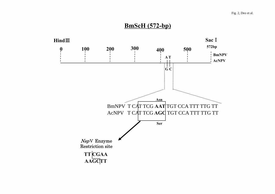
I/Hind III digestion on agarose (data not shown). The BmScH cDNA (Fig. 2) was
transferred from pUC18/BmScH into the p143 cDNA of the AcNPV bacmid by
homologous recombination (as described in the Materials and Methods section).
BmScH isolated from the hybrid NPV bacmid was confirmed by PCR using primers
(Table 1). The hybrid NPV bacmid PCR product was further analyzed by the restriction
enzyme Nsp V that digests at TT/CGAA nucleotide sequences (Fig. 2), giving two
bands of 500 bp and 100 bp. PCR products of hybrid NPV and BmNPV showed the two
expected bands of 500 bp and 100 bp, but that of AcNPV was not digested by the Nsp V
restriction enzyme, giving one band (Fig. 3).
11
3.2. Application of hybrid NPV bacmid in Bac-to-Bac system
The prepared hybrid NPV bacmid DNA was successfully transformed into
DH10Bac competent cells containing pHelper, encoding for transposase, to create
DH10B/hybrid NPV bacmid/pHelper transformant cells. pDEST-8/GFPuv-β3GnT2, as a
transfer vector, was transformed into DH10B/hybrid NPV bacmid/pHelper competent
cells to create a recombinant hybrid NPV-me-GFPuv-β3GnT2 bacmid. A successful
transfer of cDNA from the destination vector to the hybrid NPV bacmid took place,
which proves that this system works successfully. Using the hybrid
NPV-me-GFPuv-β3GnT2 bacmid, cotransfection was performed and recombinant virus
was prepared, which can infect both B. mori Bm5 cells as well as Sf 9 cells efficiently.
The infected Bm5 and Sf 9 cells were bigger in size compared with mock-transfected
cells owing to the occluded virions that are embedded within polyhedral inclusion
bodies (data not shown). This indicates that the hybrid NPV can be infectious to Sf 9
and Bm5 cells.
To validate the application of hybrid NPV in the Bac-to Bac system, Bm5 cells
infected with the hybrid NPV-me-GFPuv-β3GnT2 bacmid virus were observed under a
fluorescence microscope and they showed a distinct GFPuv fluorescence (Fig. 4B). The
same cells were also viewed under a normal light microscope and showed the expected
infected cell structure because of the occluded virions (Fig. 4A). This result indicates
that this hybrid bacmid can be applied to the Bac-to-Bac system.

12
3.3. Expression of fusion protein in silkworm larvae using hybrid bacmid
The efficiency of the hybrid bacmid was further verified by infecting silkworm
larvae with the hybrid NPV-me-GFPuv-β3GnT2 bacmid virus. The hybrid
NPV-me-GFPuv-β3GnT2 bacmid virus was injected into silkworms using a syringe. The
larvae appeared greenish at 48 hr postinfection and the fluorescence intensity further
increased with the optimum result at 96 hr postinfection (II in Fig. 5A). At 96 hr
postinfection, the larvae were dissected, and hemolymph fluid was collected to check
the expression of the fusion protein by SDS-PAGE (12%) (lane 1 in Fig. 5B). A
fluorescent band of approximately 75 kDa corresponding to the expected size of the
GFPuv fusion protein was observed for GFPuv-β3GnT2.
4. Discussion
We have developed a new hybrid bacmid capable of infecting a broad range of
host insect cells. A highly homologous region (BmScH) of 572-bp exists within both the
helicase gene of BmNPV and p143 of AcNPV. BmScH was inserted into the AcNPV
bacmid using a pUC18-based transfer vector. A unique Nsp V restriction enzyme site
exists in the 572-bp region of the BmScH fragment of bacmid-BmNPV; thus,
homologous recombination between pUC18/BmScH and AcNPV bacmid was easily
selected and confirmed (Fig. 2). The hybrid bacmid was infectious to both S. frugiperda
and T. ni cell lines. GFPuv fusion protein was expressed successfully in silkworm larvae
using this hybrid bacmid system. The use of the GFPuv fusion protein assisted us to
13
easily monitor the progress of infection.
We could easily propagate the hybrid bacmid in E. coli like any other plasmid and
easily screen for the bacmid carrying inserts with a very high efficiency and precision.
The new hybrid bacmid was utilized in the Bac-to-Bac expression system. Using the
Gateway system, transfer vectors can be prepared with ease and the foreign gene of
interest can be transferred to the hybrid bacmid by site-specific homologous
recombination.
The hybrid NPV bacmid is altogether simple and realistically feasible for use in
large-scale applications. It can be easily managed in E. coli, which has no biohazard
safety concerns, in addition to the baculovirus-based expression system. The
biotechnology industry requires the use of expensive human proteins of medicinal value
and also those for other research studies. In this study, we concur with other leading
research groups around the world, which are focused on meeting the growing demand in
the biotechnology field on this idea.
Acknowledgment
This work was supported in part by the Program of Basic Research Activities for
Innovative Biosciences (PROBRAIN), Japan. The authors thank Ms. Ayano Kageshima
for helping with the preparation of this manuscript.
References
14
Argaud, O., Croizier, L., Lopez-Ferber, M., Croizier, G., 1998. Two key mutations in the
host-range specific domain of the p143 gene of Autographa californica nuclear
polyhedrosis virus are required to kill Bombyx mori larvae. J. Gen. Virol. 79,
931-935.
Croizier, G., Croizier, L., Argaud, O., Poudevigne, D., 1994. Extension of Autographa
californica nuclear polyherosis virus host range by interspecific replacement of a
short DNA sequence in the p143 helicase gene. Proc. Natl. Acad. Sci., USA, 91,
48-52.
Kamita, S.G., Maeda, S., 1977. Sequencing of the putative DNA helicase-encoding gene
of the Bombyx mori nuclear polyhedrosis virus and fine-mapping of a region
involved in host range expansion. Gene 190, 173-179.
Kato, T., Murata, T., Usui, T., Park, E.Y., 2003. Improvement of GFPuv-β3GnT2 fusion
protein production by suppressing protease in baculovirus expression system. Biosci.
Biotechnol. Biochem. 67(11), 2388-2395.
Kato, T., Park, E.Y., Murata, T., Usui, T., 2004. Efficient production of human
β-1, 3-N-acetylglucosaminyltransferase-2 fused with green fluorescent protein in
insect cell, Biochem. Eng. J. 19, 15-23.
Kondo, A., Maeda, S., 1991. Host range expansion by recombination of the baculovirus
Bombyx mori nuclear polyhedrosis virus and Autographa californica nuclear
polyhedrosis virus. J. Virol. 65, 3625-3632.
Lu, A., Carstens, E.B., (1991). Nucleotide sequence of a gene essential for viral DNA
replication in the baculovirus Autographa californica nuclear polyhedrosis virus.
15
Virol. 181, 336-347.
Luckow, V.A., 1991. Cloning and expression of heterologous genes in insect cells with
baculovirus vectors. In: Prokop, A., Bajpai, R. K., Ho, C. S., (Eds.), Recombinant
DNA Technology and Applications. McGraw-Hill, Inc. New York, 1991.
Luckow, V.A., Lee, S.C., Barry, G.F., Olins, P.O., 1993. Efficient generation of
infectious recombinant baculoviruses by site-specific transposon-mediated insertion
of foreign genes into a baculovirus genome propagated in Escherichia coli. J. Virol.
67, 4566-4579.
Maeda, S., Kamita, S.G., Kondo, A., 1993. Host range expansion of Autographa
californica nuclear polyhedrosis virus (NPV) following recombination of a
0.6-kilobase-pair DNA fragment originating from Bombyx mori NPV. J. Virol. 67,
6234-6238.
Mori, H., Nakazawa, H., Shirai, N., Shibata, N., Sumida, M., Matsubara, F. J., 1992.
Foreign gene expression by a baculovirus vector with an expanded host range. J.
Gen. Virol. 73, 1477-1880.
Motohashi, T., Shimojima, T., Fukagawa, T., Maenaka, K., Park, E.Y., 2005. Efficient
large-scale protein production of larvae and pupae of silkworm nuclear polyhedrosis
virus (BmNPV) bacmid system. Biochem. Biophys. Res. Com. 326, 564-569.
Muneta, Y., Zhao, H.K., Inumaru, S., Mori, Y., 2003. Large-scale production of porcine
mature interleukin-18 (IL-18) in silkworms using a hybrid baculovirus expression
system. Immunol. 65, 21-223.
O’Relly, D.R., Miller, L.K., Luckow, V.A., 1922. Baculovirus Expression Vectors A
16
Laboratory Manual, Oxford University Press, pp. 216-234.
Sanger, F., Nicklen, S., Coulson, A.R., 1977. DNA sequencing with chain-terminating
inhibitors. Proc. Natl. Acad. Sci., USA, 74, 5463-5467.

17
Legends for Figures
Fig. 1. BmNPV genome map with restriction sites showing DNA helicase gene ORF78.
A 572-bp fragment (BmScH) was isolated from BmNPV genome using PCR with
primers flanked by Sac I/Hind III restriction sites and ligated into pUC18 at the Sac
I/Hind III restriction sites.
Fig. 2. Highly homologous area in 572-bp BmScH fragment of DNA helicase gene of
BmNPV and p143 gene of AcNPV. Asn and Ser denote asparagine and serine,
respectively. The Nsp V restriction enzyme site is shown in the BmScH fragment.
Fig. 3. 1.5% agarose gel showing PCR products. Lane M denotes λ-Hind III marker.
Lanes 1, 3, and 5 denote PCR products from wild-type BmNPV, wild-type hybrid
NPV, and wild-type AcNPV, respectively. Lanes 2, 4, and 6 denote Nsp V digested
PCR products from wild-type BmNPV, wild-type hybrid NPV, and wild-type
AcNPV, respectively.
Fig. 4. Bm5 cells at 3 days postinfection with hybrid NPV/me-GFPuv-β3GnT2. A and B
were obtained using a light microscope and a fluorescence microscope, respectively.
Fig. 5. Silkworm larvae infected with hybrid NPV/me-GFPuv-β3GnT2 at 3 days
postinfection. A. Mock-infected silkworm larvae (I) and hybrid

18
NPV/me-GFPuv-β3GnT2 infected silkworm (II). B. Fluorescent image of
SDS-PAGE (12%) of fluorescent GFPuv fusion protein. M denotes GFP fluorescent
marker. Lane 1 denotes GFPuv fusion protein band of 75 kDa from silkworm
hemolymph.
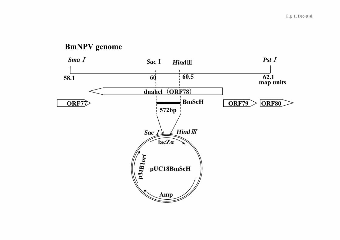
Fig. 1, Deo et al.
SmaⅠ PstⅠHindⅢSacⅠ
BmNPV genome
58.1 62.1map units
60.560
dnahel(ORF78)
ORF77 ORF79 ORF80BmScH572bp
dnahel(ORF78)
lacZαSacⅠ HindⅢ
pUC18BmScH
Amp
BmScH (572-bp)
Fig. 2, Deo et al.
HindⅢ SacⅠ
0 572bp100 200 300 400 500A T
G C
BmNPVAcNPV
BmNPV T CAT TCG AAT TGT CCA TTT TTG TTAcNPV T CAT TCG AGC TGT CCA TTT TTG TT
Asn
Ser
NspⅤ EnzymeNspⅤ Enzyme Restriction site
TT CGAAAAGCTT

Fig. 3, Deo et al.
M 1 2 3 4 5 6
0.5 kbp0.6 kbp
0.1 kbp
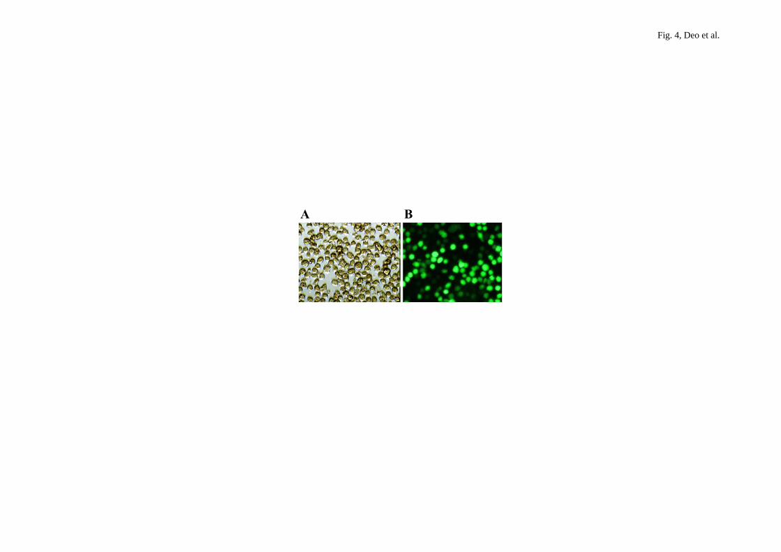
Fig. 4, Deo et al.
A BA B
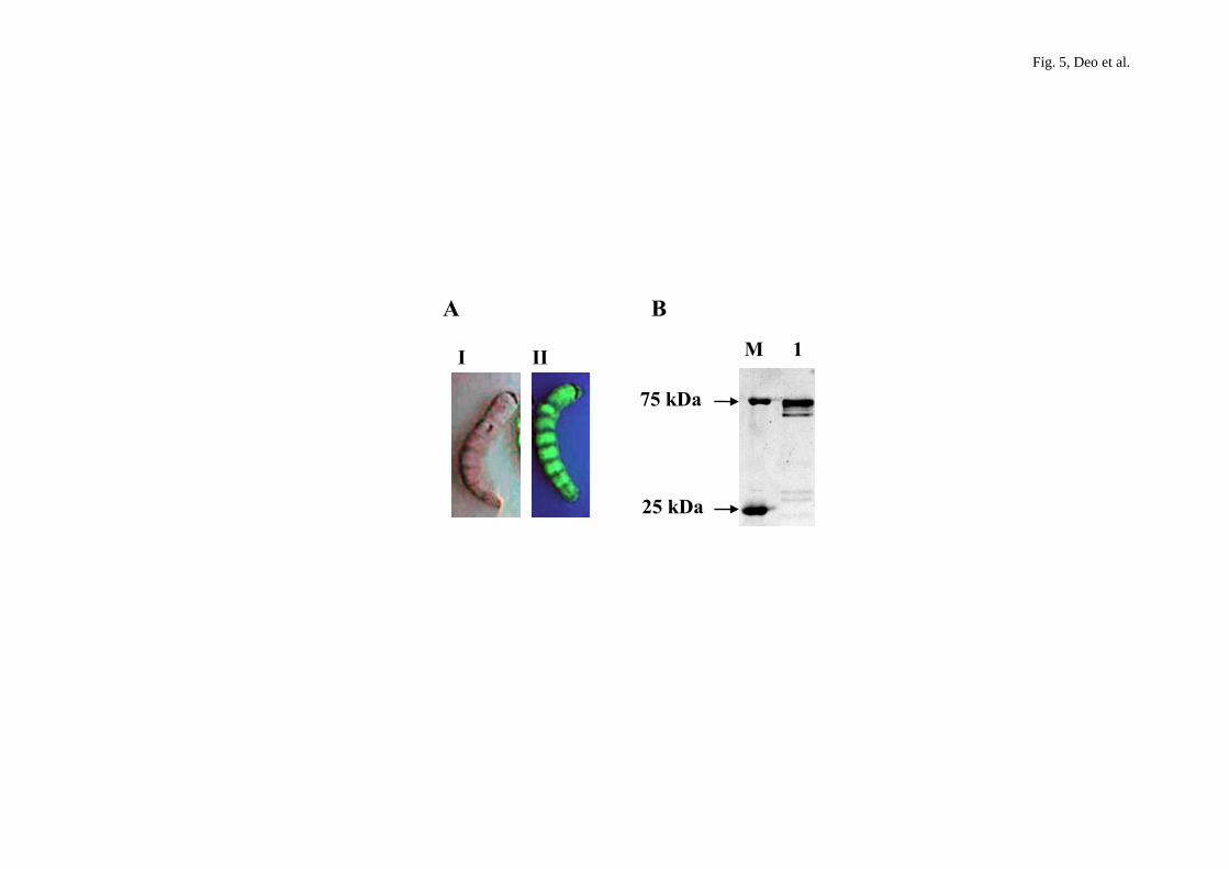
Fig. 5, Deo et al.
A B
I II M 1
75 kDa
25 kDa

Table 1
Primers used to prepare BmScH fragment for hybrid NPV bacmid and to identify the GFPuv-β3GnT2 cDNA using PCR.
Primer-name Sequence (5’→3’)
BmDhel Sac 1-f ATGCTGAGCTCGTCGGTGCTAGTAGATA
BmDhel Hind III-r GAATGAAGCTTTGAAACGATGTGACCT
me-β3GnT2-Forward
GGGGTACCATGAAATTCTTAGTCAACGTTGCCCTTGTTTTTATGGTCGTATACATTTCTTACATCTATGCCGGACGCGGGGTTC
TCATCATCA C
M13-Reverse
CAGGAAACAGCTATGACC
Copyright © 2022 FDOKUMEN