The DEXD/H-box RNA helicase RHII/Gu is a co-factor for c-Jun-activated transcription
10
Jukka Westermarck 1 , 2 , 3 , Carsten Weiss 1 , 4 , Rainer Saffrich 1 , Ju ¨ rgen Kast 1 , 5 , Anna-Maria Musti 1 , 6 , Matthias Wessely 1 , Wilhelm Ansorge 1 , Bertrand Se ´ raphin 1 , 7 , Matthias Wilm 1 , Benigno C.Valdez 8 and Dirk Bohmann 1 , 3 , 4 1 EMBL, D-69117 Heidelberg, Germany, 2 Turku Centre for Biotechnology, University of Turku and A ˚ bo Akademi University, 20520 Turku, Finland, 4 Department of Biomedical Genetics, University of Rochester, Rochester, NY 14642, 8 Department of Pharmacology, Baylor College of Medicine, Houston, TX 77030, USA, 6 Universita ` della Calabria, I-87100 Rende (CS), Italy and 7 CGM-CNRS, F-91198 Gif sur Yvette Cedex, France 5 Present address: Biomedical Research Centre, Vancouver, BC, Canada V6T 1Z3 3 Corresponding authors e-mail: [email protected] or jukwes@utu.fi Tandem affinity purification (TAP) and mass spectro- metric peptide sequencing showed that the DEAD-box RNA helicase RHII/Gu is a functional interaction partner of c-Jun in human cells. The N-terminal tran- scription activation region of, c-Jun interacts with a C-terminal domain of RHII/Gu. This interaction is stimulated by anisomycin treatment in a manner that is concurrent with, but independent of, c-Jun phos- phorylation. A possible explanation for this effect is provided by the observation that RHII/Gu translo- cates from nucleolus to nucleoplasm upon anisomycin or UV treatment or when JNK signaling is activated by overexpression of a constitutively active form of MEKK1 kinase. Several experiments show that the RNA helicase activity of RHII/Gu supports c-Jun- mediated target gene activation: dominant-negative forms of RHII/Gu, as well as a neutralizing antibody against the enzyme, significantly interfered with c-Jun target gene activity but not with transcription in gen- eral. These findings clarify the mechanism of c-Jun- mediated transcriptional regulation, and provide evi- dence for an involvement of RHII/Gu in stress response and in RNA polymerase II-catalyzed tran- scription in mammalian cells. Keywords: c-Jun/helicase/RHII/Gu/TAP Introduction The AP-1 family transcription factor c-Jun plays a pivotal role in the genetic responses of cells to a number of extracellular stimuli, including stress insults, apoptotic and differentiation signals. Many of these signals are trans- duced to c-Jun by signaling pathways that culminate in the activation of Jun-N-terminal kinase (JNK), causing the phosphorylation and activation of c-Jun (Leppa ¨ and Bohmann, 1999; Davis, 2000). In spite of a large body of literature on the topic, the molecular mechanism by which the transcriptionally active form of c-Jun stimulates mRNA synthesis is still incom- pletely understood, but it presumably involves the recruit- ment of specific co-factors. Several c-Jun-interacting proteins have been described (for a review see Chinenov and Kerppola, 2001). Examples are the co-activators CBP and p300, which bind to the N-terminal transactivation domain of c-Jun in vitro and enhance AP-1-mediated promoter activation (Chinenov and Kerppola, 2001). Remarkably, none of the proteins reported to bind to the transactivation domain of c-Jun and to facilitate c-Jun- mediated transcription has been shown to interact with c-Jun in in vivo binding assays. In that regard, a further development of methods for the identification of proteins that participate in c-Jun-mediated gene activation in vivo is required. Recent progress in mass spectrometric protein sequen- cing technology as well as the rapid growth of protein and genome databases have made direct approaches to map protein–protein interactions feasible. Because the identi- fication of interacting proteins by a biochemical route is not based on a transcriptional read-out, as is the case in yeast two-hybrid assays, it is also amenable to the study of transcription factor complexes. Results Identification of RHII/Gu RNA helicase as an interaction partner of c-Jun by tandem affinity purification In order to isolate proteins that interact with the trans- activation domain of c-Jun in human cells, we employed the tandem affinity purification (TAP) method (Rigaut et al., 1999). Briefly, the protein of interest is fused to two ligand-binding domains, one derived from a calmodulin- binding peptide, the other from protein A (Figure 1A). This construction endows the bait protein with high affinity for both calmodulin and IgG affinity purification resins. The calmodulin-binding peptide and protein A affinity tags are separated by the recognition sequence for tobacco etch virus (TEV) protease, permitting proteolytic elution of the fusion protein from the IgG affinity resin (Rigaut et al., 1999; Puig et al., 2001). The tagged protein, potentially in a complex with interacting factors, can be purified from crude extracts by two consecutive affinity chromatographic steps. We developed a mammalian expression vector that codes for a fusion protein consisting of amino acids 1–223 of c-Jun linked to the TAP tandem affinity purification domain (c-Jun 1–223 TAP, Figure 1A). For a typical experi- ment such as the one shown in Figure 1B, 10 8 293 cells The DEXD/H-box RNA helicase RHII/Gu is a co-factor for c-Jun-activated transcription The EMBO Journal Vol. 21 No. 3 pp. 451–460, 2002 ª European Molecular Biology Organization 451
-
Upload
independent -
Category
Documents
-
view
0 -
download
0
Transcript of The DEXD/H-box RNA helicase RHII/Gu is a co-factor for c-Jun-activated transcription

Jukka Westermarck1,2,3, Carsten Weiss1,4,Rainer Saffrich1, JuÈ rgen Kast1,5,Anna-Maria Musti1,6, Matthias Wessely1,Wilhelm Ansorge1, Bertrand Se raphin1,7,Matthias Wilm1, Benigno C.Valdez8 andDirk Bohmann1,3,4
1EMBL, D-69117 Heidelberg, Germany, 2Turku Centre forBiotechnology, University of Turku and AÊ bo Akademi University,20520 Turku, Finland, 4Department of Biomedical Genetics,University of Rochester, Rochester, NY 14642, 8Department ofPharmacology, Baylor College of Medicine, Houston, TX 77030,USA, 6UniversitaÁ della Calabria, I-87100 Rende (CS), Italy and7CGM-CNRS, F-91198 Gif sur Yvette Cedex, France
5Present address: Biomedical Research Centre, Vancouver, BC,Canada V6T 1Z3
3Corresponding authorse-mail: [email protected] or jukwes@utu.®
Tandem af®nity puri®cation (TAP) and mass spectro-metric peptide sequencing showed that the DEAD-boxRNA helicase RHII/Gu is a functional interactionpartner of c-Jun in human cells. The N-terminal tran-scription activation region of, c-Jun interacts with aC-terminal domain of RHII/Gu. This interaction isstimulated by anisomycin treatment in a manner thatis concurrent with, but independent of, c-Jun phos-phorylation. A possible explanation for this effect isprovided by the observation that RHII/Gu translo-cates from nucleolus to nucleoplasm upon anisomycinor UV treatment or when JNK signaling is activatedby overexpression of a constitutively active form ofMEKK1 kinase. Several experiments show that theRNA helicase activity of RHII/Gu supports c-Jun-mediated target gene activation: dominant-negativeforms of RHII/Gu, as well as a neutralizing antibodyagainst the enzyme, signi®cantly interfered with c-Juntarget gene activity but not with transcription in gen-eral. These ®ndings clarify the mechanism of c-Jun-mediated transcriptional regulation, and provide evi-dence for an involvement of RHII/Gu in stressresponse and in RNA polymerase II-catalyzed tran-scription in mammalian cells.Keywords: c-Jun/helicase/RHII/Gu/TAP
Introduction
The AP-1 family transcription factor c-Jun plays a pivotalrole in the genetic responses of cells to a number ofextracellular stimuli, including stress insults, apoptotic anddifferentiation signals. Many of these signals are trans-duced to c-Jun by signaling pathways that culminate inthe activation of Jun-N-terminal kinase (JNK), causing the
phosphorylation and activation of c-Jun (LeppaÈ andBohmann, 1999; Davis, 2000).
In spite of a large body of literature on the topic, themolecular mechanism by which the transcriptionally activeform of c-Jun stimulates mRNA synthesis is still incom-pletely understood, but it presumably involves the recruit-ment of speci®c co-factors. Several c-Jun-interactingproteins have been described (for a review see Chinenovand Kerppola, 2001). Examples are the co-activators CBPand p300, which bind to the N-terminal transactivationdomain of c-Jun in vitro and enhance AP-1-mediatedpromoter activation (Chinenov and Kerppola, 2001).Remarkably, none of the proteins reported to bind to thetransactivation domain of c-Jun and to facilitate c-Jun-mediated transcription has been shown to interact withc-Jun in in vivo binding assays. In that regard, a furtherdevelopment of methods for the identi®cation of proteinsthat participate in c-Jun-mediated gene activation in vivo isrequired.
Recent progress in mass spectrometric protein sequen-cing technology as well as the rapid growth of protein andgenome databases have made direct approaches to mapprotein±protein interactions feasible. Because the identi-®cation of interacting proteins by a biochemical route isnot based on a transcriptional read-out, as is the case inyeast two-hybrid assays, it is also amenable to the study oftranscription factor complexes.
Results
Identi®cation of RHII/Gu RNA helicase asan interaction partner of c-Jun by tandemaf®nity puri®cationIn order to isolate proteins that interact with the trans-activation domain of c-Jun in human cells, we employedthe tandem af®nity puri®cation (TAP) method (Rigautet al., 1999). Brie¯y, the protein of interest is fused to twoligand-binding domains, one derived from a calmodulin-binding peptide, the other from protein A (Figure 1A).This construction endows the bait protein with highaf®nity for both calmodulin and IgG af®nity puri®cationresins. The calmodulin-binding peptide and protein Aaf®nity tags are separated by the recognition sequence fortobacco etch virus (TEV) protease, permitting proteolyticelution of the fusion protein from the IgG af®nity resin(Rigaut et al., 1999; Puig et al., 2001). The tagged protein,potentially in a complex with interacting factors, can bepuri®ed from crude extracts by two consecutive af®nitychromatographic steps.
We developed a mammalian expression vector thatcodes for a fusion protein consisting of amino acids 1±223of c-Jun linked to the TAP tandem af®nity puri®cationdomain (c-Jun1±223TAP, Figure 1A). For a typical experi-ment such as the one shown in Figure 1B, 108 293 cells
The DEXD/H-box RNA helicase RHII/Gu is a co-factorfor c-Jun-activated transcription
The EMBO Journal Vol. 21 No. 3 pp. 451±460, 2002
ã European Molecular Biology Organization 451
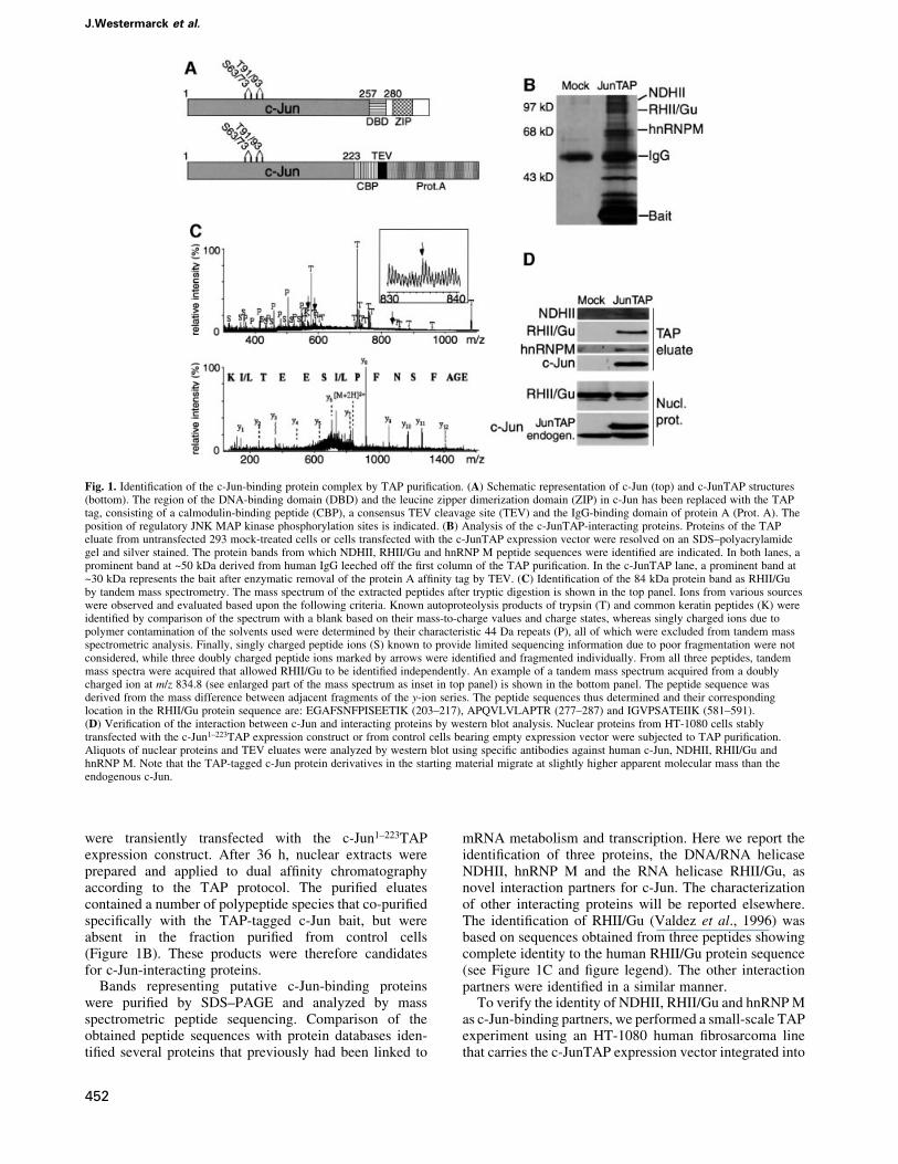
were transiently transfected with the c-Jun1±223TAPexpression construct. After 36 h, nuclear extracts wereprepared and applied to dual af®nity chromatographyaccording to the TAP protocol. The puri®ed eluatescontained a number of polypeptide species that co-puri®edspeci®cally with the TAP-tagged c-Jun bait, but wereabsent in the fraction puri®ed from control cells(Figure 1B). These products were therefore candidatesfor c-Jun-interacting proteins.
Bands representing putative c-Jun-binding proteinswere puri®ed by SDS±PAGE and analyzed by massspectrometric peptide sequencing. Comparison of theobtained peptide sequences with protein databases iden-ti®ed several proteins that previously had been linked to
mRNA metabolism and transcription. Here we report theidenti®cation of three proteins, the DNA/RNA helicaseNDHII, hnRNP M and the RNA helicase RHII/Gu, asnovel interaction partners for c-Jun. The characterizationof other interacting proteins will be reported elsewhere.The identi®cation of RHII/Gu (Valdez et al., 1996) wasbased on sequences obtained from three peptides showingcomplete identity to the human RHII/Gu protein sequence(see Figure 1C and ®gure legend). The other interactionpartners were identi®ed in a similar manner.
To verify the identity of NDHII, RHII/Gu and hnRNP Mas c-Jun-binding partners, we performed a small-scale TAPexperiment using an HT-1080 human ®brosarcoma linethat carries the c-JunTAP expression vector integrated into
Fig. 1. Identi®cation of the c-Jun-binding protein complex by TAP puri®cation. (A) Schematic representation of c-Jun (top) and c-JunTAP structures(bottom). The region of the DNA-binding domain (DBD) and the leucine zipper dimerization domain (ZIP) in c-Jun has been replaced with the TAPtag, consisting of a calmodulin-binding peptide (CBP), a consensus TEV cleavage site (TEV) and the IgG-binding domain of protein A (Prot. A). Theposition of regulatory JNK MAP kinase phosphorylation sites is indicated. (B) Analysis of the c-JunTAP-interacting proteins. Proteins of the TAPeluate from untransfected 293 mock-treated cells or cells transfected with the c-JunTAP expression vector were resolved on an SDS±polyacrylamidegel and silver stained. The protein bands from which NDHII, RHII/Gu and hnRNP M peptide sequences were identi®ed are indicated. In both lanes, aprominent band at ~50 kDa derived from human IgG leeched off the ®rst column of the TAP puri®cation. In the c-JunTAP lane, a prominent band at~30 kDa represents the bait after enzymatic removal of the protein A af®nity tag by TEV. (C) Identi®cation of the 84 kDa protein band as RHII/Guby tandem mass spectrometry. The mass spectrum of the extracted peptides after tryptic digestion is shown in the top panel. Ions from various sourceswere observed and evaluated based upon the following criteria. Known autoproteolysis products of trypsin (T) and common keratin peptides (K) wereidenti®ed by comparison of the spectrum with a blank based on their mass-to-charge values and charge states, whereas singly charged ions due topolymer contamination of the solvents used were determined by their characteristic 44 Da repeats (P), all of which were excluded from tandem massspectrometric analysis. Finally, singly charged peptide ions (S) known to provide limited sequencing information due to poor fragmentation were notconsidered, while three doubly charged peptide ions marked by arrows were identi®ed and fragmented individually. From all three peptides, tandemmass spectra were acquired that allowed RHII/Gu to be identi®ed independently. An example of a tandem mass spectrum acquired from a doublycharged ion at m/z 834.8 (see enlarged part of the mass spectrum as inset in top panel) is shown in the bottom panel. The peptide sequence wasderived from the mass difference between adjacent fragments of the y-ion series. The peptide sequences thus determined and their correspondinglocation in the RHII/Gu protein sequence are: EGAFSNFPISEETIK (203±217), APQVLVLAPTR (277±287) and IGVPSATEIIK (581±591).(D) Veri®cation of the interaction between c-Jun and interacting proteins by western blot analysis. Nuclear proteins from HT-1080 cells stablytransfected with the c-Jun1±223TAP expression construct or from control cells bearing empty expression vector were subjected to TAP puri®cation.Aliquots of nuclear proteins and TEV eluates were analyzed by western blot using speci®c antibodies against human c-Jun, NDHII, RHII/Gu andhnRNP M. Note that the TAP-tagged c-Jun protein derivatives in the starting material migrate at slightly higher apparent molecular mass than theendogenous c-Jun.
J.Westermarck et al.
452

its genome. Immunoblot analyses with NDHII-, RHII/Gu-and hnRNP M-speci®c antibodies revealed cross-reactiveprotein species of the expected molecular masses in therelevant eluate fraction (Figure 1D, upper panel). Weconclude that NDHII, RHII/Gu and hnRNP M wereretained on the column by means of their interaction withc-JunTAP. These results validate the mass spectrometricidenti®cation of NDHII, RHII/Gu and hnRNP M as c-Jun-binding proteins. Furthermore, the experiment con®rmsthat the interaction of c-Jun with the analyzed proteins isnot restricted to 293 cells and occurs when the c-JunTAPfusion protein is expressed at levels comparable withendogenous c-Jun as they are present in the stably trans-fected HT-1080 cells.
DEAD-box RNA helicases have been identi®ed previ-ously as auxiliary factors in transcription and translationprocesses (Eisen and Lucchesi, 1998; Luking et al., 1998;Linder and Daugeron, 2000). NDHII, for example, bindsCREB, a leucine zipper transcription factor and a distantrelative of c-Jun. NDHII mediates an interaction betweenRNA polymerase II (pol II) and CREB-binding protein(CBP) and supports CREB-mediated transcription acti-vation in reporter gene assays, an effect that requires theATPase function of the enzyme (Nakajima et al., 1997). Itis thus not surprising that NDHII also interacts with c-Jun.It is conceivable that this enzyme supports c-Jun tran-scription in a manner comparable with its role in CREBtarget gene activation.
Interestingly, one other newly identi®ed c-Jun inter-action partner, RHII/Gu, is a human DEAD-box RNAhelicase (Figure 1B and D). Unlike NDHII, however,RHII/Gu has not been implicated previously in RNApol II-mediated transcription. The presence of RHII/Gu
in a complex with c-Jun thus prompted further studieson the role of this enzyme in AP-1-regulated geneexpression.
RHII/Gu binds c-Jun directlyTo study whether the interaction between RHII/Gu andc-Jun is mediated by direct protein±protein contact, in vitropull-down assays were performed. Different bacteriallyexpressed GST±RHII/Gu fusion proteins were immobi-lized on glutathione±beads and subsequently incubatedwith various sources of c-Jun. Immunoblot analysesrevealed that both bacterially expressed c-Jun(c-JunHis6) and c-Jun present in 293 cell nuclear extractswere retained speci®cally on the immobilized C-terminaldomain of RHII/Gu (amino acids 646±801; Figure 2A,lanes 3 and 6). However, no interaction of c-Jun with theN-terminal part of RHII/Gu or with GST alone could bedetected (Figure 2A, lanes 1, 2, 4 and 5). This result showsthat the interaction observed by the TAP method can alsobe reconstituted in vitro and, importantly, that it is directand does not rely on any intermediary proteins that mighthave been present in 293 or HT-1080 cells. Furthermore,these results show that RHII/Gu protein may interact withthe non-fused wild-type form of c-Jun present in nuclearextracts.
Analysis of deletion mutants of RHII/Gu in the pull-down assay revealed that the c-Jun interaction domain onRHII/Gu resides in the most C-terminal part of the pro-tein between amino acids 749 and 801 (Figure 2A).Importantly, the interaction between c-Jun and GST±Gu749±801 was not affected by addition of high concentra-tions of ethidium bromide or RNase A, or by 600 mMNaCl (Figure 2B), demonstrating that the interaction
Fig. 2. Direct protein±protein interaction between RHII/Gu and c-Jun. (A) Mapping of the c-Jun-binding domain of RHII/Gu. Bacterially expressedGST±RHII/Gu proteins were immobilized on glutathione±beads and incubated with either bacterially expressed full-length c-JunHis6 protein or withnuclear proteins from unstimulated 293 cells. After extensive washing, proteins bound to the beads were eluted in protein sample buffer and analyzedby western blotting with c-Jun (top) or GST antibodies (bottom). A schematic representation of the structure of RHII/Gu and summary of the bindingdata are shown below. (B) The interaction between RHII/Gu and c-Jun is not mediated by DNA or RNA. GST pull-down experiments betweenGST±RHII/Gu 749±801 and c-JunHis6 were performed in the absence or presence of ethidium bromide (10 or 50 mg/ml), 40 mg/ml RNase A or NaCl(300 or 600 mM).
RNA helicase RHII/Gu is a co-factor for c-Jun
453

between the recombinant proteins is not mediated bycontaminating DNA or RNA, and is stable at moderatelyhigh ionic strength.
Interaction between endogenous c-Junand RHII/GuAfter showing that RHII/Gu and c-Jun interact in vitro orafter overexpression of c-Jun in transfected cells, it wasimportant to show that the interaction between the proteinsat their endogenous expression levels also occurs in cells.To this end, we used two independent approaches. First,we performed co-immunoprecipitation studies on nuclearextracts from non-transfected 293 and HT-1080 cells.Immunoblotting con®rmed that c-Jun antiserum ef®cientlyco-immunoprecipitated endogenous RHII/Gu and c-Junfrom both cell lines, whereas no precipitation of either ofthe proteins was observed with control pre-immune serum(PI) (Figures 3A and 4B). This experiment demonstratesthat c-Jun and RHII/Gu proteins interact in human cells attheir endogenous level of expression.
In the next experiment, we used a DNA af®nitypuri®cation assay to examine whether RHII/Gu interactswith DNA-bound c-Jun. A biotinylated double-strandedDNA oligonucleotide probe, containing a consensus AP-1-binding site, retrieved both c-Jun and RHII/Gu fromextracts of non-transfected HT-1080 and 293 cells(Figure 3B and data not shown). In contrast, no signi®cantbinding of either c-Jun or RHII/Gu proteins was observedwith a control oligonucleotide carrying a mutated AP-1-binding site (Figure 3B). This ®nding indicates that thebinding of c-Jun to DNA and to RHII/Gu is not mutuallyexclusive, and c-Jun could conceivably deliver RHII/Gu toa transcription unit.
Anisomycin stimulates c-Jun±RHII/Gu interactionThe part of c-Jun that is present in c-Jun1±223TAP carriesregulatory phosphorylation sites for the MAP kinase JNK.To examine whether the interaction between RHII/Gu and
c-Jun might be in¯uenced by the phosphorylation state ofthe latter, the binding of RHII/Gu to different phosphoryl-ation site mutants of c-Jun1±223TAP was examined(Figure 4A). In c-Jun1±223AspTAP, the JNK phosphoryl-ation sites were replaced with aspartic acid to create amimic of phosphorylated c-Jun (Treier et al., 1995). Ac-Jun1±223AlaTAP construct, with alanine substitutions ofthe phosphorylation sites, was generated in order tocreate an uninduced, inactive form of c-Jun. 293 cellswere transiently transfected with c-Jun1±223AlaTAP,c-Jun1±223TAP or c-Jun1±223AspTAP. In addition, thesecells received an expression vector for DMEKK1, aconstitutively active JNKKK that induces high JNKactivity and phosphorylation of both endogenous c-Junand wild-type c-Jun1±223TAP protein (Figure 4A). Probingof the eluate fractions with anti-RHII/Gu serum showedthat all three c-Jun1±223TAP proteins were expressed andrecovered at comparable levels and co-puri®ed withapproximately the same amounts of RHII/Gu (Figure 4A,lanes 2±4). This ®nding indicates that c-Jun phosphoryl-ation does not in¯uence binding to RHII/Gu. However, itdoes not exclude the possibility that the interactionbetween the proteins might be regulated by signals thatincrease the transcriptional activity of c-Jun, in a mannerthat is not mediated by the c-Jun phosphorylation sites. Totest this idea, non-transfected HT-1080 cells were treatedwith anisomycin and the amount of RHII/Gu in c-Jun co-immunoprecipitates was measured by western blotting.Anisomycin treatment induces potent activation of theJNK pathway and prominent phosphorylation of theimmunoprecipitated c-Jun protein (Figure 4B).Interestingly, more RHII/Gu was co-immunoprecipitatedwith c-Jun from anisomycin-treated cells, whereas theamount of c-Jun in the immunoprecipitate was identical intreated and control cells. These results suggest thatanisomycin-induced signaling regulates the interactionbetween c-Jun and RHII/Gu. To con®rm that this effect isindependent of c-Jun phosphorylation, as suggested by theexperiment shown in Figure 4A, we tested whetheranisomycin treatment would also increase RHII/Gu bind-ing to a c-Jun derivative that lacks the relevant phos-phorylation sites. For this purpose, we used a stableHT-1080 cell line expressing the c-Jun1±223AspTAP fusionprotein, and performed TAP puri®cation from untreated oranisomycin-treated cells. As shown in Figure 4C, aniso-mycin treatment signi®cantly increased binding of endo-genous RHII/Gu to c-Jun1±223AspTAP, but did not causeincreased expression of RHII/Gu protein or an elevatedrecovery of c-Jun1±223AspTAP in the eluate of the af®nitycolumn (Figure 4C).
JNK signaling regulates subnuclear localizationof RHII/GuThe results above show that the stoichiometry of inter-action between endogenous c-Jun and RHII/Gu proteinscan be increased by cellular signaling that induces JNKactivity. As this effect does not depend on c-Jun phos-phorylation on the known JNK substrate sites, it is possiblethat it is mediated by a JNK-induced change in theproperties of RHII/Gu. RHII/Gu protein has been reportedpreviously to be a predominantly nucleolar protein.Interestingly, treatment of cells with cytotoxic drugsresults in translocation of RHII/Gu from nucleolus to
Fig. 3. Interaction between endogenous RHII/Gu and c-Jun proteinsin 293 and HT-1080 cells. (A) RHII/Gu speci®cally co-immuno-precipitates with c-Jun. Nuclear extracts from non-transfectedsubcon¯uent 293 cells were subjected to immunoprecipitation (IP) witheither speci®c rabbit anti-c-Jun antiserum or rabbit pre-immune serum(PI). Immunoprecipitated proteins were analyzed by western blotting(IB) using anti-RHII/Gu or anti-c-Jun antibodies. Similar amounts ofstarting material were used in each lane. (B) RHII/Gu and c-Jun forma protein complex on AP-1-binding sites. Whole-cell extracts fromnon-transfected subcon¯uent HT-1080 cells were incubated with5¢-biotinylated double-stranded 25mer oligonucleotides carrying eithera consensus or a mutated AP-1-binding site. The oligonucleotides wererecovered using streptavidin-conjugated magnetic beads. Boundproteins were visualized by western blot using anti-RHII/Gu or anti-c-Jun antibodies. Similar amounts of starting materials were used ineach lane. Both panels show representative examples of two or threeexperiments with similar results.
J.Westermarck et al.
454

nucleoplasm (Perlaky et al., 1997). This suggests thatRHII/Gu has both nucleolar and nucleoplasmic functions.Thus, we investigated whether JNK activation might alsocause accumulation of RHII/Gu in the nucleoplasm, whichwould provide an explanation for the JNK-dependentstimulation of c-Jun±RHII/Gu interaction. We found, inaddition to the prevalent nucleolar stores, a signi®cant
fraction of RHII/Gu in the nucleoplasm of both 293 andHT-1080 cells. Next, we examined if activation of the JNKpathway might cause an enhanced translocation of RHII/Gu into the nucleoplasm. To this end, HT-1080 cells wereseeded on glass coverslips and, after treatment withanisomycin (5 mg/ml), UVC (20 J/m2) or actinomycin D(0.2 mM) for 1 h, cells were ®xed and immunostained withRHII/Gu antisera. As reported earlier, actinomycin Dtreatment induced signi®cant translocation of RHII/Gufrom nucleolus to nucleoplasm, resulting in uniformnucleoplasmic staining (Figure 5A). Interestingly, treat-ment of cells with anisomycin and UVC also increasednucleoplasmic levels of RHII/Gu (Figure 5A).
Even though anisomycin and UVC treatments havebeen used as prototypical activators of JNK signaling, theyin¯uence multiple cellular processes. In order to studyspeci®cally the role of JNK signaling in RHII/Gu trans-location, HT-1080 cells were transfected with RHII/Gu±green ¯uorescent protein (GFP) alone or together
Fig. 4. The interaction between c-Jun and RHII/Gu is increased uponstress signaling. (A) Phosphorylation of c-Jun in JNK phosphorylationsites is not required for interaction between c-Jun and RHII/Gu.Subcon¯uent 293 cells were transiently co-transfected with vectorsdirecting the expression of DMEKK1 and different c-JunTAPconstructs, containing either wild-type c-Jun sequences (Wt), orderivatives thereof in which the MAP kinase phosphorylation sites atposition 63, 73, 91 and 93 were mutated to alanine (Ala) or asparticacid (Asp) residues. Nuclear proteins were prepared and subjected toIgG af®nity puri®cation and TEV cleavage. Aliquots of nuclearproteins and TEV eluates were analyzed by western blot using speci®cantibodies against human c-Jun or RHII/Gu. (B) The interactionbetween endogenous c-Jun and RHII/Gu proteins is increased uponstress signaling. Nuclear extracts from untreated or anisomycin-treated(5 mg/ml, 1 h) subcon¯uent HT-1080 cells were subjected to immuno-precipitation with either c-Jun antiserum or rabbit pre-immune serum(PI). The amounts of c-Jun and RHII/Gu in the immunoprecipitateswere analyzed by western blotting with RHII/Gu and c-Jun antibodies.Phosphorylation of c-Jun in the anisomycin-treated samples is apparentby the mobility shift of the protein. Similar amounts of startingmaterial were used for each lane. (C) Anisomycin-induced increase ofRHII/Gu binding is independent of c-Jun phosphorylation state.Subcon¯uent HT-1080 cells stably expressing c-Jun1±223AspTAPwere treated for 1 h with anisomycin (5 mg/ml), or left untreated, andnuclear proteins were subjected to IgG af®nity puri®cation and TEVcleavage. The amounts of c-Jun and RHII/Gu proteins in the TEVeluate and nuclear proteins used as a starting material were examinedby western blot using RHII/Gu and c-Jun antibodies. Phosphorylationof endogenous c-Jun in the anisomycin-treated samples is discernableby mobility shift of the protein. All panels show representativeexamples of two or three experiments with similar results.
Fig. 5. JNK activity regulates subnuclear localization of RHII/Gu.(A) Chemical activators of JNK signaling cause nucleoplasmicretention of RHII/Gu in HT-1080 cells. Serum-starved HT-1080 wereleft untreated (CTL), treated with anisomycin (5 mg/ml), UVC (20 J/m2) or actinomycin D (0.2 mM) for 1 h, cells were ®xed,immunostained with RHII/Gu antisera and analyzed by ¯uorescencemicroscopy. (B) Overexpression of constitutively active MEKK1causes nucleoplasmic retention of RHII/Gu in HT-1080 cells. HT-1080cells were transiently co-transfected with an expression constructcoding for DMEKK1 together with RHII/Gu±GFP, and after 24 h cellswere serum starved for 12 h, after which subcellular localization ofRHII/Gu±GFP fusion protein was studied by ¯uorescence microscopy.Hoechst staining was used to visualize the morphology of nuclei.Shown are representative images of four experiments showing similarresults.
RNA helicase RHII/Gu is a co-factor for c-Jun
455

with an expression vector for DMEKK1. Activation ofJNK signaling by DMEKK1 resulted in markedlyincreased nucleoplasmic staining of RHII/Gu±GFP(Figure 5B). Taken together, these ®ndings provideevidence that in addition to the well-established functionof activating c-Jun by direct phosphorylation, JNK alsopromotes the translocation of RHII/Gu from the nucleolusto the nucleoplasm. This mechanism provides a plausibleexplanation for the observed stimulation of the c-Jun andRHII/Gu interaction in response to anisomycin-inducedJNK signaling.
RHII/Gu contributes to transcription activationby c-JunThe observation that RHII/Gu preferentially interacts withc-Jun in vivo in conditions that coincide with target geneactivation raises the possibility that it supports thisprocess. To test this idea, we performed experiments inwhich HT-1080 cells were microinjected with an expres-sion construct for a c-JunAsp±estrogen receptor fusionprotein (c-JunAspER) and an AP-1-responsive LacZreporter construct. In addition, the injected cells receivedan anti-RHII/Gu antibody, which blocks helicase activity,or a control antibody. The c-JunAspER was used for theseexperiments because it allows a rapid induction of atranscriptionally active c-Jun protein without engagingother signaling pathways. Treatment of c-JunAspER-expressing cells with 1 mM estradiol, 12 h after injection,
signi®cantly increased the fraction of b-galactosidase-positive cells (Figure 6A), indicating that activation of thec-Jun protein stimulated the TRELacZ reporter. Estradioltreatment had no effect on TRELacZ reporter activity incells that did not express c-JunAspER (data not shown).Signi®cantly, when the RHII/Gu neutralizing antibodywas co-injected, c-JunAspER-dependent TRELacZ pro-moter activation was essentially abolished. The suppres-sive effect of the RHII/Gu neutralizing antibody was notdue to a general suppression of transcription, as it did notinterfere with the activity of an RSVLacZ reporter, whichis insensitive to changes in AP-1 activity (Figure 6A). Thisis strong evidence that the endogenous RHII/Gu in theinjected cells supports transcriptional activation by c-Jun.
The neutralizing RHII/Gu antibody that interfered withc-Jun-dependent transcription speci®cally inhibits theRNA helicase activity of RHII/Gu (Valdez et al., 1997).This suggests that the helicase activity of RHII/Gu isrequired for c-Jun-mediated transcriptional activation. Toaddress the role of helicase function of the RHII/Gu inc-Jun-mediated transcriptional activation more speci®c-ally, we generated mammalian expression vectors codingfor full-length RHII/Gu in which either the helicase or theATPase domain of RHII/Gu was inactivated by pointmutations. We have shown previously that both mutationscompletely abolish the RNA helicase activity of RHII/Guin vitro (Valdez et al., 1997). To study the effects of themutants on c-Jun-mediated promoter activation, we used
Fig. 6. RNA helicase and ATPase activities of RHII/Gu contribute to transcription regulation by c-Jun. (A) A RHII/Gu neutralizing antibody inhibitsc-Jun-induced activation of an AP-1 reporter in HT-1080 cells. Subcon¯uent HT-1080 cells were microinjected with a c-JunAspER expression constructalong with either an AP-1 reporter plasmid (TRELacZ) or a non-AP-1 responsive control reporter (RSVLacZ), and either an af®nity-puri®ed humanRHII/Gu neutralizing antibody (aRHII/Gu) or control human IgG (IgG). At 12 h after injection, cells were treated with 1 mM estradiol (E2) andincubation was continued for 18 h. Thereafter, cells were ®xed and stained for b-galactosidase activity. The bar graph represents the percentage ofLacZ-positive cells among the injected cells (average 6 SD of three independent experiments), **P <0.02 (Student's t-test). (B) The helicase activityof RHII/Gu is required for stimulation of c-Jun activity. 293 cells were transiently transfected with c-JunGal4 or ElkGal4 expression constructstogether with either wild-type (Guwt), helicase mutant (Gu311) or ATPase mutant (Gu312) forms of RHII/Gu (left and right panels). Subcon¯uent 293cells carrying a stably integrated 53Gal4 reporter construct were transiently transfected with c-JunGal4 expression construct alone or in combinationwith the either wild-type (Guwt) or ATPase mutant (Gu312) form of RHII/Gu (middle panel). Luciferase activity in cell lysates was measured 24 hafter transfection. Transfection ef®ciency was monitored by co-transfecting the cells with a Ubi±Renilla luciferase construct. Shown are meanvalues 6 SD of three or four independent experiments, each in duplicate, *P <0.05; **P <0.02 (Student's t-test). (C) Inhibition of RHII/Gu activityimpairs c-Jun-mediated induction of mRNA expression. 293 cells were transiently transfected with the 53Gal4 reporter construct, the Ubi±Renillaexpression vector and c-JunGal4 expression construct alone or together with Gu312. After 24 h, cell lysates from parallel samples were prepared andluciferase activity assays were performed along with northern blots to quantitate the abundance of luciferase and Renilla luciferase mRNAs. Equalloading of mRNA was con®rmed by ethidium bromide staining of the rRNA.
J.Westermarck et al.
456

constructs in which the N-terminal transactivation domainof c-Jun was fused to the Gal4 DNA-binding domain. Theeffects of the overexpressed proteins were determined bymeasurement of 53UAS luciferase reporter activity.Overexpression of wild-type RHII/Gu had no effect onc-JunGal4-mediated promoter activation (Figure 6B),probably due to a high abundance and non-limitingconcentration of the endogenous RHII/Gu in these cells.In accordance with the results of the microinjectionexperiment, both the helicase mutant (Gu311) and theATPase mutant (Gu312) forms of RHII/Gu, however,signi®cantly suppressed the c-JunGal4-induced promoteractivation (P-values <0.05 and <0.02, Figure 6B).Importantly, overexpression of the wild-type and mutantforms of RHII/Gu had no signi®cant effect on the basal
activity of the 53UAS luciferase reporter (data not shown)or when the luciferase reporter was activated by Gal4-Elk1, a derivative of a transcription factor that is unrelatedto c-Jun (Figure 6B). These results suggest that thehelicase activity of RHII/Gu speci®cally facilitates c-Jun-mediated transcription.
To investigate whether the results of the transfectionexperiments could be repeated in a chromatin context andwere not in¯uenced by the episomal state of the reporterconstructs, we expressed c-JunGal4 alone or together withwild-type and the Gu312 mutant of RHII/Gu, in a 293 cellline bearing a genomically integrated 53UAS luciferasereporter. In these cells, a similar signi®cant suppressiveeffect of Gu312 on c-JunGal4-induced reporter activationwas observed (P-value <0.02, Figure 6B).
In addition to the proposed role of DEAD-box RNAhelicases in transcription, some of these enzymes promotemRNA translation (Chuang et al., 1997; Luking et al.,1998; Linder and Daugeron, 2000). We wanted to deter-mine whether RHII/Gu might affect reporter expression atthe translational rather than the transcriptional level. 293cells were transfected with the 53UAS luciferase reporter,along with c-JunGal4 in the absence or presence of theRHII/Gu mutant Gu312, and both luciferase activity andluciferase mRNA expression were determined from par-allel samples. As before, Gu312 inhibited c-JunGal4-dependent induction of luciferase activity (Figure 6B).Furthermore, Gu312 expression signi®cantly (by 75%)decreased the abundance of luciferase mRNA as measuredby northern blots (Figure 6C), indicating that helicaseactivity of RHII/Gu is involved in the transcriptionalregulation of gene expression. In accordance with previousexperiments, inhibition of helicase activity of RHII/Gu didnot have any non-speci®c inhibitory effect on the expres-sion of ubiquitin promoter-driven Renilla luciferasemRNA, which was transcribed from an internal controlvector (Figure 6C).
RHII/Gu promotes c-Jun-mediated neuronaldifferentiation of PC12 cellsWe wanted to study whether RHII/Gu is required forregulation of complex cellular programs induced by c-Jun.c-Jun potently induces neuronal differentiation of PC12cells (LeppaÈ et al., 2001). To study whether RHII/Guactivity is involved in this differentiation function, weperformed an experiment in which PC12 cells weremicroinjected with an expression vector for hemagglutinin(HA) epitope-tagged c-Jun together with either a controlantibody or a neutralizing antibody against RHII/Gu. At48 h after injection, the effect of the RHII/Gu antibody onc-Jun-mediated neuronal differentiation of the PC12 cellswas measured. Injection of PC12 cells with controlantibody alone did not have any effect on the cellphenotype, whereas prominent neurite outgrowth wasobserved in cells injected with c-Jun expression constructtogether with control antibody (Figure 7A). Remarkably,injection of the RHII/Gu neutralizing antibody stronglydecreased c-Jun-induced neurite outgrowth, showing thatRHII/Gu activity is required for induction of the neuronaldifferentiation program initiated by c-Jun in these cells(Figure 7A and B). Importantly, comparable numbers ofHA epitope-positive cells were observed in both groups ofcells (data not shown), indicating that the RHII/Gu
Fig. 7. RHII/Gu activity supports c-Jun-mediated PC12 celldifferentiation. (A) Neurite outgrowth in microinjected PC12 cells.PC12 cells were microinjected with FITC-labeled dextran or HA-c-JunAsp expression construct together with control IgG or RHII/Gublocking antibodies (aGu) as indicated. After 48 h, cells were ®xedand cells expressing HA-c-JunAsp were identi®ed by staining with anti-HA antibody and FITC-labeled secondary antibody (bright nuclei).The morphology of the cells was visualized by actin staining.(B) Quanti®cation of neurite outgrowth. The percentage of HA-c-JunAsp-injected cells with neurites 3- or 4-fold the cell length amongmicroinjected (FITC-positive) cells is shown. The graph is arepresentative example out of three experiments with similar results.
RNA helicase RHII/Gu is a co-factor for c-Jun
457

antibody did not have adverse effects on the survival ofinjected PC12 cells. These results provide further evidencein support of a contribution of endogenous RHII/Gu toc-Jun-mediated cellular processes and imply that RHII/Gusupports the activation of endogenous c-Jun target genes,in addition to synthetic reporter genes.
Discussion
Many fundamental functions of the eukaryotic cell such astranscription initiation, splicing, replication and DNArepair are catalyzed by multiprotein complexes consistingof many dozens of subunits with enzymatic as well asregulatory functions (Hirose and Manley, 2000; Lemonand Tjian, 2000). To learn more about the biology of theseprocesses and the multiprotein complexes involved, theidenti®cation of their subunits and the characterization oftheir interactions is essential. The data presented heredocument how the TAP technology that was developedinitially to characterize protein interactions in yeast can beadapted successfully for transiently and stably transfectedmammalian tissue culture systems. Based on its genericnature (Rigaut et al., 1999; Puig et al., 2001), this methodshould be widely applicable to the identi®cation andcharacterization of known or suspected protein±proteininteractions in higher eukaryotic cells.
We have identi®ed a group of proteins that bind to c-Junand provide evidence that the DEAD-box RNA helicaseRHII/Gu is a novel transcriptional co-activator of c-Jun.Several of the other proteins identi®ed as c-Jun interactionpartners have established or supposed roles in thetranscriptional regulation of gene expression, making itplausible that we have identi®ed a set of proteins thatfacilitate c-Jun-mediated promoter activation and mRNAsynthesis.
The data presented in this study indicate that the RNAhelicase activity of RHII/Gu is involved speci®cally in thestimulation of c-Jun-mediated reporter activation and foractivation of endogenous c-Jun targets during PC12 cellneuronal differentiation. The fact that the substratespeci®city of RHII/Gu is limited to RNA±RNA andRNA±DNA duplexes suggests that it acts at a step oftranscription different from DNA helicases such as NDHII(Nakajima et al., 1997; Eisen and Lucchesi, 1998). Basedon our reporter gene assays, the RNA helicase activity ofRHII/Gu facilitates ef®cient mRNA synthesis but, incontrast to other RNA helicases such as eIF-4A and theyeast RNA helicase Ded1p (Chuang et al., 1997; Linderand Daugeron, 2000), it appears not to be involved intranslational regulation of gene expression. In addition,since the reporter genes used in this study do not containintrons, it is evident that the stimulatory effect of RHII/Guon their expression cannot be explained by effects at thelevel of pre-mRNA splicing as has been shown for yeastDEAD-box PRP proteins (Luking et al., 1998). Further-more, the ®nding that overexpression of mutant forms ofRHII/Gu equally potently suppressed c-Jun-mediatedtranscriptional activation in both an episomal and achromatin context suggests that RHII/Gu is not involvedin chromatin decondensation.
The mechanism by which RHII/Gu participates inc-Jun-stimulated mRNA synthesis remains a matter ofspeculation at this point. Interestingly, it was shown
recently that the vaccinia virus DEXD/H protein NPH-IIcan displace protein from RNA (Jankowsky et al., 2001).This might suggest an additional mechanism to explainhow these proteins could in¯uence gene transcription, forexample by facilitating promoter clearance. Further con-ceivable possibilities for a role of RHII/Gu in support ofc-Jun include assisting in the packaging of the RNA intohnRNPs or even a function in transcription-coupled DNArepair or the removal of a stalled polymerase fromdamaged templates.
Our data show that the binding of RHII/Gu to c-Junincreases in response to anisomycin treatment by a c-Junphosphorylation-independent mechanism. This observa-tion could be explained by the JNK-regulated trans-location of RHII/Gu from nucleoli to nucleoplasm. Such amodel would not be without precedent. In addition tobeing a site for rRNA synthesis and assembly, thenucleolus is now recognized as a store for proteins withlatent nucleoplasmic functions (e.g. Mdm2; Visintin andAmon, 2000). It has been established that nucleolarproteins may shuttle continuously between nucleolus andnucleoplasm (Phair and Misteli, 2000). Interestingly, theC-terminal domain of RHII/Gu that binds to c-Jun alsoserves as a nucleolar localization signal for RHII/Gu (Ouet al., 1999), indicating that the interaction between RHII/Gu and c-Jun might prevent relocalization of RHII/Gu tothe nucleolus.
A ®nal point of note is that both c-Jun and RHII/Gu havebeen shown to be necessary for cell growth and/or survival(Ou et al., 1999; Davis, 2000). These ®ndings indicate thatthe interaction between RHII/Gu and c-Jun might berequired for the regulation of c-Jun target genes importantfor cell cycle progression. Further studies will be requiredto elucidate a potential function of interaction betweenc-Jun and RHII/Gu in cell growth and transformation.
Materials and methods
Plasmids and construction of expression vectorsBacterial expression plasmids for His6-tagged c-Jun (Treier et al., 1994),mammalian HA-tagged c-JunAsp (Treier et al., 1995), constitutivelyactive DMEKK1 (Whitmarsh et al., 1995), GST±RHII/Gu fusion proteins(Valdez et al., 1996) and reporter plasmids RSVLacZ and TRELacZ(Yeung et al., 1999) have been described. Information about cloning ofthe mammalian expression constructs for RHII/Gu mutants JunAspER andc-Jun1±223TAP is available on request.
TAP puri®cationFor TAP puri®cation experiments, subcon¯uent 293 cells were transientlytransfected by the calcium phosphate method with 5 mg of CMVJunTAPconstructs, 2 mg of DMEKK1 or 7 mg of empty cytomegalovirus (CMV)-driven expression construct per 10 cm plate. c-JunTAP complexes werepuri®ed using a published procedure (Rigaut et al., 1999; Puig et al.,2001) with minor modi®cations. High salt nuclear extractions from mock-or Jun1±223TAP-transfected cells were prepared as described (Andrewsand Faller, 1991) and subsequently adjusted to IgG-binding conditions:180 mM NaCl, 10 mM Tris±HCl pH 8.0, 0.2% NP-40, 0.5 mMdithiothreitol (DTT), complete protein inhibitors (Roche), 10 mM b-glycerolphosphate and 20 mM NaF. Diluted extracts were rotatedovernight at 4°C with 100 ml of IgG matrix (Amersham Biotech), afterwhich the beads were washed extensively in binding buffer. Washedbeads were resuspended in TEV cleavage buffer (10 mM Tris±HClpH 8.0, 150 mM NaCl, 0.3% NP-40, 0.5 mM EDTA, 0.5 mM DTT), and5±15 ml of recombinant TEV enzyme (150 U; Life Technologies) wasadded to the mixture. After rotation for 2 h at 16°C, the TEV eluate fromthe IgG column was recovered and adjusted to calmodulin-bindingconditions: 45 mM Tris±HCl pH 8.0, 150 mM NaCl, 0.7 mM Mg-acetate, 0.7 mM imidazole, 2.5 mM CaCl2, 0.2 % NP-40, 10 mM
J.Westermarck et al.
458

b-mercaptoethanol, and rotated for 2 h at 4°C with 50 ml of calmodulinaf®nity resin (Stratagene). After binding, sedimented beads were washedextensively with calmodulin-binding buffer. Bound proteins wererecovered by boiling the calmodulin beads for 3 min in protein samplebuffer and loaded onto a preparative 12% SDS±polyacrylamide gel.Proteins were detected by silver staining (Shevchenko et al., 1996).
Peptide sequences of c-Jun-speci®c interaction partners were identi®edas described previously (Bouveret et al., 2000) except that a Q-Tof massspectrometer (Micromass) was used for peptide sequencing by tandemmass spectrometry. Protein bands were excised from the silver-stainedgel, reduced, alkylated and digested overnight with trypsin, and peptidesequences were obtained by nano-electrospray tandem mass spectrometryand by comparison with a comprehensive non-redundant protein databaseusing the program PeptideSearch.
For western blot analysis, aliquots of nuclear proteins or TEV eluatesafter IgG af®nity puri®cation and TEV cleavage were loaded onto 12%SDS±polyacrylamide gels and transferred to nitrocellulose membrane.
GST pull-down experimentsPull-down assays with puri®ed JunHis6, a histidine-tagged recombinantfull-length c-Jun, were performed by incubating 1 mg of soluble JunHis6
protein with molar equivalent amounts (1±3 mg) of GST±RHII/Gudeletions immobilized on glutathione±Sepharose beads in 0.5 ml of buffercontaining 20 mM Tris±HCl pH 7.4, 0.2 mM EDTA, 0.1 M NaCl and1 mM DTT. In experiments in which nuclear proteins containing c-Junwere used, nuclear proteins were prepared from 293 cells (Andrews andFaller, 1991), adjusted to the binding conditions described above. Inexperiments in which ethidium bromide, RNase A or higher concentra-tions of NaCl were used, these reagents were added to the bindingreaction prior to addition of the recombinant proteins. Bound proteinswere boiled in sample buffer, resolved by SDS±PAGE and immuno-blotted with anti-GST and anti-c-Jun antibodies.
Co-immunoprecipitations and DNA af®nity puri®cation assayProtein G±Sepharose (Pharmacia) beads were agitated on a tumbler for 2 hat 4°C with c-Jun antiserum or with rabbit pre-immune serum in 20 mMHEPES±KOH pH 7.5, 400 mM NaCl, 0.25 mM EGTA, 1.5 mM MgCl2,0.25% NP-40, protease inhibitors (Roche), 10 mM b-glycerolphosphate,20 mM NaF and 0.5 mM DTT. Before co-immunoprecipitation, 293 andHT-1080 nuclear extracts were sonicated and pre-cleared for 2 h at 4°Cwith rabbit pre-immune serum immobilized to protein G±Sepharose(Pharmacia) beads, and supernatants from the pre-cleared samples wereincubated with immobilized c-Jun antiserum or control rabbit pre-immune serum overnight at 4°C. Thereafter, sedimented beads werewashed four times with 50 mM Tris±HCl pH 7.4, 150 mM NaCl, 0.3%NP-40 and 0.5 mM DTT, and bound proteins were analyzed by westernblotting using mouse anti-c-Jun or human anti-RHII/Gu antibodies. Forthe DNA af®nity puri®cation assay, HT-1080 cells were lysed in RIPAbuffer [50 mM Tris±HCl pH 7.4, 150 mM NaCl, 1% NP-40, 1 mMEDTA, 1 mM phenylmethylsulfonyl ¯uoride (PMSF), protease inhibitors(Roche), 10 mM b-glycerolphosphate, 20 mM NaF, 0.5 mM DTT]. Thecell lysates were then sonicated and incubated with 3 mg of biotinylateddouble-stranded oligonucleotides at 4°C for 3 h. Oligonucleotide±proteincomplexes were recovered using streptavidin-conjugated magnetic beads(Dynal) and washed extensively. The following double-strandedoligomers containing either wild-type or mutated AP-1-binding sites(underlined) were used: (AP-1wt) 5¢-biotinTAAAGCATGAGTCAGA-CACCTCTG-3¢ or (AP-1mut) 5¢-biotinTAAAGCAGGGCCTCAGACA-CCTCTG-3¢.
Proteins bound to DNA were resolved by SDS±PAGE and analyzed bywestern blotting using c-Jun and RHII/Gu antibodies.
Reporter gene assays293 cells were transfected at 50±70% con¯uency by the calciumphosphate method with 1 mg of reporter plasmid pf2Luc (Stratagene)together with CMVGal4DBD, CMVJunGal4 (Stratagene),CMVElk1Gal4 (pFA2-Elk1, Stratagene) (10 ng each), pHook-Flag-Gu,pHook-FLAG-Gu311 or pHook-FLAG-Gu312 (150 ng each) expressionconstructs. Control cultures were co-transfected in parallel withequivalent amounts of the corresponding empty expression vector.Transfection ef®ciency was controlled by co-transfection with 10 ng ofubiquitin promoter-driven Renilla luciferase reporter construct. At 24 hafter transfection, cells were washed twice with phosphate-buffered saline(PBS) and lysed with passive lysis buffer (Promega). Fire¯y luciferaseand Renilla luciferase activities were measured separately.
Microinjection reporter assaysHT-1080 cells were seeded on CELLocate coverslips (Eppendorf) tofacilitate identi®cation of control IgG and RHII/Gu neutralizing antibody-injected cells after staining. A total of 100±150 cells were co-injectedwith the JunAspER expression construct (100 mg/ml) and TRELacZreporter construct (200 mg/ml) or with RSVLacZ control vector (5 mg/ml)together with 90 ng/ml of either puri®ed human IgG or af®nity-puri®edhuman RHII/Gu neutralizing antibody (Valdez et al., 1996, 1997). At 12 hafter microinjection, cells were treated with 1 mM estradiol andincubation was continued for 18 h. Cells were washed thereafter withPBS, ®xed with 2% paraformaldehyde (PFA) and LacZ stained asdescribed previously (Rose et al., 1999). Cells were inspected by lightmicroscopy and the injected cells that exhibit blue staining were countedas positive.
PC12 cell differentiation assayPC12 cells were microinjected and stained as described previously(LeppaÈ et al., 2001). Brie¯y, 100±150 PC12 cells were injected withCMV HA c-JunAsp expression construct (Treier et al., 1995) together with90 ng/ml of puri®ed human IgG or af®nity-puri®ed human RHII/Guneutralizing antibody (Valdez et al., 1997). Control cells were injectedwith puri®ed human IgG together with ¯uorescein isothiocyanate (FITC)-labeled dextran (Molecular Probes). At 48 h after injection, cells were®xed with 2% PFA, and nuclear expression of HAc-JunAsp was visualizedby staining of cells with a monoclonal anti-HA antibody (Roche) andFITC-conjugated secondary antibody (Dianova). The morphology of cellswas visualized using tetramethylrhodamine isothiocyanate (TRITC)-labeled phalloidin (Sigma). For visualization of the cells and quanti®-cation of neurite outgrowth, mounted samples were examined using aZeiss LSM410 confocal imaging system.
Acknowledgements
We thank Dr F.Stewart, Dr D.Rose and Dr R.Davis for providing thecDNA encoding the estrogen receptor-binding domain, the TRELacZreporter and DMEKK1 expression constructs, respectively; Dr F.Grosseand Dr J.-P.Fuchs for providing us with the NDHII and hnRNP Mantibodies, respectively; Dr S.LeppaÈ for advice on PC12 differentiationassays; and members of our laboratory, especially U.Gritzan, J.Brenneckeand M.Bonoli, for their technical help and suggestions. This work wassupported by the Academy of Finland (project No. 43019), CultureFoundation of Finland and Astra-Zeneca (J.W.), P.F. Biotecnologie-CNR-Italy (A.-M.M.), and the National Institutes of Health GrantDK52341 (B.C.V.).
References
Andrews,N.C. and Faller,D.V. (1991) A rapid micropreparationtechnique for extraction of DNA-binding proteins from limitingnumbers of mammalian cells. Nucleic Acids Res., 19, 2499.
Bouveret,E., Rigaut,G., Shevchenko,A., Wilm,M. and Seraphin,B.(2000) A Sm-like protein complex that participates in mRNAdegradation. EMBO J., 19, 1661±1671.
Chinenov,Y. and Kerppola,T.K. (2001) Close encounters of many kinds:Fos±Jun interactions that mediate transcription regulatory speci®city.Oncogene, 20, 2438±2452.
Chuang,R.Y., Weaver,P.L., Liu,Z. and Chang,T.H. (1997) Requirementof the DEAD-box protein ded1p for messenger RNA translation.Science, 275, 1468±1471.
Davis,R. (2000) Signal transduction by the JNK group of MAP kinases.Cell, 103, 239±252.
Eisen,A. and Lucchesi,J.C. (1998) Unraveling the role of helicases intranscription. BioEssays, 20, 634±641.
Hirose,Y. and Manley,J.L. (2000) RNA polymerase II and theintegration of nuclear events. Genes Dev., 14, 1415±1429.
Jankowsky,E., Gross,C.H., Shuman,S. and Pyle,A.M. (2001) Activedisruption of an RNA±protein interaction by a DExH/D RNA helicase.Science, 291, 121±125.
Lemon,B. and Tjian,R. (2000) Orchestrated response: a symphony oftranscription factors for gene control. Genes Dev., 14, 2551±2569.
LeppaÈ,S. and Bohmann,D. (1999) Diverse functions of JNK signalingand c-Jun in stress response and apoptosis. Oncogene, 18, 6158±6162.
LeppaÈ,S., Eriksson,M., Saffrich,R., Ansorge,W. and Bohmann,D. (2001)Complex functions of AP-1 transcription factors in differentiation andsurvival of PC12 cells. Mol. Cell. Biol., 21, 4369±4378.
RNA helicase RHII/Gu is a co-factor for c-Jun
459

Linder,P. and Daugeron,M.C. (2000) Are DEAD-box proteins becomingrespectable helicases? Nature Struct. Biol., 7, 97±99.
Luking,A., Stahl,U. and Schmidt,U. (1998) The protein family of RNAhelicases. Crit. Rev. Biochem. Mol. Biol., 33, 259±296.
Nakajima,T., Uchida,C., Anderson,S.F., Lee,C.G., Hurwitz,J., Parvin,J.D.and Montminy,M. (1997) RNA helicase A mediates association of CBPwith RNA polymerase II. Cell, 90, 1107±1112.
Ou,Y., Fritzler,M.J., Valdez,B.C. and Rattner,J.B. (1999) Mapping andcharacterization of the functional domains of the nucleolar proteinRNA helicase II/Gu. Exp. Cell Res., 247, 389±398.
Perlaky,L., Valdez,B.C. and Busch,H. (1997) Effects of cytotoxic drugson translocation of nucleolar RNA helicase RH-II/Gu. Exp. Cell Res.,235, 413±420.
Phair,R.D. and Misteli,T. (2000) High mobility of proteins in themammalian cell nucleus. Nature, 404, 604±609.
Puig,O., Caspary,F., Rigaut,G., Rutz,B., Bouveret,E., Bragado-Nilsson,E.,Wilm,M. and SeÂraphin,B. (2001) The tandem af®nity puri®cation (TAP)method: a general procedure of protein complex puri®cation. Methods,24, 218±229.
Rigaut,G., Shevchenko,A., Rutz,B., Wilm,M., Mann,M. and Seraphin,B.(1999) A generic protein puri®cation method for protein complexcharacterization and proteome exploration. Nature Biotechnol., 17,1030±1032.
Rose,D.W., Mullen,T.-M., Rosenfeld,M.G. and Glass,C.K. (1999)Functional characterization of coactivators using mammalian cellmicroinjection. In Picard,D. (ed.), Nuclear Receptors: A PracticalApproach. IRL Press, Oxford, UK, pp. 72±93.
Shevchenko,A., Wilm,M., Vorm,O. and Mann,M. (1996) Massspectrometric sequencing of proteins silver-stained polyacrylamidegels. Anal. Chem., 68, 850±858.
Treier,M., Staszewski,L.M. and Bohmann,D. (1994) Ubiquitin-dependent c-Jun degradation in vivo is mediated by the deltadomain. Cell, 78, 787±798.
Treier,M., Bohmann,D. and Mlodzik,M. (1995) JUN cooperates with theETS domain protein pointed to induce photoreceptor R7 fate in theDrosophila eye. Cell, 83, 753±760.
Valdez,B.C., Henning,D., Busch,R.K., Woods,K., Flores-Rozas,H.,Hurwitz,J., Perlaky,L. and Busch,H. (1996) A nucleolar RNAhelicase recognized by autoimmune antibodies from a patient withwatermelon stomach disease. Nucleic Acids Res., 24, 1220±1224.
Valdez,B.C., Henning,D., Perumal,K. and Busch,H. (1997) RNA-unwinding and RNA-folding activities of RNA helicase II/GuÐtwoactivities in separate domains of the same protein. Eur. J. Biochem.,250, 800±807.
Visintin,R. and Amon,A. (2000) The nucleolus: the magician's hat forcell cycle tricks. Curr. Opin. Cell Biol., 12, 372±377.
Whitmarsh,A.J., Shore,P., Sharrocks,A.D. and Davis,R.J. (1995)Integration of MAP kinase signal transduction pathways at theserum response element. Science, 269, 403±407.
Yeung,K. et al. (1999) Suppression of Raf-1 kinase activity and MAPkinase signalling by RKIP. Nature, 401, 173±177.
Received May 2, 2001; revised November 16, 2001;accepted November 29, 2001
J.Westermarck et al.
460