Docking Studies of Pakistani HCV NS3 Helicase: A Possible Antiviral Drug Target
12
Docking Studies of Pakistani HCV NS3 Helicase: A Possible Antiviral Drug Target Kaneez Fatima 1 , Shilu Mathew 2 , Mohd Suhail 3 , Ashraf Ali 3 , Ghazi Damanhouri 3 , Esam Azhar 3 , Ishtiaq Qadri 3 * 1 IQ Institute of Infection and Immunity, Lahore, Punjab, Pakistan, 2 Center of Excellence in Genomic Medicine Research, King Abdul Aziz University, Jeddah, Saudi Arabia, 3 King Fahd Medical Research Center, King Abdul Aziz University, Jeddah, Saudi Arabia Abstract The nonstructural protein 3 (NS3) of hepatitis C virus (HCV) helicase is believed to be essential for viral replication and has become an attractive target for the development of antiviral drugs. The study of helicase is useful for elucidating its involvement in positive sense single-stranded RNA virus replication and to serve as templates for the design of novel antiviral drugs. In recent years, several models have been proposed on the conformational change leading to protein movement and RNA unwinding. Some compounds have been recently reported to inhibit the helicase and these include small molecules, RNA aptamers and antibodies. The current study is designed to help gain insights for the consideration of potential inhibitors for Pakistani HCV NS3 helicase protein. We have cloned, expressed and purified HCV NS3 helicase from Pakistani HCV serum samples and determined its 3D structure and employed it further in computational docking analysis to identify inhibitors against HCV genotype 3a (GT3a),including six antiviral key molecules such as quercetin, beta-carotene, resveratrol, catechins, lycopene and lutein. The conformation obtained after docking showed good hydrogen bond (HBond) interactions with best docking energy for quercetin and catechins followed by resveratrol and lutein. These anti-helicase key molecules will offer an alternative attraction to target the viral helicase, due to the current limitation with the interferon resistance treatment and presences of high rate of resistance in anti-protease inhibitor classes. Citation: Fatima K, Mathew S, Suhail M, Ali A, Damanhouri G, et al. (2014) Docking Studies of Pakistani HCV NS3 Helicase: A Possible Antiviral Drug Target. PLoS ONE 9(9): e106339. doi:10.1371/journal.pone.0106339 Editor: Shama Ahmad, University of Colorado, Denver, United States of America Received March 30, 2014; Accepted July 19, 2014; Published September 4, 2014 Copyright: ß 2014 Fatima et al. This is an open-access article distributed under the terms of the Creative Commons Attribution License, which permits unrestricted use, distribution, and reproduction in any medium, provided the original author and source are credited. Data Availability: The authors confirm that all data underlying the findings are fully available without restriction. All data are included within the paper and its Supporting Information files. Funding: This work was funded by IQ Institute of Infection and Immunity (IQI3) and Miraj Foundation. The funders had no role in study design, data collection and analysis, decision to publish, or preparation of the manuscript. Competing Interests: The authors have declared that no competing interests exist. * Email: [email protected] Introduction Hepatitis C virus (HCV) is one of the major causative agent of chronic hepatitis which leads to liver cirrhosis, hepato cellular carcinoma, and liver failure and the most significant cause for liver transplantation [1,2]. It is estimated that about 3% of the world’s population (,180 million people) are affected with HCV [3] and 10 million people are believed to be infected by HCV alone in Pakistan [4]. HCV RNA genome encodes a single open reading frame that is translated into 3,000 amino acids (AA) poly protein and cleaved into 10 mature proteins. HCV genome translated into 4 structural (Core, E1 E2 and p7), and 6 important nonstructural (NS) proteins: NS2, NS3, NS4A, NS4B, NS5A, and NS5B [5], which coordinate the intracellular processes of the viral life cycle. Among the NS proteins, NS3 is a multifunctional protein (1–631 AA) with serine protease activity at the N-terminal (1–180 AA) and a nucleoside-triphosphatase (NTPase) dependent RNA helicase activity (NS3 NTPase/helicase) at the C-terminal (181–631 AA) [6]. Among all HCV proteins, NS3/NS4A serine protease and helicase are effective drug targets to develop anti-HCV agents [7]. The basic role NS3/NS4A is to cleave virus at different functional points as well as involved in viral replication. NS3 RNA helicase affects two different steps in the virus life cycle: (a) RNA replication step of virion in which NS3 is required to unwind the double- stranded RNA intermediate during RNA-dependent replication, that enables the movement of HCV NS5B polymerase [8], (b) NS3 assists in virus assembly and can also act as a scaffold for interaction with viral or cellular cofactors [9,10]. The crystal structure of HCV helicase shows that it consists of motifs I, Ia, II, III, IV, V, and VI, which are highly conserved. These motifs are located in the ATP binding cleft, and some project residues located at the nucleic acid binding site. Recently two NS3 protease inhibitors have been approved as a standard care for HCV GT1 affected patients by providing treatment with triple therapy (Peglated-Interferon - a, ribavirin and boceprevir or telaprevir [11] that are available in the market under the brand name Victrelis for boceprevir or Incivek and Incivo for telaprevir. In patients with GT1chronic HCV infection, the treatments with telaprevir/boceprevir based triple therapy are standard-of-care. However, more efficacious direct-acting antivi- rals (DAA) (Interferon (IFN)-based new DAAs) are available and interferon-free (IFN-free) regimens are imminent in near future. Imminent treatments for individuals infected by HCV will likely involve combinations of compounds that inhibit multiple viral PLOS ONE | www.plosone.org 1 September 2014 | Volume 9 | Issue 9 | e106339
-
Upload
independent -
Category
Documents
-
view
1 -
download
0
Transcript of Docking Studies of Pakistani HCV NS3 Helicase: A Possible Antiviral Drug Target

Docking Studies of Pakistani HCV NS3 Helicase: APossible Antiviral Drug TargetKaneez Fatima1, Shilu Mathew2, Mohd Suhail3, Ashraf Ali3, Ghazi Damanhouri3, Esam Azhar3,
Ishtiaq Qadri3*
1 IQ Institute of Infection and Immunity, Lahore, Punjab, Pakistan, 2 Center of Excellence in Genomic Medicine Research, King Abdul Aziz University, Jeddah, Saudi Arabia,
3 King Fahd Medical Research Center, King Abdul Aziz University, Jeddah, Saudi Arabia
Abstract
The nonstructural protein 3 (NS3) of hepatitis C virus (HCV) helicase is believed to be essential for viral replication and hasbecome an attractive target for the development of antiviral drugs. The study of helicase is useful for elucidating itsinvolvement in positive sense single-stranded RNA virus replication and to serve as templates for the design of novelantiviral drugs. In recent years, several models have been proposed on the conformational change leading to proteinmovement and RNA unwinding. Some compounds have been recently reported to inhibit the helicase and these includesmall molecules, RNA aptamers and antibodies. The current study is designed to help gain insights for the consideration ofpotential inhibitors for Pakistani HCV NS3 helicase protein. We have cloned, expressed and purified HCV NS3 helicase fromPakistani HCV serum samples and determined its 3D structure and employed it further in computational docking analysis toidentify inhibitors against HCV genotype 3a (GT3a),including six antiviral key molecules such as quercetin, beta-carotene,resveratrol, catechins, lycopene and lutein. The conformation obtained after docking showed good hydrogen bond (HBond)interactions with best docking energy for quercetin and catechins followed by resveratrol and lutein. These anti-helicase keymolecules will offer an alternative attraction to target the viral helicase, due to the current limitation with the interferonresistance treatment and presences of high rate of resistance in anti-protease inhibitor classes.
Citation: Fatima K, Mathew S, Suhail M, Ali A, Damanhouri G, et al. (2014) Docking Studies of Pakistani HCV NS3 Helicase: A Possible Antiviral Drug Target. PLoSONE 9(9): e106339. doi:10.1371/journal.pone.0106339
Editor: Shama Ahmad, University of Colorado, Denver, United States of America
Received March 30, 2014; Accepted July 19, 2014; Published September 4, 2014
Copyright: � 2014 Fatima et al. This is an open-access article distributed under the terms of the Creative Commons Attribution License, which permitsunrestricted use, distribution, and reproduction in any medium, provided the original author and source are credited.
Data Availability: The authors confirm that all data underlying the findings are fully available without restriction. All data are included within the paper and itsSupporting Information files.
Funding: This work was funded by IQ Institute of Infection and Immunity (IQI3) and Miraj Foundation. The funders had no role in study design, data collectionand analysis, decision to publish, or preparation of the manuscript.
Competing Interests: The authors have declared that no competing interests exist.
* Email: [email protected]
Introduction
Hepatitis C virus (HCV) is one of the major causative agent of
chronic hepatitis which leads to liver cirrhosis, hepato cellular
carcinoma, and liver failure and the most significant cause for liver
transplantation [1,2]. It is estimated that about 3% of the world’s
population (,180 million people) are affected with HCV [3] and
10 million people are believed to be infected by HCV alone in
Pakistan [4]. HCV RNA genome encodes a single open reading
frame that is translated into 3,000 amino acids (AA) poly protein
and cleaved into 10 mature proteins. HCV genome translated into
4 structural (Core, E1 E2 and p7), and 6 important nonstructural
(NS) proteins: NS2, NS3, NS4A, NS4B, NS5A, and NS5B [5],
which coordinate the intracellular processes of the viral life cycle.
Among the NS proteins, NS3 is a multifunctional protein (1–631
AA) with serine protease activity at the N-terminal (1–180 AA) and
a nucleoside-triphosphatase (NTPase) dependent RNA helicase
activity (NS3 NTPase/helicase) at the C-terminal (181–631 AA)
[6].
Among all HCV proteins, NS3/NS4A serine protease and
helicase are effective drug targets to develop anti-HCV agents [7].
The basic role NS3/NS4A is to cleave virus at different functional
points as well as involved in viral replication. NS3 RNA helicase
affects two different steps in the virus life cycle: (a) RNA replication
step of virion in which NS3 is required to unwind the double-
stranded RNA intermediate during RNA-dependent replication,
that enables the movement of HCV NS5B polymerase [8], (b) NS3
assists in virus assembly and can also act as a scaffold for
interaction with viral or cellular cofactors [9,10]. The crystal
structure of HCV helicase shows that it consists of motifs I, Ia, II,
III, IV, V, and VI, which are highly conserved. These motifs are
located in the ATP binding cleft, and some project residues located
at the nucleic acid binding site.
Recently two NS3 protease inhibitors have been approved as a
standard care for HCV GT1 affected patients by providing
treatment with triple therapy (Peglated-Interferon - a, ribavirin
and boceprevir or telaprevir [11] that are available in the market
under the brand name Victrelis for boceprevir or Incivek and
Incivo for telaprevir. In patients with GT1chronic HCV infection,
the treatments with telaprevir/boceprevir based triple therapy are
standard-of-care. However, more efficacious direct-acting antivi-
rals (DAA) (Interferon (IFN)-based new DAAs) are available and
interferon-free (IFN-free) regimens are imminent in near future.
Imminent treatments for individuals infected by HCV will likely
involve combinations of compounds that inhibit multiple viral
PLOS ONE | www.plosone.org 1 September 2014 | Volume 9 | Issue 9 | e106339

targets. HCV helicase is an attractive target with no available drug
candidates in clinical trials. Herein we describe an integrated
strategy for identifying fragment inhibitors using computational
techniques. Due to increase in HCV infection cases and lack of
effective therapies, there is a need to develop specific compounds
that can target the HCV [12]. Therefore, this study was planned
to molecularly characterize the Pakistani HCV helicase protein.
We cloned, purified HCV helicase, determined its 3D structure
and docked with different available inhibitors chosen from the
family of bioflavonoids. The flavonoids are significant source for
developing new antiviral agents. Using computational docking
study, we determined active inhibitors against genotype 3a (GT3a)
NS3 helicase strain to pave a way to treat HCV patients in
Pakistan.
Methodology and principal findings
2.1 Cloning and expression of Pakistani HCV helicaseThe HCV helicase in this study was derived from our reported
HCV NS3/NS4A expression clone (Accession no. FJ839678) [13],
obtained from the Pakistani HCV serum samples collected from
the Holy Family Hospital, Rawalpindi and were a kind gift from
Dr. Omar Ahmad and Dr. Zahid. The NS3/NS4A of genotype 3a
(GT3a) was PCR amplified by using site specific primers following
the cloning into pET28 (a) expression vector. In this construct, the
last six amino acid residues ‘‘DLEVTT’’ from the 39 end of
helicase have been deleted to get the high yield of helicase
expression and purification. The set of primers were designed for
cloning into pET28 (a) vector by using site specific restriction
endonucleases: Forward primer (NS3hF 59-AAAGCTAGCT-
CAACTCCTCCTGCTGTTCCACAG-39 the NheI site is un-
derlined) and reverse primer (NS3hR-CCCGCGGCCGCTTA-
AGCTGACATGCTTGCCATGATGTA-39 the NotI site is
underlined). The pET-NS3-helicase construct was carrying His6
tag at C-terminus of the protein to facilitate purification, and was
expressed in Escherichia coli, Rosetta DE3.
The expression of this construct was tested at a small scale by
using the method optimized in Dr. Charles M. Rice lab [14]. A
discrete protein band of 50 kDa was identified on sodium dodecyl
sulfate-polyacrylamide gel electrophoresis (SDS-PAGE) in parallel
to the control NS3h ‘Con1-1b genotype’ shown in Figure 1. After
assessment of protein expression level at lab scale, the protein was
expressed at large scale for purification. The expression was
induced by addition of 1.0 mM IPTG at 30uC for4 h. The
bacterial cell culture was harvested by centrifugation at 3000 rpm
for 20 min and 16 g pellet was re-suspended in re-suspension
buffer(5 M NaCl, 2 mM b-imidazole, 10% IGEPAL, 100 mM
PMSF, 14 M b-ME, 10 mg/ml DNase I (Fermentas, Cat#EN0523), 5 mg/ml RNase A (Fermentas, Cat #EN0531), and
50 mg/ml lysozyme). The cells were lysed by freezing and
thawing, followed by sonication using sonifier for 90 s/pulse at
level one for the first pulse following the 4–5 pulses for 30 seconds.
The NS3h protein with His6 tag was bound to the Nickel
Sepharose pre-equilibrated with there-suspension buffer and
washed with the washing buffer (500 mM NaCl, 20 mM
imadizole, 20 mM Tris–HCl (pH 8.5) and 1 mM b-ME). The
bound protein was eluted with elution buffer (500 mM NaCl,
240 mM imidazole, 20 mM Tris pH 8.5 and 1 mM b-ME). The
eluted protein was subjected to Sodium dodecylsulphate-poly-
acrylamide gel electrophoresis (SDS-PAGE) and stained with
Coomassie Brilliant Blue R-250 (CBBR-250). The fractions were
pooled for the following purification steps.
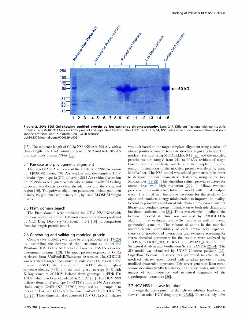
2.2 Purification of NS3 helicase by Fast protein liquidchromatography (FPLC)
Affinity Chromatography was performed by using Nickel-
Nitrilotriacetic acid (Ni-NTA kit). After semi purification from
NI-NTA kit, the respective protein was further purified by FPLC
using the Ion Exchange Column Chromatography method
optimized in Rice lab [14]. The samples were prepared, filtered
and collected in a syringe with the injection needle and run for
overnight. The purified protein was collected and the concentra-
tion was measured by Bradford assay. The desired eluted protein
was found to be present with traces of other non-specific protein
(Figure 2). Approximately 14 mg/ml of purified (.95%) protein
was obtained following the purification. The purified protein was
stored at 220uC for further study.
2.3 Structure of Pakistani HCV NS3 helicaseThe first crystal structure of HCV NS3 was first purified by Yao
et al in 1999 [11,15]. The structure of the HCV NS3 helicase was
determined by multiple isomorphous replacement (MIR) com-
bined with anomalous scattering (MIRAS). The protein consists of
three domains separated by a series of clefts. The reported
structures of HCV helicase shared similar global conformation
consisting of three domains and can be viewed as a Y-shaped
molecule, the most N-terminal domain (domain 1) and the middle
domain (domain 2) are above the C-terminal domain (domain 3)
Figure 1. 4–20% gradient SDS-PAGE for expression of HCV NS3helicase protein GT3a. Lane 1, Molecular Mass Marker; Lane 2–3, NS3helicase GT3a expression strain 01 and strain 02 respectively; Lane 4,Control Con1 GT1bhelicase.doi:10.1371/journal.pone.0106339.g001
Docking of Pakistani HCV NS3 Helicase
PLOS ONE | www.plosone.org 2 September 2014 | Volume 9 | Issue 9 | e106339
[15]. The sequence length of GT3a NS3/NS4A is 705 AA, with a
chain length 1–651 AA consists of protein NS3 and 651–705 AA
positions holds protein NS4A [13].
2.4 Pairwise and phylogenetic alignmentThe target FASTA sequence of the GT3a NS3/NS4(Accession
no: FJ839678) having 705 AA residues and the template HCV
domain of genotype 1a (GT1a) having 3011 AA residues(Accession
no: P27958) were aligned by pair wise alignment with CLC drug
discovery workbench to define the identities and the conserved
region [16]. The pairwise alignment parameters include gap open
penalty 10, gap extension penalty 0.1, by using BLOSUM weight
matrix.
2.5 Pfam domain searchThe Pfam domain were predicted for GT3a NS3/NS4Awith
the score and e-value from 100 most common domains predicted
by CLC Drug Discovery Workbench 1.0.2 [16] by comparison
from full length protein model.
2.6 Generating and validating modeled proteinComparative modeling was done by using Modeler 9.2 [17–20]
by assembling the determined rigid structure to model the
Pakistani HCV GT3a NS3 helicase from the FASTA sequence
determined as target [13]. The input protein sequence of GT3a
retrieved from UniProtKB/Swissprot (Accession No. C1KIT2)
was screened as target from structural databases [13]. Based on the
protein BLAST, the UniProtKB: C1KIT2 shared highest
sequence identity (83%) and the total query coverage (89%)with
X-Ray structure of HCV isolated from genotype 1 (PDB ID:
1CU1) which has been elucidated at 2.50 A0 [11]. The HCV NS3
helicase domain of genotype 1a (GT1a) strain, is 476 AA residues
chain length (UniProtKB: P27958) was used as a template to
model the Pakistani GT3a NS3 helicase (UniProtKB ID: C1KIT2)
[13,21]. Three dimensional structure of HCV GT3a NS3 helicase
was built based on the target-template alignment using a subset of
atomic positions from the template structure as guiding factor. Ten
models were built using MODELLER 9.12 [20] and the modeled
protein residues ranged from 210 to 651AA residues of target
based upon the similarity match with the template. Further,
energy minimization of the modeled protein was done by using
ModRefiner. The NS3 model was refined geometrically in order
to decrease the side chain steric clashes by using online tool
ModRefiner [19,22]. This algorithm refines protein structure for
atomic level with high resolution [22]. It follows two-step
procedure for constructing full-atom model with initial C-alpha
trace. The initial step builds the backbone for the available C-
alpha and conducts energy minimization to improve the quality.
Second step involves addition of side chain atoms from a rotamer
library and conducts energy minimization to both side chains and
backbone conformations [22]. The stereo chemical quality of the
helicase modeled structure was analyzed by PROCHECK
algorithm that evaluates residue by residue as well as overall
geometrical structure. The volume of atoms in the modeled
macromolecule, compatibility of each amino acid sequence,
statistics of non-bonded interactions and extensive screening for
stereo chemical parameters for the residues were analyzed by
PROVE, VERIFY_3D, ERRAT and WHAT_CHECK from
Structural Analysis and Verification Server (SAVES) [23,24]. The
3D model was visualized by UCSF Chimera package [25].
SuperPose Version 1.0 server was performed to calculate 3D
modeled helicase superimposed with template protein by using
modified quaternion approach. This server generates Root mean
square deviation (RMSD) statistics, PDB coordinates, interactive
images of both sequence and structural alignment of the
superimposed structures [26].
2.7 HCV NS3 helicase inhibitorsThough the development of the helicase inhibitor has been far
slower than other HCV drug targets [27,28]. There are only a few
Figure 2. 20% SDS Gel showing purified protein by ion exchange chromatography. Lane 2–7, Different fractions with non-specificproteins; Lane 8–10, NS3 helicase GT3a purified and separated fractions after FPLC; Lane 11 & 14, NS3 helicase with low concentration and non-specific proteins; Lane 15, Control Con1 GT1b helicase.doi:10.1371/journal.pone.0106339.g002
Docking of Pakistani HCV NS3 Helicase
PLOS ONE | www.plosone.org 3 September 2014 | Volume 9 | Issue 9 | e106339

classes of helicase inhibitors that have been reported to slow HCV
RNA replication in cell culture system of viral replication. HCV
helicase inhibitors reported to act as antiviral include nucleoside
mimics [29] triphenylmethanes [30], acridones [31,32], amidi-
noanthracyclines [33], tropolones [34], symmetrical benzimid-
azoles [35–37] and primuline derivatives [38]. A very few studies
have been reported targeting NS3 helicase by using flavonoid.
Bioflavonoids are the secondary metabolites occurring naturally
from plants; possess a wide spectrum of antiviral properties
including the inhibition of viral replication, translation and other
Figure 3. Two-dimensional structure of antiviral agents generated against HCV GT3a NS3 helicase.doi:10.1371/journal.pone.0106339.g003
Figure 4. Structures of HCV NS3 Helicase (A) Ribbon form of 1CU1 protein, (domain A; dark blue and domain B; purple) (B) crystalstructure of helicase 1A1V. (C) Superimposed macromolecule of 1CU1 (yellow) and 1A1V (pink). (D) Ramachandran contour plot of helicase NS3.doi:10.1371/journal.pone.0106339.g004
Docking of Pakistani HCV NS3 Helicase
PLOS ONE | www.plosone.org 4 September 2014 | Volume 9 | Issue 9 | e106339

Docking of Pakistani HCV NS3 Helicase
PLOS ONE | www.plosone.org 5 September 2014 | Volume 9 | Issue 9 | e106339

steps of infection [39]. Bioflavonoids such as quercetin, beta-
carotene, resveratrol, catechins, lycopene and lutein were chosen
to study the interaction with the modeled HCV helicase. These
effective bioactive compounds have been reported to interfere with
many disease associated biochemical processes in vitro [40].
Quercetin is reported in inhibiting initial stage of viral replication
[41]. Previous studies have demonstrated that the bioflavonoid
quercetin blocks HCV proliferation by inhibiting through internal
ribosomal entry site (IRES) - mediated translation of the HCV
viral genome [40]. Flavonoids are well known for their significant
antiviral inhibition activity, associated with no cytotoxicity [40].
Catechins being a polyphenolic compound from green tea were
best evaluated for altering physical property of influenza viral
membrane and voted for direct virucidal activity [42,43]. Studies
have reported beta-carotene: a major precursor for vitamin A if
maintained through proper supplementation is beneficial to boost
the immune against viral and tumor surveillance [44]. In recent
years, deep research is carried out for resveratrol produced by
certain plants on various stimuli, is well known for its antiviral
activity through various molecular pathways [45]. Though
numerous epidemiological studies have pointed that consuming
abundant carotenoids calm down the risk of various cancer, the
protective mechanism of carotenoids based on their anti-oxidant
capability, makes it a promising chemo preventive molecule that
can be targeted for antiviral character. Lutein and lycopene,
abundant in fruits and vegetables also possess strong anti-oxidant
property [46]. Lutein inhibition activity against HBV full-length
promoter was exposed in vitro, through data which introduced
lutein exerts antivirus effects via inhibition of HBV transcription
cycle [47]. Therefore, the listed flavonoids are well known to stand
high for their anti-oxidant property, which led us to recruit them
as inhibitors [42]. The chemical structures of selected compounds
are represented in Figure 3. The 2D structures were downloaded
from PUBCHEM in SDF format which were converted to PDB by
using NCI Online SMILES Translator [48].
2.8 In silico dockingFurther refinement of the retrieved targeted compounds, were
docked with the modeled GT3a NS3 helicase by using CLC Drug
Discovery Workbench software package [16]. Basic set ups like
protein preparation, ligand preparation, detecting cavities, recep-
tor grid generation and targeted ligand docking were performed.
Docking wizard was customized by using default MolDock
optimizer algorithm with 200 numbers of runs. The docking
Figure 5. Pairwise alignment between HCV GT1a and GT3a NS3/NS4A by using CLC drug discovery workbench. Amino acid pairwisealignment between target (705 AA) and template (3011 AA) showed 80.28% similar for conserved regions.doi:10.1371/journal.pone.0106339.g005
Figure 6. BLAST alignment and phylogenetic tree of GT3a NS3 helicase. (A) The green line indicates the template sequence with maximumsimilarity with the target sequence (B) The amino acid substitution rate between GT3a NS3/NS4A and HCV GT1a is 0.05%.doi:10.1371/journal.pone.0106339.g006
Docking of Pakistani HCV NS3 Helicase
PLOS ONE | www.plosone.org 6 September 2014 | Volume 9 | Issue 9 | e106339

parameters include population size 50, maximum iterations 2000,
scaling factor 0.50, crossover rate 0.90 and RMSD thresholds for
similar cluster poses were set as 1.00. CLC Drug Discovery
Workbench uses a standard precision mode to determine the
favorable binding poses, which detects various flexible ligand
conformations while holding protein as rigid structure during
docking. Quercetin, beta-carotene, resveratrol, catechins, lycopene
and lutein, were docked into active site recognized in the
macromolecule cavity. For comparative analysis, ten active
compounds were selected as they were reported in inhibiting the
HCV NS3 helicase activity. Maximum of 10 poses for each
conformations were generated by using default parameter of CLC
Drug Discovery Workbench. Docking studies were carried out to
predict the binding affinities based on scoring functions. On the
Table 1. PFAM domain search table.
Start End Domain Accession Score E-value
15 190 Trypsin PF00089 278.10 0.54
62 432 Major Facilitator Superfamily PF07690 2100.10 0.85
98 434 Aminotransferase class I and II PF00155 2165.10 0.92
122 137 ATPase family associated with various cellular activities (AAA) PF00004 20.30 0.23
159 173 Trypsin PF00089 21.10 0.35
209 352 DEAD/DEAH box helicase PF00270 231.80 2.10E-4
222 231 DEAD/DEAH box helicase PF00270 21.50 0.63
400 489 Helicase conserved C-terminal domain PF00271 2.30 5.70E-4
480 489 Helicase conserved C-terminal domain PF00271 20.70 0.92
doi:10.1371/journal.pone.0106339.t001
Figure 7. Docking conformations and binding pockets of HCVGT3a NS3 helicase with different inhibitors. (A) Three-dimensionalrepresentation of quercetin with target macromolecule and its hit residues (B) Beta-carotene represents no interactions in its constraints with themodeled structure. The Hbond formation is shown in stick mode (green). (C) Best pose of the compound resveratrol forming seven HBonds withthree-dimensional structure of helicase at its binding sites GLN 315, GLU 453, ARG 484, PRO 472, ASP 474, ARG 501 (D) Three-dimensionalrepresentation of catechins and target molecule with eleven HBonds network. The Hbond formation is shown in stick mode (yellow) and theconstraint is shown in green color. (E) Zero interactions between lycopene and modeled GT3a NS3 helicase (F) binding of lutein at THR 226, LEU 575and SER 578 residues. Lutein forming three HBonds with high HBond energy 22.5461 kcal?mol21.doi:10.1371/journal.pone.0106339.g007
Docking of Pakistani HCV NS3 Helicase
PLOS ONE | www.plosone.org 7 September 2014 | Volume 9 | Issue 9 | e106339

basis of hydrogen interaction and docking score, the best ranked
compounds were selected and their binding residues were
observed by using CLC drug discovery visualization tool [49].
Result and Discussion
3.1 Structural alignment and domain analysisThe BLASTp search program revealed several sequence
homologous to helicase polyprotein (UniProtKB id: C1KIT2)
but HCV helicase domain (PDB code 1A1V, represented in
Figure 4A) was chosen as the best template for modeling GT3a
helicase. The multiple sequence alignment score between the
target (705 AA) and template (3011 AA) showed 80.28% pairwise
similarity by using CLC drug discovery workbench shown in
Figure 5. The AA substitution rate between GT3a NS3/NS4A
and HCV genotype 1a (GT1a) is 0.05% also shown in Figure 6.
The Pfam domain predicted for GT3a NS3/NS4Ais presented in
Table 1 with the score and e-value.
Therefore we generated a 3D macromolecule from the target
GT3a NS3from 210 to 651AAas they were aligned and modeled
from chain A containing 476 AA sequence length (1A1V) of
helicase of GT1a by homology modeling procedure. The template
3D structure of NS3 helicase domain of 1A1V with its chain A and
B is represented in ribbon structure in Figure 4A. The modeled
GT3a NS3helicase structure and its Ramachandran plot contain-
ing the phi and psi values are shown in Figure 4B and 4D with
93.7% residues in the most favored regions, followed by 6.0% in
the additionally allowed region and 0.00% in disallowed region.
The RMSD value of the superimposed modeled and template
structure is 1.27A0 (presented in Figure 4C). The calculated
RMSD value is between the main-chain atom of model and
template, which indicates close homology, ensuring good reliabil-
ity of the model helicase [22].
3.2 Binding Interaction of HCV NS3 helicaseThe docking studies of GT3a NS3helicase were carried out to
define the binding pockets, inhibitors interactions, and their
specificity and energy requirement. Our in silico docking study
using MolDock scores observed for the modeled GT3a NS3
helicase and the six ligands are summarized in Table 2. Ten
different conformations with best pose between protein ligand
interactions were generated based on HBond distances. The best-
ranked docked conformation was considered for binding affinity
study by measuring HBond interactions. Depending on two
parameters, the potential inhibitors were chosen based on 1)
details of HBonds of best-ranked pose and 2) binding energy
predicted between docked flavonoids and the protein. In our
analysis, the compounds catechins and quercetin fitted well in
active pockets of GT3a NS3 modeled helicase presented in
Figure 7, denoting minimum docking energy values and formed
maximum number of HBonds compared with other flavonoid.
The best pose of interaction for quercetin showed fourteen
HBonds interactions with its active site residues such as SER 251,
ASN 249, GLU 311, GLN 480, ALA 433, LEU 434, ASP 432,
ASP 425, ARG 484, GLN 480, ILE 379 and ASP 474. In the
docked complex, quercetin and catechins revealed H Bonds
formation with similar active site residues ARG 484 and ASP 432,
which denote region for NS3 binding motifs. Catechins interacted
with GLN 315, GLU 453, SER 477, ARG 481, HIS 313 and VAL
426 with minimum docking energy (Figure 7C). The HBonds
formed between each flavonoid compound and the modeled NS3
helicase with its docking energy, MolDock score, HBond energy
and number of residue interaction with labels are denoted in
Figure 8. Per residue interaction of HCV GT3a NS3 helicase at specific domains (A) Helicase ATP-binding (B) NS3-binding (C) DECHbox. The modeled GT3a NS3 helicase protein is shown in cartoon and colored in gray. Binding cavities and HBonds are represented in green color.doi:10.1371/journal.pone.0106339.g008
Docking of Pakistani HCV NS3 Helicase
PLOS ONE | www.plosone.org 8 September 2014 | Volume 9 | Issue 9 | e106339

Table 2. The HBond energy values for both quercetin and
catechins were 29.4 kcal?mol21and 27.2 kcal?mol21. The other
two inhibitors like resveratrol displayed seven HBonds interaction
with HBond energy and docking energy as 27.2 kcal?mol21
and295.41 kcal?mol21, whereas lutein exhibited only three
HBonds interaction with THR 226, LEU 575 and SER 578 with
high HBond energy 22.54 kcal?mol21. Beta-carotene and lyco-
pene showed no interactions with the active residues of the
modeled helicase might be due to their long chain of carbon atoms
and presence of isoprene units as they both are members of
tetraterpenes, synthesized biochemically. The docking interactions
and the number of HBonds formed between each bioflavonoid are
shown in (Video S1). The MolDock score increased with respect to
the number of hydrocarbon atoms in the targeted compounds.
The number of torsions angles chosen for ligand to fit into the
binding pockets of active site residues also showed higher for
isoprenoid compounds. Since the docking result suggests that the
CLC drug discovery software reproduced appropriate conforma-
tions of selected flavonoid inside the binding pockets of NS3
helicase active site suggesting a competitive inhibition of the
helicase. The ATP binding position (1217–1369 AA), NS3 binding
motifs site (1679–1690 AA) and DECH box (1316–1319 AA) of
the template 1A1V was compared to determine the frequent
binding interaction at the conserved sites of the respective regions
of the modeled helicase (Figure 8). The binding modes depict ILE
379 and SER 251 reside DECH box region. Frequent binding was
spotted at NS3 binding site by quercetin, resveratrol and lutein.
Only quercetin and catechins showed HBonds interaction at
DECH box. The interaction of specific residues with the potential
regions was confirmed from GT1a through sequence annotation
along with feature and region description of residue positions. The
confirmation obtained after docking showed good energy binding
and docking energy for quercetin, catechins, resveratrol and lutein.
Quercetin and catechins demonstrated stronger in silico inhibition
of the infectious virion helicase compared to resveratrol. Lutein
demonstrates mild interaction compared to other bioflavonoid.
Therefore the flavonoid compounds that presented high HBond
interaction and closest binding energy values to modeled GT3a
NS3helicase were considered to be the best results. This order of
specificity indicates that the flavonoids catechins and quercetin
followed by resveratrol and lutein showed good inhibition activity
towards the modeledGT3a NS3 helicase.
Very few in silico studies have shown different inhibitors
targeted against helicase and their binding specificities. A list of insilico study carried out against viral helicase as targets from HCV
is shown in Table 3. Though HCV comes under the family of
flaviviridae, its helicase is considered as template for comparative
study with other vital viruses. Use of natural photochemical from
plants is a promising therapy, reported by a study were medicinal
plant Amelanchier alnifolia and its component quercetin followed
by 3-galactoside and 3-glucoside showed interactions with protease
and helicase respectively [50]. Drug therapy target against NS3
resistant variants R155K and V36M was also reported with
impact on conformation of the beta-barrel domain of the viral
protein. This domain is involved in substrate binding and in active
site binding pocket [51]. Therefore, our docked results provide
potential information for new inhibitors analogues targeted
towards helicase, to fight against HCV through drug-flavonoid
pharmacokinetic interactions.
3.3 Comparative docking study from bioassay hitcompounds
A comparative study of the interaction of active ten compounds
from PubChem bioassay were considered for docking with the
Ta
ble
2.
Tar
ge
ted
resi
du
es
inin
tera
ctio
nan
dth
eir
do
ckin
gsc
ore
.
Na
me
of
the
De
riv
ati
ve
Mo
lDo
ckS
core
RM
SD
Hb
on
dIn
tera
ctio
ns
Do
ckin
gS
core
Na
me
Of
the
bo
nd
Nu
mb
er
of
hy
dro
ge
nb
on
ds
Qu
erc
eti
n2
86
.68
63
37
.51
82
9.4
56
71
42
94
.86
08
UN
K-O
UN
K-C
SER
25
1-O
ASN
24
9-O
GLU
31
1-O
GLN
48
0-O
ALA
43
3-O
LEU
43
4-O
ASP
43
2-O
ASP
42
5-O
AR
G4
84
-NG
LN4
80
-OIL
E3
79
-HA
SP4
74
-O
Be
ta-C
aro
ten
e2
13
3.2
04
57
.49
64
21
49
.24
90
21
30
.60
1-
-
Re
sve
rato
l2
89
.88
86
37
.12
95
27
.34
95
72
95
.41
49
UN
K-O
UN
K-C
GLU
45
3-O
AR
G4
84
-NP
RO
47
2-O
ASP
47
4-O
AR
G5
01
-O
Cat
ech
ins
28
8.3
72
24
1.8
00
82
7.2
84
51
12
99
.55
67
UN
K-O
UN
K-C
GLN
31
5-O
GLU
45
3-O
SER
47
7-O
AR
G4
81
-NA
RG
48
4-N
ASP
43
2-O
HIS
31
3-O
VA
L4
26
-O
Lyco
pe
ne
21
13
.58
23
6.6
53
42
13
6.2
84
02
10
9.6
35
--
Lute
in2
12
0.0
47
42
.43
08
22
.54
61
32
11
9.7
47
UN
K-O
TH
R2
26
-OLE
U5
75
-OSE
R5
78
-O
do
i:10
.13
71
/jo
urn
al.p
on
e.0
10
63
39
.t0
02
Docking of Pakistani HCV NS3 Helicase
PLOS ONE | www.plosone.org 9 September 2014 | Volume 9 | Issue 9 | e106339

modeled GT3a NS3 has been presented in Figure 9 exploiting the
interaction of the bound drug. Molecular docking studies were
performed to provide further insight into various other drug
interactions which were identified as inhibitors of the hepatitis C
virus NS3 through fluorescence-based primary biochemical high
throughput screening. The lowest score was selected corresponded
to the best docking pose as well as the number of HBonds formed.
The docked conformations of the ten hit compounds in the GT3a
NS3complex are shown in Figure 9. Table 4 denotes the
interaction energy and the HBonds formed with the modeled
residues. Thus, it has been observed that the number of residues
involved in binding is higher is maximum for bioflavonoids
catechins and quercetin whereas in the case of ten hit compounds
selected the number of residues perturbed was found to be
minimum. This observation indicates that the bioflavonoids such
as catechins and quercetin bound in binding pockets of the protein
complex.
Perspectives on the Future Directions
This study as proved an impetus to initiate broader screening
for key molecules as HCV helicase inhibitors. The lead
Table 3. Inhibitors of helicase available until now and their binding specificities.
Drugs Residues References
Ivermectin T408, D409 [52]
Quercetin,3-galactoside, 3- glucoside ALA157, HIS528 [53]
Mercapto compounds CYS431,ARG393,ARG481 [54]
VX-950,tri-andtetra peptides, hexapeptides Q526A, H528A, H528S [55]
Dihydropyrols, Phenylalanine analogs, Thiophenes, Benzofurans,Phenylpropanyl Benzamides, Benzimidazoles, Indoles
ASP318,SER556,ASN291, SER 367, SER476, TYR477 [56]
doi:10.1371/journal.pone.0106339.t003
Figure 9. Comparing the docking interaction with ten hit compounds with HCV GT3a NS3 helicase such as 1) SID 17513061 2) SID3716320 3) SID 17513201 4) SID 17401675 5) SID 49666882 6) SID 24818609 7) SID 24827353 8) SID 49732586 9) SID 4257236 10)SID 49817864 obtained from PubChem Bioassay.doi:10.1371/journal.pone.0106339.g009
Table 4. Comparison of active hit compounds docked against modeled HCV NS3 GT3a.
Compounds Molecular Formula Interaction energy (kcal/mole) HBonds
SID 17513061 C16H11NO4S 285.165 1
SID 3716320 C20H12N2O4 290.566 4
SID 17513201 C17H14N2O2S 265.266 2
SID 17401675 C14H14N4O2 2110.633 5
SID 49666882 C19H18N2O5S3 296.326 2
SID 24818609 C11H11BrN2O3 289.623 1
SID 24827353 C20H18ClN3O4 292.366 4
SID 49732586 C21H16BrNO5 270.362 4
SID 4257236 C18H21N5 294.238 6
SID 49817864 C22H17N3O8S 289.365 3
doi:10.1371/journal.pone.0106339.t004
Docking of Pakistani HCV NS3 Helicase
PLOS ONE | www.plosone.org 10 September 2014 | Volume 9 | Issue 9 | e106339

compounds screened in this study should be a promising starter for
large scale screening from the list of more than one million
compounds from Ligand info meta database using the virtual
screening protocols. Recent advances in understanding the
molecular basis for helicase action might also spur interest in
rationally designing compounds that should target key motif or
clefts. HCV helicase is clearly an undeveloped drug target, which
is envisaged with all the recent advances the pharmaceutical
industry may track new attempts to find better helicase inhibitors.
Such continued work and recent advances in the field will
hopefully soon result in the discovery of more compounds for use
in the laboratory and clinical trials. Deeper biochemical under-
standing of the complex nature of helicases will eventually help to
define the molecular mechanisms of RNA helicases which will be a
vital to attain an efficacious drug against human and animal
pathogens and the ultimate assault on viral infections with their
induced pathogenesis such as chronic liver disease. In summary we
cloned, expressed, purified and docked GT3a NS3 helicase as a
model with inhibitors. This study will pave a way to study in depth
the molecular interactions of helicase of other HCV genotypes and
help for screening specific antiviral inhibitors from combinatorial
chemical library and random biological library.
Supporting Information
Video S1 Computational docking study with possibleantiviral drugs and analysis of complex interactions.
(MP4)
Acknowledgments
We are thankful to Dr. Charles M. Rice and Dr. Meigang G.U.,
Rockefeller University, New York, NY for providing the facilities and
control protein samples, respectively. We are also thankful to Dr. Ali
Abdullah, Dr. Zainab Younas and Nadia Rashid of IQ institute of
Infection and Immunity (IQI3) for the critical review of this manuscript.
We also thank Shiny Mathew in various software installations and
optimizing the use of the application.
Author Contributions
Conceived and designed the experiments: IQ. Performed the experiments:
KF. Analyzed the data: KF IQ SM. Contributed reagents/materials/
analysis tools: KF IQ. Wrote the paper: KF SM IQ. Proof reading of the
manuscript: MS SM AA GD EA.
References
1. Ashfaq UA, Javed T, Rehman S, Nawaz Z, Riazuddin S (2011) An overview of
HCV molecular biology, replication and immune responses. Virol J 8: 161.
2. Mathew S, Ali A, Abdel-Hafiz H, Fatima K, Suhail M, et al. (2014) Biomarkersfor virus-induced hepatocellular carcinoma (HCC). Infect Genet Evol.
3. Naggie S (2012) Management of hepatitis C virus infection: the basics. Top
Antivir Med 20: 154–161.
4. Hamid S, Umar M, Alam A, Siddiqui A, Qureshi H, et al. (2004) PSG consensusstatement on management of hepatitis C virus infection–2003. J Pak Med Assoc
54: 146–150.
5. Bartenschlager R, Ahlborn-Laake L, Mous J, Jacobsen H (1994) Kinetic andstructural analyses of hepatitis C virus polyprotein processing. J Virol 68: 5045–
5055.
6. Kim DW, Gwack Y, Han JH, Choe J (1995) C-terminal domain of the hepatitisC virus NS3 protein contains an RNA helicase activity. Biochem Biophys Res
Commun 215: 160–166.
7. Ashfaq UA, Khan SN, Nawaz Z, Riazuddin S (2011) In-vitro model systems to
study Hepatitis C Virus. Genet Vaccines Ther 9: 7.
8. Piccininni S, Varaklioti A, Nardelli M, Dave B, Raney KD, et al. (2002)
Modulation of the hepatitis C virus RNA-dependent RNA polymerase activity
by the non-structural (NS) 3 helicase and the NS4B membrane protein. J BiolChem 277: 45670–45679.
9. Ranji A, Boris-Lawrie K (2010) RNA helicases: emerging roles in viral
replication and the host innate response. RNA Biol 7: 775–787.
10. Jones DM, McLauchlan J (2010) Hepatitis C virus: assembly and release of virusparticles. J Biol Chem 285: 22733–22739.
11. Yao N, Reichert P, Taremi SS, Prosise WW, Weber PC (1999) Molecular views
of viral polyprotein processing revealed by the crystal structure of the hepatitis Cvirus bifunctional protease-helicase. Structure 7: 1353–1363.
12. Lopez-Labrador FX (2008) Hepatitis C Virus NS3/4A Protease Inhibitors.
Recent Pat Antiinfect Drug Discov 3: 157–167.
13. Fatima K, Tahir M, Qadri I (2011) Development of robust in vitro serineprotease assay based on recombinant Pakistani HCV NS3-4A protease. Virus
Res 160: 230–237.
14. Gu M, Rice CM (2010) Three conformational snapshots of the hepatitis C virus
NS3 helicase reveal a ratchet translocation mechanism. Proc Natl AcadSci U S A 107: 521–528.
15. Kim JL, Morgenstern KA, Griffith JP, Dwyer MD, Thomson JA, et al. (1998)
Hepatitis C virus NS3 RNA helicase domain with a bound oligonucleotide: thecrystal structure provides insights into the mode of unwinding. In: Structure 6:
89–100.
16. CLC Inc A, Denmark.
17. Eswar N, Webb B, Marti-Renom MA, Madhusudhan MS, Eramian D, et al.(2006) Comparative protein structure modeling using Modeller. Curr Protoc
Bioinformatics Chapter 5: Unit 5 6.
18. Eswar N, Webb B, Marti-Renom MA, Madhusudhan MS, Eramian D, et al.(2007) Comparative protein structure modeling using MODELLER. Curr
Protoc Protein Sci Chapter 2: Unit 2 9.
19. Fiser A, Do RK, Sali A (2000) Modeling of loops in protein structures. ProteinSci 9: 1753–1773.
20. Marti-Renom MA, Stuart AC, Fiser A, Sanchez R, Melo F, et al. (2000)
Comparative protein structure modeling of genes and genomes. Annu Rev
Biophys Biomol Struct 29: 291–325.
21. Yao N, Hesson T, Cable M, Hong Z, Kwong AD, et al. (1997) Structure of the
hepatitis C virus RNA helicase domain. Nat Struct Biol 4: 463–467.
22. Xu D, Zhang Y (2011) Improving the physical realism and structural accuracy ofprotein models by a two-step atomic-level energy minimization. Biophys J 101:
2525–2534.
23. Bowie JU, Luthy R, Eisenberg D. (1991) A method to identify protein sequences
that fold into a known three-dimensional structure. Science 12: 164–170.
24. Luthy R, Bowie JU, Eisenberg D (1992) Assessment of protein models withthree-dimensional profiles. Nature 356: 83–85.
25. Pettersen EF, Goddard TD, Huang CC, Couch GS, Greenblatt DM, et al.
(2004) UCSF Chimera–a visualization system for exploratory research and
analysis. J Comput Chem 25: 1605–1612.
26. Maiti R, Van Domselaar GH, Zhang H, Wishart DS (2004) SuperPose: a simpleserver for sophisticated structural superposition. Nucleic Acids Res 32: W590–
594.
27. Belon CA, Frick DN (2009) Helicase inhibitors as specifically targeted antiviral
therapy for hepatitis C. Future Virol 4: 277–293.
28. Cho HS, Ha NC, Kang LW, Chung KM, Back SH, et al. (1998) Crystalstructure of RNA helicase from genotype 1b hepatitis C virus. A feasible
mechanism of unwinding duplex RNA. The Journal of biological chemistry 273:15045–15052.
29. Frick DN (2007) The hepatitis C virus NS3 protein: a model RNA helicase and
potential drug target. Curr Issues Mol Biol 9: 1–20.
30. Gemma S, Butini S, Campiani G, Brindisi M, Zanoli S, et al. (2011) Discovery of
potent nucleotide-mimicking competitive inhibitors of hepatitis C virus NS3helicase. Bioorg Med Chem Lett 21: 2776–2779.
31. Chen CS, Chiou CT, Chen GS, Chen SC, Hu CY, et al. (2009) Structure-based
discovery of triphenylmethane derivatives as inhibitors of hepatitis C virus
helicase. J Med Chem 52: 2706–2723.
32. Stankiewicz-Drogon A, Dorner B, Erker T, Boguszewska-Chachulska AM(2010) Synthesis of new acridone derivatives, inhibitors of NS3 helicase, which
efficiently and specifically inhibit subgenomic HCV replication. J Med Chem53: 3117–3126.
33. Manfroni G, Paeshuyse J, Massari S, Zanoli S, Gatto B, et al. (2009) Inhibition
of subgenomic hepatitis C virus RNA replication by acridone derivatives:
identification of an NS3 helicase inhibitor. J Med Chem 52: 3354–3365.
34. Krawczyk M, Wasowska-Lukawska M, Oszczapowicz I, Boguszewska-Cha-chulska AM (2009) Amidinoanthracyclines - a new group of potential anti-
hepatitis C virus compounds. Biol Chem 390: 351–360.
35. Belon CA, High YD, Lin TI, Pauwels F, Frick DN (2010) Mechanism and
specificity of a symmetrical benzimidazolephenylcarboxamide helicase inhibitor.Biochemistry 49: 1822–1832.
36. Najda-Bernatowicz A, Krawczyk M, Stankiewicz-Drogon A, Bretner M,
Boguszewska-Chachulska AM (2010) Studies on the anti-hepatitis C virusactivity of newly synthesized tropolone derivatives: identification of NS3 helicase
inhibitors that specifically inhibit subgenomic HCV replication. Bioorg Med
Chem Lett 18: 5129–5136.
37. Phoon CW, Ng PY, Ting AE, Yeo SL, Sim MM (2001) Biological evaluation ofhepatitis C virus helicase inhibitors. Bioorg Med Chem Lett 11: 1647–1650.
38. Tunitskaya VL, Mukovnya AV, Ivanov AA, Gromyko AV, Ivanov AV, et al.
(2011) Inhibition of the helicase activity of the HCV NS3 protein by symmetricaldimeric bis-benzimidazoles. Bioorg Med Chem Lett 21: 5331–5335.
Docking of Pakistani HCV NS3 Helicase
PLOS ONE | www.plosone.org 11 September 2014 | Volume 9 | Issue 9 | e106339

39. Li K, Frankowski KJ, Belon CA, Neuenswander B, Ndjomou J, et al. (2012)
Optimization of potent hepatitis C virus NS3 helicase inhibitors isolated fromthe yellow dyes thioflavine S and primuline. J Med Chem 55: 3319–3330.
40. Khachatoorian R, Arumugaswami V, Raychaudhuri S, Yeh GK, Maloney EM,
et al. (2012) Divergent antiviral effects of bioflavonoids on the hepatitis C viruslife cycle. Virology 433: 346–355.
41. Kaul TN, Middleton E, Jr., Ogra PL (1985) Antiviral effect of flavonoids onhuman viruses. J Med Virol 15: 71–79.
42. Calland N, Dubuisson J, Rouille Y, Seron K (2012) Hepatitis C virus and natural
compounds: a new antiviral approach? Viruses 4: 2197–2217.43. Song JM, Lee KH, Seong BL (2005) Antiviral effect of catechins in green tea on
influenza virus. Antiviral Res 68: 66–74.44. Santos MS, Meydani SN, Leka L, Wu D, Fotouhi N, et al. (1996) Natural killer
cell activity in elderly men is enhanced by beta-carotene supplementation.Am J Clin Nutr 64: 772–777.
45. Campagna M, Rivas C (2010) Antiviral activity of resveratrol. Biochem Soc
Trans 38: 50–53.46. Khachik F, Beecher GR, Smith JC Jr (1995) Lutein, lycopene, and their
oxidative metabolites in chemoprevention of cancer. J Cell Biochem Suppl 22:236–246.
47. Pang R, Tao JY, Zhang SL, Zhao L, Yue X, et al. (2010) In vitro antiviral
activity of lutein against hepatitis B virus. Phytother Res 24: 1627–1630.48. Santos E, Nebreda AR (1989) Structural and functional properties of ras
proteins. FASEB journal: official publication of the Federation of AmericanSocieties for Experimental Biology 3: 2151–2163.
49. Thomsen RC, M H (2006) MolDock: A New Technique forHigh-AccuracyMolecular Docking. J Med Chem 49: 3315–3321.
50. Perchyonok VTSZO T (2013) Protective Effect of Conventional Antioxidant (b-
Carotene, Resveratrol and Vitamin E) in Chitosan-Containing Hydrogels
Against Oxidative Stress and Reversal of DNA Double Stranded Breaks Induced
by Common Dental Composites: In-Vitro Model. Open Nanoscience Journal 7.
51. Khan M, Masoud MS, Qasim M, Khan MA, Zubair M, et al. (2013) Molecular
screening of phytochemicals from Amelanchier Alnifolia against HCV NS3
protease/helicase using computational docking techniques. Bioinformation 9:
978–982.
52. Mastrangelo E, Pezzullo M, De Burghgraeve T, Kaptein S, Pastorino B, et al.
(2012) Ivermectin is a potent inhibitor of flavivirus replication specifically
targeting NS3 helicase activity: new prospects for an old drug. J Antimicrob
Chemother 67(8).
53. Khan M, Masoud MS, Qasim M, Khan MA, Zubair M, et al. (2013) Molecular
screening of phytochemicals from Amelanchier Alnifolia against HCV NS3
protease/helicase using computational docking techniques. Bioinformation
9(19).
54. Vlachakis D, Kossida S (2013) Molecular modeling and pharmacophore
elucidation study of the Classical Swine Fever virus helicase as a promising
pharmacological target. PeerJ Jun 11;1:e85.
55. Dahl G, Sandstrom A, Akerblom E, Danielson UH. (2007) Effects on protease
inhibition by modifying of helicase residues in hepatitis C virus nonstructural
protein 3. FEBS J 274(22).
56. Elhefnawi M, ElGamacy M, Fares M (2012) Multiple virtual screening
approaches for finding new Hepatitis c virus RNA-dependent RNA polymerase
inhibitors: Structure-based screens and molecular dynamics for the pursue of
new poly pharmacological inhibitors. BMC Bioinformatics (Suppl 17): S5.
Docking of Pakistani HCV NS3 Helicase
PLOS ONE | www.plosone.org 12 September 2014 | Volume 9 | Issue 9 | e106339