The adnAB locus, encoding a putative helicase-nuclease activity, is essential in Streptomyces
Upload
uni-wuerzburgCategory
view
4download
0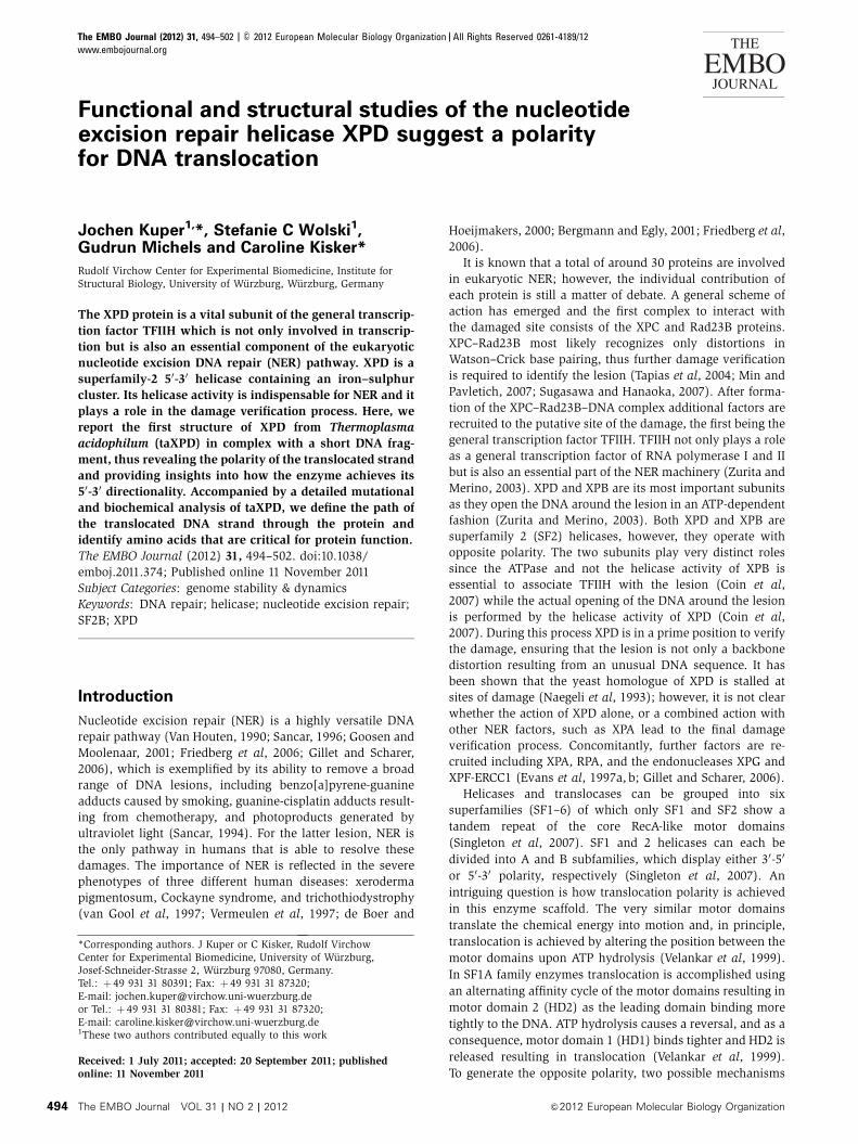
Functional and structural studies of the nucleotideexcision repair helicase XPD suggest a polarityfor DNA translocation
Jochen Kuper1,*, Stefanie C Wolski1,Gudrun Michels and Caroline Kisker*
Rudolf Virchow Center for Experimental Biomedicine, Institute forStructural Biology, University of Wurzburg, Wurzburg, Germany
The XPD protein is a vital subunit of the general transcrip-
tion factor TFIIH which is not only involved in transcrip-
tion but is also an essential component of the eukaryotic
nucleotide excision DNA repair (NER) pathway. XPD is a
superfamily-2 50-30 helicase containing an iron–sulphur
cluster. Its helicase activity is indispensable for NER and it
plays a role in the damage verification process. Here, we
report the first structure of XPD from Thermoplasma
acidophilum (taXPD) in complex with a short DNA frag-
ment, thus revealing the polarity of the translocated strand
and providing insights into how the enzyme achieves its
50-30 directionality. Accompanied by a detailed mutational
and biochemical analysis of taXPD, we define the path of
the translocated DNA strand through the protein and
identify amino acids that are critical for protein function.
The EMBO Journal (2012) 31, 494–502. doi:10.1038/
emboj.2011.374; Published online 11 November 2011
Subject Categories: genome stability & dynamics
Keywords: DNA repair; helicase; nucleotide excision repair;
SF2B; XPD
Introduction
Nucleotide excision repair (NER) is a highly versatile DNA
repair pathway (Van Houten, 1990; Sancar, 1996; Goosen and
Moolenaar, 2001; Friedberg et al, 2006; Gillet and Scharer,
2006), which is exemplified by its ability to remove a broad
range of DNA lesions, including benzo[a]pyrene-guanine
adducts caused by smoking, guanine-cisplatin adducts result-
ing from chemotherapy, and photoproducts generated by
ultraviolet light (Sancar, 1994). For the latter lesion, NER is
the only pathway in humans that is able to resolve these
damages. The importance of NER is reflected in the severe
phenotypes of three different human diseases: xeroderma
pigmentosum, Cockayne syndrome, and trichothiodystrophy
(van Gool et al, 1997; Vermeulen et al, 1997; de Boer and
Hoeijmakers, 2000; Bergmann and Egly, 2001; Friedberg et al,
2006).
It is known that a total of around 30 proteins are involved
in eukaryotic NER; however, the individual contribution of
each protein is still a matter of debate. A general scheme of
action has emerged and the first complex to interact with
the damaged site consists of the XPC and Rad23B proteins.
XPC–Rad23B most likely recognizes only distortions in
Watson–Crick base pairing, thus further damage verification
is required to identify the lesion (Tapias et al, 2004; Min and
Pavletich, 2007; Sugasawa and Hanaoka, 2007). After forma-
tion of the XPC–Rad23B–DNA complex additional factors are
recruited to the putative site of the damage, the first being the
general transcription factor TFIIH. TFIIH not only plays a role
as a general transcription factor of RNA polymerase I and II
but is also an essential part of the NER machinery (Zurita and
Merino, 2003). XPD and XPB are its most important subunits
as they open the DNA around the lesion in an ATP-dependent
fashion (Zurita and Merino, 2003). Both XPD and XPB are
superfamily 2 (SF2) helicases, however, they operate with
opposite polarity. The two subunits play very distinct roles
since the ATPase and not the helicase activity of XPB is
essential to associate TFIIH with the lesion (Coin et al,
2007) while the actual opening of the DNA around the lesion
is performed by the helicase activity of XPD (Coin et al,
2007). During this process XPD is in a prime position to verify
the damage, ensuring that the lesion is not only a backbone
distortion resulting from an unusual DNA sequence. It has
been shown that the yeast homologue of XPD is stalled at
sites of damage (Naegeli et al, 1993); however, it is not clear
whether the action of XPD alone, or a combined action with
other NER factors, such as XPA lead to the final damage
verification process. Concomitantly, further factors are re-
cruited including XPA, RPA, and the endonucleases XPG and
XPF-ERCC1 (Evans et al, 1997a, b; Gillet and Scharer, 2006).
Helicases and translocases can be grouped into six
superfamilies (SF1–6) of which only SF1 and SF2 show a
tandem repeat of the core RecA-like motor domains
(Singleton et al, 2007). SF1 and 2 helicases can each be
divided into A and B subfamilies, which display either 30-50
or 50-30 polarity, respectively (Singleton et al, 2007). An
intriguing question is how translocation polarity is achieved
in this enzyme scaffold. The very similar motor domains
translate the chemical energy into motion and, in principle,
translocation is achieved by altering the position between the
motor domains upon ATP hydrolysis (Velankar et al, 1999).
In SF1A family enzymes translocation is accomplished using
an alternating affinity cycle of the motor domains resulting in
motor domain 2 (HD2) as the leading domain binding more
tightly to the DNA. ATP hydrolysis causes a reversal, and as a
consequence, motor domain 1 (HD1) binds tighter and HD2 is
released resulting in translocation (Velankar et al, 1999).
To generate the opposite polarity, two possible mechanismsReceived: 1 July 2011; accepted: 20 September 2011; publishedonline: 11 November 2011
*Corresponding authors. J Kuper or C Kisker, Rudolf VirchowCenter for Experimental Biomedicine, University of Wurzburg,Josef-Schneider-Strasse 2, Wurzburg 97080, Germany.Tel.: þ 49 931 31 80391; Fax: þ 49 931 31 87320;E-mail: [email protected] Tel.: þ 49 931 31 80381; Fax: þ 49 931 31 87320;E-mail: [email protected] two authors contributed equally to this work
The EMBO Journal (2012) 31, 494–502 | & 2012 European Molecular Biology Organization | All Rights Reserved 0261-4189/12
www.embojournal.org
The EMBO Journal VOL 31 | NO 2 | 2012 &2012 European Molecular Biology Organization
EMBO
THE
EMBOJOURNAL
THE
EMBOJOURNAL
494

can be envisioned. The helicase scaffold could bind the DNA
with a different orientation using a similar translocation
mechanism, or the DNA is bound in the same orientation
in both subfamilies with the translocation mechanism being
altered. The latter has been shown recently for SF1A and
SF1B enzymes (Saikrishnan et al, 2009) with a reversed order
of tight binding for HD1 and HD2 resulting in opposite
translocation polarity. For SF2 enzymes, it has so far not
been possible to derive similar conclusions.
XPD belongs to the SF2B subfamily of helicases and
contains a 4Fe–4S cluster (FeS) in an auxiliary domain, that
is vital for its helicase activity and might be involved in the
damage recognition process (Rudolf et al, 2006; Fan et al,
2008; Liu et al, 2008; Wolski et al, 2008). Although apo
structures of SF2A and SF2B members are available, no
DNA-bound structure of an SF2B enzyme has been reported
so far which would allow to pin point the underlying trans-
location mechanism in this family. We have chosen
Thermoplasma acidophilum XPD (taXPD) as a representative
member of SF2B helicases and a homologue of human XPD
as a model enzyme. After determining the structure of
apo-taXPD (Wolski et al, 2008) we have now solved the
first structure of an XPD–DNA complex. To identify function-
ally important regions of the protein and determine the
DNA-binding regions of taXPD, we performed a detailed
mutational and biochemical analysis to address mechanisti-
cally important questions on translocation polarity, helicase
function and damage recognition.
Results
The taXPD DNA structure
In order to obtain insights into the molecular events leading
to DNA translocation of taXPD and the detailed nature of the
DNA-binding mechanism we have solved the crystal structure
of taXPD in the presence of a 22-mer single-stranded DNA
(ssDNA) oligonucleotide. The structure was refined at a
resolution of 2.2 A to an R factor of 19.5% (Rfree¼ 25.1%)
with good stereochemistry as indicated by the Ramachandran
statistics (Table I). After molecular replacement using the apo
structure of taXPD (pdb entry 2VSF) as search model we
obtained clear electron density for the additional N-terminal
helix (residues 5–23) that has been introduced in the expres-
sion construct used in this study (Figure 1A and C;
Supplementary Figure S1) thus representing the full-length
T. acidophilum XPD. This additional helix contains the Q-
motif that is also present in FancJ and several other helicases.
The overall fold of the helicase scaffold does not change
significantly when compared with the structure of apo-taXPD
(Wolski et al, 2008) and can be superimposed with a root
mean square deviation of 0.86 A using 572 Ca atoms. The
only major difference is an eight-residue loop (residues
421–429) that is disordered in the taXPD–DNA complex
structure. In addition to the electron density for the
N-terminal a-helix four other significant electron density
features were observed (Supplementary Figure S2). Three
of them could be identified as sulphate molecules originating
from the crystallization solution. The first sulphate molecule
is located in the ATP-binding pocket of the Walker A motif,
representing a possible phosphate-binding site, most likely the
a-phosphate (Figure 2A). This position is in good agreement
with the position of the phosphates of the nucleotide-bound
Table I Data collection and refinement statistics
Space group P65
Wavelength (l) 0.972Cell dimensionsa, b, c (A) 79.1, 79.1, 175.7a, b, g (deg) 90, 90, 120Resolution (A) 44.5–2.2 (2.32–2.2)Completeness (%) 100 (100)Rsym 0.085 (0.583)oI/sI4 9.4 (2.1)Redundancy 3.8 (3.8)
RefinementResolution (A) 52–2.2No. of reflections 29 606Rcryst/Rfree 0.195/0.251No. of atoms
Protein/ions/DNA 4933/16/96Water 138
B factors (A2)XPD 45.6DNA 75.9
Bond lengths (A) 0.012Bond angles (deg) 1.736Ramachandran statistics (%) 95.4/4.1/0.5
Values in parentheses refer to the highest resolution shell.Rsym¼Shkl{S|I�oI4|/SoI4}Rcryst¼S||Fo|�|Fc||/|Fo| where Fo and Fc are the observed andcalculated structure factors, respectively. I/sI indicates the averageof the intensity divided by its average standard deviation. Rfree is thesame as Rcryst for 5% of the data randomly omitted from refinement.B-factor analysis was performed on the full B factors after TLSrefinement. Ramachandran statistics indicate the fraction of resi-dues in the favoured, allowed, and outlier regions of theRamachandran diagram.
Figure 1 The XPD–DNA complex. (A) Overall structure of XPDwith the two RecA-like domains in yellow and red, the FeS clusterdomain in cyan, and the arch domain in green. The DNA identifiedin the electron density is shown in orange. (B) Enlarged viewshowing the tetranucleotide visualized in the structure with itscarbon atoms in light blue. Residues interacting with the DNA areshown with their carbon atoms in grey and hydrogen bonds areindicated by dashed green lines. (C) Side view of the taXPD–DNAcomplex. Colour scheme is similar to (A). The cleft where the DNAis bound is indicated with arrows. The additional N-terminal helixharbouring the Q-motif is shown in grey. (D) Combination ofexperimentally verified DNA (orange) with modelled DNA (grey).The colour scheme for XPD is as described above.
Crystal structure of an XPD–DNA complexJ Kuper et al
&2012 European Molecular Biology Organization The EMBO Journal VOL 31 | NO 2 | 2012 495

structures of NS3 helicase in the presence of the nucleotide
analogue AMPPNP and UvrB in the presence of ATP
(Supplementary Figure S3). The second sulphate is located
directly in the basic groove at the exit of the pore and forms
hydrogen bonds with the hydroxyl group of Y166, the Nzatom of K170, the NZ1 atom of R88, and the Ne2 atom of
H198 at equal distances of B2.8 A (Figure 2B). The last
sulphate site is located in the arch domain at a-helix 13 and
is additionally involved in crystal contacts (data not shown).
It is, therefore, unlikely that it represents a physiologically
relevant site and will not be discussed further.
The fourth and largest feature was identified as a four base
fragment originating from the 22-mer ssDNA oligonucleotide
that has been utilized for co-crystallization. The sequence
could be mapped from the electron density as TACG and
is positioned in the 50 region of the DNA substrate
(Figure 1A and B). The fragment is located at a cleft in HD2
(Figure 1A and B) that has been formerly predicted as a DNA-
binding site (Wolski et al, 2008). The protein–DNA interface
buries an accessible surface area of 370 A2 and is comprised
in near equal parts of non-polar and polar residues, with the
latter group consisting of equal ratios of charged and un-
charged polar residues as analysed by PROTORP (Reynolds
et al, 2009). The most prominent protein–DNA interactions
are mediated through Trp549 and Arg584 (Figure 1B). The
indole ring of Trp549 forms p-stacking interactions with the
adenine base at a distance of 3.7 A and is responsible for most
of the non-polar surface interactions. The NZ1 and NZ2
atoms of Arg584 form hydrogen bonds with the phosphate
backbone of the adenine and cytosine bases. Asp582 is not
directly located at the interface but is stabilizing Arg584 via
hydrogen bonds and thereby keeps it in position for DNA
binding. Although no direct interaction of Phe538 with the
cytosine base can be observed (Figure 1B), one can speculate
that this residue is involved in stabilizing larger bases such as
adenine or guanine.
Most importantly, however, the electron density enabled
us to identify the polarity of the crystallized DNA fragment.
This is of paramount interest since there has been no
structural information for an SF2B helicase bound to DNA
so far, and consequently the detailed mechanism of translo-
cation and unwinding could not be deduced for these en-
zymes. The 50-end is pointing away from the HD2 domain
into the solvent, whereas the 30 end is pointing towards HD1
(Figure 1A and B). Comparing the DNA fragment with our
previously proposed DNA model, one can observe that the
DNA strand in the protein–DNA complex is directly extended
in our model (Figure 1D). Taking the directionality of the
fragment into account it clearly demonstrates how the trans-
located strand is situated within the helicase scaffold.
Biochemical properties of taXPD variants
Based on the crystal structure of the taXPD–DNA complex,
our previously proposed DNA-binding model (Wolski et al,
2008), and sequence alignments (Supplementary Figure S4)
we have rationally designed several taXPD point mutations.
These variants provide a basis to assess how the path of the
translocated DNA strand extends from the experimentally
verified stretch of ssDNA and identify functionally important
regions of the protein that also apply to eukaryotic XPDs. We
generated the variants listed in Table II and characterized the
ssDNA-binding properties via biolayer interferometry, their
ability to hydrolyse ATP and their helicase activity. To rule out
artefacts due to misfolding or instability every variant was
subjected to thermal unfolding studies and only proteins that
exhibited an unfolding transition similar to the wild-type
protein were analysed further (Supplementary Figure S5).
Wild-type taXPD displays a dissociation constant (KD) of
Figure 2 Putative phosphate positions in the binary XPD–DNAcomplex. (A) The first sulphate molecule is located in the ATP-binding pocket of the Walker A motif in HD1 and is shown in allbonds representation. Hydrogen bonds are indicated by dashedgreen lines. (B) The second sulphate molecule is located in closeproximity to the FeS cluster, in the basic groove at the exit ofthe pore.
Table II Binding constants, ATPase activity, and helicase activity
Variant KD (nM) ATPase activity(mol ATP*s�1*mol�1 XPD)
Helicase activity(Rel. fluorescence change*s�1)
Class
Wild type 61±3.5 2.0±0.6 333.1±20.7R59A 384±83.7 0.1±0.03 218.3±15.6 I +R88H 298±57.9 3.1±0.5 357.7±30.4 II +E107A 403±79.7 2.9±0.4 273.6±16.6 II +F133A 353±43.1 1.8±0.80 901.8±21.5 II (+)Y166A 145±67.7 4.7±1.8 36.3±4.5 II +K170A 2122±403 3.8±1.2 1054.2±59.9 II +F326A 594±48.9 3.7±1.1 17.5±2.3 II (+)Y425E 3323±850 0.17±0.09 35.6±5.4 I +W549S 28 256±22 839 ND 106.3±22.8 I (+)D582N 697±78.5 0.12±0.06 ND I +R584E 2260±366.7 0.07±0.02 ND I +
Activities for the relevant parameters are given as indicated. ND indicates that no activity could be detected, + in the class row refers toconserved residue and (+) indicates residues where a homologue can be found in close vicinity as indicated in Supplementary Figure S4.
Crystal structure of an XPD–DNA complexJ Kuper et al
The EMBO Journal VOL 31 | NO 2 | 2012 &2012 European Molecular Biology Organization496

61 nM for the ssDNA substrate used in our study, indicating a
high-affinity binding to this target (Table II). This is compar-
able to the ssDNA-binding affinity of Sulfolobus acido-
caldarius XPD (Fan et al, 2008).
In our previously published model (Wolski et al, 2008), we
proposed a path for the translocated strand along the two
motor domains (HD1 and HD2) which then leads through a
pore created by the arch domain, HD1, and the FeS domain
(Figures 1D and 3). The protein–DNA model is shown in a
surface representation in Figure 3 and the variants under
investigation are depicted on the surface in different colours
according to their DNA-binding phenotype (red, B450-fold
reduction; orange at least 35-fold reduction; green at least
5-fold reduction compared with the wild-type protein (the
only exception is Y166A which displays only an B2.5-fold
reduction)). The proposed DNA-binding region is in very
good agreement with those variants showing a significantly
decreased affinity for ssDNA. The strongest effects on ssDNA
binding were observed in the second motor domain (HD2)
where Tyr425, Trp549, Asp582, and Arg584 are located at the
side of a small cleft on its surface (Figure 3A and B). This cleft
is formed by a-helices 22 and 24 (for a reference to the
secondary structure elements, see Supplementary Figure S1A
and B) on one side and a loop located between b-strand 14
and the 310-helix 3 on the opposite side. The W549S variant
displays the strongest phenotype with a KD of only 28mM
(Table II), an affinity which is almost a thousand times
reduced compared with the wild-type protein. This result is
in very good agreement with our crystallographic data, since
the observed ssDNA fragment is bound in this cleft.
In the proposed model, the ssDNA further extends between
the two motor domains as indicated by the reduced affinity of
the R59A variant. Arg59 is strictly conserved and located in
HD1 at the interface between HD1 and HD2 and points
towards the channel formed by these two domains
(Table II; Figure 3A and B). Our model suggests that the
DNA is protruding through the pore formed by HD1, the arch
domain, and the iron–sulphur cluster domain. We designed
four variants within or in close proximity to the pore to cover
the space presumably occupied by DNA, namely E107A,
R88H, Y166A, and F326A (Figure 3A and C). All four variants
display a decreased binding affinity (2.5-fold for Y166A and
between 5-fold and 8-fold for the other three mutants)
indicating that these residues are involved in protein–DNA
interactions and thus further support the model that the
translocating strand is passing through the pore (Table II;
Figure 3A and C). After passage through the pore the trans-
located DNA strand most likely interacts with residues lo-
cated in a pronounced basic depression that is formed by two
a-helices (a5 and a8) from the FeS domain and a-helix 10
from HD1. In this depression, the exchange of Lys170 to
alanine reduces the DNA-binding affinity at least B30-fold
(Table II; Figure 3A and C), thus strongly supporting the
notion that this region also interacts with the DNA. Phe133 is
located at the very end of this depression and demarks the
outer end of the protein at this side. Interestingly, the F133A
variant shows significantly decreased affinity for ssDNA
indicating its involvement in DNA binding (Table II; Figure
3A and C). Taken together, the location and binding affinities
of the different variants suggest that the path of ssDNA
stretches nearly over the complete length of the protein
covering a distance of B67 A, which could thus accommo-
date around 20 bases of ssDNA.
In addition to the ssDNA-binding analysis, we performed
ATPase activity measurements in the presence and absence of
ssDNA. In the absence of ssDNA, the ATPase activity of
taXPD is hardly detectable (data not shown). Upon addition
of 0.5 mM ssDNA, the specific activity of wild-type taXPD is
2.0 mol ATP*s�1*mol�1 XPD (Table II) and is thus approxi-
mately four times higher than that of XPD from S. acidocal-
darius (Fan et al, 2008). Interestingly, the variants can clearly
be divided into two classes with respect to their location and
phenotype; class I is defined by variants located in the motor
domains of the protein and, using the orientation displayed in
Figure 1A, in ‘front’ of the pore while class II is defined by
variants in the arch domain, the FeS domain, and HD1 which
are located in or ‘behind’ the pore. As shown above, all class I
variants (Table II) display a significantly decreased affinity for
ssDNA which is accompanied by a significant loss of ssDNA-
induced ATPase activity. The most strongly affected variant is
W549S that displays no measurable ATPase activity and also
exhibits the highest KD value. Following the proposed path of
the ssDNA up to the pore and ending with R59A, all variants
Figure 3 Model of the XPD–DNA complex and location of the XPDvariants. (A) Model of the XPD–DNA complex using the apo-XPDstructure with the protein shown in a transparent surface represen-tation. The different variants are shown as CPK models in green,orange, and red, indicating the strength of reduction for DNAbinding compared with the wild-type protein (red B450-fold re-duction; orange B35-fold reduction; green B5-fold reduction (Y166is an exception and displays only an B2.5-fold reduction) relative tothe wild-type protein). The surface of the subdomains of XPD iscoloured as follows: HD1 (yellow), HD2 (red), arch (green), and FeS(cyan). The modelled ssDNA is shown in a cartoon representation.Experimental ssDNA is shown with an orange backbone and thessDNA model in black. (B) Close-up view into the area of HD2where the cleft is located. (C) Residues located at the pore, or inclose proximity to the pore, after rotation of the molecule by 1801 asindicated by the arrow.
Crystal structure of an XPD–DNA complexJ Kuper et al
&2012 European Molecular Biology Organization The EMBO Journal VOL 31 | NO 2 | 2012 497

display at least a ten-fold reduction in ATPase activity.
However, mutants located within or behind the pore display
a different phenotype, and the correlation between weakened
DNA binding and reduced ATPase activities no longer exists.
In these variants, the ATPase activity remains high despite
impaired ssDNA binding. The first class II variant, Y166A, is
located directly in the pore, and displays an elevated activity
of 4.7 mol ATP*s�1*mol�1 compared with 2.0 mol ATP*s�1*
mol�1 for the wild type. Likewise the K170A, F326A, R88H,
and E107A variants exhibit a significantly elevated ATPase
activity that is at least 50% increased compared with the
wild-type protein (Table II). The activity of F133A is, how-
ever, comparable to wild-type taXPD.
We also investigated the dsDNA unwinding capability of
wild-type XPD and its variants (Table II; Figure 4) utilizing a
fluorescence-based assay followed by native PAGE analysis to
substantiate our results. A typical kinetic measurement of
taXPD is shown in the inset of Figure 4. Using the nomencla-
ture of class I and class II variants, it becomes evident that all
class I variants (R59A, Y425E, W549S, D582N, and R584E), in
addition to their defects in DNA binding and ATPase activity,
are also impaired in their ability to unwind dsDNA (Figure 4).
Notably, the two variants D582N and R584E that mimic muta-
tions in human XPD that lead to XP display no measurable
activity. The phenotype of the class II variants, however, dis-
plays a more complex pattern. For the R88H and E107A
variants, helicase activity seems to be unaltered compared
with wild-type XPD, despite the defects in DNA binding and
altered ATPase activity. Interestingly, while F133A and K170A
also show a decreased binding affinity for ssDNA accompanied
by wild-type (F133A) or elevated (K170A) ATPase activity, both
mutants exhibit a significantly increased helicase activity.
K170A displays the highest helicase activity (B3-fold elevated
compared with wild type) and combined with the elevated
ATPase activity it can be concluded that in this case the higher
turnover rate for ATP also results in a higher helicase activity.
In contrast, the Y166A and F326A variants also display an
elevated ATPase activity but show a highly reduced (F326A,
B20-fold and Y166A, B9-fold) helicase activity.
Discussion
Recently, three structures of apo-XPD from archaeal
organisms have been reported which revealed the overall
architecture of this important helicase. All groups proposed a
similar path for ssDNA within the protein scaffold (Fan et al,
2008; Liu et al, 2008; Wolski et al, 2008). Other studies have
implicated that the 30 end of the DNA is located close to the
FeS domain (Pugh et al, 2008; Honda et al, 2009). However,
the polarity and exact location of the ssDNA and thus vital
information about strand separation and ratcheting elements
in the helicase remained unclear. The structure of our taXPD–
DNA complex combined with our mutational data defines the
path and polarity of the translocated ssDNA with respect to
the helicase scaffold (Figure 1D). Furthermore, we have
identified variants incapable of unwinding dsDNA thus defin-
ing important functional parts of the helicase. This has major
implications for the mechanism of translocation along DNA,
how directionality is achieved and the elements involved in
unwinding of dsDNA (Figure 5).
For SF1 family helicases (exemplified by PcrA and RecD2),
it has been shown recently that both subgroups, that is, SF1A
and SF1B helicases bind DNA in a similar orientation but
translocate in opposite directions along the DNA through
inverted actions of the RecA-like motor domains (Saikrishnan
et al, 2009). XPD displays a comparable DNA-binding orien-
tation as observed in the 30-50 SF2A helicases NS3 and Hel308
for the translocated strand (Kim et al, 1998; Buttner et al,
2007). Our structure, therefore, suggests that also in SF2
superfamily helicases the orientation of the DNA is
unchanged, whereas directionality is achieved via different
means. Since the overall layout of the motor domains is
similar between SF1A/B and SF2A enzymes, the mechanism
of translocation in the 50-30 direction could occur by analo-
gous means as in SF1B family helicases, namely the reversed
order of gripping between the motor domains.
Figure 4 Helicase activity of taXPD and its variants. The activity ofwild-type XPD and the variants is shown in a bar diagram. The insetshows a typical kinetic measurement; ND indicates that the activitywas not detectable.
Figure 5 Different functionalities within the XPD protein. Thesurface representation of XPD and the four views highlight differentpossible functions of specific residues within the protein. The redarea is located in HD2 and may play a role in ratcheting, whereasthe yellow area depicts residues abolishing helicase activity. Thegreen areas show residues generating a hyperactive helicase.The arrows indicate the location of important features discussedin the text. The two arrows for the basic groove denote the start andend point of the groove.
Crystal structure of an XPD–DNA complexJ Kuper et al
The EMBO Journal VOL 31 | NO 2 | 2012 &2012 European Molecular Biology Organization498

We previously identified a wedge-like feature in HD2 as a
potential DNA-binding element (Wolski et al, 2008). Our
present data unambiguously show that this feature is critical
for DNA binding, since the corresponding amino acids Trp549
and Phe538, which directly interact with the bound DNA, are
located in this wedge-like element. However, from the polar-
ity of the bound DNA it can be excluded that strand separa-
tion takes place at this position. The 50-end of the DNA in the
XPD–DNA complex at this position is pointing towards the
solvent, whereas the 30-end points towards HD1 (Figure 1A
and D). This result, in combination with the binding data of
the accompanying mutational analysis, clearly defines the
directionality of the DNA and its path within the protein. As a
consequence, the 50-end of the DNA is located at the outer
part of HD2 and its 30-end is situated at the other end of the
protein, ‘behind’ the pore at the aforementioned basic depres-
sion (Figure 5). Since XPD is known to translocate in the 50-30
direction, strand separation most likely occurs at the 30-end of
the translocated strand, namely at the FeS domain which is in
line with data implicating that the FeS domain binds close to
the 30-end of DNA (Pugh et al, 2008; Honda et al, 2009).
The observed DNA-binding site in HD2 could provide the
necessary grip on the DNA for ratcheting. Notably, neither
Trp549 nor Phe538 is positioned in a strictly conserved region
of the sequence alignment (Supplementary Figure S4).
However, in very close vicinity to Trp549 other hydrophobic
amino acids such as a highly conserved phenylalanine posi-
tioned three residues C-terminally could potentially fulfil this
role in the eukaryotic enzyme. Additionally, the importance
of this feature in XPD is also underlined by the XP-causing
mutations D681N and R683W/E that correspond to Asp582
and Arg584 in taXPD, the latter directly interacting with the
phosphate backbone of the bound DNA. Both variants show
severe effects with respect to DNA binding, ATPase, and
helicase activity. Tyr425 also belongs to this feature and
displays a strongly decreased binding and ATPase phenotype
as well as reduced helicase activity when mutated to a
glutamate. Unfortunately, Tyr425 is part of a disordered
loop in the protein–DNA complex, and therefore no further
conclusions can be derived regarding the role of this residue
with respect to the formation of a protein–DNA complex.
Taken together, this region comprises amino-acid residues
that display the most significant phenotypes when analysed
for ssDNA binding. This observation could at least partially
explain why only at this position ssDNA from the 22-mer was
observed in the crystal structure and not also further down in
our predicted path of DNA. The remainder of the ssDNA
might just be too flexible to be observed or could have been
attacked by trace amounts of DNase which was used during
XPD purification. The flexibility could also be enforced not
only by the lower impact on binding of residues down the
predicted path of ssDNA but also due to the conformation the
helicase adopts in this complex. TaXPD seems to be in a
preprocessive state when compared with NS3 and Hel308
(Wolski et al, 2008), which might also lower the affinity for
the ssDNA further down the path.
A highly interesting position in the protein is marked
by the putative phosphate-binding site bridging the FeS
domain and HD1 (Figure 2B). Three of the four residues
(Supplementary Figure S4; Figure 2B) interacting with the
sulphate are strictly conserved and have been mutated result-
ing in three different phenotypes. R88H, Y166A and K170A all
display decreased ssDNA binding, at least to a certain extent,
and a significantly elevated ATPase activity; however, the
influence of the exchanges on the helicase activity of XPD
varies greatly. While the R88H variant seems to be unaf-
fected, the K170A variant displays an increased helicase
activity and the Y166A variant displays a significantly re-
duced activity. At the same time, the latter mutant displays
the smallest difference compared with the wild-type protein
with respect to ssDNA binding of all three variants. It seems
that this area marks a ‘hot spot’ for helicase regulation in
both a positive and negative fashion, indicating a high
functional importance of this site. The actual mechanism of
regulation remains elusive in the absence of a complete XPD
DNA structure that would also cover this region of the
protein. However, it is tempting to speculate that Tyr166
could be involved in stacking interactions thereby promoting
strand translation. Together with another highly conserved
aromatic side chain, Tyr185, it forms a hydrophobic pocket
indicating a possible binding site for DNA bases. A double
mutant of Tyr166 and Tyr185 to alanine shows a decreased
ssDNA-binding phenotype and a severely impaired ATPase
activity (data not shown). However, the stability of the
variant was affected as well indicating structural effects
resulting from these amino-acid exchanges, which renders
the exact interpretation of the phenotype difficult. Lys170 on
the other hand seems to negatively regulate helicase activity,
which was also observed for the fourth residue involved in
the putative phosphate-binding site, namely His198. This
residue was mutated by Pugh et al and shows a behaviour
analogous to Lys170 underlining the functional relevance of
this motif (Pugh et al, 2012). Notably, K170A shows the
highest impairment in ssDNA binding among the class II
mutants. The reduced interaction with the DNA substrate
seems to directly increase the helicase and ATPase activity.
Lys170 could therefore act as an anchor negatively regulating
activity. Another variant showing a similar phenotype is
F133A. Here, we observed a highly elevated helicase activity
but without the increase of ATPase activity indicating a
different mechanism of regulation.
Arg88, the third residue involved in the ‘hot spot’ does not
show an alteration in helicase activity when mutated to
histidine despite its higher ATPase activity. This finding is
in contrast to previous studies with the enzyme from
S. acidocaldarius (Rudolf et al, 2006; Liu et al, 2008). The
difference can be explained by a decay of the FeS cluster that
seems to be less stable in the Solfulobus enzyme. This would
also likely explain the missing helicase activity of the human
enzyme (Dubaele et al, 2003). Arg88 bridges the ‘hot spot’
sulphate position directly with the FeS cluster through hydro-
gen bonds formed between the guanidinium group of Arg88
with the sulphate and the FeS cluster coordinating Cys113,
respectively, thus underlining the importance of Arg88. In
taXPD, the FeS cluster seems to be more stable so that it is not
dramatically affected by the R88H exchange. When mutated
to alanine, however, the helicase activity is abolished, which
is likely due to an alteration of the FeS cluster (Pugh et al,
2012).
The F326A substitution displays a similar phenotype as
observed for the Y166A mutant with a significant reduction in
helicase activity, an increase in ATPase activity and a mod-
erate ssDNA-binding phenotype. Interestingly, Phe326 is
located between the arch and the FeS domain where it is
Crystal structure of an XPD–DNA complexJ Kuper et al
&2012 European Molecular Biology Organization The EMBO Journal VOL 31 | NO 2 | 2012 499

partly embedded in their connecting interface. The phenyl
ring of Phe326 is pointing towards the pore and might be
involved in stacking interactions with bases of the DNA.
Additionally, this mutation may affect the required flexibility
between the arch and the FeS domain, thereby reducing
helicase activity. This is the first time that the arch domain
is implicated in helicase activity demonstrating that not only
the FeS domain is of functional importance with respect to
dsDNA unwinding but also the relative orientation of the two
domains or the ‘ease’ to separate the two domains may be
important as well. It was shown in in-vitro studies that XPD
unwinds dsDNA substrates containing a bubble (Rudolf et al,
2010). Furthermore, in vivo the protein has to be capable of
binding to DNA without the presence of loose ends in close
proximity. Therefore, the pore has to be opened upon forma-
tion of the protein–DNA complex, which has to occur at the
interface between the arch and the FeS cluster domain.
The exact position of initial dsDNA unwinding often
referred to as a ‘wedge’ like feature could not be localized
so far. Although it is likely that it is located in the FeS domain,
no residue in an exposed position to perform this task could
be identified. Phe326 may not be sufficiently exposed to
assume this role when compared with similar elements in
other structures. However, rearrangements of the arch do-
main during DNA binding and unwinding are most likely to
occur that could lead to differences in the exposure of certain
residues, which could then fulfil the task of a wedge.
Class I and II variants are clearly separated by their
location within the protein (see above). Interestingly class I
variants display mainly impairment in ssDNA binding com-
bined with decreased ATPase and helicase activity, whereas
most class II variants display decreased ssDNA and elevated
ATPase activity and a variety of helicase phenotypes. Based
on the overall architecture of taXPD, it is tempting to spec-
ulate that class I variants negatively affect the translocation
and ATPase activity of taXPD via impairing the interplay
between the two motor domains. Class II variants, despite
their impairment in binding or interaction with ssDNA, show
mostly elevated ATPase activity, which could be due to a de-
coupling effect. Since the translocation of ssDNA between the
two motor domains is most likely not affected by these
variants the reduced interaction with ssDNA upstream of
the motor domains could result in a reduction of pulling
force on the substrate that results in elevated ssDNA-induced
ATPase activity.
During eukaryotic NER, the helicase activity of XPD is
exclusively responsible for unwinding the DNA around the
damage (Coin et al, 2007). In-vivo and in-vitro data further
suggest that XPD is involved in damage verification. It was
shown that translocation of the yeast XPD homologue Rad3 is
stalled at sites of damage in vivo (Naegeli et al, 1993). More
recently, XPD from Ferroplasma acidarmanus was shown to
be stalled at CPD lesions on the translocated strand in vitro
(Mathieu et al, 2010). This stalling was accompanied by an
elevated ATPase activity. A similar behaviour can be ob-
served for UvrB, the prokaryotic NER protein responsible
for damage verification (Wang et al, 2006).
In restriction protection assays, Mathieu et al (2010) de-
monstrated that the DNA was susceptible to cleavage 16
bases prior to the damage (50-30direction) and 15 bases after
the damage, whereas it was protected at a site close to
the damage. In eukaryotic NER, the length of the excised
fragment on average is around 27 bp while displaying an
asymmetrically excised damage that is always located 2–9
bases from the 30-end (Wood, 1999, 2010). An extrapolation
of these observations to our DNA-binding model suggests
that the site of damage verification should be located in or
close to the pore (Figure 5). The pore is placed asymmetri-
cally with respect to the two motor domains and, more
importantly, with respect to the translocated DNA strand
and puts it closer to the 30-end of the translocated DNA.
This positioning would be in line with the observed restric-
tion protection data of the CPD damage and, therefore, the
pore represents a structural feature likely involved in damage
verification. The pore size and diameter is not fixed but
probably varies in response to ATP hydrolysis (Wolski et al,
2010). The above-described ‘hot spot’ and Phe326 are located
in the pore in direct proximity to the DNA model and the FeS
cluster and show helicase regulating phenotypes. This activ-
ity would also be necessary during the damage induced
stalling of the helicase that could be regulated using the
sites identified in this study.
In conclusion, our data provide the first detailed insights
into the translocation mechanism of an SF2B helicase and
reveal how polarity is achieved. Combined with the biochem-
ical analysis of XPD important tasks of this helicase during
NER can also be rationalized. We have generated loss and
gain of function variants that pinpoint towards important
functional regions of the protein and will provide the frame-
work for further analyses.
Materials and methods
Mutagenesis, expression, and purification of taXPDThe plasmid used for expression and mutagenesis was describedearlier (Wolski et al, 2008) using the alternative start codonresulting in a 19 amino-acid N-terminal extension. TaXPD mutantswere generated using the Quick-Change site directed mutagenesiskit (Stratagene). The reactions were carried out as suggested by themanufacturer’s instructions. All mutants were verified by double-stranded sequencing. TaXPD WT and variants were expressed as N-terminally His-tagged proteins in Escherichia coli BL21-CodonPlus(DE3)-RIL cells (Stratagene) by induction with 0.1 mM isopropyl-b-thiogalactoside at 141C for 18 h. The proteins were purified tohomogeneity by metal affinity chromatography (Ni-NTA, Invitro-gen) followed by size-exclusion chromatography (HiLoad 26/60Superdex 200 prep grade; GE Healthcare) in 20 mM Tris (pH 8),200 mM NaCl and 10 mM MgCl2. The proteins were concentrated to5 mg ml�1 based on a theoretical molar absorption coefficient of65140 M�1 cm�1. Expressed proteins were checked for syproorange-based thermal unfolding (Ericsson et al, 2006) using aMX3005P real-time PCR cycler (Stratagene). Protein concentrationwas between 0.1 and 0.5 mg ml�1 and a final concentration 5�sypro orange in 30 ml 20 mM Tris (pH 8), 200 mM NaCl and 10 mMMgCl2 was used.
Crystallization and structure solutionXPD was co-crystallized with a 22-mer oligonucleotide (50-cccagtacgacggccagtgcgc-30). The protein was incubated with the DNA in a1:1.2 molar ratio at room temperature for 30 min. Crystals weregrown by vapour diffusion in hanging drops containing equalvolumes of a protein–DNA complex in 20 mM Tris (pH 8), 200 mMNaCl and 10 mM MgCl2 and a reservoir solution consisting of 50 mMMES (pH 6.5), 10 mM MgSO4 and 15% 2-Methyl-2, 4-pentanediolequilibrated against the reservoir solution at 201C. Prior to datacollection, the crystals were cryoprotected by sequential transferinto mother liquor containing increasing amounts of MPD at a finalconcentration of 30%. The crystals were flash cooled in liquidnitrogen and data collection was performed at 100 K. The data setwas collected at beamline BM14 (ESRF) at a wavelength of 0.972 Ato a resolution of 2.2 A. All data were indexed and processed using
Crystal structure of an XPD–DNA complexJ Kuper et al
The EMBO Journal VOL 31 | NO 2 | 2012 &2012 European Molecular Biology Organization500

Mosflm and Scala (Leslie, 1992, 2006; Evans, 2006). The crystalsbelong to space group P65 with unit cell dimensions ofa¼ b¼ 79.1 A and c¼ 175.7 A. Initial phases were obtained bymolecular replacement with 2VSF as a search model using theprogram PHASER (McCoy et al, 2007). The model was improved byalternating rounds of manual model building using Coot (Emsleyand Cowtan, 2004) and maximum likelihood refinement usingRefmac5 (Bailey, 1994). All non-water ligands were added at theend of refinement to minimize model bias and use the best electrondensity for interpretation. The Ramachandran analysis wasperformed using the program Rampage (Bailey, 1994).
ssDNA taXPD biolayer interferometry binding assayReal-time binding assays between ssDNA (50-gactacgtactgttacggctccatctctaccgcaatcaggccagatctgc-30) and purified taXPD wild type andvariants were performed using biolayer interferometry on an OctetRED system (Fortebio). This system monitors interference of lightreflected from the surface of a fibre optic sensor to measure thethickness of molecules bound to the sensor surface. 30-BiotynilatedDNA was obtained from Biomers and coupled to kinetic gradestreptavidin biosensors (Fortebio) at a concentration of 100 nM.Sensors coated with ssDNA were allowed to bind taXPD in reactionbuffer (20 mM Tris pH 8, 150 mM NaCl, 10 mM MgCl2, 1 mM DTTand 1 mg ml�1 BSA) at different taXPD concentrations ranging from0.5 to 5 mM. Measurements were carried out in triplicates and withdifferent protein batches. Binding kinetics were calculated using theOctet Data Analysis Software 6.3, with a 1:1 binding model tocalculate the association rate constants. Binding affinities werecalculated as the ratio of dissociation and association rateconstants.
In-vitro ATPase assayTaXPD ATPase activity was measured with an in-vitro ATPase assay,in which ATP consumption is coupled to the oxidation of NADH viapyruvate kinase and lactate dehydrogenase activities. Activitieswere measured at 371C in 50 ml solution, containing 1.5 U pyruvatekinase, 1.9 U lactate dehydrogenase, 2 mM phosphoenolpyruvateand 0.15 mM NADH, 10 mM KCl, 20 mM MgCl2, 0.5 mM DTT,0.5 mM CaCl2 and 60 mM HEPES (pH 7.2). ssDNA (50-gctcgagtctagactgcagttgagagcttgctaggacggatccctcgagg-30) was added at a finalconcentration of 0.5mM. The assay was carried out under saturatingconcentrations of ATP (2 mM), using XPD wild type and variants ata concentration range of 200–2000 nM. For catalytic measurements,the mix of all reagents, with the exception of ATP, was preincubatedat 371C until a stable base line was achieved. Enzyme catalysis wasinitiated by the addition of ATP. The activity profiles were measuredat 340 nm using an Agilent 8453 diode array spectrophotometer.The initial velocities were recorded and ATP consumption wasdetermined using the molar extinction coefficient of NADH. Themeasurements were carried out in triplicates and with differentprotein batches.
In-vitro helicase assayHelicase activity was analysed utilizing a fluorescence-basedhelicase assay (Martinez-Senac and Webb, 2005). We used an openfork substrate with a cy3 label at the 30-end of the translocatedstrand oligonucleotide (50-agctaccatgcctgcacgaattaagcaattcgtaatcatggtcatagct-30-cy3) and a dabcyl modification on the 50-end of theopposite strand (Dabcyl-50-agctatgaccatgattacgaattgcttggaatcctgacgaactgtag-30). This resulted in a quenching of the cy3 fluorescencethat is removed upon unwinding of the substrate. Assays werecarried out in 20 mM Tris pH 8.5, 10 mM NaCl, 5 mM MgCl2, 1 mMEDTA and 2 mM DTT. 800 nM of the protein was mixed with40 nM open fork substrate and 800 nM capture oligonucleotide(50-caattcgtaatcatggtc-30). After a stable baseline was reached, thereaction was subsequently started with the addition of 500mM ATP.In the absence of XPD, no significant increase in fluorescence afterthe addition of ATP could be observed. Kinetics were recorded witha Flouromax4 fluorescence spectrometer (Horiba Jobin Yvon) andmonitored for at least 10 min or until the reaction was completedwhere possible. Fluorescence was detected at an excitationwavelength of 550 nm (slid width 2 nm) and an emissionwavelength of 570 nm (slid width 2 nm). Initial velocities werefitted with Origin8 and represent the averages of at least twodifferent reactions and two independent protein batches. In order toverify the unwinding of the substrate helicase reactions weremonitored on native polyacrylamide gels using a PHAROS FXscanner (Bio-Rad).
Accession codesCoordinates and structure factors for the XPD–DNA complex havebeen deposited in the Protein Data Bank (4a15).
Supplementary dataSupplementary data are available at The EMBO Journal Online(http://www.embojournal.org).
Acknowledgements
We thank the staff of the European synchrotron radiation facility atbeamline BM14 for technical support. This research was supportedby grants through the Deutsche Forschungsgemeinschaft (KI-562/2and Forschungszentrum FZ-82) to CK.
Author contributions: SCW, JK and CK conceived and designedthe experiments. SCW, JK and GM performed the experiments andanalysed the data. CK and JK wrote the manuscript.
Conflict of interest
The authors declare that they have no conflict of interest.
References
Bailey S (1994) The CCP4 suite—programs for protein crystallogra-phy. Acta Cryst D 50: 760–763
Bergmann E, Egly JM (2001) Trichothiodystrophy, a transcriptionsyndrome. Trends Genet 17: 279–286
Buttner K, Nehring S, Hopfner KP (2007) Structural basis for DNAduplex separation by a superfamily-2 helicase. Nat Struct Mol Biol14: 647–652
Coin F, Oksenych V, Egly JM (2007) Distinct roles for the XPB/p52and XPD/p44 subcomplexes of TFIIH in damaged DNA openingduring nucleotide excision repair. Mol Cell 26: 245–256
de Boer J, Hoeijmakers JH (2000) Nucleotide excision repair andhuman syndromes. Carcinogenesis 21: 453–460
Dubaele S, Proietti De Santis L, Bienstock RJ, Keriel A, Stefanini M,Van Houten B, Egly JM (2003) Basal transcription defect discri-minates between xeroderma pigmentosum and trichothiodystro-phy in XPD patients. Mol Cell 11: 1635–1646
Emsley P, Cowtan K (2004) Coot: model-building tools for molecu-lar graphics. Acta Crystallogr D Biol Crystallogr 60: 2126–2132
Ericsson UB, Hallberg BM, Detitta GT, Dekker N, Nordlund P (2006)Thermofluor-based high-throughput stability optimization ofproteins for structural studies. Anal Biochem 357: 289–298
Evans E, Fellows J, Coffer A, Wood RD (1997a) Open complexformation around a lesion during nucleotide excision repairprovides a structure for cleavage by human XPG protein. EMBOJ 16: 625–638
Evans E, Moggs JG, Hwang JR, Egly J-M, Wood RD (1997b)Mechanism of open complex and dual incision formation byhuman nucleotide excision repair factors. EMBO J 16: 6559–6573
Evans P (2006) Scaling and assessment of data quality. ActaCrystallogr D Biol Crystallogr 62: 72–82
Fan L, Fuss JO, Cheng QJ, Arvai AS, Hammel M, Roberts VA, CooperPK, Tainer JA (2008) XPD helicase structures and activities:insights into the cancer and aging phenotypes from XPD muta-tions. Cell 133: 789–800
Friedberg EC, Walker GC, Siede W, Wood RD, Schultz RA,Ellenberger T (2006) DNA Repair and Mutagenesis, 2nd edn,Washington, D.C.: ASM Press
Gillet LC, Scharer OD (2006) Molecular mechanisms of mammalianglobal genome nucleotide excision repair. Chem Rev 106: 253–276
Goosen N, Moolenaar GF (2001) Role of ATP hydrolysis by UvrAand UvrB during nucleotide excision repair. Res Microbiol 152:401–409
Crystal structure of an XPD–DNA complexJ Kuper et al
&2012 European Molecular Biology Organization The EMBO Journal VOL 31 | NO 2 | 2012 501

Honda M, Park J, Pugh RA, Ha T, Spies M (2009) Single-moleculeanalysis reveals differential effect of ssDNA-binding proteins onDNA translocation by XPD helicase. Mol Cell 35: 694–703
Kim JL, Morgenstern KA, Griffith JP, Dwyer MD, Thomson JA,Murcko MA, Lin C, Caron PR (1998) Hepatitis C virus NS3 RNAhelicase domain with a bound oligonucleotide: the crystal struc-ture provides insights into the mode of unwinding. Structure 6:89–100
Leslie AG (2006) The integration of macromolecular diffractiondata. Acta Crystallogr D Biol Crystallogr 62: 48–57
Leslie AGW (1992) Recent changes to the MOSFLM package forprocessing film and image plate data. Joint CCP4 + ESF-EAMCBNewsletter on Protein Crystallography 35: 27–33
Liu H, Rudolf J, Johnson KA, McMahon SA, Oke M,Carter L, McRobbie AM, Brown SE, Naismith JH, White MF(2008) Structure of the DNA repair helicase XPD. Cell 133:801–812
Martinez-Senac MM, Webb MR (2005) Mechanism of translocationand kinetics of DNA unwinding by the helicase RecG.Biochemistry 44: 16967–16976
Mathieu N, Kaczmarek N, Naegeli H (2010) Strand- and site-specificDNA lesion demarcation by the xeroderma pigmentosum group Dhelicase. Proc Natl Acad Sci USA 107: 17545–17550
McCoy AJ, Grosse-Kunstleve RW, Adams PD, Winn MD, Storoni LC,Read RJ (2007) Phaser crystallographic software. J ApplCrystallogr 40: 658–674
Min JH, Pavletich NP (2007) Recognition of DNA damage by theRad4 nucleotide excision repair protein. Nature 449: 570–575
Naegeli H, Bardwell L, Friedberg EC (1993) Inhibition of Rad3 DNAhelicase activity by DNA adducts and abasic sites: implicationsfor the role of a DNA helicase in damage-specific incision of DNA.Biochemistry 32: 613–621
Pugh RA, Honda M, Leesley H, Thomas A, Lin Y, Nilges MJ,Cann IK, Spies M (2008) The iron-containing domain isessential in Rad3 helicases for coupling of ATP hydrolysis toDNA translocation and for targeting the helicase to the single-stranded DNA-double-stranded DNA junction. J Biol Chem 283:1732–1743
Pugh RA, Wu CG, Spies M (2012) Regulation of translocationpolarity by helicase domain 1 in SF2B helicases. EMBO J 31:503–514
Reynolds C, Damerell D, Jones S (2009) ProtorP: a protein-proteininteraction analysis server. Bioinformatics 25: 413–414
Rudolf J, Makrantoni V, Ingledew WJ, Stark MJ, White MF (2006)The DNA repair helicases XPD and FancJ have essential iron-sulfur domains. Mol Cell 23: 801–808
Rudolf J, Rouillon C, Schwarz-Linek U, White MF (2010) Thehelicase XPD unwinds bubble structures and is not stalled by
DNA lesions removed by the nucleotide excision repair pathway.Nucleic Acids Res 38: 931–941
Saikrishnan K, Powell B, Cook NJ, Webb MR, Wigley DB (2009)Mechanistic basis of 50-30 translocation in SF1B helicases. Cell137: 849–859
Sancar A (1994) Mechanisms of DNA excision repair. Science 266:1954–1956
Sancar A (1996) DNA excision repair. Annu Rev Biochem 65: 43–81Singleton MR, Dillingham MS, Wigley DB (2007) Structures and
mechanism of helicases and nucleic acid. Annu Rev Biochem76: 23–50
Sugasawa K, Hanaoka F (2007) Sensing of DNA damage by XPC/Rad4: one protein for many lesions. Nat Struct Mol Biol 14: 887–888
Tapias A, Auriol J, Forget D, Enzlin JH, Scharer OD, Coin F,Coulombe B, Egly JM (2004) Ordered conformational changesin damaged DNA induced by nucleotide excision repair factors.J Biol Chem 279: 19074–19083
van Gool AJ, van der Horst GT, Citterio E, Hoeijmakers JH (1997)Cockayne syndrome: defective repair of transcription? EMBO J16: 4155–4162
Van Houten B (1990) Nucleotide excision repair in Escherichia coli.Microbiol Rev 54: 18–51
Velankar SS, Soultanas P, Dillingham MS, Subramanya HS, WigleyDB (1999) Crystal structures of complexes of PcrA DNA helicasewith a DNA substrate indicate an inchworm mechanism. Cell 97:75–84
Vermeulen W, de Boer J, Citterio E, van Gool AJ, van der Horst GT,Jaspers NG, de Laat WL, Sijbers AM, van der Spek PJ, SugasawaK, Weeda G, Winkler GS, Bootsma D, Egly JM, Hoeijmakers JH(1997) Mammalian nucleotide excision repair and syndromes.Biochem Soc Trans 25: 309–315
Wang H, DellaVecchia MJ, Skorvaga M, Croteau DL, Erie DA, VanHouten B (2006) UvrB domain 4, an autoinhibitory gate forregulation of DNA binding and ATPase activity. J Biol Chem281: 15227–15237
Wolski SC, Kuper J, Hanzelmann P, Truglio JJ, Croteau DL,Van Houten B, Kisker C (2008) Crystal structure of the FeScluster-containing nucleotide excision repair helicase XPD. PLoSBiol 6: e149
Wolski SC, Kuper J, Kisker C (2010) The XPD helicase: XPanDingarchaeal XPD structures to get a grip on human DNA repair. BiolChem 391: 761–765
Wood RD (1999) DNA damage recognition during nucleotideexcision repair in mammalian cells. Biochimie 81: 39–44
Wood RD (2010) Mammalian nucleotide excision repair proteins andinterstrand crosslink repair. Environ Mol Mutagen 51: 520–526
Zurita M, Merino C (2003) The transcriptional complexity of theTFIIH complex. Trends Genet 19: 578–584
Crystal structure of an XPD–DNA complexJ Kuper et al
The EMBO Journal VOL 31 | NO 2 | 2012 &2012 European Molecular Biology Organization502
Copyright © 2022 FDOKUMEN