functional modulation of base excision repair (ber) by ... - Uniud
221
UNIVERSITÁ DEGLI STUDI DI UDINE CORSO DI DOTTORATO DI RICERCA IN SCIENZE BIOMEDICHE E BIOTECNOLOGICHE CICLO XXVI TESI DI DOTTORATO DI RICERCA FUNCTIONAL MODULATION OF BASE EXCISION REPAIR (BER) BY NON-CANONICAL DNA REPAIR ENZYMES: THE CASE OF NUCLEOPHOSMIN Dottorando: MATTIA POLETTO Relatore: Prof. GIANLUCA TELL ANNO ACCADEMICO 2013/2014
-
Upload
khangminh22 -
Category
Documents
-
view
0 -
download
0
Transcript of functional modulation of base excision repair (ber) by ... - Uniud

UNIVERSITÁ DEGLI STUDI DI UDINE
CORSO DI DOTTORATO DI RICERCA IN
SCIENZE BIOMEDICHE E BIOTECNOLOGICHE
CICLO XXVI
TESI DI DOTTORATO DI RICERCA
FUNCTIONAL MODULATION OF BASE EXCISION
REPAIR (BER) BY NON-CANONICAL DNA REPAIR
ENZYMES: THE CASE OF NUCLEOPHOSMIN
Dottorando: MATTIA POLETTO
Relatore: Prof. GIANLUCA TELL
ANNO ACCADEMICO 2013/2014


To Michela

TABLE OF CONTENTS
TESI DI DOTTORATO DI POLETTO MATTIA DISCUSSA PRESSO L’UNIVERSITÁ DI UDINE
TABLE OF CONTENTS
PREFACE.............................................................................................................................................. 1
ABSTRACT............................................................................................................................................ 3
INTRODUCTION ................................................................................................................................. 4
1. The endless knot: DNA damage and DNA repair systems ....................................................... 4
1.1. Seek and destroy: from DNA lesions to repair systems ...................................................... 4
1.2. The DNA damage response ................................................................................................ 6
2. Base excision repair. A pathway for sneaky lesions ................................................................. 9
2.1. Exploring the BER pathway ............................................................................................. 10
2.2. Coordination of BER. The power is nothing without control ........................................... 12
2.3. BER as a target for anti-cancer therapy .......................................................................... 16
2.4. Nucleoli and BER. The ribosome factory revisited .......................................................... 16
3. Planet of the APE1(s) .............................................................................................................. 18
3.1. Protein structure and function .......................................................................................... 18
3.2. The N-terminal domain: an overlooked molecular device for APE1 ............................... 21
3.3. APE1 and pathology ......................................................................................................... 23
4. NPM1: a diverse protein .......................................................................................................... 25
4.1. Protein structure and function .......................................................................................... 26
4.2. NPM1 and cancer: oncogene or tumor suppressor? ........................................................ 28
4.3. NPM1, a novel player in the DNA damage response? ..................................................... 29
5. The APE1/NPM1 interaction in pathology ............................................................................. 31
AIMS .................................................................................................................................................... 35
RESULTS ............................................................................................................................................ 36
1. Targeting the APE1/NPM1 interaction ................................................................................... 36
1.1. An AlphaScreen®-based high-throughput screening for the identification of low
molecular weight disruptors of the APE1/NPM1 interaction ....................................................... 36
1.2. Selection of molecules able to impair the APE1/NPM1 interaction in living cells .......... 38
1.3. Characterization of the molecular target of the top hits compounds ............................... 41
1.4. Selected APE1/NPM1 inhibitors sensitize tumor cells to genotoxins and display anti-
proliferative activity ..................................................................................................................... 43
1.5. Investigating the mechanisms of action of the APE1/NPM1 inhibitors ........................... 46

TABLE OF CONTENTS
TESI DI DOTTORATO DI POLETTO MATTIA DISCUSSA PRESSO L’UNIVERSITÁ DI UDINE
2. Investigating the role(s) of NPM1 during the DDR ............................................................... 51
2.1. NPM1 depletion leads to BER protein upregulation ....................................................... 51
2.2. NPM1 regulates BER protein localization and dynamics during nucleolar stress .......... 54
2.3. Active redistribution of APE1 from nucleoli protects cells from cisplatin....................... 59
2.4. APE1 is involved in the ribosome biogenesis process ..................................................... 64
2.5. NPM1-mediated BER modulation is dependent on the activation of the p14Arf
/Mule axis ..
.......................................................................................................................................... 68
2.6. BER capacity is positively modulated by NPM1 .............................................................. 72
2.7. Depletion of NPM1 does not significantly impair NHEJ- or HR-mediated DNA repair . 76
DISCUSSION...................................................................................................................................... 80
1. The APE1/NPM1 interaction as a promising target for anti-tumor therapy ........................ 81
2. NPM1 is a novel player in the BER ........................................................................................ 83
3. BER and the nucleolus: speculations and perspectives on a brand new world .................... 85
EXPERIMENTAL METHODS.......................................................................................................... 89
1. Recombinant protein expression and purification ................................................................. 89
2. AlphaScreen®-based high-throughput screening assay ......................................................... 89
3. Cell culture and siRNA transfection ....................................................................................... 90
4. Chemical reagents and viability assays ................................................................................... 90
5. Immuno-fluorescence, Proximity Ligation Assay (PLA) and confocal microscopy ............. 91
6. In vivo assessment of the APE1 redox activity ....................................................................... 92
7. Surface Plasmon Resonance (SPR) Experiments .................................................................. 92
8. Preparation of cell extracts, Western blotting and co-immuno-precipitation ....................... 93
9. rRNA maturation kinetics ....................................................................................................... 93
10. DNA damage accumulation assays ......................................................................................... 94
11. BER assays ............................................................................................................................... 94
12. DSB-repair assays .................................................................................................................... 95
13. Real-time PCR ......................................................................................................................... 96
14. Statistical analyses ................................................................................................................... 97
REFERENCES ................................................................................................................................... 98
APPENDIX ....................................................................................................................................... 115
ABBREVIATIONS ........................................................................................................................... 117
PAPERS PUBLISHED DURING THE Ph.D. COURSE ............................................................... 118

PREFACE
1
TESI DI DOTTORATO DI POLETTO MATTIA DISCUSSA PRESSO L’UNIVERSITÁ DI UDINE
PREFACE
The stability of our genome is granted by a complex array of DNA repair systems that
counteract DNA damage. DNA damage and DNA repair are necessarily linked to oncology, as the
vast majority of genomic mutations originate from unrepaired or aberrantly repaired DNA lesions.
Genome instability is an initiating factor during tumorigenesis, but plays also a fundamental role
during tumor progression, because certain tumors can become reliant on a subset of DNA repair
pathways to thrive. Interestingly, in the current clinical practice almost every therapeutic approach to
cancer induces DNA damage. The study of DNA repair pathways is therefore crucial to understand
the tumor response to conventional anti-cancer therapy and to develop novel approaches that exploit
DNA repair pathway weaknesses in tumor cells.
This thesis work will focus on one particular DNA repair pathway (namely, the base excision
repair – BER) that, in the last decade, has emerged as novel and appealing target for anti-cancer
therapy. One of the components of BER that might be suitable for drug development is the
apurinic/apyrimidinic endonuclease 1 (APE1), an enzyme that is critical to the pathway. APE1, in
fact, is the major abasic endonuclease in mammalian cells, but it also regulates gene expression
through redox-dependent and independent modulation of transcription. Currently, many laboratories
are focusing on the development of specific APE1 inhibitors targeting the many functions of the
protein: these molecules are very promising candidates for anti-cancer therapy, both as single agents
and as adjuvants in combination therapy. However, the main limitation in translating these inhibitors
from bench top to bedside is represented by the fact that APE1 is ubiquitously expressed, and
systemic inhibition of this essential protein would likely have deleterious consequences.
A proteomic study carried out in the laboratory that hosted me during my PhD project
revealed that the APE1 interactome is far more diverse than expected; among the proteins interacting
with APE1, in fact, there are factors involved in the ribosome biogenesis and in different RNA
processing steps. APE1, moreover, was unexpectedly found to accumulate within nucleoli through the
interaction with the phosphoprotein nucleophosmin (NPM1), thus suggesting a putative role for
APE1 during ribosome biogenesis. These studies are rather pioneering, as in this decade the scientific
community is strongly reconsidering the view of the nucleolus as a mere ribosome factory. A novel
concept of the nucleolus as a stress sensor is emerging, and a growing number of studies are revealing
the presence of DNA repair factors among the proteins populating this organelle. Yet, their precise
function still represents a question mark.
PREFACE
2
TESI DI DOTTORATO DI POLETTO MATTIA DISCUSSA PRESSO L’UNIVERSITÁ DI UDINE
During my PhD studies I contributed to the thorough characterization of the APE1/NPM1
interaction, revealing its relevance to cancer. We discovered that this protein-protein association
controls the APE1 catalytic activity during BER and, importantly, we observed that an aberrant
APE1/NPM1 interaction occurring in a subset of acute myeloid leukemia (AML) patients sensitizes
cells to DNA-damaging agents. Crucially, impairment of the APE1/NPM1 interaction hinders cancer
cell proliferation. Therefore, we believe that targeting this molecular association might prove
efficacious in treating cancers that overexpress both APE1 and NPM1, because these tumors might
rely on an increased APE1/NPM1 interaction for survival and proliferation.
The characterization of the APE1/NPM1 interaction revealed a substantial gap into our
knowledge of the NPM1 biology. Many evidences in literature point to a function of this
multifunctional protein during the DNA damage response (DDR); nevertheless our knowledge
concerning the exact role(s) of NPM1 in this context is still scanty. The discovery of the APE1/NPM1
association represented the first relevant hint to a possible direct role for NPM1 in the BER pathway.
As it will become clear in this thesis, however, working on the sole APE1/NPM1 interaction was like
scratching the tip of an iceberg.

ABSTRACT
3
TESI DI DOTTORATO DI POLETTO MATTIA DISCUSSA PRESSO L’UNIVERSITÁ DI UDINE
ABSTRACT
The base excision DNA repair pathway (BER) is an essential cellular process that deals with
small lesions originating from oxidation and alkylation of DNA, which occurs spontaneously, as a
result of the intracellular metabolism, or after exposure to genotoxins (e.g. anti-tumor drugs).
Unfortunately, dysregulation of BER enzymes is observed in some tumors and can foster cancer cells
survival upon DNA damage caused by chemo- and radio-therapy. Recent findings highlighted an
unexpected complexity of BER. Apparently unrelated protein components are now listed as novel
modulators of the pathway; in addition, the individuation of BER factors within nucleoli opens
questions on potential roles for BER proteins beyond DNA repair.
The interaction between the apurinic/apyrimidinic endonuclease 1 (APE1) and
nucleophosmin (NPM1) is a perfect example of the BER complexity. As the main AP-endonuclease
in mammalians, APE1 is central to BER; yet, through the association with NPM1, APE1 accumulates
within nucleoli in tumor cells. Here it possibly takes part to RNA processing, a function that is
somehow unrelated to the canonical APE1 role in BER. Furthermore, the APE1/NPM1 interaction
modulates both the BER and the nucleolar roles of APE1, being essential for the proliferation of
cancer cells. On the other hand, despite the growing body of evidence linking NPM1 and the DNA
damage response, it is not clear whether NPM1 could play a direct role in the modulation of the
overall BER.
My PhD project was aimed at the thorough characterization of the APE1/NPM1 association
and of the general role of NPM1 in BER. A first part of this thesis will describe the development of
small molecules able to disrupt the APE1/NPM1 interaction in tumor cells as novel tools for
investigation and for translational purposes. The second part of this work will focus on the
characterization of NPM1 as modulator of the BER. This thesis work led to the individuation of low
molecular weight compounds targeting the APE1/NPM1 interaction and displaying anti-tumor
properties. In addition, I demonstrate here for the first time the implication of NPM1 as multi-level
modulator of the BER pathway, providing new insights into the role of BER proteins within nucleoli.

INTRODUCTION
4
TESI DI DOTTORATO DI POLETTO MATTIA DISCUSSA PRESSO L’UNIVERSITÁ DI UDINE
INTRODUCTION
1. The endless knot: DNA damage and DNA repair systems
The genetic information required to build a functional organism is carved on an intrinsically
unstable substrate called DNA. The chemical nature of the DNA makes this molecule a powerful
system to carry information; yet the very same composition of nucleic acids renders our genes
susceptible to alterations. Byproducts of cellular metabolism, chemotherapeutic drugs, UV-radiation
continuously expose our genome to life-threatening mutations. Consequently, in order to grant the
integrity of DNA, cells have evolved an array of DNA repair systems. This chapter will introduce the
concept of DNA damage, briefly reviewing our current knowledge of the complex network of
pathways that are commonly referred to as “DNA repair systems”.
1.1. Seek and destroy: from DNA lesions to repair systems
The cellular environment is constantly targeted by DNA-damaging threats; endogenous (e.g.
reactive oxygen species – ROS1) as well as exogenous (e.g. ionizing radiation – IR) stimuli are
common sources of DNA lesions [1]. The magnitude of the issue is nicely exemplified by a crude
estimate of 105 DNA-damaging events per day in a single human cell; this implies that DNA repair
systems must work at the striking rate of roughly 1016
-1018
repair events per day in an adult human
organism [2]. Unrestricted accumulation of DNA lesions can induce mutagenesis (base substitutions,
insertions, deletions and chromosomal rearrangements), contributing to cell death, cancer,
neurodegeneration and aging. For this reason our cells possess several DNA repair systems; each of
them operates on a particular subtype of DNA lesion, often with overlaps in the damage selectivity
[3]. The spectrum of possible DNA lesions is wide: at a first glimpse DNA damage can be broadly
categorized into (i) single nucleobases modifications (e.g. oxidation, alkylation, generation of abasic
(AP) sites), (ii) damage to the phosphate backbone (i.e. single and double-strand breaks) and (iii)
inter/intra-strand crosslinks [2]. Fig. 1 schematically lists the main DNA repair pathways, along with
the type of lesion that is usually dealt with by each pathway.
1 A list of the abbreviations has been placed at the end of the thesis, for clarity.

INTRODUCTION
5
TESI DI DOTTORATO DI POLETTO MATTIA DISCUSSA PRESSO L’UNIVERSITÁ DI UDINE
Fig. 1 – Schematic representation of the major DNA repair pathways in eukaryotic cells. Repair
pathways or crucial enzymes are reported at the top, while the respective lesion is indicated at the
bottom. APTX: aprataxin; BER: base excision repair; DSBR: double-strand break repair; HR:
homologous recombination; MGMT: O6-methylguanine-DNA methyltransferase; MMR: mismatch
repair; NER: nucleotide excision repair; NHEJ: nonhomologous end joining; PNKP: polynucleotide
kinase 3’-phosphatase; SSBR: single-strand break repair; TDP1: tyrosyl-DNA phosphodiesterase 1; G-
Me: O
6-methylguanine; TˆT: thymine dimer; I: inosine; U: uracil; G
o: 8-oxoguanine. Source: [3].
The DNA repair systems acting in eukaryotic cells are usually organized into multi-step
pathways, where groups of enzymes work in serial manner in order to resolve specific target lesions.
Indeed, while prokaryotes and lower eukaryotes display several direct reversal enzymes, the only
lesions directly reverted in mammals are O6-alkylguanine adducts. This kind of damage, mainly
introduced by endogenous and exogenous methylating agents (e.g. S-adenosylmethionine [1] and N-
methylnitrosourea [4], respectively), is generally removed by a single suicide enzyme, namely
MGMT [5]. Complex photo-adducts and other helix-distorting lesions are usually produced by
environmental mutagens (e.g. UV irradiation, benzopyrene) by reactions with endogenous products of
lipid peroxidation (e.g. malendialdehyde) or by certain chemotherapeutic drugs (e.g. cisplatin). The
nucleotide excision repair (NER) pathway deals with such bulky lesions, removing a patch of DNA
spanning several nucleotides around the site of damage and replacing it with undamaged nucleotides
[6]. Bases mismatches and insertion-deletion loops are generally removed by the mismatch repair
system (MMR); this pathway is in charge for the detection and elimination of replication errors, as
well as of deleterious repair intermediates arising from the homologous recombination (HR) repair
[7]. Single nucleotide lesions constitute a subtle modification of the DNA sequence that seldom leads
to significant distortion in the double-helix structure. Bases modifications occur very frequently in
eukaryotic cells, as mitochondrial respiration can foster the production of ROS, leading to nucleotides
oxidation. Typical examples of nucleobase lesions are oxidation derivatives (e.g. 7,8-dihydro-8-
oxoguanine – 8-oxo-dG) or alkylation byproducts (e.g. 3-methyladenine); the latter being mainly
introduced by chemotherapeutic alkylating agents (e.g. temozolomide). The base excision repair
(BER) pathway is the DNA repair system that detects these small lesions and removes the damaged
nucleotide(s). This particular DNA repair pathway is central to this thesis and will be described in
great detail in the next chapter. DNA strand breaks are very toxic and mutagenic lesions which can

INTRODUCTION
6
TESI DI DOTTORATO DI POLETTO MATTIA DISCUSSA PRESSO L’UNIVERSITÁ DI UDINE
induce chromosomal rearrangements. It has been calculated that a single double-strand break (DSB)
is sufficient to arrest the cell cycle of a eukaryotic cell [8]. In order to cope with such cytotoxic
lesions our cells have two DSB repair systems, namely the nonhomologous end joining (NHEJ) and
the HR pathways. The two enzymatic systems are preferentially used in distinct cell cycle phases (i.e.
HR prevails over NHEJ during the S and G2 phases); with NHEJ being the major DSBs repair system
in higher eukaryotes [3].
Notably, each DNA damaging agent induces a widespread spectrum of lesions, and this is
particularly evident for anti-cancer agents. Cisplatin, for instance, generates DNA adducts, interstrand
crosslinks (ICLs) and oxidative-stress [9, 10]. It is therefore clear that a single DNA repair pathway
cannot cope with a DNA damaging challenge as an isolated entity. Accordingly, several evidences
point to the existence of diverse cross-talks between DNA repair pathways. BER, just to name one, is
tightly and dynamically connected to the single-strand break repair (SSBR) pathway, as single-strand
breaks (SSBs) are repair intermediates generated during the BER process itself [11]. In addition, also
the NER and the MMR pathways have recently been suggested to share connection points with the
BER, especially when DNA lesions are very heterogeneous, as in the case of cisplatin [10-13] or 5-
fluorouracil [14].
In conclusion, the description of DNA repair pathways as static arrays of sequential
operations is nowadays outdated. Rather, DNA repair pathways must be regarded as dynamic systems
that co-operate and sometimes compete to repair DNA with extreme plasticity, adapting to the
existing damage load and characteristics.
1.2. The DNA damage response
As our knowledge of DNA repair pathways deepened, the definition of “DNA repair system”
itself evolved. Currently it seems more appropriate to name “DNA damage response” (DDR) the
complex signaling network that is responsible for the maintenance of the genome stability. The
current concept of DDR is centered onto DNA checkpoints as key entities that monitor DNA integrity
throughout the cell cycle, working together with canonical DNA repair pathways. DNA checkpoints
are therefore not responsible for DNA damage sensing per se (this is a task carried out by DNA repair
pathways), their role is rather to grant an appropriate response to any kind of insult the cell genome
might take. The typical behavior of a coordinated DDR involves: halting the cell cycle to allow
adequate time to repair DNA or to trigger apoptosis; preventing the generation of secondary lesions,
by directing the primary lesion to the most appropriate repair pathway and by boosting that pathway;
modifying transcription [5].

INTRODUCTION
7
TESI DI DOTTORATO DI POLETTO MATTIA DISCUSSA PRESSO L’UNIVERSITÁ DI UDINE
The DDR works at least at five levels (Fig. 2): sensors recognize DNA lesions and chromatin
abnormalities. Usually, the detection of the lesion is directly carried out by DNA repair proteins.
Proximal transducer kinases, such as the ataxia telangiectasia mutated (ATM) and the ataxia
telangiectasia and Rad3 related (ATR), mediate the very first DNA damage signaling event. Distal
transducer kinases (e.g. Chk1 and Chk2) are then responsible for the amplification of the signaling
cascade. The importance of phosphorylation at this stage of the DDR is highlighted by the finding
that DNA damage elicits over 900 distinct phosphorylation events, involving more than 700
substrates [15]. Mediators determine both the temporal and the spatial progression of the DDR by
coordination of the phosphorylation events, whereas effectors determine the outcome of the DDR. A
typical example of effector molecule is the p53 tumor suppressor, the stabilization of which is
quantitatively linked to the DNA damage load and may lead to cell cycle arrest, apoptosis or
senescence [16, 17].
Fig. 2 – Schematic description of the canonical DNA damage response. Genomic instability
induced either by direct DNA damage (here represented by a DSB) or by replicative stress is detected
by specific sensors (e.g. the Mre11-Rad50-Nbs1 or “MRN complex”, or the Rad9-Rad1-Hus1 or “911
complex”) and immediately transduced to proximal kinases (i.e. ATM, ATR). Mediators determine the
spatial and temporal activation of the DDR through coordination of the phosphorylation cascade to the
distal transducers (i.e. Chk1, Chk2). Effectors govern the final outcome of the response through direct
and indirect modulation of the cell cycle engines. Adapted from [18].

INTRODUCTION
8
TESI DI DOTTORATO DI POLETTO MATTIA DISCUSSA PRESSO L’UNIVERSITÁ DI UDINE
It is worth mentioning that in the last decades the nucleolus has emerged as novel and
unusual site where the DDR might originate. In particular, it appears that some kinds of cell stress,
DNA damage in particular, are sensed through this organelle, leading to p53 stabilization [19-21].
This topic is central to this thesis and it will be thoroughly discussed in a dedicate paragraph (2.4
“Nucleoli and BER. The ribosome factory revisited”).

INTRODUCTION
9
TESI DI DOTTORATO DI POLETTO MATTIA DISCUSSA PRESSO L’UNIVERSITÁ DI UDINE
2. Base excision repair. A pathway for sneaky lesions
As mentioned in the first chapter, the BER pathway canonically deals with small non-bulky
lesions that do not induce major distortion in the double-helix structure. Examples of such lesions are
oxidized or alkylated DNA nucleobases, but also deamination products, such as uracil or inosine [3].
The importance of this pathway is underlined by the fact that DNA damage resolved by BER is very
common in our cells; current estimates suggest that there are more than 100 kinds of oxidative base
modifications, potentially arising as the result of ROS attack ([22] and references therein). In
addition, the occurrence of abnormal nucleobases can be highly mutagenic, as some lesions do not
block the DNA polymerase, which can incorporate the wrong nucleotide. This can lead, for instance,
to transversion mutations in the case of 8-oxo-dG:A mispairings [23, 24]. Other modified bases (e.g.
thymine glycol), conversely, directly hinder the progression of DNA or RNA polymerase, triggering
apoptotic responses ([3] and Fig. 3).
Fig. 3 – Frequency and mutagenic potential of some common bases and sugar lesions. The graph
depicts the chemical composition of a subset of canonical BER substrates. The mutagenic potential
refers to the likelihood of inaccurate duplication or bypass over the lesion; as a general rule, with few
exceptions, the greater the blocking potential, the lower the mutagenic capacity. Source: [25].
The following paragraphs will give an overview of the BER pathway, with particular
emphasis on the coordination of this complex DNA repair system. The current implication of BER
enzymes in tumorigenesis and therapy resistance, as well as the possibility to target BER enzymes for

INTRODUCTION
10
TESI DI DOTTORATO DI POLETTO MATTIA DISCUSSA PRESSO L’UNIVERSITÁ DI UDINE
anti-cancer therapy, will be briefly outlined. This section will end with a short glimpse on our current
knowledge of the non-canonical roles of the BER proteins, providing some preliminary consideration
concerning the still puzzling presence of BER enzymes within nucleoli.
2.1. Exploring the BER pathway
The BER pathway is an essential DNA repair system in higher eukaryotes; accordingly,
homozygous knockout of the core BER factors (apurinic/apyrimidinic endonuclease 1 – APE1, DNA
polymerase – Pol, X-ray repair cross-complementing 1 – XRCC1, DNA ligase I – LigI and DNA
ligase III – LigIII) results in embryonic or early post-natal lethality [3]. Enzymatic and non-enzymatic
components of the pathway co-operate in a sequential manner following five major steps: (i)
recognition and excision of the damaged base, (ii) incision of the resulting AP site to generate a nick
on the DNA backbone, (iii) processing of the DNA ends surrounding the nick, (iv) filling of the
nucleotide gap and (v) sealing of the nick ([3, 26] and Fig. 4).
Fig. 4 – The BER pathway: importance of its modulation to avoid the formation of aborted
intermediates. Schematic representation of the BER-mediated DNA repair. Each stage involves the
intervention of one or more proteins (indicated by ellipses and boxes). The canonical separation of the
pathway in two sub-pathways (short and long patch BER) is depicted. Note that in the absence of
proper coordination of the BER, each enzymatic step can generate an aborted intermediate that, in turn,
can lead to different deleterious consequences. Adapted from [11, 27].

INTRODUCTION
11
TESI DI DOTTORATO DI POLETTO MATTIA DISCUSSA PRESSO L’UNIVERSITÁ DI UDINE
As already mentioned, BER copes with small and non-bulky damaged bases; therefore, in
order to detect such “sneaky” lesions, cells possess a pool of specialized enzymes called DNA
glycosylases. These proteins are thought to scan the DNA helix, recognizing and excising the
damaged base in a lesion-specific manner. DNA glycosylases can be categorized into monofunctional
or bifunctional, depending on their mechanism of action. While monofunctional DNA glycosylases
(e.g. the uracil-DNA glycosylase – UNG) simply cleave the C1’-N-glycosidic bond, generating an
AP-site, bifunctional enzymes have an associated -lyase activity (e.g. the 8-oxoguanine DNA
glycosylase – OGG1) that, in addition to the elimination of the damaged base, cleaves the DNA
backbone leaving a 3’-,-unsaturated aldehyde blocking moiety. An additional family of DNA
glycosylases (represented by the human NEIL1 and NEIL2 enzymes) is also able to operate a ,-
elimination reaction, leaving a 3’-phosphate-blocked nick on the DNA backbone [28]. Higher
eukaryotes display a vast array of DNA glycosylases (at least 11 in human [28]), with significant
redundancy in their damage selectivity. Accordingly, single knockout of many enzymes of this class
is not lethal per se, even if accumulation of unrepaired DNA lesions occurs [29, 30].
The base excision step generally leaves behind an AP-site that is immediately recognized and
processed by an AP-endonuclease, which cleaves 5’ to the baseless site, nicking the DNA backbone
and generating a 3’-OH and a 5’-dRP (deoxyribonucleotide-phosphate) moiety. In metazoans the
major AP-endonuclease activity is ascribed to APE1, a multifunctional enzyme that will be described
in great detail in the following chapter of this thesis. APE1 incision is usually sufficient to generate
the DNA ends suitable for the conclusion of the repair process; further oxidation of the DNA termini,
base-excision operated by bifunctional glycosylases, or complex repair intermediates, however,
require the intervention of other end-processing enzymes such as aprataxin (APTX), the
polynucleotide kinase 3’-phosphatase (PNKP) or the tyrosyl-DNA phosphodiesterase 1 (TDP1). The
dRP-lyase activity of Pol and minor APE1 activities (see below) may also contribute to the “end-
cleaning” process. These end-tailoring enzymes convert the SSB to a single-nucleotide gap that can
be efficiently filled in and re-ligated [26, 28, 31-34].
After the end-processing step, the repair process can take two different sub-pathways.
Typically, BER proceeds via the short-patch (SP) pathway, engaging Pol to replace the missing
nucleotide, followed by the XRCC1-LigIII complex which is responsible for the ligation of the nick
[35]. Under some circumstances, such as the presence of a 5’-moiety that is refractory to the Pol
lyase activity, low ligation efficiency, or during the S phase of the cell cycle (i.e. when replication-
associated proteins are more abundant), BER can be completed through a strand displacement-
dependent gap filling process called long-patch (LP) pathway [3, 36]. LP-BER relies on replicating
polymerases such as DNA polymerase and ,in a complex with the sliding clamp PCNA

INTRODUCTION
12
TESI DI DOTTORATO DI POLETTO MATTIA DISCUSSA PRESSO L’UNIVERSITÁ DI UDINE
(proliferating cell nuclear antigen); Pol is able to carry out a strand displacement-dependent gap
filling as well, but only after stimulation by specific protein-protein interactions [37]. During LP-BER
a stretch of 2-12 nucleotides generated during the synthesis process is removed by the flap
endonuclease FEN1 and subsequent intervention of the PCNA-associated LigI seals the nick [37, 38].
As mentioned in the first chapter, the BER is not an isolated process, but it shares protein
components with at least two sub-pathways, namely SSBR and nucleotide incision repair (NIR) [27].
SSBs are generated by a plethora of cues, including ROS, ionizing radiation, radiomimetic drugs and
topoisomerase I inhibitors, but are also unavoidable intermediates generated during BER processing.
The SSBR pathway initiates through recruitment of the poly(ADP-ribose) polymerase PARP1, which
recognizes exposed SSBs and modulates the repair process through enzymatic ADP-ribosylation of
protein substrates. Interestingly, many BER proteins (e.g. XRCC1, Pol) interact with PARP1 ([37]
and Fig. 5). Moreover, PARP1 has been shown to enhance the BER processing of uracil and AP-sites
overall [39], highlighting the tight connection between BER and SSBR. More recently, Ischenko and
Saparbaev individuated a glycosylase-independent branch of the BER that specifically deals with a
subset of base lesions (e.g. 5-hydroxy-2’-deoxycytidine, uracil). The so called NIR pathway requires
intervention of APE1 as entry-point enzyme, through a direct incision step at the damaged base [40-
42]. Although very interesting, the physiological impact of the NIR pathway is still undergoing
investigation, as within the intracellular milieu the presence of specific DNA glycosylases would
likely render the NIR process poorly efficient.
2.2. Coordination of BER. The power is nothing without control
The concept of BER as a streamline process in which isolated enzymes carry out sequential
reactions independently of one another is well outdated. The current view of BER is that of a dynamic
intertwining of different enzymes and auxiliary proteins that operate in a highly orchestrated manner.
Temporal and spatial coordination of BER (and, in general, of any DNA repair pathway) is essential
for at least two reasons:
i. Imbalance in the BER components has been linked to genetic instability. In particular,
overexpression of several core elements of the pathway is a hallmark of cancer progression
and resistance to therapy [43-46]. These observations are possibly explained by the fact that
increased expression of a single BER factor results in competition or excessive enzymatic
activity, which is not buffered by sufficient amounts of the other proteins in the pathway.
This has been formally shown, for instance, for APE1 [47], N-alkyladenine DNA glycosylase
(AAG) [48] and Pol [49, 50]

INTRODUCTION
13
TESI DI DOTTORATO DI POLETTO MATTIA DISCUSSA PRESSO L’UNIVERSITÁ DI UDINE
ii. Abortive intermediates of the pathway display intrinsic cytotoxicity. As introduced in the
previous paragraph, in the absence of coordination the BER pathway may expose toxic
reaction products to the cellular environment. Paradoxically, unprotected intermediates (e.g.
SSBs) are much more toxic than the initial damaged bases (Fig. 4 and [51]). Therefore, the
fine synchronization of the pathway is possibly the result of an evolutionary tradeoff between
the rapid repair of mutagenic lesions and the potential hazard of deleterious intermediates that
such repair generates.
Several models have been proposed in order to explain the mechanisms that have evolved to
optimize the repair efficiency of the BER pathway. A first hypothesis dates back to 2000, when
Wilson and Kunkel presented the so called “passing the baton” model for the BER coordination [11].
This model was based on the evidence that many BER factors are tightly associated by means of
direct protein-protein interaction, or through DNA-protein interaction. Wilson and Kunkel suggested
that the reaction substrate is channeled from the DNA glycosylase to the DNA ligase without any
interruption that would expose toxic intermediates [11]. This model is supported by the evidence that
many BER proteins interact with each other, displaying increased affinity for the reaction product of
the upstream enzyme, rather than for their own substrate. A subsequent model proposed by Allinson
and colleagues suggested that reaction rates of enzymatic activities within the pathway are
concurrently tuned in order to achieve the required synchronization [39]. This model, for instance,
postulates that the DNA glycosylases turnover rate and PARP1 contribute to the coordination of the
repair process. According to this model, each kind of lesion affects differently the downstream
pathway, which flexibly optimizes the repair rates with oscillations as great as one order of magnitude
in terms of repair capacity [39].
A model that was somehow opposing the “passing the baton” paradigm postulated the
existence of pre-formed complexes of BER factors (BERosomes) that process the DNA lesion as
separate units [52, 53]. This concept was further elaborated to a “unified model”, which separated the
whole BER process into three functional processes (lesion recognition, strand scission/gap tailoring
and DNA synthesis/ligation) each carried out by one or more transient multi-protein complexes and
coordinated by scaffold proteins and post-translational modifications (PTMs) [27].
Despite the apparent divergence among the models that have been put forward to explain the
complexity of BER, each of them probably describes different aspects of a unique and highly
dynamic process. It is currently clear that the fine modulation of the BER pathway is achieved
through a complex network of more or less stable protein-protein interactions and PTMs.
Phosphorylation, acetylation, methylation, SUMOylation, as well as ubiquitination of almost every
BER component have been suggested to play a role in the modulation of the pathway [27, 37, 54].

INTRODUCTION
14
TESI DI DOTTORATO DI POLETTO MATTIA DISCUSSA PRESSO L’UNIVERSITÁ DI UDINE
Moreover, direct as well as DNA-mediated interactions among BER enzymes and non-enzymatic
scaffolds (e.g. XRCC1, PCNA) coordinate catalytic activities and dictate the selection of the sub-
pathway appropriate for each situation ([27, 37] and Fig. 5).
An emerging concept is the role of non-canonical proteins as BER modulators. Several
proteins apparently unrelated to the pathway have recently been discovered as novel unexpected
coordinators of the BER [19]. p53, for instance has been implicated in the modulation of both APE1
and Pol [37], whereas our laboratory discovered nucleophosmin (NPM1) as a new modulator of the
APE1 enzymatic activity (see below). Additional modulation of the BER pathway is also achieved
through evolutionarily acquired disordered extensions of some BER components, as will be discussed
later.
Fig. 5 – The complexity of the BER protein-protein interaction network. Canonical BER proteins
are highlighted. Proteins detected within the same complex are connected by solid lines. Dashed lines
connect proteins that do not share a complex, but that functionally influence each other. Stimulatory
(closed arrows), inhibitory (open arrows) or stimulatory/inhibitory (closed arrows with a rectangle)
effects are indicated. Lines without arrows indicate interaction in the absence of known effect. Source:
[37].
BER enzymes are generally abundant proteins endowed with a relatively long half-life. Given
the highly dynamic environment that every organism faces, it is clear that the amount of BER proteins
must constantly oscillate and adjust to the steady-state DNA damage load. It is currently accepted that
the enzymes engaged in the repair process are stabilized, while excessive components not involved in
DNA repair are targeted for proteasomal degradation. The recent identification of several factors
belonging to the ubiquitin-proteasome system as modulators of the BER stability shed new light on

INTRODUCTION
15
TESI DI DOTTORATO DI POLETTO MATTIA DISCUSSA PRESSO L’UNIVERSITÁ DI UDINE
the quick regulation of the BER dynamics ([28, 54, 55] and references therein). The turnover of Pol,
for instance, is the result of a balance between mono- or poly-ubiquitination reactions (mediated by
the Mcl-1 ubiquitin ligase E3 – Mule and CHIP, respectively) and de-ubiquitination activities (carried
out by the ubiquitin specific protease USP47) [56-58]. Interestingly, Mule activity is modulated
through direct binding to the p14 alternative reading frame (p14Arf
) tumor suppressor [55], a DNA
damage-responsive factor ([59-62] and Fig. 6) that is involved in the cellular protection against DNA
damage and oncogenic activation. The mechanisms controlling p14Arf
activation are only beginning to
be elucidated, and it has been proposed that, under stress conditions, this protein is able to trigger a
cell cycle delay through the stabilization of p53. This process is likely mediated by concurrent
inhibition of the E3-ligases Mule and of the mouse double minute homolog 2 (Mdm2) which, under
basal condition, target p53 for proteasomal degradation [63]. The p14Arf
/Mule axis, therefore, appears
an interesting connection point between DNA damage sensing and the modulation of the BER protein
amount; the fine mechanisms controlling these fluctuations, however, still need thorough explanation.
Fig. 6 – Modulation of the BER protein stability through the p14Arf
/Mule axis. Schematic
representation of the pathway that regulates the steady-state levels of the core BER components (i.e.
Pol, XRCC1 and LigIII). BER proteins not engaged in DNA repair are constantly targeted for
proteasomal degradation through Mule-mediated mono-ubiquitination and CHIP-mediated poly-
ubiquitination. Upon DNA damage sensing, activation of the p14Arf
tumor suppressor inhibits Mule,
thus allowing quick stabilization of the BER proteins required to repair the lesion. Adapted from [28].

INTRODUCTION
16
TESI DI DOTTORATO DI POLETTO MATTIA DISCUSSA PRESSO L’UNIVERSITÁ DI UDINE
2.3. BER as a target for anti-cancer therapy
Very few diseases have been directly associated with defects in the BER pathway, perhaps as
a consequence of the already mentioned importance of the core BER components for life. Among the
few enzymes that have been linked to disease it is worth mentioning the glycosylases MUTYH and
UNG, associated with cancer predisposition and immunological disorders, respectively [3]. Several
investigators are pursuing the hypothesis that subtle variations among individuals, rather than overt
inactivation of the BER capacity, are associated with disease occurrence, likely in an environmental
exposure-dependent manner [64, 65].
A crucial feature of many cancer types is the overexpression of BER proteins, which
frequently leads to the onset of chemo- and radio-resistance ([5] and references therein).
Overexpression of APE1, for instance, has been reported to confer resistance to several
chemotherapeutics (e.g. cisplatin, bleomycin) [66, 67] as well as to ionizing radiation [45]. In addition
to these “macroscopic” expression phenotypes, more subtle variations, such as polymorphisms have
been suggested to affect predisposition to tumor development. Polymorphic variants of APE1,
XRCC1, OGG1 and Pol, for instance, have been shown to affect DNA repair capacity, increasing
susceptibility to cancer and negatively impacting on the overall survival [5, 68, 69].
Despite the presence of conflicting reports, it is possible to conclude that polymorphic
variants, as well as expression patterns of BER proteins, might be important prognostic and predictive
indicators in cancer. Several preclinical studies, moreover, have shown that downregulation of BER
components sensitizes cells to different chemotherapeutics. For these reasons it has been suggested
that the BER pathway might prove an effective druggable target to induce chemo-sensitization in
specific tumor subtypes [30, 70]. Several laboratories are currently working in order to develop
inhibitors of the core enzymes of the pathway (i.e. APE1, Pol and FEN1) that display features
suitable for translational applications [70].
2.4. Nucleoli and BER. The ribosome factory revisited
Before the turn of the century the nucleolus was merely considered the “ribosome factory” of
the cell; strikingly, proteomics analyses of the nucleolar proteome revealed that these organelles
contain a plethora of proteins that are not directly related to the classical ribosome processing
machinery [71-74]. Among these were identified several DNA repair factors (e.g. LigIII, BRCA1, the
Ku70/80 antigens, XRCC1), although their precise function in nucleoli was (and still is) poorly
understood (reviewed in [19]). Nowadays, the presence of DNA repair proteins within nucleoli is well
established ([19] and references therein). Several BER enzymes have been found to accumulate in
these subnuclear compartments and their nucleolar localization has usually been linked to functions

INTRODUCTION
17
TESI DI DOTTORATO DI POLETTO MATTIA DISCUSSA PRESSO L’UNIVERSITÁ DI UDINE
that are uncoupled from their canonical role in the BER pathway. A paradigmatic example is given by
APE1, which accumulates within nucleoli through the interaction with NPM1 and ribosomal RNA
(rRNA). The nucleolar fraction of APE1 has been suggested to play a role in the RNA quality control
processes [75] although the detailed molecular mechanisms of this non-canonical APE1 activity still
remain elusive (the reader is referred to the next chapter for further details). Very recently, also the
single-strand-selective monofunctional uracil-DNA glycosylase 1 (SMUG1) was shown to localize
within nucleoli and to take part to the rRNA processing steps [76].
An emerging aspect in the biology of the nucleolus is the function of this organelle as a
sensor for cell stress [21]. Extensive analyses documented a broad rewiring of the nucleolar proteome
upon DNA damage; DNA repair proteins, in particular, relocalize from nucleoli under stress
conditions (see for example [77-79], while the topic has been recently reviewed in [19]. The observed
reorganization of the nucleolar proteome, fascinatingly, appears to be highly specific and selective,
depending on the DNA damaging stimulus and ranging widely terms of both order of magnitude and
proteins involved [20, 80].
Two key questions are still in need of answer: what is the function of nucleolar DNA repair
proteins? Is there any functional relevance for the observed relocalization of nucleolar DNA repair
proteins upon DNA damage? This thesis will try to address these open points, with a particular
emphasis on BER enzymes.

INTRODUCTION
18
TESI DI DOTTORATO DI POLETTO MATTIA DISCUSSA PRESSO L’UNIVERSITÁ DI UDINE
3. Planet of the APE1(s)
APE1 is a key enzyme in the BER pathway and in mammalian cells. Almost every DNA
lesion processed through the BER generates a repair intermediate that eventually requires the
intervention of this protein. APE1, moreover, interacts with several BER components, both upstream
and downstream in the pathway, acting as a fundamental coordinator for the entire DNA repair
process [81].
APE1 is an abundant protein, ubiquitously expressed in every tissue [82]; the first reports on
the protein date back to the nineties, when APE1was independently discovered as the main abasic
endonuclease in cells and as a protein able to activate through a redox-dependent mechanism the
transcription factor AP-1 [83-85]. Thereafter APE1 was named APE1/Ref-1 (i.e.
apurinic/apyrimidinic endonuclease 1/Redox effector factor 1) and regarded as a multifunctional
protein, acting both as a central BER component and as a co-transcriptional modulator.
As many other core BER enzymes, the protein is essential for cell viability and embryo
development [86], whereas haploinsufficiency for APE1 has been shown to render mice
hypersensitive to oxidative stress and enhance spontaneous mutagenesis [87, 88]. Being APE1 a vital
protein, no APE1-deficient cellular models are available to date, making in vivo studies challenging.
Therefore, it is still a matter of debate which protein function (i.e. DNA repair, redox, or both) is
required for cell survival. This chapter will thoroughly describe the functions of the protein, with
particular emphasis on its overlooked and phylogenetically young N-terminal domain. The recent
discovery of novel non-canonical roles of APE1 will be discussed, especially in relation with its
unexpected nucleolar residence and its interaction with NPM1. The chapter will then summarize our
knowledge about the modulation of APE1, eventually describing the current efforts aimed at the
pharmacological targeting of APE1 in pathology.
3.1. Protein structure and function
APE1 is a relatively small protein encompassing 318 amino acids. X-ray diffraction analyses
revealed a monomeric /-sandwich globular fold that is structurally related to the E. coli
exonuclease III (ExoIII) [89]. The protein also presents an unstructured N-terminal extension (first 48
amino acids) which represents a phylogenetically young addition to the protein [90-92]. APE1 can
conventionally be separated into two functionally independent, but structurally overlapping domains.
The C-terminal globular region of the protein (residues 61-318) is mainly devoted to the enzymatic
activity and nucleic acid binding function, whereas the N-terminal portion (residues 1-127) is mainly

INTRODUCTION
19
TESI DI DOTTORATO DI POLETTO MATTIA DISCUSSA PRESSO L’UNIVERSITÁ DI UDINE
committed to the redox-dependent activity towards different transcription factors ([82, 93] and Fig.
7).
Fig. 7 – Schematic overview of the structural and functional organization of the APE1 protein.
The APE1 tripartite functional arrangement is reported, along with the main activities ascribed to each
domain. The lower part of the figure highlights the main residues known or predicted to undergo
PTMs. Black bars at the N-terminus represent the bipartite nuclear localization signal. Adapted from
[94-96].
The endonuclease function of APE1 on abasic DNA accounts for the vast majority of the total
AP-site incision activity in a whole cell extract [83, 84]. The protein’s C-terminal domain is highly
conserved and shares extensive similarity with the E. coli homolog ExoIII [89]; this globular domain
catalyzes the Mg2+
-dependent cleavage of the phosphodiester bond 5’ to the AP-site, leaving a 3’-OH
and a 5’-dRP group flanking the DNA nick. The enzymatic mechanism has been extensively analyzed
and the fundamental amino-acids involved in the catalysis have been described by multiple
mutagenesis studies [89, 92, 97, 98]. Beside the canonical activity on AP-site-containing DNA, APE1
has been reported to possess also additional functions such as 3’-phosphatase, 3’-5’ exonuclease, 3’-
phosphodiesterase, and RNaseH activity [97, 99, 100]. Most of these supplementary activities are
very weak in vitro and likely contribute to very specific DNA end-tailoring events in vivo. The 3’-
phosphodiesterase activity, for example, has been proposed to take part to the removal of fragmented
sugar moieties at DNA strand breaks induced by bleomycin [101, 102]. The AP-site incision activity
INTRODUCTION
20
TESI DI DOTTORATO DI POLETTO MATTIA DISCUSSA PRESSO L’UNIVERSITÁ DI UDINE
and the additional APE1 functions likely share critical amino acids within the C-terminal domain,
with few, reaction-specific, exceptions [103-105].
Recent and surprising findings revealed that APE1 exerts endonuclease activity also on RNA
([106] and references therein). The APE1-mediated cleavage of abasic RNA was observed
concurrently with the unexpected nucleolar accumulation of the protein and was suggested to play a
role in the rRNA processing mechanisms [75, 107, 108]. Accordingly, siRNA-mediated depletion of
APE1 in HeLa cells resulted in an accumulation of oxidized RNA species and impairment of protein
synthesis and cell growth [75]. In parallel studies APE1 was identified in rat liver polysomes as the
endonuclease responsible for the cleavage of the c-myc mRNA, thus suggesting that the APE1 RNA-
endonuclease activity might control the stability of selected target RNAs [108]. Additional mRNAs
were also demonstrated to be target of APE1, the in vivo relevance of this overlooked APE1 function,
however, remains to be elucidated [109].
APE1 is the only known DNA repair protein endowed with an associated redox function [110].
Since the first report identifying APE1 as the protein able to stimulate the DNA binding activity of
AP-1 in a redox-dependent manner [85], several laboratories detected a long list of ubiquitous and
tissue-specific transcription factors that are co-activated by APE1. Among these, there are factors
relevant to cancer development and growth (e.g. nuclear factor-B – NF-B, HIF-1, Egr-1, p53)
suggesting that pharmacological targeting of the APE1 redox activity might be an interesting
approach to anti-tumor treatment (refer to the paragraph 3.3 “APE1 and pathology”) [110]. The
mechanism underlying the APE1-mediated activation of so many structurally unrelated transcription
factors is still poorly understood [110, 111]. The C65 residue has been implicated in catalysis, with
C93 and/or C99 likely working as resolving cysteines during the reaction mechanism [110]. C65 is
however solvent-inaccessible and a catalytic mechanism involving the partial unfolding of APE1 has
been proposed [112, 113]. Curiously, the redox domain of APE1 appears to be a phylogenic gain of
function of the protein, as the zebrafish ortholog (zAPE1) lacks any detectable redox activity.
Substitution of the residue corresponding to C65 in zAPE1, however, completely restores the redox
function of the mutant, further pointing to C65 as critical residue for this accessory APE1 activity
[111]. APE1 has been suggested to modulate transcription factors with two distinct mechanisms; p53,
for instance, undergoes both redox-dependent and redox-independent modulation by APE1. While the
first mechanism enhances p53 DNA-binding activity through reduction of the transcription factor
[114], the redox-independent action of APE1 promotes p53 tetramerization [115].
In addition to its canonical activities (i.e. DNA repair and redox), APE1 has been
demonstrated to directly modulate the transcriptional rate of diverse genes. In particular, APE1 was
initially discovered as a component of a protein complex binding to the negative calcium response
INTRODUCTION
21
TESI DI DOTTORATO DI POLETTO MATTIA DISCUSSA PRESSO L’UNIVERSITÁ DI UDINE
elements (nCaRE) on the parathyroid hormone gene promoter [116] and subsequently suggested to
play a role in the modulation of the renin gene [117]. It has been proposed that the presence of nCaRE
sequences upstream the APE1 gene contributes to a negative feedback-like modulation of the APE1
expression [118]; this hypothesis, however, has not been thoroughly investigated. As nCaRE
sequences are present in many other genes, it is conceivable that the binding of APE1 to these
sequences might play a role in the transcriptional modulation of several cellular components. This is
indeed the case for the SIRT1 deacetylase, which transcription is induced upon oxidative stress by an
nCaRE-binding complex containing APE1 itself [119, 120].
3.2. The N-terminal domain: an overlooked molecular device for APE1
Only very recently the N-terminal extension of APE1 roused the interest of the scientific
community. This domain has long been barely considered in the structural analyses of the protein as
its intrinsic lack of ordered structure impairs its ability to crystallize ([90] and references therein).
Intrinsically disordered regions in DNA repair proteins are only now emerging as important
regulation points of many aspects of the BER pathway, and several studies highlighted the recent
phylogenic acquisition of disordered domains in different BER proteins [52, 90, 91].
The N-terminal region of APE1 (amino acids 1 to 48) has been shown to modulate diverse
aspects of the biology of the protein, including its roles in abasic DNA/RNA cleavage (through
regulation of the catalytic activity and the nucleic acids binding), its function as a transcriptional
modulator, and its protein-protein interaction network. In particular, it is clear that the N-terminal
extension of APE1 is required for its stable binding to several protein partners, including XRCC1,
Pol and NPM1 [75, 121, 122]. This protein region, moreover, is absolutely necessary for the
discrimination of structural elements in undamaged nucleic acids, a feature that APE1 likely exploits
during the nCaRE-mediated transcriptional modulation and to detect AP-sites [90, 119]. During part
of my PhD project I systematically investigated the APE1 binding capacity toward undamaged
nucleic acids, highlighting that secondary structures (e.g. double-stranded regions interrupted by
single-stranded local distortions) strongly enhance the APE1 affinity for DNA/RNA [90]. These
findings deepen our comprehension of the APE1-mediated transcriptional modulation through nCaRE
sequences binding [119]. Moreover, our observations support a two-step mechanism explaining APE1
detection of DNA damage: a first, low-affinity, quasi-processive scanning [123] is carried out by the
unstructured N-terminal region of APE1, which recognizes a local distortion (or an AP-site); the
interaction is then stabilized by an higher-affinity binding event that is carried out by the whole
globular domain of APE1 [90].

INTRODUCTION
22
TESI DI DOTTORATO DI POLETTO MATTIA DISCUSSA PRESSO L’UNIVERSITÁ DI UDINE
The phylogenic acquisition of critical lysine residues conferred additional flexibility to the
APE1 N-terminal domain, as confirmed by both structural and biophysical studies [90, 124]. The
presence of additional basic residues, in a very plastic structural context, suggest that APE1 could
exploit its N-terminal extension as a sort of “tail”, which can be easily adapted to bind very different
interacting partners. Moreover, as observed for other BER components, the intrinsically disordered N-
terminal region also includes residues target of PTMs and a bipartite nuclear localization signal ([125]
and Fig. 7). APE1 is modulated by means of different PTMs (reviewed in [82, 126, 127]), most of
which occur indeed within the N-terminal domain. Examples include ubiquitination [128, 129],
acetylation and proteolysis [130-132]. Among the PTMs on this domain, acetylation at K6 and 7 has
been demonstrated to modulate APE1 transactivation activity [133], while we showed that acetylation
at K27, 31, 32 and 35 is important for the tuning of different activities of APE1. In particular, the
charge status of these residues affects the nucleolar accumulation of the protein, its ability to interact
with NPM1 and the catalytic activity on abasic DNA, possibly through the modulation of product
release [90, 91, 124]. Very recently, we also demonstrated that acetylation at the K27-35 lysine
cluster affects the acetylation status of the K6/7 residues, in a cross-talk involving the SIRT1
deacetylase [124]. Furthermore, during my PhD project I analyzed the APE1 K35 acetylation pattern
in triple negative breast cancer specimens, highlighting a profound dysregulation of the APE1 PTMs
status in tumor tissue [134] (Fig. 8).
Fig. 8 – Schematic summary of the multiple regulatory functions exerted by the disordered N-
terminal domain of APE1. The ribbon representation of APE1 (grey) bound to abasic DNA (blue)
was obtained from the RCSB Protein Data Bank (1DEW) and edited using the SPDBV software
(version 4.01). The 42 N-terminal amino acids, absent in the original structure, were manually inserted;
side-chains of the critical lysine residues 27, 31, 32, 35 are highlighted (red). Adapted from [19].

INTRODUCTION
23
TESI DI DOTTORATO DI POLETTO MATTIA DISCUSSA PRESSO L’UNIVERSITÁ DI UDINE
In summary, the N-terminal extension of APE1 represents a novel molecular device that the
protein acquired during phylogenesis. The evolutionary pressure exerted by an increasingly complex
environment endowed APE1 with new residues which modulate the protein in many different ways
(e.g. regulating APE1 activities, subcellular localization, interacting partners, PTMs). Due to its
diverse effect on APE1, and based on the involvement of APE1 PTMs pattern in cancer, the N-
terminal domain could be considered as a novel and interesting target to pharmacologically modulate
different functions of the protein.
3.3. APE1 and pathology
The general importance of APE1 to the cell is conceivable, due to the multiplicity of cellular
activities exerted by the protein. In accordance with its pivotal role, defects in APE1 activity and
expression have been connected to cancer and neurodegeneration [135].
As already elucidated in the paragraph 2.3 (“BER as a target for anti-cancer therapy”)
alterations in the whole BER pathway and dysregulation of APE1, in particular, have been linked to
cancer onset and progression. APE1 expression in human tumors shows a complex and heterogeneous
pattern, being predominantly nuclear, with cytoplasmic and nuclear-cytoplasmic staining as well [82].
The endoribonuclease activity of APE1, its role in the mitochondrial BER, as well as its redox
activity on newly synthesized transcription factors might in part explain its cytoplasmic localization
[82, 105]. Alterations in the APE1 localization pattern have been observed in several tumors ([105]
and references therein). Interestingly, an increased cytoplasmic localization of the protein has been
linked to more aggressive cancer phenotypes and correlates with lower tumor differentiation,
increased angiogenesis and lymph node status [46, 136]. Elevated APE1 expression levels, moreover,
have been linked to tumor resistance to radiation- and platinum-based therapies [45, 67]. Taken
together, these observations suggest that APE1 may have substantial prognostic significance in
tumors.
Given the strong association existing between APE1 and cancer, pharmacological targeting of
APE1 has been proposed as an appealing approach to improve the current anti-tumor therapy.
Depletion or downregulation of APE1, in fact, has been show to induce apoptosis [137], cell growth
defects [138] and sensitization to several DNA-damaging agents [105, 139]; whereas the redox
activity of APE1 influences different mechanisms that are linked to cancer survival (e.g. growth,
metastasis, angiogenesis, microenvironment) [110]. In the last decade a considerable effort has been
made in order to develop small molecule inhibitors specifically targeting a single enzymatic activity
of APE1 [110, 140]. To date, (2E)-3-[5-(2,3-dimethoxy-6-methyl 1,4-benzoquinoyl)]-2-nonyl-2-
propenoic acid (E3330, APX3330) and few improved analogs are the most potent compounds

INTRODUCTION
24
TESI DI DOTTORATO DI POLETTO MATTIA DISCUSSA PRESSO L’UNIVERSITÁ DI UDINE
targeting the redox function of APE1 [141-144]. Potential applications for these inhibitors range from
adjuvant to anti-tumor therapy to ophthalmological hyper-vascularization diseases [110]. Just to give
an example, impairment of the APE1-mediated redox activation of AP-1, could have a profound
effect on tumor resistance to cisplatin, as AP-1 has been suggested to induce the expression of DNA
repair factors involved in the removal of cisplatin adducts [145]. Very recently, the specificity of
action of E3330 has been the subject of a lively debate, as it appears that at high concentration the
compound affects also the endonuclease activity of APE1 [112, 113, 146].
Parallel investigations by different groups have led to the design of potent small-molecule
inhibitors of the APE1 endonuclease function. Optimized screening campaigns and structure-activity
relationship studies have identified several structurally unrelated compounds with inhibitory activity
in the single-digit micromolar range ([105, 140, 147-150] and references therein). Use of these
inhibitors on cell lines potentiates the cytotoxicity of alkylating agents (e.g. methyl methanesulfonate
– MMS, temozolomide), leading to an accumulation of AP-sites in treated cells. A couple of
compounds (i.e. N-(3-(benzo[d]thiazol-2-yl)-6-isopropyl-4,5,6,7-tetrahydrothieno[2,3-c]pyridin-2-
yl)acetamide – compound #3 and N-(3-(benzo[d]thiazol-2-yl)-5,6-dihydro-4H-thieno[2,3-c]pyrrol-2-
yl)acetamide – compound #52), moreover, have been preliminarily tested in vivo and showed
properties amenable for further pharmacological development [148]. At present, methoxyamine (MX)
is the only APE1 inhibitor actively pursued in clinical trials for the management of advanced tumors
[151]. The molecule, however, is formally to be considered an indirect BER inhibitor, as it forms
covalent AP-site adducts that are refractory to APE1 cleavage [152].
Additional improvement and characterization of the existing APE1 inhibitors is warranted, as
they will definitely reveal useful tools to depict the relative contribution of each APE1 activity to the
cellular response to environmental stimuli. While valuable for in vitro studies, however, it is not clear
whether unrestricted systemic inhibition of the APE1 functions could achieve specificity of action
towards tumor cells. For this reason, my PhD project focused on the development and
characterization of a new class of APE1 inhibitors targeting its protein-protein interaction network,
and, in particular, the APE1/NPM1 molecular association.

INTRODUCTION
25
TESI DI DOTTORATO DI POLETTO MATTIA DISCUSSA PRESSO L’UNIVERSITÁ DI UDINE
4. NPM1: a diverse protein
Nucleophosmin (B23, numatrin, NO38, here referred to as NPM1) is a multifunctional
phosphoprotein originally identified as a mitogen-induced factor in B lymphocytes [153]. The NPM1
gene encodes for three splicing variants (B23.1, B23.2 and B23.3), that differ in their length and
subcellular localization [154]. NPM1 (B23.1) is by far the best characterized isoform and in this
thesis I will focus on this protein variant which, as previously explained, was identified as an
interacting partner of APE1.
NPM1 is an essential protein that has been associated with several cellular functions, as well
as to cancer onset and progression. From this standpoint, however, the overall picture of the role of
NPM1 is still very blurred, as the protein has been described both as a proto-oncogene and as a tumor
suppressor. Interestingly, the literature offers numerous hints pointing to a possible involvement of
NPM1 in the DDR; nonetheless, our knowledge on the subject is still very poor.
This chapter will detail what is currently known about NPM1 functions, with particular
emphasis on the still debated role of this protein in cancer. An entire section of this chapter (4.3
“NPM1, a novel player in the DNA damage response?”) will focus on our current comprehension of
NPM1 role(s) during the DDR, as this will be a central topic to this thesis.

INTRODUCTION
26
TESI DI DOTTORATO DI POLETTO MATTIA DISCUSSA PRESSO L’UNIVERSITÁ DI UDINE
4.1. Protein structure and function
NPM1 is synthesized as a 37 kDa polypeptide that can assemble into higher order oligomers.
The NPM1 monomer shows a modular structure encompassing 294 amino acids and folds into two
structurally independent domains (Fig. 9):
Fig. 9 – Schematic representation of the NPM1 primary structure. NPM1 contains two main
structured regions (the N-terminal hydrophobic and the C-terminal DNA/RNA binding domains) that
are separated by basic and acidic clusters. The subcellular localization of the protein is dynamically
modulated by one nuclear export signal, a bipartite nuclear localization signal and a nucleolar
localization signal. Source: [155].
The N-terminal part of the protein (amino acids 1-122) constitutes a highly conserved -
barrel core domain that is shared among the nucleoplasmin family of molecular chaperones; this
region is responsible for the oligomerization of NPM1 [156-158]. Five monomers assemble into a
ring-shaped oligomer; two pentamers, then, join head-to-head forming high molecular weight
multimers [156, 157]. The N-terminal core is also responsible for several protein-protein interactions
involving NPM1 (e.g. with p14Arf
or APE1) [75, 159]. Mutual interactions between p53, Mdm2 and
p14Arf
are critical for the modulation of cell proliferation and apoptosis; NPM1 likely plays a key
function in mediating the cross-talk among these factors. For instance, NPM1 regulates p53 levels
and activity through direct interaction [160] and through competition with p14Arf
for Mdm2 binding
[161, 162]. On the other hand, NPM1 appears to be the focal link between nucleolar integrity and p53
activation. Although the functional significance of the NPM1/p14Arf
interaction is still matter of
debate, it is clear that the protein-protein association contributes to the stabilization of both

INTRODUCTION
27
TESI DI DOTTORATO DI POLETTO MATTIA DISCUSSA PRESSO L’UNIVERSITÁ DI UDINE
molecules. Whether the NPM1-mediated p14Arf
sequestration within nucleoli has any functional
significance is less understood [163]. It is however evident is that, upon triggering of nucleolar stress,
redistribution of NPM1 to the nucleoplasm and ATM-induced dissociation from p14Arf
are two crucial
events leading to p53 stabilization [163, 164].
The C-terminal domain of NPM1 (amino acids 244-294) has been the subject of several
structural studies, being the protein region most frequently mutated in acute myeloid leukemia (AML
– see the next paragraph). This protein domain folds into a three-helix bundle that accounts for the
nucleic acid binding activity of NPM1 [154, 165, 166]. The structural integrity of the NPM1 C-
terminal region is tightly linked to the nucleolar localization of NPM1; it has been proposed that the
high affinity of NPM1 for ribosomal particles and for G-quadruplex DNA structures is responsible for
the nucleolar residence of the protein [154, 165, 167].
Analysis of the NPM1 primary sequence highlighted the presence of several targeting motifs
that allow continuous shuttling of the protein from nucleoli to the cytoplasm (Fig. 9 and [168]). This
feature, along with the high affinity of NPM1 for rRNA particles, and the reported involvement in the
28S rRNA maturation [169], strongly suggest a role for the protein in the ribosome processing.
NPM1, in fact, has been proposed to provide the export signals and the chaperoning capabilities (see
below) necessary for the transcription, maturation and transport of the ribosomal particles [170, 171].
There is no record of a comprehensive crystal structure of the full-length protein, so far.
Nuclear magnetic resonance and circular dichroism analyses, however, shed insight into the dynamic
organization of the unstructured regions containing one basic and two acidic stretches (Fig. 9) and
linking the NPM1 N- and C-terminal domains [154, 172, 173]. These intrinsically disordered regions
are important regulatory domains and acceptors of PTMs that modulate the NPM1 oligomerization
status and its binding to RNA and G-quadruplex structures [154, 167, 173, 174].
The structural characteristics of NPM1 make this protein a paradigm to study protein folding
dynamics. Recent analyses performed in collaboration with Dr. Marasco (University of Naples)
allowed us to unveil the remarkable thermal and chemical stability of the NPM1 core domain.
Notably, we showed that the folding of the C-terminal domain is stabilized by the presence of the
surrounding disordered regions and by the N-terminal core. This suggests that future structural studies
on NPM1 must take into account potential stabilizing effects given by the whole protein fold [172].
Different studies, moreover, demonstrated that NPM1 exerts chaperone-like activity on a variety of
substrates, protecting specific target proteins from aggregation and denaturation in vitro [175]. The
acidic portions of the protein appear necessary for the molecular chaperone activity of NPM1,
especially on histones. NPM1, in fact, has been shown to assists the proper deposition of histones on

INTRODUCTION
28
TESI DI DOTTORATO DI POLETTO MATTIA DISCUSSA PRESSO L’UNIVERSITÁ DI UDINE
chromatin in vivo, thus modulating cellular processes such as transcription and, likely, DNA repair
[176-180].
4.2. NPM1 and cancer: oncogene or tumor suppressor?
NPM1 role in cancer is multifaceted: the protein is known to suppress the early stages of
tumorigenesis, while promoting cell growth in transformed cells. Moreover, tissue- and context-
specific alterations characterize different aspects of NPM1 involvement in cancer [155]. Solid tumors,
for instance, are often characterized by overexpression of NPM1, while hematological malignancies
frequently bear mutations and chromosomal rearrangement involving the NPM1 gene. These
rearrangements have been reported to generate fusion proteins (e.g. NPM1-ALK, NPM1-MLF1,
NPM1-RAR) that can sustain cell growth in acute promyelocytic leukemia, anaplastic large cell
lymphoma and AML (reviewed in [163, 171]). The occurrence of NPM1 alterations in about one third
of adult AML patients, moreover, results in the concerted loss of one or two tryptophan residues that
act as nucleolar targeting signals, along with the generation of an additional nuclear export signal and
consequent delocalization of the protein to the cytoplasm (referred to as NPM1c+ mutation) [163].
The NPM1c+ mutation is considered a tumor initiating lesion in NPM1c+ AMLs [181, 182] and it is
thought to promote transformation through overexpression of c-myc [183] and relocalization and/or
inactivation of different interacting partners with tumor-suppressive function, such as p14Arf
[184],
Miz1 [185] and Fbw7 [183]. NPM1c+ might also act as a dominant negative form, providing a
dimerization interface and altering the subcellular localization of the wild-type protein, encoded by
the non-mutated NPM1 allele [163]. Interestingly, the NPM1c+ mutation identifies a prognostically
favorable subgroup in AML (when present in the absence of mutations at the FLT3-IDT locus),
showing higher relapse-free survival rates than AML with normal NPM1 [163]; a possible molecular
explanation for the better response to therapy in NPM1c+ AML patient was recently suggested by our
group. We demonstrated that NPM1c+ expression induces a cytoplasmic relocalization of APE1 in
AML cells. The nuclear deprivation of APE1 and the consequent BER deficiency are likely to render
AML cells more susceptible to genotoxins [186].
NPM1 levels generally correlate with proliferation; accordingly, overexpression of NPM1 is
considered a prognostic marker of recurrence and progression in solid tumors [171, 187, 188]. The
correlation between increased NPM1 levels and cancer has usually been linked to NPM1 roles in
ribosome biogenesis [171], but the interaction of NPM1 with Mdm2, p53 and p14Arf
might explain its
involvement in the modulation of tumor cell proliferation. For instance, the elevated NPM1 levels
observed in cancer may be oncogenic through negative regulation of the p14Arf
anti-oncogenic
functions [62]. NPM1, moreover, has been shown to cooperate with oncogenes in inducing cellular

INTRODUCTION
29
TESI DI DOTTORATO DI POLETTO MATTIA DISCUSSA PRESSO L’UNIVERSITÁ DI UDINE
transformation in mouse embryonic fibroblasts (MEFs) [189, 190]. Most of the conclusions drawn so
far, however, result from experimental data from cell cultures; the lack of suitable mouse models of
tumorigenesis renders our understanding of the in vivo NPM1 contribution to carcinogenesis
controversial [171].
In conclusion, NPM1 could be considered as a tumor suppressor, in that loss of protein
function (for instance, through impairment of physiological intracellular localization, or protein-
protein interaction ability) may lead to selective pressure toward loss of p53 function and onset of
tumorigenesis [63]. On the other hand, when considering the pro-proliferative role of NPM1 and the
cross-activation of diverse pathway after fusion with different protein partners, one might envision
NPM1 more as a proto-oncogene. For a comprehensive description of the duality of NPM1 in cancer
the reader is referred to [155].
4.3. NPM1, a novel player in the DNA damage response?
Extensive work has been carried out in order to establish a link between NPM1 and the DDR;
a clear picture of the exact function of NPM1 in this context, however, is still lacking. Perhaps one of
the first evidences linking NPM1 and the maintenance of genome stability was the discovery that
NPM1 associates with centrosomes during the G1 phase of the cell cycle, acting as a licensing factor.
This function of NPM1 is controlled by cyclin E/Cdk2-dependent phosphorylation and prevents
centrosome duplication prior to cell division [191, 192]. In accordance with these data, NPM1
knockout (KO) in mice leads to unrestricted centrosome duplication and overall genomic instability
[193]. Studies performed on NPM1 KO mice revealed that NPM1 is essential for embryonic
development and that NPM1 haploinsufficiency accelerates oncogenesis [193]. Loss of NPM1
function in MEFs results in stabilization of p53 and increased susceptibility to oncogenic
transformation, with parallel accumulation of DDR markers (i.e. -H2AX and phospho-ATM) [194].
These observations are consistent with several reports showing that DNA damage induces the
expression and/or the nucleoplasmic relocalization of NPM1 [21, 161, 195-198]. More recently,
NPM1 was shown to undergo phosphorylation-mediated relocalization to sites of DSBs upon IR.
Moreover, pharmacological interference with NPM1 translocation to DNA lesions prolongs DNA
damage persistence [199, 200]. In summary, there are many hints that point to a role for NPM1
during the DDR; in particular, it appears clear that NPM1 function(s) are linked to the cell capacity to
deal with DNA strand breaks.
Very recently, our laboratory proposed a possible involvement of NPM1 as novel modulator
of the BER pathway. Specifically, we demonstrated that NPM1 modulates the AP-site incision
activity of APE1 [91, 186]. Accordingly, NPM1-deficient MEFs display an increased sensitivity to

INTRODUCTION
30
TESI DI DOTTORATO DI POLETTO MATTIA DISCUSSA PRESSO L’UNIVERSITÁ DI UDINE
genotoxins that elicit a BER response (e.g. MMS, oxidizing agents, and bleomycin) [186]. These data
suggest that NPM1 may play an important role in the modulation of the early BER steps, although no
information is available on the effect of NPM1 on the overall BER capacity. This thesis work is
aimed at filling this gap, investigating the connection between NPM1 and the BER.

INTRODUCTION
31
TESI DI DOTTORATO DI POLETTO MATTIA DISCUSSA PRESSO L’UNIVERSITÁ DI UDINE
5. The APE1/NPM1 interaction in pathology
In the last few years we demonstrated that the interaction between APE1 and NPM1 affects
multiple aspects of the APE1 biology. Vice versa, the impact of this interaction on NPM1 functions,
if any, is still unknown. The characterization of the APE1/NPM1 association in different tumor cell
lines highlighted the pivotal role played by this molecular interaction in cancer cells. Existing
evidences suggest that the APE1/NPM1 interaction could be considered a novel and interesting target
for cancer therapy. Based on available data it is possible to summarize the effects of NPM1 on APE1
as follows:
NPM1 positively modulates the endonuclease function of APE1 on DNA, but decreases its
activity on AP-site-containing RNA. The result of this balance affects both cell growth and
DNA repair capacity in response to genotoxins [75, 91, 186].
NPM1 promotes the accumulation of a fraction of APE1 within nucleoli, where it possibly
functions as rRNA processing enzyme [75]. Accordingly, interfering with the nucleolar
localization of APE1 strongly impairs tumor cell growth [124].
These observations led us to propose a model explaining the APE1/NPM1 cross-talk in tumor
cells, both under basal conditions and after genotoxic stress (Fig. 10).

INTRODUCTION
32
TESI DI DOTTORATO DI POLETTO MATTIA DISCUSSA PRESSO L’UNIVERSITÁ DI UDINE
Fig. 10 – Proposed dynamic modulation of APE1 through interaction with NPM1, intracellular
compartmentalization and PTMs. APE1 is mainly localized in the cell nucleoplasm; PTMs on the N-
terminal domain (i.e. acetylation and ubiquitination) fine-tune the protein amount and activity,
homeostatically maintaining genome stability. A fraction of the non-acetylated protein accumulates
within nucleoli through the association with NPM1. Here, APE1 is possibly stored and exerts rRNA
processing-related functions. Genotoxic stress may shift the equilibrium toward the acetylated form of
APE1, which has lower affinity for RNA and NPM1, thus promoting its release from nucleoli and
increasing its nucleoplasmic BER activity. Source: [91].
PTMs on the N-terminal region play a critical role in modulating the APE1 dynamics, as the
charge status of this protein domain modulates the catalytic function of APE1 [91, 124], its nucleic
acid binding activity [90] and its association with NPM1 (and, consequently, its nucleolar
accumulation) [124]. Whether APE1 is retained within nucleoli to boost the nucleoplasmic DNA
repair capacity upon release, or to maintain cellular proliferation through control of the ribosome
processing, still remains unanswered.
Recent studies performed in our laboratory unveiled the potential implication of an aberrant
APE1/NPM1 interaction in cancer onset and development. For the first time, we observed the
occurrence of an abnormal APE1/NPM1 interaction in the cytoplasm of AML cells carrying the
NPM1c+ mutation. The expression of the NPM1c+ form was associated with significant nuclear
depletion of APE1, with increased sensitivity to genotoxins [186]. These observations lay the
molecular basis to understand the good prognostic feature of NPM1c+ AMLs. As mentioned in the

INTRODUCTION
33
TESI DI DOTTORATO DI POLETTO MATTIA DISCUSSA PRESSO L’UNIVERSITÁ DI UDINE
previous chapters, both APE1 and NPM1 overexpression is a negative prognostic feature in solid
tumors (e.g. ovarian and hepatocellular cancer). In collaboration with the Azienda Ospedaliera
Universitaria of Udine we recently carried out a retrospective study on a cohort of 73 ovarian serous
cancer specimens, and found that the expression profiles of APE1 and NPM1 were positively
correlated in this carcinoma, with higher APE1/NPM1 expression in more aggressive tumors [188].
An increased APE1/NPM1 interaction was also associated with poor differentiation and augmented
resistance to MMS in hepatic carcinoma cell lines (unpublished observations). These evidences
suggest that an elevated APE1/NPM1 interaction might increase tumor aggressiveness and resistance
to therapy. A comprehensive model emerging from the study of APE1, NPM1, and from the
characterization of their interaction, is summarized in Fig. 11.
Fig. 11 – Schematic synopsis of the relative contribution of APE1 and NPM1 during
tumorigenesis. A. Conditions of NPM1 haploinsufficiency and/or NPM1c+ expression characterize
hematological malignancies. This situation can lead to BER dysregulation due to aberrant cytoplasmic
APE1/NPM1 interaction, with intrinsic genomic instability caused by impaired APE1 and NPM1
function. Transformation is favored on this background and can be triggered by NPM1c+ expression
itself. B. In solid tumors, the presence of elevated amounts of wild-type APE1 and NPM1 may
facilitate oncogenic transformation by allowing DDR checkpoint evasion and by sustaining growth
even under persistent DNA damage (e.g. during therapy). Source: [186].

INTRODUCTION
34
TESI DI DOTTORATO DI POLETTO MATTIA DISCUSSA PRESSO L’UNIVERSITÁ DI UDINE
The apparent involvement of the APE1/NPM1 interaction in cancer development and growth
suggest that this protein-protein interaction might be a sensible target for anti-tumor therapy.
Accordingly, we demonstrated that interfering with the APE1/NPM1 interaction HeLa cells abolishes
APE1 accumulation in nucleoli and strongly impairs cell proliferation [124]. These observations
highlight the translational relevance that novel compounds targeting the APE1/NPM1 association
may have. As previously mentioned, APE1 is a ubiquitous protein and systemic administration of
APE1 inhibitors of either the redox or the endonuclease activity may not be a suitable approach to
cancer. Targeting the APE1/NPM1 interaction might be an interesting alternative approach to cancer
treatment, as tumor cell that express higher level of both APE1 and NPM1 might be more sensitive to
the inhibition of the interaction.

AIMS
35
TESI DI DOTTORATO DI POLETTO MATTIA DISCUSSA PRESSO L’UNIVERSITÁ DI UDINE
AIMS
The information gathered during the last fifteen years strongly suggest that both APE1 and
NPM1 play an important role during cancer onset and progression; the presence of high levels of
these proteins, moreover, is frequently a negative prognostic indicator and affects the response to
anti-tumor therapy. The primary task of my PhD project was the development and characterization of
novel low molecular weight compounds targeting the APE1/NPM1 interaction for translational
purposes. The experiments that were carried out in this context led to a profound characterization of
the functional relevance of the APE1/NPM1 association. These very same experiments, however,
revealed that our knowledge on basic aspects of NPM1 and BER biology is still scanty. In particular,
during my PhD project I realized that at least two fundamental questions did not find explanation in
the existing literature:
1. Which is the involvement of NPM1 in the DDR? In particular, is there any link between
NPM1 and the BER pathway beyond the control of the APE1 activity?
2. What is the purpose of the BER protein localization within nucleoli? Is the nucleolus merely
a storage site, or is there a rationale behind the nucleolar accumulation of DNA repair
factors?
In order to address these questions I systematically analyzed the BER pathway upon
depletion of NPM1; moreover, by studying the effect of genotoxins known to induce a relocalization
of NPM1 from nucleoli, I explored the consequences of nucleolar stress on the BER process.
Due to the limited amount of space, many of the data concerning the characterization of the
APE1/NPM1 interaction, which have already been published, have been included in the introductory
part of this thesis. The following chapter will describe the work undertaken during the last part of my
PhD project which is, so far, submitted for publication. For the sake of clarity, this thesis has been
divided into two sections. The first part (“Targeting the APE1/NPM1 interaction”) focuses on the
work carried out in order to develop and characterize small molecules targeting the APE1/NPM1
association. I describe the high-throughput screening developed in collaboration with the NIH
Chemical Genomics Center (NCGC – Bethesda (MD), USA) and the efforts made in order to narrow
the number of hits to three compounds that are able to disrupt the APE1/NPM1 in living cells,
displaying interesting anti-tumor properties. The second part of the thesis (“Investigating the role(s)
of NPM1 during the DDR”), instead, aims to clarify the role of NPM1 in the DDR, with particular
emphasis on its involvement in BER. The experiments performed to pursue this second task also put
forward a first tentative explanation for the role of BER proteins within nucleoli in tumor cells.
RESULTS
36
TESI DI DOTTORATO DI POLETTO MATTIA DISCUSSA PRESSO L’UNIVERSITÁ DI UDINE
RESULTS
The data reported in this chapter are the subject of two articles recently submitted for
publication:
Poletto M. et al., “Inhibitors of the APE1/NPM1 interaction that display anti-tumor
properties” (Mol Carcinogenesis, under revision)
Poletto M. et al., “Nucleophosmin modulates stability, activity and nucleolar accumulation
of base excision repair proteins” (Mol Biol Cell, under revision)
1. Targeting the APE1/NPM1 interaction
As elucidated in the introductive chapter, targeting the BER pathway, and in particular APE1,
is a promising approach to cancer treatment. Aberrant sub-cellular localization, PTM pattern and
expression levels of APE1 have been linked to tumor development and resistance to traditional
therapeutic regimens. The interaction with NPM1, moreover, regulates several aspects of the APE1
biology, ranging from the nucleolar accumulation of APE1 to its AP-site incision activity on DNA
and RNA.
The NPM1c+ AML model of aberrant APE1/NPM1 interaction shows that this molecular
association is relevant in determining the tumor response to therapy [186]. Moreover, the impairment
of the APE1/NPM1 interaction in tumor cell models strongly affects cell proliferation, suggesting that
a functional molecular association between APE1 and NPM1 is pivotal for tumor cell growth and
response to genotoxins [124]. These evidences imply that interfering with the APE1/NPM1
interaction might prove a relevant strategy to target tumor cell proliferation and/or their sensitivity to
DNA damaging agents. In addition, targeting the APE1 interactome, rather than its enzymatic
activities, might reveal more specific toward those cells which display increased APE1 and NPM1
expression levels. The recent observation of a positive correlation between the expression levels of
APE1 and NPM1 in ovarian cancer specimens strongly supports this line of research [188].
1.1. An AlphaScreen®-based high-throughput screening for the identification of
low molecular weight disruptors of the APE1/NPM1 interaction
In collaboration with the NIH Chemical Genomics Center (NCGC – Bethesda (MD), USA),
we set up a quantitative high-throughput luminescence-based screening to detect small molecules able
to interfere with the APE1/NPM1 interaction. The assay exploits the AlphaScreen® technology (the

RESULTS
37
TESI DI DOTTORATO DI POLETTO MATTIA DISCUSSA PRESSO L’UNIVERSITÁ DI UDINE
principle of which is explained in Fig. 12A) using full-length recombinant GST-tagged APE1 and
His(6)-tagged NPM1.
Fig. 12 – A high-throughput screening assay to detect APE1/NPM1 interaction inhibitors. A.
Principle of the AlphaScreen® assay. The APE1/NPM1 interaction is detected, in solution, when a
donor and an acceptor bead are brought in close proximity by the molecular association between GST-
APE1 and His(6)-NPM1. Upon excitation at 680 nm, the glutathione-coated donor beads release singlet
oxygen (1O2), which is rapidly quenched by the aqueous milieu. If a Ni
2+-coated acceptor bead is
within a range of 200 nm, the transfer culminates in light production at 520-620 nm. B. Schematic
representation of the Z’ score as measured throughout the screening; a stable score above 0.65
confirmed the high quality of the assay. C. Graphic overview of the dose-dependent activity for a
subset of positive hits from the screening. The graph reports the concentration-dependent loss of
APE1/NPM1 interaction signal for the indicated compounds, relative to the vehicle-treated samples.
Molecules here reported are the ones further characterized in downstream assays.
Milligram amounts of recombinant proteins were expressed and purified to near homogeneity
as described in the “Experimental Methods” section. The optimization of the assay was carried out by
the NCGC team, which downscaled the reaction volume in order to increase the throughput of the
assay, while minimizing reagent consumption. A pilot set of commercially available small molecule
libraries (≈12,700 compounds in total) were screened at seven-concentration dilution series (3.7 nM
to 57 M) with an excellent Z’ score (>0.65) that was maintained throughout the screening (Fig.

RESULTS
38
TESI DI DOTTORATO DI POLETTO MATTIA DISCUSSA PRESSO L’UNIVERSITÁ DI UDINE
12B). Positive compounds were rescreened for confirmation and false positives were eliminated
through a counterscreen assay measuring the AlphaScreen® signal in presence of a GST-His(6)
conjugate which served as recognition moiety bridging both the donor and the acceptor beads. The
screening led to the identification of 58 top hits that were selected on the basis of their dose-response
curves, also manually removing molecules that were likely false positives (e.g. molecules with
reactive groups, which could lead to modifications of the target proteins) (Fig. 12C). The screening
assay also gave us some information on the potency of each compound, which was represented by an
associated AC50 (Supplementary Table 1).
1.2. Selection of molecules able to impair the APE1/NPM1 interaction in living
cells
Positive hits from the primary screening were subjected to a panel of orthogonal cell-based
secondary validation assays. HeLa cells were used as a model to preliminarily test the cytotoxicity
and the solubility of each compound in the culture medium. Individual toxicity of the molecules was
tested through the MTS viability assay over a wide range of concentrations (0.1 – 100 M) and time
points (4 – 24 hours); whereas compound solubility was evaluated by assessing particle deposition
through a phase contrast microscope. These information were exploited in the subsequent assays to
reduce the likelihood of confounding effects.
The interaction with NPM1 is known to modulate the subcellular localization of APE1 [124,
186]; therefore, I decided to screen the molecules for their ability to induce a relocalization of APE1
upon cell treatment. HeLa cells were treated with test compounds in at least two different conditions
(i.e. time and concentration) that affected cell viability less than 50% (as estimated through previous
MTS assays) and the subcellular localization of APE1 was assessed through immuno-fluorescence.
Treatment with a subset of the compounds affected differently the APE1 subcellular localization
(Supplementary Table 1): while cell stimulation with some molecules (e.g. ZM 241385, troglitazone,
ketoconazole) led to a reduction in the nucleolar accumulation of APE1, treatment with some others
(e.g. fiduxosin, SB 206553, spiclomazine) elicited a relocalization of the protein to the cytoplasm
(Fig. 13).

RESULTS
39
TESI DI DOTTORATO DI POLETTO MATTIA DISCUSSA PRESSO L’UNIVERSITÁ DI UDINE
Fig. 13 – Immuno-fluorescence-based secondary screening on the putative APE1/NPM1
inhibitors. Representative immuno-fluorescence panels showing the APE1 relocalization induced in
HeLa cells by some hits from the primary screening. Left panel – The increased cytoplasmic staining
for APE1 induced by treatment with SB 206553 is visible on the cytoplasmic focal plane and using a
quantitative color look-up table rendering (Q LUT). Right panel – Absence of APE1 nucleolar
accumulation upon cell treatment with ZM 241385. Bars: 8 m.
The molecules that induced a relocalization of APE1 were selected for further validation
through a Proximity Ligation Assay (PLA). This assay couples the immuno-recognition of two targets
(i.e. APE1 and NPM1) in fixed cells and a PCR-based amplification of the interaction signal,
allowing the detection and quantitation of transient, as well as stable interactions, that appear as bright
spots detectable through confocal microscopy [186, 201, 202]. The primary AlphaScreen® assay was
carried out with N-terminally tagged proteins; in order to exclude any artifact arising from the
presence of an N-terminal tag, I exploited an already available HeLa cell line stably expressing a C-
terminally FLAG-tagged version of APE1 [124]. The presence of the tag, in this case, was functional
to the PLA assay and was already shown not to interfere with the APE1/NPM1 association [186].
HeLa cells were incubated with the putative APE1/NPM1 inhibitors under conditions that induced a
relocalization of APE1 and fixed immediately upon treatment. The extent of residual APE1/NPM1
interaction was thereby quantified through PLA (Fig. 14).

RESULTS
40
TESI DI DOTTORATO DI POLETTO MATTIA DISCUSSA PRESSO L’UNIVERSITÁ DI UDINE
Fig. 14 – PLA validation of small molecules targeting the APE1/NPM1 interaction in living cells.
HeLa cells were treated with the indicated APE1/NPM1 inhibitor and immediately fixed; PLA
reactions were carried out and scored as described in the “Experimental Methods” section. A.
Representative confocal micrographs showing a typical PLA result. The anti-FLAG staining (green)
highlights APE1 and was used to individuate nuclei; the PLA signal is visible as red dots. In the
control reaction the anti-NPM1 antibody was omitted. Fiduxosin-treated cells show a reduction in the
PLA signal. Bars: 8 m. B. The extent of APE1/NPM1 association upon treatment with a subset of
inhibitors was measured through PLA, and the effect of the inhibitors on such interaction is reported as
a boxplot graph. Left panel – Boxplot representation reporting the median cytoplasm-to-nucleus ratios
of the amount of PLA interaction spots scored in HeLa cells upon treatment with the indicated
APE1/NPM1 inhibitors. Right panel – The median number of interaction spots scored in vehicle-
treated cells was used as reference and arbitrarily set to 1.0; the relative extent of APE1/NPM1
association upon inhibition of the interaction was calculated by scoring the PLA signals in the
inhibitor-treated cells. Cell challenge with the indicated molecules determines a statistically significant
reduction in the extent of cellular APE1/NPM1 interaction. Black marks represent the median value of
the analyzed population; the red line indicates the median of the vehicle-treated cells, for easier
identification of differences. The amount and the localization of the APE1/NPM1 interaction signals
were assessed as described in the “Experimental Methods” section. N≥35, NS: not statistically
significant, *: p<0.05, **: p<0.01, ***: p<0.001.

RESULTS
41
TESI DI DOTTORATO DI POLETTO MATTIA DISCUSSA PRESSO L’UNIVERSITÁ DI UDINE
The secondary PLA-based validation screening led to the identification of eight compounds
(i.e. FSCPX, troglitazone, SB 206553, ZM 241385, rotenone, spiclomazine, fiduxosin and
myoseverin B) effectively able to interfere with the APE1/NPM1 interaction in living cells. In
accordance with the preliminary immuno-fluorescence observations (Fig. 13 and Supplementary
Table 1), a subset of compounds that induced a relocalization of APE1 to the cytoplasm was able to
relocalize also part of the interaction spots to the cytoplasm (e.g. spiclomazine, fiduxosin). Other
molecules, instead, did not show any variation in the distribution of the PLA signal, despite the
observed relocalization of APE1 (e.g. SB 206553). At this point I decided to further restrict the panel
of molecules under investigation excluding rotenone, a well-known respiratory chain poison and
myoseverin B, a microtubule-binding molecule [203], because more likely to display off target
effects.
1.3. Characterization of the molecular target of the top hits compounds
In order to understand whether the small molecules had preferential effect through APE1 or
NPM1, I pursued two parallel strategies. On one side, I tested the effect of the APE1/NPM1 inhibitors
on the APE1 redox activity in living cells; moreover, in collaboration with Dr. Daniela Marasco
(University of Naples), the in vitro binding characteristics of the small molecules towards
recombinant purified APE1 and NPM1 were investigated.
The effect of the APE1/NPM1 inhibitors on the APE1 redox function was assessed by
exploiting a cell model developed by Cesaratto et al. [204]. Briefly, JHH6 hepatocellular carcinoma
cells were transfected with a plasmid bearing a luciferase reporter which transcription is driven by an
interleukin-8 (IL-8) promoter containing NF-B binding sites. Cells were then challenged with tumor
necrosis factor- (TNF-), which triggers the NF-B-dependent IL-8 transcription through a
mechanism involving the APE1 redox-mediated activation of NF-B itself [204]. Any APE1/NPM1
inhibitor influencing the TNF--induced reporter transcription, therefore, could be inferred to affect
the APE1 redox function.

RESULTS
42
TESI DI DOTTORATO DI POLETTO MATTIA DISCUSSA PRESSO L’UNIVERSITÁ DI UDINE
Fig. 15 – Evaluation of the effect of the APE1/NPM1 inhibitors on the APE1 redox function.
Histogram showing the fold of reporter induction upon JHH6 cells stimulation with the indicated
APE1/NPM1 inhibitors (10 M) and subsequent challenge with TNF-. E3330 (100 M) was used as
positive control for APE1 inhibition. Note that some inhibitors do not display any significant effect,
while others efficiently impair the TNF--mediated reporter activation. The overall effect on the basal
promoter activity is negligible. The histogram reports the mean ± SD from at least three independent
experiments. *: p<0.05, **: p<0.01.
As described in Fig. 15, cell treatment with the APE1/NPM1 inhibitors had different effects
on the APE1 redox activity. While some inhibitors (i.e. FSCPX, ZM 241385 and SB 206553) did not
display any major effect on the TNF--induced reporter activation, others (i.e. troglitazone,
fiduxosin, spiclomazine) efficiently blunted the NF-B-mediated response, suggesting an inhibitory
effect of these molecules on the APE1 redox function. Notably, troglitazone showed an inhibitory
activity comparable to that of E3330 (a well characterized APE1 redox inhibitor used as positive
control), even at tenfold lower concentration. These observations suggest that APE1, rather than
NPM1, is the preferred binding partner for at least a subset of the APE1/NPM1 inhibitors.
In vitro binding assays carried out at the University of Naples were confirmative of these
data. Purified recombinant APE1 or NPM1 were immobilized on a surface plasmon resonance (SPR)
chip and each small molecule was tested for its protein binding activity. Although SPR experiments
were limited by the poor solubility of the compounds in the aqueous running buffer, it is clear that the
affinity of the APE1/NPM1 inhibitors is higher for APE1 than for NPM1 (Fig. 16).

RESULTS
43
TESI DI DOTTORATO DI POLETTO MATTIA DISCUSSA PRESSO L’UNIVERSITÁ DI UDINE
Fig. 16 – SPR binding experiments with the APE1/NPM1 inhibitors on recombinant purified
APE1 and NPM1. Each protein was immobilized with similar efficiency on a SPR chip and the
candidate small molecules were used as analytes. The histogram reports the protein-compound
interaction efficiency for the indicated APE1/NPM1 inhibitors at two different concentrations. The
affinity is expressed as maximal binding response adjusted for the compound molecular weight of each
molecule. Note the good dose-response for APE1 and the poor binding towards NPM1 displayed by all
the molecules. E3330 was used as positive control for APE1 binding.
Altogether, these observations suggest that the APE1/NPM1 inhibitors analyzed display
diverse effects toward the APE1 redox function. Moreover, during the inhibition mechanism, the
preferential binding partner is APE1, rather than NPM1.
1.4. Selected APE1/NPM1 inhibitors sensitize tumor cells to genotoxins and display
anti-proliferative activity
In order to investigate the potential anti-tumor properties of the APE1/NPM1 inhibitors I
assessed whether some of the molecules were able to sensitize tumor cells to different kinds of
genotoxins. MMS, the radiomimetic drug bleomycin, and the PARP inhibitor PJ34 were chosen as
genotoxic stimuli able to elicit a BER response [101, 205, 206].
As shown in Fig. 17 (panels A and B) cell exposure to either SB 206553 or spiclomazine
sensitized HeLa cells (cervical cancer) to MMS-induced cell death. Combination of MMS with
fiduxosin, ZM 241385 or FSCPX did not have any effect sensitizing effect (not shown). Furthermore,
the interaction between SB 206553 and MMS was synergistic, as assessed through the combination
index analysis [207].
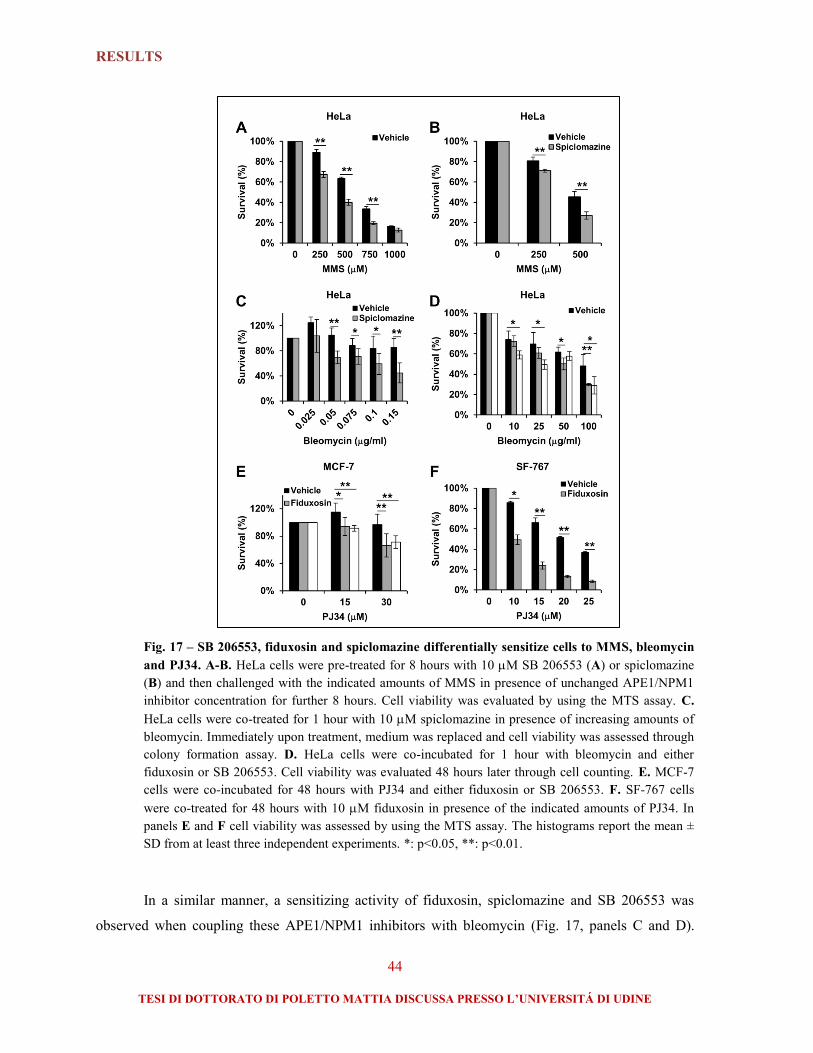
RESULTS
44
TESI DI DOTTORATO DI POLETTO MATTIA DISCUSSA PRESSO L’UNIVERSITÁ DI UDINE
Fig. 17 – SB 206553, fiduxosin and spiclomazine differentially sensitize cells to MMS, bleomycin
and PJ34. A-B. HeLa cells were pre-treated for 8 hours with 10 M SB 206553 (A) or spiclomazine
(B) and then challenged with the indicated amounts of MMS in presence of unchanged APE1/NPM1
inhibitor concentration for further 8 hours. Cell viability was evaluated by using the MTS assay. C.
HeLa cells were co-treated for 1 hour with 10 M spiclomazine in presence of increasing amounts of
bleomycin. Immediately upon treatment, medium was replaced and cell viability was assessed through
colony formation assay. D. HeLa cells were co-incubated for 1 hour with bleomycin and either
fiduxosin or SB 206553. Cell viability was evaluated 48 hours later through cell counting. E. MCF-7
cells were co-incubated for 48 hours with PJ34 and either fiduxosin or SB 206553. F. SF-767 cells
were co-treated for 48 hours with 10 M fiduxosin in presence of the indicated amounts of PJ34. In
panels E and F cell viability was assessed by using the MTS assay. The histograms report the mean ±
SD from at least three independent experiments. *: p<0.05, **: p<0.01.
In a similar manner, a sensitizing activity of fiduxosin, spiclomazine and SB 206553 was
observed when coupling these APE1/NPM1 inhibitors with bleomycin (Fig. 17, panels C and D).

RESULTS
45
TESI DI DOTTORATO DI POLETTO MATTIA DISCUSSA PRESSO L’UNIVERSITÁ DI UDINE
Also in the case of the spiclomazine/bleomycin combination, the combination index analysis
suggested the presence of a moderate synergism between these drugs. In addition, the cytotoxicity
induced by the PARP inhibitor PJ34 was enhanced by SB 206553 and fiduxosin in both the
glioblastoma-derived SF-767 cell line and the breast-cancer derived MCF-7 cell line (Fig. 17, panels
E and F).
In summary, selected APE1/NPM1 inhibitors display interesting cell sensitizing effects when
combined with DNA damaging drugs that trigger the BER activation. In order to investigate whether
the molecules displayed any anti-proliferative activity as single agents, I measured the proliferation of
HeLa cells upon acute or chronic exposure to the APE1/NPM1 inhibitors. Notably, cell treatment
with fiduxosin and spiclomazine resulted in a dose-dependent impairment of the cell growth rate, both
after short-term (24-72 hours) and long-term (10 days) treatment. SB 206553 had no significant effect
on cellular proliferation (Fig. 18).
Fig. 18 – Fiduxosin and spiclomazine show anti-proliferative activity as single agents. A. HeLa
cells were treated with vehicle (DMSO) or the indicated APE1/NPM1 inhibitor for 24, 48 or 72 hours.
Cell proliferation was determined by cell counting in at least three independent experiments and is
reported as representative growth curve from technical duplicates. *: p<0.05. B-C. HeLa cells were
seeded at low density and allowed to form colonies for 10 days in presence of increasing
concentrations of the indicated inhibitors. Survival is expressed as percentage of visible colonies
assuming the vehicle-treated cells as a reference. Fiduxosin and spiclomazine (B), but not SB 206553
(C) display a dose-dependent anti-proliferative activity. The histograms report the mean ± SD from at
least three independent experiments.

RESULTS
46
TESI DI DOTTORATO DI POLETTO MATTIA DISCUSSA PRESSO L’UNIVERSITÁ DI UDINE
1.5. Investigating the mechanisms of action of the APE1/NPM1 inhibitors
The primary screening and the orthogonal validation assays narrowed the number of
molecules under investigation to three interesting compounds (i.e. spiclomazine, SB 206553 and
fiduxosin). Based on my data, these molecules are able to disrupt the APE1/NPM1 association in
living cells and display interesting anti-tumor features, both as single-agents and in combination with
different genotoxins (Table 1)
Table 1 – Characteristics of the positive hits from the secondary validation assays. For each molecule the
table reports structure, effects on the APE1 redox function (where “++”, “+” and “-” denote the strength of the
phenotype as strong, mild or absent, respectively), presence/absence of sensitizing effect in combination with
MMS, bleomycin or PJ-34, and presence of anti-proliferative activity. NA: not available.

RESULTS
47
TESI DI DOTTORATO DI POLETTO MATTIA DISCUSSA PRESSO L’UNIVERSITÁ DI UDINE
The sensitization and anti-proliferative effects suggest that an inhibition of the APE1/NPM1
interaction may impair the function of at least one of the two interacting partners. In order to address
this issue, I investigated the ribosome biogenesis dynamics and the DNA damage accumulation in
treated HeLa cells, as biological processes that could be potentially affected by an impairment of the
APE1/NPM1 association (see also the paragraph 2.4 “APE1 is involved in the ribosome biogenesis
process”).
I first assessed whether cell treatment with the APE1/NPM1 inhibitors induced any variation
in the expression levels of the target proteins. None of the molecules tested led to downregulation of
APE1 or NPM1 (Fig. 19A); moreover, NPM1 maintained its nucleolar localization over a 72 hours
treatment with either spiclomazine or fiduxosin (Fig. 19B). Thus, it is possible to conclude that the
inhibitory effect of fiduxosin, spiclomazine and SB 206553 on the APE1/NPM1 interaction was not
provoked by a downregulation of the target proteins. Furthermore, at cytostatic spiclomazine and
fiduxosin dosage, HeLa cells did not show any major nucleolar stress (as demonstrated by the
integrity of nucleolar structures).
Fig. 19 – The APE1/NPM1 inhibitors do not induce modifications of APE1 or NPM1 expression,
or nucleolar stress. A. HeLa cells were treated with the indicated molecules (10 M for 16 hours) and
the levels of APE1 and NPM1 were assessed through Western blotting on whole cell extracts (20 g).
Cell treatment under conditions leading to the disruption of the APE1/NPM1 association does not lead
to modifications in the APE1 or NPM1 expression pattern. Tubulin was used as loading control. B.
Representative immuno-fluorescence on HeLa cells treated with either spiclomazine or fiduxosin (20
M) for the indicated time points. Staining with an anti-NPM1 antibody (red) shows the intactness of
nucleolar structures. Nuclei were counterstained with TO-PRO-3, bars 16 m.
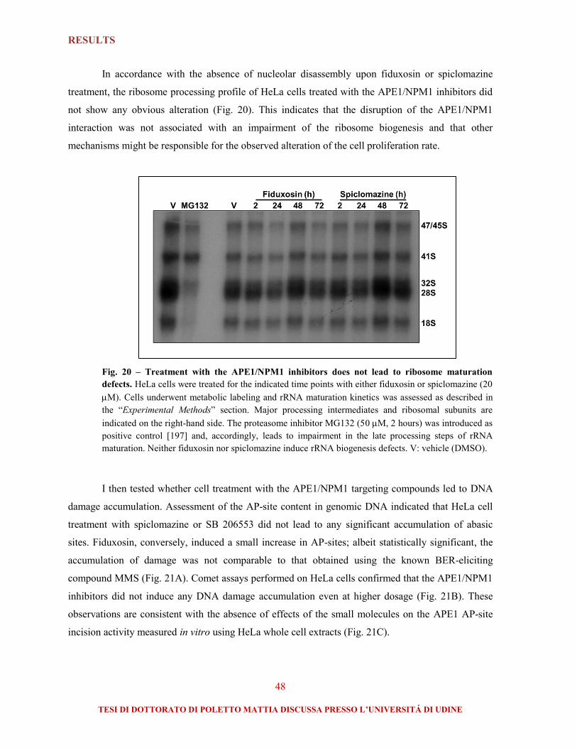
RESULTS
48
TESI DI DOTTORATO DI POLETTO MATTIA DISCUSSA PRESSO L’UNIVERSITÁ DI UDINE
In accordance with the absence of nucleolar disassembly upon fiduxosin or spiclomazine
treatment, the ribosome processing profile of HeLa cells treated with the APE1/NPM1 inhibitors did
not show any obvious alteration (Fig. 20). This indicates that the disruption of the APE1/NPM1
interaction was not associated with an impairment of the ribosome biogenesis and that other
mechanisms might be responsible for the observed alteration of the cell proliferation rate.
Fig. 20 – Treatment with the APE1/NPM1 inhibitors does not lead to ribosome maturation
defects. HeLa cells were treated for the indicated time points with either fiduxosin or spiclomazine (20
M). Cells underwent metabolic labeling and rRNA maturation kinetics was assessed as described in
the “Experimental Methods” section. Major processing intermediates and ribosomal subunits are
indicated on the right-hand side. The proteasome inhibitor MG132 (50 M, 2 hours) was introduced as
positive control [197] and, accordingly, leads to impairment in the late processing steps of rRNA
maturation. Neither fiduxosin nor spiclomazine induce rRNA biogenesis defects. V: vehicle (DMSO).
I then tested whether cell treatment with the APE1/NPM1 targeting compounds led to DNA
damage accumulation. Assessment of the AP-site content in genomic DNA indicated that HeLa cell
treatment with spiclomazine or SB 206553 did not lead to any significant accumulation of abasic
sites. Fiduxosin, conversely, induced a small increase in AP-sites; albeit statistically significant, the
accumulation of damage was not comparable to that obtained using the known BER-eliciting
compound MMS (Fig. 21A). Comet assays performed on HeLa cells confirmed that the APE1/NPM1
inhibitors did not induce any DNA damage accumulation even at higher dosage (Fig. 21B). These
observations are consistent with the absence of effects of the small molecules on the APE1 AP-site
incision activity measured in vitro using HeLa whole cell extracts (Fig. 21C).

RESULTS
49
TESI DI DOTTORATO DI POLETTO MATTIA DISCUSSA PRESSO L’UNIVERSITÁ DI UDINE
Fig. 21 – The APE1/NPM1 inhibitors do not induce any major accumulation of DNA damage in
HeLa cells. A. HeLa cells were treated with fiduxosin or spiclomazine (10 M, 48 hours), SB 206553
(20 M, 16 hours), or MMS (250 M, 16 hours) as positive control. AP-site accumulation in genomic
DNA was measured as described in the “Experimental Methods” section. The histogram reports the
mean ± SD from three measurements. *: p<0.01. B. Comet assay performed on HeLa cells treated with
fiduxosin or spiclomazine (30 M, 16 hours), SB 206553 (100 M, 16 hours), or H2O2 (200 M, 10
minutes) as positive control. The boxplot reports the median tail moment (black marks) for the
analyzed cell population. N≥100. **: p<0.001. C. Representative AP-site incision activity
measurement on HeLa whole cell extracts (50 ng) pre-incubated with either vehicle (DMSO) or
fiduxosin (5 M). The APE1/NPM1 inhibitor shows no significant effect on the APE1 endonuclease
function. The graph reports the mean ± SD from three independent assays.
Taken together, these data show that a subset of bioactive compounds, screened for their
ability to impair the APE1/NPM1 interaction, display interesting anti-tumor properties (i.e. cell
sensitization to genotoxins and anti-proliferative activity). The mechanism of action of the selected
APE1/NPM1 inhibitors is, at present, poorly understood, as treated cells do not show any impairment

RESULTS
50
TESI DI DOTTORATO DI POLETTO MATTIA DISCUSSA PRESSO L’UNIVERSITÁ DI UDINE
in the ribosome maturation process or evident accumulation of DNA damage. This suggests that the
anti-proliferative and sensitizing effects are not related to a major disruption of the BER function of
APE1 or of the NPM1 activity in ribosome processing.
The interesting anti-cancer properties of the APE1/NPM1 inhibitors certainly deserve further
characterization; additional studies aimed at the improvement of these molecules and at the
comprehension of their mechanism of action are therefore warranted.

RESULTS
51
TESI DI DOTTORATO DI POLETTO MATTIA DISCUSSA PRESSO L’UNIVERSITÁ DI UDINE
2. Investigating the role(s) of NPM1 during the DDR
Despite the valuable insights emerging from the study of NPM1 KO models, the mechanisms
underlying NPM1 function during the DDR are still poorly understood. It appears clear that NPM1
expression confers protection not only upon exogenous loads of DNA damage, but also under steady-
state conditions. Our recent work suggested that NPM1 might be a novel modulator of the BER
pathway, positively regulating the AP-site incision activity of APE1, possibly through mechanisms
involving both protein-protein and protein-nucleic acids interaction [186]. Yet, the precise
involvement of NPM1 during the overall BER processing remains unclear. Moreover, the DNA
damage-induced relocalization of NPM1 from nucleoli, suggests a possible involvement of the
protein during the DDR. It is not known, however, if NPM1 translocation has any impact on the BER
capacity.
2.1. NPM1 depletion leads to BER protein upregulation
Earlier observations from our laboratory revealed the presence of higher APE1 protein
amounts in NPM1/p53 double KO MEFs (hereafter referred to as NPM1-/-
) when compared to the
isogenic single p53 KO control MEF cells (NPM1+/+
) [186]. In order to understand whether NPM1
could be further involved in the modulation of the BER pathway downstream APE1, I compared the
protein and transcript expression profile of representative BER components in NPM1+/+
and NPM1-/-
cells. Western blotting analyses on MEF whole cell extracts revealed that BER protein levels were
largely upregulated as a consequence of NPM1 depletion (Fig. 22, panels A and B). The increased
BER protein expression, however, was not paralleled by a significantly augmented transcription rate
of BER genes (Fig. 22C), suggesting that NPM1 effect is mediated post-transcriptionally. In this
analysis Pol was a notable exception, being upregulated both at the transcript and at the protein
level.

RESULTS
52
TESI DI DOTTORATO DI POLETTO MATTIA DISCUSSA PRESSO L’UNIVERSITÁ DI UDINE
Fig. 22 – NPM1 depletion leads to BER protein upregulation. A. Representative Western blotting
analysis on NPM1+/+
and NPM1-/-
MEF whole cell extracts (60 g) showing increased BER protein
amounts in the absence of NPM1. Proteins probed are indicated on the right hand-side, tubulin was
used as loading control. B. Histogram reporting the average protein expression fold change in NPM1-/-
cells, as measured through densitometric quantification of the Western blotting signals. C. Histogram
showing the transcript fold change in NPM1-/-
cells, as measured through Real-time PCR. Bars indicate
the mean ± SD from at least three independent replicates. *: p<0.05, **: p<0.01, Polcs: DNA
polymerase catalytic subunit.
In order to rule out any confounding artifact, which may be caused by the intrinsic genetic
instability exhibited by the NPM1-/-
cells [194], I transiently depleted NPM1 in NPM1+/+
cells and
assessed the BER protein levels upon siRNA transfection. In accordance with my previous
observations, siRNA-mediated knockdown of NPM1 recapitulated the BER expression phenotype for
many of the proteins tested (Fig. 23).

RESULTS
53
TESI DI DOTTORATO DI POLETTO MATTIA DISCUSSA PRESSO L’UNIVERSITÁ DI UDINE
Fig. 23 – Transient downregulation of NPM1 induces BER protein stabilization in MEFs.
Representative Western blotting analysis on NPM+/+
whole cell extracts (50 g) showing the increment
of BER protein levels upon siRNA-mediated depletion of NPM1 (siNPM1). Proteins probed are
indicated on the right hand-side, tubulin was used as loading control. The average protein expression
fold change ± SD from at least three independent replicates is reported on the right hand-side. siScr:
scrambled control siRNA.
Complete NPM1 KO requires p53 depletion in order to prevent triggering of apoptosis [194];
the link between NPM1 expression and BER protein stability was therefore further validated in
human tumor cell lines bearing either active or inactive wild type p53 (i.e. HepG2 and HeLa,
respectively). The experiments confirmed that NPM1 downregulation leads to an increased
stabilization of BER proteins also in human cell lines, with variations in the degree of expression,
likely reflecting cell-type specific differences (Fig. 24). The discrepancies observed in fold-induction
when comparing the siRNA-mediated knockdown (Fig. 23 and Fig. 24) and the complete NPM1 KO
(Fig. 22) are probably related to the residual amount of NPM1 (about 20%) upon depletion (see for
instance FEN1, PCNA or LigI). Nonetheless, these data imply that NPM1 is a novel modulator of the
BER protein stability.

RESULTS
54
TESI DI DOTTORATO DI POLETTO MATTIA DISCUSSA PRESSO L’UNIVERSITÁ DI UDINE
Fig. 24 – Downregulation of NPM1 induces BER protein stabilization in human cell lines bearing
different p53 status. A. Representative Western blotting analyses on HeLa and HepG2 whole cell
extracts (50 g) upon treatment with scrambled (siScr) or NPM1-specific (siNPM1) siRNAs. Proteins
probed are indicated on the right hand-side, tubulin was used as loading control. B. Histogram
reporting average protein expression fold change in siNPM1-treated cells; signal from siScr-
transfected cells was taken as reference. Bars indicate the mean ± SD from at least three independent
replicates. ¥: not statistically significant (p>0.05).
2.2. NPM1 regulates BER protein localization and dynamics during nucleolar
stress
NPM1 is considered a hub factor as it mediates the nucleolar retention of different proteins
[19, 208], including p14Arf
[162] and APE1 [75], at least in transformed cells. Given the BER
dysregulation observed in NPM1-/-
MEFs I checked whether NPM1 expression affected the
subcellular localization of a subset of BER components. Strikingly, while in NPM1+/+
cells a fraction
of LigI, DNA polymerase catalytic subunit (Polcs), FEN1 and APE1 revealed a nucleolar
accumulation, the lack of NPM1 resulted in an impaired retention of these BER factors within
nucleoli (Fig. 25A).

RESULTS
55
TESI DI DOTTORATO DI POLETTO MATTIA DISCUSSA PRESSO L’UNIVERSITÁ DI UDINE
Fig. 25 – NPM1 promotes nucleolar accumulation of BER proteins. Representative immuno-
fluorescence micrographs showing the differential subcellular localization of BER factors in NPM1+/+
and NPM1-/-
cells. A. Each protein analyzed shows nucleolar accumulation in NPM1+/+
MEFs only
(white arrowheads). Nuclei were counter-stained with TO-PRO-3. B. Fibrillarin shows the typical
nucleolar staining pattern (green), whereas BER proteins (red) co-localize with fibrillarin only in
NPM1-proficient cells (yellow). The BER protein analyzed in each experiment is indicated on the left-
hand side. Bars: 16 m.

RESULTS
56
TESI DI DOTTORATO DI POLETTO MATTIA DISCUSSA PRESSO L’UNIVERSITÁ DI UDINE
Importantly, failure of BER proteins to accumulate within nucleoli was not determined by
abnormalities in the nucleolar architecture of NPM1-/-
cells, as already reported [162, 194] and
confirmed by the ordinary nucleolar fibrillarin staining (Fig. 25B). The relevance of NPM1
expression for the correct nucleolar accumulation of BER proteins was confirmed by reconstitution
experiments in NPM1-/-
MEFs: these cells were transfected with a plasmid expressing a human
FLAG-tagged NPM1 and restoration of nucleolar localization was observed for a subset of BER
components (Fig. 26A and [186]).
Fig. 26 – NPM1 co-purifies with BER protein complexes and is required for the nucleolar
accumulation of a subset of BER components. A. NPM1-/-
MEFs were transfected with either an
empty vector (mock) or with a FLAG-tagged human NPM1-expressing plasmid (pNPM1). The
representative immuno-fluorescence micrographs show the nucleolar accumulation of BER factors
(red) only in NPM1-expressing cells (green). Nuclei were counter-stained with TO-PRO-3, bars 16
m. B. HeLa cells were transfected with either an empty vector (mock) or FLAG-tagged NPM1
(NPM1) and immuno-precipitation was carried out with an anti-FLAG antibody. The immuno-purified
material was subsequently subjected to Western blotting analyses, revealing an enrichment for several
BER components. The antibodies used are indicated on the right-hand side.

RESULTS
57
TESI DI DOTTORATO DI POLETTO MATTIA DISCUSSA PRESSO L’UNIVERSITÁ DI UDINE
In addition, co-immuno-precipitation experiments performed in HeLa cells confirmed that,
under basal conditions, NPM1 can be co-purified with proteins belonging to the BER pathway (Fig.
26B). These data further suggest that NPM1 is essential for the accumulation of BER components
within nucleoli.
Several studies verified that DNA damage induces reorganization of the nucleolar proteome,
with a prevalent outflow from nucleoli (reviewed in [19]). NPM1 has been described as the main
p14Arf
interacting protein [209, 210] and the DNA damage-induced relocalization of both NPM1 and
p14Arf
is well documented [21, 59, 62, 197]. The redistribution of these proteins from nucleoli to the
nucleoplasm is believed to boost the p53-mediated response [63]. I reasoned that if NPM1 was
required for nucleolar accumulation of BER proteins, then a DNA damaging cue able to displace
NPM1 from nucleoli would affect the localization of BER enzymes as well. Canonical BER-eliciting
genotoxins (e.g. MMS, H2O2) did not affect the localization of NPM1 or APE1 (data not shown);
conversely, as already reported [59, 211, 212], cell treatment with the DNA damaging agents cisplatin
and daunorubicin, or the RNA polymerase I (Pol I) inhibitor actinomycin D led to a massive
relocalization of NPM1 and p14Arf
to the nucleoplasm. In accordance with the role of NPM1 in
promoting the nucleolar accumulation of BER proteins, cell treatment with these compounds resulted
in a complete nucleolar depletion of LigI, FEN1 and APE1 (Fig. 27 and not shown).

RESULTS
58
TESI DI DOTTORATO DI POLETTO MATTIA DISCUSSA PRESSO L’UNIVERSITÁ DI UDINE
Fig. 27 – Cisplatin and daunorubicin induce a subcellular relocalization of NPM1, p14Arf
and
BER enzymes. HeLa cells were treated with cisplatin (100 M, 6 hours, A) or daunorubicin (1 M, 2
hours, B), fixed and stained with the indicated antibodies. Representative immuno-fluorescence panels
show the nucleoplasmic relocalization of NPM1 and p14Arf
; the concomitant depletion of LigI, FEN1
and APE1 from nucleoli is highlighted by white arrowheads. Nuclei were counter-stained with TO-
PRO-3, bars 16 m.
Taken together, these observations suggest that, in immortalized cells, NPM1 is responsible
for the nucleolar accumulation of a subset of BER components; upon specific DNA damage or Pol I
inhibition, NPM1, p14Arf
and BER enzymes are released from nucleoli and relocalize to the
nucleoplasm.

RESULTS
59
TESI DI DOTTORATO DI POLETTO MATTIA DISCUSSA PRESSO L’UNIVERSITÁ DI UDINE
2.3. Active redistribution of APE1 from nucleoli protects cells from cisplatin
Cisplatin treatment has been shown to induce APE1 expression in a lung cancer cell line;
whereas depletion of APE1 sensitized the same cells to cisplatin-induced apoptosis [67]. Our and
others’ laboratories, moreover, observed interesting interrelations between cell sensitivity to cisplatin
and BER capacity ([9, 13] and unpublished results). Given the observed link between BER protein
localization and cisplatin, I decided to investigate thoroughly the functional significance of the BER
protein nucleolar accumulation and cell response to cisplatin.
It is worth highlighting that, upon cisplatin treatment, the relocalization of p14Arf
and BER
proteins takes place in the absence of complete nucleolar breakdown, as still a moderate amount of
NPM1 is retained within nucleoli (Fig. 27A, Fig. 28B). Under these conditions, however, nucleolar
stress occurs in a measurable manner, as suggested by the formation of fibrillarin caps ([197] and Fig.
28A) and by the interruption of nucleolar transcription ([197] and Fig. 28C).

RESULTS
60
TESI DI DOTTORATO DI POLETTO MATTIA DISCUSSA PRESSO L’UNIVERSITÁ DI UDINE
Fig. 28 – Cisplatin triggers nucleolar stress, which is followed by NPM1 relocalization. HeLa cells
were treated with cisplatin (100 M) and the effects on nucleolar function were monitored at different
time points. A. Representative immuno-fluorescence showing the onset of nucleolar stress 2 hours
after cisplatin addition, as evidenced by the appearance of perinucleolar fibrillarin caps (insets). Nuclei
were counter-stained with TO-PRO-3, bars 1 m. B. Representative immuno-staining for NPM1
reveals the nucleoplasmic translocation between 2 and 4 hours after cisplatin addition. Note that a
significant amount of NPM1 still populates nucleoli after 6 hours. Bars 8 m. C. Representative
fluorouridine (FUrd) incorporation assay shows the absence of transcriptional activity within nucleoli
one hour after cisplatin treatment. White arrowheads highlight the selective incorporation of FUrd in
nucleolar compartments. Bars 4 m. D. Graphical overview summarizing the timing of cisplatin
effects. Impairment of nucleolar transcription occurs within 1 hour after cisplatin addition. Fibrillarin
caps appear after 2 hours, when NPM1 starts to relocalize to the nucloeplasm. The amount of
redistributed NPM1 was measured by analyzing the immuno-fluorescence presented in panel B
through quantification of the mean nucleoplasmic NPM1 signal in randomly chosen cells (N≥20).
To exclude that the redistribution of BER proteins is merely a passive consequence of
nucleolar disassembly, I analyzed the behavior of APE1, as representative enzyme of the pathway.
Live cell imaging experiments exploiting an APE1-Dendra2 fusion construct [124, 213] revealed that
APE1 and NPM1 relocalization are uncoupled, as the first protein reduces its nucleolar accumulation
between 5 and 6 hours after cisplatin challenge (Fig. 29), whereas NPM1 redistribution to the
nucleoplasm is already visible 2 hours after cell exposure to cisplatin (Fig. 28). As already observed

RESULTS
61
TESI DI DOTTORATO DI POLETTO MATTIA DISCUSSA PRESSO L’UNIVERSITÁ DI UDINE
previously [124, 213] and confirmed by live cell imaging studies, the complete nucleolar emptying of
APE1 induced by cisplatin (Fig. 27) is an artifact arising during the permeabilization procedures
required for the immuno-fluorescence. In living cells, in fact, cisplatin is more likely to impair the
nucleolar accumulation capacity of APE1 and other BER enzymes, rather than completely abolishing
their nucleolar localization.
Fig. 29 – Cisplatin impairs the nucleolar accumulation capability of APE1 in a time-dependent
manner. Live cell imaging analysis on HeLa cells transfected with the APE1-Dendra2 construct and
treated with cisplatin show the time-dependent depletion of the protein from nucleoli (white
arrowheads). The transition occurs between 5 and 6 hours after cisplatin addition. Bars 8 m.
APE1 redistribution from nucleoli was further explored using two recently described
acetylation mutants. APE1K4pleA
, where the lysine residues K27, K31, K32 and K35 are converted to
alanine, generating a mutant protein endowed with higher AP-endonuclease activity that does not
interact with NPM1, accumulates less efficiently within nucleoli and strongly impairs cell
proliferation. In the second mutant (APE1K4pleR
) the same lysine residues are mutated to arginine,
resulting in a protein refractory to acetylation, but not affecting its nucleolar accumulation, the cell
growth rate or its ability to interact with NPM1 [124]. Remarkably, while cisplatin induced a
relocalization of APE1WT
from nucleoli, the APE1K4pleR
mutant was incapable of such redistribution
(Fig. 30A). Live cell imaging experiments confirmed the peculiar behavior of the APE1KpleR
mutant,
showing a delayed redistribution of the protein in comparison with the wild type form. Importantly,
expression of either APE1 mutant did not affect the cisplatin-induced relocalization of NPM1 (not
shown), thus confirming that this phenomenon is directly linked to the APE1 mutation. These
experiments indicate that, in addition to NPM1, other factors (e.g. rRNA) contribute to the nucleolar
accumulation of APE1. Moreover, these evidences confirm that during cisplatin-induced nucleolar
stress APE1 does not freely relocalize to the nucleoplasm as a consequence of nucleolar disassembly,
nor the protein is simply towed to the nucleoplasm by NPM1. Some kind of PTM involving lysine
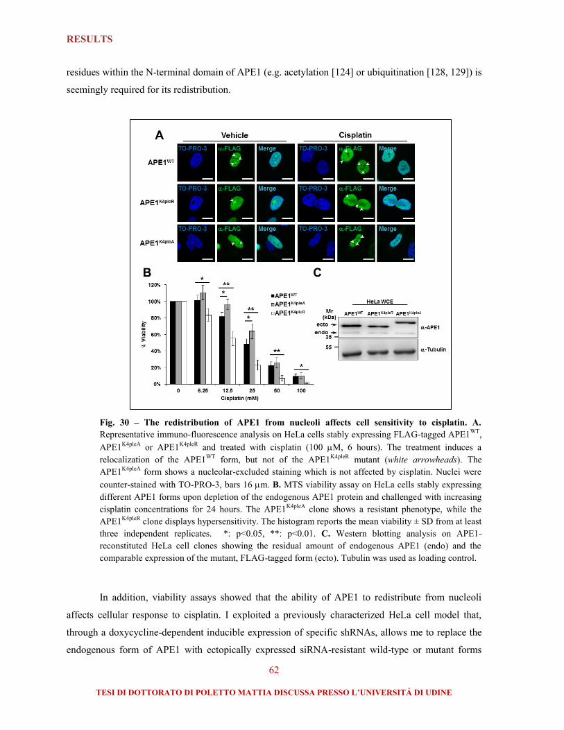
RESULTS
62
TESI DI DOTTORATO DI POLETTO MATTIA DISCUSSA PRESSO L’UNIVERSITÁ DI UDINE
residues within the N-terminal domain of APE1 (e.g. acetylation [124] or ubiquitination [128, 129]) is
seemingly required for its redistribution.
Fig. 30 – The redistribution of APE1 from nucleoli affects cell sensitivity to cisplatin. A.
Representative immuno-fluorescence analysis on HeLa cells stably expressing FLAG-tagged APE1WT
,
APE1K4pleA
or APE1K4pleR
and treated with cisplatin (100 M, 6 hours). The treatment induces a
relocalization of the APE1WT
form, but not of the APE1K4pleR
mutant (white arrowheads). The
APE1K4pleA
form shows a nucleolar-excluded staining which is not affected by cisplatin. Nuclei were
counter-stained with TO-PRO-3, bars 16 m. B. MTS viability assay on HeLa cells stably expressing
different APE1 forms upon depletion of the endogenous APE1 protein and challenged with increasing
cisplatin concentrations for 24 hours. The APE1K4pleA
clone shows a resistant phenotype, while the
APE1K4pleR
clone displays hypersensitivity. The histogram reports the mean viability ± SD from at least
three independent replicates. *: p<0.05, **: p<0.01. C. Western blotting analysis on APE1-
reconstituted HeLa cell clones showing the residual amount of endogenous APE1 (endo) and the
comparable expression of the mutant, FLAG-tagged form (ecto). Tubulin was used as loading control.
In addition, viability assays showed that the ability of APE1 to redistribute from nucleoli
affects cellular response to cisplatin. I exploited a previously characterized HeLa cell model that,
through a doxycycline-dependent inducible expression of specific shRNAs, allows me to replace the
endogenous form of APE1 with ectopically expressed siRNA-resistant wild-type or mutant forms

RESULTS
63
TESI DI DOTTORATO DI POLETTO MATTIA DISCUSSA PRESSO L’UNIVERSITÁ DI UDINE
[124, 138]. As shown in Fig. 30, the APE1K4pleR
-expressing clone was hypersensitive to cisplatin
treatment, indicating that the APE1 capacity to shuttle from nucleoli plays a critical role in dictating
cisplatin cytotoxicity. Furthermore, the expression of the APE1KpleA
form was protective at lower
doses of cisplatin (Fig. 30). Since inhibition of the APE1 catalytic activity in the APE1K4pleA
-
expressing clone did not affect its resistance to cisplatin (Fig. 31), the observed protective effect
might be ascribed to the lower proliferative capacity of these cells [124].
Fig. 31 – Inhibition of the APE1 catalytic activity in the APE1K4pleA
-expressing cells does not
affect cell sensitivity to cisplatin. HeLa cells stably expressing the APE1KpleA
form were incubated
with increasing cisplating concentrations for 24 hours, in presence of either methoxyamine (MX, 10
mM, A) or compound #3 (3.5 M, B), to block the APE1 catalytic activity. MMS (24 hours) was used
as positive control, to demonstrate the sensitizing activity of the APE1 inhibitors at the dosage tested.
The histograms report the mean ± SD of three independent replicates. *: p<0.05, **: p<0.01.
In conclusion, these experiments point to an active role of APE1 redistribution (and possibly
that of other BER proteins) upon cisplatin-induced nucleolar stress. The relocalization of APE1, in
fact, does not appear to be a passive phenomenon, but rather a modulation involving PTMs of specific
lysine residues seems to be required. Moreover, it is clear that interfering with cisplatin-induced BER
protein relocalization has a major impact on cell response to this genotoxin.

RESULTS
64
TESI DI DOTTORATO DI POLETTO MATTIA DISCUSSA PRESSO L’UNIVERSITÁ DI UDINE
2.4. APE1 is involved in the ribosome biogenesis process
The effect observed on the BER protein localization upon cisplatin, along with the concurrent
impairment in the nucleolar transcription, point to a major role for the BER components in the
maintenance of the ribosome biogenesis.
In order to gain further insights into the role of nucleolar BER enzymes, using again APE1 as
a representative enzyme of the pathway I assessed the influence of APE1 depletion on the early
ribosome biogenesis process. I transiently depleted cells of APE1 and measured the rRNA
transcription rate through two complementary assays. First, I assessed the nucleolar incorporation of
fluorouridine (FUrd), a fluorinated UTP analogue, into nascent rRNA transcripts [214, 215]. Notably,
the depletion of APE1 resulted in a strong impairment in the nucleolar FUrd uptake (Fig. 32A, Fig.
32B). This observation was corroborated by metabolic [32
P]-orthophosphate labeling experiments,
which confirmed the reduced synthesis rate of the 47S rRNA precursor in APE1-depleted cells (Fig.
32A, Fig. 32C). Altogether, these data demonstrate that the expression of APE1 is crucial for an
efficient transcription of ribosomal genes.

RESULTS
65
TESI DI DOTTORATO DI POLETTO MATTIA DISCUSSA PRESSO L’UNIVERSITÁ DI UDINE
Fig. 32 – Depletion of APE1 leads to nucleolar impairment. A. Representative Western blotting
analysis on HeLa cells treated with either a scrambled siRNA (siScr) or an APE1-specific siRNA
(siAPE1) shows the efficiency of APE1 knockdown. Tubulin was used as loading control. B.
Fluorouridine (FUrd) labeling of siScr- and siAPE1-treated HeLa cells. Left panel – representative
immuno-fluorescence showing the preferential accumulation of FUrd within transcriptionally active
nucleoli only in APE1-proficient cells (white arrowheads). Bars correspond to 8 m. Right panel –
histogram reporting the average FUrd nucleolar incorporation efficiency in siScr and siAPE1 cells.
Values reported express the average amount of FUrd-positive cells ± SD from at least three
independent experiments. C. [32
P]-phosphate metabolic labeling of nascent rRNA transcripts in HeLa
cells treated with siScr, siAPE1 or actinomycin D (as positive control) reveals the nucleolar
impairment in APE1-depleted cells. Cells were transfected with siRNAs and 48 hours later pulsed for 1
hour with [32
P]-orthophosphate, as described in the “Experimental Methods” section. Left panel – total
RNA was extracted and the amount of labeled rRNA precursors was measured through
autoradiography. Ethidium bromide staining was used as loading control. Right panel – histogram
reporting the average [32
P]-phosphate incorporation into the 47S precursor in siScr and siAPE1 cells.
Values reported express the mean incorporation ± SD from at least three independent experiments.
siScr was used as reference sample. *: p<0.01, **: p<0.001.

RESULTS
66
TESI DI DOTTORATO DI POLETTO MATTIA DISCUSSA PRESSO L’UNIVERSITÁ DI UDINE
I then checked whether the APE1 endonuclease activity of the protein might be involved in
the modulation of the ribosome biogenesis process. To this aim I exploited the inhibitor “compound
#3”, which specifically targets the endonuclease activity of the protein [148], and I assessed the
triggering of nucleolar stress phenotypes in HeLa cells. The onset of nucleolar stress upon APE1
inhibition was highlighted by the appearance of nucleolar fibrillarin caps (Fig. 33A); in addition,
FUrd incorporation analyses revealed a reduction of the nucleolar activity in HeLa cells treated with
compound #3 (Fig. 33B). Metabolic [32
P]-orthophosphate labeling, however, did not reveal any major
impairment in the 47S precursor transcription, upon APE1 inhibition (Fig. 33C). Interestingly, on the
other hand, I measured a reduced processing of mature rRNA species (Fig. 33, panels C and D).
These data indicate that the APE1 endonuclease activity might be important for the correct processing
of rRNA transcripts, rather than being involved in the transcription of the early 47S rRNA precursor.

RESULTS
67
TESI DI DOTTORATO DI POLETTO MATTIA DISCUSSA PRESSO L’UNIVERSITÁ DI UDINE
Fig. 33 – Inhibition of the APE1 endonuclease activity impairs ribosome biogenesis. HeLa cells
were treated with compound #3 (1.75 M, 24 hours) and nucleolar stress, rRNA transcription and
processing were analyzed. A. Representative immuno-fluorescence panels and insets show the
formation of fibrillarin caps upon inhibition of the APE1 endonuclease activity. Bars correspond to 1
m, nuclei were counterstained with TO-PRO-3. B. Representative immuno-fluorescence showing the
incorporation of fluorouridine (FUrd) within transcriptionally active nucleoli only in vehicle-treated
cells (white arrowheads). Bars correspond to 8 m. C. [32
P]-phosphate metabolic labeling of nascent
rRNA transcripts does not show any significant impairment in the transcription of the 47S precursor,
but reveals impairment in the processing of rRNA species following APE1 inhibition. Top panel –the
amount of labeled rRNA precursors was measured through autoradiography, as described in the
“Experimental Methods” section. Ethidium bromide staining was used as loading control. Bottom
panel – histogram reporting the average [32
P]-phosphate incorporation into the 47S and 41S precursors
in vehicle- or compound #3-treated cells. Values reported express the mean incorporation ± SD from
three independent replicates. Vehicle-treated cells were used as reference sample, *: p<0.05. D.
Analysis of the rRNA processing kinetics in HeLa cells upon [32
P]-phosphate metabolic labeling. The
analysis was carried out as in panel C; after the [32
P]-orthophosphate pulse, cells were chased for the
indicated time and intermediate rRNA species were analyzed on an agarose gel. The assay reveals a
profound impairment in the rRNA processing kinetics upon inhibition of APE1.
These experiments, coupled with previous evidences [75], strongly suggest that the
accumulation of APE1 and, possibly, of other BER enzymes within the nucleolus play an active role
in the maintenance of the correct nucleolar function of the cell.

RESULTS
68
TESI DI DOTTORATO DI POLETTO MATTIA DISCUSSA PRESSO L’UNIVERSITÁ DI UDINE
2.5. NPM1-mediated BER modulation is dependent on the activation of the
p14Arf
/Mule axis
The experiments performed so far allowed me to demonstrate that NPM1 is a novel player in
the BER pathway. NPM1 acts by promoting BER protein retention in nucleoli (Fig. 25); depletion of
NPM1, moreover, stabilizes several BER components in a post-translational manner (Fig. 22, Fig. 23
and Fig. 24). In order to explain the mechanisms underlying this last observation, I investigated the
interrelation between NPM1 and the p14Arf
/Mule axis, which has already been implicated in the
modulation of BER protein stability [55].
NPM1 is known to regulate both the stability and the nucleolar accumulation of p14Arf
. This
was demonstrated by monitoring the endogenous mouse p19Arf
ortholog in p53 KO MEFs [194] or
through transfection of ectopic p14Arf
in human tumor cell lines [162]. Consistently with these
reports, NPM1 depletion in HeLa cells decreased the stability of p14Arf
(Fig. 34A) and induced a
nucleoplasmic translocation of the residual protein (Fig. 34B).
Fig. 34 – p14Arf
is unstable and undergoes nucleoplasmic relocalization upon NPM1 depletion.
HeLa cells were transfected with either scrambled (siScr) or NPM1-specific (siNPM1) siRNAs and
subjected to Western blotting and immuno-fluorescence analyses. A. Representative Western blotting
on HeLa whole cell extracts (50 g) highlights the decreased stability of p14Arf
after NPM1
downregulation. Tubulin was used as loading control. B. Representative immuno-fluorescence analysis
on siNPM1-treated HeLa cells shows the nucleoplasmic redistribution of the residual p14Arf
. Note that
the immuno-fluorescence images were acquired at constant microscope settings, and therefore the
p14Arf
staining is saturated to allow for visualization of the downregulated p14Arf
protein in siNPM1-
trated cells. Nuclei were counter-stained with TO-PRO-3, bars 16 m.

RESULTS
69
TESI DI DOTTORATO DI POLETTO MATTIA DISCUSSA PRESSO L’UNIVERSITÁ DI UDINE
The p14Arf
tumor suppressor has been reporter to inhibit the E3-ligase activity of Mule upon
genotoxic stress [216]. I hypothesized that the chronic nucleoplasmic localization of p14Arf
, observed
in NPM-depleted cells, could trigger the stabilization of BER proteins through constitutive inhibition
of Mule. In agreement with this hypothesis, simultaneous depletion of either NPM1 and p14Arf
, or
NPM1 and Mule, efficiently suppressed the upregulation of BER components observed when
depleting NPM1 only (Fig. 35).
Fig. 35 – Interfering with the p14Arf
/Mule axis bluntens the BER stabilization effect obtained
upon NPM1 depletion. HeLa cells were transiently depleted of NPM1, p14Arf
and Mule through
specific siRNAs, as indicated. The expression levels of a subset of BER proteins was then assessed
through Western blotting on whole cell extracts (50 g) (A). B. Histogram reporting the quantitative
protein expression fold change for siNPM1, siNPM1+sip14Arf
and siNPM1+siMule relative to control
siRNAs (scrambled, sip14Arf
or siMule, respectively). The histogram shows the mean ± SD from at
least three independent experiments. The statistical analysis was performed by comparing NPM1-
depleted cells with their control counterparts (i.e. sScr, sip14Arf
and siMule, respectively). *: p<0.05,
**: p<0.01.

RESULTS
70
TESI DI DOTTORATO DI POLETTO MATTIA DISCUSSA PRESSO L’UNIVERSITÁ DI UDINE
The contribution of p14Arf
to the NPM1-mediated control of the BER protein stability was
further highlighted by knockdown studies on cell lines lacking detectable p14Arf
(i.e. U2OS and SK-
BR-3). In these cell lines the upregulation of BER components following depletion of NPM1 was
partially abolished or reduced in order of magnitude (Fig. 36). A notable exception was represented
by Pol, but this phenotype might reflect a p14Arf
-independent regulation of the protein at the
transcriptional level, as suggested by the previous Real-time PCR analysis (Fig. 22).
Fig. 36 – NPM1-mediated BER protein stabilization is partially impaired in cell lines lacking
detectable p14Arf
. U2OS (A) or SK-BR-3 (B) cells were treated with either scrambled (siScr) or
NPM1-specific (siNPM1) siRNAs and BER protein expression levels were assessed in whole cell
exctracts (50 g) using the indicated antibodies (right-hand side). The fold of protein induction upon
NPM1 depletion is indicated for each protein analyzed. HeLa whole cell extract was used as positive
control for p14Arf
expression, while tubulin was used as loading control.

RESULTS
71
TESI DI DOTTORATO DI POLETTO MATTIA DISCUSSA PRESSO L’UNIVERSITÁ DI UDINE
Additional immuno-fluorescence studies on NPM1-, p14Arf
- or Mule-depleted cells revealed a
cross-modulation existing between these three factors, which dictates their subcellular localization.
As previously demonstrated (Fig. 34), nucleolar localization of p14Arf
was promoted by NPM1, and,
to a lesser extent, by Mule expression. NPM1 nucleoplasmic accumulation was increased when
knocking down either p14Arf
or Mule, indicating that both proteins partially regulate NPM1 nucleolar
retention. NPM1 down-regulation, moreover, resulted in the depletion of the nucleolar Mule pool,
suggesting that Mule subcellular trafficking is, in turn, modulated by NPM1 (Fig. 37).
Fig. 37 – The subcellular localization of p14Arf
, Mule and NPM1 is modulated by an intricate
cross-talk. Representative immuno-fluorescence experiments on HeLa cells treated with scrambled,
p14Arf
-, NPM1- or Mule-targeting siRNAs (siScr, sip14Arf
, siNPM1 and siMule, respectively). Cells
were probed with the antibodies indicated in the upper part of the figure. Note the increased p14Arf
nucleoplasmic localization upon NPM1 and Mule knockdown, the nucleolar depletion of Mule in
NPM1-depleted cells, and the increased nucleoplasmic staining for NPM1 in p14Arf
- and Mule-
depleted cells. Nuclei were counter-stained with TO-PRO-3, bars 16 m.

RESULTS
72
TESI DI DOTTORATO DI POLETTO MATTIA DISCUSSA PRESSO L’UNIVERSITÁ DI UDINE
2.6. BER capacity is positively modulated by NPM1
Despite the higher BER protein content, NPM1-/-
MEFs have previously been shown to
display increased susceptibility to BER-eliciting genotoxins [186]. We recently linked the augmented
sensitivity to MMS, bleomycin and oxidizing agents to an impaired AP-site incision activity in
NPM1-/-
cell extracts [186]. Prompted by the observation that NPM1 depletion leads to a broad
upregulation of the BER components, here I tested whether the enzymatic steps of the BER process
downstream APE1 incision may be affected by NPM1 depletion. To this aim, I compared in vitro
gap-filling, nick-ligation and flap-incision activities at equal amounts of NPM1+/+
and NPM1-/-
whole
cell extracts (Fig. 38).
Fig. 38 – Comparison between NPM1+/+
and NPM1-/-
whole cell extracts show proficiency in gap-
filling, but no significant difference in nick-ligation or flap-incision activities upon depletion of
NPM1. BER enzymatic activities were meaured by using standard in vitro radioisotope-based assays,
as indicated in the “Experimental Methods” section; equal amounts of MEF whole cell exctracts were
compared in each reaction. BER activities were monitored in time-course reactions and, for every time
point, the percentage of substrate (S) to product (P) conversion was calculated. Graphs on the right-
hand side report the mean conversion ± SD from at least three independent replicates. For each BER
activity a representative autoradiogram is reported, along with a Western blotting showing the different
protein content among NPM1+/+
and NPM1-/-
cells. *: p<0.05, **: p<0.01.
As expected, the higher Pol content was paralleled by a small, yet significant increased
proficiency in gap-filling in NPM1-/-
MEFs. Strikingly, despite the higher amount of LigIII and

RESULTS
73
TESI DI DOTTORATO DI POLETTO MATTIA DISCUSSA PRESSO L’UNIVERSITÁ DI UDINE
FEN1, both the nick-ligation and the flap-incision activities in NPM1+/+
and NPM1-/-
cell extracts did
not differ significantly (Fig. 38). The same repair activity results were obtained using whole cell
extracts prepared by different methods (i.e. detergent vs. hypotonic extraction), suggesting that my
measurements were not affected by differential protein extraction efficiency among the two cell lines
(not shown).
In order to compare the BER enzymatic activities at equal protein content, I carried out the in
vitro BER assays after precise normalization of the Pol, LigIII and FEN1 levels in NPM1+/+
and
NPM1-/-
whole cell extracts. This experimental setup highlighted a major impairment in each BER
enzymatic step in the absence of NPM1, thus suggesting that NPM1 is a previously uncharacterized
positive modulator of the BER process (Fig. 39).
Fig. 39 – In vitro BER assays on NPM1+/+
and NPM1-/-
whole cell extracts at equal Pol, LigIII
and FEN1 content highlight the profound impairment in each BER step after depletion of
NPM1. BER enzymatic activities were meaured as indicated in Fig. 38; MEF whole cell exctracts
concentration was adjusted to compare equal protein amounts in each reaction. Graphs on the right-
hand side report the mean percentage of substrate (S) to product (P) conversion ± SD from at least
three independent replicates. For each BER activity a representative autoradiogram is reported, along
with a Western blotting showing the comparable protein content in NPM1+/+
and NPM1-/-
extracts. *:
p<0.05, **:p<0.001.

RESULTS
74
TESI DI DOTTORATO DI POLETTO MATTIA DISCUSSA PRESSO L’UNIVERSITÁ DI UDINE
Despite the remarkable BER impairment in the absence of NPM1, under experimental
conditions reflecting the protein expression pattern in living cells (i.e. without adjustment in whole
cell extract amounts), I did not observe significant variations in the BER capacity (Fig. 38).
Nonetheless, previous studies highlighted an increased sensitivity of NPM1-/-
cells to BER-eliciting
agents [186], along with the appearance of DDR markers in NPM1 KO embryos [194], as alluded to
earlier. This indicates that the in vitro biochemical assays here reported do not entirely recapitulate
the in vivo BER dynamics. To address this issue I developed a reporter-based assay suitable for the
assessment of the overall BER capacity in living cells. A luciferase reporter-containing plasmid was
heat-depurinated and transfected into living NPM1+/+
or NPM1-/-
MEFs and the luciferase activity was
used as a direct measure of BER-mediated repair. As demonstrated by digestion with recombinant
APE1, this treatment mainly generated AP-sites and SSBs with no detectable DSBs (Fig. 40B); thus,
the plasmid obtained after heat-induced depurination is a substrate suitable to measure the overall
cellular BER capacity, without confounding contributions from other DNA repair pathways.
Using this reporter-based assay I confirmed that, despite the increased BER protein
expression, NPM1-/-
cells have an impaired BER response (Fig. 40A), therefore explaining the
augmented sensitivity to genotoxins. Consistently, NPM1 depletion in NPM1+/+
MEFs recapitulated
the NPM1-/-
phenotype (Fig. 40 A and C); whereas reconstitution of NPM1-/-
cells with recombinant
human NPM1 rescued the BER deficiency (Fig. 40 A and C). Altogether, these results indicate that
NPM1 is a novel positive modulator of the BER pathway both in vitro and in vivo.

RESULTS
75
TESI DI DOTTORATO DI POLETTO MATTIA DISCUSSA PRESSO L’UNIVERSITÁ DI UDINE
Fig. 40 – NPM1 depletion impairs the BER capacity in vivo. A. Reporter-based BER assays
performed after transfection of a heat-depurinated luciferase plasmid reveal that NPM1-/-
cells have an
impaired repair capacity (left bars). siRNA-mediated depletion of NPM1 in NPM1+/+
MEFs confirms
this finding (middle bars); while transfection of human NPM1 in the NPM1-/-
background rescues BER
capacity (right bars). The histogram reports the mean luciferase activity (relative to that of intact
DNA) ± SD from at least three independent replicates. *: p<0.01. B. Agarose gel analysis on the heat-
depurinated reporter plasmid shows the formation of a small amount of spontaneously nicked DNA
after depurination. Digestion of the damaged plasmid with recombinant APE1 results in the conversion
of the plasmid into nicked and linear DNA, thus confirming the presence of AP-sites. A NdeI-
linearized plasmid was used as marker to determine the migration of the linear vector. ND: not
digested. C. Representative Western blotting analysis on MEF whole cell extracts from the experiment
reported in panel A and showing the siRNA-mediated depletion of NPM1 (siNPM1) in NPM1+/+
cells
and the reconstitution of NPM1-/-
cells (pNPM1).

RESULTS
76
TESI DI DOTTORATO DI POLETTO MATTIA DISCUSSA PRESSO L’UNIVERSITÁ DI UDINE
2.7. Depletion of NPM1 does not significantly impair NHEJ- or HR-mediated DNA
repair
As pointed out earlier, several evidences link NPM1 to DNA strand break-repair pathways
[199, 200]. In the previous chapter I thoroughly analyzed the involvement of NPM1 in the BER-
mediated DNA repair: the overall BER dysregulation in NPM1-depleted cells clearly explains the
increased sensitivity of NPM1-/-
cells to MMS, oxidizing agents, bleomycin and ionizing radiation
([186] and not shown). Some of the genotoxins used in previous studies, however, may also generate
DSBs that are not directly targeted by the BER pathway. Therefore, in order to understand whether
NPM1 has any significant relevance in the clearance of DNA strand breaks, I carried out a broad
analysis to compare the DSB-repair capacity in NPM1+/+
and NPM1-/-
cells.
Western blotting analyses on NPM1-deficient MEFs revealed major variations in the
expression levels of a small set of NHEJ components, with increased expression of the Ku70 antigen
and of DNA ligase IV (LigIV), and reduction in the DNA-dependent protein kinase catalytic subunit
(DNA-PKcs) amount (Fig. 41). Expression studies on a representative subset of HR factors,
moreover, showed a significant upregulation of Rad51 in NPM1-/-
cells (Fig. 41).
Fig. 41 – NPM1-depleted cells show upregulation of a subset of DSB-repair proteins. A.
Representative Western blotting analysis on NPM1+/+
and NPM1-/-
MEFs whole cell extracts (60 g)
shows the variation in NHEJ and HR protein amounts in the absence of NPM1. Antibodies used are
indicated on the right hand-side, tubulin was used as loading control. B. Histogram reporting the
average protein expression fold change in NPM1-/-
cells, as measured through densitometric
quantification of the Western blotting signals. Bars indicate the mean ± SD from at least three
independent replicates. *: p<0.05, **: p<0.01.

RESULTS
77
TESI DI DOTTORATO DI POLETTO MATTIA DISCUSSA PRESSO L’UNIVERSITÁ DI UDINE
As already shown for the BER pathway, variation in DSB-repair protein levels may reflect a
dysregulation in the DSB repair capacity of NPM1-deficient cells. In order to compare the NHEJ
capacity of NPM1+/+
and NPM1-/-
MEFs, I carried out in vitro end-joining (EJ) assays using linear
DNA substrates ad hoc generated through restriction of a plasmid DNA substrate. The in vitro EJ
assays demonstrated that the ligation activity in whole cell extracts from NPM1-/-
cells was
significantly higher than NPM1+/+
extracts toward DNA ends with cohesive overhangs (Fig. 42A).
The ligation efficiency of blunt-ends did not show significant differences even if a trend toward
higher activity of NPM1-/-
lysates was still present (Fig. 42A).
Fig. 42 – In vitro end-joining assays reveal a slight proficiency of NPM1-/-
whole cell extracts. A.
EJ assay on a linear DNA substrate with cohesive overhangs generated through restriction of a
pmirGLO plasmid with the ApaI endonuclease. A representative gel shows the time-dependent
conversion of the substrate (S) to oligomeric products (P1 and P2). The ApaI recognition sequence is
also reported. B. EJ assay on a linear DNA substrate with blunt-ends generated through restriction of a
pmirGLO plasmid with the EcoRV endonuclease. A representative gel shows the time-dependent
conversion of the substrate (S) to a dimeric product (P). The EcoRV recognition sequence is reported
below. The histograms (right-hand side) show the average percentage of substrate conversion ± SD
from three independent experiments. *: p<0.05, M: DNA ladder.
Similar results were obtained using different enzymes, suggesting that the observed behavior
is not linked to any sequence preference in the substrate.

RESULTS
78
TESI DI DOTTORATO DI POLETTO MATTIA DISCUSSA PRESSO L’UNIVERSITÁ DI UDINE
I then assessed the in vivo DSB repair capacity of NPM1+/+
and NPM1-/-
cells exploiting a
dual luciferase-containing pmirGLO plasmid. A single DSB was introduced within the vector by
means of restriction endonucleases; the restriction site was carefully selected in order to repress the
transcription of one selected reporter gene. MEF cells were subsequently transfected with the linear
plasmid and the luciferase activity was measured to assess the EJ activity. Using different restriction
enzymes I probed different biases in the DSB repair, such as the EJ activity on cohesive vs. blunt
ends, or the overall vs. precise EJ capacity (Fig. 43).
Fig. 43 – In vivo end-joining assays highlights the higher activity of NPM1-/-
cells toward cohesive
DNA ends. A. Schematic overview of the plasmid used in the assay with the restriction sites exploited
to generate the substrate. The restriction endonucleases used are listed in the lower part of the panel,
along with the characteristics of the substrate generated. KpnI-mediated restriction did not affect the
activity of any reporter gene and was used to obtain a linear DNA substrate to be used as reference.
PflMI was used to induce a DSB separating the SV40 promoter from the reporter gene and therefore
allowing to monitor the overall EJ efficiency (as extensive editing at the DNA damage site is much
more sustainable in a non coding region). luc2: firefly luciferase reporter; hRluc-neo fusion:
humanized Renilla luciferase-neomycin resistance reporter cassette. B. In vivo EJ assay on control wild
type (WT), LigIV-/-
and DNA-PKcs-/-
HCT116 cell lines highlights the assay performance and
dependency on the presence of LigIV and, partially, on DNA-PKcs in the case of cohesive ends. The
histogram reports the average repair efficiency ± SD from three technical replicates. *:p<0.05. C. In
vivo EJ assay on NPM1+/+
and NPM1-/-
MEFs shows the proficiency of NPM1-depleted cells during
cohesive end repair. The histogram reports the average repair efficiency ± SD from three independent
replicates. **:p<0.01.

RESULTS
79
TESI DI DOTTORATO DI POLETTO MATTIA DISCUSSA PRESSO L’UNIVERSITÁ DI UDINE
The in vivo EJ assays results were in accordance with the in vitro data (Fig. 42), highlighting
a significantly increased EJ capacity for NPM1-/-
cells over cohesive overhangs, regardless of the
repair specificity (Fig. 43). These assays do not point to any significant difference in the EJ activity
over blunt ended substrates. Reasonably, these data suggest that the presence of higher amounts of
Ku70 and LigIV slightly improves the DNA EJ capacity of NPM1-deficient cell extracts.
Differently from other mammalian cells, the EJ capacity of MEFs is extremely low (compare
the ligation efficiency in MEFs and human cell lines in Fig. 43) and the preferred DSB-repair
pathway appears to be HR [217]. Therefore, in order to assess the HR-dependent DNA repair in
NPM1-depleted MEFs, I challenged cells with the etoposide or bleomycin and measured the
formation of Rad51 foci (Fig. 44). Being Rad51 a major component of the HR pathway involved in
the homology search and pairing during the repair process, the dynamics of Rad51 foci clearance over
the time allowed me to estimate the HR-dependent DSB repair.
Fig. 44 – The HR pathway is not impaired in NPM1-/-
cells. NPM1+/+
and NPM1-/-
MEFs were
treated with etoposide (100 M, 30 minutes) and Rad51 foci-positive cells were scored immediately
(0) or after the indicated release time points. The histogram shows the mean percentage of Rad51-
positive cells ± SD from three independent experiments. Unt: untreated; *: p<0.05.
The assessment of Rad51 foci formation showed a slightly increased susceptibility of NPM1-
/- cells to etoposide (Fig. 44) and bleomycin (not shown), but similar DNA damage clearance kinetics.
Consequently, despite the higher Rad51 expression levels in NPM1-depleted cells, it is possible to
conclude that the HR pathway is not substantially affected by the absence of NPM1. These data are in
agreement with comet assay analyses that did not reveal significant differences between NPM1+/+
and
NPM1-/-
cells neither in the basal DNA damage content nor in the bleomycin-induced strand breaks
repair (not shown).

DISCUSSION
80
TESI DI DOTTORATO DI POLETTO MATTIA DISCUSSA PRESSO L’UNIVERSITÁ DI UDINE
DISCUSSION
The work described in this thesis broadens our comprehension of the BER mechanisms,
presenting, on one side, the characterization of novel molecular entities targeting the interaction
between a BER component and a non-canonical BER modulator. On the other hand, I carried out a
wide analysis on the possible involvement of NPM1 in the DNA damage response, showing how a
protein apparently unrelated to DNA repair may affect substantially the DDR.
The first part of this work led to the characterization of low molecular weight compounds
targeting the APE1/NPM1 interaction. To the best of my knowledge, this is the first attempt to target
the BER pathway through impairment of its protein-protein interaction network. The pilot-scale
strategy set up during this project will be a useful starting point for the development of APE1/NPM1
inhibitors with increased potency and specificity. These molecules will likely provide novel
translational tools and could be exploited to thoroughly characterize the biological relevance of the
APE1/NPM1 association in the tumor biology.
A parallel and comprehensive analysis of the role(s) of NPM1 in the DDR revealed a tight
link connecting NPM1 and the BER pathway. Analyses on the BER protein expression, localization
and activity demonstrated that NPM1 is clearly a novel player in the BER response to DNA damage.
The significance and the full implications of these findings, however, still remains to be elucidated, as
it appears clear that canonical BER-eliciting stimuli are not able to induce NPM1 and BER protein
relocalization. On the other hand, the observation of a link between cell response to cisplatin and
NPM1/BER protein redistribution might open novel perspectives for a thorough comprehension of
tumor resistance, explaining the involvement of the BER pathway in the scavenging of cisplatin-
induced DNA damage.

DISCUSSION
81
TESI DI DOTTORATO DI POLETTO MATTIA DISCUSSA PRESSO L’UNIVERSITÁ DI UDINE
1. The APE1/NPM1 interaction as a promising target for anti-
tumor therapy
This thesis describes, for the first time, a pilot-scale screening aimed at the identification of
low molecular weight compounds targeting the APE1/NPM1 association. In collaboration with the
NCGC, we exploited the AlphaScreen® technology using full-length recombinant proteins and
screened commercially available small molecule libraries in order to obtain putative disruptors of the
APE1/NPM1 interaction. The hits from the screening underwent further prioritization through
secondary cell-based orthogonal assays, and I obtained a set of compounds effectively able to
interfere with the APE1/NPM1 association in living cells. The data available so far do not allow me
to exclude the presence of in vivo off-target effects of the top-hit molecules, which are already known
bioactive drugs. Through my proof of concept approach, however, I successfully demonstrated that
the disruption of the APE1/NPM1 interaction can be achieved via structurally unrelated compounds
under conditions of limited acute cytotoxicity, leading to interesting anti-tumor phenotypes. The
APE1/NPM1 inhibitors, in fact, potentiate the cytotoxicity of clinically relevant genotoxins and show
anti-proliferative activity as single agents in tumor cells of different histological origin (Fig. 17 and
Fig. 18).
The mechanism of action of the APE1/NPM1 inhibitors has not been entirely characterized;
however, as the protein-protein interaction is reported to involve the N-terminal domain of both
proteins [75, 91], it is reasonable to speculate that the compounds might target the APE1 unstructured
region or the NPM1 oligomerization domain. The change in the APE1 subcellular localization
observed upon treatment with the compounds, indeed, suggests that the molecules might target the N-
terminal domain of APE1, which is responsible for the nuclear accumulation of the protein [125] as
well as for the interaction with NPM1 and rRNA [75, 90, 91]. SPR experiments (Fig. 16), and the
negative effect of some molecules towards the APE1 redox activity (Fig. 15), would support a
preference of the compounds for APE1 binding.
Curiously, among the screening hits was identified troglitazone, a PPAR agonist with anti-
diabetic properties, which was withdrawn from the market for its hepatotoxicity [218]. Troglitazone
has previously been suggested to exert anti-inflammatory action through inhibition of the NF-B
pathway [219] and various reports have described interesting anti-tumor properties of the molecule
[220, 221]. These observations might well be explained by the finding that troglitazone is a very
potent inhibitor of the APE1/NF-B axis (Fig. 15). A positive interaction between bleomycin and the
antipsychotic phenothiazine trifluoperazine was also previously reported [222]; the APE1/NPM1
inhibitor spiclomazine is a member of the phenothiazine class of drugs and, consistently, I measure a

DISCUSSION
82
TESI DI DOTTORATO DI POLETTO MATTIA DISCUSSA PRESSO L’UNIVERSITÁ DI UDINE
synergic effect between bleomycin and spiclomazine (Fig. 17). Albeit trifluoperazine has been
suggested to impair the NHEJ process [222], an involvement of APE1 [101] or of the APE1/NPM1
interaction in the elimination of bleomycin-induced DNA damage could also explain the observed
effects.
The sensitization effects measured when disrupting the APE1/NPM1 interaction in
combination with MMS, bleomycin and PJ34 (Fig. 17) are not easily explained, as the endonuclease
activity of APE1 is not directly impaired, in vitro, by the inhibitors (Fig. 21). Some of the molecules,
however, induce relocalization of APE1 to the cytoplasm (Fig. 13 and Supplementary Table 1). It is
therefore tempting to speculate that the sensitization observed toward genotoxins is driven by an
impairment of the nuclear BER capacity, as already shown for NPM1c+ AML cells [186]. The
measured effects might also reflect a possible role for the APE1/NPM1 association in the scavenging
of DNA strand-breaks, as the genotoxins used in this study are likely to lead to the accumulation of
SSBs and DSBs, in particular during the DNA replication process. Accordingly, NPM1 has been
reported to associate with PARP1 and PARP2 [223], although the functional relevance of this
interaction for DNA repair is still evanescent. Moreover, inhibition of the APE1 endonuclease activity
was demonstrated to induce synthetic lethality in a DSB-repair deficient background [224]. It is
conceivable, on the other hand, that non-canonical APE1 activities (e.g. RNA binding/cleavage,
exonuclease, or RNaseH [95]) are involved in the response to the DNA damaging agents used in my
experiments. Design of more selective APE1/NPM1 interaction inhibitors, therefore, will be useful to
specifically characterize the relevance of this protein-protein interaction in vivo.
In conclusion, in the first part of this thesis I show that known bioactive molecules selected
for their ability to displace the APE1/NPM1 association are endowed with previously uncharacterized
anti-tumor properties. Potential targets of these inhibitors might be tumors that show upregulation of
both APE1 and NPM1 levels and that might possibly rely on an increased APE1/NPM1 interaction
for resistance and/or proliferation. As proposed in the introductory section, ovarian cancer, as well as
hepatocarcinoma, might reveal sensible targets for the APE1/NPM1 inhibitors. Understanding the
specificity of action of these molecules in vivo is clearly needed at this point. The improvement of
these compounds, however, will lead to the development of new useful tools for the biological
investigation of the APE1/NPM1 interaction under physiological and pathological conditions.

DISCUSSION
83
TESI DI DOTTORATO DI POLETTO MATTIA DISCUSSA PRESSO L’UNIVERSITÁ DI UDINE
2. NPM1 is a novel player in the BER
The data presented in this thesis demonstrate that NPM1 is a novel multilevel modulator of
the BER pathway. Previous work showed that NPM1 positively modulates the expression, the activity
and the nucleolar accumulation of APE1 [186], but nothing was known about the relationship
between NPM1 and the remainder of the BER pathway. My results suggest that NPM1 operates on
the BER acting, at least at three different levels:
1) NPM1 affects the stability of BER proteins. Here I demonstrate that the lack of or
reduced NPM1 expression results in the stabilization of several BER components (Figs. 22-24). The
mechanisms underlying the BER protein upregulation still deserves further investigation; it appears
clear, however, that BER stabilization following NPM1 depletion partially requires the functionality
of the p14Arf
/Mule axis (Figs. 35 and 36). It is useful to underline that some of the expression changes
that I observe are quantitatively small and may be indirect, in that increased expression of one BER
protein may up-regulate another pathway component. The overall dysregulation of the protein
expression levels, however, might play a significant role in reducing the coordination of the BER
process, therefore affecting the repair capacity.
2) NPM1 promotes the nucleolar accumulation of a subset of BER components. The
nucleolar localization of many DNA repair proteins is well established [19, 20] and several BER
factors (including SMUG1 [76], aprataxin [225], APE1 [75, 186], FEN1 [77], PARP1/2 [223]) have
been implicated in nucleolar functions that are possibly unrelated to their “canonical” DNA repair
activity. Therefore, the novel discovery of the BER/replicative enzymes LigI and Polcs within
nucleoli was not unexpected. To the best of my knowledge, however, the prominent role of NPM1 in
maintaining the nucleolar BER pool was never reported previously, if not for APE1 [75, 186] and
PARP1/2 [223].
3) NPM1 positively modulates the cellular BER capacity. It has previously been shown by
our group that NPM1 affects the APE1 endonuclease activity through direct protein-protein
association [75, 91, 186]. In this thesis I demonstrate that NPM1 expression affects virtually each
enzymatic step downstream the APE1 intervention in the BER process (Figs. 38 and 39). In addition,
in vivo BER assays point to a significant dysregulation of the BER pathway dynamics upon NPM1
depletion (Fig. 40). A possible interpretation of these data could take into account the already
documented chaperone activity of NPM1 [176]; by means of protein-protein or nucleic acid-protein
contacts NPM1 could act as activator of the overall BER process. Nonetheless, attempts to restore the
BER activity in NPM1-/-
cell extracts using recombinant NPM1 in vitro were unsuccessful (not
shown). Thus suggesting that PTMs either of, or induced by, NPM1 may be required for BER
DISCUSSION
84
TESI DI DOTTORATO DI POLETTO MATTIA DISCUSSA PRESSO L’UNIVERSITÁ DI UDINE
modulation. Further studies aimed at elucidating the mechanisms underlying the NPM1-mediated
BER activation are clearly needed at this point. Nevertheless, the BER impairment measured in vivo
likely reflects a profound role for NPM1 in stimulating BER capacity. Additional analyses aimed at
the elucidation of the NPM1 role in the DNA DSB-repair systems did not highlight any major effect
of NPM1 depletion in the NHEJ and in the HR pathways (Figs. 42-44). Given the well-established
responsiveness of NPM1 to UV-induced DNA damage [59, 195, 226] it would be probably worth to
extend this analysis to other DNA repair pathways such as the NER.

DISCUSSION
85
TESI DI DOTTORATO DI POLETTO MATTIA DISCUSSA PRESSO L’UNIVERSITÁ DI UDINE
3. BER and the nucleolus: speculations and perspectives on a
brand new world
A peculiar aspect of my observations is the widespread relocalization of BER proteins from
nucleoli upon DNA damage or after inhibition of Pol I-mediated transcription (Fig. 27). A point that
still deserves extensive investigation is the effective role of BER components within nucleoli. It could
be argued that nucleoli may act as mere storage depots for DNA repair proteins; under this
assumption, my data would point to the BER protein redistribution as a means of increasing the
nucleoplasmic DNA repair capacity. A possible contribution of the BER process to the direct
elimination of the DNA damage induced by cisplatin was already suggested [10, 12], while
daunorubicin, actinomycin D, and cisplatin itself have been reported to promote the accumulation of
oxidative stress and strand-break markers [227-229]. Thus, the involvement of BER in the repair of
DNA damage induced by these agents appears reasonable. There might be, however, some arguments
against this hypothesis: first of all, the lack of any support of this model for canonical BER-eliciting
compounds. Alkylating and oxidizing agents, in fact, have never been reported to induce NPM1 or
BER protein translocation from nucleoli, nor have I been able to observe clear relocalization
phenomena upon treatment with MMS or H2O2. In addition, crude estimates on nucleoli
biochemically isolated from HeLa cells revealed that the BER protein content in these organelles is
very low (ranging between 0.5% and 9%, depending on the protein, personal observations). A
relocalization involving such a tiny amount of protein is unlikely to increase significantly the overall
nucleoplasmic repair capacity. If we observe the problem from another perspective, however, the
stress onset triggers a dramatic drop in the BER protein content within nucleoli, suggesting that the
nucleolar BER capacity might be severely affected under these circumstances. As a matter of fact,
nucleolar BER factors have been suggested to play a role in the rRNA quality control processes: the
enzymatic activity of the SMUG1 glycosylase has been implicated in the rRNA quality check
mechanisms [76], while nucleolar FEN1 was proposed to play a role in cell survival [77]. We showed
that interfering with the nucleolar accumulation of APE1 results in growth defects [124], whereas
depletion of APE1 leads to the accumulation of damaged rRNA species [75]. These phenotypes,
however, were not accompanied by any obvious effect on the ribosome biogenesis, possibly reflecting
a role for APE1 in the rRNA quality control process, rather than in rRNA synthesis [75]. The
protracted (5-10 days) down-regulation of APE1 in those pioneering experiments, however, could
have possibly triggered selection mechanisms that masked the early effects of APE1 depletion on the
ribosome biogenesis. The short-term (48 h) depletion experiments presented in this thesis highlight a
crucial role for APE1 in the early steps of the ribosome biogenesis process (Fig. 32). In this process,

DISCUSSION
86
TESI DI DOTTORATO DI POLETTO MATTIA DISCUSSA PRESSO L’UNIVERSITÁ DI UDINE
the ribonuclease activity of the protein might be crucial for the elimination of damaged rRNA species;
this observation, which deserves further investigation, would support the accumulation of oxidized
rRNA species previously measured upon APE1 knockdown [75]. In addition, my data suggest that
APE1’s ability to redistribute from nucleoli also affects the cellular response to cisplatin (Fig. 30).
Thus, it is conceivable that the relocalization of the BER proteins that I observe upon stimuli known
to affect the cell cycle (e.g. cisplatin, daunorubicin, actinomycin D), but not to activate a canonical
BER-mediated DNA repair, might contribute to the arrest of cell proliferation trough impairment of
the rRNA synthesis. This hypothesis, which demands further investigation, would imply a leading
role of nucleolar BER proteins in controlling cell proliferation.
As already pointed out, cisplatin is a widely used chemotherapeutic agent that generates a
broad spectrum of lesions on DNA (and possibly RNA); platinum-based compounds are particularly
used in combined chemo-radiation strategies. Intrinsic or acquired platinum resistance, however, is a
major clinical problem, since alternative agents that circumvent cancer resistance do not currently
exist [30]. It is commonly accepted that cisplatin-related adducts are eliminated by the NER pathway,
nonetheless, recent findings suggest that BER might be at least partially involved in the removal of
DNA lesions induced by crosslinking agents [10]. In addition, BER intervention on cisplatin-DNA
adducts may significantly delay the removal of these lesions, competing with the NER pathway for
the access to DNA [9]. An alternative explanation for the phenomena I observed takes into account a
BER contribution to both the DNA repair and the nucleolar function. The presence of BER proteins
within nucleoli might be necessary for rDNA/rRNA maintenance under unstressed conditions,
ensuring correct transcription of the ribosomal genes and accurate processing of the rRNA. The
reorganization of the nucleolar proteome observed upon cisplatin treatment might allow a cell cycle
arrest through depletion of BER components from nucleoli, concurrently allowing NER factors to
take over the repair of rDNA free of competing proteins. Interestingly, the importance of nucleolar
BER proteins for cisplatin resistance is in accordance with a very recent report showing that 5-
fluorouracil-induced rRNA biogenesis defects are dependent on DNA repair protein expression [14].
An additional link between BER protein relocalization and cell cycle arrest triggered by
nucleolar stress is the p14Arf
tumor suppressor. Under basal conditions, at least in cancer cells, the
majority of p41Arf
is associated with NPM1 in an inactive, nucleolar state [162, 209]. p41Arf
is an
established DNA damage sensor [59, 60, 230]: in tumor cells DNA damage promotes a transient
subnuclear redistribution of p41Arf
to the nucleoplasm, where it can boost the p53 response through
interaction with at least two E3-ubiquitin ligases, namely Mdm2 [62, 63] and Mule [216]. My results
strongly support the notion of the nucleolus as a stress sensor upstream p53. There are still conflicting
reports concerning the effect of NPM1 on p53 activation following DNA damage. While elevated

DISCUSSION
87
TESI DI DOTTORATO DI POLETTO MATTIA DISCUSSA PRESSO L’UNIVERSITÁ DI UDINE
levels of NPM1 following UV irradiation have been shown to suppress p53-mediated growth arrest,
other reports point to a positive function of NPM1 on p53. These apparently contradictory
observations suggest that NPM1 levels, together with the p41Arf
and p53 status of the cell, have to be
taken into account [63]. The observation that NPM1 downregulation triggers p41Arf
redistribution, as
happens under conditions of nucleolar stress, suggest that NPM1 act as a major sensing mechanism
for the nucleolus.
Based on my findings, the dynamics following a nucleolar stress response and involving
NPM1, p14Arf
and the BER pathway are compatible with the speculative model depicted in Fig. 45:
Fig. 45 – A model for BER modulation during nucleolar stress. A. Under unstressed conditions
NPM1 localizes within nucleoli, promoting the accumulation of a subset of BER enzymes, along with
the p14Arf
tumor suppressor and a sub-population of Mule. Basal BER capacity is maintained through
constitutive Mule activity, while cell cycle progression is ensured by p53 destabilization and
degradation. In this scenario, BER factors exert also nucleolus-specific functions that grant cellular
proliferation. B. Upon triggering of nucleolar stress, relocalization of NPM1 p14Arf
and BER
components occurs. This may lead to cell cycle delay through p14Arf
-mediated inhibition of Mule and
MDM2 and consequent p53 stabilization. Cell cycle arrest is also enforced by BER protein
redistribution from nucleoli. Simultaneously, BER capacity can be increased through NPM1-mediated
stabilization of BER proteins and enhancement of enzymatic activity.
Under basal conditions NPM1 may function as a nucleolar gatekeeper, retaining a small
subpopulation of BER proteins within nucleoli, along with the p14Arf
tumor suppressor. The
nucleoplasmic BER capacity is homeostatically maintained through constitutive degradation of BER

DISCUSSION
88
TESI DI DOTTORATO DI POLETTO MATTIA DISCUSSA PRESSO L’UNIVERSITÁ DI UDINE
factors exceeding the physiological threshold [54]. This mechanism limits the accumulation of DNA
repair proteins, thus granting a steady state DNA repair level. The triggering of nucleolar stress leads
to NPM1 release to the nucleoplasm, along with p14Arf
and BER proteins. It is likely that this event
fosters a p53-dependent cell cycle arrest that follows the p53 stabilization induced by NPM1- and
p14Arf
-mediated sequestration of Mdm2 [160, 161, 231, 232]. The exit of BER proteins from nucleoli
might contribute to the arrest of cell proliferation through impairment of the normal rRNA biogenesis
process. Concurrently, p14Arf
may help to increase the BER protein stability through inhibition of the
Mule E3-ligase activity.
The signaling cascade following nucleolar stress should grant a cell cycle delay sufficient to
carry out DNA repair; concurrently the NPM1-mediated activation of BER enzymes in the
nucleoplasm could promote faster elimination of the DNA damage. The possible involvement of
NPM1 in the modulation of other DSB-repair pathways is suggested by my experiments and by
previous work [194, 199, 200], moreover, earlier observations highlighted how the rewiring of the
nucleolar proteome upon DNA damage involves several DNA repair proteins belonging to different
DNA repair pathways (reviewed in [19]). Therefore, the speculative model depicted in Fig. 45 might
be also applicable to other DNA repair pathways, together with the BER.
It is worth pointing out that the proposed model may be important for tumor cells that are
characterized by a nucleolar accumulation of p14Arf
. It has been suggested, in fact, that the
localization of p14Arf
in primary cells might not entirely recapitulate what observed in transformed
cell models [60]. Nonetheless, the data presented in this thesis link for the first time the NPM1/p14Arf
network and BER, providing a plausible explanation for the observed relocalization of DNA repair
proteins from nucleoli upon nucleolar stress. Further comprehension of the precise functional
significance of DNA repair protein within the nucleolus under basal conditions, however, deserves
more extensive investigation.

EXPERIMENTAL METHODS
89
TESI DI DOTTORATO DI POLETTO MATTIA DISCUSSA PRESSO L’UNIVERSITÁ DI UDINE
EXPERIMENTAL METHODS
1. Recombinant protein expression and purification
For large scale production of recombinant proteins in E. coli I made use of a Bio-Flo110
(NewBrunswick) fermentor. GST-APE1 was expressed using a pGEX-3X vector, while His(6)-NPM1
was expressed using a pET-15b [75, 91]. Purification of recombinant proteins was carried out
essentially as described previously [75, 91], using an ÄKTA Purifier FPLC system (GE Healthcare)
equipped with GSTrapTM
FF and HisTrapTM
HP chromatographic columns. Extensive dialysis was
carried out to remove any trace of elution buffer and the quality of the purification was checked by
Coomassie-stained SDS-PAGE.
2. AlphaScreen®-based high-throughput screening assay
The entire AlphaScreen®-based screening was optimized and carried out at the NIH Chemical
Genomics Center (NCGC – Bethesda (MD), USA). Protein-protein interaction assessment for GST-
APE1 and His(6)Tag-NPM1 was carried out in 1,536-well plates screening the following libraries
(number of compounds is indicated in parentheses): FDA Pharmaceutical Collections (2,816),
MicroSource Spectrum collection (1,408), Tocris/TimTec (1,395), and bioactive compounds from
Sigma Aldrich LOPAC1280 (1,280), FDA-Tocris-KU-DP (1,376), Prestwick (1,120), BU-GP-
BioMol (1,302), Kinacore (2,037) and NCGC chemistry analogues. Briefly, 3 µl of GST-APE1 (125
nM final) was dispensed by BioRAPTR (Beckman Coulter) flying reagent dispenser, followed by
addition of 23 nl of DMSO solution of compound library members achieved by a Kalypsys pintool
[233]; after compound addition, 1 µl of His(6)Tag-NPM1 (200 nM final) was dispensed and the assay
plates were incubated for 20 minutes at room temperature. For detection, 1 µl of 20 g/ml
Glutathione donor/Ni2+
chelate acceptor AlphaScreen® bead mix was added. Plates were briefly
centrifuged and incubated for 20 minutes at room temperature. The AlphaScreen®
chemiluminescence signal was measured with an EnVision multilabel plate reader (PerkinElmer)
equipped with a 1,536 Plate HTS AlphaScreen® aperture (80 ms excitation time, 240 ms
measurement time). The signal was compared with that of DMSO-containing samples. Assay buffer
consisted of 20 mM potassium phosphate pH 7.0, 50 mM NaCI, 5 mM MgCI2, 0.01% Tween-20 and
2 mM DTT.

EXPERIMENTAL METHODS
90
TESI DI DOTTORATO DI POLETTO MATTIA DISCUSSA PRESSO L’UNIVERSITÁ DI UDINE
3. Cell culture and siRNA transfection
p53-/-
(NPM1+/+
) and p53-/-
/NPM1-/-
(NPM1-/-
) MEFs [194], HeLa, MCF-7, SF-767, HepG2,
U2OS, and SK-BR-3 cells were grown in DMEM (GE Healthcare), JHH6 cells were cultured in
William’s Medium (Sigma) and HCT116 were cultured in McCoy's 5A Medium (Invitrogen). All
media were supplemented with 10% fetal bovine serum (GE Healthcare), 100 U/ml penicillin, and
100 μg/ml streptomycin sulphate. HeLa cells reconstituted with mutant APE1 forms were grown as
already indicated elsewhere [124].
For siRNA experiments 1 x 10⁵ cells were seeded onto 6-well plates and transfected 24 hours
later with 50 pmol of either a scrambled control siRNA or the target-specific siRNA by using the
OligofectamineTM
Reagent (Invitrogen) as per manufacturer’s indications. Mouse and human NPM1
siRNAs were from Thermo Scientific (ON-TARGETplus SMARTpool), p14Arf
and Mule siRNAs
were already described [58], while the oligonuclotide sequence used for APE1 was 5’-
UACUCCAGUCGUACCAGACCU-3’. Cells were harvested 48 or 72 hours after transfection.
Transient transfection procedures were carried out using the Lipofectamine®2000 Reagent
(Invitrogen) as already described [186].
4. Chemical reagents and viability assays
Screening compounds were dissolved in DMSO as 10 mM stocks. MMS was from Sigma-
Aldrich, bleomycin sulphate was from Santa Cruz Biotechnology and PJ34 (PARP Inhibitor VIII)
was from Merck. Daunorubicin was from Bosche Scientific LLC, cisplatin was purchased from
Sigma and freshly resuspended in dimethylformamide before every use.
Cell viability was assessed through the MTS ([3-(4,5-dimethylthiazol-2-yl)-5-(3-
carboxymethoxyphenyl)-2-(4-sulfophenyl)-2H-tetrazolium, inner salt) assay (CellTiter 96® AQueous
One Solution Cell Proliferation Assay – Promega) as per manufacturer’s instructions. Briefly, 4-6 x
103 cells were seeded in 96-well plates and treated 24 hours later with the indicated compound; cell
viability was measured at the indicated time points. Bleomycin cytotoxicity and cell proliferation
were evaluated through cell counting experiments. In short, 3.2 x 10⁴ cells were seeded into 12-well
plates; 24 hours later cells were treated as indicated, medium was replaced and viability measured 48
hours later by a Trypan Blue exclusion assay or using a coulter counter (Beckman). For colony
formation assays 2 x 10² HeLa cells were plated onto 6-well plates; 24 hours later cells were exposed
to the selected compound(s) and cells were allowed to grow until formation of visible colonies (9-11

EXPERIMENTAL METHODS
91
TESI DI DOTTORATO DI POLETTO MATTIA DISCUSSA PRESSO L’UNIVERSITÁ DI UDINE
days) and stained as described earlier [75]. All the viability assays were performed in triplicate or
quadruplicate and reproduced at least twice in independent experimental sessions.
5. Immuno-fluorescence, Proximity Ligation Assay (PLA) and
confocal microscopy
For immuno-fluorescence analyses cells grown on glass coverslips were fixed with 4%
paraformaldehyde for 20 min and extracted for 5 min in PBS/Triton X-100 0.1% at room temperature.
Cells were saturated with 10% fetal bovine serum in TBS supplemented with 0.1% Tween 20 and
incubated over night with primary antibodies. A complete list of the antibodies used can be found in
the Supplementary Table 2. Rhodamine Red-, DyLightTM
549- or AlexaFluor®488-conjugated
secondary antibodies (Jackson ImmunoResearch) were used for detection. Nuclei were counterstained
with TO-PRO-3 (Invitrogen).
To monitor the interaction between APE1 and NPM1 in living cells, I used the in situ
Proximity Ligation Assay kit (Olink Bioscience). HeLa cells stably expressing a FLAG-tagged form
of APE1 [124] were seeded on glass slides, treated with the selected compounds and
fixed/permeabilized immediately after the treatment as indicated above. Cells were incubated with a
FITC-conjugated mouse anti-FLAG antibody for 3h at 37°C and then with a rabbit anti-NPM1
overnight at 4°C. PLA reactions were carried out following the manufacturer’s instructions. Technical
controls, represented by the omission of either the anti-NPM1 or the anti-FLAG primary antibodies,
resulted in the complete loss of the PLA signal.
Fluorouridine (FUrd) incorporation to assess nucleolar transcription was monitored as
described in [214, 215] with minor modifications. Briefly, cells were incubated with 2 mM FUrd
(Sigma) in DMEM for 15 minutes at 37°C. Cells were then washed with ice-cold PBS and fixed with
4% paraformaldehyde for 10 minutes at room temperature. FUrd was visualized after cell
permeabilization with 0.5% Triton X-100 for 5 minutes at room temperature and incubation with a
monoclonal anti-BrdU antibody (Sigma) for 1 hour; cells were washed twice with PBS, and then
stained for 1 hour with an AlexaFluor®488-conjugated anti-mouse secondary antibody (Jackson
ImmunoResearch).
Cells were visualized through a Leica TCS SP laser-scanning confocal microscope (Leica
Microsystems) equipped with a 488-nm argon laser, a 543-nm HeNe laser, and a 63X oil objective
(HCX PL APO 63X Leica). Multi-color images were captured through sequential scanning.
Determination and scoring of PLA signals was performed by sectioning the whole cell height
into six focal stacks, which were averaged and combined into a single image. This procedure allowed

EXPERIMENTAL METHODS
92
TESI DI DOTTORATO DI POLETTO MATTIA DISCUSSA PRESSO L’UNIVERSITÁ DI UDINE
me to detect the PLA signals present throughout the cell, regardless of their subcellular localization.
At least 35 randomly selected cells per condition were analyzed following this procedure; PLA-spots
present in each single cell were then scored using the BlobFinder software (Center for Image
Analysis, Uppsala University, Uppsala, Sweden). The staining for FLAG-APE1 was used to identify
cell nuclei, therefore allowing me to distinguish between nuclear and cytoplasmic interaction signals.
6. In vivo assessment of the APE1 redox activity
The APE1 redox activity was assessed as already described [204]. In brief, 1.1 x 10⁴ JHH6
cells were seeded in 96-well plates, 24 hours later cells were co-transfected with 78.4 ng of pIL-8
(human IL-8 promoter – driven firefly luciferase reporter), 1.6 ng of pRL-CMV (Renilla reporter, as a
reference for transfection efficiency) and 120 ng of pUC9 carrier plasmid, using the
Lipofectamine®2000 Reagent (Invitrogen) as per manufacturer’s instructions. One day after
transfection cells were pre-treated with the selected APE1/NPM1 inhibitor (10 M, 5 hours) or with
E3330 (Sigma) (100 M, 4 hours) in serum-free medium and subsequently challenged with 2000
U/ml TNF- (Preprotech) for further 3 hours. The activity of luciferases was measured using the
Dual-Glo® Luciferase Assay System (Promega).
7. Surface Plasmon Resonance (SPR) Experiments
SPR analyses were performed in collaboration with Dr. Daniela Marasco, at University of
Naples, Italy. Real time binding assays were performed on a Biacore T-100 SPR instrument (GE
Healthcare). Full length recombinant APE1 and NPM1 were bound at similar immobilization levels
(2600 and 2800 RU, respectively) on a CM5 Biacore sensor chip in 10 mM sodium acetate, pH 5.5,
by using the EDC/NHS chemistry, with a flow rate of 5 μl/min and an injection time of 7 min, as
previously described [91]. Binding assays were carried out by injecting 100 L of small molecule
analyte, at 60 l/min, in a running buffer composed of HBS (10 mM HEPES, 150 mM NaCl, 3 mM
EDTA, pH 7.4), 0.1 mM TCEP and 10% DMSO. The BIAevaluation analysis package (version 4.1,
GE Healthcare), was used to subtract the signal from the reference channel.

EXPERIMENTAL METHODS
93
TESI DI DOTTORATO DI POLETTO MATTIA DISCUSSA PRESSO L’UNIVERSITÁ DI UDINE
8. Preparation of cell extracts, Western blotting and co-immuno-
precipitation
Whole cell extracts (WCE) and Western blotting analyses were performed as already
described [75]. A complete list of the antibodies used can be found in the Supplementary Table 2.
Protein concentration was determined by using the Protein Assay reagent (Bio-Rad) and images were
acquired and quantified using a Chemidoc XRS video-densitometer.
Cell extracts for in vitro BER assays were prepared by resuspending cells in 50 mM Tris pH
7.4, 1 mM EDTA, 0.5 mM DTT, Protease Inhibitors Cocktail (Sigma), 1 mM PMSF; cell suspension
was freeze-thawed and KCl was added to a final concentration of 222 mM. After 30 minutes on ice
the cell lysate was cleared by centrifugation and supplemented with glycerol (10% final
concentration) and stored at -80°C.
For co-immuno-precipitation assays cells were transfected with FLAG-tagged NPM1 and, 24
hours later, lysed in 50 mM Tris-HCl (pH 7.4), 150 mM NaCl, 1 mM EDTA, 1% Triton X-100
supplemented with protease inhibitor cocktail (Sigma), 0.5 mM PMSF, 1 mM NaF, and 1 mM
Na3VO4 under gentle rocking for 20 minutes at 4°C. The cell extract was clarified by centrifugation
and co-immuno-precipitation was carried out for 3 hours using the anti-FLAG M2 affinity gel
(Sigma) as per manufacturer’s indications. Co-immuno-precipitated material was eluted by incubation
with 0.15 mg/ml FLAG peptide in Tris-buffered saline (TBS) and analyzed through Western blotting.
9. rRNA maturation kinetics
rRNA processing was monitored essentially as described by Burger and colleagues [197]
with minor modifications. Briefly, 5 x 10⁵ HeLa cells were incubated with the indicated inhibitors and
metabolically labeled with [32
P]-orthophosphate (Perkin Elmer). For metabolic labeling cells were
harvested by trypsinization and phosphate-depleted under rocking at 37°C for 1 hour in presence of
unchanged drug concentrations, by incubation in phosphate-free DMEM (Invitrogen) supplemented
with 10% dialyzed FBS. Medium was replaced with phosphate-free DMEM supplemented with 10%
dialyzed FBS containing 15 Ci/ml [32
P]-orthophosphate and cells were labeled for 1 hour. Medium
was again replaced with normal DMEM supplemented with 10% FBS in presence of unchanged drug
concentrations and total RNA was isolated after 2 hours using the Trizol® Reagent (Invitrogen). RNA
was separated on an agarose-formaldehyde gel loading the same amount of radioactivity per lane.
Gels were vacuum-dried and subjected to autoradiography. To assess the 47S transcription rate RNA
was isolated immediately after the [32
P]-orthophosphate pulse. Ethidium bromide staining of the 28S

EXPERIMENTAL METHODS
94
TESI DI DOTTORATO DI POLETTO MATTIA DISCUSSA PRESSO L’UNIVERSITÁ DI UDINE
specie was used as loading control. Actinomycin D (1 g/ml) was used as positive control and was
introduced for 1 hour during phosphate depletion.
10. DNA damage accumulation assays
Genomic DNA for AP-sites accumulation analysis was isolated from 1 x 10⁶ HeLa cells by
using the Get pureDNA Kit (Dojindo). AP-sites content was measured by using the DNA Damage
Quantification Kit (Dojindo) as per manufacturer’s indications. Briefly, 1 g of genomic DNA was
labeled with a biotinylated aldehyde reactive robe (ARP) at for 1 hour at 37°C and ARP-DNA was
purified following the manufacturer’s instructions. The amount of labeled ARP-DNA was then
detected through a colorimetric reaction and eventually measured using a calibration curve provided
with the kit.
Alkali comet assay was carried out essentially as described in [234]. Briefly, HeLa cells were
exposed to the indicated concentrations of the APE1/NPM1 inhibitors and washed twice with ice-cold
PBS. Approximately 3 x 10³ cells were embedded in 0.5% low melting point agarose (Cambrex) in
PBS and spread on microscope slides. Slides were placed in cold lysis solution (2.5 M NaCl, 100 mM
EDTA, 10 mM Tris pH 10.0, 1% Triton X-100) for 1 hour at 4°C, and washed in cold 0.4 M Tris (pH
7.4). Next, the slides were incubated in alkali solution (300 mM NaOH, 1 mM EDTA, final pH 13.0)
for 30 minutes at 4°C and then electrophoresed horizontally for 30 minutes at 25 V, 350 mA at 4°C.
Slides were briefly neutralized with 0.4 M Tris (pH 7.4), stained with ethidium bromide (2 g/ml)
and viewed on a Ziess Axiovert 200 M fluorescent microscope (Zeiss). The analysis of the comet tail
was carried out using the Comet Assay IV software (Perceptive Instruments) to determine the Olive
tail moment. At least 100 cells per experimental condition were scored.
11. BER assays
The AP-incision activity in HeLa whole cell extracts was monitored as described earlier [235]
with modifications. Briefly, HeLa cell extracts were prepared through lysis in 50 mM Tris-HCl pH
7.4, 150 mM NaCl, 1 mM EDTA, 1% Triton X-100, 0.5 mM PMSF and protease inhibitor cocktail
(Sigma), for 20 minutes at 4°C and subsequent clarification; the supernatant was collected as whole
cell extract. Enzymatic reactions were carried out using 50 ng of whole cell extract in a buffer
containing 50 mM HEPES pH 7.5, 50 mM KCl, 1 mM MgCl2, and 1 mM DTT; the extract was pre-
incubated for 15 minutes at 22°C with the selected inhibitor, reactions were started by adding 100 nM
double stranded abasic DNA substrate (obtained by annealing of a 34F oligonucleotide 5’-

EXPERIMENTAL METHODS
95
TESI DI DOTTORATO DI POLETTO MATTIA DISCUSSA PRESSO L’UNIVERSITÁ DI UDINE
CTGCAGCTGATGCGCFGTACGGATCCCCGGGTAC-3’, where F indicates a tetrahydrofuran
residue, with a complementary 34G oligonucleotide 5’-
GTACCCGGGGATCCGTACGGCGCATCAGCTGCAG-3’) and incubated at 37°C for the indicated
time. Reactions were halted by addition of formamide buffer (96% formamide, 10 mM EDTA with
traces of bromophenol blue and xylene cyanol), separated onto a 20% denaturing polyacrylamide gel
and stained with SYBR® Gold (Invitrogen) as per manufacturer’s indications. The percentage of
substrate converted to product was determined after background subtraction using standard
phosphorimager analysis and the ImageQuant software.
In vitro WCE-based BER assays were carried out by using standard [32
P]-radiolabeled
oligonucleotide substrates essentially as described in [235]. Briefly, all reactions were performed in
the same reaction buffer (50 mM HEPES pH 7.5, 100 mM KCl, 10% glycerol, 1 mM DTT) at 37°C
for the indicated time; oligonucleotide substrates were already described in [235, 236]. Flap-incision
reactions were carried out using 1 g of WCE; gap-filling reactions were carried out using 3 g of
WCE and nick-ligation assays were completed with 5 g of WCE. Gap-filling reactions were
supplemented with 0.5 mM dCTP, nick-ligation reactions were instead supplemented with 1 mM
ATP. Reactions were stopped with 1 volume of formamide buffer and analyzed by electrophoresis on
15% denaturing polyacrylamide gels or 20% sequencing gels (for gap-filling reactions). The
percentage of substrate converted to product was determined as above.
For reporter-based in vivo BER assays a pCMV/Luc plasmid was in vitro depurinated by
incubation at 65°C for 40 minutes in 0.1 M sodium citrate and 0.5 M NaCl. DNA was ethanol-
precipitated and resuspended in water for transfection. 1 x 10⁴ cells were seeded onto 96 wells plates
and transfected with 200 ng of either damaged or undamaged pCMV/Luc along with 8 ng of intact
pCMV/RL (encoding for the Renilla luciferase, as a reference for transfection efficiency) using the
Lipofectamine2000® Reagent (Invitrogen) as per manufacturer’s indications. Luciferase activity was
measured 8 hours after transfection by using the Dual-Glo® Luciferase Assay System (Promega);
repair efficiency was expressed as percentage of luciferase signal normalized to the undamaged
plasmid signal.
12. DSB-repair assays
In vitro NHEJ assays were performed using MEF whole cell extracts prepared as described in
[237]. Briefly, cells were freeze-thawed and cell pellet was lysed for 10 minutes on ice in 45 mM
HEPES-KOH pH 7.8, 0.4 M KCl, 1 mM EDTA, 0.2% Triton X-100, 0.1 mM DTT, 10% glycerol,
protease inhibitors. After centrifugation at 20,000 x g for 10 minutes the supernatant was diluted four-

EXPERIMENTAL METHODS
96
TESI DI DOTTORATO DI POLETTO MATTIA DISCUSSA PRESSO L’UNIVERSITÁ DI UDINE
fold with 45 mM HEPES-KOH pH 7.8, 0.25 mM EDTA, 2% glycerol, 0.3 mg/ml BSA, aliquoted and
stored at -80°C. NHEJ enzymatic assays were performed in a total volume of 10 l in a reaction
buffer containing 20 mM HEPES-KOH pH 7.5, 10 mM MgCl2, 80 mM KCl, 1 mM ATP, and 1 mM
DTT. Each reaction was carried out at 25°C using 2 g of whole cell extract and 10 ng of plasmid
DNA substrate. Reactions were halted by adding SDS (0.05%) and EDTA (50 mM), RNA and
proteins were then removed by incubation with 0.5 mg/ml RNase A (10 minutes at 25°C) followed by
30 minutes at 37°C in presence of proteinase K (0.2 mg/ml). Samples were then resolved onto a 0.7%
agarose gel and DNA was detected with SYBR® Gold staining.
For the reporter-based in vivo NHEJ assays a linear DNA substrate was generate through
restriction or a dual luciferase-containing pmirGLO plasmid; after digestion, the restricted vector was
separated by undigested forms and gel-purified. The day before transfection cells (6 x 10³ MEFs or
2.5 x 10⁴ HCT116) were plated onto 96-well plates and allowed to adhere for 24 hours; cells were
then transfected with 150 ng of DNA substrate and luciferase activity was read 24 hours post-
transfection. The presence of two reporter genes allowed me to calculate the repair capacity as the
ratio between the luciferase activity of the cleaved reporter and the activity of the intact reporter. The
intact reporter was here used as reference signal to normalize for transfection efficiency. A reference
sample (i.e. the undamaged substrate, corresponding to 100% repair activity) was obtained through
linearization of the pmirGLO plasmid with the KpnI restriction endonuclease, which did not damage
any reporter in the vector.
Rad51 foci formation was assessed after cell treatment with etoposide (100 M, 30 minutes)
or bleomycin (100 g/ml, 1 hour) and release for the indicated time points. Cells were stained with an
-Rad51 antibody and scored by manually counting cells containing more than three foci. At least
100 cells per condition were scored.
13. Real-time PCR
Real-time PCR analyses were performed as described [75], using a CFX96TM
Real-Time PCR
Detection System (BioRad). Primers sequences are listed in the Supplementary Table 3. FEN1 and
XRCC1 primers sequences were from [238] and [239], respectively. Gene expression analysis was
carried out using the CFX ManagerTM
Software (BioRad) and using ACTB and GAPDH as reference
genes.

EXPERIMENTAL METHODS
97
TESI DI DOTTORATO DI POLETTO MATTIA DISCUSSA PRESSO L’UNIVERSITÁ DI UDINE
14. Statistical analyses
Statistical analyses were carried out by using the Student’s t test available with the Microsoft
Office Excel package. p<0.05 was considered as statistically significant.

REFERENCES
98
TESI DI DOTTORATO DI POLETTO MATTIA DISCUSSA PRESSO L’UNIVERSITÁ DI UDINE
REFERENCES
1. Instability and decay of the primary
structure of DNA. Lindahl T. 1993, Nature.,
Vol. 362, pp. 709-715.
2. Chemistry and biology of DNA repair.
Schärer OD. 2003, Angew Chem Int Ed
Engl., Vol. 42, pp. 2946-2974.
3. DNA repair mechanisms in dividing and
non-dividing cells. Iyama T, Wilson DM 3rd.
2013, DNA Repair (Amst)., Vol. 12, pp. 620-
636.
4. Implication of localization of human
DNA repair enzyme O6-methylguanine-DNA
methyltransferase at active transcription sites
in transcription-repair coupling of the
mutagenic O6-methylguanine lesion. Ali RB,
Teo AK, Oh HK, Chuang LS, Ayi TC, Li
BF. 1998, Mol Cell Biol., Vol. 18, pp. 1660-
1669.
5. Kelley MR. DNA repair in cancer
therapy. Elsevier Inc., 2012. ISBN: 978-0-12-
384999-1.
6. Molecular mechanisms of mammalian
global genome nucleotide excision repair.
Gillet LC, Schärer OD. 2006, Chem Rev.,
Vol. 106, pp. 253-276.
7. The multifaceted mismatch-repair
system. Jiricny J. 2006, Nat Rev Mol Cell
Biol., Vol. 7, pp. 335-346.
8. Sensitivity and selectivity of the DNA
damage sensor responsible for activating p53-
dependent G1 arrest. Huang LC, Clarkin
KC, Wahl GM. 1996, Proc Natl Acad Sci
USA., Vol. 93, pp. 4827-4832.
9. Novel role of base excision repair in
mediating cisplatin cytotoxicity.
Kothandapani A, Dangeti VSMN, Brown
AR, Banze LA, Wang XH, Sobol RW,
Patrick SM. 2011, J. Biol. Chem., Vol. 286,
pp. 14564-14574.
10. A novel link to base excision repair?
Wilson DM 3rd, Seidman MM. 2010, Trends
Biochem Sci., Vol. 35, pp. 247-252.
11. Passing the baton in base excision
repair. Wilson SH, Kunkel TA. 2000, Nat
Struct Biol., Vol. 7, pp. 176-178.
12. Evidence for base excision repair
processing of DNA interstrand crosslinks.
Kothandapani A, Patrick SM. 2013, Mutat
Res., Vols. 743-744, pp. 44-52.
13. Epistatic role of base excision repair
and mismatch repair pathways in mediating
cisplatin cytotoxicity. Kothandapani A,
Sawant A, Dangeti VS, Sobol RW, Patrick
SM. 2013, Nucleic Acids Res., Vol. 41, pp.
7332-7343.
14. Base excision repair AP endonucleases
and mismatch repair act together to induce
checkpoint-mediated autophagy. Sengupta T,
Torgersen ML, Kassahun H, Vellai T,
Simonsen A, Nilsen H. 2013, Nat Commun.,
p. 4:2578. doi: 10.1038/ncomms3674.
15. ATM and ATR substrate analysis
reveals extensive protein networks responsive
to DNA damage. Matsuoka S, Ballif BA,
Smogorzewska A, McDonald ER 3rd,
Hurov KE, Luo J, Bakalarski CE, Zhao Z,
Solimini N, Lerenthal Y, Shiloh Y, Gygi SP,
Elledge SJ. 2007, Science., Vol. 316, pp.
1160-1166.
16. ATM-Dependent Downregulation of
USP7/HAUSP by PPM1G Activates p53
Response to DNA Damage. Khoronenkova
SV, Dianova II, Ternette N, Kessler BM,

REFERENCES
99
TESI DI DOTTORATO DI POLETTO MATTIA DISCUSSA PRESSO L’UNIVERSITÁ DI UDINE
Parsons JL, Dianov GL. 2012, Mol Cell.,
Vol. 45, pp. 801-813.
17. Basal Dynamics of p53 Reveal
Transcriptionally Attenuated Pulses in
Cycling Cells. Loewer A, Batchelor E,
Gaglia G, Lahav G. 2010, Cell, Vol. 142, pp.
89-100.
18. Targeting checkpoint kinase 1 in
cancer therapeutics. Tse AN, Carvajal R,
Schwartz GK. 2007, Clin Cancer Res., Vol.
13, pp. 1955-1960.
19. Emerging Roles of the Nucleolus in
Regulating the DNA Damage response: the
non-canonical DNA repair enzyme APE1/Ref-
1 as a paradigmatical example. Antoniali G,
Lirussi L, Poletto M, Tell G. 2014, Antioxid
Redox Signal., Vol. 20, pp. 621-639.
20. Quantitative proteomics and dynamic
imaging of the nucleolus reveal distinct
responses to UV and ionizing radiation.
Moore HM, Bai B, Boisvert FM, Latonen L,
Rantanen V, Simpson JC, Pepperkok R,
Lamond AI, Laiho M. 2011, Mol Cell
Proteomics., Vol. 10. doi:
10.1074/mcp.M111.009241
21. Disruption of the nucleolus mediates
stabilization of p53 in response to DNA
damage and other stresses. Rubbi CP,
Milner J. 2003, EMBO J., Vol. 22, pp. 6068-
6077.
22. Free radical-induced damage to DNA:
mechanisms and measurement. Dizdaroglu
M, Jaruga P, Birincioglu M, Rodriguez H.
2002, Free Radic Biol Med., Vol. 32, pp.
1102-1115.
23. Insertion of specific bases during DNA
synthesis past the oxidation-damaged base 8-
oxodG. Shibutani S, Takeshita M, Grollman
AP. 1991, Nature., Vol. 349, pp. 431-434.
24. Transcriptional mutagenesis induced
by 8-oxoguanine in mammalian cells.
Brégeon D, Peignon PA, Sarasin A. 2009,
PLoS Genet., Vol. 5, p. e1000577.
25. The mechanics of base excision repair,
and its relationship to aging and disease.
Wilson DM 3rd, Bohr VA. 2007, DNA
Repair (Amst)., Vol. 6, pp. 544-559.
26. Early steps in the DNA base
excision/single-strand interruption repair
pathway in mammalian cells. Hegde ML,
Hazra TK, Mitra S. 2008, Cell Res., Vol. 18,
pp. 27-47.
27. A unified view of base excision repair:
lesion-dependent protein complexes regulated
by post-translational modification. Almeida
KH, Sobol RW. 2007, DNA Repair (Amst).,
Vol. 6, pp. 695-711.
28. Mammalian base excision repair: the
forgotten archangel. Dianov GL, Hübscher
U. 2013, Nucleic Acids Res., Vol. 41, pp.
3483-3490.
29. DNA glycosylases: in DNA repair and
beyond. Jacobs AL, Schär P. 2012,
Chromosoma., Vol. 121, pp. 1-20.
30. Targeting base excision repair to
improve cancer therapies. Sharma RA,
Dianov GL. 2007, Mol Aspects Med., Vol.
28, pp. 345-374.
31. Mammalian base excision repair and
DNA polymerase beta. SH, Wilson. 1998,
Mutat Res., Vol. 407, pp. 203-215.
32. AP endonuclease-independent DNA
base excision repair in human cells.
Wiederhold L, Leppard JB, Kedar P,
Karimi-Busheri F, Rasouli-Nia A, Weinfeld
M, Tomkinson AE, Izumi T, Prasad R,
Wilson SH, Mitra S, Hazra TK. 2004, Mol
Cell., Vol. 15, pp. 209-220.
33. Tyrosyl-DNA phosphodiesterase 1
(TDP1) repairs DNA damage induced by
topoisomerases I and II and base alkylation in

REFERENCES
100
TESI DI DOTTORATO DI POLETTO MATTIA DISCUSSA PRESSO L’UNIVERSITÁ DI UDINE
vertebrate cells. Murai J, Huang SY, Das
BB, Dexheimer TS, Takeda S, Pommier Y.
2012, J Biol Chem., Vol. 287, pp. 12848-
12857.
34. Aprataxin, causative gene product for
EAOH/AOA1, repairs DNA single-strand
breaks with damaged 3'-phosphate and 3'-
phosphoglycolate ends. Takahashi T, Tada
M, Igarashi S, Koyama A, Date H,
Yokoseki A, Shiga A, Yoshida Y, Tsuji S,
Nishizawa M, Onodera O. 2007, Nucleic
Acids Res., Vol. 35, pp. 3797-3809.
35. DNA polymerase beta promotes
recruitment of DNA ligase III alpha-XRCC1 to
sites of base excision repair. Parsons JL,
Dianova II, Allinson SL, Dianov GL. 2005,
Biochemistry., Vol. 44, pp. 10613-10619.
36. Proliferating cell nuclear antigen
facilitates excision in long-patch base excision
repair. Gary R, Kim K, Cornelius HL, Park
MS, Matsumoto Y. 1999, J Biol Chem., Vol.
274, pp. 4354-4363.
37. Protein-protein interactions and
posttranslational modifications in mammalian
base excision repair. Fan J, Wilson DM 3rd.
2005, Free Radic Biol Med., Vol. 38, pp.
1121-1138.
38. Interaction between PCNA and DNA
ligase I is critical for joining of Okazaki
fragments and . Levin DS, McKenna AE,
Motycka TA, Matsumoto Y, Tomkinson
AE. 2000, Curr Biol., Vol. 10, pp. 919-922.
39. Orchestration of base excision repair
by controlling the rates of enzymatic activities.
Allinson SL, Sleeth KM, Matthewman GE,
Dianov GL. 2004, DNA Repair (Amst)., Vol.
3, pp. 23-31.
40. Alternative nucleotide incision repair
pathway for oxidative DNA damage. Ischenko
AA, Saparbaev MK. 2002, Nature., Vol. 415,
pp. 183-187.
41. Major oxidative products of cytosine
are substrates for the nucleotide incision
repair pathway. Daviet S, Couvé-Privat S,
Gros L, Shinozuka K, Ide H, Saparbaev M,
Ishchenko AA. 2007, DNA Repair (Amst).,
Vol. 6, pp. 8-18.
42. Uracil in duplex DNA is a substrate for
the nucleotide incision repair pathway in
human cells. Prorok P, Alili D, Saint-Pierre
C, Gasparutto D, Zharkov DO, Ishchenko
AA, Tudek B, Saparbaev MK. 2013, Proc
Natl Acad Sci USA., Vol. 39, pp. E3695-3703.
doi: 10.1073/pnas.1305624110.
43. DNA polymerase beta expression
differences in selected human tumors and cell
lines. Srivastava DK, Husain I, Arteaga CL,
Wilson SH. 1999, Carcinogenesis., Vol. 20,
pp. 1049-1054.
44. Altered expression of the DNA repair
protein, N-methylpurine-DNA glycosylase
(MPG) in breast cancer. Cerda SR, Turk
PW, Thor AD, Weitzman SA. 1998, FEBS
Lett., Vol. 431, pp. 12-18.
45. Altered Expression of Ape1/ref-1 in
Germ Cell Tumors and Overexpression in NT2
Cells Confers Resistance to Bleomycin and
Radiation. Robertson KA, Bullock HA, Xu
Y, Tritt R, Zimmerman E, Ulbright TM,
Foster RS, Einhorn LH, Kelley MR. 2001,
Cancer Res., Vol. 61, pp. 2220-2225.
46. Human AP endonuclease I (HAPI)
protein expression in breast cancer correlates
with lymph node status and angiogenesis.
Kakolyris S, Kaklamanis L, Engels K, Fox
SB, Taylor M, Hickson ID, Gatterl KC,
Harris AL. 1998, Br J Cancer., Vol. 77, pp.
1169-1173.
47. APE1 overexpression in XRCC1-
deficient cells complements the defective
repair of oxidative single strand breaks but
increases genomic instability. Sossou M,
Flohr-Beckhaus C, Schulz I, Daboussi F,

REFERENCES
101
TESI DI DOTTORATO DI POLETTO MATTIA DISCUSSA PRESSO L’UNIVERSITÁ DI UDINE
Epe B, Radicella JP. 2005, Nucleic Acids
Res., Vol. 33, pp. 298-306.
48. Overexpression of N-methylpurine-
DNA glycosylase in Chinese hamster ovary
cells renders them more sensitive to the
production of chromosomal aberrations by
methylating agents--a case of imbalanced
DNA repair. Coquerelle T, Dosch J, Kaina
B. 1995, Mutat Res., Vol. 336, pp. 9-17.
49. Overexpression of DNA polymerase
beta in cell results in a mutator phenotype and
a decreased sensitivity to anticancer drugs.
Canitrot Y, Cazaux C, Fréchet M,
Bouayadi K, Lesca C, Salles B, Hoffmann
JS. 1998, Proc Natl Acad Sci USA., Vol. 95,
pp. 12586-12590.
50. Deregulated DNA polymerase beta
induces chromosome instability and
tumorigenesis. Bergoglio V, Pillaire MJ,
Lacroix-Triki M, Raynaud-Messina B,
Canitrot Y, Bieth A, Garès M, Wright M,
Delsol G, Loeb LA, Cazaux C, Hoffmann
JS. 2002, Cancer Res., Vol. 62, pp. 3511-
3514.
51. DNA damages processed by base
excision repair: biological consequences. SS,
Wallace. 1994, Int J Radiat Biol., Vol. 66, pp.
579-589.
52. Functions of disordered regions in
mammalian early base excision repair
proteins. Hegde ML, Hazra TK, Mitra S.
2010, Cell Mol Life Sci., Vol. 67, pp. 3573-
3587.
53. Specific interaction of DNA polymerase
beta and DNA ligase I in a multiprotein base
excision repair complex from bovine testis.
Prasad R, Singhal RK, Srivastava DK,
Molina JT, Tomkinson AE, Wilson SH.
1996, J Biol Chem., Vol. 271, pp. 16000-
16007.
54. Co-ordination of base excision repair
and genome stability. Parsons JL, Dianov
GL. 2013, DNA Repair (Amst)., Vol. 12, pp.
326-333.
55. The emerging role of Mule and ARF in
the regulation of base excision repair.
Khoronenkova SV, Dianov GL. 2011, FEBS
Lett., Vol. 585, pp. 2831-2835.
56. USP47 Is a Deubiquitylating Enzyme
that Regulates Base Excision Repair by
Controlling Steady-State Levels of DNA
Polymerase b. Parsons JL, Dianova II,
Khoronenkova SV, Edelmann MJ, Kessler
BM, Dianov GL. 2011, Mol Cell., Vol. 41,
pp. 609-615.
57. CHIP-Mediated Degradation and DNA
Damage-Dependent Stabilization Regulate
Base Excision Repair Proteins. Parsons JL,
Tait PS, Finch D, Dianova II, Allinson SL,
Dianov GL. 2008, Mol Cell., Vol. 29, pp.
477-487.
58. Ubiquitin ligase ARF-BP1/Mule
modulates base excision repair. Parsons JL,
Tait PS, Finch D, Dianova II, Edelmann
MJ, Khoronenkova SV, Kessler BM,
Sharma RA, McKenna WG, Dianov GL.
2009, EMBO J., Vol. 28, pp. 3207-3215.
59. DNA damage disrupts the p14ARF-
B23(nucleophosmin) interaction and triggers
a transient subnuclear redistribution of
p14ARF. Lee C, Smith BA, Bandyopadhyay
K, Gjerset RA. 2005, Cancer Res., Vol. 65,
pp. 9834-9842.
60. p14ARF is a component of the p53
response following ionizing irradiation of
normal human fibroblasts. Khan S, Guevara
C, Fujii G, Parry D. 2004, Oncogene., Vol.
23, pp. 6040-6046.
61. Regulation of p14ARF through
subnuclear compartmentalization. Gjerset

REFERENCES
102
TESI DI DOTTORATO DI POLETTO MATTIA DISCUSSA PRESSO L’UNIVERSITÁ DI UDINE
RA, Bandyopadhyay K. 2006, Cell Cycle.,
Vol. 5, pp. 686-690.
62. Human p14(Arf): an exquisite sensor of
morphological changes and of short-lived
perturbations in cell cycle and in nucleolar
function. David-Pfeuty T, Nouvian-Dooghe
Y. 2002, Oncogene., Vol. 21, pp. 6779-6790.
63. DNA damage, p14ARF, nucleophosmin
(NPM/B23), and cancer. RA, Gjerset. 2006, J
Mol Histol., Vol. 37, pp. 239-251.
64. Estimating the effect of human base
excision repair protein variants on the repair
of oxidative DNA base damage. Sokhansanj
BA, Wilson DM 3rd. 2006, Cancer Epidemiol
Biomarkers Prev., Vol. 15, pp. 1000-1008.
65. Variation in base excision repair
capacity. Wilson DM 3rd, Kim D, Berquist
BR, Sigurdson AJ. 2011, Mutat Res., Vol.
711, pp. 100-112.
66. Impairment of APE1 function enhances
cellular sensitivity to clinically relevant
alkylators and antimetabolites. McNeill DR,
Lam W, DeWeese TL, Cheng YC, Wilson
DM 3rd. 2009, Mol Cancer Res., Vol. 7, pp.
897-906.
67. APE1 overexpression is associated with
cisplatin resistance in non-small cell lung
cancer and targeted inhibition of APE1
enhances the activity of cisplatin in A549
cells. Wang D, Xiang DB, Yang XQ, Chen
LS, Li MX, Zhong ZY, Zhang YS. 2009,
Lung Cancer., Vol. 66, pp. 298-304.
68. Functional characterization of Ape1
variants identified in the human population.
Hadi MZ, Coleman MA, Fidelis K,
Mohrenweiser HW, Wilson DM 3rd. 2000,
Nucleic Acids Res., Vol. 28, pp. 3871-3879.
69. Common polymorphisms and somatic
mutations in human base excision repair
genes in ovarian and endometrial cancers.
Pieretti M, Khattar NH, Smith SA. 2001,
Mutat Res., Vol. 432, pp. 53-59.
70. Base Excision Repair: Contribution to
Tumorigenesis and Target in Anticancer
Treatment Paradigms. Illuzzi JL, Wilson III
DM. 2012, Curr Med Chem., Vol. 19, pp.
3922-3936.
71. Directed proteomic analysis of the
human nucleolus. Andersen JS, Lyon CE,
Fox AH, Leung AK, Lam YW, Steen H,
Mann M, Lamond AI. 2002, Curr Biol., Vol.
12, pp. 1-11.
72. The multifunctional nucleolus. Boisvert
FM, van Koningsbruggen S, Navascués J,
Lamond AI. 2007, Nat Rev Mol Cell Biol.,
Vol. 8, pp. 574-585.
73. Deciphering the human nucleolar
proteome. Couté Y, Burgess JA, Diaz JJ,
Chichester C, Lisacek F, Greco A, Sanchez
JC. 2006, Mass Spectrom Rev., Vol. 25, pp.
215-234.
74. Functional proteomic analysis of
human nucleolus. Scherl A, Couté Y, Déon
C, Callé A, Kindbeiter K, Sanchez JC,
Greco A, Hochstrasser D, Diaz JJ. 2002,
Mol Biol Cell., Vol. 13, pp. 4100-4109.
75. APE1/Ref-1 Interacts with NPM1
within Nucleoli and Plays a Role in the rRNA
Quality Control Process. Vascotto C, Fantini
D, Romanello M, Cesaratto L, Deganuto M,
Leonardi A, Radicella JP, Kelley MR,
D’Ambrosio C, Scaloni A, Quadrifoglio F,
Tell G. 2009, Mol Cell Biol, Vol. 29, pp.
1834-1854.
76. The human base excision repair
enzyme SMUG1 directly interacts with DKC1
and contributes to RNA quality control.
Jobert L, Skjeldam HK, Dalhus B,
Galashevskaya A, Vågbø CB, Bjørås M,
Nilsen H. 2013, Mol Cell., Vol. 49, pp. 339-
345.

REFERENCES
103
TESI DI DOTTORATO DI POLETTO MATTIA DISCUSSA PRESSO L’UNIVERSITÁ DI UDINE
77. Nucleolar localization and dynamic
roles of flap endonuclease 1 in ribosomal
DNA replication and damage repair. Guo Z,
Qian L, Liu R, Dai H, Zhou M, Zheng L,
Shen B. 2008, Mol Cell Biol., Vol. 28, pp.
4310-4319.
78. Delocalization of nucleolar poly(ADP-
ribose) polymerase-1 to the nucleoplasm and
its novel link to cellular sensitivity to DNA
damage. Rancourt A, Satoh MS. 2009, DNA
Repair (Amst)., Vol. 8, pp. 286-297.
79. DNA ends alter the molecular
composition and localization of Ku
multicomponent complexes. Adelmant G,
Calkins AS, Garg BK, Card JD, Askenazi
M, Miron A, Sobhian B, Zhang Y, Nakatani
Y, Silver PA, Iglehart JD, Marto JA,
Lazaro JB. 2012, Mol Cell Proteomics., Vol.
11, pp. 411-421.
80. Cellular dynamics and modulation of
WRN protein is DNA damage specific.
Karmakar P, Bohr VA. 2005, Mech Ageing
Dev., Vol. 126, pp. 1146-1158.
81. Mammalian DNA base excision repair
proteins: their interactions and role in repair
of oxidative DNA damage. Izumi T,
Wiederhold LR, Roy G, Roy R, Jaiswal A,
Bhakat KK, Mitra S, Hazra TK. 2003,
Toxicology., Vol. 193, pp. 43-65.
82. The Intracellular Localization of
APE1/Ref-1: More than a Passive
Phenomenon? Tell G, Damante G, Caldwell
D, Kelley MR. 2005, Antioxid Redox Signal.,
Vol. 7, pp. 367-384.
83. Cloning and expression of APE, the
cDNA encoding the major human apurinic
endonuclease: definition of a family of DNA
repair enzymes. Demple B, Herman T, Chen
DS. 1991, Proc Natl Acad Sci USA., Vol. 88,
pp. 11450-11454.
84. Isolation of cDNA clones encoding a
human apurinic/apyrimidinic endonuclease
that corrects DNA repair and mutagenesis
defects in E. coli xth (exonuclease III)
mutants. Robson CN, Hickson ID. 1991,
Nucleic Acids Res., Vol. 19, pp. 5519-5523.
85. Identification and characterization of
Ref-1, a nuclear protein that facilitates AP-1
DNA-binding activity. Xanthoudakis S,
Curran T. 1992, EMBO J., Vol. 11, pp. 653-
665.
86. The redox/DNA repair protein, Ref-I, is
essential for early embryonic development in
mice. Xanthoudakis S, Smeyne RJ, Wallace
JD, Curran T. 1996, Proc Natl Acad Sci
USA., Vol. 93, pp. 8919-8923.
87. Heterozygosity for the mouse Apex
gene results in phenotypes associated with
oxidative stress. Meira LB, Devaraj S, Kisby
GE, Burns DK, Daniel RL, Hammer RE,
Grundy S, Jialal I, Friedberg EC. 2001,
Cancer Res., Vol. 61, pp. 5552-5557.
88. Spontaneous mutagenesis is enhanced
in Apex heterozygous mice. Huamani J,
McMahan CA, Herbert DC, Reddick R,
McCarrey JR, MacInnes MI, Chen DJ,
Walter CA. 2004, Mol Cell Biol., Vol. 24, pp.
8145-8153.
89. The crystal structure of the human
DNA repair endonuclease HAP1 suggests the
recognition of extra-helical deoxyribose at
DNA abasic sites. Gorman MA, Morera S,
Rothwell DG, de La Fortelle E, Mol CD,
Tainer JA, Hickson ID, Freemont PS. 1997,
EMBO J., Vol. 16, pp. 6548-6558.
90. Role of the unstructured N-terminal
domain of the hAPE1 (human
apurinic/apyrimidinic endonuclease 1) in the
modulation of its interaction with nucleic
acids and NPM1 (nucleophosmin). Poletto M,
Vascotto C, Scognamiglio PL, Lirussi L,

REFERENCES
104
TESI DI DOTTORATO DI POLETTO MATTIA DISCUSSA PRESSO L’UNIVERSITÁ DI UDINE
Marasco D, Tell G. 2013, Biochem J., Vol.
452, pp. 545-557.
91. Critical lysine residues within the
overlooked N-terminal domain of human
APE1 regulate its biological functions.
Fantini D, Vascotto C, Marasco D,
D’Ambrosio C, Romanello M, Vitagliano L,
Pedone C, Poletto M, Cesaratto L,
Quadrifoglio F, Scaloni A, Radicella JP,
Tell G. 2010, Nucleic Acids Res, Vol. 38, pp.
8239-8256.
92. Two divalent metal ions in the active
site of a new crystal form of human
apurinic/apyrimidinic endonuclease, Ape1:
implications for the catalytic mechanism.
Beernink PT, Segelke BW, Hadi MZ,
Erzberger JP, Wilson DM 3rd, Rupp B.
2001, J Mol Biol., Vol. 307, pp. 1023-1034.
93. The redox and DNA-repair activities of
Ref-1 are encoded by nonoverlapping
domains. Xanthoudakis S, Miao GG,
Curran T. 1994, Proc Natl Acad Sci USA.,
Vol. 91, pp. 23-27.
94. Deletion analysis of human AP-
endonuclease: minimum sequence required for
the endonuclease activity. Izumi T, Mitra S.
1998, Carcinogenesis., Vol. 19, pp. 525-527.
95. Understanding different functions of
mammalian AP endonuclease (APE1) as a
promising tool for cancer treatment. Tell G,
Fantini D, Quadrifoglio F. 2010, Cell Mol
Life Sci., Vol. 67, pp. 3589-3608.
96. The many functions of APE1/Ref-1: not
only a DNA repair enzyme. Tell G,
Quadrifoglio F, Tiribelli C, Kelley MR.
2009, Antioxid Redox Signal., Vol. 11, pp.
601-620.
97. Site-directed mutagenesis of the human
DNA repair enzyme HAP1: identification of
residues important for AP endonuclease and
RNase H activity. Barzilay G, Walker LJ,
Robson CN, Hickson ID. 1995, Nucleic
Acids Res., Vol. 23, pp. 1544-1550.
98. DNA-bound structures and mutants
reveal abasic DNA binding by APE1 and DNA
repair coordination. Mol CD, Izumi T, Mitra
S, Tainer JA. 2000, Nature., Vol. 403, pp.
451-456.
99. A novel action of human
apurinic/apyrimidinic endonuclease: excision
of L-configuration deoxyribonucleoside
analogs from the 3' termini of DNA. Chou
KM, Kukhanova M, Cheng YC. 2000, J Biol
Chem., Vol. 275, pp. 31009-31015.
100. APE1 is the major 3'-
phosphoglycolate activity in human cell
extracts. Parsons JL, Dianova II, Dianov
GL. 2004, Nucleic Acids Res., Vol. 32, pp.
3531-3536.
101. Distinct roles of Ape1 protein in the
repair of DNA damage induced by ionizing
radiation or bleomycin. Fung H, Demple B.
2011, J Biol Chem., Vol. 286, pp. 4968-4977.
102. Bleomycins: towards better
therapeutics. Chen J, Stubbe J. 2005, Nat
Rev Cancer., Vol. 5, pp. 102-112.
103. Insight into mechanisms of 3'-5'
exonuclease activity and removal of bulky
8,5'-cyclopurine adducts by
apurinic/apyrimidinic endonucleases.
Mazouzi A, Vigouroux A, Aikeshev B,
Brooks PJ, Saparbaev MK, Morera S,
Ishchenko AA. 2013, Proc Natl Acad Sci
USA., Vol. 110, pp. E3071-80. doi:
10.1073/pnas.1305281110.
104. Genetic and biochemical
characterization of human AP endonuclease 1
mutants deficient in nucleotide incision repair
activity. Gelin A, Redrejo-Rodríguez M,
Laval J, Fedorova OS, Saparbaev M,
Ishchenko AA. 2010, PLoS One., Vol. 5, p.
e12241. doi: 10.1371/journal.pone.0012241.

REFERENCES
105
TESI DI DOTTORATO DI POLETTO MATTIA DISCUSSA PRESSO L’UNIVERSITÁ DI UDINE
105. Human AP endonuclease 1 (APE1):
from mechanistic insights to druggable target
in cancer. Abbotts R, Madhusudan S. 2010,
Cancer Treat Rev., Vol. 36, pp. 425-435.
106. Intrusion of a DNA repair protein in
the RNome world: is this the beginning of a
new era? Tell G, Wilson III DM, Lee CH.
2010, Mol Cell Biol., Vol. 30, pp. 366-371.
107. Characterization of the
endoribonuclease active site of human
apurinic/apyrimidinic endonuclease 1. Kim
WC, Berquist BR, Chohan M, Uy C, Wilson
III DM, Lee CH. 2011, J Mol Biol., Vol. 411,
pp. 960-971.
108. Identification of
Apurinic/apyrimidinic endonuclease 1 (APE1)
as the endoribonuclease that cleaves c-myc
mRNA. Barnes T, Kim WC, Mantha AK,
Kim SE, Izumi T, Mitra S, Lee CH. 2009,
Nucleic Acids Res., Vol. 37, pp. 3946-3958.
109. RNA-cleaving properties of human
apurinic/apyrimidinic endonuclease 1 (APE1).
Kim WC, King D, Lee CH. 2010, Int J
Biochem Mol Biol., Vol. 1, pp. 12-25.
110. APE1/Ref-1 role in redox signaling:
translational applications of targeting the
redox function of the DNA repair/redox
protein APE1/Ref-1. Kelley MR, Georgiadis
MM, Fishel ML. 2012, Curr Mol Pharmacol.,
Vol. 5, pp. 36-53.
111. Evolution of the redox function in
mammalian apurinic/apyrimidinic
endonuclease. Georgiadis MM, Luo M,
Gaur RK, Delaplane S, Li X, Kelley MR.
2008, Mutat Res., Vol. 643, pp. 54-63.
112. Inhibition of apurinic/apyrimidinic
endonuclease I's redox activity revisited.
Zhang J, Luo M, Marasco D, Logsdon D,
LaFavers KA, Chen Q, Reed A, Kelley MR,
Gross ML, Georgiadis MM. 2013,
Biochemistry., Vol. 52, pp. 2955-2966.
113. Interactions of apurinic/apyrimidinic
endonuclease with a redox inhibitor: evidence
for an alternate conformation of the enzyme.
Su D, Delaplane S, Luo M, Rempel DL, Vu
B, Kelley MR, Gross ML, Georgiadis MM.
2011, Biochemistry., Vol. 50, pp. 82-92.
114. Identification of redox/repair protein
Ref-1 as a potent activator of p53. Jayaraman
L, Murthy KG, Zhu C, Curran T,
Xanthoudakis S, Prives C. 1997, Genes
Dev., Vol. 11, pp. 558-570.
115. Redox factor 1 (Ref-1) enhances
specific DNA binding of p53 by promoting p53
tetramerization. Hanson S, Kim E, Deppert
W. 2005, Oncogene., Vol. 24, pp. 1641-1647.
116. A redox factor protein, ref1, is
involved in negative gene regulation by
extracellular calcium. Okazaki T, Chung U,
Nishishita T, Ebisu S, Usuda S, Mishiro S,
Xanthoudakis S, Igarashi T, Ogata E. 1994,
J Biol Chem., Vol. 269, pp. 27855-27862.
117. Implication of Ref-1 in the repression
of renin gene transcription by intracellular
calcium. Fuchs S, Philippe J, Corvol P,
Pinet F. 2003, J Hypertens., Vol. 21, pp. 327-
335.
118. Human AP-endonuclease 1 and
hnRNP-L interact with a nCaRE-like
repressor element in the AP-endonuclease 1
promoter. Kuninger DT, Izumi T,
Papaconstantinou J, Mitra S. 2002, Nucleic
Acids Res., Vol. 30, pp. 823-829.
119. SIRT1 gene expression upon
genotoxic damage is regulated by APE1
through nCaRE promoter elements. Antoniali
G, Lirussi L, D’Ambrosio C, Dal Piaz F,
Vascotto C, Casarano E, Marasco D,
Scaloni A, Fogolari F, Tell G. 2013, Mol
Biol Cell. [Epub ahead of print].
120. Negative regulation of the major
human AP-endonuclease, a multifunctional

REFERENCES
106
TESI DI DOTTORATO DI POLETTO MATTIA DISCUSSA PRESSO L’UNIVERSITÁ DI UDINE
protein. Izumi T, Henner WD, Mitra S.
1996, Biochemistry., Vol. 35, pp. 14679-
14683.
121. XRCC1 coordinates the initial and
late stages of DNA abasic site repair through
protein-protein interactions. Vidal AE,
Boiteux S, Hickson ID, Radicella JP. 2001,
EMBO J., Vol. 20, pp. 6530-6539.
122. Probing conformational changes in
Ape1 during the progression of Base Excision
Repair. Yu E, Gaucher SP, Hadi MZ. 2010,
Biochemistry., Vol. 49, pp. 3786-3796.
123. Human apurinic/apyrimidinic
endonuclease is processive. Carey DC,
Strauss PR. 1999, Biochemistry., Vol. 38, pp.
16553-16560.
124. Nucleolar accumulation of APE1
depends on charged lysine residues that
undergo acetylation upon genotoxic stress and
modulate its BER activity in cells. Lirussi L,
Antoniali G, Vascotto C, D'Ambrosio C,
Poletto M, Romanello M, Marasco D, Leone
M, Quadrifoglio F, Bhakat KK, Scaloni A,
Tell G. 2012, Mol Biol Cell., Vol. 23, pp.
4079-4096.
125. Analysis of nuclear transport signals
in the human apurinic/apyrimidinic
endonuclease (APE1/Ref1). Jackson EB,
Theriot CA, Chattopadhyay R, Mitra S,
Izumi T. 2005, Nucleic Acids Res., Vol. 33,
pp. 3303-3312.
126. Human apurinic/apyrimidinic
endonuclease 1. Li M, Wilson DM 3rd. 2014,
Antioxid Redox Signal., Vol. 20, pp. 678-707.
127. Posttranslational modification of
mammalian AP endonuclease (APE1). Busso
CS, Lake MW, Izumi T. 2010, Cell Mol Life
Sci., Vol. 67, pp. 3609-3620.
128. Ubiquitination of mammalian AP
endonuclease (APE1) regulated by the p53-
MDM2 signaling pathway. Busso CS,
Iwakuma T, Izumi T. 2009, Oncogene., Vol.
28, pp. 1616-1625.
129. Ubiquitin ligase UBR3 regulates
cellular levels of the essential DNA repair
protein APE1 and is required for genome
stability. Meisenberg C, Tait PS, Dianova II,
Wright K, Edelmann MJ, Ternette N,
Tasaki T, Kessler BM, Parsons JL, Tae
Kwon Y, Dianov GL. 2011, Nucleic Acids
Res., Vol. 40, pp. 701-711.
130. Cleaving the oxidative repair protein
Ape1 enhances cell death mediated by
granzyme A. Fan Z, Beresford PJ, Zhang D,
Xu Z, Novina CD, Yoshida A, Pommier Y,
Lieberman J. 2003, Nat Immunol., Vol. 4,
pp. 145-153.
131. Human apurinic/apyrimidinic
endonuclease (Ape1) and its N-terminal
truncated form (AN34) are involved in DNA
fragmentation during apoptosis. Yoshida A,
Urasaki Y, Waltham M, Bergman AC,
Pourquier P, Rothwell DG, Inuzuka M,
Weinstein JN, Ueda T, Appella E, Hickson
ID, Pommier Y. 2003, J Biol Chem., Vol.
278, pp. 37768–37776.
132. Granzyme K degrades the redox/DNA
repair enzyme Ape1 to trigger oxidative stress
of target cells leading to cytotoxicity. Guo Y,
Chen J, Zhao T, Fan Z. 2008, Mol
Immunol., Vol. 45, pp. 2225-2235.
133. Role of acetylated human AP-
endonuclease (APE1/Ref-1) in regulation of
the parathyroid hormone gene. Bhakat KK,
Izumi T, Yang SH, Hazra TK, Mitra S.
2003, EMBO J., Vol. 22, pp. 6299-6309.
134. Acetylation on critical lysine residues
of Apurinic/apyrimidinic endonuclease 1
(APE1) in triple negative breast cancers.
Poletto M, Di Loreto C, Marasco D, Poletto
E, Puglisi F, Damante G, Tell G. 2012,
Biochem Biophys Res Commun., Vol. 424,
pp. 34-39.

REFERENCES
107
TESI DI DOTTORATO DI POLETTO MATTIA DISCUSSA PRESSO L’UNIVERSITÁ DI UDINE
135. The multifunctional DNA repair/redox
enzyme Ape1/Ref-1 promotes survival of
neurons after oxidative stress. Vasko MR,
Guo C, Kelley MR. 2005, DNA Repair
(Amst)., Vol. 4, pp. 367-379.
136. Subcellular Localization of APE1/Ref-
1 in Human Hepatocellular Carcinoma:
Possible Prognostic Significance. Di Maso V,
Avellini C, Crocè LS, Rosso N, Quadrifoglio
F, Cesaratto L, Codarin E, Bedogni G,
Beltrami CA, Tell G, Tiribelli C. 2007, Mol.
Med., Vols. 13, pp. 89-96.
137. Two essential but distinct functions of
the mammalian abasic endonuclease. Izumi
T, Brown DB, Naidu CV, Bhakat KK,
Macinnes MA, Saito H, Chen DJ, Mitra S.
2005, Proc Natl Acad Sci USA., Vol. 102, pp.
5739-5743.
138. Genome-wide analysis and proteomic
studies reveal APE1/Ref-1 multifunctional role
in mammalian cells. Vascotto C, Cesaratto
L, Zeef LA, Deganuto M, D’Ambrosio C,
Scaloni A, Romanello M, Damante G,
Taglialatela G, Delneri D, Kelley MR,
Mitra S, Quadrifoglio F, Tell G. 2009,
Proteomics., Vol. 9, pp. 1058-1074.
139. A role for the human DNA repair
enzyme HAP1 in cellular protection against
DNA damaging agents and hypoxic stress.
Walker LJ, Craig RB, Harris AL, Hickson
ID. 1994, Nucleic Acids Res., Vol. 22, pp.
4884-4889.
140. Small molecule inhibitors of DNA
repair nuclease activities of APE1. Wilson
DM 3rd, Simeonov A. 2010, Cell Mol Life
Sci., Vol. 67, pp. 3621-3631.
141. The Ape-1/Ref-1 redox antagonist
E3330 inhibits the growth of tumor
endothelium and endothelial progenitor cells:
therapeutic implications in tumor
angiogenesis. Zou GM, Karikari C, Kabe Y,
Handa H, Anders RA, Maitra A. 2009, J
Cell Physiol., Vol. 219, pp. 209-218.
142. High-performance affinity beads for
identifying drug receptors. Shimizu N,
Sugimoto K, Tang J, Nishi T, Sato I,
Hiramoto M, Aizawa S, Hatakeyama M,
Ohba R, Hatori H, Yoshikawa T, Suzuki F,
Oomori A, Tanaka H, Kawaguchi H,
Watanabe H, Handa H. 2000, Nat
Biotechnol., Vol. 18, pp. 877-881.
143. Design and synthesis of novel quinone
inhibitors targeted to the redox function of
apurinic/apyrimidinic endonuclease 1/redox
enhancing factor-1 (Ape1/ref-1). Nyland RL,
Luo M, Kelley MR, Borch RF. 2010, J Med
Chem., Vol. 53, pp. 1200-1210.
144. Functional analysis of novel
analogues of E3330 that block the redox
signaling activity of the multifunctional AP
endonuclease/redox signaling enzyme
APE1/Ref-1. Kelley MR, Luo M, Reed A, Su
D, Delaplane S, Borch RF, Nyland RL 2nd,
Gross ML, Georgiadis MM. 2011, Antioxid
Redox Signal., Vol. 14, pp. 1387-1401.
145. Redox regulation of DNA repair:
implications for human health and cancer
therapeutic development. Luo M, He H,
Kelley MR, Georgiadis MM. 2010, Antioxid
Redox Signal., Vol. 12, pp. 1247-1269.
146. NMR studies reveal an unexpected
binding site for a redox inhibitor of AP
endonuclease 1. Manvilla BA, Wauchope O,
Seley-Radtke KL, Drohat AC. 2011,
Biochemistry., Vol. 50, pp. 10540-10549.
147. Diverse small molecule inhibitors of
human apurinic/apyrimidinic endonuclease
APE1 identified from a screen of a large
public collection. Dorjsuren D, Kim D,
Vyjayanti VN, Maloney DJ, Jadhav A,
Wilson DM 3rd, Simeonov A. 2012, PLoS
One., Vol. 7, p. e47974. doi:
10.1371/journal.pone.0047974.

REFERENCES
108
TESI DI DOTTORATO DI POLETTO MATTIA DISCUSSA PRESSO L’UNIVERSITÁ DI UDINE
148. Synthesis, biological evaluation, and
structure-activity relationships of a novel class
of apurinic/apyrimidinic endonuclease 1
inhibitors. Rai G, Vyjayanti VN, Dorjsuren
D, Simeonov A, Jadhav A, Wilson DM 3rd,
Maloney DJ. 2012, J Med Chem., Vol. 55, pp.
3101-3112.
149. Novel small-molecule inhibitor of
apurinic/apyrimidinic endonuclease 1 blocks
proliferation and reduces viability of
glioblastoma cells. Bapat A, Glass LS, Luo
M, Fishel ML, Long EC, Georgiadis MM,
Kelley MR. 2010, J Pharmacol Exp Ther.,
Vol. 334, pp. 988-998.
150. Pharmacophore guided discovery of
small-molecule human apurinic/apyrimidinic
endonuclease 1 inhibitors. Zawahir Z,
Dayam R, Deng J, Pereira C, Neamati N.
2009, J Med Chem., Vol. 52, pp. 20-32.
151. Therapeutic impact of methoxyamine:
blocking repair of abasic sites in the base
excision repair pathway. Liu L, Gerson SL.
2004, Curr Opin Investig Drugs., Vol. 5, pp.
623-627.
152. Processing in vitro of an abasic site
reacted with methoxyamine: a new assay for
the detection of abasic sites formed in vivo.
Rosa S, Fortini P, Karran P, Bignami M,
Dogliotti E. 1991, Nucleic Acids Res., Vol.
19, pp. 5569-5574.
153. "Numatrin," a nuclear matrix protein
associated with induction of proliferation in B
lymphocytes. Feuerstein N, Mond JJ. 1987, J
Biol Chem., Vol. 262, pp. 11389-11397.
154. Intrinsically disordered regions of
nucleophosmin/B23 regulate its RNA binding
activity through their inter- and intra-
molecular association. Hisaoka M, Nagata
K, Okuwaki M. 2014, Nucleic Acids Res.,
Vol. 42, pp. 1180-1195.
155. Nucleophosmin and cancer. Grisendi
S, Mecucci C, Falini B, Pandolfi PP. 2006,
Nat Rev Cancer., Vol. 7, pp. 493-505.
156. The crystal structure of
nucleoplasmin-core: implications for histone
binding and nucleosome assembly. Dutta S,
Akey IV, Dingwall C, Hartman KL, Laue
T, Nolte RT, Head JF, Akey CW. 2001, Mol
Cell., Vol. 8, pp. 841-853.
157. Crystal structure of human
nucleophosmin-core reveals plasticity of the
pentamer-pentamer interface. Lee HH, Kim
HS, Kang JY, Lee BI, Ha JY, Yoon HJ,
Lim SO, Jung G, Suh SW. 2007, Proteins.,
Vol. 69, pp. 672-678.
158. The structure and function of Xenopus
NO38-core, a histone chaperone in the
nucleolus. Namboodiri VM, Akey IV,
Schmidt-Zachmann MS, Head JF, Akey
CW. 2004, Structure., Vol. 12, pp. 2149-2160.
159. Essential role of the B23/NPM core
domain in regulating ARF binding and B23
stability. Enomoto T, Lindström MS, Jin A,
Ke H, Zhang Y. 2006, J Biol Chem., Vol.
281, pp. 18463-18472.
160. Nucleophosmin regulates the stability
and transcriptional activity of p53. Colombo
E, Marine JC, Danovi D, Falini B, Pelicci
PG. 2002, Nat Cell Biol., Vol. 4, pp. 529-533.
161. Nucleolar protein NPM interacts with
HDM2 and protects tumor suppressor protein
p53 from HDM2-mediated degradation.
Kurki S, Peltonen K, Latonen L, Kiviharju
TM, Ojala PM, Meek D, Laiho M. 2004,
Cancer Cell., Vol. 5, pp. 465-475.
162. Nucleophosmin (B23) targets ARF to
nucleoli and inhibits its function. Korgaonkar
C, Hagen J, Tompkins V, Frazier AA,
Allamargot C, Quelle FW, Quelle DE. 2005,
Mol Cell Biol., Vol. 25, pp. 1258-1271.

REFERENCES
109
TESI DI DOTTORATO DI POLETTO MATTIA DISCUSSA PRESSO L’UNIVERSITÁ DI UDINE
163. Acute myeloid leukemia carrying
cytoplasmic/mutated nucleophosmin (NPMc +
AML): biologic and clinical features. Falini
B, Nicoletti I, Martelli MF, Mecucci C.
2007, Blood., Vol. 109, pp. 874-885.
164. Functional interplay between the
DNA-damage-response kinase ATM and ARF
tumour suppressor protein in human cancer.
Velimezi G, Liontos M, Vougas K,
Roumeliotis T, Bartkova J, Sideridou M,
Dereli-Oz A, Kocylowski M, Pateras IS,
Evangelou K, Kotsinas A, Orsolic I, Bursac
S, Cokaric-Brdovcak M, Zoumpourlis V,
Kletsas D, Papafotiou G, Klinakis A,
Volarevic S, Gu W, Bartek J, Halazonetis
TD, Gorgoulis VG. 2013, Nat Cell Biol., Vol.
15, pp. 967-977.
165. Structural consequences of
nucleophosmin mutations in acute myeloid
leukemia. Grummitt CG, Townsley FM,
Johnson CM, Warren AJ, Bycroft M. 2008,
J Biol Chem., Vol. 283, pp. 23326-23332.
166. Folding mechanism of the C-terminal
domain of nucleophosmin: residual structure
in the denatured state and its
pathophysiological significance. Scaloni F,
Gianni S, Federici L, Falini B, Brunori M.
2009, FASEB J., Vol. 23, pp. 2360-2365.
167. Nucleophosmin mutations alter its
nucleolar localization by impairing G-
quadruplex binding at ribosomal DNA.
Chiarella S, De Cola A, Scaglione GL,
Carletti E, Graziano V, Barcaroli D, Lo
Sterzo C, Di Matteo A, Di Ilio C, Falini B,
Arcovito A, De Laurenzi V, Federici L.
2013, Nucleic Acids Res., Vol. 5, pp. 3228-
3239.
168. Major nucleolar proteins shuttle
between nucleus and cytoplasm. Borer RA,
Lehner CF, Eppenberger HM, Nigg EA.
1989, Cell., Vol. 56, pp. 379-390.
169. Preferential cleavage in pre-
ribosomal RNA byprotein B23
endoribonuclease. Savkur RS, Olson MO.
1998, Nucleic Acids Res., Vol. 26, pp. 4508-
4515.
170. NPM1/B23: A Multifunctional
Chaperone in Ribosome Biogenesis and
Chromatin Remodeling. Lindström MS.
2011, Biochem Res Int., p. 2011:195209. doi:
10.1155/2011/195209.
171. Nucleophosmin and its complex
network: a possible therapeutic target in
hematological diseases. Colombo E, Alcalay
M, Pelicci PG. 2011, Oncogene, Vol. 30, pp.
2595-2609.
172. Role of mutual interactions in the
chemical and thermal stability of
nucleophosmin NPM1 domains. Marasco D,
Ruggiero A, Vascotto C, Poletto M,
Scognamiglio PL, Tell G, Vitagliano L.
2013, Biochem Biophys Res Commun., Vol.
430, pp. 523-528.
173. Structure of nucleophosmin DNA-
binding domain and analysis of its complex
with a G-quadruplex sequence from the c-
MYC promoter. Gallo A, Lo Sterzo C, Mori
M, Di Matteo A, Bertini I, Banci L, Brunori
M, Federici L. 2012, J Biol Chem., Vol. 287,
pp. 26539-26548.
174. G-quadruplex DNA recognition by
nucleophosmin: new insights from protein
dissection. Scognamiglio PL, Di Natale C,
Leone M, Poletto M, Vitagliano L, Tell G,
Marasco D. Submitted.
175. Nucleolar protein B23 has molecular
chaperone activities. Szebeni A, Olson MO.
1999, Protein Sci., Vol. 8, pp. 905-912.
176. The multifunctional protein
nucleophosmin (NPM1) is a human linker
histone H1 chaperone. Gadad SS, Senapati
P, Syed SH, Rajan RE, Shandilya J,

REFERENCES
110
TESI DI DOTTORATO DI POLETTO MATTIA DISCUSSA PRESSO L’UNIVERSITÁ DI UDINE
Swaminathan V, Chatterjee S, Colombo E,
Dimitrov S, Pelicci PG, Ranga U, Kundu
TK. 2011, Biochemistry., Vol. 50, pp. 2780-
2789.
177. Transcription regulation of the rRNA
gene by a multifunctional nucleolar protein,
B23/nucleophosmin, through its histone
chaperone activity. Murano K, Okuwaki M,
Hisaoka M, Nagata K. 2008, Mol Cell Biol.,
Vol. 28, pp. 3114-3126.
178. Human histone chaperone
nucleophosmin enhances acetylation-
dependent chromatin transcription.
Swaminathan V, Kishore AH, Febitha KK,
Kundu TK. 2005, Mol Cell Biol., Vol. 25, pp.
7534-7545.
179. Acetylated NPM1 localizes in the
nucleoplasm and regulates transcriptional
activation of genes implicated in oral cancer
manifestation. Shandilya J, Swaminathan V,
Gadad SS, Choudhari R, Kodaganur GS,
Kundu TK. 2009, Mol Cell Biol., Vol. 29, pp.
5115-5127.
180. Regulation of nucleolar chromatin by
B23/nucleophosmin jointly depends upon its
RNA binding activity and transcription factor
UBF. Hisaoka M, Ueshima S, Murano K,
Nagata K, Okuwaki M. 2010, Mol Cell Biol.,
Vol. 30, pp. 4952-4964.
181. Mutant nucleophosmin and
cooperating pathways drive leukemia
initiation and progression in mice. Vassiliou
GS, Cooper JL, Rad R, Li J, Rice S, Uren
A, Rad L, Ellis P, Andrews R, Banerjee R,
Grove C, Wang W, Liu P, Wright P,
Arends M, Allan Bradley. 2011, Nat Genet.,
Vol. 43, pp. 470-475.
182. Myeloid leukemia-associated
nucleophosmin mutants perturb p53-
dependent and independent activities of the
Arf tumor suppressor protein. den Besten W,
Kuo ML, Williams RT, Sherr CJ. 2005, Cell
Cycle., Vol. 4, pp. 1593-1598.
183. Nucleophosmin and its AML-
associated mutant regulate c-Myc turnover
through Fbw7 gamma. Bonetti P, Davoli T,
Sironi C, Amati B, Pelicci PG, Colombo E.
2008, J Cell Biol., Vol. 182, pp. 19-26.
184. Delocalization and destabilization of
the Arf tumor suppressor by the leukemia-
associated NPM mutant. Colombo E,
Martinelli P, Zamponi R, Shing DC, Bonetti
P, Luzi L, Volorio S, Bernard L, Pruneri G,
Alcalay M, Pelicci PG. 2006, Cancer Res.,
Vol. 66, pp. 3044-3050.
185. A ribosomal protein L23-
nucleophosmin circuit coordinates Mizl
function with cell growth. Wanzel M, Russ
AC, Kleine-Kohlbrecher D, Colombo E,
Pelicci PG, Eilers M. 2008, Nat Cell Biol.,
Vol. 10, pp. 1051-1061.
186. Functional regulation of the
Apurinic/apyrimidinic endonuclease APE1 by
Nucleophosmin (NPM1): Impact on tumor
biology. Vascotto C, Lirussi L, Poletto M,
Tiribelli M, Damiani D, Fabbro D,
Damante G, Demple B, Colombo E, Tell G.
2013, Oncogene., doi: 10.1038/onc.2013.251.
[Epub ahead of print]
187. Increased expression of
nucleophosmin/B23 in hepatocellular
carcinoma and correlation with
clinicopathological parameters. Yun JP,
Miao J, Chen GG, Tian QH, Zhang CQ,
Xiang J, Fu J, Lai PBS. 2007, Br J Cancer.,
Vol. 96, pp. 477-484.
188. Expression and prognostic
significance of APE1/Ref1 and NPM1 proteins
in high-grade ovarian serous cancer.
Londero AP, Tell G, Marzinotto S, Orsaria
M, Capodicasa V, Poletto M, Vascotto C,
Mariuzzi L. 2013, Am J Clin Pathol., in
press.

REFERENCES
111
TESI DI DOTTORATO DI POLETTO MATTIA DISCUSSA PRESSO L’UNIVERSITÁ DI UDINE
189. Nucleophosmin suppresses oncogene-
induced apoptosis and senescence and
enhances oncogenic cooperation in cells with
genomic instability. Li J, Sejas DP, Burma S,
Chen DJ, Pang Q. 2007, Carcinogenesis.,
Vol. 28, pp. 1163-1170.
190. Nucleophosmin interacts directly with
c-Myc and controls c-Myc-induced
hyperproliferation and transformation. Li Z,
Boone D, Hann SR. 2008, Proc Natl Acad Sci
USA., Vol. 105, pp. 18794-18799.
191. Nucleophosmin/B23 is a target of
CDK2/cyclin E in centrosome duplication.
Okuda M, Horn HF, Tarapore P,
Tokuyama Y, Smulian AG, Chan PK,
Knudsen ES, Hofmann IA, Snyder JD,
Bove KE, Fukasawa K. 2000, Cell., Vol.
103, pp. 127-140.
192. Specific phosphorylation of
nucleophosmin on Thr(199) by cyclin-
dependent kinase 2-cyclin E and its role in
centrosome duplication. Tokuyama Y, Horn
HF, Kawamura K, Tarapore P, Fukasawa
K. 2001, J Biol Chem., Vol. 276, pp. 21529-
21537.
193. Role of nucleophosmin in embryonic
development and tumorigenesis. Grisendi S,
Bernardi R, Rossi M, Cheng K, Khandker
L, Manova K, Pandolfi PP. 2005, Nature.,
Vol. 437, pp. 147-153.
194. Nucleophosmin is required for DNA
integrity and p19Arf protein stability.
Colombo E, Bonetti P, Lazzerini Denchi E,
Martinelli P, Zamponi R, Marine JC, Helin
K, Falini B, Pelicci PG. 2005, Mol Cell Biol.,
Vol. 25, pp. 8874-8886.
195. Involvement of nucleophosmin/B23 in
the response of HeLa cells to UV irradiation.
Wu MH, Chang JH, Chou CC, Yung BY.
2002, Int J Cancer., Vol. 97, pp. 297-305.
196. A proteomic approach to identify
early molecular targets of oxidative stress in
human epithelial lens cells. Paron I, D'Elia
A, D'Ambrosio C, Scaloni A, D'Aurizio F,
Prescott A, Damante G, Tell G. 2004,
Biochem J., Vol. 378, pp. 929-937.
197. Chemotherapeutic drugs inhibit
ribosome biogenesis at various levels. Burger
K, Mühl B, Harasim T, Rohrmoser M,
Malamoussi A, Orban M, Kellner M,
Gruber-Eber A, Kremmer E, Hölzel M,
Eick D. 2010, J Biol Chem., Vol. 16, pp.
12416-12425.
198. A novel interaction of nucleophosmin
with BCL2-associated X protein regulating
death evasion and drug sensitivity in human
hepatoma cells. Lo SJ, Fan LC, Tsai YF, Lin
KY, Huang HL, Wang TH, Liu H, Chen
TC, Huang SF, Chang CJ, Lin YJ, Yung
BY, Hsieh SY. 2013, Hepatology., Vol. 57,
pp. 1893-1905.
199. Recruitment of phosphorylated NPM1
to sites of DNA damage through RNF8-
dependent ubiquitin conjugates. Koike A,
Nishikawa H, Wu W, Okada Y,
Venkitaraman AR, Ohta T. 2010, Cancer
Res., Vol. 70, pp. 6746-6756.
200. The novel chemical entity YTR107
inhibits recruitment of nucleophosmin to sites
of DNA damage, suppressing repair of DNA
double-strand breaks and enhancing
radiosensitization. Sekhar KR, Reddy YT,
Reddy PN, Crooks PA, Venkateswaran A,
McDonald WH, Geng L, Sasi S, Van Der
Waal RP, Roti JL, Salleng KJ, Rachakonda
G, Freeman ML. 2011, Clin Cancer Res.,
Vol. 17, pp. 6490-6499.
201. Use of proximity ligation to screen for
inhibitors of interactions between vascular
endothelial growth factor A and its receptors.
Gustafsdottir SM, Wennström S,
Fredriksson S, Schallmeiner E, Hamilton

REFERENCES
112
TESI DI DOTTORATO DI POLETTO MATTIA DISCUSSA PRESSO L’UNIVERSITÁ DI UDINE
AD, Sebti SM, Landegren U. 2008, Clin
Chem., Vol. 54, pp. 1218-1225.
202. High content screening for inhibitors
of protein interactions and post-translational
modifications in primary cells by proximity
ligation. Leuchowius KJ, Jarvius M,
Wickström M, Rickardson L, Landegren U,
Larsson R, Söderberg O, Fryknäs M,
Jarvius J. 2010, Mol Cell Proteomics., Vol. 9,
pp. 178-183.
203. Myoseverin, a microtubule-binding
molecule with novel cellular effects. Rosania
GR, Chang YT, Perez O, Sutherlin D, Dong
H, Lockhart DJ, Schultz PG. 2000, Nat
Biotechnol., Vol. 18, pp. 304-308.
204. Correction: Specific Inhibition of the
Redox Activity of Ape1/Ref-1 by E3330 Blocks
Tnf-Α-Induced Activation of Il-8 Production in
Liver Cancer Cell Lines. Cesaratto L,
Codarin E, Vascotto C, Leonardi A, Kelley
MR, Tiribelli C, Tell G. 2013, PLoS One.,
Vol. 8, pp. doi: 10.1371/annotation/a610ee5c-
525c-4d3c-b6b7-a4cde7b8db54.
205. BER, MGMT, and MMR in defense
against alkylation-induced genotoxicity and
apoptosis. Kaina B, Ochs K, Grösch S, Fritz
G, Lips J, Tomicic M, Dunkern T,
Christmann M. 2001, Prog Nucleic Acid Res
Mol Biol., Vol. 68, pp. 41-54.
206. Treatment with the PARP-inhibitor
PJ34 causes enhanced doxorubicin-mediated
cell death in HeLa cells. Magan N, Isaacs RJ,
Stowell KM. 2012, Anticancer Drugs., Vol.
23, pp. 627-637.
207. Theoretical basis, experimental
design, and computerized simulation of
synergism and antagonism in drug
combination studies. TC, Chou. 2006,
Pharmacol Rev., Vol. 58, pp. 621-681.
208. Nucleolar targeting: the hub of the
matter. Emmott E, Hiscox JA. 2009, EMBO
Rep., Vol. 10, pp. 231-238.
209. Physical and functional interactions
of the Arf tumor suppressor protein with
nucleophosmin/B23. Bertwistle D, Sugimoto
M, Sherr CJ. 2004, Mol Cell Biol., Vol. 24,
pp. 985-996.
210. Tumor suppressor ARF degrades B23,
a nucleolar protein involved in ribosome
biogenesis and cell proliferation. Itahana K,
Bhat KP, Jin A, Itahana Y, Hawke D,
Kobayashi R, Zhang Y. 2003, Mol Cell.,
Vol. 12, pp. 1151-1164.
211. Cyclosporin A and verapamil
enhancement of daunorubicin-produced
nucleolar protein B23 translocation in
daunorubicin-resistant and -sensitive human
and murine tumor cells. Sweet P, Chan PK,
Slater LM. 1989, Cancer Res., Vol. 49, pp.
677-680.
212. Nucleolar protein B23 translocation
after doxorubicin treatment in murine tumor
cells. Chan PK, Aldrich MB, Yung BY.
1987, Cancer Res., Vol. 47, pp. 3798-3801.
213. Combining RNAi and in vivo confocal
microscopy analysis of the photoconvertible
fluorescent protein Dendra2 to study a DNA
repair protein. Tell G, Di Piazza M,
Kamocka MM, Vascotto C. 2013,
Biotechniques., Vol. 55, pp. 198-203.
214. The ATM repair pathway inhibits
RNA polymerase I transcription in response to
chromosome breaks. Kruhlak M, Crouch
EE, Orlov M, Montaño C, Gorski SA,
Nussenzweig A, Misteli T, Phair RD,
Casellas R. 2007, Nature., Vol. 447, pp. 730-
734.
215. In vivo run-on assays to monitor
nascent precursor RNA transcripts. Percipalle

REFERENCES
113
TESI DI DOTTORATO DI POLETTO MATTIA DISCUSSA PRESSO L’UNIVERSITÁ DI UDINE
P, Louvet E. 2012, Methods Mol Biol., Vol.
809, pp. 519-533.
216. ARF-BP1/Mule is a critical mediator
of the ARF tumor suppressor. Chen D, Kon
N, Li M, Zhang W, Qin J, Gu W. 2005,
Cell., Vol. 121, pp. 1071-1083.
217. Homologous and non-homologous
recombination differentially affect DNA
damage repair in mice. Essers J, van Steeg
H, de Wit J, Swagemakers SM, Vermeij M,
Hoeijmakers JH, Kanaar R. 2000, EMBO J.,
Vol. 19, pp. 1703-1710.
218. Troglitazone, but not rosiglitazone,
damages mitochondrial DNA and induces
mitochondrial dysfunction and cell death in
human hepatocytes. Rachek LI, Yuzefovych
LV, Ledoux SP, Julie NL, Wilson GL. 2009,
Toxicol Appl Pharmacol., Vol. 240, pp. 348-
354.
219. Nuclear factor-kappaB suppressive
and inhibitor-kappaB stimulatory effects of
troglitazone in obese patients with type 2
diabetes: evidence of an antiinflammatory
action? Aljada A, Garg R, Ghanim H,
Mohanty P, Hamouda W, Assian E,
Dandona P. 2001, J Clin Endocrinol Metab.,
Vol. 86, pp. 3250-3256.
220. Peroxisome proliferator-activated
receptor gamma ligand troglitazone induces
cell cycle arrest and apoptosis of
hepatocellular carcinoma cell lines.
Yoshizawa K, Cioca DP, Kawa S, Tanaka
E, Kiyosawa K. 2002, Cancer., Vol. 95, pp.
2243-2251.
221. Troglitazone induces apoptosis via the
p53 and Gadd45 pathway in vascular smooth
muscle cells. Okura T, Nakamura M,
Takata Y, Watanabe S, Kitami Y, Hiwada
K. 2000, Eur J Pharmacol., Vol. 407, pp. 227-
235.
222. The antipsychotic drug trifluoperazine
inhibits DNA repair and sensitizes non–small
cell lung carcinoma cells to DNA double-
strand break–induced cell death. Polischouk
AG, Holgersson A, Zong D, Stenerlow B,
Karlsson HL, Moller L, Viktorsson K,
Lewensohn R. 2007, Mol Cancer Ther., Vol.
6, pp. 2303-2309.
223. PARP-1 and PARP-2 interact with
nucleophosmin/B23 and accumulate in
transcriptionally active nucleoli. Meder VS,
Boeglin M, de Murcia G, Schreiber V. 2005,
J Cell Sci., Vol. 118, pp. 211-222.
224. Synthetic lethal targeting of DNA
double-strand break repair deficient cells by
human apurinic/apyrimidinic endonuclease
inhibitors. Sultana R, McNeill DR, Abbotts
R, Mohammed MZ, Zdzienicka MZ, Qutob
H, Seedhouse C, Laughton CA, Fischer PM,
Patel PM, Wilson III DM, Madhusudan S.
2012, Int J Cancer., Vol. 131, pp. 2433-2444.
225. Nucleolar localization of aprataxin is
dependent on interaction with nucleolin and
on active ribosomal DNA transcription.
Becherel OJ, Gueven N, Birrell GW,
Schreiber V, Suraweera A, Jakob B,
Taucher-Scholz G, Lavin MF. 2006, Hum
Mol Genet., Vol. 15, pp. 2239-2249.
226. Identification of nucleolin and
nucleophosmin as genotoxic stress-responsive
RNA-binding proteins. Yang C, Maiguel DA,
Carrier F. 2002, Nucleic Acids Res., Vol. 30,
pp. 2251-2260.
227. Induction of metallothionein by zinc
protects from daunorubicin toxicity in rats. Ali
MM, Frei E, Straub J, Breuer A, Wiessler
M. 2002, Toxicology., Vol. 179, pp. 85-93.
228. Actinomycin D induces histone
gamma-H2AX foci and complex formation of
gamma-H2AX with Ku70 and nuclear DNA
helicase II. Mischo HE, Hemmerich P,
REFERENCES
114
TESI DI DOTTORATO DI POLETTO MATTIA DISCUSSA PRESSO L’UNIVERSITÁ DI UDINE
Grosse F, Zhang S. 2005, J Biol Chem., Vol.
280, pp. 9586-9594.
229. Effect of naringin on the DNA damage
induced by daunorubicin in mouse hepatocytes
and cardiocytes. Cariño-Cortés R, Alvarez-
González I, Martino-Roaro L, Madrigal-
Bujaidar E. 2010, Biol Pharm Bull., Vol. 33,
pp. 697-701.
230. Supramolecular complex formation
between Rad6 and proteins of the p53 pathway
during DNA damage-induced response.
Lyakhovich A, Shekhar MP. 2003, Mol Cell
Biol., Vol. 23, pp. 2463-2475.
231. The Ink4a tumor suppressor gene
product, p19Arf, interacts with MDM2 and
neutralizes MDM2's inhibition of p53.
Pomerantz J, Schreiber-Agus N, Liégeois
NJ, Silverman A, Alland L, Chin L, Potes J,
Chen K, Orlow I, Lee HW, Cordon-Cardo
C, DePinho RA. 1998, Cell., Vol. 92, pp.
713-723.
232. ARF promotes MDM2 degradation
and stabilizes p53: ARF-INK4a locus deletion
impairs both the Rb and p53 tumor
suppression pathways. Zhang Y, Xiong Y,
Yarbrough WG. 1998, Cell., Vol. 92, pp.
725-734.
233. Compound Management for
Quantitative High-Throughput Screening.
Yasgar A, Shinn P, Jadhav A, Auld D,
Michael S, Zheng W, Austin CP, Inglese J,
Simeonov A. 2008, JALA Charlottesv Va.,
Vol. 13, pp. 79-89.
234. A simple technique for quantitation of
low levels of DNA damage in individual cells.
Singh NP, McCoy MT, Tice RR, Schneider
EL. 1988, Exp. Cell Res., Vol. 175, pp. 184-
191.
235. Inhibition of Ape1 nuclease activity by
lead, iron, and cadmium. McNeill DR,
Narayana A, Wong HK, Wilson DM 3rd.
2004, Environ Health Perspect., Vol. 112, pp.
799-804.
236. Properties of and substrate
determinants for the exonuclease activity of
human apurinic endonuclease Ape1. Wilson
DM 3rd. 2003, J Mol Biol., Vol. 330, pp.
1027-1037.
237. Inter-individual differences in repair
of DNA base oxidation, measured in vitro with
the comet assay. Collins AR, Dusinská M,
Horváthová E, Munro E, Savio M, Stĕtina
R. 2001, Mutagenesis., Vol. 16, pp. 297-301.
238. Kinetics of endogenous mouse FEN1
in base excision repair. Kleppa L, Mari PO,
Larsen E, Lien GF, Godon C, Theil AF,
Nesse GJ, Wiksen H, Vermeulen W, Giglia-
Mari G, Klungland A. 2012, Nucleic Acids
Res., Vol. 40, pp. 9044-9059.
239. XRCC1 suppresses somatic
hypermutation and promotes alternative
nonhomologous end joining in Igh genes.
Saribasak H, Maul RW, Cao Z, McClure
RL, Yang W, McNeill DR, Wilson DM 3rd,
Gearhart PJ. 2011, J Exp Med., Vol. 208, pp.
2209-2216.

APPENDIX
115
TESI DI DOTTORATO DI POLETTO MATTIA DISCUSSA PRESSO L’UNIVERSITÁ DI UDINE
APPENDIX
Supplementary Table 1 – Characteristics of the top hit molecules screened for their ability to inhibit the
APE1/NPM1 interaction. For every compound analyzed the commercial or common name is indicated, as well
as the AC50 measured during the primary screening, the conditions tested for the immuno-fluorescence assays,
and the observed APE1 relocalization phenotype, if present. NA: not available, “-”: no effect.
Compound ID Common or commercial name AC50 [M] Treatment conditions Phenotype
NCGC00016600-01 Clofazimine 2.26 4.5 M, 2 and 8h -
NCGC00142391-04 Folic acid 2.67 100 and 500 M, 4 and 16h -
NCGC00025170-01 SU 4312 2.99 1 M, 8 and 16h -
NCGC00015447-04 FSCPX 3.16 10 M, 8 and 16h No nucleolar
accumulation
NCGC00238831-01 NA 4.50 100 M, 4 and 8h -
NCGC00013628-01 Rolitetracycline 5.97 10 M, 8 and 16h -
NCGC00091268-01 Oxytetracycline 8.91 100 M, 8 and 16h -
NCGC00167776-01 NA 8.91 1 M, 8 and 16h -
NCGC00015478-02 GW 5074 9.47 10 M, 2 and 8h -
NCGC00165830-01 Myoseverin B 11.2 100 M, 4 and 8h No nucleolar accumulation
NCGC00025330-01 SB 408124 11.2 10 M, 8 and 16h -
NCGC00167819-01 NA 11.2 10 M, 2 and 8h -
NCGC00024246-05 Daunorubicinum 11.9 1 M, 2h No nucleolar
accumulation
NCGC00025000-03 Ketoconazole 11.9 10 M, 8 and 16h No nucleolar accumulation
NCGC00017358-05 Rotenone 12.6 1 and 10 M, 16h No nucleolar
accumulation
NCGC00240013-01 NA 12.6 10 M, 8 and 16h -
NCGC00016069-04 UK 14304 13.4 10 and 100 M, 4-8-16h -
NCGC00015747-06 Nicardipine 13.4 10 M, 8 and 16h -
NCGC00015280-03 C 8863 14.1 1 M, 16h or 10M, 2h -
NCGC00092382-02 GW405833 14.1 10 M, 8 and 16h -
NCGC00240145-01 NA 14.1 10 M, 8 and 16h -
NCGC00159457-02 Troglitazone 15.0 10 M, 2-8-16h No nucleolar
accumulation
NCGC00159344-02 Carubicin 15.0 1 M, 2h No nucleolar accumulation
NCGC00167493-01 Manidipine 15.8 10 M, 8 and 16h -
NCGC00159958-01 NA 15.9 10 M, 8 and 16h Cyt. re-localization
NCGC00165892-01 NA 15.9 10 M, 8 h -
NCGC00159542-01 CGP 71683 15.9 10 M, 2h No nucleolar
accumulation
NCGC00159584-01 NA 17.8 1 and 10 M, 8h -
NCGC00162178-02 Fiduxosin 17.8 10 M, 8 and 16h Cyt. re-localization
NCGC00090903-01 SASP 18.9 100 M, 8 and 16h -
NCGC00015392-04 DMCM 18.9 10 and 100 M, 2h -
NCGC00159758-01 NA 19.8 10 M, 8 and 16h -
NCGC00165880-01 NA 20.0 10 M, 2 and 8h -
NCGC00167835-01 NA 20.0 10 M, 2 and 8h -
NCGC00014648-01 Spiclomazine 21.2 10 M, 2 and 4h Cyt. re-localization
NCGC00024955-02 ZM 241385 21.2 10 M, 8 and 16h No nucleolar accumulation
NCGC00163174-01 N-(4-bromobenzyl)-5-methoxytryptamine 21.2 10 M, 2 and 8h -
NCGC00025014-01 (S)(+)-Niguldipine 21.2 10 M, 2 and 8h Cyt. re-localization
NCGC00015193-05 CGS-15943 21.2 10 M, 8 and 16h -
NCGC00161419-02 Trimetrexate glucuronate 22.4 100 M, 2 and 8h -
NCGC00094031-02 Ro 90-7501 22.4 100 M, 8 and 16h No nucleolar
accumulation
NCGC00167826-01 NA 22.4 10 M, 2 and 8h -
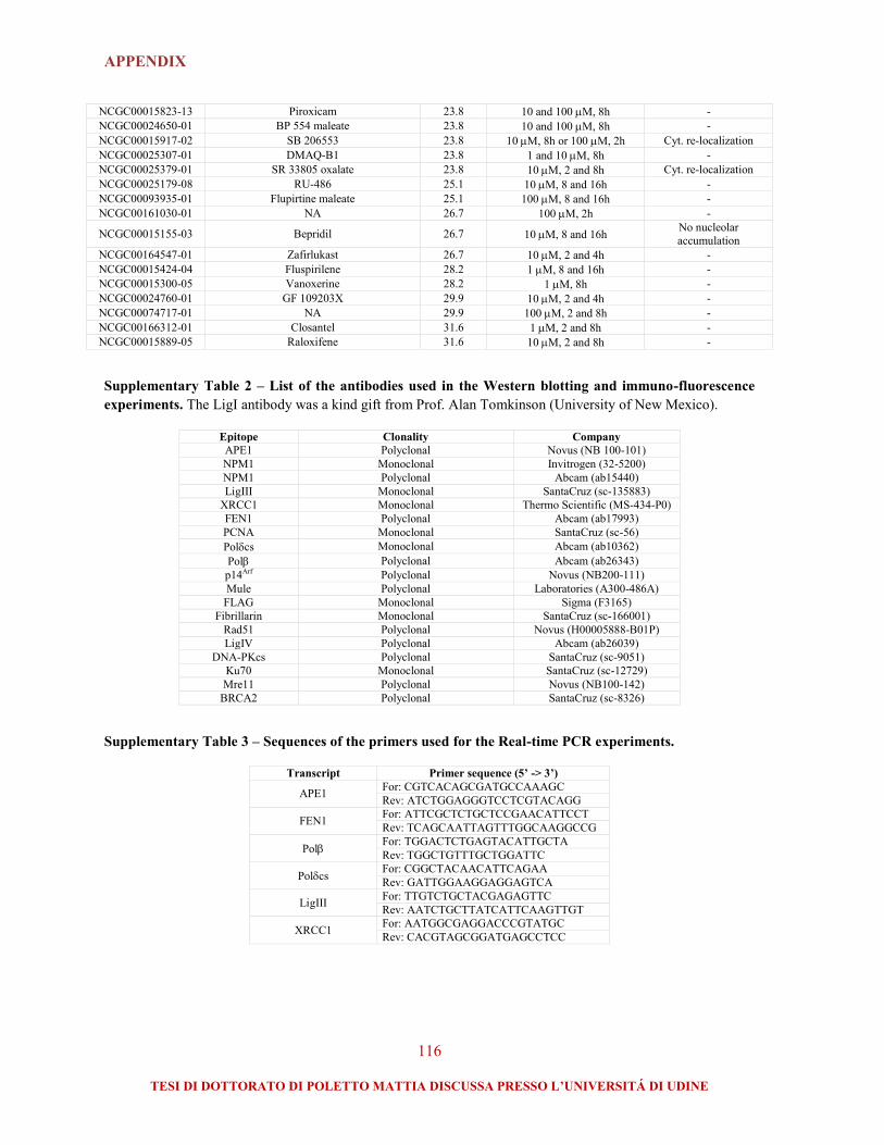
APPENDIX
116
TESI DI DOTTORATO DI POLETTO MATTIA DISCUSSA PRESSO L’UNIVERSITÁ DI UDINE
NCGC00015823-13 Piroxicam 23.8 10 and 100 M, 8h -
NCGC00024650-01 BP 554 maleate 23.8 10 and 100 M, 8h -
NCGC00015917-02 SB 206553 23.8 10 M, 8h or 100M, 2h Cyt. re-localization
NCGC00025307-01 DMAQ-B1 23.8 1 and 10 M, 8h -
NCGC00025379-01 SR 33805 oxalate 23.8 10 M, 2 and 8h Cyt. re-localization
NCGC00025179-08 RU-486 25.1 10 M, 8 and 16h -
NCGC00093935-01 Flupirtine maleate 25.1 100 M, 8 and 16h -
NCGC00161030-01 NA 26.7 100 M, 2h -
NCGC00015155-03 Bepridil 26.7 10 M, 8 and 16h No nucleolar
accumulation
NCGC00164547-01 Zafirlukast 26.7 10 M, 2 and 4h -
NCGC00015424-04 Fluspirilene 28.2 1 M, 8 and 16h -
NCGC00015300-05 Vanoxerine 28.2 1 M, 8h -
NCGC00024760-01 GF 109203X 29.9 10 M, 2 and 4h -
NCGC00074717-01 NA 29.9 100 M, 2 and 8h -
NCGC00166312-01 Closantel 31.6 1 M, 2 and 8h -
NCGC00015889-05 Raloxifene 31.6 10 M, 2 and 8h -
Supplementary Table 2 – List of the antibodies used in the Western blotting and immuno-fluorescence
experiments. The LigI antibody was a kind gift from Prof. Alan Tomkinson (University of New Mexico).
Epitope Clonality Company
APE1 Polyclonal Novus (NB 100-101)
NPM1 Monoclonal Invitrogen (32-5200)
NPM1 Polyclonal Abcam (ab15440)
LigIII Monoclonal SantaCruz (sc-135883)
XRCC1 Monoclonal Thermo Scientific (MS-434-P0)
FEN1 Polyclonal Abcam (ab17993)
PCNA Monoclonal SantaCruz (sc-56)
Polcs Monoclonal Abcam (ab10362)
Pol Polyclonal Abcam (ab26343)
p14Arf Polyclonal Novus (NB200-111)
Mule Polyclonal Laboratories (A300-486A)
FLAG Monoclonal Sigma (F3165)
Fibrillarin Monoclonal SantaCruz (sc-166001)
Rad51 Polyclonal Novus (H00005888-B01P)
LigIV Polyclonal Abcam (ab26039)
DNA-PKcs Polyclonal SantaCruz (sc-9051)
Ku70 Monoclonal SantaCruz (sc-12729)
Mre11 Polyclonal Novus (NB100-142)
BRCA2 Polyclonal SantaCruz (sc-8326)
Supplementary Table 3 – Sequences of the primers used for the Real-time PCR experiments.
Transcript Primer sequence (5’ -> 3’)
APE1 For: CGTCACAGCGATGCCAAAGC
Rev: ATCTGGAGGGTCCTCGTACAGG
FEN1 For: ATTCGCTCTGCTCCGAACATTCCT
Rev: TCAGCAATTAGTTTGGCAAGGCCG
Pol For: TGGACTCTGAGTACATTGCTA
Rev: TGGCTGTTTGCTGGATTC
Polcs For: CGGCTACAACATTCAGAA
Rev: GATTGGAAGGAGGAGTCA
LigIII For: TTGTCTGCTACGAGAGTTC
Rev: AATCTGCTTATCATTCAAGTTGT
XRCC1 For: AATGGCGAGGACCCGTATGC
Rev: CACGTAGCGGATGAGCCTCC

ABBREVIATIONS
117
TESI DI DOTTORATO DI POLETTO MATTIA DISCUSSA PRESSO L’UNIVERSITÁ DI UDINE
ABBREVIATIONS
5’-dRP: 5’-deoxyribonucleotide-phosphate
8-oxo-dG: 7,8-dihydro-8-oxoguanine
AAG: N-alkyladenine DNA glycosylase
AML: acute myeloid leukemia
AP: apurinic/apyrimidinic (site)
APE1: apurinic/apyrimidinic endonuclease 1
APTX: aprataxin
ATM: ataxia telangiectasia mutated
ATR: ataxia telangiectasia and Rad3 related
BER: base excision repair
Compound #3: N-(3-(benzo[d]thiazol-2-yl)-6-
isopropyl-4,5,6,7-tetrahydrothieno[2,3-c]pyridin-2-
yl)acetamide
DDR: DNA damage response
DNA-PKcs: DNA-dependent protein kinase
catalytic subunit
DSB: double-strand break
E3330: (2E)-3-[5-(2,3-dimethoxy-6-methyl 1,4-
benzoquinoyl)] -2-nonyl-2-propenoic acid
EJ: end-joining
ExoIII: exonuclease III
FEN1: flap endonuclease 1
HR: homologous recombination
ICLs: interstrand crosslinks
IL-8: interleukin 8
IR: ionizing radiation
KO: knockout
LigI: DNA ligase I
LigIII: DNA ligase III
LigIV: DNA ligase IV
LP: long-patch
Mdm2: mouse double minute homolog 2
MEFs: mouse embryonic fibroblasts
MMR: mismatch repair
MMS: methyl methanesulfonate
MTS: [3-(4,5-dimethylthiazol-2-yl)-5-(3-
carboxymethoxyphenyl)-2-(4-sulfophenyl)-2H-
tetrazolium, inner salt
Mule: Mcl-1 ubiquitin ligase E3
MX: methoxyamine
nCaRE: negative calcium response elements
NF-B: nuclear factor-B
NIR: nucleotide incision repair
NER: nucleotide excision repair
NHEJ: nonhomologous end joining
NPM1: nucleophosmin
OGG1: 8-oxoguanine DNA glycosylase
p14Arf
: p14 alternative reading frame
PARP: poly(ADP-ribose) polymerase
PCNA: proliferating cell nuclear antigen
PNKP: polynucleotide kinase 3’-phosphatase
Pol I: RNA polymerase I
Pol: DNA polymerase
Polcs: DNA polymerase catalytic subunit
PTMs: post-translational modifications
ROS: reactive oxygen species
rRNA: ribosomal RNA
SMUG1: single-strand-selective monofunctional
uracil-DNA glycosylase 1
SP: short-patch
SSBR: single-strand break repair
SSBs: single-strand breaks
TDP1: tyrosyl-DNA phosphodiesterase 1
TNF-: tumor necrosis factor-
WCE: Whole cell exctract
XRCC1: X-ray repair cross-complementing 1
UNG: uracil-DNA glycosylase
zAPE1: zebrafish APE1

PAPERS PUBLISHED DURING THE Ph.D. COURSE
118
TESI DI DOTTORATO DI POLETTO MATTIA DISCUSSA PRESSO L’UNIVERSITÁ DI UDINE
PAPERS PUBLISHED DURING THE
Ph.D. COURSE
Poletto M., Di Loreto C., Marasco D., Poletto E., Damante G. and Tell G. “Acetylation on
critical lysine residues of apurinic/apyrimidinic endonuclease 1 (APE1) in triple negative breast
cancers” Biochem Biophys Res Commun. – (2012) 424(1):34-9.
Lirussi L., Antoniali G., Vascotto C., D’Ambrosio C., Poletto M., Romanello M., Marasco D.,
Leone M., Quadrifoglio F., Bhakat K., Scaloni A. and Tell G. “Nucleolar accumulation of APE1
depends on charged lysine residues that undergo acetylation upon genotoxic stress and modulate
its BER activity in cells” Mol Biol Cell. – (2012) 23(20):4079-96.
Marasco D., Ruggiero A., Vascotto C., Poletto M., Scognamiglio P. L., Tell G. and Vitagliano L.
“Role of mutual interactions in the chemical and thermal stability of Nucleophosmin NPM1
domains” Biochem Biophys Res Commun. – (2013) 430(2):523-8.
Poletto M., Vascotto C., Scognamiglio P. L., Marasco D. and Tell G. “Role of the unstructured
N-terminal domain of the hAPE1 (human apurinic/apyrimidinic endonuclease 1) in the
modulation of its interaction with nucleic acids and NPM1 (nucleophosmin)” Biochem J. –
(2013) 452(3):545-57.
Vascotto C., Lirussi L., Poletto M., Tiribelli M., Damiani D., Fabbro D., Damante G., Demple
B., Colombo E. and Tell G. “Functional regulation of the apurinic/apyrimidinic endonuclease 1
by nucleophosmin: impact on tumor biology” Oncogene. – (2013) Jul 8. doi:
10.1038/onc.2013.251. [Epub ahead of print].
Londero A. P., Orsaria M., Tell G., Marzinotto S., Capodicasa V., Poletto M., Vascotto C., Sacco
C. and Mariuzzi L. “Expression and prognostic significance of APE1/Ref1 and NPM1 proteins in
high-grade ovarian serous cancer” Am J Clin Pathol. – (2014) 141(3):404-14.
Antoniali G., Lirussi L., Poletto M. and Tell G. “Emerging roles of the nucleolus in regulating
the DNA damage response: the non-canonical DNA repair enzyme APE1/Ref-1 as a
paradigmatical example” Antioxid Redox Signal. – (2014) 20(4):621-39. Review.
Scognamiglio P. L., Di Natale C., Leone M., Poletto M., Vitagliano L., Tell G. and Marasco D.
“G-quadruplex DNA recognition by nucleophosmin: New insights from protein dissection”
(Biochim Biophys Acta, accepted for publication).
The work presented in this thesis is included in:
Poletto M., Dorjsuren D., Malfatti M.C., Scognamiglio P.L., Marasco D., Jadhav A., Maloney
D.J., Wilson 3rd
D.M., Simeonov A. and Tell G. “Inhibitors of the APE1/NPM1 interaction that
display anti-tumor properties” (Mol Carcingenesis, under revision).
Poletto M., Lirussi L., Wilson D. M. 3rd
and Tell G. “Nucleophosmin (NPM1) modulates the
stability and activity of DNA base excision repair proteins and is required for their nucleolar
accumulation” (Mol Biol Cell, under revision).

Biochemical and Biophysical Research Communications 424 (2012) 34–39
Contents lists available at SciVerse ScienceDirect
Biochemical and Biophysical Research Communications
journal homepage: www.elsevier .com/locate /ybbrc
Acetylation on critical lysine residues of Apurinic/apyrimidinic endonuclease 1(APE1) in triple negative breast cancers
Mattia Poletto a, Carla Di Loreto a, Daniela Marasco b,c, Elena Poletto a, Fabio Puglisi a,Giuseppe Damante a, Gianluca Tell a,⇑a Department of Medical and Biological Sciences, University of Udine, 33100 Udine, Italyb Department of Biological Sciences, University of Naples ‘‘Federico II’’, 80134 Naples, Italyc Institute of Biostructures and Bioimaging, National Research Council, 80134 Naples, Italy
a r t i c l e i n f o
Article history:Received 2 June 2012Available online 16 June 2012
Keywords:Apurinic/apyrimidinic endonuclease/redoxeffector factor 1 (APE1/Ref-1)Base excision repair (BER)AcetylationTriple negative breast cancer (TNBC)
0006-291X/$ - see front matter � 2012 Elsevier Inc. Ahttp://dx.doi.org/10.1016/j.bbrc.2012.06.039
Abbreviations: APE1, Apurinic/apyrimidinic endontional modification; nCaRE, negative calcium responserepair; TNBC, triple negative breast cancer; PARP, PMMS, methyl-methanesulphonate.⇑ Corresponding author. Address: Department of Me
University of Udine, P.le Kolbe 4, 33100 Udine, Italy.E-mail address: [email protected] (G. Tell).
a b s t r a c t
Protein acetylation plays many roles within living cells, modulating metabolism, signaling and cellresponse to environmental stimuli, as well as having an impact on pathological conditions, such as cancerpathogenesis and progression. The Apurinic/apyrimidinic endonuclease APE1 is a vital protein that exertsmany functions in mammalian cells, acting as a pivotal enzyme in the base excision repair (BER) pathwayof DNA lesions, as transcriptional modulator and being also involved in RNA metabolism. As an eclecticand abundant protein, APE1 is extensively modulated through post-translational modifications, includingacetylation. Many findings have linked APE1 to cancer development and onset of chemo- and radio-resis-tance. Here, we focus on APE1 acetylation pattern in triple negative breast cancer (TNBC). We describethe validation and characterization of a polyclonal antibody that is specific for the acetylation on lysine35 of the protein. Finally, we use the new antibody to analyze the APE1 acetylation pattern on a cohort ofTNBC specimens, exploiting immunohistochemistry. Our findings reveal a profound deregulation of APE1acetylation status in TNBC, opening new perspectives for future improvements on treatment and progno-sis of this molecular subtype of breast carcinomas.
� 2012 Elsevier Inc. All rights reserved.
1. Introduction
The complexity of the proteome extends far beyond the merenumber of protein-coding genes: several mechanisms that actdownstream transcription and translation greatly expand theplethora of functions that a single protein-coding sequence canachieve. Among these, post-translational modifications (PTMs) ofproteins modulate nearly all processes within living cells. Morethan 200 different PTMs have been reported: from the covalentfunctionalization of proteins with relatively large modifiers (e.g.ubiquitin), to the alteration of target polypeptides with tiny chem-ical moieties (e.g. methylation or acetylation) [1]. Since its discov-ery in histones 40 years ago, lysine acetylation has beeninvestigated as an essential PTM that act modulating histones-DNA interactions and, ultimately, chromatin accessibility. In thelast decades, however, the significance of protein acetylation has
ll rights reserved.
uclease1; PTM, post-transla-elements; BER, base excision
oly(ADP-ribose) Polymerase;
dical and Biological Sciences,Fax: +39 0432 494301.
increasingly become broader, as many groups have pointed outthe importance of this PTM for several non-histone proteins [2].Acetylation, in fact, has profound consequences on protein func-tions and regulates many physiological processes such as cellularmetabolism, migration and signaling, as well as pathological condi-tions, such as cancer occurrence and progression [2,3].
The Apurinic/apyrimidinic endonuclease/redox effector factor-1(APE1/Ref-1, hereafter APE1) is an essential and multifunctionalprotein, firstly discovered as central enzyme in the base excision re-pair (BER) pathway of DNA lesions [4,5]. APE1 acts as the main aba-sic endonuclease within mammalian cells, contributing to themaintenance of genomic stability against alkylative and oxidativeDNA damage. As the acronym suggests, the protein has also beenseparately identified as a transcriptional modulator, able to activatea broad range of transcription factors through a redox-mediatedmechanism. In addition to these ‘‘classical’’ functions, APE1 is in-volved in other cellular processes such as RNA metabolism and tran-scriptional repression through the binding of negative calciumresponse elements (nCaRE) sequences within gene promoters[6,7]. APE1 is an abundant protein (106–107 copies/cell) and is en-dowed with a relatively long half-life (approximately 8 h in HeLacells), therefore, to cope with a rapidly changing environment, cellsmust rely on a fast way to modulate APE1 activities and localization.

M. Poletto et al. / Biochemical and Biophysical Research Communications 424 (2012) 34–39 35
Hence, it is not unexpected that the protein undergoes several PTMsincluding phosphorylation, ubiquitination, proteolysis, S-nitrosa-tion and acetylation [4,8]. Several acetylation sites have been foundwithin the APE1 poorly structured N-terminal region. In particular,acetylation/deacetylation on K6 and K7 has been linked to the mod-ulation of several APE1 activities, including its affinity to the nCaREsequences, the YB-1-mediated MDR1 gene activation and also theBER function of the protein, upon genotoxic insult [9–11]. Recentevidences from our laboratory, moreover, highlight the role of thelysine cluster between K27 and K35 in the modulation of APE1 func-tions. The acetylation status of these residues, in fact, appears to bemodulated by genotoxic stress and likely influences the accessibil-ity of K6 and K7 to lysine acetyl-transferases and deacetylases, sug-gesting the existence of a cross-talk between different acetylationhot-spots of the protein. The modification of lysines within the basicK27 to K35 cluster, has been also linked to a modulation of APE1endonuclease activity, subcellular localization, nucleic acids bind-ing affinity and protein–protein interaction [12] and (Lirussi et al.,unpublished results). Moreover, acetylation of the K27 to K35 clusterinduced by alkylating treatment with methyl-methanesulphonate(MMS), is associated with an increased DNA repair activity ofAPE1 which may account for the reduced sensitivity of some cancercells to DNA damaging agents (Lirussi et al., unpublished results). Asa central enzyme in the BER pathway, APE1 has in fact been linked totumorigenesis and onset of chemo- and radio-resistance in severalmalignancies. In particular, aberrant APE1 localization and expres-sion patterns, have been related to an increased tumor aggressive-ness and recurrence, lymph node positivity and poor prognosis[13–16]. However, there are no data linking the acetylation statusof APE1 and tumor progression.
In this paper, we investigated the status of APE1 acetylation, onthe K27 to K35 cluster, in triple negative breast cancer (TNBC). Wedeveloped and characterized a polyclonal antibody specific foracetylated APE1 that we exploited to analyze, through immunohis-tochemistry, a cohort of TNBC specimens. Our data point to a pro-found deregulation of APE1 acetylation status in specific subtype ofbreast carcinoma and represent a descriptive starting point for fu-ture translational studies devoted to improve treatment and prog-nosis of this disease.
2. Materials and methods
2.1. Peptide synthesis and purification
Different APE1 peptide regions (Table 1) containing K27/K31/K32/K35 were chemically synthesized in the acetylated and non-acetylated form. Solid phase peptide synthesis was performed onan automated synthesizer Syro I (MultiSynTech), following Fmocmethodologies [17]. Peptides were purified by preparative RP-HPLCon a Shimadzu LC-8A, and analyzed through mass spectrometry. LC-ESI-Ion Trap (IT)-MS analysis was carried out on a Surveyor HPLCsystem connected to a LCQ DECA XP IT (Thermo Scientific).
Table 1Sequences of the peptides used in this study.
Peptide name Sequencea
25–38 25KSKTAAKKNDKEAA38
25–3827,31,32,35Ac 25KSK(Ac)TAAK(Ac)K(Ac)NDK(Ac)EAA38
24–39 24KKSKTAAKKNDKEAAG39
24–3927,31,32,35Ac 24KKSK(Ac)TAAK(Ac)K(Ac)NDK(Ac)EAAG39
14–38 14GDELRTEPEAKKSKTAAKKNDKEAA38
14–3827,31,32,35Ac 14GDELRTEPEAKKSK(Ac)TAAK(Ac)K(Ac)NDK(Ac)EAA38
14–3831,32,35Ac 14GDELRTEPEAKKSKTAAK(Ac)K(Ac)NDK(Ac)EAA38
14–3827,31,32Ac 14GDELRTEPEAKKSK(Ac)TAAK(Ac)K(Ac)NDKEAA38
14–3835Ac 14GDELRTEPEAKKSKTAAKKNDK(Ac)EAA38
a Numbers indicate the position of the peptide within APE1 amino acidicsequence. ‘‘Ac’’ denotes acetylated residues. K27/K31/K32/K35 are bolded.
2.2. Generation of polyclonal antibody specific for acetylated APE1
Based on the identified acetylation sites at K27/K31/K32/K35, wesynthesized a peptide bearing the APE1 25–38 sequence and acet-ylated lysine residues, which was conjugated through the N-termi-nal CG dipeptide with keyhole limpet hemocyanin (KLH), i.e. KLH-CG25KSK(Ac)TAAK(Ac)K(Ac)NDK(Ac)EAA38. Polyclonal antibodiesagainst this peptide were raised in rabbits following standardprocedures (PRIMM).
2.3. In vitro acetylation of recombinant APE1
Recombinant full length APE1 mutants were previously de-scribed (Lirussi et al., unpublished results). Both recombinantproteins or APE1 peptides were non-enzymatically acetylatedby incubation with 5 mM acetyl-CoA [18] and (Lirussi et al.,unpublished results), in HAT buffer (50 mM Tris–HCl pH 8.0,0.1 mM EDTA, 5% glycerol, 1 mM DTT) for 2 h at 30 �C. Reactionwas stopped by adding Laemmli sample buffer and subjectedto immunoblotting using the indicated antibodies. The acetyla-tion of proteins and peptides was confirmed through MSanalysis.
2.4. Cell culture, transfection and immuno-precipitation
Cell culture, transfection and immuno-precipitation have beenperformed by using already published methodologies [12].
2.5. Immuno-fluorescence and immunohistochemistry
Immuno-fluorescences with the anti-APE1K27–35Ac (PRIMM)have been carried out following previously described procedures[12]. Briefly, cells were paraformaldehyde-fixed for 20 min at RTand permeabilized for 5 min using a PBS, 0.5% (v/v) Triton X-100solution. After saturation with 10% (v/v) foetal bovine serum(Euroclone) for 30 min, slides were incubated with the polyclonalanti-APE1K27–35Ac antibody (1:100 for 3 h at 37 �C), followed by asecondary anti-rabbit Dyt-Light549 conjugated (Jackson Immuno-Research). Microscope slides were visualized through a TCS SPlaser-scanning confocal microscope (Leica).
Core biopsies of triple negative breast tumors were examinedby immunohistochemistry. All tissues were formalin-fixed for16–24 h. Five micrometers formalin-fixed paraffin-embedded tis-sue sections, mounted on Superfrost slides (Surgipath) wereimmunohistochemically stained by using the peroxidase/DAB PlusDako REAL™ EnVision™ Detection System (Dako A/S). Antigen re-trieval was performed in a water bath at 98 �C with 0.01 M citratebuffer, at pH 6.0 for 40 min. Endogenous peroxidase activity wasblocked by incubation in the Peroxidase Block solution (Dako A/S) for 10 min. Primary mouse monoclonal anti-APE1 antibody[20] diluted 1:200 (optimum working dilution) and primary rabbitpolyclonal anti-APE1K27–35Ac (PRIMM) antiserum diluted 1:100were applied to each slide and incubated, respectively, for60 min at room temperature and overnight at 4 �C. After washing,slides were incubated with the peroxidase/DAB Plus Dako REAL™EnVision™ Detection System (Dako A/S) according to manufac-turer’s guidelines. For reaction visualization, 3-3 diaminobenzidinetetrahydrochloride was used as chromogen. The sections werecounterstained with Mayer haematoxylin. Normal breast tissuesections were used as positive control, whereas negative controlwas performed by replacing the primary antibody with PBS. Con-trols were included in each staining run. Nuclear immunostainingwas quantitatively evaluated by using light microscopy: the entiresection was scanned at high-power magnification (400�) and thepercentage of positive cells was scored.

C B
17
11
Mr(kDa)
- Acetyl-CoA (5 mM)+ + + 25 25-38 (µg)25 10 5
WB: APE1K27-35Ac
Acetyl-CoA (5 mM) - + - +
Ponceau S staining
36
Mr(kDa)
36
- + - + - +
36
Ponceau S Staining
WB: Ac-Lys36
WB: APE1K27-35Ac
WB: APE1K27-35Ac
17
11
Mr(kDa)
A
D
14-38
25-38
24-39
2 µg 4 6 2 4 6
WB: APE1K27-35Ac
- + Mr(kDa)
36
36
MMS (0.5 mM)
WB: FLAG
WB: APE1K27-35Ac
E
32 µm 32 µm 32 µm
No competition 25-3827,31,32,35 Ac 25-38F
Fig. 1. The anti-APE1K27–35Ac antibody is specific for APE1 acetylation at K27/K31/K32/K35. (Panel A) Schematic view of the position of the acetyl-lysine residues in the primarysequence of APE1. Lysines residues targeted by the antibody are indicated by an arrowhead. (Panel B) Western blotting analyses on synthetic APE1 peptides. Left panel: 10 lgof chemically synthesized 25–38 or tetra-acetylated 25-3827,31,32,35Ac were separated onto a discontinuous Tris-tricine SDS–PAGE and then subjected to immunoblotting.Right panel: the indicated amounts of BSA-conjugated 24–39 APE1 peptide, or unconjugated 14–38 and 25–38 APE1 peptides (either in their non-acetylated or tetra-acetylated forms) were slot-blotted on PVDF membrane and subjected to immunoblotting. (Panel C) Western blotting analysis on the in vitro acetylated 25–38 peptide;increasing amounts of the non-acetylated 25–38 peptide were in vitro acetylated as described in Section 2 and separated onto a discontinuous Tris-tricine SDS–PAGE prior toimmunoblotting analysis. All Western blotting analyses were performed using the anti-APE1K27–35Ac antibody. (Panel D) The anti-APE1K27–35Ac antibody recognizes in vitroacetylated full length APE1. 2.5 lg of recombinant APE1 and its lysine to alanine mutants were in vitro acetylated as described in Section 2 and probed with either acommercial anti-Acetyl-lysine antibody (Millipore) or the anti-APE1K27–35Ac antibody. Ponceau-S staining was used as a loading control for normalization. (Panel E) The anti-APE1K27–35Ac antibody detects changes in the APE1 acetylation pattern upon MMS treatment. FLAG-APE1 was immuno-precipitated from HeLa cells treated for 4 h with MMS,as indicated, was probed using the anti-APE1K27–35Ac antibody. Anti-FLAG (Sigma) was used as loading control. (Panel F) Immuno-fluorescence on HeLa cells performed usingeither the anti-APE1K27–35Ac alone, or in presence of a 50-fold molar excess of 25–3827,31,32,35Ac or 25–38 peptides. Images were captured maintaining fixed photomultiplierand laser parameters.
36 M. Poletto et al. / Biochemical and Biophysical Research Communications 424 (2012) 34–39
2.6. Western blotting analyses
Protein and peptides were separated onto 12% sodium dodecylsulphate–polyacrylamide gel electrophoresis (SDS–PAGE) or onto adiscontinuous Tris-tricine SDS–PAGE [19] and transferred to nitro-
cellulose or PVDF membranes (Perkin Elmer). Western blottingwas carried out as already described [20], using 5% (w/v) bovineserum albumin (BSA) as blocking agent and revealed using ECLchemiluminescence procedure (GE Healthcare). Images wereacquired through a ChemiDoc XRS video-densitometer (Bio-Rad).

M. Poletto et al. / Biochemical and Biophysical Research Communications 424 (2012) 34–39 37
2.7. Surface plasmon resonance (SPR)
The BIAcore 3000 SPR system for Real time binding assay andrelated reagents were from GE Healthcare. The polyclonal anti-APE1K27–35Ac antibody was immobilized at a concentration of25 lg/mL in 10 mM acetate buffer pH 5.0 (flow rate 5 lL/min, timeinjection 7 min) on a CM5 Biacore sensor chip, using EDC/NHSchemistry following the manufacturer’s instructions [21]. Residualreactive groups were deactivated by treatment with 1 M ethanol-amine hydrochloride, pH 8.5. Reference channel was prepared byactivating with EDC/NHS and deactivating with ethanolamine.Binding assays were carried out at 20 lL/min, with 4.5 min con-tact-time, APE1 peptides were diluted in running-buffer, HBS(10 mM HEPES, 150 mM NaCl, 3 mM EDTA, pH 7.4) 0.1 mM TCEP.Analyte injections of 90 lL were performed at the indicated con-centrations. The BIAevaluation analysis package (v4.1, GE Health-care) implemented by instrument software was used to subtractthe signal of the reference channel and to evaluate kinetic andthermodynamic parameters of complexes.
A
B
0
10
20
30
20 80 140 200 260 320 380 440 500Time (s)
Res
pons
e(R
U)
0
40
20 80 140 200 260 320 380 440 500Time (s)
Res
pons
e(R
U)
Fig. 2. The anti-APE1K27–35Ac antibody displays a major specificity for the acetylatedlysine 35. (Panel A) Representative overlay sensorgrams relative to a typical SPRexperiment of immobilized anti-APE1K27–35Ac at a 2000RU level with either A theAPE1 14–3827,31,32,35Ac (5–800 nM) peptide or B the APE1 14-3827,31,32Ac (0.1–1000 lM) peptide.
3. Results and discussion
3.1. Specificity of anti-APE1K27–35Ac antibody
To gain new insights into the role of acetylation in the func-tional modulation of APE1, we developed a polyclonal antibodyin rabbit. The antibody (hereafter referred to as anti-APE1K27–
35Ac) was developed to recognize the tetra-acetylated form of theAPE1 N-terminal 27 to 35 lysines stretch (Fig. 1A). The anti-APE1K27–35Ac antibody specificity was tested both by Western blot-ting and slot blotting, using a panel of chemically synthesized APE125–38 or 24–39 peptides (Table 1) that were either acetylated dur-ing synthesis (Fig. 1B) or in vitro acetylated after synthesis (Fig. 1C).Immunoblotting analyses confirmed the ability of the antibody todiscriminate the acetylated peptides from their non-acetylatedcounterparts. The ability of the anti-APE1K27–35Ac antibody to rec-ognise APE1 acetylation within a full-length protein backgroundwas investigated using purified recombinant lysine to alanineAPE1 mutants expressed in Escherichia coli (i.e. APE1WT, the doublemutants APE127,35Ala and APE131,32Ala, the quadruple mutantAPE127,31,32,35Ala and the deletion mutant APE1ND33, that lacks theentire 1 to 33 N-terminal domain) (Lirussi et al., unpublished re-sults). Recombinant proteins were in vitro acetylated using acet-yl-CoA and subjected to Western blotting (Fig. 1D). Analysesrevealed that the anti-APE1K27–35Ac is able to detect APE1 acetyla-tion only in presence of an acetylatable (i.e. not mutated) lysineresidue at position 35 (Fig. 1D – upper panel). The specificity ofthe anti-APE1K27–35Ac antibody was also compared to that of acommercial anti-Acetyl-lysine antibody (Millipore); this antibodyis also able to discriminate between acetylated APE1 forms, thoughwith a different pattern. In particular, all APE1 mutants are de-tected by the Acetyl-lysine antibody when in their acetylated form,in accordance with the broad specificity of the antibody and withthe presence of other acetylatable residues within APE1 (e.g. K6
and K7) (Fig. 1D – lower panel). Altogether these data suggest thatthe anti-APE1K27–35Ac specifically recognizes APE1 acetylationwithin the 27 to 35 lysines stretch, with a particular affinity towardthe acetylated K35 residue. This latter point is further highlightedby the strong signal obtained with the APE1ND33 deletion mutant,which conserves only K35, that is possibly located in a moreaccessible position. To further validate the specificity of theanti-APE1K27–35Ac antibody, we used immuno-precipitated APE1samples obtained from HeLa cells treated with MMS, to increasethe acetylation status of the K27 to K35 cluster (Lirussi et al., unpub-lished results). Fig. 1E shows that the anti-APE1K27–35Ac efficiently
detects changes in the acetylation pattern of the protein. We finallyassessed the staining pattern of the antibody using immuno-fluo-rescence on HeLa cells: the selectivity of the staining was verifiedthrough competition studies using the acetylated 25–38 peptide(Fig. 1F).
Surface Plasmon resonance (SPR) biosensors are ideally suitedfor antibody characterization and quantitative immunoassaydetection. Here, we exploited SPR to measure the affinity of theantibody for the acetyl-lysine residues within APE1 amino acids27 to 35 (Fig. 2). To characterize the kinetic values for the anti-APE1K27–35Ac we employed different APE1 peptides as ligands(Table 1). SPR data confirmed what observed through Westernblotting, showing that acetylated K35 is the preferential target sitefor immunorecognition, with a measured KD in the sub-micromo-lar range (Table 2). Notably, the lack of acetylation on K35 only, re-sults in a dramatic reduction in the antibody affinity for thecognate peptide (Fig. 2B, Table 2 and data not shown). Therefore,these analyses lead us to conclude that the developed antibodyhas major specificity toward the acetylated K35 residue of APE1.
3.2. Analysis of APE1 acetylation in triple negative breast Cancer
The APE1K27–35Ac antibody was used to study APE1 acetylationin a cohort of triple negative breast cancer (TNBC) specimens. Thistype of breast cancer comprises tumors that lack expression ofestrogen receptor (ER), progesterone receptor (PR) and overexpres-sion/gene amplification of epidermal growth factor receptor type 2(HER2) [22]. TNBC account for approximately 15% of all invasivebreast cancers and often show aggressive phenotypical character-istics, such as a high histologic grade [23,24]. To date, no specificmolecular targets have been identified in TNBC and, accordingly,no specific targeted therapies have been developed [25]. Therefore,the prognosis of patients with TNBC is usually poorer than that of

Table 2Kinetic and thermodynamic parameters of anti-APE1K27–35Ac toward peptidesdescribed in Table 1.
Peptide Ka � 104 (Ms) Kd � 10�3 (1/s) KD (M)
14–38 NDa ND �10�3
14–3835Ac 2.39 6.52 270 ± 8 � 10�9
14–3831,32,35Ac 4.70 7.71 164 ± 3 � 10�9
14–3827,31,32Ac ND ND �10�3
14–3827,31,32,35Ac 3.62 5.10 141 ± 3 � 10�9
a ND: Not Determined.
38 M. Poletto et al. / Biochemical and Biophysical Research Communications 424 (2012) 34–39
patients with other subtypes of breast cancer [26,27]. Various find-ings, however, indicate that a substantial diversity in biology, prog-nosis and treatment sensitivity can be observed among tumordefined as TNBC [28,29]. A cohort of 103 TNBC was investigatedfor levels of total APE1 and acetylated APE1 by using immunohis-tochemistry. Fig. 3 shows representative immunostainings andquantitation of positivity: overexpression of total APE1 wasdetected in TNBC compared to normal breast tissue, with 90% oftumor specimens showing an APE1 nuclear positivity higher than80% (Fig. 3B). This result is in accordance with previous works thatreport a correlation between APE1 overexpression and tumoraggressiveness in a variety of cancers [4,6]. In contrast, immuno-staining of acetylated APE1 was very heterogeneous among TNBCspecimens: samples showing either low or high nuclear positivitywere observed (T1 and T2, respectively, in Fig. 3A). The heteroge-neity of APE1K27–35Ac immunostaining among TNBC is quantita-
APE1
APE1K27-35Ac
N A
B
APE1
Class ofpositivity (%):
Perc
enta
geof
posi
tive
cells
80
60
40
20
Fig. 3. Immunostaining of TNBC specimens for total APE1 and acetylated APE1. (Panel A)antibody against total (APE1) and acetylated (APE1K27–35Ac) APE1. Positivity is indicatedAPE1 and acetylated APE1. In x and y axes are indicated different classes of positivity animmunostaining is indicated below the respecting bars.
tively captured in Fig. 3B. These data suggest that in TNBC,concomitantly with total APE1 overexpression, a specific deregula-tion of the acetylation status of this protein is occurring. APE1deregulation in breast carcinoma (present data and [30]) may belinked to an impairment in its redox activity, being instrumentalin altering estrogen-responsive gene expression in breast cancercell lines [31].
Member of the Poly(ADP-ribose) Polymerase (PARP) family arecentral to specific DNA damage repair pathways, particularly theBER. PARP inhibition has been shown to enhance the cytotoxicityof agents that generate single-strand breaks in DNA, such as radi-ation and certain chemotherapy drugs (i.e. alkylating agents)[32]. In addition, PARP inhibitors may induce cell death through‘‘synthetic lethality’’, if the DNA repair mechanisms that rescueBER-deficient cells are themselves impaired, as in BRCA-deficientbreast cancers [32,33]. Because of the phenotypic similarities be-tween some TNBC and most BRCA1-associated breast cancers,some have hypothesized that TNBC might also be sensitive to PARPinhibition [32]. Interestingly, it has been recently demonstratedthat BRCA1 may be involved in regulation of the expression levelsof DNA repair genes such as APE1, OGG1 and NTH1 [34]. Thus,APE1 represents an ideal candidate target for developing new ther-apeutic strategies for breast cancer treatment. In particular, dereg-ulation of APE1 observed in breast cancer specimens might beexploited in order to induce synthetic lethality in tumors with de-fects in other DNA repair pathway, as recently pointed out [33].Our observations, therefore, represent a descriptive starting pointfor future investigations; it would be interesting to understand
T1 T2
APE1K27-35Ac
Representative cases of normal breast tissue (N) and TNBC (T1 and T2) stained withby the brown colour. (Panel B) Quantitative analysis of nuclear positivity for totald percentage of TNBC present within each class, respectively. APE1 or APE1K27–35Ac

M. Poletto et al. / Biochemical and Biophysical Research Communications 424 (2012) 34–39 39
whether the deregulation of APE1 acetylation status is paralleledby an impairment of its functions, in order to design novel thera-peutic approaches to breast carcinoma.
Conflict of Interest
Authors declare that they have no conflict of interest pending.
Acknowledgments
The authors thank Dr. Lisa Lirussi for technical help in someexperiments. This work was supported by the Associazione Italianaper la Ricerca sul Cancro (IG10269) and Ministero dell’Istruzione,dell’Università e della Ricerca (FIRB_RBRN07BMCT and PRIN2008_CCPKRP_003 to G.T.).
References
[1] C.T. Walsh, S. Garneau-Tsodikova, G.J. Gatto, Protein posttranslationalmodifications: the chemistry of proteome diversifications, Angew. Chem. Int.Ed. Engl. 44 (2005) 7342–7372.
[2] X.J. Yang, E. Seto, Lysine acetylation: codified crosstalk with otherposttranslational modifications, Mol. Cell 31 (2008) 449–461.
[3] P. Close, C. Creppe, M. Gillard, et al., The emerging role of lysine acetylation ofnon-nuclear proteins, Cell. Mol. Life Sci. 67 (2010) 1255–1264.
[4] G. Tell, G. Damante, D. Caldwell, et al., The intracellular localization of APE1/Ref-1: more than a passive phenomenon?, Antioxid Redox Signal. 7 (2005)367–384.
[5] C. Vascotto, L. Cesaratto, L.A. Zeef, et al., Genome-wide analysis and proteomicstudies reveal APE1/Ref-1 multifunctional role in mammalian cells, Proteomics9 (2009) 1058–1074.
[6] G. Tell, D. Fantini, F. Quadrifoglio, Understanding different functions ofmammalian AP endonuclease (APE1) as a promising tool for cancertreatment, Cell. Mol. Life Sci. 67 (2010) 3589–3608.
[7] G. Tell, D.M. Wilson 3rd, C.H. Lee, Intrusion of a DNA repair protein in theRNome world: is this the beginning of a new era?, Mol Cell. Biol. 30 (2010)366–371.
[8] C.S. Busso, M.W. Lake, T. Izumi, Posttranslational modification of mammalianAP endonuclease (APE1), Cell. Mol. Life Sci. 67 (2010) 3609–3620.
[9] K.K. Bhakat, T. Izumi, S.H. Yang, et al., Role of acetylated human AP-endonuclease (APE1/Ref-1) in regulation of the parathyroid hormone gene,EMBO J. 22 (2003) 6299–6309.
[10] T. Yamamori, J. DeRicco, A. Naqvi, et al., SIRT1 deacetylates APE1 and regulatescellular base excision repair, Nucleic Acids Res. 38 (2010) 832–845.
[11] S. Sengupta, A.K. Mantha, S. Mitra, et al., Human AP endonuclease (APE1/Ref-1)and its acetylation regulate YB-1-p300 recruitment and RNA polymerase IIloading in the drug-induced activation of multidrug resistance gene MDR1,Oncogene 30 (2011) 482–493.
[12] D. Fantini, C. Vascotto, D. Marasco, et al., Critical lysine residues within theoverlooked N-terminal domain of human APE1 regulate its biologicalfunctions, Nucleic Acids Res. 38 (2010) 8239–8256.
[13] S. Kakolyris, L. Kaklamanis, K. Engels, et al., Human AP endonuclease I (HAPI)protein expression inbreast cancer correlates with lymph node status andangiogenesis, Br. J. Cancer 77 (1998) 1169–1173.
[14] K.A. Robertson, H.A. Bullock, Y. Xu, et al., Altered expression of Ape1/ref-1 ingerm cell tumors and overexpression in NT2 cells confers resistance tobleomycin and radiation, Cancer Res. 61 (2001) 2220–2225.
[15] V. Di Maso, C. Avellini, L.S. Crocè, Subcellular localization of APE1/Ref-1 inhuman hepatocellular carcinoma: possible prognostic significance, Mol. Med.13 (2007) 89–96.
[16] Q. Sheng, Y. Zhang, R. Wang, et al., Prognostic significance of APE1 cytoplasmiclocalization in human epithelial ovarian cancer, Med. Oncol. 29 (2012) 1265–1271.
[17] G.B. Fields, R.L. Noble, Solid phase peptide synthesis utilizing 9-fluorenylmethoxycarbonyl amino acids, Int. J. Pept. Protein Res. 35 (1990)161–214.
[18] G.J. Garbutt, E.C. Abraham, Non-enzymatic acetylation of human hemoglobins,Biochim. Biophys. Acta 670 (1981) 190–194.
[19] H. Schägger, Tricine–SDS–PAGE, Nat. Protoc. 1 (2006) 16–22.[20] C. Vascotto, D. Fantini, M. Romanello, et al., APE1/Ref-1 interacts with NPM1
within nucleoli and plays a role in the rRNA quality control process, Mol. Cell.Biol. 29 (2009) 1834–1854.
[21] B. Johnsson, S. Lofas, G. Lindquist, Immobilization of proteins to acarboxymethyldextran-modified gold surface for biospecific interactionanalysis in surface plasmon resonance sensors, Anal. Biochem. 198 (1991)268–277.
[22] W.D. Foulkes, I.E. Smith, J.S. Reis-Filho, Triple-negative breast cancer, N. Engl. J.Med. 363 (2010) 1938–1948.
[23] E.A. Rakha, J.S. Reis-Filho, I.O. Ellis, Basal-like breast cancer: a critical review, J.Clin. Oncol. 26 (2008) 2568–2581.
[24] J.S. Reis-Filho, A.N. Tutt, Triple negative tumours: a critical review,Histopathology 52 (2008) 108–118.
[25] C. Andreetta, A.M. Minisini, M. Miscoria, et al., First-line chemotherapy with orwithout biologic agents for metastatic breast cancer, Crit. Rev. Oncol. Hematol.76 (2010) 99–111.
[26] C. Liedtke, C. Mazouni, K.R. Hess, et al., Response to neoadjuvant therapy andlong-term survival in patients with triple-negative breast cancer, J. Clin. Oncol.26 (2008) 1275–1281.
[27] R. Dent, M. Trudeau, K.I. Pritchard, et al., Triple-negative breast cancer: clinicalfeatures and patterns of recurrence, Clin. Cancer Res. 13 (2007) 4429–4434.
[28] C. Oakman, E. Moretti, G. Pacini, et al., Triple negative breast cancer: aheterogeneous subgroup defined by what it is not, Eur. J. Cancer 47 (Suppl 3)(2011) S370–S372.
[29] C.X. Ma, J. Luo, M.J. Ellis, Molecular profiling of triple negative breast cancer,Breast Dis. 32 (2010) 73–84.
[30] F. Puglisi, F. Barbone, G. Tell, et al., Prognostic role of Ape/Ref-1 subcellularexpression in stage I-III breast carcinomas, Oncol. Rep. 9 (2002) 11–17.
[31] C.D. Curtis, D.L. Thorngren, Y.S. Ziegler, et al., Apurinic/apyrimidinicendonuclease 1 alters estrogen receptor activity and estrogen-responsivegene expression, Mol. Endocrinol. 23 (2009) 1346–1359.
[32] M.R. Kelley, DNA Repair In Cancer Therapy, first ed., Molecular Targets andClinical Applications, Academic Press, 2012.
[33] R. Sultana, D.R. McNeill, R. Abbotts, et al., Synthetic lethal targeting of DNAdouble-strand break repair deficient cells by human apurinic/apyrimidinicendonuclease inhibitors, Int. J. Cancer (2012), http://dx.doi.org/10.1002/ijc.27512.
[34] T. Saha, M. Smulson, E.M. Rosen, BRCA1 regulation of base excision repairpathway, Cell Cycle 9 (2010) 2471–2472.

Volume 23 October 15, 2012 4079
MBoC | ARTICLE
Nucleolar accumulation of APE1 depends on charged lysine residues that undergo acetylation upon genotoxic stress and modulate its BER activity in cellsLisa Lirussia,*, Giulia Antonialia,*, Carlo Vascottoa, Chiara D’Ambrosiob, Mattia Polettoa, Milena Romanelloa, Daniela Marascoc,d, Marilisa Leoned, Franco Quadrifoglioa, Kishor K. Bhakate, Andrea Scalonib, and Gianluca TellaaDepartment of Medical and Biological Sciences, University of Udine, 33100 Udine, Italy; bProteomics and Mass Spectrometry Laboratory, Istituto di Ricerche per il Sistema Produzione Animale in Ambiente Mediterraneo, National Research Council, 80147 Naples, Italy; cDepartment of Biological Sciences, University of Naples “Federico II,” 80134 Naples, Italy; dInstitute of Biostructures and Bioimaging, National Research Council, 80134 Naples, Italy; eDepartment of Biochemistry and Molecular Biology, University of Texas Medical Branch, Galveston, TX 77555
ABSTRACT Apurinic/apyrimidinic endonuclease 1 (APE1) is the main abasic endonuclease in the base excision repair (BER) pathway of DNA lesions caused by oxidation/alkylation in mammalian cells; within nucleoli it interacts with nucleophosmin and rRNA through N-termi-nal Lys residues, some of which (K27/K31/K32/K35) may undergo acetylation in vivo. Here we study the functional role of these modifications during genotoxic damage and their in vivo relevance. We demonstrate that cells expressing a specific K-to-A multiple mutant are APE1 nucleolar deficient and are more resistant to genotoxic treatment than those expressing the wild type, although they show impaired proliferation. Of interest, we find that genotoxic treatment induces acetylation at these K residues. We also find that the charged status of K27/K31/K32/K35 modulates acetylation at K6/K7 residues that are known to be involved in the coordination of BER activity through a mechanism regulated by the sirtuin 1 deacetylase. Of note, structural studies show that acetylation at K27/K31/K32/K35 may account for local confor-mational changes on APE1 protein structure. These results highlight the emerging role of acetylation of critical Lys residues in regulating APE1 functions. They also suggest the exis-tence of cross-talk between different Lys residues of APE1 occurring upon genotoxic dam-age, which may modulate APE1 subnuclear distribution and enzymatic activity in vivo.
INTRODUCTIONApurinic/apyrimidinic endonuclease 1/redox effector factor-1 (APE1) plays a central role in the maintenance of genome stability and re-dox signaling (Bapat et al., 2009, 2010; Tell et al., 2010a; Wilson and Simeonov, 2010). Through its C-terminal domain (residues 61–318), it acts as an essential enzyme in the base excision repair (BER) path-way of DNA damages caused by both endogenous and exogenous oxidizing/alkylating agents, including many chemotherapeutic drugs. In combination with thioredoxin (Ueno et al., 1999; Seemann and Hainaut, 2005) and through its N-terminal domain (residues 1–127), it also functions as a regulatory redox agent to maintain cancer-related transcription factors (Egr-1, NF-κB, p53, HIF-1α, AP-1, and Pax proteins) in an active reduced state (Hirota et al., 1997;
Monitoring EditorKarsten WeisUniversity of California, Berkeley
Received: Apr 17, 2012Revised: Jul 25, 2012Accepted: Aug 16, 2012
This article was published online ahead of print in MBoC in Press (http://www .molbiolcell.org/cgi/doi/10.1091/mbc.E12-04-0299) on August 23, 2012.*These authors contributed equally to this workThe authors declare that they have no conflict of interests.Address correspondence to: Gianluca Tell ([email protected]).
© 2012 Lirussi et al. This article is distributed by The American Society for Cell Biology under license from the author(s). Two months after publication it is avail-able to the public under an Attribution–Noncommercial–Share Alike 3.0 Unported Creative Commons License (http://creativecommons.org/licenses/by-nc-sa/3.0).“ASCB®,” “The American Society for Cell Biology®,” and “Molecular Biology of the Cell®” are registered trademarks of The American Society of Cell Biology.
Abbreviations used: APE1/Ref-1, apurinic/apyrimidinic endonuclease/redox effector factor 1; BER, base excision repair; MMS, methyl methanesulfonate; MTS, 3-(4-5-dimethylthiazol-2-yl)-5-(3-carboxymethoxyphenyl)-2-(4-sulfophenyl)-2H-tet-razolium salt; NPM1, nucleophosmin 1; SIRT1, sirtuin 1; TBHP, tert-butyl hydroper-oxide.
http://www.molbiolcell.org/content/suppl/2012/08/20/mbc.E12-04-0299.DC1.htmlSupplemental Material can be found at:

4080 | L. Lirussi et al. Molecular Biology of the Cell
mutants are reintroduced into APE1-silenced cell clones to deter-mine the role of these crucial amino acids during cell response to genotoxic treatment.
RESULTSPositively charged K27/K31/K32/K35 residues are essential for APE1 nucleolar accumulation through stabilization of protein interaction with NPM1 and rRNAWe previously demonstrated that charged K residues, located within the unstructured APE1 N-terminal domain (i.e., K24/K25/K27/K31/K32), are crucial for APE1 interaction with rRNA and NPM1 and for modu-lating its catalytic activity on abasic DNA through regulation of prod-uct binding. Of interest, some of these critical amino acids (i.e., K27/K31/K32 in addition to K35) may undergo in vivo acetylation under basal conditions (Fantini et al., 2010). We hypothesized that the de-gree of positive charges, modified by acetylation at these residues, may modulate APE1’s different functions through its redirection to different substrates and/or stimulation of its DNA-repair enzymatic activity (Fantini et al., 2010). To address this issue, we inspected the role of the positively charged residues within the region 27–35 in modulating the interaction between APE1 and NPM1. Colocaliza-tion experiments in HeLa cells transiently transfected with either FLAG-tagged, wild-type APE1 (APE1WT), a K-to-A mutant (APE1K4pleA) in which the positive charges have been removed as in the case of constitutive acetylation, or a nonacetylatable K-to-R APE1 mutant (APE1K4pleR) showed that APE1K4pleA mutant has a marked exclusion from the nucleoli apparent in all expressing cells (Figure 1A). Silenc-ing of the endogenous APE1 protein did not alter either the ability of the APE1WT and APE1K4pleR proteins to accumulate within the nu-cleolar compartment or the inability of the APE1K4pleA to accumulate within the nucleolus (unpublished data). These data thus demon-strate that APE1WT and APE1K4pleR nucleolar accumulation is not the consequence of their overexpression.
To complement these observations and also to exclude a possi-ble contribution of the FLAG tag used to generate recombinant ec-topic proteins, we exploited a live-cell imaging system as suggested by Schnell et al. (2012). APE1 cDNA was cloned into a pDendra2 vector to express APE1 in fusion with the green photoconvertible fluorophore Dendra (Chudakov et al., 2007). As reported in Figure 1B, the fluorophore alone is present in both the cytoplasm and the nuclear compartment, but it is completely excluded from the nucle-oli, as demonstrated by quantitative fluorescence signal analyses. In contrast, whereas the expression of Dendra in fusion with APE1WT and APE1K4pleR results in an efficient accumulation of the protein within the nucleolar compartment, the Dendra-APE1K4pleA mutant displays a homogeneous nuclear distribution. This approach con-firms immunofluorescence-based data on the reduced accumula-tion of FLAG-tagged APE1K4pleA within the nucleoli and supports the evidence that the nucleolar accumulation of APE1 protein does not depend on the protein abundance nor does it depend on the spe-cific tag used to generate the ectopic recombinant proteins (see also Supplemental Figure S1).
We supported our immunofluorescence data with biochemical interaction experiments. Coimmunoprecipitation analysis showed that the APE1K4pleA mutant has a significantly reduced interaction with NPM1 (Figure 2A), in accordance with its impaired nucleolar accumulation. The altered electrophoretic mobility observed for the APE1K4pleA mutant (Figure 2A) was further confirmed by SDS–PAGE analysis of recombinant proteins expressed in Escherichia coli and purified by chromatography. The effect is likely due to alteration of its overall charge, since electrospray ionization mass spectrometry analysis confirmed the correctness of protein mass values, and the
Wei et al., 2000; Ziel et al., 2004; Gray et al., 2005; Pines et al., 2005; Tell et al., 2005, 2010a). APE1 can also act as a transcriptional repres-sor through indirect binding to negative Ca2+-response elements (nCaRE), which are regulated by K6/K7 acetylation (Bhakat et al., 2003). Recently APE1 was demonstrated to bind/cleave abasic RNA (Vascotto et al., 2009b; Fantini et al., 2010; Tell et al., 2010b) and to control c-Myc expression by cleaving its mRNA (Barnes et al., 2009). These discoveries pointed to a new function of APE1 in regulating gene expression through posttranscriptional mechanisms and brought to light the fact that this protein is a possible target for anti-cancer therapy.
In this context, we showed that the first 35 amino acids in the nonstructured N-terminal domain of APE1 are required for a stable interaction with rRNA, nucleophosmin 1 (NPM1), and other proteins involved in ribosome biogenesis/RNA processing (Vascotto et al., 2009b; Tell et al., 2010b). In particular, K residues within the protein region spanning amino acids 24–35 are involved in the interaction of APE1 with both rRNA and NPM1 and also regulate its in vitro enzy-matic activity (Fantini et al., 2010). Of interest, some of these critical K residues, namely K27/K31/K32/K35, undergo in vivo acetylation. These results suggest that protein–protein interactions and/or post-translational modifications (PTMs) involving the APE1 N-terminal domain may play important roles in vivo in coordinating and fine-tuning the protein’s BER activity and functions on rRNA metabolism. Recently it was also demonstrated that APE1 K6/K7 may undergo acetylation during cell response to genotoxic treatment (Fantini et al., 2008) and that the acetylation status of these K residues, con-trolled by the sirtuin 1 (SIRT1) deacetylase activity, should be impor-tant in modulating protein DNA-repair function by regulating the kinetics of its interaction with other enzymes involved in BER, for example, XRCC1 (Yamamori et al., 2010).
APE1 is mainly a nuclear protein and is critical for controlling cel-lular proliferative rates (Fung and Demple, 2005; Izumi et al., 2005; Vascotto et al., 2009b). We also showed that a considerable amount of APE1 is accumulated within the nucleoli of different cell lines (Vascotto et al., 2009b; Fantini et al., 2010), but its role within this compartment is unknown. Cytoplasmic, mitochondrial, and endo-plasmic reticulum localizations have also been ascertained (Tell et al., 2001; Szczesny and Mitra, 2005; Chattopadhyay et al., 2006; Grillo et al., 2006; Mitra et al., 2007). APE1 is an abundant and rela-tively stable protein in mammalian cells (Tell et al., 2009, 2010a). Fine-tuning of the multiple APE1 functions may therefore depend on the modulation of its PTMs and, eventually, on its interactome. Although a functional role has been determined for some PTMs (K6/K7 acetylation and K24/K25/K27 ubiquitination; Bhakat et al., 2003; Fantini et al., 2008; Busso et al., 2009), the identity and importance of various interacting partners in modulating APE1 biological func-tions are still under investigation (Parlanti et al., 2007; Busso et al., 2009; Vascotto et al., 2009b). APE1 may affect cell growth by di-rectly acting on rRNA quality control mechanisms; in particular, APE1 interaction with NPM1 may affect its activity over rRNA mole-cules. However, many aspects of this new function are undefined (Vascotto et al., 2009b; Tell et al., 2010b).
In this study, we address the biological role of APE1 acetylation at K27/K31/K32/K35 and, in light of recent evidence showing the emerg-ing function of the nucleolus as a central sensor of protein trafficking during DNA repair after genotoxic treatment (Nalabothula et al., 2010), the role of the nucleolus itself on the APE1 protective function toward genotoxic damage. We used a reconstitution strategy with APE1 mutants in which the charged Lys residues were replaced by either Ala or Arg to mimic constitutive acetylated (APE1K4pleA) and nonacetylatable (APE1K4pleR) protein forms, respectively. These

Volume 23 October 15, 2012 Functional relevance of APE1 nucleolar accumulation | 4081
difference in their apparent mobility ob-served in SDS–PAGE was abolished when separating various mutants in urea-contain-ing denaturing gels (Supplemental Figure S2 and unpublished data). It was also ob-served for other K-to-A mutants of APE1 (Fantini et al., 2010). We then evaluated the contribution of the single K residues to the extent of APE1 nucleolar localization (Supplemental Figure S3) and the ability to interact with NPM1 (Figure 2B). Double mu-tants APE1K27/35A, APE1K31/32A, APE1K27/35R, and APE1K31/32R and a deletion mutant lack-ing 33 amino acids at the protein N-terminus (APE1NΔ33) were compared with APE1WT. Whereas APE1WT, APE1K27/35R, and APE1K31/32R displayed a nucleolar and nucleoplasmic staining, APE1K27/35A and APE1K31/32A showed two alternative stain-ings, exhibiting nucleolar/nucleoplasmic or only nucleoplasmic positivity, respectively (Supplemental Figure S3). Coimmunopre-cipitation experiments (Figure 2B) were in accordance with immunofluorescence anal-yses and showed a substantial reduction of the interaction in the case of APE1K4pleA and APE1NΔ33 mutants and a moderate impair-ment for APE1K27/35A and APE1K31/32A mu-tants. Glutathione S-transferase (GST) pull-down assays with recombinant purified proteins confirmed that these results were due to effects on direct interaction between APE1 and NPM1 (Figure 2C). This confirmed previous hypotheses (Fantini et al., 2010). We also checked the effect of the K-to-A mutation on the ability of APE1 to bind nu-cleolar rRNA (Vascotto et al., 2009b). As ex-pected, rRNA–chromatin immunoprecipita-tion (ChIP) analyses showed that K-to-A mutations significantly alter APE1 binding to
FIGURE 1: Positively charged K27/K31/K32/K35 residues are required for APE1 nucleolar accumulation. (A) Confocal microscopy of HeLa cells transfected with APE1WT, APE1K4pleA, or APE1K4pleR FLAG-tagged proteins and stained with antibodies against NPM1 (red) and ectopic FLAG-APE1 (green). Overlap of staining (yellow) demonstrated colocalization of the two proteins. Of note, the APE1K4pleA mutant was completely excluded from nucleoli (white arrowheads), whereas both APE1WT and the APE1K4pleR were accumulated within. Images are representative of 100% of transfected cells. (B) HeLa cells transfected with pDendra2-N empty
A
B Dendra APE1WT
APE1K4pleRAPE1K4pleA
Flu
ore
scen
ce in
ten
sity
Line lenght
m5 10 15 20
50
100
150
200
25 m
Flu
ore
scen
ce in
ten
sity
Line lenght
m5 10 15 20
50
100
150
200
25 m
Flu
ore
scen
ce in
ten
sity
Line lenght
m5 10 15 20
50
100
150
200
25 m
Flu
ore
scen
ce in
ten
sity
Line lenght
m5 10 15 20
50
100
150
200
25 m
NucleolusNucleolus
Nucleolus
APE1WT
NPM1 Merged
APE1K4pleR
APE1K4pleA
8 m 8 m
4 m
4 m
4 m 4 m
4 m 4 m
8 m
FLAG
µ µ µ
µµµ
µ µ µ
µµ
µ µ
µ
µ µ
µ
vector or encoding APE1 (wild-type and mutants) in fusion with Dendra fluorophore were analyzed with a live confocal microscopy workstation. Cells transfected with empty vector (Dendra) showed diffuse green fluorescence within cytoplasm and nucleus, with exclusion of the nucleolar compartment. APE1WT and APE1K4pleR mutant mainly localized within the nuclear compartment and accumulated within nucleoli. In contrast, APE1K4pleA did not show any nucleolar accumulation. Images were captured by using the same settings (488-nm laser at 10% of intensity and PMT at 760 V). Fluorescence intensity analysis was carried out on a 25-μm-long line (white). Graphs represent the fluorescence intensity measured through a cross section of the nucleus and demonstrate an incremental fluorescence in corresponding nucleoli only in the case of APE1WT and APE1K4pleR-Dendra fusion protein–expressing cells.

4082 | L. Lirussi et al. Molecular Biology of the Cell
FIGURE 2: Positively charged K27/K31/K32/K35 residues are required for NPM1 interaction. (A) The APE1K4pleA mutant shows impaired interaction with NPM1. Coimmunoprecipitation (CoIP) analysis on HeLa cells transfected with APE1WT, APE1K4pleA, or APE1K4pleR FLAG-tagged proteins with endogenous NPM1. Western blot analysis was used to quantify the interaction among the different APE1 forms and NPM1 by using specific antibodies. IB, immunoblot. (B) K27/K31/K32/K35 residues are responsible for proper interaction of APE1 with NPM1. CoIP analysis on HeLa cells transfected with FLAG-tagged APE1WT, APE1NΔ33, or the APE1 double (APE1K27/35A, APE1K31/32A, APE1K27/35R, and APE1K31/32R) or quadruple (APE1K4pleA and APE1K4pleR) K-to-A or K-to-R mutants. Western blot analysis was performed on total cell extracts (left) and on immunoprecipitated material (right) with specific antibody for endogenous NPM1. Normalized coimmunoprecipitated amounts of NPM1 are indicated under each relative bar. Anti-FLAG staining was used as loading control. (C) Interaction of K-to-A APE1 mutants with NPM1 and rRNA. A 150-pmol amount of each bait GST-APE1 recombinant purified protein (rAPE1) was incubated with an equimolar amount of recombinant NPM1, as described in Materials and Methods. After GST pull-down, samples underwent Western blot analysis by using the indicated antibodies (right). NPM1 band intensities were normalized vs. those of GST-APE1 proteins, and the resulting values, expressed as percentage of bound with respect to APE1WT, were plotted in the histogram (left). Each bar represents the mean of two independent experiments whose variation was <10%. (D) Interaction of K-to-A APE1 mutants with NPM1 and rRNA. After transfection of HeLa cells, total cell lysates were prepared as described in Materials and Methods, and an RNA-ChIP assay was performed to measure the rRNA-binding activity of the different APE1 mutants. Immunoprecipitated samples underwent either Western blot analysis (right) or RNA extraction and quantification by reverse transcription and quantitative PCR analysis, using 18S and 47S rRNA–specific primers. Data are presented as a fold percentage of the amount of total 18S or 47S rRNA with respect to the amount of immunoprecipitated FLAG-tagged APE1 protein, respectively. The resulting values are plotted in the histograms (left), showing the average values with SD of three independent experiments. (E) Nucleoplasmic interaction between APE1 and NPM1 is affected by K-to-A mutation on APE1. PLA technology was used to evaluate in vivo the effect of positively charged K27/K31/K32/K35 residues on APE1 interaction with NPM1 in the nuclear compartment. Stable HeLa clones expressing APE1WT, APE1K4pleA, or APE1K4pleR FLAG-tagged proteins were seeded on a glass coverslip, and PLA reaction was carried out using anti-FLAG and anti-NPM1 antibodies. 4′,6-Diamidino-2-phenylindole staining is used as a reference for the nuclei. The histogram represents the average number of PLA blobs scored for at least 30 cells per slide. Asterisks represent a significant difference between APE1WT and mutants.
A
B
C
D
GST-Pulldown
Mr(kDa)
input
36
36
IB: NPM1
IB: FLAG
36
36
Mr(kDa)
IP α-FLAG
1.00 - 0.87 0.60 0.45 0.68 0.14 0.69NPM1 Co-IP amount(relative units)
36
IB: FLAG
IB: NPM1
IP α-FLAGinput
36
36 36
Mr(kDa)
Mr(kDa)
RNA ChIP
18S47S
% IN
PU
T
0
0.02
0.04
0.06
0.08
0.10
0.12
INPUTIB:FLAG
IPIB: APE1
36
36
Mr(kDa)
Pu
lldo
wn
NP
M1
(%)
Bait:0
20
40
60
80
100
120Mr
(kDa)72
55
Pull-down: α-GST
IB: GST
IB: NPM1
28 IB: GST
36
E
0
10
20
30
40
50
60
Nu
mb
er o
f P
LA
blo
bs
*
***
*p < 0.0001** p < 0.05
WT K4pleA K4pleR
MergedPLA
APE1WT
APE1K4pleA
APE1K4pleR
10 µm 10 µm 10 µm
10 µm 10 µm 10 µm
10 µm 10 µm 10 µm
DAPI

Volume 23 October 15, 2012 Functional relevance of APE1 nucleolar accumulation | 4083
rRNA molecules (Figure 2D) and that a partial removal of positive charges in the region 27–35 strongly affects the protein binding to rRNA molecules.
Interaction between APE1 and NPM1 may also occur in the nu-cleoplasmic compartment of cells. We measured the effect of the K-to-A mutation on the nucleoplasmic interaction of APE1 with NPM1 through proximity ligation assay (PLA) analysis, which allows in situ detection of two proteins that are at interacting distance of <40 nm (Weibrecht et al., 2010). Data displayed in Figure 2E show that nucleoplasmic interaction between APE1 and NPM1 was sig-nificantly affected by the K-to-A mutation. Taken together, these data demonstrate that charged lysines within the 27–35 region are essential for APE1 maintenance within the nucleoli and for a proper/stable interaction of this protein with NPM1 or rRNA molecules. Moreover, our data suggest that APE1 must lose the positive charge at more than two K residues (among K27/K31/K32/K35) to get a signifi-cant reduction of the APE1/NPM1 interaction in the nucleus and loss of APE1 nucleolar accumulation.
Loss of APE1 nucleolar accumulation causes impairment of cell proliferationAPE1 protects cells against genotoxic damaging agents (Tell and Wilson, 2010). To clarify the biological relevance of the APE1 nucle-olar accumulation, we estimated the effect of the expression of the nucleolar-deficient form of APE1 (i.e., APE1K4pleA) on cell viability with respect to APE1WT and APE1K4pleR mutant. To test the effects of the mutant proteins and exclude the contribution of the endoge-nous one, we used inducible APE1-silenced (through small interfer-ing RNA [siRNA] technology) HeLa cells, which were reconstituted with siRNA-resistant APE1 ectopic proteins in place of the endog-enous one (Figure 3A; Vascotto et al., 2009a). The levels of the ec-topic proteins expressed by the different cell clones used for the following experiments were comparable to that of the wild-type endogenous one before silencing, as demonstrated by quantita-tive Western blot analysis (Supplemental Figure S4); the extent of the residual endogenous protein was <10%. Quantification of the nuclear amount of ectopic proteins after doxycycline treatment, demonstrated by Western blot analysis with a calibration curve, gave the following results (expressed as nanograms of APE1 per microgram of nuclear extract): 23.22 ± 6.04 for APE1WT, 18.63 ± 4.84 for APE1K4pleA, and 17.69 ±4.60 for APE1K4pleR (Supplemental Figure S4B). Therefore these cell lines represent a reliable system for testing our hypothesis. On the basis of previous data showing that nucleolar APE1 may act as a cleansing factor in rRNA quality control, possibly affecting cellular proliferation through an impair-ment of the overall protein synthesis machinery (Vascotto et al., 2009b; Tell et al., 2010b), we investigated the effect of the nucleo-lar-deficient form APE1K4pleA on cell proliferation rate under basal conditions. Of interest, by cell counting assays performed on re-constituted cell clones, we obtained proof that APE1K4pleA acts as a loss-of-function mutation in terms of cell proliferation, whereas APE1K4pleR behaves similarly to APE1WT (Figure 3B). These data, confirmed by using at least two different clones for each mutant cell line, suggest that nucleolar APE1 is required for appropriate control of cell proliferation, perhaps through its role in rRNA me-tabolism (Tell et al., 2010b). Colony formation assays confirmed cell proliferation data (Figure 3C). However, although the cell number in each colony of APE1K4pleR-expressing cells was always similar to that expressing APE1WT, these cells grew in a more widespread manner, possibly due to an altered migrating phenotype and/or an intercellular adhesion pattern. These data were indicative that abo-lition of the acetylatable residues of APE1 at K27/K31/K32/K35 may
significantly affect cell biology, even though it is also possible that the lower expression level of the APE1K4pleR (∼76%) with respect to APE1WT may also have an effect on this phenotype.
Increased APE1 acetylation at K27/K31/K32/K35 residues upon genotoxic damageAPE1 acetylation at K6/K7 is known to be enhanced after genotoxic insult by methyl methanesulfonate (MMS) and it has been shown to play a role in modulating APE1 interaction with XRCC1, possibly coordinating different enzymatic steps in the BER pathway (Yamamori et al., 2010). We therefore tested whether MMS treat-ment, which promotes generation of DNA damage specifically re-paired through BER, may also induce APE1 acetylation at K27/K31/K32/K35 residues. Immunopurified APE1 samples from control and MMS-treated cells were separated by SDS–PAGE and excised bands and then analyzed by peptide mapping experiments. Semi-quantitative nano–electrospray linear ion trap tandem mass spec-trometry (nanoLC-ESI-LIT-MS/MS) analysis was performed on iden-tical quantities of immunopurified APE1WT-FLAG protein samples obtained from HeLa cells before and after MMS treatment (Supple-mental Figure S5). In particular, we evaluated the amount of the peptides (15–33)Ac3 and (15–35)Ac4 in each APE1 endoprotease AspN digest and compared them to that of the nonmodified coun-terparts. Analysis was performed by extracting and integrating the corresponding nanoLC-ESI-LIT-MS peak areas equivalent to the as-signed m/z values for the acetylated and nonacetylated peptides in the same total ion chromatogram. After MMS treatment, the amount of the peptide (15–33)Ac3 was significantly increased, and the pep-tide (15–35)Ac4 was almost doubled as compared with that of the nonmodified counterparts (Figure 4A). The MMS-induced acetyla-tion on the aforementioned residues was further demonstrated by Western blotting using a commercial anti–Ac-Lys antibody on im-munopurified proteins from HeLa cells transiently transfected with the FLAG-tagged APE1WT and the nonacetylatable APE1K4pleR forms. It is striking that a significant increase of APE1 acetylation was observed after MMS treatment but mainly for APE1WT rather than for APE1K4pleR (Supplemental Figure S6). These data show that, besides increasing the acetylation status of K6/K7 (see later discus-sion and Yamamori et al., 2010), MMS treatment also promotes acetylation at K27/K31/K32/K35 residues. The mild increase of Lys acetylation level in the mutant APE1K4pleR may possibly be ascribed to K6/K7 acetylation (see later discussion).
We then investigated the cellular distribution pattern of acety-lated APE1 at K27/K31/K32/K35 residues by using an ad hoc–devel-oped antibody that specifically recognized the peptide 25–38 fully acetylated at K27/K31/K32/K35, hereafter referred to as anti-APE1K27-35Ac (Poletto et al., 2012). This antibody was particularly efficient in rec-ognizing acetylation at K35 with a measured affinity in the nanomolar range (196 nM for the tetra-acetylated stretch vs. 26,800 nM for the nonacetylated one, as assessed by surface plasmon resonance (SPR; Poletto et al., 2012). Preincubation of the antibody with the acetylated and nonacetylated peptides confirmed the specificity of the antibody (Poletto et al., 2012). The distribution pattern of APE1WT, APE1K4pleA, and APE1K4pleR was compared by using anti-FLAG and anti-APE1K27-35Ac antibodies. Of interest, staining with the anti-APE1K27-35Ac antibody gave a pattern quite similar (i.e., nucleo-lar exclusion) to that of the anti-FLAG antibody but only in the case of the APE1K4pleA mutant (Figure 4B). PLA was also carried out to demonstrate the in vivo occurrence of acetylation on endogenous APE1 by using either the anti-APE1K27-35Ac antibody alone (as a control) or together with an anti-APE1 antibody (Figure 4C). This assay clearly highlighted the proximity between the target of both

4084 | L. Lirussi et al. Molecular Biology of the Cell
FIGURE 3: Expression of the nucleolus-deficient APE1 mutant causes impaired cell proliferation. (A) Generation of reconstituted cell lines expressing APE1K4pleA and APE1K4pleR mutants. HeLa cells were stably transfected with the inducible siRNA vectors and siRNA resistant APE1WT, APE1K4pleA, and APE1K4pleR–expressing vectors, as previously described (Vascotto et al., 2009a; Fantini et al., 2010). Expression of ectopic APE1 forms without silencing and after the suppression of endogenous APE1 expression after 10 d of treatment with doxycycline (Doxy) was assayed by Western blotting on total cellular extracts with an anti-APE1 antibody. Normalized expression levels for each clone of ectopic and endogenous APE1 protein after the silencing are indicated under each relative band. β-Tubulin was used as loading control. (B) Cell proliferation assays for APE1-reconstituted cell lines. APE1-expressing cell clones were seeded in 60-mm Petri dishes. Growth was followed by measuring cell numbers at various times upon doxycycline treatment, as indicated. Cells were harvested at the indicated times, stained with trypan blue, and counted in triplicate. Data, expressed as cell number, are the mean ± SD of three independent experiments. (C) Colony formation assays for APE1-reconstituted cell lines. After 8 d of doxycycline treatment, 200 cells of APE1WT and the indicated APE1 mutants were seeded in 60-mm Petri dishes and grown for 8 d in medium supplemented with doxycycline to promote endogenous APE1 silencing. Then cells were stained with crystal violet and images captured by using a Leica S8 microscope with 80× magnification. Data, expressed as number of cells per colony, are the mean ± SD of 10 colonies for each clone.
C
B
APE1K4pleA APE1K4pleRAPE1WT
Cel
l nu
mb
er (
x105 )
WTK4pleAK4pleR
0 2 4 6 8 10 12 14
Days
0
2
4
6
8
10
12
14
16
18
A
1.00
1.23
1.00
1.09
1.00
0.67
0.09 0.08 0.06 Normalized endogenous APE1(relative units)
Normalized ectopic APE1
36
55
IB: APE1
IB: Tubulin
Mr(kDa)
WT K4pleA K4pleR
- + - + - + Doxycycline
ecto
endo
Ave
rag
e o
f ce
ll n
um
ber
per
co
lon
y
WT K4pleA K4pleR0
40
80
120
160

Volume 23 October 15, 2012 Functional relevance of APE1 nucleolar accumulation | 4085
antibodies, further suggesting that, in vivo, APE1 is acetylated at K27/K31/K32/K35 resi-dues and that these acetylated forms are excluded from the nucleoli. Taken together, these data suggest that acetylation at these charged amino acids may control the nucle-olar/nucleoplasmic distribution of APE1 within cells.
Removal of positively charged K27/K31/K32/K35 residues increases APE1 DNA-repair activity in vivoWe tested whether abolition of positive charges on acetylatable residues may affect the APE1 DNA repair function in cells, as previously demonstrated in vitro by using recombinant purified proteins bearing two different clusters of K-to-A mutation (i.e., on residues 24/25/27 and on residues 24/25/27/31/32; Fantini et al., 2010). For this purpose we used nuclear extracts from reconstituted cell clones after precise nor-malization for the APE1 ectopic nuclear con-tent with a titration curve (as shown in Sup-plemental Figure S4). This normalization for APE1 ectopic protein expression allowed comparison of the enzymatic activities of the different protein mutants in the nuclear fractions from each clone. Figure 5A shows that the AP-endonuclease activity of the APE1K4pleA mutant, measured through cleav-age assays performed with nuclear extracts, was significantly increased with respect to APE1WT-and APE1K4pleR-expressing cells. Moreover, Figure 5B shows a lower amount of abasic DNA lesions accumulated after MMS treatment by the APE1K4pleA- as com-pared with the APE1WT-expressing cells. These results suggest that removal of posi-tive charges at Lys 27–35, as exerted by acetylation, may result in a more enzymati-cally active protein.
FIGURE 4: Genotoxic treatment promotes APE1 acetylation at K27/K31/K32/K35 residues. (A) Relative quantitative changes of APE1 acetylation at K27/K31/K32/K35 after MMS treatment. Mass spectrometry analysis of acetylated peptides present in the endoprotease AspN digest of FLAG-tagged APE1 WT purified from HeLa cells (see Materials and Methods for details). Histograms indicate the relative amounts of the peptides (15–33)Ac3 and (15–35)Ac4 with respect to their nonmodified counterparts before and after MMS treatment. Identical ionization tendencies were assumed for each peptide pair. Each bar represents the mean of three independent experiments. (B) Acetylated APE1 at K27/K31/K32/K35 residues is present within cell nucleoplasm but is excluded from nucleoli. Confocal microscopy of HeLa cells transfected with
A
C
B
Arb
itra
ry u
nit
s
*p<0.05
*
*
(15-35) Ac4(15-33) Ac3
0
1
2
3
4
5
6
7
8
9
0.5 mM MMS (8 h) - + - +
11 m
APE1K27-25Ac
PLA Merged
11 m 11 m
11 m
APE1
11 m 11 m
Anti-APE1K27-35Ac
and anti-APE1
Anti-APE1K27-35Ac
control
4 m
4 m
4 m 4 m
4 m 4 m
4 m 4 m 4 m
APE1WT
APE1K27-35Ac Merged
APE1K4pleR
APE1K4pleA
FLAG
APE1WT, APE1K4pleA, or APE1K4pleR FLAG-tagged proteins and stained with antibodies against endogenous APE1K27–35Ac (anti-APE1K27–35Ac, red) and ectopic APE1 FLAG-tagged (green). Overlap of staining (yellow) demonstrated the codetection of the two protein forms. Images are representative of 100% of transfected cells. (C) APE1 acetylation at K27/K31/K32/K35 occurs in vivo. PLA signal obtained using the anti-APE1K27–
35Ac antibody together with the anti-APE1 antibody on fixed HeLa cells. A technical control, using the anti-APE1K27–35Ac antibody alone, was introduced (top). Nuclei were counterstained using an Alexa Fluor 488–conjugated secondary anti-rabbit (recognizing the endogenous APE1K27–35Ac, in the control reaction) or anti-mouse antibody (recognizing the endogenous APE1 protein in the PLA reaction; green).

4086 | L. Lirussi et al. Molecular Biology of the Cell
FIGURE 5: Abolition of positively charges K27/K31/K32/K35 residues increases APE1 DNA-repair activity. (A) Nuclear extracts from APE1K4pleA mutant present an increased AP endonuclease activity on abasic DNA. AP-site incision activity of the nuclear extracts from HeLa-reconstituted cell clones was tested using an AP endonuclease activity assay as described in Material and Methods. The nuclear APE1 content in each clone was precisely quantified through a titration curve obtained using purified recombinant APE1 (Supplemental Figure S4). Nuclear ectopic APE1 protein levels were then normalized between the different cell clones in order to compare their relative enzymatic activities. Top left, concentration-dependent conversion of an AP site–containing DNA substrate (S) to the incised product (P). A representative image of the denaturing polyacrylamide gel of the enzymatic reactions is shown. The amounts (femtomoles) of APE1 used in the reaction and the percentage of substrate converted into product, as determined by standard phosphorimager analysis, are indicated. NE, no cell extract control. Bottom left, time-dependent kinetics of APE1 (2.15 ng) endonuclease activity from nuclear extracts of the different reconstituted cell clones. The image of a representative gel analysis is shown (bottom). Right, graph depicting the time-course kinetics of APE1 from incision results shown on the left. Average values are plotted ± SD of three independent experiments. Asterisks represent a
A
B
NE 12 24 48 96 12 24 48 96 964812 24WT K4pleA K4pleR
S
P
fmol
% conv0 0 0 17 100 10 28 90 100 0 0 14 100
NEWT K4pleA
min2.5 5 10 20 40 2.5 5 10 20 40
S
P
% conv0 0 14 19 32 55 39 61 100 100 100
D
C
AP
sit
es/1
05 bp
0.5 mM MMS (4 h) - -+ +WT K4pleA
0
30
25
20
15
10
5
*
*
*p<0.05
WTK4pleA*p<0.05
**p<0.01
% C
on
vers
ion
Time (min)
*
*
** ** **
2.5 5 10 20 4000
20
40
60
80
100
120
Cel
l via
bili
ty r
elat
ive
to
un
trea
ted
(%)
**
*
*p<0.001**p<0.05
MMS (mM)0 0.6 1.00.50.1
100
80
40
20
60
00.2 0.3 0.4
WTK4pleAK4pleR
*
** **
**
0.7 0.8 0.9
E
Cel
l via
bili
ty r
elat
ive
to
un
trea
ted
(%)
**
*p<0.001**p<0.05
TBHP (mM)0 0.30.250.05 0.1 0.15 0.2
WTK4pleAK4pleR
***
*
0.4
100
80
40
20
60
0
120
0.35
**
0.125 0.175MMS (mM)
Cel
l gro
wth
rel
ativ
e to
u
ntr
eate
d (%
)
0
10
20
30
40
50
**
*
WTK4pleAK4pleR
*p<0.001
0.100
60*
**
*
*

Volume 23 October 15, 2012 Functional relevance of APE1 nucleolar accumulation | 4087
amino acids during MMS treatment is unknown. We thus checked whether the charged status of K27/K31/K32/K35 residues might play a role in modulating the acetylation status of K6/K7 through the contribution of SIRT1. First, we evaluated the effect of the modifi-cation at K27/K31/K32/K35 on the ability of SIRT1 to deacetylate K6/K7 in APE1. We found that recombinant purified APE1 protein is nonenzymatically acetylated after incubation with acetyl-CoA, as demonstrated for other proteins (Garbutt and Abraham, 1981). Thus, to obtain a significant amount of acetylated recombinant APE1 protein on K6/K7 residues, we treated purified recombinant rAPE1 obtained from E. coli with acetyl-CoA, as described in the Supplemental Information. Then we treated in vitro–acetylated rAPE1WT, rAPE1K4pleA, rAPE1K27/35A, or rAPE1K31/32A with purified re-combinant SIRT1 protein and measured the acetylation level on K6/K7 through a specific antibody that recognizes only acetylation at these residues (Fantini et al., 2008; Sengupta et al., 2011). Of interest, although APE1WT was efficiently deacetylated by SIRT1 at K6/K7 (Figure 6A), the APE1K4pleA mutant did not show any deacety-lation in this region; concomitant K-to-A substitutions at positions 27 and 35 (APE1K27/35A mutant) or at positions 31 and 32 (APE1K31/32A mutant) caused an intermediate effect.
We then verified the ability of SIRT1 to directly deacetylate APE1 at K27/K31/K32/K35 residues through in vitro deacetylation assays car-ried out on acetylated purified recombinant rAPE1WT or mutant pro-teins as substrates (Supplemental Figure S7). Protein acetylation level was monitored by using the anti-APE1K27-35Ac (Figure 6B). Incu-bation with recombinant-purified SIRT1 protein revealed a marked decrease in acetyl-APE1 signal but only when APE1WT was used. These data were corroborated by qualitative peptide mapping ex-periments on in vitro–acetylated rAPE1WT before and after incuba-tion with SIRT1. This analysis demonstrated that SIRT1 was indeed able to deacetylate in vitro–acetylated rAPE1WT at least at K25/K27/K32 residues (Supplemental Table S1).
The ability of SIRT1 to deacetylate in vitro APE1 at K27/K31/K32/K35 was also analyzed on APE1 (25–38) peptides (Figure 6C), which were chemically synthesized either in their nonacetylated or tetra-acety-lated form at these K residues. These peptides were used in a com-petitive fluorescence-based assay in which a fluorogenic p53 acety-lated peptide was used as SIRT1 substrate (Marcotte et al., 2004). As expected, we observed a dose-dependent decrease in the
Cell viability experiments, after MMS treatment, were carried out to verify the enzymatic data. Thus, the effect of K27/K31/K32/K35 mu-tation on cell viability after MMS treatment was measured by 3-(4-5-dimethylthiazol-2-yl)-5-(3-carboxymethoxyphenyl)-2-(4-sul-fophenyl)-2H-tetrazolium salt (MTS) and clonogenic assays using the reconstituted cell clones. The MTS data (Figure 5C) showed that the APE1K4pleA-mutant–expressing cells were significantly more resistant to MMS treatment than those expressing the nonacetylatable APE1K4pleR mutant. Of note, clonogenic assay experiments (Figure 5D) confirmed these results and highlighted the relevant acetylation occurring at the K residues, as demonstrated by the higher sensitiv-ity of APE1K4pleR-expressing cells after MMS treatment. We extended our observations on the protective function of the APE1K4pleA mutant by using tert-butyl-hydroperoxide (TBHP) as a reactive oxygen spe-cies (ROS) generator (Lazzé et al., 2003). We recently demonstrated that APE1 knockdown sensitizes HeLa cells to TBHP treatment (Li et al., 2012). Therefore we now measured the sensitivity of the differ-ent reconstituted cell clones to TBHP in a dose–response experi-ment (Figure 5E). As in the case of MMS treatment, expression of the APE1K4pleA mutant exerted a protective function with regard to TBHP treatment with respect to APE1WT-expressing cells. Similarly to MMS treatment, the APE1K4pleR-mutant–expressing clone evidenced even more sensitivity than the APE1WT-expressing one. Taken together, these data show that acetylation at residues K27/K31/K32/K35 is asso-ciated with cell response to DNA damage and confers protection to genotoxic treatment. The increased activity of the APE1K4pleA mutant may explain its proficient protective effect in vivo against genotoxic treatment, even though further explanatory mechanisms, such as its altered protein association in the cell or its possible higher stability (Vascotto et al., 2011), could also be invoked.
K6/K7 deacetylation by SIRT1 is modulated by the charged status of K27/K31/K32/K35residuesIt was recently demonstrated that APE1 K6/K7 may undergo acety-lation during cell response to genotoxic treatment (Fantini et al., 2008; Yamamori et al., 2010) and that modulation of the acetyla-tion status of these residues through SIRT1 deacetylase activity is important for the regulation of APE1 DNA-repair function after MMS treatment (Yamamori et al., 2010). However, the molecular mechanism regulating the SIRT1-mediated modification at these
significant difference between APE1WT and APE1K4pleA. (B) Accumulation of genomic abasic (AP) lesions after MMS treatment (0.5 mM, 4 h) of reconstituted cell clones with APE1WT and APE1K4pleA mutant as measured by an aldehyde-reactive probe. APE1WT and APE1K4pleA-expressing HeLa cells were grown in medium supplemented with Doxy (10 d) to silence endogenous APE1 expression and were treated with 0.5 mM MMS for 4 h. Counting at the AP sites was performed by using the AP-site quantification kit, as described in Materials and Methods. In the histogram, data are expressed as number of AP sites per 105 base pairs and represent the mean ± SD of four independent experiments. Asterisks represent a significant difference between the two conditions (untreated and MMS-treated cells). (C) Effects of APE1 acetylation mutants on cell viability after MMS treatment in reconstituted cells. APE1WT, APE1K4pleA-, and APE1K4pleR-expressing cells were grown in medium supplemented with Doxy to silence APE1 endogenous protein and treated with increasing concentrations of MMS for 8 h; the cytotoxic effect of this compound was determined by the MTS assay (see Materials and Methods for details). Each point, shown as percentage viability with respect to untreated clones, represents the mean ± SD of four observations, repeated in at least two independent assays. Asterisks represent a significant difference between APE1K4pleA and APE1K4pleR mutants. (D) Cell growth as measured by colony survival assay. One thousand cells of APE1WT-, APE1K4pleA-, and APE1K4pleR-expressing clones treated with increasing concentrations of MMS for 8 h were seeded in Petri dishes and then treated with Doxy for 10 d to silence endogenous APE1. Data, expressed as the percentage of change with respect to untreated clones, are the mean ± SD of three independent experiments. (E) Effects of APE1 acetylation mutants on cell viability after TBHP treatment in APE1WT-, APE1K4pleA-, and APE1K4pleR-expressing cells grown in medium supplemented with Doxy to silence APE1 endogenous protein. Cells were treated with increasing concentrations of TBHP for 6 h, and the cytotoxic effects were determined by the MTS assay. Each point, shown as percentage viability with respect to untreated clones, represents the mean ± SD of four observations, repeated in at least two independent assays. Asterisks represent a significant difference between APE1K4pleA and APE1K4pleR mutants.

4088 | L. Lirussi et al. Molecular Biology of the Cell
FIGURE 6: SIRT1 deacetylase activity on K6/K7 depends on the charged status of K27/K31/K32/K35 residues. (A) K6/K7 acetylation, modulated by SIRT1, depends on the charged status of K27/K31/K32/K35 residues. Western blot analysis on purified recombinant APE1 proteins in vitro acetylated with acetyl-CoA and then incubated in the presence/absence of recombinant GST-SIRT1, as indicated. The analysis was carried out using an antibody specific for acetylated K6/K7 APE1 (top). The histogram reports data obtained from densitometric quantification of the bands for each APE1 protein after normalization on Ponceau S staining (bottom). Data shown are the mean of two independent experimental sets whose variation was <10%. (B) SIRT1 deacetylates rAPE1 at K27/K31/K32/K35. Left, Western blot analysis on the in vitro–acetylated and deacetylated purified recombinant APE1, further subjected to MS analysis (Supplemental Table S1). rAPE1WT was incubated with 0.5 mM acetyl-CoA and then with recombinant SIRT1 protein, as shown. Samples were separated onto 10% SDS–PAGE, and Western blot analysis was performed by using the anti-APE1K27-35Ac antibody. Ponceau S staining was used as loading control. Right, the histogram shows the densitometric quantification of the
A
B
C
36
36
36
Mr(kDa)
Acetyl-CoA (0.5 mM)GST-SIRT1 (500 ng)-
--+
++
Ponceau S Staining
IB: Ac-Lys
IB: APE1K27-35Ac
*p<0.05
Ace
tyla
ted
AP
E1K
27-3
5Ac /
tota
l A
PE
1(f
old
)
0
0.5
1.0
1.5
2.0
2.5
3.0
3.5
*
**
Ace
tyla
ted
AP
E1K
6/7 /
tota
l A
PE
1 (f
old
)1.4
1.2
1.0
0.8
0.6
0.4
0.2
0
rAPE1AcAPE1 SIRT1AcAPE1 + SIRT1
Acetyl-CoA (0.5 mM)SIRT1 (50 ng)
Mr(kDa)
rK4pleA
--
+-
++
IB: APE1K6K7Ac
36
36 Ponceau
rK27/35A
--
+-
++
rK31/32A
--
+-
++
rWT
--
+-
++
Log C, (µM)-2.5 -1.5 -0.5 0.5 1.5 2.5
0
5000
10000
15000
Em
issi
on
460
nm
25-38 no acetyl25-38 acetyl
rAPE1AcAPE1 SIRT1AcAPE1 + SIRT1

Volume 23 October 15, 2012 Functional relevance of APE1 nucleolar accumulation | 4089
could be responsible for APE1 subnuclear trafficking. Thus, we examined the subnuclear distribution of SIRT1 on c-myc-SIRT1–transfected cells through immunofluorescence analysis. We found that transiently transfected c-myc-SIRT1 mainly localized in the nu-cleoplasmic compartment and was not found in the nucleoli (Figure 8A, left); quantification of the endogenous SIRT1 protein in bio-chemically purified nucleoli confirmed its absence from this subnu-clear compartment (Figure 8A, right). Evaluation of the acetylation status of APE1 present in nucleolar or nucleoplasmic fractions under basal conditions showed that APE1 acetylated at K6/K7 is mainly present within the nucleoplasm but almost absent in the nucleolus (Figure 8B). Of note, these findings may have important implications for SIRT1-mediated deacetylation at K6/K7 and demonstrate the possibility that APE1 acetylation modulates the protein’s subnuclear distribution and enzymatic functions, corroborating our previous work (Fantini et al., 2010).
It is known that Lys acetylation may control local conformational stability of proteins, thus affecting their activity, subcellular distribu-tion, and protein–protein interaction network. To examine the effect of acetylation on the local structure of the N-terminal APE1 region, we analyzed the conformational behavior of the protein portion of residues 14–38, which contain K27/K31/K32/K35 residues acetylatable in vivo. To this end, we chemically synthesized and purified four peptides bearing differential acetylation at positions 27, 31, 32, and 35 (Supplemental Table S2). To evaluate the effect of acetylation on peptide conformation, we undertook structural analysis of these peptides in solution by far-ultraviolet (UV) circular dichroism (CD) and nuclear magnetic resonance (NMR) spectroscopy. Figure 8C shows the overlay of CD spectra of APE1(14–38) and APE1(14–38)K27/31/32/35Ac in aqueous buffer, indicating a minimum at ∼200 nm and a shoulder at ∼220 nm. These features suggest the presence of mixed conformational states in which an unfolded state coexists with a certain helical content. The propensity of this domain to adopt helical conformation was also confirmed by trifluoroethanol (TFE) titration experiments (Supplemental Figure S8A). In particular, the CD spectrum of the tetra-acetylated peptide showed a mini-mum at 220 nm that was deeper than that of the nonacetylated counterpart, suggesting a role for the acetyl groups in determining structural changes in this protein region. An intermediate behavior was observed for the monoacetylated and triacetylated peptides, confirming the role of this K modification in modulating the confor-mation of the N-terminal APE1 region (unpublished data).
To further address this point, we carried out additional NMR ex-periments. In aqueous buffer, the one-dimensional (1D) spectra pre-sented poor signal dispersion (Figure 8D, left), confirming a rather disordered state for both the tetra-acetylated and nonacetylated peptides. This observation was further strengthened by the analysis of two-dimensional (2D) [1H, 1H] total correlation spectroscopy (TOCSY) spectra (Griesinger et al., 1988; Figure 8D, middle), and 2D [1H, 1H] rotating frame nuclear Overhauser effect spectroscopy (ROESY) spectra (Bax and Davis, 1985; Figure 8D, right). In the latter case, the presence of a restricted set of cross-peaks in the NH-aliphatic side-chain proton correlation region made unfeasible the process of sequential resonance assignments, as often occurs for small, flexible
fluorescence signal by using different amounts of the tetra-acety-lated peptide (25–38); the nonacetylated counterpart was used as a negative control substrate. Best data fitting was observed with a one-site competition equation, which provided an IC50 value of 4.6 ± 0.3 μM. These data support the conclusion that the acety-lated APE1 region spanning amino acids 27–35 is a substrate for SIRT1 deacetylase activity. Taken together, these results demon-strate that SIRT1’s ability to efficiently deacetylate APE1 at K6/K7 residues relies on the presence of positive charges at K27/K31/K32/K35. Moreover, SIRT1 is also able to bind and possibly deacetylate in vitro–acetylated APE1 K27/K31/K32/K35 residues.
Cross-talk between the charged state of K27/K31/K32/K35 and the acetylation status of K6/K7 residues through SIRT1To understand whether SIRT1 activity on acetylated K6/K7 residues might be modulated in vivo by MMS as a function of the charged status of K27/K31/K32/K35, we performed coimmunoprecipitation experiments on transiently transfected HeLa cells. As shown in Figure 7A, an intact N-terminal domain is required for stable APE1 binding to SIRT1. The APE1/SIRT1 association was induced after MMS treatment and was abolished in the case of both APE1NΔ33 and APE1K4pleA mutants. Substitution of K27/K31/K32/K35 residues with nonacetylatable R residues was ineffective, suggesting that SIRT1 binding depends on the presence of positively charged amino acids spanning the APE1 region 27–35. Measurements of K6/K7 acetylation status with the specific antibody (performed on the same samples) clearly showed that, in agreement with the binding data, K6/K7 residues resulted in more acetylation both un-der basal and after MMS treatment but only in the case of the APE1K4pleA mutant. In addition, the acetylation status of APE1WT and APE1K4pleR, both under basal conditions and after MMS treat-ment, was comparable (Figure 7B), supporting the existence of cross-talk between the charged status of K27/K31/K32/K35 and the acetylation level of K6/K7.
We then investigated the cross-talk between K6/K7 and K27/K31/K32/K35 acetylation status through siRNA experiments. HeLa cell lines stably expressing both endogenous APE1 and APE1WT, APE1K4pleA, and APE1K4pleR ectopic forms were silenced for SIRT1 expression as described in Materials and Methods. The acetylation level of K6/K7 was then measured through Western blotting. Data shown in Figure 7C demonstrate that the K6/K7 acetylation level of the ectopic APE1 forms was increased ∼30–40% upon SIRT1 silencing but only in the case of APE1WT- and APE1K4pleR-expressing cells, whereas acetylation of K6/K7 of APE1K4pleA was <10%. As a control, the acetylation level of endogenous APE1 always increased (∼60–70%) upon SIRT1 si-lencing in all the cell lines tested (unpublished data). Taken together, these data demonstrate that the acetylation/charged status of K27/K31/K32/K35 residues controls the stability of the SIRT1/APE1 com-plex, thus playing a role in the acetylation level of K6/K7.
Possible relevance of acetylation for APE1 subnuclear distribution and protein local conformationThe data suggest that a coordinated acetylation/deacetylation dy-namic modulated by SIRT1 may occur within the cell nucleus and
band intensities, after normalization to the nonacetylated APE1 form, of the in vitro–acetylated and deacetylated purified APE1 mutants. Data are the mean ± SD of three independent replicates. Asterisks represent a significant difference. (C) The acetylated APE1 25–38 peptide is a substrate of SIRT1 activity. Purified APE1 peptides (25–38) either in fully acetylated form at K27/K31/K32/K35 residues or not acetylated were analyzed as competitors in an in vitro deacetylase SIRT1 assay based on a fluorogenic acetylated peptide derived from p53 (region 379–382). Dose–response signals allowed an estimated IC50 value of 4.6 ± 0.3 μM of the acetylated APE1 25–38 region with respect to the p53 peptide.
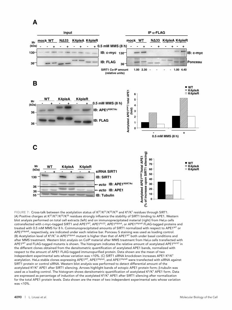
4090 | L. Lirussi et al. Molecular Biology of the Cell
FIGURE 7: Cross-talk between the acetylation status of K27/K31/K32/K35 and K6/K7 residues through SIRT1. (A) Positive charges at K27/K31/K32/K35 residues strongly influence the stability of SIRT1 binding to APE1. Western blot analysis performed on total cell extracts (left) and on immunoprecipitated material (right) from HeLa cells cotransfected with c-myc–tagged SIRT1 and APE1WT, APE1NΔ33, APE1K4pleA, or APE1K4pleR FLAG-tagged proteins and treated with 0.5 mM MMS for 8 h. Coimmunoprecipitated amounts of SIRT1 normalized with respect to APE1WT or APE1K4pleR, respectively, are indicated under each relative bar. Ponceau S staining was used as loading control. (B) Acetylation level of K6/K7 in APE1K4pleA mutant is higher than that of APE1WT both under basal conditions and after MMS treatment. Western blot analysis on CoIP material after MMS treatment from HeLa cells transfected with APE1WT and FLAG-tagged mutants is shown. The histogram indicates the relative amount of acetylated APE1K6/K7 in the different clones obtained from the densitometric quantification of acetylated APE1 bands, normalized with respect to the amount of APE1 FLAG-tagged immunopurified protein. Data shown are the mean of two independent experimental sets whose variation was <10%. (C) SIRT1 siRNA knockdown increases APE1 K6/K7 acetylation. HeLa stable clones expressing APE1WT, APE1K4pleA, and APE1K4pleR were transfected with siRNA against SIRT1 protein or control siRNA. Western blot analysis was performed to detect differential amount of the acetylated K6/K7 APE1 after SIRT1 silencing. Arrows highlight bands of ectopic APE1 protein form; β-tubulin was used as a loading control. The histogram shows densitometric quantification of acetylated K6/K7 APE1 form. Data are expressed as percentage of induction of the acetylated K6/K7 APE1 after SIRT1 silencing after normalization for the total APE1 protein levels. Data shown are the mean of two independent experimental sets whose variation was <10%.
C
A
BWTK4pleAK4pleR
Mr(kDa)
36
36
IB: APE1K6K7Ac
IB: FLAG
- + - + - +WT K4pleA K4pleR
0.5 mM MMS (8 h)
0.5 mM MMS (8 h)
Ace
tyla
ted
AP
E1K
6K7 /
to
tal A
PE
1(f
old
)- +
0
1
2
3
4
5
6
0.5 mM MMS (8 h)
IB: c-myc130
Ponceau
Mr(kDa)
SIRT1 Co-IP amount(relative units)
1.00 2.30 1.00 4.40- - - -
36
130
IB: FLAG
mock WT
+--
N 33
+-
K4pleA
+-
K4pleR
+-
mock WT
+--
N 33
+-
K4pleA
+-
K4pleR
+-
IB: c-myc
input IP -FLAG
Ace
tyla
ted
AP
E1K
6K7 /
tota
l AP
E1
(% o
f in
crea
se u
po
n S
IRT
1 si
len
cin
g)
siRNA SIRT1+-WT
IB: APE1K6K7Ac
IB: Tubulin
IB: SIRT1130
55
36ecto
Mr(kDa)
+-K4pleA
+-K4pleR
36 ecto IB: APE1
80
70
60
50
40
30
20
10
0
WTK4pleAK4pleR
36

Volume 23 October 15, 2012 Functional relevance of APE1 nucleolar accumulation | 4091
to our previous data obtained in vitro with recombinant purified pro-teins (Fantini et al., 2010). In many cases, the presence of the unstruc-tured N-terminal domain of APE1 seemed essential for interaction of APE1 with different substrates (i.e., nucleic acids or proteins). Interac-tion also increased after histone deacetylase (HDAC) inhibition, which promotes APE1 acetylation at K6/K7 residues (Bhakat et al., 2003; Yamamori et al., 2010). This evidence strongly support the no-tion that this unstructured domain is responsible for the modulation of APE1’s different functions through the recruitment of APE1 in dif-ferent protein complexes by means of various amino acid side-chain modification events. From an evolutionary perspective, it can be hy-pothesized that mammalian APE1 activity was made adjustable (through PTMs and/or interaction with other proteins), or expanded toward other substrates, with the acquisition of a protein N-terminus containing positively charged residues, without major modifications on the enzyme catalytic site, which retained the “canonical” function toward abasic DNA. The existence of such an N-terminal extension in only mammals may suggest its evolutionary significance in the face of increased functional complexity. For the noncomplexed pro-tein in solution, the intrinsic lack of a secondary structure associated with this domain can confer functional advantages to mammalian APE1, including the ability to bind to different protein targets (e.g., NPM1, XRCC1, CSB, RNA, etc.; Vidal et al., 2001; Wong et al., 2007; Vascotto et al., 2009a; Tell et al., 2010a), thus allowing efficient con-trol over the thermodynamics in the binding process to different sub-strates. Because the protein’s N-terminus is required for the stabiliza-tion of APE1 interaction with NPM1 or rRNA and for the control of the overall endonuclease activity (possibly decreasing the rate of product release once in the positively charged state; Fantini et al., 2010; Figure 5), this region-specific multitasking function can provide a “trigger” for molecular regulation with important biological signifi-cance. This should be regarded, however, in light of BER coordina-tion to prevent formation of harmful, unprotected DNA strand breaks. Therefore subcellular distribution of APE1 and its enzymatic activity seem to be finely tuned to demand and in a time-dependent manner through multiple interactions with various protein partners and the coordinated occurrence of different PTMs, such as acetyla-tion and ubiquitination (Busso et al., 2009, 2011). The observation that acetylation at K27/K31/K32/K35 residues may favor a transition to-ward a more organized conformation (Supplemental Figure S8) would support the notion that this modification may significantly modulate the interaction with several protein partners on a structural basis. In addition, acetylation at the mentioned K residues may pro-foundly affect APE1 protein stability, based on a recent report show-ing that the same K residues can be also polyubiquitinated by UBR3, targeting the protein for degradation by the proteasome (Meisenberg et al., 2011). Given that acetylation competes with ubiquitination for the same K residues, it may represent the switch for controlling pro-tein turnover rate. In this context, the acetylation at these residues that we observed during cell response to genotoxicants may stabilize protein half-life, thus preventing its degradation.
Few studies have reported on the effect of acetylation on the structure of small, ordered (Hughes and Waters, 2006; Liu and Duan, 2008) and disordered (Smet-Nocca et al., 2010) peptides. Molecular dynamic simulations carried out on a peptide from the histone H3 N-terminal tail in the nonacetylated and doubly acetylated forms revealed that acetylation, although not appreciably disturbing the overall structure of the most-populated states, influenced peptide stability (Liu and Duan, 2008). The effect of acetylation at K residues on the conformational properties of small random-coil peptides from the histone H4 N-terminal tail and from nonhistone thymine DNA glycosylase indicated that acetylation had little effect on the
peptides that tumble very rapidly in solution. On the other hand, a detailed comparison of the 1D NMR data for the nonacetylated and tetra-acetylated APE1 peptides (Figure 8D, left) indicated that acety-lation causes a small but clear improvement of spectral dispersion, which can be particularly appreciated in the NH region (Figure 8D, top left). Moreover, the intensity of ROE cross-peaks is increased in the spectra of APE1(14-38)K27/31/32/35Ac peptide as compared with the nonmodified counterpart (Figure 8D, right). This evidence, together with small chemical shift changes, point toward the presence of more organized conformations for the tetra-acetylated peptide in aqueous solution, in agreement with CD data. Similar conclusions are drawn from the analysis of NMR data for APE1(14–38) and APE1(14–38)K27/31/32/35Ac peptides in phosphate:TFE solution (Supplemental Figure S8B), which suggested again the higher propensity of this domain in its acetylated form to adopt a more ordered conformation in contrast to the nonmodified counterpart. Also in this case, monoacetylated and triacetylated peptides showed an intermediate behavior (unpublished data). These data suggest that acetylation may account for local conformational changes on APE1 structure that may modulate its binding ability to different substrates.
DISCUSSIONAPE1 is an unusually abundant DNA-repair protein in mammalian cells, with a wide nuclear distribution and an essential role in the BER pathway of DNA lesions (Tell and Wilson, 2010). We calculated that HeLa cells express ∼4 × 10⁷ molecules per cell (Supplemental Figure S4A), whereas other enzymes of the BER pathway, such as Pol β or XRCC1, are present at an extent of <1/10 (Demple and DeMott, 2002; Parsons et al., 2008). Therefore APE1 involvement in pre-formed DNA-repair complexes may only partially explain the energy cost used to maintain such high protein concentration within the cells. Recently we found that APE1 may interact with rRNA and with proteins involved in RNA metabolism and is associated with nucleo-lar structures through its interaction with NPM1 (Vascotto et al., 2009b; Tell et al., 2010b). Interaction with rRNA and NPM1 strictly depends on the positive charge of K residues within the APE1 region 24–35 placed within the unstructured protein N-terminal domain, as demonstrated by the inability of the corresponding K-to-A mutants (resembling constitutive acetylation at these residues) to stably bind both rRNA and NPM1 (Fantini et al., 2010). Of interest, some of these amino acids (i.e., K27/K31/K32/K35), which have been acquired during evolution, may undergo in vivo acetylation (Fantini et al., 2010). We conjectured that, under physiological conditions, non-acetylated APE1 may be stored in the nucleolar compartment through its binding to NPM1 and rRNA, but the in vivo relevance of APE1 nucleolar accumulation was still unclear. This study was aimed at addressing this issue.
We also found that APE1K4pleA binds poorly to NPM1 and rRNA in vivo and as a result is unable to accumulate within the nucleoli, whereas it is present in the nucleoplasm. Moreover, reconstitution of HeLa cells with this mutant gave improved protection from geno-toxic damage induced by alkylating agents, such as MMS, and oxida-tive stress-generating compounds, such as TBHP, through increased DNA-repair activity. As expected for a regulated pheno menon such as the response to a genome insult, an in vivo–augmented acetyla-tion at K27/K31/K32/K35 residues was observed during cell response to genotoxic damage. Furthermore, cross-talk involving the SIRT1 deacetylase occurred within cells between acetylation at K27/K31/K32/K35 and at K6/K7 residues. Therefore we hypothesized that genotoxic stress may shift the equilibrium between the nonacetylated and acetylated APE1 forms toward the latter, which would result in the most enzymatically active one on abasic DNA. This also corresponds

4092 | L. Lirussi et al. Molecular Biology of the Cell
FIGURE 8: Nucleolar APE1 hypoacetylated on K6/K7 and conformational impact of acetylation at K27/K31/K32/K35 on APE1 local structure. (A) SIRT1 resides within the nucleoplasm of HeLa cells. Left, confocal microscopy of HeLa cells cotransfected with green fluorescent protein–fused APE1 (APE1GFP) and c-myc-SIRT1 after fixing and staining with antibodies against NPM1 (red) and c-myc-SIRT1 (top, green; bottom, red). SIRT1, clearly excluded from nucleoli,
A
B C 1.0
-10.0
-5.0
0
260200 220 240Wavelength (nm)
deg
cm
2d
mo
l -1 r
es-1
* 10
3
APE1 (14-38)APE1 (14-38) K27/31/32/35Ac
IB: APE1K6K7Ac
IB: APE1
Mr(kDa)
36
36
Nucleus Nucleoli
Ace
tyla
ted
AP
E1K
6K7
(%)
10
2030
4050
6070
80
0
Mr(kDa)
IB: Nucleolin95
IB: NPM136
IB: APE136
130 IB: SIRT1
SIRT1 myc Merged
NPM1
APE1GFP
4 m 4 m
2 m2 m
4 m
2 m
D
APE1(14-38)K27/31/32/35AcAPE1(14-38) APE1(14-38)K27/31/32/35Ac APE1(14-38)
1 H p
pm
1 H p
pm
2.0
2.2
3.0
3.2
1.6
1.8
2.4
2.6
2.8
1H ppm 8.08.28.28.4
2.0
1H ppm 8.08.28.2 8.4
2.2
3.0
3.2
1.6
1.8
2.6
2.8
2.4
1H ppm8.8 7.08.0
APE1(14-38)
Acetyl methyl groups
1.62.02.4 1H ppm
APE1(14-38)K27/31/32/35Ac

Volume 23 October 15, 2012 Functional relevance of APE1 nucleolar accumulation | 4093
This is also underway in our laboratory. Of note, data obtained in this work not only show that nucleoli may act as a storage site so that an appropriate amount of APE1 is readily available for maintenance of genome stability, but also emphasize that nucleolar accumulation of APE1 controls cell proliferation, possibly through its rRNA cleans-ing function. Compatible with this, APE1K4pleA-expressing cells, un-der basal conditions, showed impairment in proliferation rate with respect to APEWT-expressing ones (Figure 3). Therefore it can be speculated that nucleolar APE1 is responsible for functional activity of the nucleolus in ribosome biogenesis. APE1 release from the nu-cleoli after genotoxic treatment may constitute a signal to block ac-tive protein synthesis and allow activation of the proper DNA-repair mechanisms. Experiments are in progress along these lines to ad-dress this in light of the possibility that acetylation may control the APE1 trafficking from nucleoli to nucleoplasm.
In conclusion, our data shed light on novel molecular aspects highlighting the multifunctional nature of APE1 in regulating differ-ent biological outcomes and point to acetylation as an important mechanism for the fine-tuning of protein functions, subcellular dis-tribution, and stability. They also emphasize the need for additional investigation of the APE1 N-terminal domain in order to understand the structural details of the regulatory mechanisms for this multi-functional protein. In addition, recent evidence on APE1 acetylation pattern in triple-negative breast cancer reveals that, concomitantly with total APE1 overexpression, a profound deregulation of APE1 acetylation status occurs under pathological conditions (Poletto et al., 2012). This underscores the biological relevance of our find-ings and the need for future investigation.
MATERIALS AND METHODSInducible APE1 knockdown and generation of APE1 knock-in cell linesInducible silencing of endogenous APE1 and reconstitution with mutant proteins in HeLa cell clones was performed as described (Vascotto et al., 2009a,b) and as reported in the Supplemental Information. For inducible shRNA experiments, doxycycline (1 μg/ml; Sigma-Aldrich, St. Louis, MO) was added to the cell culture medium, and cells were grown for 10 d. All biological data were reproduced in at least two different cell clones for each model.
Cell culture and transient transfection with plasmids or siRNA knockdownHeLa cells were grown in DMEM (Invitrogen, Carlsbad, CA) supple-mented with 10% fetal bovine serum (EuroClone, Milan, Italy), 100 U/ml penicillin, and 100 μg/ml streptomycin sulfate. One day
overall polypeptide structure, while inducing local conformational changes (Smet-Nocca et al., 2010). In agreement with these studies, APE1(14–38) and APE1(14–38)K27/31/32/35Ac became disordered, as shown by the lack of ROE (Bax and Davis, 1985) cross-peak patterns, which are characteristic of ordered secondary structure elements. However, small differences in the NMR spectra (Figure 8 and Sup-plemental Figure S8), concerning both chemical shift values and signal intensities, seem to indicate that at least local conformational changes may occur following acetylation. We cannot ignore that these changes may be important for the interaction of APE1 with SIRT1 deacetylase and with NPM1; they highlight the role that the charged status of K residues within this region may play at the pro-tein structural level. Based on our data, it can be speculated that full acetylation of K27–35 may reduce SIRT1 binding to APE1, thus delay-ing its enzymatic activity on K6/K7. This mechanism could represent a way to coordinate the kinetics of the overall acetylation status of the protein. According to this hypothesis, SIRT1 should first deacety-late K27–35Ac before deacetylating K6/K7Ac. Our ability to identify APE1 peptides with varying amounts of acetylation on K27–35 sup-ports the existence of a dynamic equilibrium between multiple acetylated forms of the protein within cells and thus its functional regulatory role. Moreover, the presence of SIRT1, found exclusively in the nucleoplasm but not in the nucleoli (similar to the APE1 acety-lated on K27–35 and the reduced nucleolar presence of APE1 acety-lated on K6/K7; Figure 8, A and B), suggests that acetylation of APE1 may force its exit from nucleoli to nucleoplasm, where it can be deacetylated by SIRT1. This model is further supported by the sig-nificantly reduced interaction of AcAPE1K27-35 with NPM1 (Figure 2). Further studies to identify the acetyltransferase able to acetylate APE1 within the nucleoli are in progress. Furthermore, this work sup-ports findings by Yu et al. (2010), who demonstrated that these K residue conformational adjustments were concomitant with DNA binding and catalysis or with interaction with Pol β.
The nucleolar role of APE1 storage and regulation, as described here, may have profound biological consequences during cell re-sponse to stressor signals, especially in light of recent evidence pointing to the nucleolus as a central hub in DNA damage (Nalabothula et al., 2010). Accordingly, the nucleolus seems respon-sible for actively transmitting signals to the molecular complex regu-lating p53 activity mediated by ARF–NPM1 interaction (Colombo et al., 2002; Lee et al., 2005; Nalabothula et al., 2010), and thus it is involved in the maintenance of genome stability. A careful elucida-tion of the NPM1-ARF-p53 signaling networks and their involve-ment in the DNA-repair pathway coordinated by APE1 is an impor-tant subject for molecular carcinogenesis and deserves further study.
colocalizes with APE1 in the nucleoplasm. The inner part of nucleoli, marked in the granular zone by NPM1, is negative for SIRT1. Right, biochemical isolation of nucleoli confirms that SIRT1 localizes in the nucleoplasmic fraction and it is excluded from nucleoli, where nucleolin, NPM1, and APE1 reside (see Materials and Methods for details). Nucleolin was used as a positive control for nuclear and nucleolar compartment. (B) Acetylated K6/K7-containing APE1 is enriched within the nucleoplasmic compartment with respect to nucleoli. After normalization for total APE1 protein amount, the levels of acetylated APE1 in nucleolar and nucleoplasmic fractions were analyzed through Western blotting. APE1 acetylated at K6/K7 residues is predominately present within the nucleoplasmic fraction, whereas it is reduced in the nucleolar fraction. The histogram indicates the relative percentage amount of acetylated APE1 obtained from the densitometric quantification of acetylated APE1 bands normalized with respect to the amount of total APE1 in each fraction. Each bar represents the mean of two independent experiments whose variation was <10%. (C) CD spectra of the APE1(14–38) and APE1(14–38)K27/31/32/35Ac peptides in 10 mM phosphate buffer, pH 7. (D) Comparison of 1D (left), 2D [1H, 1H] TOCSY (middle), and 2D [1H, 1H] ROESY (right) spectra of APE1(14–38) and APE1(14–38)K27/31/32/35Ac peptides in 10 mM phosphate buffer, pH 7.0. Two different expansions of the proton 1D spectrum are shown; the region between 0.8 and 2.4 ppm, in the lower inset, contains signals from side-chain protons. Acetyl methyl groups originate a peak around 2 ppm, which can be clearly seen in the spectrum of the acetylated peptide; peaks of backbone and side-chain NH atoms appear between 7.0 and 8.8 ppm in the upper inset. For 2D [1H, 1H] TOCSY and 2D [1H, 1H] ROESY experiments the HN-aliphatic protons correlation region of the spectra are reported.

4094 | L. Lirussi et al. Molecular Biology of the Cell
ing the ECL enhanced chemiluminescence procedure (GE Health-care Piscataway, NJ) or Western Lightning Ultra (PerkinElmer, Waltham, MA). Normalization was performed by using a monoclonal anti-tubulin antibody (Sigma-Aldrich). Blots were quantified by using a Chemidoc XRS video densitometer (Bio-Rad, Hercules, CA).
Plasmids and expression of recombinant proteinsExpression and purification of recombinant proteins from E. coli were performed as previously described (Vascotto et al., 2009b; Fantini et al., 2010). Where recombinant proteins were used for in vitro assays, the acronym rAPE1 is used.
GST pull-down assayA 150-pmol amount of either GST-tagged rAPE1WT or mutant pro-teins was added to 15 μl of glutathione–Sepharose 4B beads (GE Healthcare), together with equimolar amounts of recombinant NPM1. Binding was performed in PBS, supplemented with 1 mM dithiothreitol (DTT) and 0.5 mM phenylmethylsulfonyl fluoride (PMSF) for 2 h, under rotation, at 4°C. Beads were washed three times with PBS, supplemented with 0.1% (vol/vol) Igepal CA-630 (Sigma-Aldrich), 1 mM DTT, and 0.5 mM PMSF and resuspended in Laemmli sample buffer for Western blot analysis.
DNA–RNA ChIP assaysDNA–RNA ChIP assays were carried out by using a modified version of a protocol described earlier (Gilbert et al., 2000) and as detailed in the Supplemental Information.
Enzymatic fluorescence assaysTo examine in vitro deacetylase activity on the acetylated APE1 re-gion 25–38, we used the SIRT Fluorescent Activity Assay Kit (Biomol, Plymouth, PA). Optimizing manufacturer’s instructions, we used white plates (OPTI PLATE; PerkinElmer) with 384 wells at reduced volume (total reaction volume, 20 μl). Purified peptides were incu-bated in 25 mM Tris-HCl, pH 8.0, 2.7 mM KCl, 137 mM NaCl, 1 mM MgCl2, and 1 mg/ml bovine serum albumin containing the enzyme (0.04 U/μl) and 25 μM Fluor de Lys-p53 peptide substrate (Arg-His-Lys-Lys [Ac], from region 379–382 of human p53) in the presence/absence of 250 μM NAD+ for 30 min at 37°C. Deacetylase activity was measured in arbitrary fluorescence units at 460 nm. Dose–response experiments were carried out by using a 0–500 μM range of peptide concentration. Data fitting was performed using the GraphPad Prism 4 software, version 4.02 (GraphPad, La Jolla, CA). Data were in triplicate/duplicate from three independent assays.
Immunofluorescence confocal and proximity ligation analysesImmunofluorescence procedures and PLA were carried out as de-scribed earlier (Vascotto et al., 2009b, 2011). To study the interaction between APE1 and NPM1 in vivo, we used the in situ Proximity Ligation Assay technology (Olink Bioscience, Uppsala, Sweden). After incubation with monoclonal anti-APE1 (1:50) or anti-FLAG anti-body (1:200) for 3 h at 37°C, cells were incubated with polyclonal anti-NPM1 (1:200) overnight at 4°C. PLA was performed following manufacturer’s instructions. Technical controls, represented by the omission of anti-NPM1 primary antibody, resulted in the complete loss of PLA signal. Cells were visualized through a Leica TCS SP laser-scanning confocal microscope (Leica Microsystems, Wetzlar, Germany). Determination of PLA signals was performed using BlobFinder software (Center for Image Analysis, Uppsala University, Uppsala, Sweden). PLA technology was also used to detect acety-lated APE1 at K27/K31/K32/K35 residues. Cells were incubated with the
before transfection, cells were seeded in 10-cm plates at a density of 3.0 × 10⁶ cells/plate. Cells were then transiently transfected with the indicated plasmids using the Lipofectamine 2000 reagent (Invitrogen), according to the manufacturer’s instructions. Cells were harvested either 24 or 48 h after transfection, as indicated.
For SIRT1-knockdown experiments, HeLa clones were trans-fected with 150 nM siRNA siGENOME SMART pool or negative control siRNA 5′-CCAUGAGGUCAUGGUCUGdTdT-3′ (Dharmacon, Lafayette, CO), using Oligofectamine (Invitrogen). After 72 h the cells were harvested.
Preparation of total cell extracts and anti-FLAG coimmunoprecipitationPreparation of total cell lysates and coimmunoprecipitation analyses were performed as described (Vascotto et al., 2009a,b).
Determination of AP endonuclease activity and abasic site assayDetermination of APE1 AP endonuclease activity was performed using an oligonucleotide cleavage assay, as described previously (Vascotto et al., 2009b) and detailed in the Supplemental Information.
Mass spectrometric analysis of APE1 acetylationCharacterization of APE1 acetylation was performed on the immu-nopurified protein obtained from APE1-FLAG–expressing HeLa cells (Vascotto et al., 2009b). APE1 was resolved by SDS–PAGE; corresponding protein bands were excised, S-alkylated, and di-gested with endoprotease AspN (Fantini et al., 2010). Digest ali-quots were directly analyzed by nanoLC-ESI-LIT-MS/MS using an LTQ XL mass spectrometer (ThermoFisher Scientific, Waltham, MA) equipped with a Proxeon nanospray source connected to an Easy-nanoLC (ThermoFisher Scientific; Arena et al., 2010; Scippa et al., 2010), and analysis of APE1 acetylation was performed as described (D’Ambrosio et al., 2006) and detailed in the Supplemental Infor-mation. A semiquantitative measurement of the amino acid modifi-cation was obtained by extracting and integrating nanoLC-ESI-LIT-MS peak areas corresponding to m/z values of the modified and nonmodified peptides in the same total ion chromatogram (Salzano et al., 2011). Modification extent was then assayed by evaluating the peak area of the modified peptide with respect to that of the modified peptide plus that of the nonmodified peptide, assuming identical ionization properties for modified and nonmodified spe-cies. All these analyses were performed in triplicate.
Antibodies for immunofluorescence and immunoblottingAntibodies used were anti-NPM1 monoclonal, anti-nucleolin mono-clonal (Zymed, Invitrogen), anti-FLAG peroxidase-conjugated, anti-GST peroxidase-conjugated, anti-SIRT1 polyclonal (Abcam, Cambridge, MA), anti–c-myc (Santa Cruz Biotechnology, Santa Cruz, CA), and anti–β-tubulin monoclonal (Sigma-Aldrich). Anti-APE1 monoclonal (Vascotto et al., 2009a) and anti-APE1K6K7Ac (Bhakat et al., 2003) were described previously. Anti-APE1K27-35Ac polyclonal anti-body was generated by PRIMM (Milan, Italy; Poletto et al., 2012).
Western blot analysesFor Western blot analyses, the indicated amounts of cell extracts were resolved in 12% SDS–PAGE and transferred to nitrocellulose membranes (Schleicher & Schuell BioScience, Dassel, Germany). Membranes were blocked with 5% (wt/vol) nonfat dry milk in phos-phate-buffered saline (PBS) containing 0.1% (vol/vol) Tween 20 and probed with the indicated antibodies; blots were developed by us-

Volume 23 October 15, 2012 Functional relevance of APE1 nucleolar accumulation | 4095
REFERENCESArena S, Renzone G, Novi G, Paffetti A, Bernardini G, Santucci A, Scaloni
A (2010). Modern proteomic methodologies for the characterization of lactosylation protein targets in milk. Proteomics 10, 3414–3434.
Bapat A, Fishel ML, Kelley MR (2009). Going Ape as an approach to cancer therapeutics. Antioxid Redox Signal 11, 651–668.
Bapat A, Glass LS, Luo M, Fishel ML, Long EC, Georgiadis MM, Kelley MR (2010). Novel small-molecule inhibitor of apurinic/apyrimidinic endonu-clease 1 blocks proliferation and reduces viability of glioblastoma cells. J Pharmacol Exp Ther 334, 988–998.
Barnes T, Kim WC, Mantha AK, Kim SE, Izumi T, Mitra S, Lee CH (2009). Iden-tification of apurinic/apyrimidinic endonuclease 1 (APE1) as the endoribo-nuclease that cleaves c-myc mRNA. Nucleic Acids Res 37, 3946–3958.
Bartels C, Xia T, Billeter M, Gunthert P, Wüthrich K (1995). The program XEASY for computer-supported NMR spectral analysis of biological macromolecules. J Biomol NMR 6, 1–10.
Bax A, Davis DG (1985). Practical aspects of two-dimensional transverse NOE spectroscopy. J Magn Reson 63, 207–213.
Bhakat KK, Izumi T, Yang SH, Hazra TK, Mitra S (2003). Role of acetylated human AP-endonuclease (APE1/Ref-1) in regulation of the parathyroid hormone gene. EMBO J 1, 6299–6309.
Busso CS, Iwakuma T, Izumi T (2009). Ubiquitination of mammalian AP endonuclease (APE1) regulated by the p53-MDM2 signaling pathway. Oncogene 28, 1616–1625.
Busso CS, Wedgeworth CM, Izumi T (2011). Ubiquitination of human AP-endonuclease 1 (APE1) enhanced by T233E substitution and by CDK5. Nucleic Acids Res 39, 8017–8028.
Chattopadhyay R, Wiederhold L, Szczesny B, Boldogh I, Hazra TK, Izumi T, Mitra S (2006). Identification and characterization of mitochondrial abasic (AP)-endonuclease in mammalian cells. Nucleic Acids Res 34, 2067–2076.
Chudakov DM, Lukyanov S, Lukyanov KA (2007). Tracking intracellular pro-tein movements using photoswitchable fluorescent protein PS-CFP2 and Dendra2. Nat Protoc 2, 2024–2032.
Colombo E, Marine JC, Danovi D, Falini B, Pelicci PG (2002). Nucleophos-min regulates the stability and transcriptional activity of p53. Nat Cell Biol 4, 529–533.
Dalvit C (1998). Efficient multiple-solvent suppression for the study of the interaction of organic solvents with biomolecules. J Biomol NMR 11, 437–444.
D’Ambrosio C, Arena S, Fulcoli G, Scheinfeld MH, Zhou D, D’Adamio L, Scaloni A (2006). Hyperphosphorylation of JNK-interacting protein 1, a protein associated with Alzheimer disease. Mol Cell Proteomics 5, 97–113.
Demple B, DeMott MS (2002). Dynamics and diversions in base excision DNA repair of oxidized abasic lesions. Oncogene 21, 8926–8934.
Fantini D et al. (2008). APE1/Ref-1 regulates PTEN expression mediated by Egr1. Free Radic Res 42, 20–29.
anti-APE1K27–35Ac rabbit-polyclonal antibody diluted 1:1500 and then with a mouse-monoclonal anti-APE1 antibody (1:27). PLA was sub-sequently carried out following manufacturer’s instructions.
Confocal analyses of APE1-Dendra fusion protein through in vivo live imagingFor in vivo APE1-Dendra trafficking studies, HeLa cells were seated on glass-bottom Petri dishes (thickness #1.5; WillCo Wells, Amsterdam, Netherlands), transfected with APE1-Dendra con-structs, and grown in the presence of DMEM without phenol red. A Leica TCS SP laser-scanning confocal microscope was equipped with a heating system (Incubator S) and a CO2 controller (CTI Controller 3700 digital) to maintain cells in optimal growing con-ditions. Images were captured 24 h after transfection using a 63× oil fluorescence objective. For Dendra green fluorescence acqui-sition a 488-nm argon laser was regulated at 10% of power with PTM 750 V.
Cell viability, cell growth, and clonogenic assaysCell viability was measured by using the MTS assay (Celltiter 96 Aqueous One solution cell proliferation assay; Promega, Madison, WI) on HeLa cells stably expressing APE1WT, APE1K4pleA, and APE1K4pleR proteins grown in 96-well plates. After MMS (Sigma-Aldrich) treatment or TBHP (Sigma-Aldrich), the MTS solution was added to each well and the plates were incubated for 2 h. Absor-bance was measured at 490 nm by using a multiwell plate reader. The values were standardized to wells containing media alone.
Cell growth assays were performed as described (Vascotto et al., 2009a,b) and detailed in the Supplemental Information, and clono-genic assays were performed according to Plumb (1999) and essen-tially as described previously (Vascotto et al., 2009a,b).
Circular dichroism spectroscopyCD spectra were recorded on a Jasco J-810 spectropolarimeter (Jasco, Tokyo, Japan) at 25°C in the far-UV region from 190 to 260 nm. Each spectrum was obtained by averaging three scans, subtracting contributions from the corresponding blanks, and converting the sig-nal to mean residue ellipticity in units of deg-cm2 dmol−1 res−1. Other experimental settings were 20 nm/min scan speed, 2.0 nm band-width, 0.2 nm resolution, 50 mdeg sensitivity, and 4 s response. Pep-tide concentration was kept at 100 μM, and a 0.1 cm path-length quartz cuvette was used. Spectra were acquired in 10 mM phosphate buffer, pH 7.0, containing various percentages of TFE.
NMR spectroscopyNMR samples were prepared by dissolving APE1 peptides (1.5– 2 mg) either in 600 μl of a 10 mM phosphate buffer, pH 7, contain-ing 10% (vol/vol) D2O or in a mixture of 10 mM phosphate buf-fer:2-2-2 trifluorethanol-d3 (98% deuterium; Armar Chemicals, Döttingen, Switzerland) 70:30 (vol/vol). The 2D [1H, 1H] TOCSY spectra (1024 × 256 total data points, 32 scans per t1 increment, 70 ms mixing time; Griesinger et al., 1988) were recorded at 298 K on a Varian UNITYINOVA 600 spectrometer (Palo Alto, CA) equipped with a cold-probe. The 1D proton (128 scans and a relaxation delay of 1.5 s) and 2D [1H, 1H] ROESY (2048 × 256 total data points, 64 scans per t1 increment, 200 ms mixing time) spectra were ac-quired at 298 K on a Varian UNITYINOVA 400 spectrometer provided with z-axis pulsed-field gradients and a triple-resonance probe. Water signal was suppressed by means of either double pulsed field gradient selective echo techniques (Dalvit, 1998) or continu-ous wave irradiation. Varian software VNMRJ 1.1D was implemented for spectral processing. The programs MestRe-C2.3a (Universidade
ACKNOWLEDGMENTSWe thank Paolo Peruzzo for generation of mutant recombinant pro-teins, K. Irani for providing SIRT1-encoding plasmids, and Pablo Ra-dicella for helpful comments on the manuscript. We also thank Julie Driscol for excellent help in editing the manuscript. This work was supported by the Associazione Italiana per la Ricerca sul Cancro (IG10269) and the Ministero dell’Istruzione, dell’Università e della Ricerca (FIRB_RBRN07BMCT and PRIN2008_CCPKRP_003 to G.T.; PRIN2008_CCPKRP_002 and FIRB2008_RBNE08YFN3_003 to A.S.). This work was also supported by a UICC Yamagiwa-Yoshida Memo-rial International Cancer Study Grant to G.T. and by the Regione Friulia Venezia Giulia for the Project MINA under the Programma per la Cooperazione Transfrontaliera Italia-Slovenia 2007–2013.
de Santiago de Compostela, Santiago de Compostela, Spain) and NEASY (Bartels et al., 1995; www.nmr.ch) were used for analysis of 1D and 2D NMR data, respectively.
Statistical analysesStatistical analyses were performed by using the Excel (Microsoft, Redmond, WA) data analysis program for Student’s t test. p < 0.05 was considered as statistically significant.

4096 | L. Lirussi et al. Molecular Biology of the Cell
Scippa GS, Rocco M, Ialicicco M, Trupiano D, Viscosi V, Di Michele M, Arena S, Chiatante D, Scaloni A (2010). The proteome of lentil (Lens culinaris Medik.) seeds: discriminating between landraces. Electrophoresis 31, 497–506.
Seemann S, Hainaut P (2005). Roles of thioredoxin reductase 1 and APE/Ref-1 in the control of basal p53 stability and activity. Oncogene 24, 3853–3863.
Sengupta S, Mantha AK, Mitra S, Bhakat KK (2011). Human AP endonu-clease (APE1/Ref-1) and its acetylation regulate YB-1-p300 recruitment and RNA polymerase II loading in the drug-induced activation of multi-drug resistance gene MDR1. Oncogene 30, 482–493.
Smet-Nocca C, Wieruszeski JM, Melnyk O, Benecke A (2010). NMR-based detection of acetylation sites in peptides. J Pept Sci 16, 414–423.
Szczesny B, Mitra S (2005). Effect of aging on intracellular distribution of abasic (AP) endonuclease 1 in the mouse liver. Mech Ageing Dev 126, 1071–1078.
Tell G, Crivellato E, Pines A, Paron I, Pucillo C, Manzini G, Bandiera A, Kelley MR, Di Loreto C, Damante G (2001). Mitochondrial localization of APE/Ref-1 in thyroid cells. Mutat Res 485, 143–152.
Tell G, Damante G, Caldwell D, Kelley MR (2005). The intracellular localiza-tion of APE1/Ref-1: more than a passive phenomenon. Antioxid Redox Signal 7, 367–384.
Tell G, Fantini D, Quadrifoglio F (2010a). Understanding different functions of mammalian AP endonuclease (APE1) as a promising tool for cancer treatment. Cell Mol Life Sci 67, 3589–3608.
Tell G, Quadrifoglio F, Tiribelli C, Kelley MR (2009). The many functions of APE1/Ref-1: not only a DNA-repair enzyme. Antioxid Redox Signal 11, 601–620.
Tell G, Wilson DM 3rd (2010). Targeting DNA repair proteins for cancer treatment. Cell Mol Life Sci 67, 3569–3572.
Tell G, Wilson DM 3rd, Lee CH (2010b). Intrusion of a DNA repair protein in the RNome world: is this the beginning of a new era. Mol Cell Biol 30, 366–371.
Ueno M, Masutani H, Arai RJ, Yamauchi A, Hirota K, Sakai T, Inamoto T, Yamaoka Y, Yodoi J, Nikaido T (1999). Thioredoxin-dependent redox reg-ulation of p53-mediated p21 activation. J Biol Chem 274, 35809–35815.
Vascotto C et al. (2011). Knock-in reconstitution studies reveal an unex-pected role of Cys-65 in regulating APE1/Ref-1 subcellular trafficking and function. Mol Biol Cell 22, 3887–3901.
Vascotto C et al. (2009a). Genome-wide analysis and proteomic studies reveal APE1/Ref-1 multifunctional role in mammalian cells. Proteomics 9, 1058–1074.
Vascotto C et al. (2009b). APE1/Ref-1 interacts with NPM1 within nucleoli and plays a role in the rRNA quality control process. Mol Cell Biol 29, 1834–1854.
Vidal AE, Boiteux S, Hickson ID, Radicella JP (2001). XRCC1 coordinates the initial and late stages of DNA abasic site repair through protein-protein interactions. EMBO J 20, 6530–6539.
Wei SJ, Botero A, Hirota K, Bradbury CM, Markovina S, Laszlo A, Spitz DR, Goswami PC, Yodoi J, Gius D (2000). Thioredoxin nuclear transloca-tion and interaction with redox factor-1 activates the activator protein-1 transcription factor in response to ionizing radiation. Cancer Res 60, 6688–6695.
Weibrecht I, Leuchowius KJ, Clausson CM, Conze T, Jarvius M, Howell WM, Kamali-Moghaddam M, Söderberg O (2010). Proximity ligation assays: a recent addition to the proteomics toolbox. Expert Rev Proteomics 7, 401–409.
Wilson DM 3rd, (2010). Small molecule inhibitors of DNA repair nuclease activities of APE1. Cell Mol Life Sci 67, 3621–3631.
Wong HK, Muftuoglu M, Beck G, Imam SZ, Bohr VA, Wilson DM 3rd (2007). Cockayne syndrome B protein stimulates apurinic endonuclease 1 activity and protects against agents that introduce base excision repair intermediates. Nucleic Acids Res 35, 4103–4113.
Yacoub A, Kelley MR, Deutsch WA (1997). The DNA repair activity of hu-man redox/repair protein APE/Ref-1 is inactivated by phosphorylation. Cancer Res 57, 5457–5459.
Yamamori T, DeRicco J, Naqvi A, Hoffman TA, Mattagajasingh I, Kasuno K, Jung SB, Kim CS, Irani K (2010). SIRT1 deacetylates APE1 and regulates cellular base excision repair. Nucleic Acids Res 38, 832–845.
Yu E, Gaucher SP, Hadi MZ (2010). Probing conformational changes in Ape1 during the progression of base excision repair. Biochemistry 49, 3786–3796.
Ziel KA, Campbell CC, Wilson GL, Gillespie MN (2004). Ref-1/Ape is critical for formation of the hypoxia-inducible transcriptional complex on the hypoxic response element of the rat pulmonary artery endothelial cell VEGF gene. FASEB J 18, 986–988.
Fantini D et al. (2010). Critical lysine residues within the overlooked N-ter-minal domain of human APE1 regulate its biological functions. Nucleic Acids Res 38, 8239–8256.
Fung H, Demple B (2005). A vital role for APE1/Ref1 protein in repairing spontaneous DNA damage in human cells. Mol Cell 17, 463–470.
Garbutt GJ, Abraham EC (1981). Non-enzymatic acetylation of human hemoglobins. Biochim Biophys Acta 670, 190–194.
Gilbert SL, Pehrson JR, Sharp PA (2000). XIST RNA associates with specific regions of the inactive X chromatin. J Biol Chem 275, 36491–36494.
Gray MJ, Zhang J, Ellis LM, Semenza GL, Evans DB, Watowich SS, Gallick GE (2005). HIF-1alpha, STAT3, CBP/p300 and Ref-1/APE are com-ponents of a transcriptional complex that regulates Src-dependent hypoxia-induced expression of VEGF in pancreatic and prostate carcino-mas. Oncogene 24, 3110–3120.
Griesinger C, Otting G, Wüthrich K, Ernst RR (1988). Clean TOCSY for 1H spin system identification in macromolecules. J Am Chem Soc 110, 7870–7872.
Grillo C, D’Ambrosio C, Scaloni A, Maceroni M, Merluzzi S, Turano C, Altieri F (2006). Cooperative activity of Ref-1/APE and ERp57 in reductive acti-vation of transcription factors. Free Radic Biol Med 41, 1113–1123.
Hirota K, Matsui M, Iwata Z, Nishiyama A, Mori K, Yodoi J (1997). AP-1 transcriptional activity is regulated by a direct association between thioredoxin and Ref-1. Proc Natl Acad Sci USA 94, 3633–3638.
Hughes RM, (2006). Effects of lysine acetylation in a beta-hairpin peptide: comparison of an amide-pi and a cation-pi interaction. J Am Chem Soc 128, 13586–13591.
Izumi T, Brown DB, Naidu CV, Bhakat KK, Macinnes V, Saito H, Chen DJ, Mitra S (2005). Two essential but distinct functions of the mammalian abasic endonuclease. Proc Natl Acad Sci USA 102, 5739–5743.
Lazzé MC, Pizzala R, Savio M, Stivala LA, Prosperi E, Bianchi L (2003). Antho-cyanins protect against DNA damage induced by tert-butyl-hydroperox-ide in rat smooth muscle and hepatoma cells. Mutat Res 535, 103–115.
Lee C, Smith BA, Bandyopadhway K, Gjerset RA (2005). DNA damage dis-rupts the p14ARF-B23(nucleophosmin) interaction and triggers a tran-sient subnuclear redistribution of p14ARF. Cancer Res 65, 9834–9842.
Li M, Vascotto C, Xu S, Dai N, Qing Y, Zhong Z, Tell G, Wang D (2012). Hu-man AP endonuclease/redox factor APE1/ref-1modulates mitochondrial function after oxidative stress by regulating the transcriptional activity of NRF1. Free Radic Biol Med 53, 237–248.
Liu H, Duan Y (2008). Effects of posttranslational modifications on the structure and dynamics of histone H3 N-terminal Peptide. Biophys J 94, 4579–4585.
Marcotte PA, Richardson PL, Guo J, Barrett LW, Xu N, Gunasekera A, Glaser KB (2004). Fluorescence assay of SIRT protein deacetylases using an acetylated peptide substrate and a secondary trypsin reaction. Anal Biochem 332, 90–99.
Meisenberg C et al. (2011). Ubiquitin ligase UBR3 regulates cellular levels of the essential DNA repair protein APE1 and is required for genome stability. Nucleic Acids Res 40, 701–711.
Mitra S, Izumi T, Boldogh I, Bhakat KK, Chattopadhyay R, Szczesny B (2007). Intracellular trafficking and regulation of mammalian AP-endonuclease 1 (APE1), an essential DNA repair protein. DNA Repair 6, 461–469.
Nalabothula N, Indig FE, Carrier F (2010). The nucleolus takes control of protein trafficking under cellular stress. Mol Cell Pharmacol 2, 203–212.
Parlanti E, Locatelli G, Maga G, Dogliotti E (2007). Human base excision repair complex is physically associated to DNA replication and cell cycle regulatory proteins. Nucleic Acids Res 35, 1569–1577.
Parsons JL, Tait PS, Finch D, Dianova II, Allinson SL, Dianov GL (2008). CHIP-mediated degradation and DNA damage-dependent stabilization regulate base excision repair proteins. Mol Cell 29, 477–487.
Pines A, Bivi N, Romanello M, Damante G, Kelley MR, Adamson ED, D’Andrea P, Quadrifoglio F, Moro L, Tell G (2005). Cross-regulation be-tween Egr-1 and APE/Ref-1 during early response to oxidative stress in the human osteoblastic HOBIT cell line: evidence for an autoregulatory loop. Free Radic Res 39, 269–281.
Poletto M, Loreto CD, Marasco D, Poletto E, Puglisi F, Damante G, Tell G (2012). Acetylation on critical lysine residues of apurinic/apyrimidinic en-donuclease 1 (APE1) in triple negative breast cancers. Biochem Biophys Res Commun 424, 34–39.
Plumb JA (1999). Cell sensitivity assays: clonogenic assay. Methods Mol 28, 17–23.
Salzano AM, Renzone G, Scaloni A, Torreggiani A, Ferreri C, Chatgilialoglu C (2011). Human serum albumin modifications associated with reductive radical stress. Mol Biosyst 7, 889–898.
Schnell U, Dijk F, Sjollema KA, Giepmans BN (2012). Immunolabeling arti-facts and the need for live-cell imaging. Nat Methods 9, 152–158.

Biochemical and Biophysical Research Communications 430 (2013) 523–528
Contents lists available at SciVerse ScienceDirect
Biochemical and Biophysical Research Communications
journal homepage: www.elsevier .com/locate /ybbrc
Role of mutual interactions in the chemical and thermal stability of nucleophosminNPM1 domains
Daniela Marasco a,b,⇑, Alessia Ruggiero a, Carlo Vascotto c, Mattia Poletto c,Pasqualina Liana Scognamiglio a,b, Gianluca Tell c, Luigi Vitagliano a,⇑a Institute of Biostructures and Bioimaging, CNR via Mezzocannone 16, I-80134 Napoli, Italyb Department of Biological Sciences, University ‘‘Federico II’’ of Naples, Via Mezzocannone 16, 80134 Naples, Italyc Department of Medical and Biological Sciences, University of Udine, 33100 Udine, Italy
a r t i c l e i n f o
Article history:Received 13 November 2012Available online 8 December 2012
Keywords:Structure–stability relationshipsAcute myeloid leukemiaProtein denaturationProtein secondary structure content
0006-291X/$ - see front matter � 2012 Elsevier Inc. Ahttp://dx.doi.org/10.1016/j.bbrc.2012.12.002
Abbreviations: AML, acute myeloid leukemia; NPMfull length Nucleophosmin; NPM1N-Ter, residues 1–residues 188–294 of NPM1; NPM1C-Ter53, residues 24residues 226–294 of NPM1; Pep225–243, residues 225–⇑ Corresponding authors.
E-mail addresses: [email protected] (D.unina.it (L. Vitagliano).
a b s t r a c t
Nucleophosmin (NPM1) is a key factor involved in fundamental biological processes. Mutations involvingthe NPM1 gene are the most frequent molecular alterations in acute myeloid leukemia. Here we report abiophysical characterization of NPM1 and of its domains in order to gain insights into the role that inter-domain interactions plays in the protein stabilization. Thermal denaturation analyses show that theN-terminal domain is endowed with an exceptional thermal stability, as it does not unfold in the inves-tigated temperature range (20–105 �C). This finding is corroborated by chemical denaturation experi-ments showing that this domain is not significantly affected by the addition of 8 M urea. These resultsare consistent with the chaperone function of NPM1. In line with literature data, the other folded domainof the NPM1, a 3-helix bundle domain located at the C-terminus, shows a lower stability. Interestingly,the chemical and thermal stability of this latter domain, which embeds natural mutations related to acutemyeloid leukemia, is influenced by the presence of other regions of the protein. Small but significant sta-bilizations of the C-terminal 3-helix bundle are provided by the adjacent unfolded fragment as well as bythe rest of the protein.
� 2012 Elsevier Inc. All rights reserved.
1. Introduction
The large majority of proteins are multidomain proteins, as a re-sult of complex gene duplication events [1]. In these proteins, eachdomain may fulfill its own function independently, or may operatein a concerted manner with its neighbors. For practical reason,most of biochemical and structural studies are concentrated onthe analysis of individual domains. Therefore, the mutual influenceof individual domains in determining the functional properties ofmultidomain proteins is an open question in many cases.
Nucleophosmin (NPM1), also known as nucleolar phosphopro-tein B23 or numatrin, is a multidomain protein that is implicatedin many fundamental biological processes [2,3]. Physiologically,NPM1, despite its nucleolar localization, shuttles constantly acrossnucleolar, nucleoplasmic and cytoplasmic compartments. This
ll rights reserved.
1, Nucleophosmin; NPM1_fl,117 of NPM1; NPM1C-Ter108,3–294 of NPM1; NPM1C-Ter70,243 of NPM1.
Marasco), luigi.vitagliano@
shuttling activity of NPM1 is critical for most of its functions,including regulation of ribosome biogenesis and control ofcentrosome duplication. Deregulation of NPM1 is implicated inthe pathogenesis of several human malignancies. Indeed, NPM1has been described both as an oncogene and as a tumor suppressordepending on the cell type [4–6]. It is important to note that muta-tions involving the NPM1 gene are the most frequent molecularalteration in acute myeloid leukemia (AML) accounting for about60% of cases (i.e. one-third of adult AML) [7].
From the molecular point of view, NPM1 has a modular struc-ture (Fig. 1). The analysis of the protein sequence shows the pres-ence of distinct domains [8]. The N-terminal domain (NPM1N-Ter
residues 1–117) is the oligomerization domain, that plays a crucialrole in the molecular chaperone activity of the protein [2]. The highresolution three-dimensional structure of this domain shows thatthe NPM1N-Ter pentamer is formed by monomers that fold intoan eight-stranded b-barrel with a jellyroll topology [9]. The analy-sis of the crystal packing has also suggested that NPM1N-Ter has apropensity to form decamers through the association of twodistinct pentamers [9]. These NPM1 oligomers are the structuralunits that engage in interaction with several proteins includingprotamines, protamine-like type proteins, APE1/Ref-1, etc. [10,11].

Fig. 1. Schematic organization of NPM1 domains. Models of the folded domains at the N- (residues 9–117) and the C-terminal (residues 243–294) of the protein are reportedin the insets. The DNA binding region recently identified by Federici et al. is shown in cyan [13].
524 D. Marasco et al. / Biochemical and Biophysical Research Communications 430 (2013) 523–528
The C-terminal region of the protein contains the structural ele-ments responsible for RNA/DNA recognition. NMR investigationshave shown that the last 53 C-terminal residues (NPM1C-Ter53) foldin a 3-helix bundle [12]. Interestingly, mutagenesis studies, carriedout on the protein region containing last 70 C-terminal residues(NPM1C-Ter70), have shown that the high affinity of NPM1 forDNA is due to the region proceeding the 3-helix bundle (residues225–243) [13], although it has been recently demonstrated thatno direct interaction is established by this fragment with theDNA [14]. The central portion of NPM1 is characterized by thepresence of two acid domains (residues 119–133 and 161–188). Fi-nally, the protein contains two nuclear export and two nuclearlocalization signals.
It is important to note that the interest for this protein is notonly dictated by its multiple biological functions. In recent years,this protein, and in particular its C-terminal region NPM1C-Ter53,has been a valuable subject for understanding basic questions re-lated to protein folding and stability [15,16]. In this scenario, wehere report an analysis of the thermal and chemical stability ofNPM1 full length and some of its domains. Present analyses dem-onstrate that the N-terminal domain NPM1N-Ter is endowed withan uncommon thermal stability. Moreover, the completely differ-ent secondary structure content of the folded N- and C-terminaldomains and the presence of Trp residues exclusively in theC-terminal region of the protein have provided the opportunityto selectively analyze the folding state of different NPM1 domains,and, specifically, to evaluate the stability of the 3-helix bundle indifferent contexts. Our analysis suggests that the stability of thismotif, that embeds mutation sites related to AML, is affected bythe rest of the protein.
2. Materials and methods
2.1. Preparation of recombinant NPM1, NPM1N-Ter, and NPM1C-Ter108
NPM1 full-length (NPM1_fl residues 1–294), NPM1N-Ter (resi-dues 1–117), and NPM1C-Ter108 (residues 188–294) constructs wereexpressed and purified as HisTag-fusion proteins in Escherichia coliBL21(DE3) following or adapting the procedure previously de-scribed [11]. The quality of purification was checked by coomas-sie-stained SDS–polyacrylamide gel electrophoresis (PAGE)analysis. Extensive dialysis against PBS was performed to removeany trace of imidazole from the purified proteins.
2.2. Chemical synthesis of the fragment 225–243 (Pep225–243)
The fragment 225–243 (QESFKKQEKTPKTPKGPSS) embeddingthe K229–K230 motifs, that are essential for the high affinityDNA binding by NPM1, was chemically synthesized. Reagents forpeptide synthesis (Fmoc-protected amino acids and resins,activation and deprotection reagents) were from Novabiochem
(Laufelfingen, Switzerland) and InBios (Napoli, Italy). Solvents forpeptide synthesis and HPLC analyses were from Romil (Dublin,Ireland); reversed phase columns for peptide analysis and the LC–MS system were from ThermoFisher (Milan, Italy). Solid phase pep-tide syntheses were performed on a fully automated multichannelpeptide synthesizer Syro I (Multisynthech, Germany). PreparativeRP-HPLC were carried out on a Shimadzu LC-8A, equipped with aSPD-M10 AV detector and with a Phenomenex C18 Jupiter column(50 � 22 mm ID; 10 lm). LC–MS analyses were carried out on a LCQDECA XP Ion Trap mass spectrometer equipped with a OPTON ESIsource, operating at 4.2 kV needle voltage and 320 �C with a com-plete Surveyor HPLC system, comprised of MS pump, an autosam-pler and a photo diode array (PDA). Narrow bore 50 � 2 mm C18BioBasic LC–MS columns were used for these analyses.
The peptide was synthesized employing the solid phase methodon a 50 lmol scale following standard Fmoc strategies [17]. Crudeproduct was purified by RP-HPLC applying a linear gradient of 0.1%TFA CH3CN in 0.1% TFA water from 5% to 65% over 12 min using asemi-preparative 2.2 � 5 cm C18 column at a flow rate of 20 mL/min. Peptide purity and identity were confirmed by LC–MS andonce purified it was lyophilized and stored at �20 �C until use.
2.3. CD spectroscopy and thermal denaturation
Far-UV CD spectra were recorded on a Jasco J-810 spectropolar-imeter (JASCO Corp) in the wavelength interval 195–260 nm. Foreach spectrum the signal was converted to mean residue ellipticityin units of deg cm2 dmol�1 res�1.
Experiments were performed employing protein concentrationsof 16 lM in 50 mM phosphate buffer at pH 7.0, 1 mM DTT using a0.1 cm path-length quartz cuvette. Pep225–243 at 100 lM concen-tration was tested using the same buffer and identical instrumen-tal experimental conditions.
Thermal denaturation experiments were performed by follow-ing the CD signal at 214 nm for NPM1N-Ter and at 222 nm forNPM1_fl and NPM1C-Ter108.
Data were collected at 0.2 nm resolution, 20 nm/min scanspeed, 1.0 nm bandwidth and 4 s response. A Peltier temperaturecontroller was used to set up the temperature of the sample.
2.4. Chemically induced denaturation
A buffer solution containing 50 mM of sodium phosphate (pH7.0) 1 mM DTT was used in chemical denaturation experiments.Urea was purchased from Sigma and further purified by recrystal-lization from ethanol/water (1:1) mixtures. Stock solutions of ureawere mixed with protein solutions to give a constant final value ofthe protein concentration (8 lM). The final concentration of dena-turant was in the range 0.0–8.0 M. Each sample was incubatedovernight. Longer incubation times led to identical spectroscopicsignals.

Table 1Thermodinamic parameters of urea-induced denaturation of NPM1 and of itstruncated forms.
NPM1_fl NPM1C-Ter108 NPM1C-Ter70* NPM1C-Ter53
*
m Value(kcal mol�1 M�1)
0.66 ± 0.04 0.99 ± 0.03 0.93 ± 0.16 0.787 ± 0.07
DG (kcal mol�1) 2.77 3.76 3.49 2.02
* Values are taken from Ref. [13].
Fig. 2. CD spectra registered at 20 �C of the synthetic Pep225–243 (A) and NPM1_fl (B)in 50 mM phosphate buffer 50 mM (pH = 7).
D. Marasco et al. / Biochemical and Biophysical Research Communications 430 (2013) 523–528 525
Chemical denaturation profiles as a function of urea concentra-tion were followed by fluorescence spectroscopy investigationsthat were carried out at 25 �C. In order to follow the signal of theTrp residues located in the C-terminal domain, spectra for NPM1_fland NPM1C-Ter108 were recorded upon excitation at 295 nm. On theother hand, for NPM1N-Ter, which does not contain Trp residues, anexcitation at 280 nm was employed. Spectra were recorded in the300–400 nm wavelength range using a Varian Cary Eclipse spec-trofluorimeter. The determination of the m values reported inTable 1 was performed by using the linear extrapolation method(LEM) as described by Pace [18]. DG was calculated as mC1/2, whereC1/2 is the concentration of the denaturant at the denaturationmidpoint.
3. Results and discussion
3.1. NPM1 overall structure: insights from secondary structureprediction and circular dichroism studies
Previous literature studies have highlighted the modulardomain organization of NPM1. Two structured domains at theN- and the C-terminus have been identified. Interestingly these do-mains are characterized by different types of secondary structureelements (Fig. 1). Indeed, the N-terminal folded domain (�residues1–117) assumes essentially a b-structure [9], whereas the C-termi-nal folded domain (residues 243–294) forms a well defined righthanded 3-helix bundle [12].
In order to check the presence of additional structured domainswithin the protein, we performed a secondary structure predictionsession on NPM1 sequence by using the PSIPRED server (http://bio-inf.cs.ucl.ac.uk/psipred/). As shown in Fig. S1, the program predictsthe occurrence of several b-strands in the 15–120 region of theprotein, in agreement with the structural data reported on theN-terminal oligomerization domain. In line with previous observa-tions [13], the server provides a convincing prediction of helicalregions that form the C-terminal bundle. These findings provedthe reliability of the prediction method employed in this analysis.As shown in Fig. S1, PSIPRED also predicts that the central region ofthe protein is essentially unfolded, as only minor and sporadic sec-ondary structure elements with a low level of confidence are de-tected. No secondary structure elements is detected in the region225–243 (QESFKKQEKTPKTPKGPSS) preceding the three helixbundle, which contains the motif responsible for the high affinityDNA binding. The analysis of the circular dichroism spectrum ofPep225–243 confirms that this fragments is intrinsically unfoldedsince it presents a single minimum below 200 nm (Fig. 2A). Thisobservation is in line with the very recent finding that this regionis disordered in the NMR structure of NPM1C-Ter70 [14].
The circular dichroism spectrum of the full-length proteinNPM1_fl registered at 20 �C in the far-UV exhibits a rather sharpminimum centered at 205 nm and a broader one at 222 nm(Fig. 2B). These features are indicative of the presence of both aand b secondary structure elements. A quantitative estimation ofthe secondary structure content, performed by using the softwaredistributed with the Jasco-810 spectropolarimeter, suggests thatthe a-helix and the b-sheet contents are 14% and 34%, respectively.
These values are in line with those derived from the crystallo-graphic structures of the N- and C-terminal domains and byassuming that the central region is unfolded. Under this hypothe-sis, the number of residues in a-helix and in b-sheet is 45 and 75,respectively. Taking into account the total number of the residuesin the protein (294) the a-helix and the b-sheet content is 15% and26%.
Altogether these results corroborate the notion that the modu-lar structure of NPM1 is characterized by regions endowed withdistinct structural properties. Indeed, an unfolded central regionseparates two folded domains displaying different secondarystructure elements. This feature has been here exploited to followthe behavior of a single domain in the context of the entire protein.
3.2. The N-terminal domain of NPM1 is endowed with a remarkablestructural stability
In line with literature crystallographic data, the CD spectrum ofNPM1 N-terminal domain (residues 1–117 and hereafter denotedas NPM1N-Ter), characterized by the presence of a single minimumat 214 nm, is suggestive of domain assuming a b structure (Fig. 3).The analysis of NPM1N-Ter CD spectra as a function of temperatureclearly indicates that the protein is stable over a wide range oftemperatures. Indeed, no significant variations of the spectra aredetected in the interval 20–90 �C (Fig. 3A). Moreover, the signalat the minimum (214 nm) does not display any significant varia-tion up to 105 �C (Fig. 3A). The remarkable thermal stability of thisdomain is in line with the chaperone function of NPM1. Indeed,previous literature studies have unveiled the ability of this proteinto protect several proteins from aggregation and to preserve theiractivity at high temperatures.

Fig. 3. Thermally induced denaturation of NPM1N-Ter, NPM1_fl, NPM1C-Ter108, in the presence of 50 mM sodium phosphate buffer (pH 7.0) and 1 mM DTT. Panels (A, C, and E)report the evolution of the CD signal as function of temperature for NPM1N-Ter, NPM1_fl, NPM1C-Ter108, respectively. Panels (B, D, and F) report the overlay of CD spectrarecorded at different temperatures for NPM1N-Ter, NPM1_fl, NPM1C-Ter108, respectively.
526 D. Marasco et al. / Biochemical and Biophysical Research Communications 430 (2013) 523–528
3.3. Thermal stability of full-length NPM1and NPM1C-Ter108
The dependence of NPM1_fl CD spectra with temperature is re-ported in Fig. 3C. A clear transition is detected at 68.3 �C. Interest-ingly, the protein presents a significant level of residual secondarystructure upon heating (Fig. 3D). Taking into account the high ther-mal stability of NPM1N-Ter, these data may be interpreted byassuming that the observed transition corresponds to the unfold-ing of the C-terminal domain of the protein. Literature data carriedout on the isolated C-terminal 3-helix bundle NPM1C-Ter53, underthe same experimental conditions here employed to characterizethe full-length protein, indicate an unfolding temperature below60 �C (see Fig. 2 of Ref. [16]). Altogether, these observations sug-gest that the 3-helix bundle is stabilized in the protein contextby approximately 10 �C. To identify the structural determinant ofthis stabilization, we performed thermal denaturation analysison a domain containing the last 108 C-terminal residues(NPM1C-Ter108) that follows the second acidic region of the protein.Therefore, NPM1C-Ter108 includes, in addition to the 3-helix bundle,residues 223–245 implicated in the high affinity binding of DNA aswell as the fragment 188–222. As shown in Fig. 3E, NPM1C-Ter108
presents a Tm of 64.3 �C, a value that is intermediate betweenthose displayed by NPM1C-Ter53 and NPM1_fl. This finding indicatesthat the presence of both the fragment 188–243 and the N-termi-nal regions of the protein induce a stabilization of the C-terminal3-helix bundle.
3.4. Chemical stability of NPM1N-Ter, NPM1_fl, and NPM1C-Ter108
The chemical stability of NPM1 domains was checked by usingurea as denaturant. For NPM1N-Ter, upon the addition of urea, spec-tra were recorded after an excitation at 280 nm to measure thefluorescence signal of the three Tyr residues located in this domain(positions 17, 29, and 67). These denaturation experiments con-firmed the uncommon stability of the NPM1N-Ter domain high-lighted by the thermal analyses. Indeed, as shown in Fig. 4A andB, no significant variation of the fluorescence signal was detectedafter the addition of the denaturant. Moreover, the maximum ofthe fluorescence signal, located at �310 nm, suggests that thethree Tyr residues are buried in the entire range of urea concentra-tion tested (0–8 M).
The particular distribution of Trp residues, that are exclusivelylocated in the C-terminal region of the protein (positions 288 and290), allows the evaluation of the folding state of the C-terminaldomain in different contexts. Indeed, ad hoc excitation of Trp resi-dues (using a radiation with a 295 nm wavelength) could provideinformation about their local environments independently fromthe presence of other protein domains. As shown in Fig. 4C, boththe intensity and the wavelength of the peak of the fluorescencesignal exhibited by NPM1_fl are strongly affected by the additionof the denaturant. The comparison of the spectra of samples con-taining the protein incubated with either 0 or 8 M urea shows aclear increase of the peak intensity and a shift of its wavelengthto higher values. The shift of the wavelength of the maximum from325 to 355 nm is indicative of a process in which the initially bur-ied Trp residues are fully exposed after the addition of 8 M urea.The analysis of the progression of the increase of the peak intensityupon denaturant addition shows that denaturation midpoint (C1/2)occurs at [urea] = 4.2 M (Fig. 4D).
In analogous experiments (Fig. 4E and F), the C1/2 exhibited byNPM1C-Ter108 against urea denaturation is 3.8 M. This value isvirtually coincident with that found for NPM1C-Ter70 in experimentscarried out by following the CD signal at 222 nm [13]. On the otherhand, literature data show that NPM1C-Ter53 presents a much lowervalue for [urea]1/2 2.6 M.
Taken together these findings show that the presence of thefragment 225–243 and the N-terminal regions significantlystabilizes the terminal 3-helix bundle also against chemicaldenaturants. The similar stability exhibited by NPM1C-Ter70 andNPM1C-Ter108 suggests that the fragment 188–225 does not provideany further contribution to the 3-helix bundle stability.
The similarity of the denaturation process of NPM1C-Ter108 andNPM1C-Ter70 is also confirmed by the analysis of the m values andDG values reported in Table 1 Notably, NPM1_fl presents a lowerm value when compared to the truncated forms. Since the m valueis related to the change in solvent accessible surface area upondenaturation, this observation suggests that the other regions ofthe protein (the N-terminal domains and/or the central acidic re-gion) influences the denaturated state of the C-terminal domain.
In conclusion, present investigations show that the individualdomains of NPM1 are endowed with different stabilities. Indeed,the N-terminal domain is endowed with an extraordinary stability
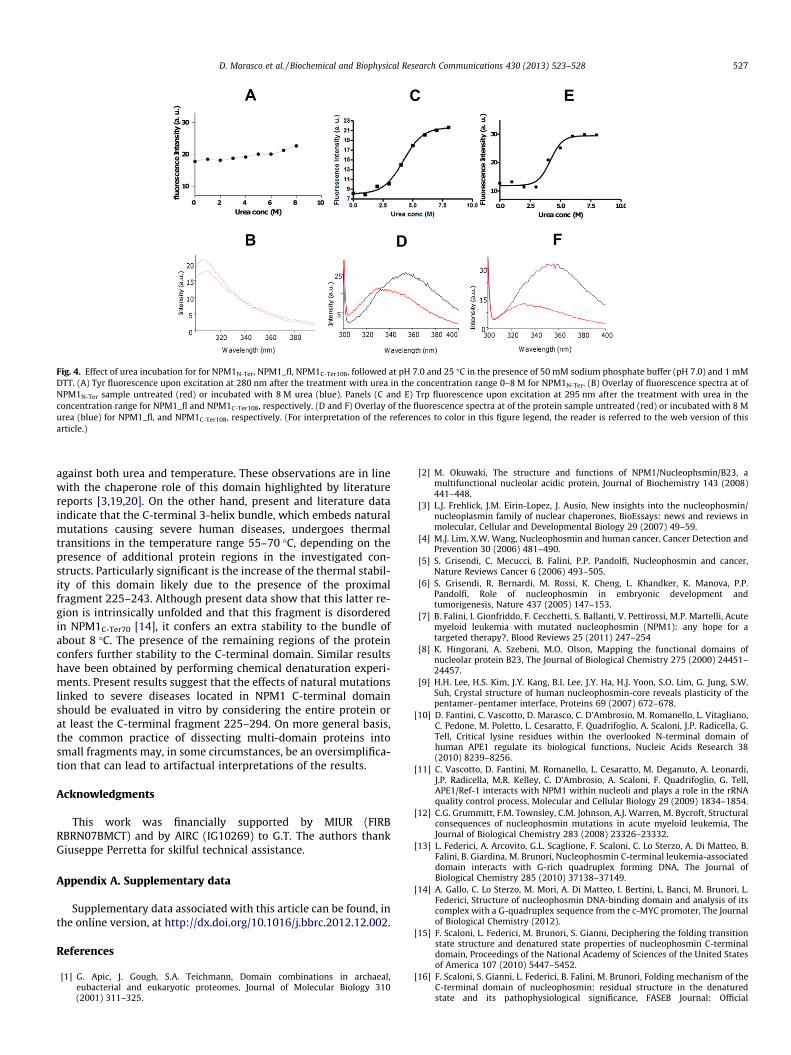
Fig. 4. Effect of urea incubation for for NPM1N-Ter, NPM1_fl, NPM1C-Ter108, followed at pH 7.0 and 25 �C in the presence of 50 mM sodium phosphate buffer (pH 7.0) and 1 mMDTT. (A) Tyr fluorescence upon excitation at 280 nm after the treatment with urea in the concentration range 0–8 M for NPM1N-Ter. (B) Overlay of fluorescence spectra at ofNPM1N-Ter sample untreated (red) or incubated with 8 M urea (blue). Panels (C and E) Trp fluorescence upon excitation at 295 nm after the treatment with urea in theconcentration range for NPM1_fl and NPM1C-Ter108, respectively. (D and F) Overlay of the fluorescence spectra at of the protein sample untreated (red) or incubated with 8 Murea (blue) for NPM1_fl, and NPM1C-Ter108, respectively. (For interpretation of the references to color in this figure legend, the reader is referred to the web version of thisarticle.)
D. Marasco et al. / Biochemical and Biophysical Research Communications 430 (2013) 523–528 527
against both urea and temperature. These observations are in linewith the chaperone role of this domain highlighted by literaturereports [3,19,20]. On the other hand, present and literature dataindicate that the C-terminal 3-helix bundle, which embeds naturalmutations causing severe human diseases, undergoes thermaltransitions in the temperature range 55–70 �C, depending on thepresence of additional protein regions in the investigated con-structs. Particularly significant is the increase of the thermal stabil-ity of this domain likely due to the presence of the proximalfragment 225–243. Although present data show that this latter re-gion is intrinsically unfolded and that this fragment is disorderedin NPM1C-Ter70 [14], it confers an extra stability to the bundle ofabout 8 �C. The presence of the remaining regions of the proteinconfers further stability to the C-terminal domain. Similar resultshave been obtained by performing chemical denaturation experi-ments. Present results suggest that the effects of natural mutationslinked to severe diseases located in NPM1 C-terminal domainshould be evaluated in vitro by considering the entire protein orat least the C-terminal fragment 225–294. On more general basis,the common practice of dissecting multi-domain proteins intosmall fragments may, in some circumstances, be an oversimplifica-tion that can lead to artifactual interpretations of the results.
Acknowledgments
This work was financially supported by MIUR (FIRBRBRN07BMCT) and by AIRC (IG10269) to G.T. The authors thankGiuseppe Perretta for skilful technical assistance.
Appendix A. Supplementary data
Supplementary data associated with this article can be found, inthe online version, at http://dx.doi.org/10.1016/j.bbrc.2012.12.002.
References
[1] G. Apic, J. Gough, S.A. Teichmann, Domain combinations in archaeal,eubacterial and eukaryotic proteomes, Journal of Molecular Biology 310(2001) 311–325.
[2] M. Okuwaki, The structure and functions of NPM1/Nucleophsmin/B23, amultifunctional nucleolar acidic protein, Journal of Biochemistry 143 (2008)441–448.
[3] L.J. Frehlick, J.M. Eirin-Lopez, J. Ausio, New insights into the nucleophosmin/nucleoplasmin family of nuclear chaperones, BioEssays: news and reviews inmolecular, Cellular and Developmental Biology 29 (2007) 49–59.
[4] M.J. Lim, X.W. Wang, Nucleophosmin and human cancer, Cancer Detection andPrevention 30 (2006) 481–490.
[5] S. Grisendi, C. Mecucci, B. Falini, P.P. Pandolfi, Nucleophosmin and cancer,Nature Reviews Cancer 6 (2006) 493–505.
[6] S. Grisendi, R. Bernardi, M. Rossi, K. Cheng, L. Khandker, K. Manova, P.P.Pandolfi, Role of nucleophosmin in embryonic development andtumorigenesis, Nature 437 (2005) 147–153.
[7] B. Falini, I. Gionfriddo, F. Cecchetti, S. Ballanti, V. Pettirossi, M.P. Martelli, Acutemyeloid leukemia with mutated nucleophosmin (NPM1): any hope for atargeted therapy?, Blood Reviews 25 (2011) 247–254
[8] K. Hingorani, A. Szebeni, M.O. Olson, Mapping the functional domains ofnucleolar protein B23, The Journal of Biological Chemistry 275 (2000) 24451–24457.
[9] H.H. Lee, H.S. Kim, J.Y. Kang, B.I. Lee, J.Y. Ha, H.J. Yoon, S.O. Lim, G. Jung, S.W.Suh, Crystal structure of human nucleophosmin-core reveals plasticity of thepentamer–pentamer interface, Proteins 69 (2007) 672–678.
[10] D. Fantini, C. Vascotto, D. Marasco, C. D’Ambrosio, M. Romanello, L. Vitagliano,C. Pedone, M. Poletto, L. Cesaratto, F. Quadrifoglio, A. Scaloni, J.P. Radicella, G.Tell, Critical lysine residues within the overlooked N-terminal domain ofhuman APE1 regulate its biological functions, Nucleic Acids Research 38(2010) 8239–8256.
[11] C. Vascotto, D. Fantini, M. Romanello, L. Cesaratto, M. Deganuto, A. Leonardi,J.P. Radicella, M.R. Kelley, C. D’Ambrosio, A. Scaloni, F. Quadrifoglio, G. Tell,APE1/Ref-1 interacts with NPM1 within nucleoli and plays a role in the rRNAquality control process, Molecular and Cellular Biology 29 (2009) 1834–1854.
[12] C.G. Grummitt, F.M. Townsley, C.M. Johnson, A.J. Warren, M. Bycroft, Structuralconsequences of nucleophosmin mutations in acute myeloid leukemia, TheJournal of Biological Chemistry 283 (2008) 23326–23332.
[13] L. Federici, A. Arcovito, G.L. Scaglione, F. Scaloni, C. Lo Sterzo, A. Di Matteo, B.Falini, B. Giardina, M. Brunori, Nucleophosmin C-terminal leukemia-associateddomain interacts with G-rich quadruplex forming DNA, The Journal ofBiological Chemistry 285 (2010) 37138–37149.
[14] A. Gallo, C. Lo Sterzo, M. Mori, A. Di Matteo, I. Bertini, L. Banci, M. Brunori, L.Federici, Structure of nucleophosmin DNA-binding domain and analysis of itscomplex with a G-quadruplex sequence from the c-MYC promoter, The Journalof Biological Chemistry (2012).
[15] F. Scaloni, L. Federici, M. Brunori, S. Gianni, Deciphering the folding transitionstate structure and denatured state properties of nucleophosmin C-terminaldomain, Proceedings of the National Academy of Sciences of the United Statesof America 107 (2010) 5447–5452.
[16] F. Scaloni, S. Gianni, L. Federici, B. Falini, M. Brunori, Folding mechanism of theC-terminal domain of nucleophosmin: residual structure in the denaturedstate and its pathophysiological significance, FASEB Journal: Official

528 D. Marasco et al. / Biochemical and Biophysical Research Communications 430 (2013) 523–528
Publication of the Federation of American Societies for Experimental Biology23 (2009) 2360–2365.
[17] G.B. Fields, R.L. Noble, Solid phase peptide synthesis utilizing 9-fluorenylmethoxycarbonyl amino acids, International Journal of Peptide andProtein Research 35 (1990) 161–214.
[18] C.N. Pace, K.L. Shaw, Linear extrapolation method of analyzing solventdenaturation curves, Proteins (Suppl. 4) (2000) 1–7.
[19] M. Okuwaki, K. Matsumoto, M. Tsujimoto, K. Nagata, Function ofnucleophosmin/B23, a nucleolar acidic protein, as a histone chaperone,Fatigue of Engineering Materials and Structures Letters 506 (2001) 272–276.
[20] A. Szebeni, M.O. Olson, Nucleolar protein B23 has molecular chaperoneactivities, Protein Science: A Publication of the Protein Society 8 (1999) 905–912.

Biochem. J. (2013) 452, 545–557 (Printed in Great Britain) doi:10.1042/BJ20121277 545
Role of the unstructured N-terminal domain of the hAPE1 (humanapurinic/apyrimidinic endonuclease 1) in the modulation of its interactionwith nucleic acids and NPM1 (nucleophosmin)Mattia POLETTO*, Carlo VASCOTTO*, Pasqualina L. SCOGNAMIGLIO†‡, Lisa LIRUSSI*, Daniela MARASCO†‡and Gianluca TELL*1
*Department of Medical and Biological Sciences, University of Udine, 33100 Udine, Italy, †Department of Pharmacy, CIRPEB (Centro Interuniversitario di Ricerca sui Peptidi Bioattivi),University of Naples ‘Federico II’, 80134 Naples, Italy, and ‡Institute of Biostructures and Bioimaging CNR, 80134 Naples, Italy
The hAPE1 (human apurinic/apyrimidinic endonuclease 1) is anessential enzyme, being the main abasic endonuclease in highereukaryotes. However, there is strong evidence to show that hAPE1can directly bind specific gene promoters, thus modulating theirtranscriptional activity, even in the absence of specific DNAdamage. Recent findings, moreover, suggest a role for hAPE1 inRNA processing, which is modulated by the interaction withNPM1 (nucleophosmin). Independent domains account for manyactivities of hAPE1; however, whereas the endonuclease and theredox-active portions of the protein are well characterized, a betterunderstanding of the role of the unstructured N-terminal region isneeded. In the present study, we characterized the requirementsfor the interaction of hAPE1 with NPM1 and undamaged nucleic
acids. We show that DNA/RNA secondary structure has an impacton hAPE1 binding in the absence of damage. Biochemical studies,using the isolated N-terminal region of the protein, reveal that thehAPE1 N-terminal domain represents an evolutionary gain offunction, since its composition affects the protein’s stability andability to interact with both nucleic acids and NPM1. Althoughrequired, however, this region is not sufficient itself to stablyinteract with DNA or NPM1.
Key words: apurinic/apyrimidinic endonuclease 1 (APE1), nuc-leophosmin (NPM1), protein–DNA interaction, protein–proteininteraction, phylogenesis.
INTRODUCTION
The hAPE1 [human APE1 (apurinic/apyrimidinic endonuclease1)] is an essential and multifaceted protein involved in the DNABER (base excision repair) pathway and in the regulation ofgene expression, acting both as a redox co-activator for severaltranscription factors and as a transcriptional repressor on nCaRE(negative calcium response elements)-containing promoters[1–3]. These biological activities are located in two distinct, butpartially overlapping, regions. The conserved C-terminal portion(residues 61–318) is responsible for the enzymatic activityon abasic DNA, whereas the N-terminal domain (residues1–127) is mainly devoted to the redox co-activating functiontowards different transcription factors such as Egr1 (early growthresponse 1), NF-κB (nuclear factor κB) and p53 [4,5]. Themechanisms of hAPE1 binding and recognition of its canonicalsubstrate, the abasic site containing DNA, have been thoroughlyinvestigated by several laboratories [6–8]. However, the notionthat hAPE1 is constitutively bound to undamaged nucleic acids isgetting stronger. Examples include the interaction of the proteinwith genomic regions, such as the nCaRE sequences [2] orthe MDR1 (multidrug resistance protein 1) gene promoter [9],and the reported association of hAPE1 with undamaged RNA[10]. Acquisition of particular secondary structures (i.e. bulges,hairpins, cruciforms, quadruplex etc.) by nucleic acids and theirregulatory roles in vivo are a well-known paradigm in molecularbiology. A previous study has carefully explored the interactionbetween hAPE1 and undamaged nucleic acids [11]; nonetheless,
potential impacts of the secondary structure of the nucleic acidon the binding affinity of hAPE1 have never been investigated.
In the last few years, an unexpected involvement of hAPE1 inRNA metabolism has been unveiled: the protein is accumulatedwithin nucleoli through interaction with NPM1 (nucleophosmin;also known as nucleolar phosphoprotein B23) and rRNA andaffects cell growth by directly acting on the RNA qualitycontrol mechanisms [10,12,13]. Notably, the N-terminal domainof hAPE1 (the first 43 residues) plays a pivotal role in this non-canonical function of the protein, serving as an adaptable devicefor protein–protein interactions and modulating its enzymaticactivity on abasic DNA, as well as its binding to RNAmolecules. Although many studies have thoroughly characterizedthe structure of the hAPE1 C-terminal globular domain, only afew have addressed the role of its N-terminal extension, perhaps asa consequence of its intrinsic disordered folding [14–16]. A basiccluster of lysine residues located within this region (Lys25–Lys35)controls the binding affinity of hAPE1 for both NPM1 and nucleicacids. Furthermore, several strands of evidence suggest that thedifferent functions of hAPE1 might be rapidly modulated in vivoby post-translational modifications on its N-terminal domain,such as ubiquitination, acetylation and proteolysis [2,17–21].Interestingly, whereas the C-terminal portion of the protein sharesextensive similarity with most of its orthologue counterparts,from Escherichia coli to mammals, its N-terminal extension isconsiderably less conserved. The importance of this protein regionin modulating the many activities of hAPE1 suggests that the N-terminal domain may be a recent acquisition during evolution
Abbreviations used: APE1, apurinic/apyrimidinic endonuclease 1; BER, base excision repair; DSP, dithiobis(succinimidyl propionate); DTT, dithiothreitol;FBS, fetal bovine serum; GST, glutathione transferase; hAPE1, human APE1; MAT, metal-affinity tag; nCaRE: negative calcium response elements; NPM1,nucleophosmin; PLA, proximity ligation assay; RU, response unit; SPR, surface plasmon resonance; zAPE1, zebrafish APE1.
1 To whom correspondence should be addressed (email [email protected]).
c© The Authors Journal compilation c© 2013 Biochemical Society
Bio
chem
ical
Jo
urn
al
ww
w.b
ioch
emj.o
rg

546 M. Poletto and others
[17], as already suggested for other BER proteins [22]. AberranthAPE1 expression and localization patterns have been linkedto neurodegeneration and cancer; therefore, many groups arecurrently directing their efforts to develop specific small-moleculemodulators of the different activities of the protein [23].
In the present study, we systematically characterized thefunctional properties of the hAPE1 N-terminal domain, exploringits nucleic acid-binding properties and its role in hAPE1–NPM1interaction. In an effort to better characterize the features guidingthe binding of hAPE1 to undamaged nucleic acids, we used acombined approach based on SPR (surface plasmon resonance),EMSA analyses and GST (glutathione transferase) pull-downexperiments. The results of the present study show that the affinityof hAPE1 for nucleic acids is enhanced by certain structuralelements and that the nucleic acid-binding activity of hAPE1 ispromoted by the N-terminal extension of the protein. This region,although necessary for both the stabilization of the hAPE1–nucleic acids and hAPE1–NPM1 interactions, shows only a weakaffinity for DNA or NPM1 when isolated from the hAPE1 globulardomain. Finally, exploiting a ‘domain swapping’ approach, wedemonstrate that lysine residues located within the N-terminaldomain of hAPE1 represent a phylogenetic gain of functionfor the protein, since the composition of this domain influencesthe stability of the protein and its ability to interact with bothnucleic acids and NPM1. Altogether, these data highlight therole for the hAPE1 N-terminal region in modulating the manyfunctions of this pleiotropic protein. Further structural analyseson this overlooked extension of the protein would be thereforeworthwhile in order to fully understand the substrate specificity ofhAPE1 and to design new anti-cancer drugs targeting this protein.
EXPERIMENTAL
Cell culture and transient transfection experiments
HeLa cells were grown in DMEM (Dulbecco’s modified Eagle’smedium) supplemented with 10% FBS (fetal bovine serum),100 units/ml penicillin and 10 mg/ml streptomycin sulfate. At1 day before transfection, cells were seeded in 10-cm platesat a density of 3×10⁶ cells/plate. Cells were then transientlytransfected with 6 μg of either pcDNA5.1 (empty vector) orpcDNA5.1-hAPE1 (full-length or amino acids 1–49) plasmidsper dish using the LipofectamineTM 2000 Reagent (Invitrogen)according to the manufacturer’s instructions. Cells were harvestedat 24 h after transfection.
Multiple sequence alignments
For the multiple sequence alignments full-length hAPE1orthologous metazoan sequences (human, Pan troglodytes,Macaca mulatta, Equus caballus, Mus musculus, Rattusnorvegicus, Canis lupus familiaris, Sus scrofa, Bos taurus,Ornithorhyncus anatinus, Salmo salar, Gasterosteus aculeatus,Danio rerio, Xenopus tropicalis and Xenopus laevis) wereretrieved from the RefSeq protein database using theBLAST algorithm (http://blast.ncbi.nlm.nih.gov/Blast.cgi) andaligned using the ClustalW2 program with default parameters(http://www.ebi.ac.uk/Tools/msa/clustalw2/ ).
Western blotting analyses
The protein samples were electrophoresed by SDS/PAGE (10–18% gels) or discontinuous Tris/Tricine SDS/PAGE [24].Proteins were then transferred on to nitrocellulose membranes(PerkinElmer), developed as described previously [12] using an
ECL (enhanced chemiluminescence) procedure (GE Healthcare)and then quantified by using a ChemiDoc XRS video-densitometer (Bio-Rad Laboratories).
Plasmids and expression of recombinant proteins
The constructs pGEX-3X-hAPE1 and pGEX-3X-zAPE1, enco-ding for the GST-fused hAPE1 and zAPE1 (zebrafish APE1)full-length proteins respectively, were provided by Dr MarkR. Kelley (Indiana University, Indianapolis, IN, U.S.A.). Theplasmid encoding for full-length GST–NPM1 was provided byDr P.G. Pelicci (European Institute of Oncology, Milan, Italy).The pTAC-MAT/hAPE1 and the pETM-20/zAPE1 constructs,encoding for C-terminal-tagged MAT (metal-affinity tag)–hAPE1and His6–zAPE1 respectively, were generated by subcloning. Allproteins were expressed in E. coli BL21(DE3) cells, inducedwith 1 mM IPTG (isopropyl β-D-thiogalactopyranoside) and thenpurified on an AKTA Purifier FPLC system (GE Healthcare)by using a GSTrap HP column (GE Healthcare) for the GST-tagged proteins, or a HisTrap HP column (GE Healthcare) for theHis6- and MAT-tagged proteins. The quality of purification waschecked by SDS/PAGE analysis. Extensive dialysis against PBSwas performed to remove any trace of imidazole from the HisTrap-purified proteins. Accurate quantification of all recombinantproteins was performed by colorimetric Bradford assays (Bio-RadLaboratories) and confirmed by SDS/PAGE and Western blottinganalysis. To remove the GST tag, GST–hAPE1 was furtherhydrolysed with Factor Xa as described previously [17]. ThezAPE1 K27 ‘swapping mutant’ and the hAPE1 N�43 and zAPE1N�36 deletion mutants, as well as the hAPE1 1–48 and 1–49constructs, were generated using the QuikChange Mutagenesis kit(Stratagene) and the respective full-length constructs as templates,following the manufacturer’s instructions and using standardsubcloning procedures.
GST pull-down assays with RNA or NPM1 andco-immunoprecipitation
The indicated amount of GST-tagged protein was mixed, togetherwith the prey protein or 10 μg of total RNA, with 10 μl ofglutathione–Sepharose 4B beads (GE Healthcare). RNA wasextracted using the TRIzol® Reagent (Invitrogen). Binding wasperformed in PBS supplemented with 5 mM DTT (dithiothreitol)and 0.5 mM PMSF for 2 h under rotation at 4 ◦C. The beadswere washed three times with washing buffer [PBS supplementedwith 0.1% Igepal CA-630 (Sigma), 5 mM DTT and 0.5 mMPMSF]. Beads were then resuspended in Laemmli sample bufferfor Western blotting analyses or in TRIzol® Reagent for RNAextraction and rRNA quantification. The levels of 28S and 18SrRNA transcripts were determined as described previously [17]by using the iScript cDNA synthesis kit (Bio-Rad Laboratories)according to the manufacturer’s instructions. Quantitative PCRwas then performed in an iQ5 multicolour real-time PCR detectionsystem (Bio-Rad Laboratories) according to the manufacturer’sprotocol. GST-tagged proteins were detected with an HRP(horseradish peroxidase)-conjugated anti-GST antibody (Sigma),whereas the prey proteins were detected using the indicatedantibodies. Co-immunoprecipitation analyses were performed asdescribed previously [12].
DSP [dithiobis(succinimidyl propionate)] cross-linking
The indicated amounts of recombinant NPM1 and hAPE1 (eitherfull-length or the amino acids 1–48 peptide), were allowed to
c© The Authors Journal compilation c© 2013 Biochemical Society

Multiple roles of unstructured APE1 N-terminal domain 547
interact at 37 ◦C for 15 min in a total volume of 10 μl; DSP (2 μgin DMSO) was added and cross-linking was carried out for 30 minat room temperature (22 ◦C). The reactions were quenched byadding 0.5 volumes of ice-cold 0.4 M ammonium acetate andincubating on ice for 10 min. Non-reducing Laemmli samplebuffer was immediately added and the samples were separatedby SDS/PAGE (8% gel).
Immunofluorescence confocal analyses and PLA (proximityligation assay)
Immunofluorescence was carried out as described previously [12].FLAG-tagged 1–49 hAPE1 was labelled using an anti-FLAGantibody and stained with a secondary anti-mouse Alexa FluorTM
488-conjugated antibody (Jackson ImmunoResearch). Nucleiwere counterstained with DAPI (4′,6-diamidino-2-phenylindole).For the PLA analyses we used the Duolink II® reagent kit (OlinkBioscience); cells were fixed in 4 % (w/v) paraformaldehyde,permeabilized with 0.25% PBS-Triton (PBS with Triton X-100)and saturated with 10% (v/v) FBS. Slides were incubated withprimary antibodies (mouse anti-FLAG, 1:100 dilution and rabbitanti-NPM1, 1:200 dilution) and then reactions were carried outfollowing the manufacturer’s instructions. Microscope slides weremounted and visualized through a Leica TCS SP laser-scanningconfocal microscope.
hAPE1–nucleic acid binding through EMSA
hAPE1 binding to nucleic acids was assessed as describedpreviously [1] with some modifications. Briefly, in order to reduceunspecific binding and to obtain more defined bound complexes,recombinant proteins or peptides were incubated at 37 ◦C with250 pmol of unlabelled poly(dT) for 15 min and then 2.5 pmol of32P-labelled probe was added. The reactions were then incubatedfor further 15 min and separated by native PAGE (6–10% gel) at150 V for 4 h.
Southwestern assays
Bait proteins were separated using a pre-cast SDS/PAGE (Bio-Rad Laboratories), electrotransferred on to a nitrocellulosemembrane and subsequently denatured and renatured by washingsix times in 1:1 serial dilutions of 6 M guanidinium/HCl in EMSAbuffer [40 mM Hepes (pH 8.0), 50 mM KCl, 2 mM EDTA, 10%(v/v) glycerol and 1 mM DTT]. The membrane was then saturatedin 5% (w/v) BSA in EMSA buffer and incubated with 50 pmol ofthe indicated 32P-labelled DNA probe at 37 ◦C, washed in EMSAbuffer to remove unspecific bound probe and finally subjected toautoradiography.
SPR
Real-time binding assays were performed on a Biacore T-100SPR instrument (GE Healthcare). Streptavidin was immobilizedon to a research-grade CM5 sensor chip using amine-couplingchemistry. The immobilization steps were carried out at a flowrate of 10 μl/min in Hepes buffer {20 mM Hepes, 150 mM NaCl,3.4 mM EDTA, 0.005 % P20 surfactant and 0.1 mM TCEP [tris-(2-carboxyethyl)phosphine]}. All surfaces were simultaneouslyactivated for 30 s with a mixture of NHS (N-hydroxysuccinimide;0.05 M) and EDC [N-ethyl-N ′-(3-dimethylaminopropyl)carbodi-imide; 0.2 M]. Streptavidin was injected at a concentration of20 μg/ml in 10 mM sodium acetate (pH 4.5) for 1 min. Ethan-
olamine (1 M, pH 8.5) was injected for 7 min to saturate the re-maining activated groups. An average of 5000 RU (response units)of streptavidin was immobilized on each flow cell. Biotinylatedoligonucleotides were injected at a concentration of 500 nM untilthe desired level of immobilization was achieved (average value of300 RU). Proteins were serially diluted in running buffer (80 nM–2.0 μM) and injected at 20 ◦C at a flow rate of 60 μl/min for 1 min.Disruption of any complex that remained bound after a 5-mindissociation was achieved using a 30 s injection of 1 M NaCl at100 μl/min. When the experimental data met the quality criteria,the kinetic parameters were estimated assuming a 1:1 bindingmodel and using version 4.1 Evaluation Software (GE Health-care). Conversely, an affinity steady-state model was applied tofit the RUmax data compared with the protein concentrations andfitting was performed with GraphPad Prism v4.00 [17].
CD
Far-UV CD spectra were recorded on a Jasco J-810spectropolarimeter (JASCO) in a 195–260 nm interval.Experiments were performed employing a protein concentrationof 5 μM in 10 mM phosphate buffer (pH 7.0), supplemented with1 mM DTT and using a 0.1 cm path-length cuvette. Thermaldenaturation profiles were obtained by measuring the temperaturedependence of the signal at 225 nm in the 20–100 ◦C range with aresolution of 0.5 ◦C and 1.0-nm bandwidth. A Peltier temperaturecontroller was used to set up the temperature of the sample withthe heating rate set to 1 ◦C/min. Data were collected at a 0.2-nmresolution, 20 nm/min scan speed and a 4 s response and werereported as the unfolded fraction against the temperature.
Statistical analyses
Statistical analyses were performed by using the Microsoft Exceldata analysis software for Student’s t test analysis. P < 0.05 wasconsidered statistically significant.
RESULTS AND DISCUSSION
The binding affinity of hAPE1 for undamaged nucleic acids isdependent on their secondary structure
Several studies have thoroughly characterized hAPE1 structureand its enzymatic activities on abasic DNA, examining itssubstrate specificity and recognition mechanisms [7,11,25–28].However, when considering undamaged structured nucleic acids,the knowledge of hAPE1 substrate selectivity is still scarce.In the present study we sought to determine whether some partic-ular structural features were favoured by hAPE1 when binding tonucleic acids. Hence, we used a biophysical approach exploitingthe SPR technique to quantitatively measure the binding affinityof hAPE1 for intact oligonucleotides [29,30]. We comparedthe binding affinity of recombinant purified full-length hAPE1toward different single-stranded DNA probes, either intrinsicallylacking secondary structure (i.e. 34dT), or having a stem andloop-like folding (e.g. Stem10 and Stem20) as predicted bybioinformatics analyses (Figure 1A and Table 1). The results ofthe SPR experiments (representative sensorgrams in Figure 1Band Supplementary Figure S1C at http://www.biochemj.org/bj/452/bj4520545add.htm) are summarized in Table 2. As alreadyobserved by Beloglazova et al. [11], an increased length inthe stem region of the oligonucleotides augmented the affinityof hAPE1 for undamaged DNA (compare the Stem10 and theStem20 sequences). This observation was confirmed by directEMSA experiments (Figure 1C) and by competition EMSA
c© The Authors Journal compilation c© 2013 Biochemical Society

548 M. Poletto and others
Figure 1 The secondary structure of the substrate determines the binding selectivity of hAPE1
(A) The predicted structures for the oligonucleotides used in the present study. Calculations were performed using either the Mfold web server (http://mfold.rna.albany.edu/?q=mfold) or the DINAMeltweb server (http://mfold.rna.albany.edu/?q=DINAMelt). The calculated minimum free energy for folding (�G) at 37◦C and 50 mM Na+ is also reported. The arrow indicates the absence of loop inthe Stem20 no loop probe. (B) Representative overlay sensorgrams relative to a typical SPR experiment for the binding of hAPE1 to immobilized Biot-Stem20. The kinetic parameters were measuredinjecting purified recombinant hAPE1 solutions at increasing concentrations (i.e. 87.5, 175, 350, 700, 1050, 1400 and 1750 nM). The K d values were estimated for each concentration using theBIAevaluation v.4.1 software. (C) Positive effect of the stem length on ability of hAPE1 to bind undamaged DNA as evaluated through EMSA using the Stem10 and Stem20 oligonucleotides (Table 1).For each reaction the indicated amount of purified recombinant hAPE1 was used. The results confirm a different binding capacity of hAPE1 towards double-stranded oligonucleotides of differentlengths. The arrow indicates the hAPE1–DNA complexes, whereas F denotes the position of the free oligonucleotide probe. wt, wild-type.
c© The Authors Journal compilation c© 2013 Biochemical Society

Multiple roles of unstructured APE1 N-terminal domain 549
Table 1 Oligonucleotide sequences used in the present study
The underlined nucleotides denote the position of the predicted loops.
Name Sequence
34dT 5′-TTTTTTTTTTTTTTTTTTTTTTTTTTTTTTTTT-3′
Stem10 5′-CGATCGACGGATCCCCGTCGATCG-3′
Stem10 3′ A 5′-CGATCGACGGATCCCCGTCGATCGAAAAAA-3′
Stem10 5′ A 5′-AAAAAACGATCGACGGATCCCCGTCGATCG-3′
Stem20 5′-ATCGATCGATCGATCGACGGATCCCCGTCGATCGATCGATCGAT-3′
Stem20 no loop Forward, 5′-ATCGATCGATCGATCGACGGAT-3′ and reverse,5′-CCCCGTCGATCGATCGATCGAT-3′
Stem20_2 5′-CGATCGATCGATCGATCGACGGATCCCCGTCGATCGATCGATCGATGC-3′
Stem20 Loop8 5′-CGATCGATCGATCGATCGACGGATCCTAGTCGATCGATCGATCGATCG-3′
Bulge Forward, 5′-CATTACTCATATCCCCGTCCATTG-3′ and reverse,5′-CAATGGACGGTACAATGAGTAATG-3′
nCaRE_B2 Forward, 5′-TTTTTGAGACGGAGTTTCGCTCTTG-3′ and reverse,5′-CAAGAGCGAAACTCCGTCTCAAAAA-3′
Table 2 Dissociation constants and kinetic parameters for the interactionof full-length hAPE1 with different undamaged oligonucleotides
Data reported were obtained through SPR analyses using hAPE1 as analyte on the indicatedbiotinylated oligonucleotide ligands. The kinetic parameters highlight the importance ofsecondary structure for the recognition of undamaged nucleic acids by hAPE1.
Ligand k on (M− 1 · s− 1×10⁵) k off (s− 1) Kd (μM)
34dT 0.004 0.112 308 +− 3.00Stem10 0.270 0.020 1.03 +− 0.02Stem20 0.292 0.007 0.25 +− 0.01Stem20 no loop 0.068 0.024 4.06 +− 0.01Stem10 3′A 0.060 0.240 6.20 +− 0.01Stem10 5′A 0.059 0.059 2.11 +− 0.03Stem20_2 0.284 0.007 0.25 +− 0.01Stem20 Loop8 0.061 0.004 0.74 +− 0.02Bulge 0.834 0.019 0.23 +− 0.02nCaRE_B2 0.270 0.105 3.90 +− 0.80
experiments demonstrating the poor competitive ability of theStem10 toward the Stem20 oligonucleotide (results not shown).The effect of the presence of an intact loop structure on the stabilityof the protein–DNA complex then was investigated by comparingthe affinities of the Stem20 and the Stem20 no loop sequences.Notably, hAPE1 showed a reduced affinity toward the ligandwithout a loop (approximately 16-fold, Table 2). Accordingly,EMSA experiments confirmed these findings, showing a morestable hAPE1–DNA complex in the presence of an intact loop(Supplementary Figures S1A and S1B). Furthermore, competitiveEMSA assays demonstrated that the Stem20 probe was a moreefficient competitor if compared with the Stem20 no loop (resultsnot shown). Binding affinities were also negatively affected by thepresence of an unstructured nucleotide stretch either 3′ or 5′ to thedouble-stranded stem region (compare Stem10 3′A and Stem105′A with the Stem10 sequence). Experimental data allowed us tospeculate that the steric hindrance of a free poly(A) tail on thedouble-stranded region bound by hAPE1 might acceleratethe dissociation rate, if protruding from the 3′-side. Whenusing the same stem length the size of the loop region affectedthe binding: the expansion from 4 to 8 nucleotides reduced theaffinity of hAPE1 approximately 3-fold (compare the Stem20_2and the Stem20 Loop8 sequence). The difference in Kd value canbe accounted for by a reduction in the association rate, suggestingthat hAPE1 preferentially approaches the DNA molecules fromthe loop side as the stem side remains invariant between thetwo oligonucleotides. A lower stability of the stem region in the
Stem20 Loop8 sequence could also reflect the decreased bindingaffinity; bioinformatic predictions, however, do not suggest anymajor difference in the unfolding free energy of the two molecules(Figure 1A). We finally investigated whether the position ofthe unstructured region might affect the binding capacity ofhAPE1 (compare Bulge, Stem20 and Stem20_2). Despite thedifferences in both the dissociation and the association rates,the overall affinity of hAPE1 for the oligonucleotides remainedunchanged, suggesting that a local denaturation of the doublehelix, rather than its location within the double-stranded region,is a feature that enhances binding of hAPE1 to undamaged DNA.To compare our measurements on synthetic oligonucleotideswith a physiologically relevant undamaged DNA substrate, weinvestigated the binding properties of hAPE1 towards an nCaREduplex sequence, namely nCaRE_B2 (G. Antoniali, L. Lirussi, C.D’Ambrosio, F. Dal Piaz, C. Vascotto, D. Marasco, A. Scaloni,F. Fogolari and G. Tell, unpublished work). This sequencedisplayed a Kd value akin to that of the unstructured Stem20no loop sequence (Table 2). It is worth pointing out that ourin vitro assays, performed with the isolated purified recombinantprotein, might underestimate the real affinity of hAPE1 for thissequence, since interactions with other protein partners [e.g.Ku70/80 and hnRNPL (heterogeneous nuclear ribonucleoproteinL)] significantly increase the protein–DNA complex stability, asalready demonstrated previously (G. Antoniali, L. Lirussi, C.D’Ambrosio, F. Dal Piaz, C. Vascotto, D. Marasco, A. Scaloni, F.Fogolari and G. Tell, unpublished work) [31].
EMSA analyses were also performed on RNA substrates totest whether the nature of the sugar backbone might affect thebinding activity of hAPE1 toward structured nucleic acids. Theresults obtained confirmed that the structural requirements forhAPE1 binding to undamaged substrates are not influenced bythe composition of the sugar backbone (results not shown). Theseobservations suggest that hAPE1 shows no apparent preferencefor DNA or RNA substrates, as long as the nucleic acid isundamaged and is endowed with a secondary structure. The resultsof the present study are in agreement with experimental datafrom other laboratories that have estimated Kd values for theinteraction of hAPE1 with structured RNA that is similar to ourvalues for DNA (∼0.9 μM) [32]. It must be noted, however, thatthese experiments do not allow the exclusion of the existence of adifferential affinity of hAPE1 for abasic site-containing DNA orRNA molecules, as has been suggested by others [12,26].
In summary, hAPE1 binding to undamaged nucleic acidsappears to be strongly affected by the presence of secondarystructural elements. In particular, the presence of a double-stranded region is a primary element required for hAPE1 binding.The occurrence of a single-stranded local distortion, moreover,greatly enhances the interaction with nucleic acids. These factorsare corroborated by studies reporting a significant reduction inthe APE activity of the protein on single-stranded unstructured(or poorly structured) substrates (e.g. [7]). The requirement of adouble-stranded DNA region is conceivable, on the basis of theobservation that hAPE1 contacts both of the DNA strands [27,28].The presence of a loop or a bulge within a double-stranded stemcould mimic a local denaturation region that resembles a baselessspot, hence explaining the increased affinity for loop-containingstructures displayed in vitro by hAPE1.
The N-terminal domain contributes to nucleic acid-bindingby hAPE1
We have shown previously that the N-terminal 33 amino acids ofhAPE1 is able itself to bind RNA oligonucleotides in solution [17].Sequence alignment analyses, performed on different metazoan
c© The Authors Journal compilation c© 2013 Biochemical Society

550 M. Poletto and others
Figure 2 The isolated N-terminal region of hAPE1 interacts weakly with nucleic acids
(A) Multiple sequence alignment between full-length APE1 protein sequences from 15 different metazoan species. Only the N-terminal region of the alignment is reported. The arrow indicates thebeginning of the high-similarity region within the C-terminal globular domain; whereas the N-terminal part of the sequences is significantly less conserved. (B) The isolated recombinant 1–48 hAPE1N-terminal peptide is able to compete (comp.) with the full-length protein for RNA binding. The binding ability of the peptide was evaluated through competition RNA pull down using GST-taggedfull-length hAPE1 as the bait and whole HeLa RNA as the prey in the presence of the indicated competitor. To quantify the amount of residual hAPE1-bound rRNA the pulled-down RNA was thenretrotranscribed and 28S or 18S rRNAs were amplified by real-time PCR. The affinity for the 18S rRNA was similar to that shown for the 28S (results not shown). The competition activity of the1–48 hAPE1 peptide was also confirmed using an untagged synthetic 1–33 hAPE1 peptide. The histogram shows the relative affinity to rRNA with respect to the GST–hAPE1 sample. The error barsrepresent the S.D. (n = 3). *P < 0.05. (C) EMSA dose–response analysis showing that the 1–48 peptide binds undamaged DNA with a low affinity when compared with the full-length hAPE1 protein.For each reaction the indicated amount of purified recombinant protein was used. F denotes the position of the free oligonucleotide probe. (D) Southwestern blot assay showing a dose–responsebinding of the nCaRE_B2 duplex to the 1–48 peptide. The indicated amounts of protein or peptide were separated by SDS/PAGE, electrotransferred on to nitrocellulose membranes (left-hand panel)and probed with 32P-labelled nCaRE_B2 (right-hand panel). Note the difference in binding affinity between the full-length hAPE1 protein and the isolated peptide. MT, Mat; wt, wild-type.
species, highlight that the phylogenetic conservation of the aminoacid sequence among APE1 proteins from different organismsstarts at position 48 in the human protein, and is significantly lower
in the region spanning amino acids 1–48 (Figure 2A). In orderto better clarify the relevance of the non-conserved N-terminaldomain in modulating the binding ability of hAPE1 to nucleic
c© The Authors Journal compilation c© 2013 Biochemical Society

Multiple roles of unstructured APE1 N-terminal domain 551
acids, we cloned and expressed in E. coli cells the 1–48 aminoacid region of hAPE1 in fusion with a C-terminal MAT (Sup-plementary Figures S2A and S2B at http://www.biochemj.org/bj/452/bj4520545add.htm) and evaluated the binding propertiesof the recombinant purified peptide toward different substrates,including DNA and RNA. We indirectly estimated the rRNA-binding activity of the peptide exploiting a GST pull-downcompetition strategy. In these experiments, the full-length GST–hAPE1 was used as the bait and total HeLa RNA as the prey,while either the recombinant purified 1–48 peptide or its syntheticuntagged 1–33 counterpart were used as soluble competitors. Thedata obtained revealed that both soluble peptides were able tocompete with full-length hAPE1 for rRNA binding, independentlyof the presence of a tag (Figure 2B).
In order to compare the nucleic acid-binding activity ofthe 1–48 peptide and that of the full-length hAPE1 protein,we used EMSA analyses. Under our experimental conditions weobserved a low-affinity binding activity for the hAPE1 N-terminalpeptide towards the nCaREB_2 duplex sequence (Figure 2Cand Supplementary Figure S2C). Although at a reduced affinity,however, the interaction with DNA was specific, as demonstratedby both Southwestern assays (Figure 2D and SupplementaryFigure S2D) and Western blotting performed after the EMSAanalysis (Supplementary Figure S2E). These results, along withthe notion that the N-terminal 1–33 amino acid portion of hAPE1is absolutely required for the stable interaction of the proteinwith nucleic acids [12,17], suggest that this lysine-rich regionmight act by stabilizing the association of hAPE1 with nucleicacids through electrostatic interactions, whereas the remaining C-terminal domain accounts for the specific binding to DNA/RNA.Our data support a two-step model to describe the mechanismthrough which hAPE1 contacts nucleic acids. The unstructuredN-terminal domain would be required for the preliminary low-affinity binding process in search of the proper lesion to berepaired, as suggested for other DNA-binding proteins [33].Recognition of a local distortion (or an abasic site) involvesand is stabilized by a higher-affinity binding event. The wholehAPE1 molecule would, at this point, become necessary for therecognition of the structured nucleic acid region. Intriguingly, thismodel nicely fits previous hypothesis proposed by Masuda et al.[34] more than a decade ago.
The N-terminal domain of hAPE1 is necessary, but not sufficient,for a stable binding to NPM1
In our previous work, we demonstrated that the N-terminal 1–33 amino acid domain of hAPE1, besides being essential forstabilizing its binding to nucleic acids, is also required for a stableinteraction with NPM1 [12] and that lysine residues spanningthe region 27–35 of hAPE1 are directly involved in stabilizingthis interaction [17]. However, we did not check whether the N-terminal domain of hAPE1 was able to directly bind to NPM1itself. In an effort to better characterize the protein–proteininteraction features of the hAPE1 N-terminal domain and itsrelative contribution to the overall binding, we analysed the abilityof the 1–48 peptide to directly interact with NPM1. We expressedboth the full-length hAPE1 and the 1–48 peptide in fusion with aMAT. The recombinant proteins were affinity purified and testedfor their binding ability toward GST-tagged NPM1 through GSTpull-down assays (Figure 3A). Although the full-length hAPE1protein was able to directly interact with NPM1 (Figure 3A,left-hand panel), we failed to detect any stable peptide–NPM1association in vitro (Figure 3A, right-hand panel), even under lessstringent binding conditions (results not shown). This negative
result prompted us to hypothesize that, if present, any peptide–NPM1 association could be too weak to be detected through adirect pull-down assay. We confirmed this hypothesis through acompetition GST pull-down assay, in which the 1–48 peptidewas used to squelch the binding occurring between the full-length hAPE1 and GST–NPM1 (Figure 3B). The peptide, aswell as an untagged full-length hAPE1, was able to displace theassociation between the MAT-tagged hAPE1 and GST–NPM1.However, whereas the competitor untagged hAPE1 was alsoefficiently recovered in the pulled-down fraction, the competitor1–48 peptide was not. This indicates that the peptide was ableto mask the hAPE1-binding site on NPM1, thus competing withthe hAPE1–NPM1 association, although its interaction with theprotein was too weak to be maintained during the washing steps.The observed competition effect could also be interpreted byassuming a masking effect of the peptide on the full-length hAPE1N-terminal region, however, to the best of our knowledge, nooligomerization ability has ever been described for hAPE1.
To definitely prove the ability of the N-terminal peptideto directly associate with NPM1, we performed an in vitrostabilization of the complex exploiting the bifunctional cross-linker DSP (Figure 3C). As expected, treatment of NPM1 alonewith DSP resulted in the stabilization of slow migrating oligomers,as documented previously [35]. Moreover, the full-length hAPE1protein and its N-terminal peptide were detected as slow migratingsignals only in the presence of both NPM1 and DSP, indicating adirect association for both with NPM1. The lower affinity of the1–48 peptide toward NPM1, in comparison with that of the full-length protein, indicates that the C-terminal domain of hAPE1contributes to the stabilization of the protein–protein interaction.
To evaluate whether the interaction between the hAPE1 N-terminal extension and NPM1 also occurs in vivo, we created aHeLa cell line expressing a FLAG-tagged 1–49 hAPE1 peptideupon treatment with doxycycline, as already described previously[36]. As assessed using Western blotting, the peptide expressionincreased after the addition of doxycycline to the culture medium(Figure 4A, upper panel). Interestingly, the ectopically expressedpeptide showed a peculiar localization pattern, being cytoplasmicand nuclear, but excluded from nucleoli (Figure 4A, lower panel),in contrast with the nuclear/nucleolar accumulation observedfor the wild-type protein both under basal conditions [12,21]and when ectopically expressed (Figure 4A, lower panel). Thisresult confirms that the C-terminal domain of hAPE1 contributesto the nucleolar accumulation of the protein within cells,despite the fact that the N-terminal domain retains the nuclearlocalization signal of the protein [37]. This phenomenon maybe explained by the relevance that the C-terminal domain hasin stabilizing both rRNA and NPM1 binding. By exploiting thiscell model, we performed co-immunoprecipitation experimentsand demonstrated the occurrence of a molecular interactionbetween NPM1 and the isolated hAPE1 N-terminal peptide.However, the FLAG-tagged 1–49 peptide was only able to co-precipitate NPM1 poorly when compared with the full-lengthprotein (Figure 4B). This result parallels our in vitro results withrecombinant purified proteins and confirms that the unstructuredN-terminal region of hAPE1 does indeed interact with NPM1,albeit with a lower affinity in the absence of the remainingC-terminal domain. We further confirmed the presence of aninteraction occurring between the FLAG-tagged 1–49 hAPE1peptide and endogenous NPM1 using a PLA [21], which allowsthe quantification of physical proximity between moleculesat a distance lower than 40 nm (Supplementary Figure S3Aat http://www.biochemj.org/bj/452/bj4520545add.htm) [38]. ThePLA signal was present in the nucleoplasmic compartment only inthe presence of both the anti-FLAG and the anti-NPM1 antibodies,
c© The Authors Journal compilation c© 2013 Biochemical Society

552 M. Poletto and others
Figure 3 The unstructured N-terminal domain of hAPE1 is required, but not sufficient, for stable binding to NPM1 in vitro
(A) A representative GST pull-down experiment showing the absence of a stable interaction, in vitro, between recombinant GST-tagged NPM1 (bait) and the MAT-tagged 1–48 hAPE1 N-terminalpeptide (prey) (right-hand panel). The same assay using full-length MAT-tagged hAPE1 is reported as a positive control (left-hand panel). The assay was performed as described in the Experimentalsection using the indicated amounts of recombinant proteins. (B) The recombinant 1–48 hAPE1 peptide is able to compete, in vitro, with the full-length hAPE1 protein for the binding to NPM1.Competition GST pull down using GST–NPM1 as the bait and MAT-tagged full-length hAPE1 as the prey. The hAPE1–NPM1 interaction was evaluated in the presence of a 10-fold excess competitor(comp.; i.e. untagged full-length hAPE1 as a positive control or MAT-tagged 1–48 peptide). The extent of interaction was assessed using an anti-MAT antibody to recognize the pulled down hAPE1prey. The competing untagged hAPE1 was visualized using an anti-APE1 antibody. The histogram shows the average of three independent experiments with the error bars representing the S.D.*P < 0.05. ¥, Residual GST contaminant in the untagged hAPE1 preparation. (C) The low-affinity binding between the N-terminal recombinant hAPE1 peptide and NPM1 is stabilized by in vitrocross-linking. Recombinant purified NPM1 was incubated alone or together with either the 1–48 peptide or full-length hAPE1 as indicated. The bifunctional cross-linker DSP was also added whereindicated. The presence of high-molecular-mass complexes was assessed by Western blotting using an anti-NPM1, an anti-APE1 or an anti-MAT antibody. The formation of high-molecular-masscomplexes in the presence of DSP denotes the ability of the 1–48 peptide to interact weakly with NPM1. MT, Mat; WB, Western blot; wt, wild-type.
being absent when using either the anti-FLAG or the anti-NPM1alone. This demonstrates the existence of an interaction betweenthe isolated hAPE1 N-terminal domain and endogenous NPM1only in the nucleoplasmic compartment. We also measured, usingPLA, the extent of endogenous hAPE1–NPM1 interaction in thepresence or absence of expression of the 1–49 peptide. In thiscase, the expression of the N-terminal region resulted in a poor,but statistically significant, competing effect (about 10% loss ininteraction, Figure 4C). Altogether, these results confirm that theunstructured hAPE1 N-terminal domain alone is required, but notsufficient, for a stable binding of hAPE1 to NPM1 both in vitro andin vivo. Notably, several studies have demonstrated that hAPE1,targeted by granzymes A and K, undergoes proteolysis in livingcells and its truncation products (hAPE1 N�31 or 35) havebeen proposed to take part in the apoptotic process [18,39,40].In light of the results of the present paper, it may be temptingto speculate that the N-terminal proteolytic products (the 1–31or 1–35 peptides) might also exert an active regulatory functionwithin cell, for example by competing with the full-length proteinfor binding to nucleic acids or protein partners, such as NPM1,
XRCC1 (X-ray repair complementing defective repair in Chinesehamster cells) [41], DNA polymerase β [42] or CSB (Cockaynesyndrome B) [43] and thus possibly acting as decoy peptide.
The evolutionarily acquired N-terminal domain of hAPE1 influencesthe stability and the binding properties of the protein
Given the importance of the unstructured N-terminal regionof hAPE1 in tuning many of the protein’s functions, the poorevolutionary conservation of this domain appears remarkable.hAPE1 belongs to the exonuclease III family of endonucleases,being 27% identical with the archetype E. coli ExoIII (xth)[14]. Although the C-terminal domain of the human protein isconserved from prokaryotes to mammals, the N-terminal portionof hAPE1, comprising the redox domain, is not [17,44]. MetazoanhAPE1 orthologues, in fact, share a unique basic N-terminalextension that heavily affects the isoelectric point of the wholeprotein. Interestingly, the overall positive charges number withinthis basic region increased during evolution (from nine in fishes
c© The Authors Journal compilation c© 2013 Biochemical Society

Multiple roles of unstructured APE1 N-terminal domain 553
Figure 4 The hAPE1 N-terminal peptide interacts weakly with endogenous NPM1 in HeLa cells
(A) Upper panels, Western blotting analysis of the expression kinetics of the FLAG-tagged 1–49 hAPE1 peptide following the doxycycline treatment of HeLa. The peptide expression is induced after2 days of treatment. Lower panel, immunofluorescence confocal analysis on HeLa cells after 5 days of doxycycline treatment shows the lack of nucleolar accumulation of the 1–49 peptide (whitearrowheads), in contrast with the nucleolar positivity shown by the full-length hAPE1. (B) The recombinant 1–49 peptide binds poorly to endogenous NPM1 in vivo. NPM1 co-immunoprecipitationcarried out using HeLa cells transfected either with an empty vector (mock) or a FLAG–full-length hAPE1- or a FLAG–1–49-expressing vector as shown. The amount of immunoprecipitated NPM1was evaluated using Western blotting with an anti-NPM1 antibody. (C) The competition effect of the 1–49 hAPE1 peptide on endogenous hAPE1–NPM1 interaction. The extent of endogenoushAPE1–NPM1 interaction was measured by PLA of HeLa cells either expressing the 1–49 hAPE1 peptide or mock transfected. The hAPE1–NPM1 association was estimated by scoring the interactionsignals in transfected cells. Peptide expression resulted in a significant competing effect, leading to a 10 % reduction in hAPE1–NPM1 interaction. The boxplot shows the relative amount ofhAPE1–NPM1 interaction in 1–49-expressing cells with respect to the mock-transfected cells +− S.D. (n = 70). *P < 0.05. IP, immuoprecipitation; WB, Western blot; wt, wild-type.
to 11 in mammals; Figure 2A; [17]), this event has possiblybeen paralleled by an improved ability of APE1 to cope witha progressively complex cellular environment.
Our recent work showed that the DNA-binding activity ofhAPE1 is substantially higher than that of the orthologousprotein from a phylogenetically distant organism, such aszebrafish (zAPE1) [17]. In the present study, we hypothesizedthat evolutionarily acquired amino acids within the N-terminaldomain of the protein could account for the increased bindingproperties of hAPE1 towards both nucleic acids and NPM1.As a proof of concept, we performed interaction experimentswith recombinant purified hAPE1 and zAPE1 enzymes. Initially,through GST pull-down experiments, we confirmed that zAPE1has a lower binding activity towards both NPM1 (Figure 5A)and rRNA (Figure 5B). We then used insertional mutagenesis togenerate a mutant recombinant zAPE1 protein (namely, zAPE1K27) bearing, after Glu26, a lysine residue followed by a TAA(threonine–alanine–alanine) spacer spanning amino acids 28–30,in order to mimic the local charge density and distribution present
within the wild-type hAPE1 N-terminal 24–35 region (Figure 5C).Interestingly, the theoretical isoelectric point, calculated for thewhole unstructured N-terminal domain, is identical in boththe wild-type human protein and the zAPE1 K27 mutant (Table 3).
The NPM1-binding activity of the purified recombinantproteins was then analysed using GST pull down with equimolaramounts of each protein (Figure 5D and Supplementary FigureS4A at http://www.biochemj.org/bj/452/bj4520545add.htm). Thedata obtained indicate that, whereas the wild-type zAPE1 is lessable to interact with NPM1 than hAPE1, the full NPM1-bindingactivity of the ‘swapping mutant’ is clearly restored. Notably, boththe hAPE1 N�43 and the zAPE1 N�36 deletion mutants, lackingthe entire N-terminal extension, showed neglectable interactionability. Although recognizing the limitations of this approachwhen evaluating the binding of zAPE1 to a human NPM1 protein(even though both human and zebrafish NPM1 share extensivesimilarity and have a nearly identical pI value), our ‘domainswapping’ approach supports the notion that the reductionin binding affinity might be a consequence of a decreased
c© The Authors Journal compilation c© 2013 Biochemical Society

554 M. Poletto and others
Figure 5 The amino acidic composition of the N-terminal region enhances the affinity of the human APE1 protein for nucleic acids and NPM1
(A) zAPE1 has a lower affinity than hAPE1 for NPM1. GST pull-down assay using equimolar amounts of recombinant GST-tagged hAPE1 or zAPE1 as baits and His-tagged NPM1 as the prey. Theamount of pulled-down NPM1 was quantified through densitometric analysis (right-hand panel). The histogram show the results from three independent experiments with the error bars representingthe S.D. (n = 3), *P < 0.05. ¥, Residual GST contaminant in the GST–hAPE1 preparation. (B) zAPE1 has lower affinity than hAPE1 for rRNA. The APE1–RNA-binding capacity was evaluated throughRNA pull down using GST-tagged full-length hAPE1 or zAPE1 as the bait and whole HeLa RNA as the prey. To quantify the amount of residual hAPE1-bound rRNA the pulled-down RNA was thenretrotranscribed and 28S or 18S rRNAs were amplified by real-time PCR. The affinity for the 18S rRNA was similar to what shown for the 28S (results not shown). The histogram reports the relativeaffinity to rRNA with respect to the GST–hAPE1 sample. Error bars represent the S.D. (n = 3). *P < 0.001. (C) Multiple sequence alignment of the N-terminal region of wild-type hAPE1, zAPE1 andthe mutant zAPE1 K27. The four-amino-acid insertion mutation in zAPE1 K27 is boxed. (D) The NPM1-binding affinity of the ‘domain swapping’ mutant measured using GST pull down. GST-taggedNPM1 was used as the bait, whereas wild-type or mutant APE1 proteins were used as preys. The amount of pulled-down preys was assessed by Western blotting using an anti-APE1 antibody andis shown as the percentage of input prey. Whereas wild-type zAPE1 and the deletion mutants N�43 hAPE1 and N�36 zAPE1 show a loss of function phenotype, the NPM1-binding capacity ofthe zAPE1 K27 mutant is restored. (E) Binding affinity of the ‘domain swapping’ mutant for the nCaRE_B2 (left-hand panel) or the Stem20 (right-hand panel) oligonucleotides as measured usingEMSA. The nucleic acid-binding activity of the proteins reflects what was observed using SPR (Table 4): the wild-type zAPE1 protein and the deletion mutants N�43 hAPE1 and N�36 zAPE1 showa reduced binding ability, whereas the zAPE1 K27 mutant displays a gain of function. F, the position of the free oligonucleotide probe. Arrows indicate the bound complex. (F) Overlay of thermalmelting profiles for recombinant hAPE1, zAPE1 and zAPE1 K27 in the 25–65◦C range shown as the unfolded fraction against temperature, measured by CD at 222 nm. The difference in thermalstability observed between the wild-type hAPE1 and zAPE1 is clearly restored by the N-terminal insertions in the zAPE1 K27 mutant. WB, Western blot; wt, wild-type.
c© The Authors Journal compilation c© 2013 Biochemical Society

Multiple roles of unstructured APE1 N-terminal domain 555
Table 3 Predicted biophysical characteristics of the recombinant proteinsused in the present study
The table reports the theoretical molecular masses and isoelectric points calculated using theExpasy pI/Mw tool (http://web.expasy.org/compute_pi/). The pI value of the N-terminal regionwas calculated restricting the analysis to the human Ala43 or the homologue Ile36 in fish. Note theidentical pI values between the wild-type (wt) hAPE1 and the zAPE1 K27 N-terminal extensions.N/A, not available.
Molecular Isoelectric point Isoelectric pointProtein mass (whole protein) (N-terminal)
hAPE1 wt 35564 8.33 9.10zAPE1 wt 34881 5.77 8.04hAPE1 N�43 31217 7.59 N/AzAPE1 N�36 31207 5.54 N/AzAPE1 K27 35252 5.96 9.10
number of positive charges within the N-terminal region ofzAPE1.
A similar functional rescue for the zAPE1 K27 mutant wasalso observed when analysing the DNA-binding activity of theproteins (Figure 5E). EMSA experiments carried out with eithera nCaRE_B2 duplex (Figure 5E, left-hand panel) or the Stem20sequence (Figure 5E, right-hand panel) revealed a reduced bindingactivity for wild-type zAPE1 and both the hAPE1 N�43 andthe zAPE1 N�36 deletion mutants, with a restored functionalityin the case of the zAPE1 K27 protein. EMSA analyses showedclear differences in the electrophoretic mobility of the hAPE1 andzAPE1 in complex with the nucleic acid substrates, suggestingthe existence of possible conformational differences betweenthe two recombinant proteins when bound to the cognate DNA.The formation of a DNA–protein complex between the hAPE1N�43 mutant and the Stem20 substrate (Figure 5E, right-handpanel), but not with the nCaRE_B2 DNA, is probably owingto the presence of a highly stable stem-loop structure withinthe Stem20 sequence, absent in the case of the nCaRE_B2duplex (also see the SPR data below). This observation againsupports the concept that the C-terminal domain of hAPE1may account for the high-affinity binding to structured nucleicacid substrates. Moreover, the complete lack of nucleic acid-binding activity observed for the hAPE1 N�33 mutant [12,17]suggests that the acidic residues in the 33–42 region might have anegative effect on the overall nucleic acid-binding capacity of theprotein.
In order to corroborate these findings, we carried out SPRexperiments on zAPE1 and its mutants (Table 4) using boththe Stem20 and nCaRE_B2 sequences, which showed differentaffinities for hAPE1 (Table 2). Interestingly, both the associationand dissociation rate constants could be determined when theStem20 was used as ligand; whereas, they were too high tobe determined with the nCaRE_B2 (in this case affinities weredetermined through Langmuir isotherm fitting; SupplementaryFigure S4B). These results further point to the relevance ofthe presence of a stable stem-loop secondary structure withinthe oligonucleotide for a robust APE1–nucleic acid interaction.This result is corroborated by the presence of a detectablebinding activity of the N-terminal truncated proteins (hAPE1N�43 and zAPE1 N�36) only for the stem-loop substrate(i.e. Stem20). Similarly to what was observed in the EMSAexperiments (Figure 5E) with both DNA substrates, the measuredKd values were higher for zAPE1 than for hAPE1 (for thekinetic parameters with the human protein see Table 2). ThezAPE1 K27 mutant, in turn, displayed an intermediate behaviour,
Table 4 Dissociation constants and kinetic parameters for the interaction ofthe mutated APE1 proteins with the Stem20 and nCaRE_B2 oligonucleotides
Results were obtained using SPR analyses using different APE1 mutants as analytes on theindicated biotinylated oligonucleotide ligands. The affinity values for the nCaRE_B2 sequencewere determined through Langmuir isotherm fitting (see the text for details). wt, wild-type.
Stem20 nCaRE_B2
Protein k on (M− 1 · s− 1×10⁵) k off (s− 1) K d (μM) K d (μM)
zAPE1 wt 0.174 0.053 3.28 +− 0.07 5.7 +− 0.2zAPE1 N�36 0.232 0.530 23.0 +− 0.5 No bindinghAPE1 N�43 0.841 0.733 10.3 +− 0.3 No bindingzAPE1 K27 0.419 0.067 1.60 +− 0.02 2.4 +− 0.9
confirming the restored binding activity of the ‘swapping mutant’(Table 4).
In order to correlate the solution stability with binding ability,we performed CD analyses on wild-type, mutant and truncatedAPE1 proteins. The crystal structures of two vertebrate redox-inactive enzymes C65A hAPE1 and zAPE1, in their N-terminallytruncated variants (C65A 40–318 hAPE1 and 33–310 zAPE1),revealed no substantial differences in the structural organizationof the globular region of APE1 [44]. This was confirmed byour analyses as the CD spectra of all our proteins exhibitedminima at 225 and 208 nm, typical of α-helices and β-strands(results not shown). This indicates that, as expected, the C-terminal globular region of the protein predominantly accountsfor the folded portion of the APE1 proteins. Thermal denaturationexperiments were carried out to investigate protein stability:the sigmoidal profile of folded fraction against temperature(Figure 5F) shows that the unfolding process was fully co-operative for all of the proteins tested. By comparing the Tm
values, however, an influence of the N-terminal extension onprotein stability could be highlighted. Indeed, whereas hAPE1has a Tm value of 41.5 ◦C, the Tm value measured for zAPE1 was46.5 ◦C. Interestingly, the mutations and insertions in the zAPE1K27 mutant effectively reduced the stability of the protein, sincethe measured Tm value (39.5 ◦C) was closer to that of hAPE1(Figure 5F). These results suggest a direct correlation betweenthe ability to recognize different oligonucleotides or proteinsubstrates and the flexibility of the N-terminal region of APE1.Moreover, these data also support the hypothesis that the alteredmobility observed in the EMSA experiments between hAPE1and zAPE1 bound to the cognate DNA sequences (Figure 5E)might be ascribed to substantial conformational differences inthe two protein–DNA complexes. Similar CD analyses carriedout on both the hAPE1 N�43 and the zAPE1 N�36 deletionmutants indicated that the differences in thermal stability betweenhuman and zebrafish proteins are already present in their globularportions (the Tm values were 44 ◦C and 50.5 ◦C respectively),despite the similar structural organization (SupplementaryFigure S4C).
Taken together, these results indicate that the amino acidcomposition and the local charge distribution within the N-terminal domain of hAPE1 are responsible for its evolutionarilyacquired binding properties towards both nucleic acids and NPM1and have an impact on the flexibility of the N-terminal regionof APE1. Therefore the results of the present study point tothe insertion of human Lys27, as well as the proper chargedistribution, as central steps in the phylogenesis of this protein.From a phylogenic perspective, the acquisition of additional lysineresidues could have provided the human protein with novel waysto fine-tune its different activities by the means of new ‘hot spots’
c© The Authors Journal compilation c© 2013 Biochemical Society
556 M. Poletto and others
that can undergo acetylation or ubiquitination [17,19–21] andregulate substrate accessibility, on the basis of protein plasticityin solution. In conclusion, in light of the results of the present studyand others [17,21,42,45], we suggest that the hAPE1 unstructuredN-terminal domain might represent a flexible device that has beenselected during evolution to specifically modulate the nucleicacid- and NPM1-binding functions of the protein. It would beinteresting to explore novel pharmacological strategies aiming atthe functional modulation of the protein using small moleculesand/or peptides that target its N-terminal region.
AUTHOR CONTRIBUTION
Mattia Poletto, Daniela Marasco and Gianluca Tell provided substantial contributions toconception and design of the study, substantial contributions to acquisition of data or toanalysis and interpretation of data, drafting the article or revising it critically for importantintellectual content, and approved the final version. Carlo Vascotto and Lisa Lirussiprovided substantial contributions to acquisition of data or to analysis and interpretationof data. Pasqualina Scognamiglio provided substantial contributions to conception anddesign, and substantial contributions to acquisition of data or to analysis and interpretationof data.
ACKNOWLEDGEMENTS
We thank Dr Mengxia Li and Professor Franco Quadrifoglio for critically reading the paperprior to submission and for their helpful comments and input and Professor A. Zagari andDr V. Granata of CEINGE-NAPOLI (Naples, Italy) for their helpful discussion on the SPRexperiments.
FUNDING
This work was supported the Associazione Italiana per la Ricerca sul Cancro (AIRC) [grantnumber IG10269] and the Ministero dell’Istruzione, dell’Universita e della Ricerca (MIUR)[grant numbers FIRB_RBRN07BMCT and PRIN2008_CCPKRP_003 (to G.T.)]
REFERENCES
1 Fung, H. and Demple, B. (2005) A vital role for Ape1/Ref1 protein in repairingspontaneous DNA damage in human cells. Mol. Cell 17, 463–470
2 Bhakat, K. K., Izumi, T., Yang, S. H., Hazra, T. K. and Mitra, S. (2003) Role of acetylatedhuman AP-endonuclease (APE1/Ref-1) in regulation of the parathyroid hormone gene.EMBO J. 1, 6299–6309
3 Vascotto, C., Cesaratto, L., Zeef, L. A., Deganuto, M., D’Ambrosio, C., Scaloni, A.,Romanello, M., Damante, G., Taglialatela, G., Delneri, D. et al. (2009) Genome-wideanalysis and proteomic studies reveal APE1/Ref-1 multifunctional role in mammaliancells. Proteomics 9, 1058–1074
4 Tell, G., Damante, G., Caldwell, D. and Kelley, M. R. (2005) The intracellular localizationof APE1/Ref-1: more than a passive phenomenon? Antioxid. Redox Signaling 7, 367–384
5 Xanthoudakis, S., Miao, G. G. and Curran, T. (1994) The redox and DNA-repair activitiesof Ref-1 are encoded by nonoverlapping domains. Proc. Natl. Acad. Sci. U.S.A. 91, 23–27
6 Erzberger, J. P., Barsky, D., Scharer, O. D., Colvin, M. E. and Wilson, III, D. M. (1998)Elements in abasic site recognition by the major human and Escherichia coliapurinic/apyrimidinic endonucleases. Nucleic Acids Res. 26, 2771–2778
7 Fan, J., Matsumoto, Y. and Wilson, III, D. M. (2006) Nucleotide sequence and DNAsecondary structure, as well as replication protein A, modulate the single-stranded abasicendonuclease activity of APE1. J. Biol. Chem. 281, 3889–3898
8 Marenstein, D. R., Wilson, III, D. M. and Teebor, G. W. (2004) Human AP endonuclease(APE1) demonstrates endonucleolytic activity against AP sites in single-stranded DNA.DNA Repair 3, 527–533
9 Sengupta, S., Mantha, A. K., Mitra, S. and Bhakat, K. K. (2011) Human AP endonuclease(APE1/Ref-1) and its acetylation regulate YB-1-p300 recruitment and RNA polymerase IIloading in the drug-induced activation of multidrug resistance gene MDR1. Oncogene 30,482–493
10 Tell, G., Wilson, III, D. M. and Lee, C. H. (2010) Intrusion of a DNA repair protein in theRNome world: is this the beginning of a new era? Mol. Cell. Biol., 30, 366–371
11 Beloglazova, N. G., Kirpota, O. O., Starostin, K. V., Ishchenko, A. A., Yamkovoy, V. I.,Zharkov, D. O., Douglas, K. T. and Nevinsky, G. A. (2004) Thermodynamic, kinetic andstructural basis for recognition and repair of abasic sites in DNA by apurinic/apyrimidinicendonuclease from human placenta. Nucleic Acids Res. 32, 5134–5146
12 Vascotto, C., Fantini, D., Romanello, M., Cesaratto, L., Deganuto, M., Leonardi, A.,Radicella, J. P., Kelley, M. R., D’Ambrosio, C., Scaloni, A. et al. (2009) APE1/Ref-1interacts with NPM1 within nucleoli and plays a role in the rRNA quality control process.Mol. Cell. Biol. 29, 1834–1854
13 Barnes, T., Kim, W. C., Mantha, A. K., Kim, S. E., Izumi, T., Mitra, S. and Lee, C. H. (2009)Identification of apurinic/apyrimidinic endonuclease 1 (APE1) as the endoribonucleasethat cleaves c-myc mRNA. Nucleic Acids Res. 37, 3946–3958
14 Gorman, M. A., Morera, S., Rothwell, D. G., de La Fortelle, E., Mol, C. D., Tainer, J. A.,Hickson, I. D. and Freemont, P. S. (1997) The crystal structure of the human DNA repairendonuclease HAP1 suggests the recognition of extra-helical deoxyribose at DNA abasicsites. EMBO J. 16, 6548–6558
15 Beernink, P. T., Segelke, B. W., Hadi, M. Z., Erzberger, J. P., Wilson, III, D. M. and Rupp, B.(2001) Two divalent metal ions in the active site of a new crystal form of humanapurinic/apyrimidinic endonuclease, Ape1: implications for the catalytic mechanism. J.Mol. Biol. 307, 1023–1034
16 Strauss, P. R. and Holt, C. M. (1998) Domain mapping of human apurinic/apyrimidinicendonuclease. Structural and functional evidence for a disordered amino terminus and atight globular carboxyl domain. J. Biol. Chem. 273, 14435–14441
17 Fantini, D., Vascotto, C., Marasco, D., D’Ambrosio, C., Romanello, M., Vitagliano, L.,Pedone, C., Poletto, M., Cesaratto, L., Quadrifoglio, F. et al. (2010) Critical lysineresidues within the overlooked N-terminal domain of human APE1 regulate its biologicalfunctions. Nucleic Acids Res. 38, 8239–8256
18 Fan, Z., Beresford, P. J., Zhang, D., Xu, Z., Novina, C. D., Yoshida, A., Pommier, Y. andLieberman, J. (2003) Cleaving the oxidative repair protein Ape1 enhances cell deathmediated by granzyme A. Nat. Immunol. 4, 145–153
19 Busso, C. S., Iwakuma, T. and Izumi, T. (2009) Ubiquitination of mammalian APendonuclease (APE1) regulated by the p53–MDM2 signaling pathway. Oncogene 28,1616–1625
20 Meisenberg, C., Tait, P. S., Dianova, I. I., Wright, K., Edelmann, M. J., Ternette, N., Tasaki,T., Kessler, B. M., Parsons, J. L., Tae Kwon, Y. and Dianov, G. L. (2011) Ubiquitin ligaseUBR3 regulates cellular levels of the essential DNA repair protein APE1 and is required forgenome stability. Nucleic Acids Res. 40, 701–711
21 Lirussi, L., Antoniali, G., Vascotto, C., D’Ambrosio, C., Poletto, M., Romanello, M.,Marasco, D., Leone, M., Quadrifoglio, F., Bhakat, K. K. et al. (2012) Nucleolaraccumulation of APE1 depends on charged lysine residues that undergo acetylation upongenotoxic stress and modulate its BER activity in cells. Mol. Biol. Cell 23, 4079–4096
22 Hedge, M. L., Izumi, T. and Mitra, S. (2012) Oxidized base damage and single-strandbreak repairin mammalian genomes: role of disordered regions and posttranslationalmodifications in early enzymes. Prog. Mol. Biol. Transl. Sci. 110, 123–153
23 Tell, G., Fantini, D. and Quadrifoglio, F. (2010) Understanding different functions ofmammalian AP endonuclease (APE1) as a promising tool for cancer treatment. Cell. Mol.Life Sci. 67, 3589–3608
24 Schagger, H. (2006) Tricine-SDS-PAGE. Nat. Protoc. 1, 16–2225 Nguyen, L. H., Barsky, D., Erzberger, J. P. and Wilson, III, D. M. (2000) Mapping the
protein–DNA interface and the metal-binding site of the major humanapurinic/apyrimidinic endonuclease. J. Mol. Biol. 298, 447–459
26 Berquist, B. R., McNeill, D. R. and Wilson, III, D. M. (2008) Characterization of abasicendonuclease activity of human Ape1 on alternative substrates, as well as effects of ATPand sequence context on AP site incision. J. Mol. Biol. 379, 17–27
27 Mol, C. D., Izumi, T., Mitra, S. and Tainer, J. A. (2000) DNA-bound structures and mutantsreveal abasic DNA binding by APE1 and DNA repair coordination. Nature 403, 451–456
28 Wilson, III, D. M., Takeshita, M. and Demple, B. (1997) Abasic site binding by the humanapurinic endonuclease, Ape, and determination of the DNA contact sites. Nucleic AcidsRes. 25, 933–939
29 Katsamba, P. S., Park, S. and Laird-Offringa, I. A. (2002) Kinetic studies of RNA–proteininteractions using surface plasmon resonance. Methods 26, 95–104
30 Law, M. J., Rice, A. J., Lin, P. and Laird-Offringa, I. A. (2006) The role of RNA structure inthe interaction of U1A protein with U1 hairpin II RNA. RNA 12, 1168–1178
31 Izumi, T., Henner, W. D. and Mitra, S. (1996) Negative regulation of the major humanAP-endonuclease, a multifunctional protein. Biochemistry 35, 14679–14683
32 Kim, W. C., Berquist, B. R., Chohan, M., Uy, C., Wilson, III, D. M. and Lee, C. H. (2011)Characterization of the endoribonuclease active site of human apurinic/apyrimidinicendonuclease 1. J. Mol. Biol. 411, 960–971
33 Vuzman, D. and Levy, Y. (2012) Intrinsically disordered regions as affinity tuners inprotein–DNA interactions. Mol. Biosyst. 8, 47–57
c© The Authors Journal compilation c© 2013 Biochemical Society

Multiple roles of unstructured APE1 N-terminal domain 557
34 Masuda, Y., Bennett, R. A. O. and Demple, B. (1998) Dynamics of the interaction ofhuman apurinic endonuclease (Ape1) with its substrate and product. J. Biol. Chem. 273,30352–30359
35 Yung, B. Y. and Chan, P. K. (1987) Identification and characterization of a hexameric formof nucleolar phosphoprotein B23. Biochim. Biophys. Acta 925, 74–82
36 Vascotto, C., Bisetto, E., Li, M., Zeef, L. A., D’Ambrosio, C., Domenis, R., Comelli, M.,Delneri, D., Scaloni, A., Altieri, F. et al. (2011) Knock-in reconstitution studies reveal anunexpected role of Cys65 in regulating APE1/Ref-1 subcellular trafficking and function.Mol. Biol. Cell 22, 3887–3901
37 Jackson, E. B., Theriot, C. A., Chattopadhyay, R., Mitra, S. and Izumi, T. (2005) Analysisof nuclear transport signals in the human apurinic/apyrimidinic endonuclease(APE1/Ref1). Nucleic Acids Res. 33, 3303–3312
38 Weibrecht, I., Leuchowius, K. J., Clausson, C. M., Conze, T., Jarvius, M., Howell, W. M.,Kamali-Moghaddam, M. and Soderberg, O. (2010) Proximity ligation assays: a recentaddition to the proteomics toolbox. Expert Rev. Proteomics 7, 401–409
39 Yoshida, A., Urasaki, Y., Waltham, M., Bergman, A. C., Pourquier, P., Rothwell, D. G.,Inuzuka, M., Weinstein, J. N., Ueda, T., Appella, E. et al. (2003) Humanapurinic/apyrimidinic endonuclease (Ape1) and its N-terminal truncated form (AN34)are involved in DNA fragmentation during apoptosis. J. Biol. Chem. 278, 37768–37776
40 Guo, Y., Chen, J., Zhao, T. and Fan, Z. (2008) Granzyme K degrades the redox/DNA repairenzyme Ape1 to trigger oxidative stress of target cells leading to cytotoxicity. Mol.Immunol. 45, 2225–2235
41 Vidal, A. E., Boiteux, S., Hickson, I. D. and Radicella, J. P. (2001) XRCC1 coordinates theinitial and late stages of DNA abasic site repair through protein–protein interactions.EMBO J. 20, 6530–6539
42 Yu, E., Gaucher, S. P. and Hadi, M. Z. (2010) Probing conformational changes inApe1 during the progression of base excision repair. Biochemistry 49, 3786–3796
43 Wong, H. K., Muftuoglu, M., Beck, G., Imam, S. Z., Bohr, V. A. and Wilson, III, D. M.(2007) Cockayne syndrome B protein stimulates apurinic endonuclease 1 activity andprotects against agents that introduce base excision repair intermediates. Nucleic AcidsRes. 35, 4103–4113
44 Georgiadis, M. M., Luo, M., Gaur, R. K., Delaplane, S., Li, X. and Kelley, M. R. (2008)Evolution of the redox function in mammalian apurinic/apyrimidinic endonuclease. Mutat.Res. 643, 54–63
45 Yamamori, T., DeRicco, J., Naqvi, A., Hoffman, T. A., Mattagajasingh, I., Kasuno, K., Jung,S. B., Kim, C. S. and Irani, K. (2010) SIRT1 deacetylates APE1 and regulates cellular baseexcision repair. Nucleic Acids Res. 38, 832–845
Received 14 August 2012/28 March 2013; accepted 2 April 2013Published as BJ Immediate Publication 2 April 2013, doi:10.1042/BJ20121277
c© The Authors Journal compilation c© 2013 Biochemical Society
ORIGINAL ARTICLE
Functional regulation of the apurinic/apyrimidinic endonuclease 1by nucleophosmin: impact on tumor biologyC Vascotto1, L Lirussi1, M Poletto1, M Tiribelli2, D Damiani2, D Fabbro1, G Damante1, B Demple3, E Colombo4 and G Tell1
Nucleophosmin 1 (NPM1) is a nucleolar protein involved in ribosome biogenesis, stress responses and maintaining genomestability. One-third of acute myeloid leukemias (AMLs) are associated with aberrant localization of NPM1 to the cytoplasm(NPM1cþ ). This mutation is critical during leukemogenesis and constitutes a good prognostic factor for chemotherapy. At present,there is no clear molecular basis for the role of NPM1 in DNA repair and the tumorigenic process. We found that the nuclearapurinic/apyrimidinic endonuclease 1 (APE1), a core enzyme in base excision DNA repair (BER) of DNA lesions, specifically interactswith NPM1 within nucleoli and the nucleoplasm. Cytoplasmic accumulation of APE1 is associated with cancers including, as weshow, NPM1cþ AML. Here we show that NPM1 stimulates APE1 BER activity in cells. We provide evidence that expression of theNPM1cþ variant causes cytoplasmic accumulation of APE1 in: (i) a heterologous cell system (HeLa cells); (ii) the myeloid cell lineOCI/AML3 stably expressing NPM1cþ ; and (iii) primary lymphoblasts of NPM1cþ AML patients. Consistent with impaired APE1localization, OCI/AML3 cells and blasts of AML patients have impaired BER activity. Cytoplasmic APE1 in NPM1cþ myeloid cells istruncated due to proteolysis. Thus, the good prognostic response of NPM1cþ AML to chemotherapy may result from thecytoplasmic relocalization of APE1 and the consequent BER deficiency. NPM1 thus has an indirect but significant role in BER in vivothat may also be important for NPM1cþ tumorigenesis.
Oncogene (2013) 0, 000–000. doi:10.1038/onc.2013.251
Keywords: APE1/Ref-1; NPM1; acute myeloid leukemia; base excision repair
INTRODUCTIONThe nucleolar protein nucleophosmin 1 (NPM1) is implicated in avariety of cellular processes including ribosome biogenesis andcentrosome duplication, and NPM1 is frequently altered in somecancers.1 NPM1 physically interacts with p53 and is believedto regulate this tumor-suppressor protein, thus contributingto maintenance of genome integrity.2,3 However, a directinvolvement of NPM1 in DNA repair has not been demonstrated.
NPM1 mutations are the most frequently known gene altera-tions in cytogenetically normal acute myeloid leukemia (AML)patients and are considered as positive prognostic factors.4
The NPM1 mutations identified so far in cytogenetically normalAML patients generate a new nuclear export signal while alsodisrupting the nucleolar localization signal (SupplementaryFigure S1), which relocalizes mutated NPM1 to the cytoplasm(NPM1cþ ).5,6 A formal in vivo demonstration that mutantNPM1cþ is endowed with oncogenic potential has beenrecently presented.7 A plausible hypothesis is that NPM1cþcauses abnormal cytoplasmic localization of important NPM1-interacting proteins that impairs their functions.1,8–15 All NPM1cþAMLs are characterized by haploinsufficient expression, with onlyone NPM1 allele affected by the mutation. NPM1cþ is dominantby forming mixed oligomers with wild-type (WT) NPM1 andmoving it to the cytoplasm. The presence of the NPM1cþmutation is a favorable marker for relapse-free and overall survivalin AML with a normal karyotype, and for complete remissionafter chemotherapy using DNA-damaging compounds (that is,daunorubicin, cytarabine and so on).
Interestingly, overexpression of WT NPM1 has beendescribed in a number of solid malignancies such as hepatic,gastric, ovarian and prostate cancers.1,16 In those cases, it has beensuggested that increased amounts of NPM1 could limitthe DNA-damage response to help effect cellular transfor-mation. However, molecular support for this hypothesis isstill lacking.
We recently demonstrated that NPM1 physically interactswith apurinic/apyrimidinic endonuclease 1/redox effectorfactor 1 (APE1/Ref-1) within nucleoli and in the nucleoplasmof tumor cells.17 APE1 has a crucial role in the maintenance ofgenome stability, in redox signaling and other processes,18 and itis a promising target for augmenting chemotherapy.18,19 APE1 isthe main apurinic/apyrimidinic (AP) endonuclease in mammaliancells and a multifunctional protein. APE1 is an essential enzyme inthe base excision DNA repair (BER) pathway for DNA damagecaused by both endogenous and exogenous oxidizing oralkylating agents, including chemotherapeutic drugs.20–23 APE1can also act as a redox-regulatory protein, maintaining cancer-related transcription factors (Egr-1, NF-kB, p53, HIF-1a, AP-1 andPax proteins) in an active reduced state,18,24–28 and it may act as atranscriptional repressor.29 APE1 binds and cleaves abasic sitesin RNA17,30,31 and can control c-Myc expression by nicking itsmRNA.32 In this context, we have shown that the first 35 aminoacids of APE1 are required for a stable interaction with NPM1 andother proteins involved in ribosome biogenesis/RNA processing,17
and for APE1 stable binding to RNA.30,33 The direct interactionwith NPM1 stimulates the enzymatic activity of APE1 on abasic
1Department of Medical and Biological Sciences, University of Udine, Udine, Italy; 2Department of Experimental and Clinical Medical Sciences, University of Udine, Udine, Italy;3Department of Pharmacological Sciences, Stony Brook University School of Medicine, Stony Brook, NY, USA and 4Department of Medicine, Surgery and Dentistry, University ofMilan, Milan, Italy. Correspondence: Professor G Tell, Department of Medical and Biological Sciences, University of Udine, Piazzale Kolbe 4, 33100 Udine, ItalyE-mail: [email protected] 27 June 2012; revised 24 April 2013; accepted 19 May 2013
Oncogene (2013), 1–12& 2013 Macmillan Publishers Limited All rights reserved 0950-9232/13
www.nature.com/onc

DNA in vitro,17,30 although the in vivo effects were not known. Thiswork presented here is aimed at filling this gap.
RESULTSNPM1� /� cells are more sensitive than control cells to DNAdamages repaired through the BER pathwayWe earlier demonstrated that NPM1 stimulates the APE1endonuclease activity in vitro by means of protein–proteininteraction.17,30 Our data clearly showed that, although the Km
of APE1 for the abasic substrate is not significantly affected bythe presence of NPM1, its catalytic activity is increased almost10-fold in the presence of a two-fold excess of NPM1 recombinantprotein. As product release by APE1 appears to be rate-
limiting,34,35 these observations suggest that interactionwith NPM1 increases the speed of this reaction step. To evaluatethe role of the APE1/NPM1 interaction in vivo, we testedthe effect of NPM1 absence on the BER capacity exploiting aNPM1� /� cell model developed on the p53� /� background forimmortalization.36
First, we checked whether NPM1 expression affected theamount of APE1 protein and its subcellular localization. APE1and NPM1 protein levels were evaluated by immunoblot analysisof nuclear and cytoplasmic fractions of p53� /� mouse embryonicfibroblasts (MEFs) with NPM1þ /þ or NPM1� /� alleles36 (Figure 1aleft and Supplementary Figure S2). Quantification of theimmunoblots (Figure 1a right and Supplementary Figure S3)showed that NPM1� /� cells had about 40% more nuclear APE1
50
100
36
36
0%
50%
100%
150%
200%
Rel
ativ
e am
ou
nt
of
nu
clea
r A
PE
1
*
* p<0.01
Mr(kDa)
MEF NCE
NPM1+/+ NPM1-/-
NPM1+/+
NPM1+/+
NPM1-/-
NPM1+/+ NPM1-/-
NPM1+/+
NPM1+/+
NPM1-/-
NPM1-/-
NPM1+/+
NPM1-/-
NPM1-/-�-LSD1
�-NPM1
�-Tubulin
�-APE1
15�m 15�m
Merged
Em
pty
NP
M1
rec.ME
F N
PM
1-/-
MEF
MMS (0.5mM) -0.00
1.00
2.00
3.00
4.00
5.00
6.00
AP
sit
es/1
05 b
p
-+ +
*
**
* p<0.05
** p<0.01
* p<0.001
**
***
1%
10%
100%
0 50 100 150 200
Cel
l su
rviv
al
Bleomycin (�g/ml)
0%
20%
40%
60%
80%
100%
0 0.25 0.5 0.75 1
Cel
l via
bili
ty
MMS (mM)
*
*
**
**
*p<0.001
**p<0.05
* **
*
* p< 0.005
0%
20%
40%
60%
80%
100%
NT 0 3 4.5 6
Cel
l via
bili
ty
Release time (Hours)
�-APE1
�-NPM1 �-APE1
15�m 15�m 15�m
15�m15�m15�m
15�m 15�m 15�m
NPM1cþ protein impairs APE1 function in AMLC Vascotto et al
2
Oncogene (2013), 1 – 12 & 2013 Macmillan Publishers Limited

than cells expressing WT NPM1 protein. This increased contentwas associated with a slightly increased APE1 transcription, asmeasured by quantitative PCR analysis (not shown). Immuno-fluorescence (IF) analysis (Figure 1b) showed that, in NPM1þ /þ
cells, APE1 localization was nuclear with a strong accumulation innucleolar structures, as previously shown for some tumor celllines.17 In contrast, in NPM1� /� cells, APE1 did not accumulatewithin nucleoli, although nucleolar structures are still intactin these cells (data not shown).36 Reexpression of NPM1 inMEF-NPM1� /� cells promoted APE1 nucleolar accumulation(Figure 1c and Supplementary Figure S4), demonstrating thatinteraction with NPM1 is essential for APE1 nucleolar distribution.Moreover, recent data from our laboratory also showed thatexpression of an APE1 mutant not able to interact with NPM1 wasunable to accumulate within nucleoli, demonstrating that afunctional interaction with NPM1 is required for physicalaccumulation of APE1 within nucleoli.37
To test the functional relevance of NPM1 in BER, we nextassayed the sensitivity of the NPM1� /� cells to differentgenotoxic treatments known to cause DNA lesions that arerepaired through the BER pathway.38 First, we used methylmethanesulfonate (MMS), which is a monofunctional methylatingagent causing lesions repaired predominantly by BER pathway.39
Cell viability experiments showed that NPM1� /� cells weresignificantly more sensitive than NPM1þ /þ cells to MMS(Figure 1d). We then evaluated the ability of the MEFs toefficiently remove AP sites from the DNA of MMS-treated cells.As shown in Figure 1e, NPM1� /� cells retained a significantlyhigher level of AP sites following MMS treatment than didNPM1þ /þ cells. Notably, this difference was also apparent underbasal conditions, further pointing to an impairment in the AP-siteincision step within the BER pathway in NPM1� /� cells.
BER removes also damages caused by reactive oxygen species,such as oxidized bases (for example, 8-oxoG). Thus, we performedcell viability experiments with acute doses of H2O2 as genotoxicagent, combined with treatment of the cells with methoxyamine,an inhibitor of APE1-dependent repair.40 Data reported inSupplementary Figure S5A clearly showed that NPM1� /� cellswere significantly more sensitive to this treatment than NPM1þ /þ
cells. To confirm this result with an alternative oxidizing agent, weused KBrO3, a carcinogenic agent known to induce predominantly8-oxoG lesions on DNA after reduction of the bromate by cellularthiols, such as glutathione, or reduced cysteines.41 As displayed in
Figure 1f, treatment with KBrO3, NPM1� /� cells were significantlyless viable than control cells at all the release time points.
We also determined the sensitivity of NPM1� /� cells to theradiomimetic bleomycin (BLM), as still another DNA-damagingagent that requires BER intervention.42 BLM, which targets bothsingle-stranded DNA and double-stranded DNA, as well asRNA molecules, causes formation of 4-oxidized AP sites or30-phosphoglycolate products that account for the majority ofthe BLM-induced DNA lesions.42,43 Cell viability was measured onMEFs after a 1-h treatment with increasing doses of BLM, followedby 48 h of recovery. Data reported in Figure 1g andSupplementary Figure S5B clearly confirmed the protectivefunction exerted by NPM1 against BLM treatment. Notably, thedifferential sensitivity of the two isogenic cell lines was notapparent when using the poly (ADP-ribose) polymerase-inhibitorPJ34 (Supplementary Figure S6A) or upon ultraviolet-C irradiation,which promotes DNA damages repaired through NER(Supplementary Figure S6B). Comet assay analysis performedunder basal condition (data not shown) did not reveal anysignificant difference between the steady-state level of DNAdamage of the two isogenic cell lines. The latter observationssuggest that, if any, only a marginal involvement of the otherpathways, besides BER, may account for the observed effects oncell viability exerted by the genotoxicants used in this study,which, besides AP sites, may cause DNA-strand breaks.
Collectively, these data indicate that NPM1 is essential for cellprotection from different genotoxic treatments that elicit a BERresponse.
The lower APE1 BER activity of NPM1� /� cells is rescued by NPM1reconstitutionTo define the BER defect accompanying NPM1 deficiency, wemeasured the APE1 enzymatic activity in nuclear extracts from thetwo MEF cell lines. Endonuclease assays showed that despite thehigher expression levels of APE1 protein in NPM1� /� cells, theirAP endonuclease activity did not significantly differ from that ofthe control cells (Figure 2a). Treatment of the two cell lines withtwo different APE1 inhibitors (that is, methoxyamine39 andcompound 344) alone demonstrated that NPM1� /� cells weresignificantly more sensitive to AP endonuclease activity inhibition,confirming the hypothesis that APE1 functional activity, undergenotoxic conditions, strictly depends on NPM1 expression
Figure 1. Absence of NPM1 is associated to a higher sensitivity of cells to genotoxic treatments causing DNA lesions repaired through BER.(a) Left: representative western blotting analysis of MEF-NPM1þ /þ and NPM1� /� nuclear extracts of three independent subfractionationexperiments. Nuclear cell extracts of 5 mg of the two cell lines were analyzed through western blotting for the expression of APE1 and NPM1content. Lysine demethylase (LSD1) was used as nuclear marker and loading control, whereas tubulin was used to evaluate cytoplasmiccontaminations. Right: relative percentages of chemiluminescence intensity of APE1 signals are reported in the histogram showing asignificant increase of APE1 nuclear expression in NPM1� /� clone (40±9%) with respect to NPM1þ /þ cells. Histograms, showing the amountof APE1 nuclear protein, represent the mean±s.d. values of three independent experiments. (b) APE1 subcellular localization was detectedthrough confocal analysis on MEF-NPM1þ /þ and NPM1� /� cells using a specific monoclonal a-APE1 antibody and a secondary antibody a-mouse conjugated with Rhodamine-Red. Yellow arrows indicate APE1 accumulation within nucleoli. A diffused nuclear staining withoutnucleolar accumulation could be observed in the NPM1� /� cells. (c) Confocal analysis of APE1/NPM1 colocalization in MEF-NPM1� /� afterreexpression of NPM1. NPM1� /� cells transfected with the empty vector (Empty) showed a uniform nuclear staining for APE1 (red). On thecontrary, NPM1� /� cells transfected with pCMV5.1 vector expressing NPM1 in fusion with a C-term FLAG (NPM1 rec.) show APE1accumulation within nucleolar structure and colocalization with NPM1 (yellow arrows). (d) MTS assay was used to evaluate cell viability ofMEF-NPM1þ /þ and NPM1� /� cells after treatment with increasing amounts of MMS for 8 h. Absence of NPM1 negatively affects cell viabilitymaking NPM1� /� cells more sensitive to genotoxic damages. Mean±s.d. values are the result of three independent experiments. (e) Theamount of abasic (AP) sites was measured in MEF-NPM1þ /þ and NPM1� /� cells under basal conditions and after treatment with 0.5mM ofMMS for 4 h. NPM1� /� cells display a significantly higher amount of AP sites if compared with NPM1þ /þ cells. Treatment with an alkylatingagent leads to the accumulation of AP sites in NPM1� /� cells (5.14±0.25/105 bp) if compared with NPM1þ /þ cells (3.58±0.20/105 bp).Mean±s.d. values are the result of three independent experiments. (f ) MTS assay was used to evaluate cell viability of MEF-NPM1þ /þ andNPM1� /� cells. After Potassium bromate (40mM for 30min) treatment, cells were allowed to recover for the indicated periods of time.Absence of NPM1 negatively affects cell viability, making NPM1� /� cells more sensitive to genotoxic damages. Mean±s.d. values are theresult of three independent experiments. (g) Trypan blue cell counting was used to measure cytotoxicity of BLM in MEF cells. Mean±s.d.values are the result of three independent experiments.
NPM1cþ protein impairs APE1 function in AMLC Vascotto et al
3
& 2013 Macmillan Publishers Limited Oncogene (2013), 1 – 12

(Figures 2b and c). We circumstantiated these findings bymeasuring the endonuclease activity of APE1, using nuclearextracts of MEF cells after normalization for APE1 content.
Consistent with our previous in vitro findings,17 APE1endonuclease activity in NPM1� /� extracts was significantlylower than that from NPM1þ /þ cells (Figure 2d). Thus, the
36
36
Mr(kDa)
50
100
36
Mr(kDa)
35
50
Mr(kDa)
0%
20%
40%
60%
80%
100%
0 2 4 6 8 10
Pro
du
ct f
orm
atio
n
Minutes
MEF NCE
�-APE1
�-LSD1
�-Tubulin
0 1 2.5 5 7.5 10
NPM1+/+ NPM1+/+
1 2.5 5 7.5 10
NPM1-/-
NPM1-/-
S
P
Minutes
0%
20%
40%
60%
80%
100%
Cel
l via
bili
ty
0 2.5 5.0 7.5 10.0
*
*p<0.05
* * *
0%
20%
40%
60%
80%
100%
0
Su
rviv
al (
%)
Compound 3 (�M)
*p<0.05
**
1.25 2.5 5
0%
20%
40%
60%
80%
100%
0 5 10 15 20
Pro
du
ct f
orm
atio
n
Minutes
** p<0.001* *
Minutes 0 1 5 102 15 20
S
P
1 5 102 15 20
�-APE1
Empty NPM1 rec.
MEF NPM1-/-
�-NPM1
Minutes 0 1 5 10
MEF NPM1-/-
NPM1 rec.
2 15 20
S
P
1 5 102 15 20
Empty
0%
20%
40%
60%
80%
100%
0 5 10 15 20
Pro
du
ct f
orm
atio
n
Minutes
NPM1 rec.
*p<0.001
*
*
* *
Empty
*
*
*
**
0%
20%
40%
60%
80%
100%
NT 0.25 0.5 0.75 1.0
Cel
l via
bili
ty
MMS (mM)
EmptyNPM1 rec.
* p<0.0001
* * p<0.005
�-NPM1
�-Tubulin
Empty NPM1 rec.
MEF NPM1-/-
NPM1+/+
NPM1+/+
NPM1-/-
NPM1-/-
MX (mM)
NPM1+/+
NPM1-/-
NPM1+/+ NPM1-/-
NPM1+/+
NPM1-/-
NPM1cþ protein impairs APE1 function in AMLC Vascotto et al
4
Oncogene (2013), 1 – 12 & 2013 Macmillan Publishers Limited

increased APE1 nuclear content observed in NPM1� /� cells couldreflect a compensatory mechanism to maintain overall BER activitythat still remains insufficient to efficiently correct genotoxic damage.To confirm that the APE1 defect observed in NPM1� /� cellswas due to NPM1 loss, rather than some other effect of theknockout gene, we restored NPM1 in the NPM1� /� cell lines byectopic expression of the WT protein from a plasmid vector andmeasured the AP endonuclease activity in these cells (Figure 2e).The results showed that NPM1 reexpression in NPM1� /� cellsincreased their AP endonuclease activity when compared withcontrol cells transfected with the empty expression vector. Webiologically circumstantiated these biochemical data, by performingcell viability experiments in stable cell lines reexpressing the WTNPM1 protein in the NPM1� /� background. Data reported inFigure 2f confirmed the hypothesis made, clearly showing thatreexpression of NPM1 significantly reduced the sensitivity ofNPM1� /� cells to genotoxic treatment.
Collectively, our data demonstrate that NPM1 is a novel indirecteffector of BER by regulating APE1 localization and activity in vivo.
Ectopic expression of the NPM1cþ AML protein causes increasedcytoplasmic APE1 localization in HeLa cellsWe examined the oncological relevance of our findings by testingthe effect of the NPM1cþ mutation on BER, and by studying theeffect of NPM1cþ on the APE1–NPM1 interaction. To address thelatter, we assessed the extent of APE1–NPM1 interaction using theproximity ligation assay (PLA).45 HeLa cells expressing theNPM1cþmutant protein showed relocalization of NPM1 to thecytoplasmic compartment of the protein. As expected, the controlWT protein presented its typical nucleolar staining (Figure 3a). Thespecificity of the PLA analysis was tested by comparing the abilityof NPM1 to interact with APE1WT, or with an APE1ND33-deletionmutant that has lower affinity for NPM1.17 Specificity was clearlydemonstrated (Supplementary Figure S7 and SupplementaryMovies) by the presence of nuclear and nucleolar PLA spots onlywhen APE1WT was expressed. When the APE1ND33 protein wasinstead expressed, the PLA signals were reduced in number andrelocalized to the cytoplasmic compartment in accordance withAPE1ND33 pancellular localization. We then tested the ability of theNPM1cþ 46 mutant protein to interact with Flag-tagged APE1protein. In control cells (NPM1WT), the APE1–NPM1 interactionoccurred primarily in the nucleolar and nucleoplasmic compart-ments, whereas the interaction with NPM1cþ protein was mostly
apparent in the cytoplasmic compartment (Figure 3b). Quantifica-tion of these results, through scoring of the PLA signals, showedthat the NPM1cþ mutation significantly reduced the APE1–NPM1association within the nuclei of transfected cells, leading to asignificant increase of the APE1–NPM1 interaction within thecytoplasmic compartment. Accordingly, only in the case of cellsexpressing the NPM1cþmutant protein, the APE1 staining wasextranuclear, consistent with the PLA signal distribution (Figures3b and c). These observations indicate the existence of a directcorrelation between expression of the NPM1cþ protein and thepresence of APE1 in the cytoplasm.
NPM1cþ expression is associated with altered APE1 proteinstability in both AML cell lines and blasts from AML patientsWe assessed the biochemical relevance of the APE1 cytoplasmicrelocalization due to NPM1cþ expression in the myeloid cell lineOCI/AML3, which stably expresses a NPM1cþmutant protein.47
We analyzed the distribution of APE1 using IF and byquantification of the nuclear and cytoplasmic levels of APE1 insubcellular fractions of OCI/AML3 and control OCI/AML2 cell linesthrough western blot analysis. The mRNA expression levels ofAPE1 were similar between the two cell lines, as demonstrated byquantitative PCR analyses (data not shown). IF analysis showedthat NPM1cþ expression was associated with APE1 accumulationin the cytoplasmic compartment of OCI/AML3 cells (Figure 4a).Immunoblot analysis indicated that a significant amount oftruncated APE1 protein could be detected in the whole and inthe cytoplasmic extracts of OCI/AML3 cell line (Figure 4b).Strikingly, accumulation of this truncated protein form was alsoapparent in blasts from AML patients (Figure 4c), confirming thebiological relevance of this observation. Several groups havedemonstrated that APE1 is targeted by granzymes A and K, togenerate truncation products (APE1ND31-35) that functionallyinactivate APE1 in DNA repair and to take part in apoptosis.48–50
We have also recently shown that expression of non-cleavableproficient form of APE1 protein protects cells from genotoxicstresses,51 thus confirming that the N-terminal deletion of theprotein causes its functional impairment in vivo. This truncatedprotein corresponded to the APE1ND33 truncation derivativealready described,48–50 but was due to a specific proteolyticactivity, independent of granzyme A protein, which is notexpressed in these cell lines (Lirussi et al., in preparation).
Figure 2. Absence of NPM1 negatively affects APE1 endonuclease activity. (a) The same amount of whole-cell extracts (600 ng) from MEF-NPM1þ /þ and NPM1� /� was used to evaluate APE1’s endonuclease activity in the two cell lines. A representative image shows theconversion of the substrate (S) to product (P) operated by APE1. In the diagram on the right, the mean±s.d. values of the densitometricanalyses of three independent experiments are reported. (b) MTS assay was used to evaluate cell viability of MEF-NPM1þ /þ and NPM1� /�
cells after treatment with increasing amount of methoxyamine (MX) for 24 h. Absence of NPM1 negatively affects cell viability, makingNPM1� /� cells more sensitive to the damage. Mean±s.d. values are the result of three independent experiments. (c) WST-8 assay was used toevaluate the sensitization of MEF-NPM1þ /þ and NPM1� /� cells to the inhibition of the APE1 endonuclease activity. Cells were incubatedwith the indicated amounts of the APE1-inhibitor compound 3, and viability was measured 24 h later. Mean±s.d. values are the result of threeindependent experiments. (d) Left: on the basis of western blotting analysis of MEF-NPM1þ /þ and NPM1� /� cells reported in Figure 1a,normalized amount of nuclear cell extract (NCE) was loaded onto SDS–PAGE to obtain equal amounts of APE1. Immunoblot analysisconfirmed the presence of the same amount of APE1 protein in the nuclear extracts of the two cell lines. LSD1 was used as nuclear marker,whereas tubulin was used to evaluate cytoplasmic contaminations. Right: normalized amount of NCE from both cell lines was used to evaluateAPE1 endonuclease activity. A representative image shows the conversion of the substrate (S) to product (P) operated by APE1. In the diagrambelow are reported the mean±s.d. values of the densitometric analysis of three independent experiments. (e) Left: MEF-NPM1� /� cells weretransiently transfected with pCMV5.1 vector expressing NPM1 in fusion with a C-term FLAG (NPM1 rec.) or the empty vector (Empty) ascontrol. Total cell extracts of 7.5 mg from Empty and NPM1-transfected cells were separated onto SDS–PAGE, and western blotting analysis wasperformed using a-NPM1 and a-APE1 antibodies. Right: cell extracts of 50 ng from Empty and NPM1-reconstituted MEF-NPM1� /� cells wereused to evaluate APE1 endonuclease activity. The representative image reported shows the conversion of the substrate (S) to product(P) by the endonuclease activity of APE1. In the diagram below are reported the mean±s.d. values of the densitometric analyses of threeindependent experiments. (f ) Left: MEF-NPM1� /� cells were stably transfected with p3X-FLAG-CMV vector expressing NPM1 in fusion with aC-term FLAG (NPM1) or the empty vector (Empty) as control. Total cell extracts of 7.5 mg of each clone were separated onto SDS–PAGE, andwestern blotting analysis was performed using a-NPM1 and a-tubulin antibodies. Right: MTS assay was used to evaluate cell viability of MEF-NPM1� /� cells reconstituted with NPM1 after treatment with increasing amounts of MMS for 8 h. Reexpression of NPM1 rescues cell viability,making NPM1� /� reconstituted cells less sensitive to genotoxic damages. Mean±s.d. values are the result of three independent experiments.
NPM1cþ protein impairs APE1 function in AMLC Vascotto et al
5
& 2013 Macmillan Publishers Limited Oncogene (2013), 1 – 12
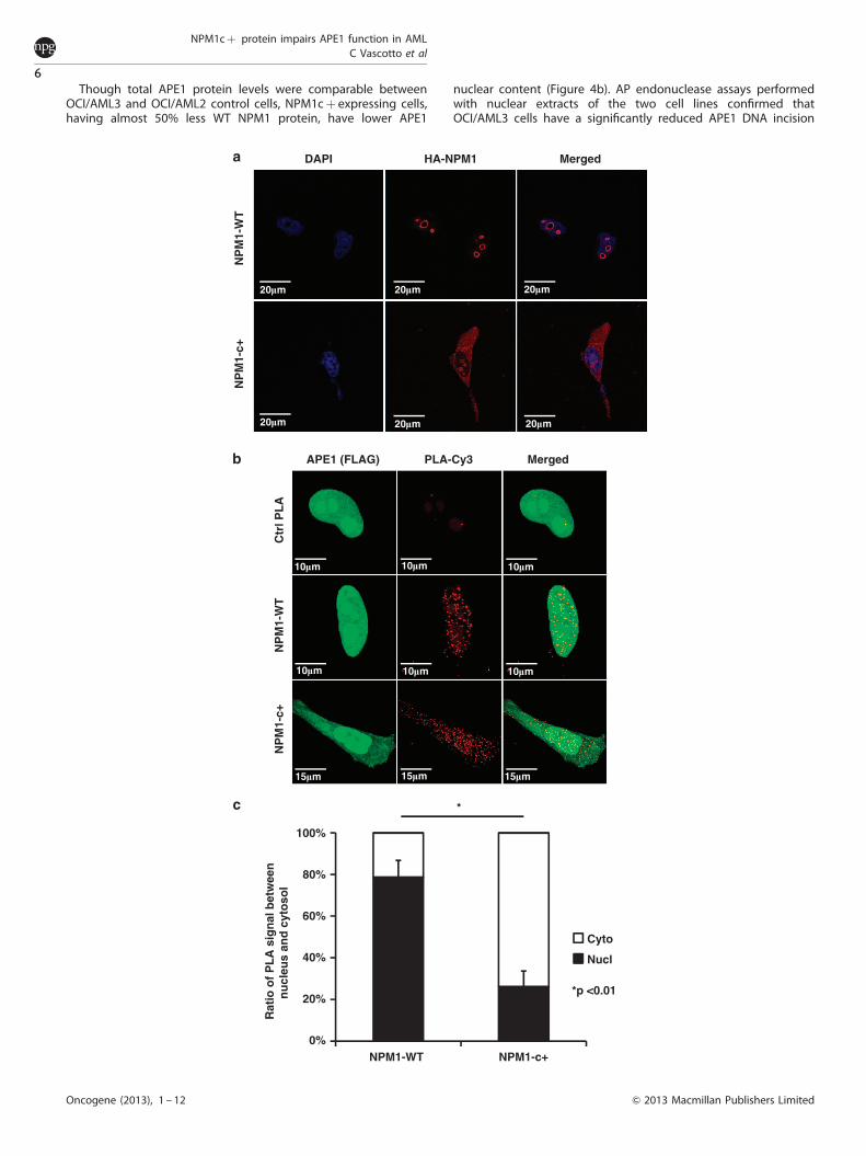
Though total APE1 protein levels were comparable betweenOCI/AML3 and OCI/AML2 control cells, NPM1cþ expressing cells,having almost 50% less WT NPM1 protein, have lower APE1
nuclear content (Figure 4b). AP endonuclease assays performedwith nuclear extracts of the two cell lines confirmed thatOCI/AML3 cells have a significantly reduced APE1 DNA incision
0%
20%
40%
60%
80%
100%
NPM1-WT NPM1-c+
Rat
io o
f P
LA
sig
nal
bet
wee
nn
ucl
eus
and
cyt
oso
l
Cyto
Nucl
*p <0.01
*
Ctr
l PL
AN
PM
1-W
TN
PM
1-c+
APE1 (FLAG) PLA-Cy3 Merged
DAPI HA-NPM1 Merged
NP
M1-
WT
NP
M1-
c+
20�m 20�m 20�m
20�m20�m20�m
10�m 10�m 10�m
10�m10�m10�m
15�m 15�m 15�m
NPM1cþ protein impairs APE1 function in AMLC Vascotto et al
6
Oncogene (2013), 1 – 12 & 2013 Macmillan Publishers Limited

activity (Figure 4d). We then evaluated the ability of the AML cellsto efficiently remove AP sites from the DNA of MMS-treated cells.As shown in Figure 4e, OCI/AML3 cells retained a significantlyhigher level of AP sites both under basal conditions and followingMMS treatment than did OCI/AML2 cells. Cell-viability experimentswere in agreement with these observations showing that OCI/AML3 cells were significantly more sensitive to MMS treatmentthan OCI/AML2 cells (Figure 4f).
To provide additional mechanistic insights into the role of APE1delocalization in NPM1cþ cells and to prove which of the APE1known functions (that is, DNA repair or redox) are affected byNPM1cþ expression accounting for the higher sensitivity of OCI/AML3 cells to genotoxicants, we performed sensitization experi-ments to MMS treatment with specific APE1 inhibitors. Methoxy-amine, as an APE1 DNA-repair inhibitor, and the well-known E3330redox inhibitor52 were used for this purpose. Data obtained(Supplementary Fig S8) show that treatment with the DNA-repairinhibitor, but not with the E3330 redox inhibitor, sensitizes OCI/AML2 cells to MMS treatment and phenocopies the response ofthe NMP1cþ expressing cells. Moreover, this experiment demon-strates that the functional impairment of the APE1 DNA repairin NPM1cþ cells does not further increase the sensitivity ofNPM1cþ cells to genotoxic damage.
Altogether, these data allow to conclude that APE1 functionalsuppression by NPM1cþ induced relocalization is important forregulating cellular sensitivity to DNA damage in NPM1cþ cells.
Blasts from AML patients, expressing the NPM1cþmutant protein,have increased cytoplasmic APE1–NPM1 interaction and showimpaired BER activityWe tested whether the altered interaction between APE1 andNPM1cþ observed upon ectopic expression of the mutantprotein in cell lines also occurs in blasts from AML patientsconstitutively expressing the NPM1cþ protein. IF and PLAanalyses (Figures 5a and b) clearly showed that the NPM1cþmutations leads to a reduction of the APE1/NPM1 interaction innuclei, coupled to a strong increase of the APE1/NPM1 associationin the cytoplasms of AML blasts. This phenotype is associated withthe relocalization of APE1 protein to the cytoplasm, as previouslyobserved in a heterologous cell system (Figure 3b).
To evaluate the physiological relevance of the altered interac-tion between APE1 and NPM1cþ , we compared the sensitivity ofblasts from AML patients expressing either the WT or the cþ formof NPM1 with that of healthy donors to DNA-alkylation damageinduced by MMS. Cell-viability experiments demonstrated thatblasts from AML patients expressing the NPM1cþ form weresignificantly more sensitive to MMS than healthy control cells andAML-WT NPM1 blasts (Figure 5c). These data support theconclusion that alteration of APE1 distribution due to theNPM1cþ mutation has a significant impact on the BER activityin vivo and has a functional relevance in AML pathogenesis.
DISCUSSIONHere we demonstrated that NPM1 has an indirect but significantrole in BER through functional regulation of the APE1 endonu-clease activity. This observation is the first evidence highlighting aclear function of NPM1 in DNA repair, and these new resultsprovide a possible alternative explanation for a role of NPM1 inthe maintenance of genomic stability in addition to its knowneffect on p53 stability.2,3 It has been recently shown thathomologous recombination-impaired BRCA-deficient cells arehypersensitive to poly (ADP-ribose) polymerase inhibitors thatblock repair of single-strand breaks, a subpathway of BER.Interestingly, NPM1 functionally interacts with BRCA2 having arole in centrosome duplication.53 Therefore, it could be speculatedthat the differential sensitivity of NPM1� /� cells to genotoxicantsused in this study could be ascribed to an impaired ability of thesecells to repair DNA-strand breaks. However, our data showing nodifferential sensitivity to poly (ADP-ribose) polymerase inhibitors,together with a similar efficiency of repair of strand breaks (asmeasured through comet assays) by the two isogenic cell lines,would exclude this hypothesis, leaving BER impairment as themain process affected by the loss of NPM1 expression. Moreover,our findings may illuminate the prompt response tochemotherapy of AML expressing the NPM1cþ mutation. Ourdata support the hypothesis that some biological effects ofNPM1cþ may be exerted through impaired functions ofimportant NPM1-interacting proteins caused by their abnormalcytoplasmic localization.1,8–13
APE1 subcellular distribution within different cell types is mainlynuclear and critically controls the cellular proliferation rate,54–56
however, in some tumors APE1 can be cytoplasmic or nuclear/cytoplasmic.18,28 Increased expression of APE1 is associated withdifferent tumorigenic processes.28 In particular, a higherexpression of APE1, with consequently increased BER activity,has been described in several cancer types,28 where it correlateswith a higher tumor aggressiveness and is associated withchemoresistance.18 Extranuclear APE1 has a role withinmitochondria in repairing mitochondrial DNA damage57–59 andin controlling the intracellular reactive oxygen species productionthrough inhibition of Rac1.60,61 However, a possible causal role ofan increased APE1 expression in tumorigenesis, as well as themechanisms responsible for the unusual APE1 accumulation in thecytoplasmic compartment, has never been investigated in detail.
The observations reported herein provide a molecular explana-tion for the delocalization of APE1 to the cytoplasm observed inAML tumors associated with NPM1cþ . Because APE1 interactswith many different partners involved in RNA metabolism,17,31,37
the occurrence of extranuclear APE1 localization may be explainedon the basis of altered expression levels of its protein partners thatmay undergo delocalization, as is the case of NPM1cþ itself.Alternatively, increased expression of APE1 cytoplasmic partners,rather than mutations occurring in APE1 itself, may directly affectthe distribution of the protein. Notably, no polymorphisms
Figure 3. NPM1 mutation alters APE1 localization in HeLa cells. (a) Representative confocal microscopy images of HeLa cells transientlytransfected with pcDNA-HA vectors expressing WT NPM1 or the NPM1cþ mutant. Twenty-four hours after transfection cells were fixed andstained with a primary antibody a-HA-Tag and a secondary antibody a-rabbit conjugated with Rhodamine Red. In cells expressing WT NPM1,the ectopic HA-tagged protein localizes, as expected, only within the granular component of nucleoli. On the contrary, cells expressingNPM1cþ mutant show also strong cytoplasm positivity. (b) PLA technology was used on HeLa cells to quantify the occurrence of in vivointeraction between APE1 and NPM1. Cells were transiently transfected with APE1-FLAG and NPM1-HA-WT or the NPM1cþ mutant. Confocalmicroscopy analysis highlights the presence of distinct fluorescent red dots (PLA signals), indicating the occurrence of in vivo interactionbetween APE1-FLAG and NPM1-HA, whereas green fluorescence shows APE1-FLAG-tagged protein localization. PLA control is represented bycells incubated only with a-FLAG antibody. In cells transfected with WT NPM1, APE1 accumulates within the nucleus where also PLA signalsare mostly present. In NPM1cþ expressing cells, a diffused cytoplasmic APE1 staining could be observed along with the relocalization of PLAsignal. (c) Blob Finder software was used to quantify the relative distribution of PLA signals between nucleus and cytoplasm of cells transfectedwith WT NPM1 or NPM1cþmutant. In NPM1cþ expressing cells, most of the interaction between APE1 and NPM1 occurs within thecytoplasm (nuclear: 26±7%), whereas in WT NPM1 cells, PLA signal is predominantly nuclear (nuclear: 79±8%). Reported results are the meanof 30 cells for each condition.
NPM1cþ protein impairs APE1 function in AMLC Vascotto et al
7
& 2013 Macmillan Publishers Limited Oncogene (2013), 1 – 12

36
55
36
100
36
55
36
36
APE1 MergedDAPI
OC
I/AM
L 2
OC
I/AM
L 3
15�m 15�m 15�m
15�m15�m15�m
OCI/AML
2 3
Mr(kDa)
**
AML Patient
WT c+
WCEOCI/AML
2 3 2 3 2 3
CCEOCI/AML
NCEOCI/AMLMr
(kDa)
**
MMS 4h (mM)
*
**
* p<0.05
** p<0.001
0
5
10
15
20
25
30
35OCI/AML2OCI/AML3
**
AP
sit
es/1
05 b
p
0 0.5 1.0
Minutes 0 1 5 102 15 20
S
P
1 5 102 15 20
OCI/AML 2 OCI/AML 3
0%
20%
40%
60%
80%
0 5 10 15 20
Pro
du
ct f
orm
atio
n
Minutes
*
* p<0.05 *
*
OCI/AML 2OCI/AML 3
0%
20%
40%
60%
80%
100%
OCI/AML2
OCI/AML3
*
*
**
***
*p<0.001
**p<0.005
MMS 8h (mM)
0 0.50.1 0.2 0.3 0.4
*
**
***p<0.05
Cel
l via
bili
ty
�-APE1
�-NPM1
�-NPM1c+
�-Tubulin
�-LSD1
�-Actin
�-APE1
�-Actin
Figure 4. NPM1cþ mutation alters APE1 subcellular localization and affects its endonuclease activity in OCI/AML3 cell line. (a) Representativeimages of fluorescence microscopy analysis of APE1 on OCI/AML2 (WT NPM1) and OCI/AML3 (NPM1cþ ) cell lines. A total of 2� 104 cells werecentrifuged onto glass slides using a cytospin, and IF was performed as reported in Materials and Methods section. A secondary a-mouseRhodamine Red-conjugated antibody was used to visualize APE1. Nuclei are stained with 4’,6-diamidino-2-phenylindole. A predominant cytoplasmicpositivity of APE1 could be observed in OCI/AML3 cells only, linking NPM1cþ mutation with APE1 cytoplasmic relocalization. (b) In all, 10mg of totalcell extract (WCE), 10mg of cytoplasmic cell extract (CCE) and 5mg of nuclear cell extract (NCE) of OCI/AML2 and 3 were separated onto SDS–PAGE,immunoblotted and analyzed for their content of APE1 and NPM1 (WT and cþ ). NPM1cþ protein was detected with a specific antibodyrecognizing only the NPM1cþmutant form.47 Western blotting analysis showed the presence of truncated APE1 protein (*) only in the whole andcytoplasmic extracts from OCI/AML3 cell line. Actin was used as loading control, whereas tubulin and LSD1 were used as markers of cytoplasmic andnuclear fraction enrichment, respectively. (c) Total cell extracts of 10mg from OCI/AML2 and 3 and of blast from AML patients expressing WT NPMand NPM1cþ mutant were separated onto SDS–PAGE, immunoblotted and analyzed for their APE1 content. The expression of NPM1cþmutantform is associated with the presence of truncated APE1 protein (*) in extracts from both the OCI/AML3 cell line and blasts from a patient carrying thismutation. Actin was used as loading control. (d) OCI/AML2 and OCI/AML3 nuclear cell extracts of 50ng were used to evaluate the APE1endonuclease activity. A representative image shows the conversion of the substrate (S) to product (P) operated by APE1. OCI/AML3 cells showlower AP-incision activity, in accordance with the decreased nuclear APE1 content. The diagram below reports the mean±s.d. values obtained fromthe densitometric analysis of three independent experiments. (e) The amount of abasic (AP) sites was measured in OCI/AML2 and OCI/AML3 cellsunder basal conditions and after treatment with 0.5 and 1mM MMS for 4h. OCI/AML3 cells display a significantly higher amount of AP sites ifcompared with OCI/AML2 cells. Mean±s.d. values are the result of three independent experiments. (f ) CellTiter-Glo Luminescent Cell Viability assaywas used to evaluate the sensitization of OCI/AML2 and OCI/AML3 cells to MMS. Cells were incubated with the indicated amounts of MMS for 8h.Expression of NPM1cþ negatively affects cell viability. Mean±s.d. values are the result of three independent experiments.
NPM1cþ protein impairs APE1 function in AMLC Vascotto et al
8
Oncogene (2013), 1 – 12 & 2013 Macmillan Publishers Limited
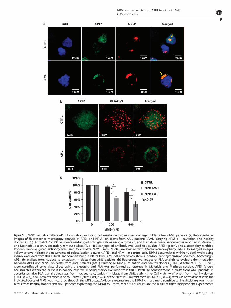
APE1 MergedDAPI NPM1
CT
RL
AM
L
10�m 10�mC
TR
L
APE1 PLA-Cy3 Merged
AM
L
*
*
*p<0.05
0%
20%
40%
60%
80%
100%
120%
0 200 500
Cel
l via
bili
ty
CTRL
NPM1-WT
NPM1-c+
10�m 10�m
10�m10�m10�m10�m
5�m 5�m 5�m
5�m5�m5�m
MMS (�M)
Figure 5. NPM1 mutation alters APE1 localization, reducing cell resistance to genotoxic damage in blasts from AML patients. (a) Representativeimages of fluorescence microscopy analysis of APE1 and NPM1 on blasts from AML patients (AML) carrying NPM1cþ mutation and healthydonors (CTRL). A total of 2� 104 cells were centrifuged onto glass slides using a cytospin, and IF analyses were performed as reported in Materialsand Methods section. A secondary a-mouse-Alexa Fluor 488-conjugated antibody was used to visualize APE1 (green), and a secondary a-rabbit-Rhodamine-conjugated antibody was used to visualize NPM1 (red). Nuclei are stained with 4’,6-diamidino-2-phenylindole. In merged images,yellow arrows indicate the occurrence of colocalization between APE1 and NPM1. In control cells, NPM1 accumulates within nucleoli while beingmainly excluded from this subcellular compartment in blasts from AML patients, which show a predominant cytoplasmic positivity. Accordingly,APE1 delocalizes from nucleus to cytoplasm in blasts from AML patients. (b) Representative images of PLA analysis to evaluate the interactionbetween APE1 and NPM1 on blasts from AML patients (AML) carrying NPM1cþ mutation and healthy donors (CTRL). A total of 2.5� 104 cellswere centrifuged onto glass slides using a cytospin, and PLA was performed as reported in Materials and Methods section. APE1 (green)accumulates within the nucleus in control cells while being mainly excluded from this subcellular compartment in blasts from AML patients. Inaccordance, also PLA signal delocalizes from nucleus to cytoplasm in blasts from AML patients. (c) Cell viability of blasts from healthy donors(CTRL, n¼ 3), AML patients expressing WT NPM1 (NPM1-WT, n¼ 3) or the NPM1cþmutant form (NPM1cþ , n¼ 4) after 4h of treatment with theindicated doses of MMS was measured through the MTS assay. AML cells expressing the NPM1-cþ are more sensitive to the alkylating agent thanblasts from healthy donors and AML patients expressing the NPM1-WT form. Mean±s.d. values are the result of three independent experiments.
NPM1cþ protein impairs APE1 function in AMLC Vascotto et al
9
& 2013 Macmillan Publishers Limited Oncogene (2013), 1 – 12
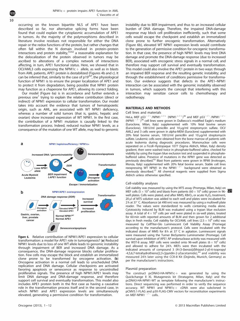
occurring on the known bipartite NLS of APE1 have beendescribed so far, nor alternative splicing forms have beenfound that could explain the cytoplasmic accumulation of APE1in tumors. As the majority of the polymorphisms described inliterature involve residues not responsible for either the DNArepair or the redox functions of the protein, but rather changes thatoften fall within the N domain involved in protein–proteininteractions and protein stability,33,62 a reasonable explanation forthe delocalization of the protein observed in tumors may beascribed to alterations of a complex network of interactionsaffecting, in turn, APE1 functional status. Here, we showed that inOCI/AML3 cells expressing the NPM1cþ allele, as well as in blastsfrom AML patients, APE1 protein is destabilized (Figures 4b and c). Itcan be inferred that, similarly to the case of p19Arf, the physiologicalfunction of NPM1 is to ensure the proper localization of APE1 andto protect it from degradation, being possible that NPM1 proteinmay function as a chaperone for APE1, allowing its correct folding.
Our model (Figure 6a) is in accordance and further extends aprevious one1 trying to explain the relative contribution (direct orindirect) of NPM1 expression to cellular transformation. Our modeltakes into account the evidence that tumors of hematopoieticorigin, such as AML, are associated with WT NPM1 deficiency,whereas a number of solid tumors (that is, gastric, hepatic andovarian) show increased expression of WT NPM1. In the first case,the contribution of a NPM1 mutation is causally linked to thetransformation process. Indeed, reduced nuclear NPM1 levels, as aconsequence of the mutation of one WT allele, may lead to genomic
instability due to BER impairment, and thus to an increased cellularburden of DNA damage. Therefore, the impaired DNA-damageresponse may block cell proliferation inefficiently, such that somecells would escape the checkpoint and establish an immortalizedclone prone to further oncogenic transformation. Alternatively(Figure 6b), elevated WT NPM1 expression levels would contributeto the generation of permissive condition for oncogenic transforma-tion. In that case, the presence of high NPM1 levels may limit DNAdamage and promote the DNA-damage response (due to increasedBER), associated with oncogenic stress signals in a normal cell, andtherefore may support cell survival and eventually transformation.This model could also involve the APE1 protein in two ways: throughan impaired BER response and the resulting genetic instability; andthrough the establishment of conditions permissive for transforma-tion. Our evidence suggests that defects in the APE1–NPM1interaction can be associated with the genomic instability observedin tumors, which supports the concept that interfering with thisinteraction may sensitize cancer cells to chemotherapy andradiotherapy.
MATERIALS AND METHODSCell lines and materialsHeLa, MEF p53� /� /NPM1þ /þ (NPM1þ /þ )36 and MEF p53� /� /NPM1� /�
(NPM1� /� )36 cell lines were grown in Dulbecco’s modified Eagle’s medium(Euroclone, Milan, Italy) supplemented with 10% fetal bovine serum(Euroclone), 100 U/ml penicillin and 10mg/ml streptomycin sulfate. OCI/AML2 and 3 cells were grown in alpha-MEM (Euroclone) supplemented with20% fetal bovine serum, 100 U/ml penicillin and 10mg/ml streptomycinsulfate. Leukemic cells were obtained from the bone marrow of patients withacute leukemia during diagnostic procedures. Mononuclear cells wereseparated on a Ficoll–Hystopaque 1077 (Sigma Aldrich, Milan, Italy) densitygradient, then were washed twice in phosphate-buffered saline, checked forviability by using the trypan blue exclusion test and suspended in phosphate-buffered saline. Presence of mutations in the NPM1 gene was detected aspreviously described.63 Blast from patients were grown in RPMI (Invitrogen,Monza, Italy) supplemented with 20% fetal bovine serum. Stable cell linesreexpressing WT NPM1 in the NPM1� /� background were obtained aspreviously described.17 All chemical reagents were supplied from SigmaAldrich unless otherwise specified.
Cell-viability analysesCell viability was measured by using the MTS assay (Promega, Milan, Italy) onMEF cells (5� 103 cells) and blasts from patients (60� 103 cells) grown in 96-well plates. Cells were plated, and after MMS, KBrO3 or acute H2O2 treatment,20ml of MTS solution was added to each well and plates were incubated for2 h at 37 1C. Absorbance (at 490 nm) was measured by using a multiwell platereader. The values were standardized to wells containing media alone.Cytotoxicity induced by BLM was evaluated using a trypan blue exclusionassay. A total of 4� 104 cells per well were plated in six-well plates, treatedfor 60 min with reported amounts of BLM and then grown for 2 additionaldays in fresh media. Cell viability for OCI/AML cell lines (2.5� 104 cells) wasmeasured by CellTiter-Glo Luminescent Cell Viability Assay (Promega)according to the manufacturer’s protocol. Cells were incubated with theindicated doses of MMS for 8 h at 37 1C in agitation. Luminescent signalswere measured using the Turner BioSystems Luminometer (Promega). Cellsurvival upon inhibition of APE1 AP endonuclease activity was measured withthe WST-8 assay. MEF cells were seeded onto 96-well plates (6� 103 cells)and allowed to adhere for 24 h. MEFs were then incubated with theindicated amounts of compound 3 (N-(3-(benzo[d]thiazol-2-yl)-6-isopropyl-4,5,6,7-tetrahydrothieno[2,3-c]pyridin-2-yl)acetamide),44 and viability wasmeasured 24 h later using the CCK-8 Kit (Dojindo, Munich, Germany) asper the manufacturer’s instructions.
Plasmid preparationThe construct pcDNA3-HA-NPM1cþ was generated by using theQuickchange II XL Mutagenesis kit (Stratagene, Milan, Italy) and thepcDNA3-HA-NPM1-WT as template following the manufacturer’s instruc-tions. Direct sequencing was performed in order to verify the sequenceaccuracy. WT NPM1 and NPM1cþ cDNA were also subcloned inpCMV5.1-FLAG and p3X-FLAG-CMV vectors for reconstitution experimentson MEF-NPM1� /� cells.
Transformation
Acute MyeloidLeukemia
Loss NPM1allele
Increased DNA / genomicinstability
BER impairment APE1 function
Senescence/apoptosis
Escape / immortalization
Oncogeneactivation
DDR
NPM1 haploinsufficiency / NPM1c+
NPM1/APE1 overexpressionTransformation
Elevated NPM1WT
Elevated APE1
Gastric, ovarian,liver, prostate
cancer
Oncogeneactivation
DNA damage response (DDR)
Senescence/apoptosis
Normal NPM1WT
Normal APE1
Increasedsurvival
Increasedproliferation
Figure 6. Relative contribution of NPM1/APE1 expression to cellulartransformation: a model for APE1 role in tumorigenesis? (a) ReducedNPM1 levels due to loss of one WT allele leads to genomic instabilitythrough impairment of BER and increased DNA damage. As aconsequence, the DNA-damage response blocks cellular prolifera-tion. Few cells may escape the block and establish an immortalizedclone prone to be transformed by oncogene activation. (b)Oncogene activation in a normal cell leads to unscheduled DNAreplication and DNA damage. Cellular checkpoints are activatedfavoring apoptosis or senescence as response to uncontrolledproliferative signals. The presence of high NPM1/APE1 levels maylimit DNA damage and DNA-damage response, and thereforesupport cell survival and eventually transformation. This model alsoincludes APE1 protein both in the first case as having a causativerole in the transformation process itself and in the second case, inwhich NPM1 and APE1 expression levels are simultaneouslyelevated, generating a permissive condition for transformation.
Q1
NPM1cþ protein impairs APE1 function in AMLC Vascotto et al
10
Oncogene (2013), 1 – 12 & 2013 Macmillan Publishers Limited
AP-sites measurementsGenomic DNA was isolated from 2� 106 cells by using the DNAzol Reagent(Invitrogen), and then concentration and purity were determined spectro-photometrically. Samples of 0.1 mg/ml of genomic DNA were analyzed toquantify abasic damage in chromosomal DNA using the DNA DamageQuantification Kit (Dojindo) according to the manufacturer’s instructions.
IF and PLA analysisFor IF analysis on HeLa and MEFs, cells were grown on glass coverslips,whereas blasts and OCI/AML 2 and 3 cells were centrifuged onto glassslides using a cytospin. Then, cells were fixed with 4% (wt/vol)paraformaldehyde for 20 min at room temperature and permeabilizedfor 5 min with phosphate-buffered saline–0.25% (vol/vol) Triton X-100.Cells were incubated for 30 min with 10% fetal bovine serum in TBS–0.1%(vol/vol) Tween 20 (blocking solution) to block unspecific binding of theantibodies. Cells were then incubated with primary antibodies diluted inblocking solutions: monoclonal-a-APE1 (diluted 1:50, 3 h, 37 1C), polyclonala-NPM1 (diluted 1:200, overnight, 4 1C), monoclonal a-FLAG-FITC-conjugated (diluted 1:100, 3 h, 37 1C) and polyclonal a-HA (diluted 1:500,overnight, 4 1C). Then cells were washed three times for 5 min each withTBS–0.1% (vol/vol) Tween 20 (Washing Solution) and incubated withsecondary antibody: a-mouse or a-rabbit Rhodamine Red-conjugated orAlexa Fluor 488-conjugated (diluted 1:200, 2 h, 25 1C) (Jackson Immuno,West Grove, PA, USA). After washing three times, coverslips were mountedon microscope slides with a mounting media containing 4’,6-diamidino-2-phenylindole and an anti-fade reagent. For PLA analysis on blasts frompatient’s primary antibody a-APE1 was previously labeled with the Zenona-Mouse IgG Labeling Kit (Invitrogen) using the Alexa Fluor 488 accordingto the manufacturer’s instructions.
To study the interaction of APE1 with NPM1 in vivo, we used the in situPLA technology (Olink Bioscience, Uppsala, Sweden). After incubation withmonoclonal a-APE1 (1:50) or a-FLAG antibody (1:100) for 3 h at 37 1C, cellswere incubated with polyclonal a-NPM1 (1:200) or polyclonal a-HAantibody (Sigma-Aldrich) (1:500) overnight at 4 1C. PLA was performedfollowing the manufacturer’s instructions and as previously reported.36
Technical controls, represented by the omission of a-NPM1/a-HA primaryantibody, resulted in the complete loss of PLA signal. Images werecaptured through a confocal microscope, and quantification of PLA signalwas performed using the Blob Finder software (Center for Image Analysis,Uppsala University).
Cells were visualized through a Leica TCS SP laser-scanningconfocal microscope (Leica Microsystems, Milan, Italy) or a DMB6000Binverted microscope equipped with a DFC300FX digital camera(Leica Microsystems).
Preparation of cell extracts and protein quantificationCell extracts were prepared as already described in Vascotto et al.17 and asreported in Supplementary Information.
Cell transfection and western blotting analysesPlasmids were prepared and used for transfection and western blottinganalysis, as already described in Fantini et al.30 and as reported inSupplementary Information.
Incision assays on abasic double-stranded DNAAPE1 endonuclease activity was evaluated as already described17 and asreported in Supplementary Information.
Statistical analysisStatistical analysis on biological data was performed using the MicrosoftExcel data analysis program for Student’s t-test analysis.
ABBREVIATIONSAML, acute myeloid leukemia; APE1/Ref-1, apurinic/apyrimidinicendonuclease 1/redox effector factor 1; BER, base excision DNArepair; NPM1, nucleophosmin 1.
CONFLICT OF INTERESTThe authors declare no conflict of interest.
ACKNOWLEDGEMENTSWe greatly thank the National Center for Advancing Translational Sciences (NCATS)and Dr David M Wilson III for providing the APE1 specific inhibitor (compound 3). Wealso thank Dr Malgorzata M Kamocka from the Indiana University School of Medicine,Department of Medicine, Division of Nephrology, Indiana Center for BiologicalMicroscopy, Indianapolis, IN, USA. for her helpful suggestions during IF analyses, DrLaura Cesaratto for cell-viability assays and Sofia Tell for kind support. This work wassupported by grants from: AIRC (IG10269) and MIUR (FIRB_RBRN07BMCT andPRIN2008_CCPKRP_003) to GT. This work has been also supported by a UICCYamagiwa-Yoshida Memorial International Cancer Study Grant to GT.
REFERENCES1 Colombo E, Alcalay M, Pelicci PG. Nucleophosmin and its complex network:
a possible therapeutic target in hematological diseases. Oncogene 2011; 30:2595–2609.
2 Colombo E, Marine JC, Danovi D, Falini B, Pelicci PG. Nucleophosmin regulates thestability and transcriptional activity of p53. Nat Cell Biol 2002; 4: 529–533.
3 Lee C, Smith BA, Bandyopadhyay K, Gjerset RA. DNA damage disrupts thep14ARF-B23 (nucleophosmin) interaction and triggers a transient subnuclearredistribution of p14ARF. Cancer Res 2005; 65: 9834–9842.
4 Mrozek K, Marcucci G, Paschka P, Whitman SP, Bloomfield CD. Clinical relevanceof mutations and gene-expression changes in adult acute myeloid leukemia withnormal cytogenetics: are we ready for a prognostically prioritized molecularclassification? Blood 2007; 109: 431–448.
5 Falini B, Bolli N, Shan J, Martelli MP, Liso A, Pucciarini A et al. Both carboxy-terminus NES motif and mutated tryptophan(s) are crucial for aberrant nuclearexport of nucleophosmin leukemic mutants in NPMcþ AML. Blood 2006; 107:4514–4523.
6 Pianta A, Fabbro D, Damiani D, Tiribelli M, Fanin R, Franzoni A et al. Two novelNPM1 mutations in a therapy-responder AML patient. Hematol Oncol 2010; 28:151–155.
7 Vassiliou GS, Cooper JL, Rad R, Li J, Rice S, Uren A et al. Mutant nucleophosminand cooperating pathways drive leukemia initiation and progression in mice.Nat Genet 2011; 43: 470–475.
8 den Besten W, Kuo ML, Williams RT, Sherr CJ. Myeloid leukemia-associatednucleophosmin mutants perturb p53-dependent and independent activities ofthe Arf tumor suppressor protein. Cell Cycle 2005; 4: 1593–1598.
9 Colombo E, Martinelli P, Zamponi R, Shing DC, Bonetti P, Luzi L et al. Delocali-zation and destabilization of the Arf tumor suppressor by the leukemia-associatedNPM mutant. Cancer Res 2006; 66: 3044–3050.
10 Bonetti P, Davoli T, Sironi C, Amati B, Pelicci PG, Colombo E. Nucleophosmin andits AML-associated mutant regulate c-Myc turnover through Fbw7 gamma.J Cell Biol 2008; 182: 19–26.
11 Cilloni D, Messa F, Rosso V, Arruga F, Defilippi I, Carturan S et al. Increase sensi-tivity to chemotherapeutical agents and cytoplasmatic interaction between NPMleukemic mutant and NF-kappaB in AML carrying NPM1 mutations. Leukemia2008; 22: 1234–1240.
12 Gurumurthy M, Tan CH, Ng R, Zeiger L, Lau J, Lee J et al. Nucleophosmin interactswith HEXIM1 and regulates RNA polymerase II transcription. J Mol Biol 2008; 378:302–317.
13 Wanzel M, Russ AC, Kleine-Kohlbrecher D, Colombo E, Pelicci PG, Eilers M. Aribosomal protein L23-nucleophosmin circuit coordinates Mizl function with cellgrowth. Nat Cell Biol 2008; 10: 1051–1061.
14 Falini B, Nicoletti I, Martelli MF, Mecucci C. Acute myeloid leukemia carryingcytoplasmic/mutated nucleophosmin (NPMcþ AML): biologic and clinicalfeatures. Blood 2007; 109: 874–885.
15 Falini B, Martelli MP, Bolli N, Sportoletti P, Liso A, Tiacci E et al. Acute myeloidleukemia with mutated nucleophosmin (NPM1): is it a distinct entity? Blood 2011;117: 1109–1120.
16 Londero AP, Orsaria M, Tell G, Marzinotto S, Capodicasa V, Poletto M et al.Expression and prognostic significance of APE1/Ref1 and NPM1 proteins inovarian serous cancer; submitted.
17 Vascotto C, Fantini D, Romanello M, Cesaratto L, Deganuto M, Leonardi A et al.APE1/Ref-1 interacts with NPM1 within nucleoli and plays a role in the rRNAquality control process. Mol Cell Biol 2009; 29: 1834–1854.
18 Tell G, Fantini D, Quadrifoglio F. Understanding different functions of mammalianAP endonuclease (APE1) as a promising tool for cancer treatment. Cell Mol Life Sci2010; 67: 3589–3608.
19 Wilson 3rd DM, Simeonov A. Small molecule inhibitors of DNA repair nucleaseactivities of APE1. Cell Mol Life Sci 2010; 67: 3621–3631.
20 Xanthoudakis S, Curran T. Identification and characterization of Ref-1, a nuclearprotein that facilitates AP-1 DNA-binding activity. EMBO J 1992; 11: 653–665.
NPM1cþ protein impairs APE1 function in AMLC Vascotto et al
11
& 2013 Macmillan Publishers Limited Oncogene (2013), 1 – 12

21 Xanthoudakis S, Miao GG, Curran T. The redox and DNA-repair activities of Ref-1are encoded by nonoverlapping domains. Proc Natl Acad Sci USA 1994; 91: 23–27.
22 Ueno M, Masutani H, Arai RJ, Yamauchi A, Hirota K, Sakai T et al. Thioredoxin-dependent redox regulation of p53-mediated p21 activation. J Biol Chem 1999;274: 35809–35815.
23 Seemann S, Hainaut P. Roles of thioredoxin reductase 1 and APE/Ref-1 in thecontrol of basal p53 stability and activity. Oncogene 2005; 24: 3853–3863.
24 Hirota K, Matsui M, Iwata Z, Nishiyama A, Mori K, Yodoi J. AP-1 transcriptionalactivity is regulated by a direct association between thioredoxin and Ref-1.Proc Natl Acad Sci USA 1997; 94: 3633–3638.
25 Wei SJ, Botero A, Hirota K, Bradbury CM, Markovina S, Laszlo A et al. Thioredoxinnuclear translocation and interaction with redox factor-1 activates the activatorprotein-1 transcription factor in response to ionizing radiation. Cancer Res 2000;60: 6688–6695.
26 Ziel KA, Campbell CC, Wilson GL, Gillespie MN. Ref-1/Ape is critical for formationof the hypoxia-inducible transcriptional complex on the hypoxic responseelement of the rat pulmonary artery endothelial cell VEGF gene. FASEB J 2004; 18:986–988.
27 Gray MJ, Zhang J, Ellis LM, Semenza GL, Evans DB, Watowich SS et al. HIF-1alpha,STAT3, CBP/p300 and Ref-1/APE are components of a transcriptional complex thatregulates Src-dependent hypoxia-induced expression of VEGF in pancreatic andprostate carcinomas. Oncogene 2005; 24: 3110–3120.
28 Tell G, Damante G, Caldwell D, Kelley MR. The intracellular localization of APE1/Ref-1: more than a passive phenomenon? Antioxid Redox Signal 2005; 7: 367–384.
29 Bhakat KK, Izumi T, Yang SH, Hazra TK, Mitra S. Role of acetylated humanAP-endonuclease (APE1/Ref-1). in regulation of the parathyroid hormone gene.EMBO J 2003; 22: 6299–6309.
30 Fantini D, Vascotto C, Marasco D, D’Ambrosio C, Romanello M, Vitagliano L et al.Critical lysine residues within the overlooked N-terminal domain ofhuman APE1 regulate its biological functions. Nucleic Acids Res 2010; 38:8239–8256.
31 Tell G, Wilson 3rd DM, Lee CH. Intrusion of a DNA repair protein in the RNomeworld: is this the beginning of a new era? Mol Cell Biol 2010; 30: 366–371.
32 Barnes T, Kim WC, Mantha AK, Kim SE, Izumi T, Mitra S et al. Identification ofapurinic/apyrimidinic endonuclease 1 (APE1) as the endoribonuclease thatcleaves c-myc mRNA. Nucleic Acids Res 2009; 37: 3946–3958.
33 Poletto M, Vascotto C, Scognamiglio PL, Lirussi L, Marasco D, Tell G. Role of theunstructured N-terminal domain of the human apurinic/apyrimidinic endonu-clease 1 (hAPE1) in the modulation of its interaction with nucleic acids andnucleophosmin (NPM1). Biochem J 2013; 452: 545–557.
34 Mol CD, Izumi T, Mitra S, Tainer JA. DNA-bound structures and mutants revealabasic DNA binding by APE1 and DNA repair coordination (corrected). Nature2000; 403: 451–456.
35 Masuda Y, Bennett RA, Demple B. Dynamics of the interaction of human apurinicendonuclease (Ape1) with its substrate and product. J Biol Chem 1998; 273:30352–30359.
36 Colombo E, Bonetti P, Lazzerini Denchi E, Martinelli P, Zamponi R, Marine JC et al.Nucleophosmin is required for DNA integrity and p19Arf protein stability. Mol CellBiol 2005; 25: 8874–8886.
37 Lirussi L, Antoniali G, Vascotto C, D’Ambrosio C, Poletto M, Romanello M et al.Nucleolar accumulation of APE1 depends on charged lysine residues thatundergo acetylation upon genotoxic stress and modulate its BER activity in cells.Mol Biol Cell 2012; 23: 4079–4096.
38 Svilar D, Goellner EM, Almeida KH, Sobol RW. Base excision repair and lesion-dependent subpathways for repair of oxidative DNA damage. Antioxid RedoxSignal 2011; 14: 2491–2507.
39 Kaina B, Ochs K, Grosch S, Fritz G, Lips J, Tomicic M et al. BER, MGMT, and MMR indefense against alkylation-induced genotoxicity and apoptosis. Prog Nucleic AcidRes Mol Biol 2001; 68: 41–54.
40 Jiang Y, Guo C, Fishel ML, Wang ZY, Vasko MR, Kelley MR. Role of APE1 in dif-ferentiated neuroblastoma SH-SY5Y cells in response to oxidative stress: use ofAPE1 small molecule inhibitors to delineate APE1 functions. DNA Repair (Amst)2009; 8: 1273–1282.
41 Ballmair D, Epe B. DNA damage by bromate: mechanism and consequences.Toxicology 2006; 221: 166–171.
42 Chen J, Stubbe J. Bleomycins: towards better therapeutics. Nat Rev Cancer 2005; 5:102–112.
43 Fung H, Demple B. Distinct roles of Ape1 protein in the repair of DNA damageinduced by ionizing radiation or bleomycin. J Biol Chem 2011; 286: 4968–4977.
44 Rai G, Vyjayanti VN, Dorjsuren D, Simeonov A, Jadhav A, Wilson 3rd DM et al.Synthesis, biological evaluation, and structure-activity relationships of a novelclass of apurinic/apyrimidinic endonuclease 1 inhibitors. J Med Chem 2012; 55:3101–3112.
45 Weibrecht I, Leuchowius KJ, Clausson CM, Conze T, Jarvius M, Howell WM et al.Proximity ligation assays: a recent addition to the proteomics toolbox. Expert RevProteomics 2010; 7: 401–409.
46 Falini B, Mecucci C, Tiacci E, Alcalay M, Rosati R, Pasqualucci L et al. GIMEMA AcuteLeukemia Working Party. Cytoplasmic nucleophosmin in acute myelogenousleukemia with a normal karyotype. N Engl J Med 2005; 352: 254–266Erratum in:N Engl J Med 2005; 352: 740.
47 Quentmeier H, Martelli MP, Dirks WG, Bolli N, Liso A, Macleod RA et al. Cell lineOCI/AML3 bears exon-12 NPM gene mutation-A and cytoplasmic expressionof nucleophosmin. Leukemia 2005; 19: 1760–1767.
48 Yoshida A, Urasaki Y, Waltham M, Bergman AC, Pourquier P, Rothwell DG et al.Human apurinic/apyrimidinic endonuclease (Ape1) and its N-terminal truncatedform (AN34) are involved in DNA fragmentation during apoptosis. J Biol Chem2003; 278: 37768–37776.
49 Fan Z, Beresford PJ, Zhang D, Xu Z, Novina CD, Yoshida A et al. Cleavingthe oxidative repair protein Ape1 enhances cell death mediated by granzyme A.Nat Immunol 2003; 4: 145–153.
50 Guo Y, Chen J, Zhao T, Fan Z. Granzyme K degrades the redox/DNA repair enzymeApe1 to trigger oxidative stress of target cells leading to cytotoxicity. MolImmunol 2008; 45: 2225–2235.
51 Vascotto C, Bisetto E, Li M, Zeef LA, D’Ambrosio C, Domenis R et al. Knock-inreconstitution studies reveal an unexpected role of Cys-65 in regulating APE1/Ref-1 subcellular trafficking and function. Mol Biol Cell 2011; 22: 3887–3901.
52 Kelley MR, Luo M, Reed A, Su D, Delaplane S, Borch RF et al. Functional analysis ofnovel analogues of E3330 that block the redox signaling activity of the multi-functional AP endonuclease/redox signaling enzyme APE1/Ref-1. Antioxid RedoxSignal 2011; 14: 1387–1401.
53 Wang HF, Takenaka K, Nakanishi A, Miki Y. BRCA2 and nucleophosmin coregulatecentrosome amplification and form a complex with the Rho effector kinaseROCK2. Cancer Res 2011; 71: 68–77.
54 Fung H, Demple B. A vital role for APE1/Ref1 protein in repairing spontaneousDNA damage in human cells. Mol. Cell 2005; 17: 463–470.
55 He T, Weintraub NL, Goswami PC, Chatterjee P, Flaherty DM, Domann FE et al.Redox factor-1 contributes to the regulation of progression from G0/G1 to S byPDGF in vascular smooth muscle cells. Am J Physiol Heart Circ Physiol 2003; 285:H804–H812.
56 Izumi T, Brown DB, Naidu CV, Bhakat KK, Macinnes MA, Saito H et al. Two essentialbut distinct functions of the mammalian abasic endonuclease. Proc Natl Acad SciUSA 2005; 102: 5739–5743.
57 Szczesny B, Tann AW, Longley MJ, Copeland WC, Mitra S. Long patch baseexcision repair in mammalian mitochondrial genomes. J Biol Chem 2008; 283:26349–26356.
58 Szczesny B, Tann AW, Mitra S. Age- and tissue-specific changes in mitochondrialand nuclear DNA base excision repair activity in mice: Susceptibility of skeletalmuscles to oxidative injury. Mech Ageing Dev 2010; 131: 330–337.
59 Tell G, Crivellato E, Pines A, Paron I, Pucillo C, Manzini G et al. Mitochondriallocalization of APE/Ref-1 in thyroid cells. Mutat Res 2001; 485: 143–152.
60 Angkeow P, Deshpande SS, Qi B, Liu YX, Park YC, Jeon BH et al. Redox factor-1: anextra-nuclear role in the regulation of endothelial oxidative stress and apoptosis.Cell Death Differ 2002; 9: 717–725.
61 Ozaki M, Suzuki S, Irani K. Redox factor-1/APE suppresses oxidative stress byinhibiting the rac1 GTPase. FASEB J 2002; 16: 889–890.
62 Yu ET, Hadi MZ. Bioinformatic processing to identify single nucleotide poly-morphism that potentially affect Ape1 function. Mutat Res 2011; 722: 140–146.
63 Pitiot AS, Santamarıa I, Garcıa-Suarez O, Centeno I, Astudillo A, Rayon C et al.A new type of NPM1 gene mutation in AML leading to a C-terminal truncatedprotein. Leukemia 2007; 21: 1564–1566.
Supplementary Information accompanies this paper on the Oncogene website (http://www.nature.com/onc)
NPM1cþ protein impairs APE1 function in AMLC Vascotto et al
12
Oncogene (2013), 1 – 12 & 2013 Macmillan Publishers Limited

Am J Clin Pathol 2014;141:000 11 DOI: 10.1309/AJCPIDKDLSGE26CX 1
© American Society for Clinical Pathology
AJCP / Original ArticleCM
E/SAM
Expression and Prognostic Significance of APE1/Ref-1 and NPM1 Proteins in High-Grade Ovarian Serous Cancer
Ambrogio P. Londero, MD,1 Maria Orsaria, MD,2 Gianluca Tell, MSc,2 Stefania Marzinotto, MSc,2 Valentina Capodicasa, MD, PhD,1 Mattia Poletto, MSc,2 Carlo Vascotto, PhD,2 Cosimo Sacco, MD,3 and Laura Mariuzzi, MD2
From the 1Deparment of Experimental Clinical and Medical Science, Clinic of Obstetrics and Gynecology, University of Udine, Udine, Italy; 2Department of Medical and Biological Sciences, University of Udine, Udine, Italy; and 3Clinic of Oncology, University Hospital of Udine, Udine, Italy.
Key Words: APE1/Ref-1; NPM1; Survival; Prognostic factors; Ovarian cancer
DOI: 10.1309/AJCPIDKDLSGE26CX
ABSTRACT
Objectives: To correlate the expression profile of human apurinic endonuclease/redox factor 1 (APE1/Ref-1) with that of nucleolar/nucleoplasmic protein nucleophosmin 1 (NPM1) in association with the aggressiveness and progression of high-grade ovarian serous cancer.
Methods: Retrospective study analyzing a tissue microarray of 73 women affected by high-grade ovarian serous cancer. Protein expression was assessed by immunohistochemistry on primary tumor masses and synchronous peritoneal metastases if present.
Results: APE1/Ref-1 and NPM1 showed a significant correlation in ovarian serous cancer. Patients with a poorer outcome showed a significant overexpression of nuclear NPM1 protein. A Cox proportional hazards multivariate regression model revealed NPM1 expression to be independently significant for overall survival in high-grade ovarian serous cancers after correcting for stage, age, cytoreduction completeness, and platinum resistance.
Conclusions: APE1/Ref-1 interacts with NPM1 to control the DNA damage repair system, and it is likely that this interaction plays a defining role in high-grade ovarian serous carcinoma. A high NPM1 immunohistochemical expression was independently correlated with a shorter survival period and thus appears to be an important prognostic factor.
Ovarian cancer occurs relatively rarely (accounting for 3.1% of female cancers in our region of Italy), but it is a leading cause of death among gynecologic malignancies as a result of three key tensions: the advanced stage of the tumor at diagnosis, the high frequency of recurrences, and its highly chemoresistant nature. Despite an improvement in survival in the past years—due to a more effective combination of surgery and empirically optimized cytotoxic drugs—the over-all cure rate remains low.1 A hallmark of high-grade serous carcinoma is defective homologous DNA repair. The new-est breakthrough in treatment is poly adenosine diphosphate ribose polymerase inhibitors,2 which suppress the ability of cells to repair single-strand DNA breaks, leading to cell death (for tumor cells that lack homologous repair capability). The use of alternative DNA repair inhibitors as a therapeutic
Upon completion of this activity you will be able to:•describetherolesofapurinicendonuclease/redoxfactor1(APE1/Ref-1)andnucleolar/nucleoplasmicproteinnucleophosmin1(NPM1)intheDNAbaseexcisionrepairpathway.
• describeAPE1/Ref-1andNPM1interactioninovarianhigh-gradeserouscancer.
• predictoverallsurvivalofovarianserouscancerbasedonlowvshighexpressionofNPM1.
TheASCPisaccreditedbytheAccreditationCouncilforContinuingMedicalEducationtoprovidecontinuingmedicaleducationforphysicians.TheASCPdesignatesthisjournal-basedCMEactivityforamaximumof1 AMA PRA Category 1 Credit ™perarticle.Physiciansshouldclaimonlythecreditcommensuratewiththeextentoftheirparticipationintheactiv-ity.ThisactivityqualifiesasanAmericanBoardofPathologyMaintenanceofCertificationPartIISelf-AssessmentModule.
Theauthorsofthisarticleandtheplanningcommitteemembersandstaffhavenorelevantfinancialrelationshipswithcommercialintereststodisclose.
Questionsappearonp000.Examislocatedatwww.ascp.org/ajcpcme.
PROOF

2 Am J Clin Pathol 2014;141:0002 DOI: 10.1309/AJCPIDKDLSGE26CX
© American Society for Clinical Pathology
Londero et al / APE1/Ref-1 and NPM1 in Ovarian Serous Cancer
strategy presents an interesting avenue for exploration, and the study of the DNA repair system in ovarian cancer is the first step in this direction.
DNA repair mechanisms appear to play out both in the development and chemoresistance of ovarian cancer.1,3 In this article, we focus on the immunohistochemical expression of two proteins involved in DNA repair mechanisms: human apurinic endonuclease/redox factor 1 (APE1/Ref-1) and nucleolar/nucleoplasmic protein nucleophosmin 1 (NPM1).
The DNA base excision repair (BER) pathway repairs DNA damage caused by various agents, including chemo-therapeutics such as bleomycins, cisplatin, alkylating agents, and ionizing radiations, generating abasic (AP) sites.4-8 APE1/Ref-1 is the main apurinic/apyrimidinic endonuclease in mammalian cells and catalyzes the removal of the abasic site, a rate-limiting step in BER.9 It does this by hydrolyzing the 5′ phosphodiester bond of the AP site to generate a DNA inter-mediate that contains a single-strand break with 3′-hydroxyl and deoxy-ribose-5′-phosphate (5′-dRp) termini. However, APE1/Ref-1 can generate a 3′-hydroxyl terminus in other ways: through its 3′-5′ exonuclease activity, APE1/Ref-1 can remove a 3′-phospho-a,b-unsaturated aldehyde, formed by complex glycosylases and by radiation.4 APE1/Ref-1 can also remove a 3′ terminal phosphate (through its 3′ phosphatase activity), produced by glycosylases NEIL1 and NEIL2.9 As such, the APE1/Ref-1 enzyme has a key responsibility for the incision of AP sites and the generation of a 3′-OH, a primer for Pol-b.4 Upregulation of this ubiquitous and essential pro-tein is at the core of chemoresistance in several tumors, such as hepatic, prostate, and ovarian cancers; accordingly, the protein is attracting attention since it offers a promising target for combination cancer therapy.4,10
While APE1/Ref-1 essentiality is attributed to its spe-cific role in BER,11 it has a secondary independent cellular function as a redox factor, coactivating several transcription factors involved in cancer promotion and progression, such as nuclear factor (NF)–kB. It thus plays a causative role in a range of inflammatory processes.11,12 Several recent advances have centered on the development of new APE1/Ref-1 inhibitors that interfere with either the DNA repair function or the redox activity of the protein.4,10 However, since it is hard to separate the roles played by each different APE1/Ref-1 function in tumor chemoresistance and identify how a specific inhibitor would discriminate between untransformed and neoplastic cells, these methods potentially present some functional limitations.
NPM1 is an abundant protein that resides specifically within the granular region of the nucleolus. It is involved in a variety of cellular processes, including centrosome duplication, maintenance of the genome integrity, and ribo-some biogenesis.13 It also has a chaperone activity regulated by phosphorylation14 and an endoribonuclease activity at a
specific site of the spacer region between the 5.8S and the 28S ribosomal RNAs (rRNAs).15 Notably, it has recently been demonstrated that the role of NPM1 in centrosome duplica-tion is triggered by its interaction with BRCA2, a gene with germline mutations that are known to be linked to a predispo-sition to ovarian cancer.16
Recent studies have discovered and characterized the molecular interaction between NPM1 and APE1/Ref-1, which modulates a new function of APE1/Ref-1 in rRNA metabo-lism.17,18 This interaction involves Lys27/31/32/35 residues of APE1/Ref-1 and the oligomerization domain of NPM1. Observations prove that NPM1 has a significant stimulatory effect on APE1/Ref-1 DNA repair activity in BER and that without this interaction, the proliferation of tumor cell lines is affected, thus providing a new means through which APE1/Ref-1 can affect cell growth and gene expression in cancer cells. Accordingly, NPM1–/– cells impair BER and react more sensitively to alkylating agents.19 These findings suggest that an aberrant APE1/Ref-1–NPM1 functional interaction can be linked with the genomic instability of cancers and imply that interfering with the APE1/Ref-1–NPM1 dynamic might allow for improved sensitization of patients to chemotherapy.
Ovarian cancer, as well as hepatocellular carcinoma, is characterized by altered expression and subcellular distribu-tion of APE1/Ref-1 in relation to normal tissue,20,21 with hepatocellular carcinoma also showing increased NPM1 expression levels.22 Data are not, however, currently avail-able concerning either the expression of NPM1 in ovarian cancer or a possible association between APE1/Ref-1 and NPM1 expression levels and cancer progression. This study was aimed at filling in this gap. APE1/Ref-1 localization is eminently nuclear, while in these tumors, it can be cyto-plasmic and nuclear/cytoplasmic. Elevated levels of APE1/Ref-1 have been linked to chemotherapy resistance, poor prognosis, and survival11,20 and are related to a higher repair activity, suggesting that BER may account for the onset of chemoresistance.23-25
In this study, by assessing a cohort comprising 73 patients affected by high-grade ovarian serous carcinoma, we correlated the expression profile of APE1/Ref-1 with that of NPM1 in association with the aggressiveness and progression of the tumor.
Materials and Methods
Patients and SpecimensFrom the registry of the Pathology Department of the
University Hospital of Udine, Udine, Italy, a total of 105 patients who had a pathologic diagnosis of ovarian serous car-cinoma between January 2003 and July 2008 were identified.
PROOF

Am J Clin Pathol 2014;141:000 33 DOI: 10.1309/AJCPIDKDLSGE26CX 3
© American Society for Clinical Pathology
AJCP / Original Article
We focused solely on cases involving primary interventions with a diagnosis of high-grade ovarian serous carcinoma and excluded patients who underwent chemotherapy before sur-gery. This retrospective study was conducted in accord with the code of ethics of the 1975 Declaration of Helsinki. The histopathologic grading of these cases was evaluated using the Malpica system26 (low and high grade), and we decided to include in this study only high-grade ovarian serous carci-noma, in accordance with literature that considers low- and high-grade serous carcinomas as two different types of car-cinoma.27 This resulted in a total population of 73 patients. Excluded cases consisted of 20 patients with intraperitoneal recurrences, three who underwent neoadjuvant therapy after laparoscopic staging but before primary surgery, and nine affected by low-grade ovarian serous cancer. All cases were classified into stages according to the International Federa-tion of Gynecology and Obstetrics (FIGO) staging system28 (stages I, II, III, and IV) and the TNM classification of malignant tumors for ovarian cancer.29 Other information, such as clinical history and treatment of each patient, was gathered from clinical files. Data collected included age at intervention, date and radicality of intervention, contingent assumption of any hormonal or nonhormonal therapy, par-ity, gravidity, last menses before intervention, and informa-tion on clinical follow-up activities up to January 2011. In archived clinical records, follow-up information consisted of data collected during medical examination (unexplained weight loss, presence of ascites), diagnostic imaging (ultra-sound, computed tomography [CT], or positron emission tomography–fludeoxyglucose/CT), blood tests (increase of serum CA125), and second-look surgery. We also made detailed notes about any first-line chemotherapy. During the study period, patients were treated with either single-agent chemotherapy (carboplatin) or platinum-based combina-tion chemotherapy (carboplatin and paclitaxel) ❚Table 1❚. Patients with FIGO stage IA or IB with low-grade tumors were not treated with adjuvant chemotherapy (National Com-prehensive Cancer Network guidelines, http://www.nccn.org/professionals/physician_gls/f_guidelines.asp). Platinum resistance was defined as disease progression during first-line chemotherapy or relapse within 6 months after treatment.30
Preparation of Tissue Microarray SlidesThe tissue microarray (TMA) technique allows us to
simultaneously analyze multiple formalin-fixed, paraf-fin-embedded tissues. H&E-stained sections were care-fully examined to select representative regions from each selected donor block. TMAs were created using a Beecher (Sun Prairie, WI) tissue microarrayer, and a cylindrical, thin-walled needle (inner diameter of 1.5 mm) was used for coring tissues and transferring cored samples into array cavities in the recipient block. A total of 73 blocks were
available, and for each block, we performed two core biop-sies representing the primary carcinoma (which involved one or both ovaries) and the metastasis, where a speci-men was available. From the recipient block, we obtained 4-µm-thick transverse sections.
For antigen retrieval and deparaffinization, slides were heated for 20 minutes at 98°C in Target Retrieval Solution (low pH, K8005; DAKO, Glostrup, Denmark) with PT-link (DAKO). The slides were then incubated at room tempera-ture in hydrogen peroxide (H2O2) for 10 minutes to block endogenous peroxidase activity. The sections were rinsed in phosphate-buffered saline and then incubated in a wet cham-ber at room temperature for 1 hour with the following primary antibodies: monoclonal APE1/Ref-1 (diluted 1:300; Novus Biologicals, Cambridge, England)18 and NPM1 (diluted 1:200; Invitrogen Life Technologies, Milan, Italy). A DAKO REAL EnVision Rabbit/Mouse (K5007) was used as a second antibody. Horseradish peroxidase activity was detected using DAKO REAL 3,3′-diaminobenzidine + chromogen (K5007) as substrate for three minutes in accordance with the manufac-turer’s instructions. Sections were counterstained with hema-toxylin with a cover slip. Sections incubated with nonimmune rabbit serum instead of the primary antibody were used as negative controls.
❚Table 1❚Population Descriptiona
Characteristic Value
Age at intervention, mean (SD), y 63.6 (12.2)Menopausal status (n = 73) 56 (77)Family history of cancer (n = 73) 11 (15)Adjuvant chemotherapy (n = 73) 70 (96)Survival after intervention, median (IQR), mo 39 (22-57)Death from cancer (n = 73) 43 (59)FIGO staging (n = 73) IA 7 (9) IB 0 IC 4 (6) IIA 3 (4) IIB 4 (6) IIC 6 (8) IIIA 7 (9) IIIB 14 (19) IIIC 26 (36) IV 2 (3)Cytoreduction among stage FIGO III (n = 47) Complete 39 (83) Incomplete 8 (17)First-line chemotherapy (n = 73) Carboplatin monotherapy 9 (12) Carboplatin combination therapy 61 (84) No chemotherapy 3 (4)Five-year overall survival, % (95% CI) FIGO I-II 42 (24-75) FIGO III-IV 31 (20-49)
CI, confidence interval; FIGO, International Federation of Gynecology and Obstetrics; IQR, interquartile range.
a Data are presented as number (%) unless otherwise indicated.
PROOF

4 Am J Clin Pathol 2014;141:0004 DOI: 10.1309/AJCPIDKDLSGE26CX
© American Society for Clinical Pathology
Londero et al / APE1/Ref-1 and NPM1 in Ovarian Serous Cancer
Semiquantitative analysis of the immunohistochemical staining was performed independently by two pathologists (M.O. and L.M.), and the staining was semiquantitatively evaluated to produce an H-score (the product of the actual percentage of positive-stained cells and intensity score—evaluated as 3 = strong, 2 = moderate, and 1 = weak—giv-ing a possible range of 0-300). In case of discrepancies, the pathologists together reviewed the specimen and gave a final shared score. In case of disagreement, they reviewed the case with a third pathologist.
Statistical AnalysisData were analyzed using R (version 2.14.1, R Founda-
tion for Statistical Computing, Vienna, Austria), and P < .05 was considered significant. For all proteins analyzed, the staining was investigated in terms of percentage of positive cells and H-score. Normality of variables was tested with the Kolmogorov-Smirnov test. Nonparametric data were presented with the median value and the interquartile range (IQR), whereas parametric data were described with the mean value and its standard deviation. The Kruskal-Wallis test and the one-way analysis of variance for continuous variables were used for the variance analysis where appropriate. For comparison of multiple categorical variables, the c2 test was used. For bivariate analysis, the following statistical tests were applied: the Wilcoxon test, t test, Spearman r, and Kendall t for continuous variables and the c2 and Fisher exact tests for categorical variables. In this work except where otherwise specified, the H-score of the primary ovarian tumor was used for survival analysis. Overall survival was analyzed using Kaplan-Meier curves and with a log-rank test being performed to test for trends. In the Kaplan-Meier survival analysis, we divided our population into two sets: one where the studied proteins were expressed at a level higher than the median value of the distribution and the other in which the expression was lower. Last, we performed Cox proportional hazards univariate and multivariate regression analysis. All potentially influencing factors and their interactions were accommodated in a single statistical model, except when the interaction term was nonsignificant (in which case, we ana-lyzed the no-interaction model). All P values presented also have been adjusted for multiple comparisons using the Holm-Bonferroni correction.
Results
Description of the PopulationIn Table 1, we present the characteristics of our popu-
lation. Twelve percent received single-agent carboplatin chemotherapy as their first-line treatment; 84% underwent
carboplatin combination chemotherapy. The remaining 4% received no chemotherapy (FIGO IA or IB). On the basis of our retrospective analysis, we found that nine patients were platinum resistant at first-line chemotherapy. We also noted that all patients who had platinum resistance were classed as FIGO stage III or higher. We therefore experienced a 12% prevalence of platinum resistance among the total population and an 18% prevalence for the subpopulation of patients at FIGO stage III and higher.
APE1/Ref-1, NPM1 Protein Expression, and Overall Survival
We found APE1/Ref-1 and NPM1 proteins to be expressed in the nucleus of high-grade ovarian serous can-cer cells with median (IQR) H-scores, respectively, of 100 (20-133) and 10 (0-77) ❚Image 1A❚, ❚Image 1B❚, ❚Image 1C❚, and ❚Image 1D❚. Both primary ovarian mass and peritoneal metastasis expressed these two proteins in a comparable man-ner. We recorded median (IQR) APE1/Ref-1 H-scores of 100 (20-133) in the primary ovarian cancer and 80 (40-130) in peritoneal locations (P = .442). Similarly, the median (IQR) NPM1 H-score was 10 (0-77) in the primary ovarian location and 8 (0-80) in the peritoneal metastasis (again, the difference was not significant; P = .765). Furthermore, APE1/Ref-1 and NPM1 H-scores in our specimens displayed a significant positive correlation, with a r of 0.39 (95% confidence inter-val [CI], 0.19-0.56; P < .05) ❚Image 1E❚. All nine patients with platinum resistance presented a significantly higher nuclear APE1/Ref-1 H-score (190; IQR, 190-195) than did platinum-sensitive patients (120; IQR, 74-142) (P < .05). Moreover, the NPM1 H-score was nonsignificantly higher in platinum-resistant patients (median [IQR], 90 [80-100]) than in platinum-sensitive patients (52 [20-100]) (P = .300).
We then focused specifically on the set of patients who expressed NPM1 or APE1/Ref-1 at levels higher than the median value of the distribution (data not shown), and we noted differences in overall survival, which we detail below ❚Figure 1A❚ and ❚Figure 1B❚ (P < .05).
A significantly decreased overall survival rate in the group of women expressing NPM1 at levels higher than the median value of the distribution in the ovarian primary tumor (P < .05) was found (as shown in Figure 1B). We also observed a significant decreased survival rate among women with a nuclear expression of APE1/Ref-1 higher than the median value of the distribution (see Figure 1A). However, this significant difference was not confirmed in the multi-variate model, as shown below ❚Table 2❚. Furthermore, the five-year overall survival rate was 51% (95% CI, 36%-71%) for patients with an NPM1 H-score lower than the median value of the distribution, while for patients expressing NPM1 at levels higher than the median, the same rate was 17% (95% CI, 7%-40%) (P < .05). In ❚Image 2❚, we present
PROOF
Am J Clin Pathol 2014;141:000 55 DOI: 10.1309/AJCPIDKDLSGE26CX 5
© American Society for Clinical Pathology
AJCP / Original Article
❚Image 1❚ Human apurinic endonuclease/redox factor 1 (APE1/Ref-1) and nucleolar/nucleoplasmic protein nucleophosmin 1 (NPM1) expression in ovarian serous cancer and correlation between APE1/Ref-1 and NPM1 expression. We show one case with a high APE1/Ref-1 and NPM1 H-score (case 1, A and C) and one case with a low APE1/Ref-1 and NPM1 H-score (case 2, B and D). A, APE1/Ref-1 expression shown by immunohistochemistry in high-grade ovarian serous cancer (high H-score = above the 50th percentile of the distribution). APE1/Ref-1 staining was observed in the tumor cell nucleus (×200; inset, ×400). B, APE1/Ref-1 expression shown by immunohistochemistry in high-grade ovarian serous cancer (low H-score = under the 50th percentile of the distribution) (×200; inset, ×400). C, NPM1 expression shown by immunohistochemistry in high-grade ovarian serous cancer (high H-score = above the 50th percentile of the distribution). NPM1 staining was observed in the tumor cell nucleus and nucleoli (×200; inset, ×400). D, NPM1 expression shown by immunohistochemistry in high-grade ovarian serous cancer (low H-score = under the 50th percentile of the distribution) (×200; inset, ×400). E, Correlation between human APE1/Ref-1 and NPM1 H-score values (least squares line and Pearson test).
A B
C D
0
50
100
0 50 100
APE1/Ref-1 (H-Score)
NP
M1
(H-S
core
)
150 200 250
150
E
PROOF

6 Am J Clin Pathol 2014;141:0006 DOI: 10.1309/AJCPIDKDLSGE26CX
© American Society for Clinical Pathology
Londero et al / APE1/Ref-1 and NPM1 in Ovarian Serous Cancer
more details about H-score distribution and show that with increasing NPM1 H-score, it will decrease the chance of survival (Image 2A).
We performed Cox proportional hazards univariate and multivariate regression analysis for our population of high-grade ovarian serous cancers, analyzing the two studied proteins in context with the main variables known to affect survival (Table 2). Our results demonstrated that high NPM1 H-score values were associated with a higher probability of death than were low H-score values (P < .05) in both uni-variate and multivariate analysis when we corrected for FIGO staging, age at intervention, complete surgical cytoreduction
at first intervention, and platinum resistance at first-line che-motherapy (Table 2).
We then looked in detail at FIGO stages in high-grade serous carcinomas and the significant impact that high nuclear NPM1 H-scores had on survival ❚Figure 2A❚ and ❚Figure 2B❚. There was no significant difference in NPM1 H-score values between the FIGO stages (Figure 2B). In addition, there was a clear division among high-grade ovarian serous cancers for patients at FIGO stages III to IV: those with low NPM1 nucle-ar expression had a significantly longer overall survival period than those with a high NPM1 expression (P < .05) (Figure 2A). Only in 66% of cases with peritoneal metastases was
02040
H-score under50th percentileH-score above50th percentile
0 10 20 30
Months of Follow-up
40
Ove
rall
Sur
viva
l (%
)
50 60
6080
100
All high-gradeserous cancersH-score under50th percentileH-score above50th percentile
02040
0 10 20 30
Months of Follow-up
40
Ove
rall
Sur
viva
l (%
)
50 60
6080
100
A
B
❚Figure 1❚ Kaplan-Meier curves of overall survival and human apurinic endonuclease/redox factor 1 (APE1/Ref-1) or nucleolar/nucleoplasmic protein nucleophosmin 1 (NPM1) expression in high-grade ovarian serous cancer (P values refer to log-rank test). A, Nuclear H-score of APE1/Ref1 above the 50th percentile of the distribution (H-score >100, dashed line) showed a significantly lower survival than the subgroup with H-scores under the 50th percentile of the distribution (solid line). APE1/Ref-1 nuclear staining, P = .042. B, Women with a nuclear NPM1 H-score above the 50th percentile of the distribution (H-score >10, dashed line) showed a significantly lower survival than the subgroup with H-scores under the 50th percentile of the distribution (solid line). NPM1 nuclear staining, P = .001.
❚Table 2❚Cox Proportional Hazards Regression Models in High-Grade Cancers
Univariate Multivariate
Characteristic HR (95% CI) P Value HR (95% CI) P Value P Valuea
APE1/Ref-1 H-score >100b 1.86 (1.01-3.40) <.05 1.74 (0.88-3.41) .110 .148NPM1 H-score >10b 2.65 (1.42-4.95) <.05 2.98 (1.46-6.08) <.05 <.05Age at intervention 1.01 (0.98-1.03) .584 1.03 (1.0-1.06) .074 .148FIGO III-IV 1.6 (1.06-2.42) <.05 1.65 (1.02-2.67) <.05 .127Complete cytoreduction 0.43 (0.21-0.85) <.05 0.41 (0.20-0.83) <.05 .055Platinum resistance 4.87 (2.19-10.83) <.05 9.07 (3.61-22.76) <.05 <.05
APE1/Ref-1, human apurinic endonuclease/redox factor 1; CI, confidence interval; FIGO, International Federation of Gynecology and Obstetrics; HR, hazard ratio; NPM1, nucleophosmin 1.
a Holm-Bonferroni correction.b H-score value higher than the median of the distribution.
PROOF

Am J Clin Pathol 2014;141:000 77 DOI: 10.1309/AJCPIDKDLSGE26CX 7
© American Society for Clinical Pathology
AJCP / Original Article
B
C D
Fourth quartileH-score >77
HR = 5.03 (95% CI:2.28-11.11; P < .05)
Third quartileH-score >10-77
First and second quartileH-score 0-10 Reference
HR = 2.31 (95% CI:1.11-4.79; P < .05)
0% 20% 40% 60% 80% 100%
A
❚Image 2❚ Nucleolar/nucleoplasmic protein nucleophosmin 1 (NPM1) and human apurinic endonuclease/redox factor 1 (APE1/Ref-1) expression in ovarian serous cancer. A, NPM1 H-score distribution and Cox proportional hazards analysis. We found an increased hazard ratio (HR) in cases with an H-score in the third or fourth quartile of the distribution, with a greater risk of reduced survival in the fourth quartile of the distribution compared with the third quartile. B-D, Some examples of NPM1 H-score distribution. B, NPM1 expression shown by immunohistochemistry in high-grade ovarian serous cancer (fourth quartile of the distribution H-score >77). NPM1 staining was observed in the tumor cell nucleus and nucleoli (×200; inset, ×400). C, NPM1 expression shown by immunohistochemistry in high-grade ovarian serous cancer (third quartile of the distribution H-score >10-77) (×200; inset, ×400). D, NPM1 expression shown by immunohistochemistry in high-grade ovarian serous cancer (first to second quartiles of the distribution H-score 0-10) (×200; inset, ×400).
a tissue sample of peritoneal spread present in our archives. Anyway, we also found the NPM1 H-score of peritoneal metastases to be correlated with survival. In fact, an NPM1 H-score higher than the median value of the distribution had a hazard ratio (HR) of 1.88 (95% CI, 0.85-4.15; P = .117) and the NPM1 H-score higher than the third quartile value of the distribution had an HR of 3.73 (95% CI, 1.37-10.15; P < .05). Delving deeper, we found that among women at FIGO
stages III to IV, those who expressed low nuclear NPM1 had a five-year overall survival rate of 48% (95% CI, 31%-76%), while those in the subgroup with high levels experienced a five-year overall survival rate of just 15% (5%-41%) (P < .05). We observed the same general pattern in overall survival again for patients at FIGO stages I to II in high-grade ovarian serous cancers, but in these cases, our findings were without statistical significance.
PROOF

8 Am J Clin Pathol 2014;141:0008 DOI: 10.1309/AJCPIDKDLSGE26CX
© American Society for Clinical Pathology
Londero et al / APE1/Ref-1 and NPM1 in Ovarian Serous Cancer
permissive condition for further oncogenic transformation, with its indirect stimulatory effect on APE1/Ref-1 being a key dynamic.19,31,32 High NPM1 levels may both limit damage to DNA and promote DNA repair (due to increased BER), associated with oncogenic stressor signals activation in a normal cell, so that it may support, in association with an increased APE1/Ref-1 expression level, cell survival as the cancer develops. Our evidence suggests that APE1/Ref-1–NPM1 proteins are linked to cancer aggressiveness, which supports the concept that interfering with the APE1/
Discussion
Our study revealed a link between an overexpression of nuclear NPM1 protein and poor outcomes for women diagnosed with high-grade ovarian serous cancer. Interest-ingly, we found a significant positive correlation between APE1/Ref-1 and NPM1 H-scores, possibly lending support to the emerging hypothesis that the DNA repair function of these two proteins may contribute to a patient’s chemore-sistance. The relative contribution of NPM1 overexpression to cancer progression may be due to its role in generating a
❚Image 2❚ (cont) E, APE1/Ref-1 H-score distribution and Cox proportional hazards analysis. We found an increased HR in cases with an H-score in the fourth quartile of the distribution. F-H, Some examples of APE1/Ref-1 H-score distribution. F, APE1/Ref-1 expression shown by immunohistochemistry in high-grade ovarian serous cancer (fourth quartile of the distribution H-score >133). APE1/Ref-1 staining was observed in the tumor cell nucleus (×200; inset, ×400). G, APE1/Ref-1 expression shown by immunohistochemistry in high-grade ovarian serous cancer (third quartile of the distribution H-score >100-133) (×200; inset, ×400). H, APE1/Ref-1 expression shown by immunohistochemistry in high-grade ovarian serous cancer (first to second quartiles of the distribution H-score 0-100) (×200; inset, ×400).
F
Fourth quartileH-score >133
HR = 2.61 (95% CI:1.32-5.16; P < .05)
Third quartileH-score >100-133
First and second quartileH-score 0-10 Reference
HR = 1.25 (95% CI:0.56-2.78; P = .583)
0% 20% 40% 60% 80% 100%
E
G H
PROOF

Am J Clin Pathol 2014;141:000 99 DOI: 10.1309/AJCPIDKDLSGE26CX 9
© American Society for Clinical Pathology
AJCP / Original Article
NPM1 H-scores and shortened survival periods, which to our knowledge has been evidenced in ovarian cancer for the first time, is a key strength of our study. Data presented herein could influence the clinical management (prognosis determi-nation) and planning of future therapies with respect to drug development and testing on ovarian cancer by presenting new avenues for exploration. Our study also benefitted from a relatively long follow-up period (from a minimum of three years to a maximum of eight years) and from the statistical power gained by analyzing a large number of affected women in the TMA model.
To conclude, we have presented the first study linking NPM1 expression and its prognostic role in overall survival for high-grade ovarian serous carcinoma. Our preliminary data demonstrate that the level of NPM1 expression in the primary tumor and peritoneal metastases influences survival and strongly suggests that its interaction with APE1/Ref-1 plays an important role in ovarian cancer. In particular, the NPM1 immunohistochemical assay would allow specialists to more accurately define a prognosis for patients with high-grade ovarian serous carcinoma.
Address reprint requests to Dr Londero: Dept. of Experimental Clinical and Medical Science, Clinic of Obstetrics and Gynecology, University of Udine, Piazzale SM della Misericordia, 15–33100 Udine, Italy; [email protected]. This work was supported by grants from the Associazione Italiana per la Ricerca sul Cancro (IG10269) and Ministero Istruzione Università e Ricerca (FIRB_RBRN07BMCT and
Ref-1–NPM1 interaction might enable improved sensiti-zation of cancer cells to chemotherapy and radiotherapy. Corroborating this hypothesis, we found a higher nuclear expression of APE1/Ref-1 in platinum-resistant cancers than in platinum-sensitive ones.
In this article, we have not inspected which of the known APE1/Ref-1 functions are associated with NPM1 overexpres-sion. It is plausible that both BER and rRNA activities may be involved, thus affecting cell proliferation and cell survival. Work along these lines is currently ongoing.
The APE1/Ref-1 interactome network is gaining signifi-cance in the research field for understanding ovarian cancer development. This has recently been underlined by the dis-covery that another known protein binding partner (ERp57)33 plays an important role in chemoresistance mechanisms in ovarian cancer by modulating the attachment of microtubules to chromosomes following paclitaxel treatment. An interac-tion with the NPM1 protein is also involved.34,35
The main limitation of this study is its retrospective design. This prevented us from accurately assessing cases of disease-free survival and from building a true picture of che-motherapy effectiveness that accounted for the emergence of tumor chemoresistance during the follow-up period. Another limitation was the retrospective application of platinum resis-tance definitions at first-line chemotherapy. The definition we used likely underestimates the real prevalence in our popula-tion—to illustrate, platinum resistance rates in the existing literature for FIGO stage III or higher sit at about 30%.36 Conversely, the strong correlation between high nuclear
FIGO I-IIFIGO III-IV (all)FIGO III-IV low NPM1FIGO III-IV high NPM1
0
20
40
0 12 24 36 48 60
Months of Follow-up
Ove
rall
Sur
viva
l (%
)
60
80
100
FIGOI-II
NP
M1
H-S
core
FIGOIII-IV
0
50
100
150
A B
❚Figure 2❚ Kaplan-Meier curves of overall survival, International Federation of Gynecology and Obstetrics (FIGO) staging in high-grade ovarian serous cancer, and nucleolar/nucleoplasmic protein nucleophosmin 1 (NPM1) expression in high-grade ovarian serous cancer. A, Stage III to IV and NPM1 expression in high-grade ovarian serous cancer: looking at NPM1 expression among the stage III to IV cases (dot-dash line), we identified two subgroups. The subgroup of women with a nuclear NPM1 H-score above the 50th percentile of the distribution (H-score >10, dashed line) showed a significantly lower survival than the subgroup with H-scores under the 50th percentile of the distribution (solid line) (P < .05, log-rank test). B, NPM1 H-score in stages I to II and III to IV in high-grade ovarian serous cancer (P = .151, Wilcoxon test).
PROOF

10 Am J Clin Pathol 2014;141:00010 DOI: 10.1309/AJCPIDKDLSGE26CX
© American Society for Clinical Pathology
Londero et al / APE1/Ref-1 and NPM1 in Ovarian Serous Cancer
14. Szebeni A, Hingorani K, Negi S, et al. Role of protein kinase CK2 phosphorylation in the molecular chaperone activity of nucleolar protein b23. J Biol Chem. 2003;278:9107-9115.
15. Savkur RS, Olson MO. Preferential cleavage in pre-ribosomal RNA byprotein B23 endoribonuclease. Nucleic Acids Res. 1998;26:4508-4515.
16. Wang HF, Takenaka K, Nakanishi A, et al. BRCA2 and nucleophosmin coregulate centrosome amplification and form a complex with the Rho effector kinase ROCK2. Cancer Res. 2011;71:68-77.
17. Tell G, Wilson DM III, Lee CH. Intrusion of a DNA repair protein in the RNome world: is this the beginning of a new era? Mol Cell Biol. 2010;30:366-371.
18. Vascotto C, Fantini D, Romanello M, et al. APE1/Ref-1 interacts with NPM1 within nucleoli and plays a role in the rRNA quality control process. Mol Cell Biol. 2009;29:1834-1854.
19. Vascotto C, Lirussi L, Poletto M, et al. Functional regulation of the apurinic/apyrimidinic endonuclease 1 by nucleophosmin: impact on tumor biology. Oncogene. 2013. doi:10.1038/onc.2013.251.
20. Di Maso V, Avellini C, Crocè LS, et al. Subcellular localization of APE1/Ref-1 in human hepatocellular carcinoma: possible prognostic significance. Mol Med. 2007;13:89-96.
21. Kakolyris S, Kaklamanis L, Giatromanolaki A, et al. Expression and subcellular localization of human AP endonuclease 1 (HAP1/Ref-1) protein: a basis for its role in human disease. Histopathology. 1998;33:561-569.
22. Yun JP, Miao J, Chen GG, et al. Increased expression of nucleophosmin/B23 in hepatocellular carcinoma and correlation with clinicopathological parameters. Br J Cancer. 2007;96:477-484.
23. Bobola MS, Blank A, Berger MS, et al. Apurinic/apyrimidinic endonuclease activity is elevated in human adult gliomas. Clin Cancer Res. 2001;7:3510-3518.
24. Robertson KA, Bullock HA, Xu Y, et al. Altered expression of Ape1/Ref-1 in germ cell tumors and overexpression in NT2 cells confers resistance to bleomycin and radiation. Cancer Res. 2001;61:2220-2225.
25. Rossi O, Carrozzino F, Cappelli E, et al. Analysis of repair of abasic sites in early onset breast cancer patients. Int J Cancer. 2000;85:21-26.
26. Malpica A, Deavers MT, Lu K, et al. Grading ovarian serous carcinoma using a two-tier system. Am J Surg Pathol. 2004;28:496-504.
27. Vang R, Shih IM, Kurman RJ. Ovarian low-grade and high-grade serous carcinoma: pathogenesis, clinicopathologic and molecular biologic features, and diagnostic problems. Adv Anat Pathol. 2009;16:267-282.
28. Heintz APM, Odicino F, Maisonneuve P, et al. Carcinoma of the ovary: FIGO 26th Annual Report on the Results of Treatment in Gynecological Cancer. Int J Gynaecol Obstet. 2006;95(suppl 1):S161-S192.
29. Gospodarowicz M, Wittekind C, Sobin L, et al. TNM Classification of Malignant Tumours. 7th ed. New York, NY: Wiley-Blackwell; 2009.
30. Al-Attar A, Gossage L, Fareed KR, et al. Human apurinic/apyrimidinic endonuclease (APE1) is a prognostic factor in ovarian, gastro-oesophageal and pancreatico-biliary cancers. Br J Cancer. 2010;102:704-709.
31. Fantini D, Vascotto C, Marasco D, et al. Critical lysine residues within the overlooked N-terminal domain of human APE1 regulate its biological functions. Nucleic Acids Res. 2010;38:8239-8256.
PRIN2008_CCPKRP_003) to G.T. This study had also financial support from the University of Udine. This study was presented at Società Italiana di Anatomia Patologica e Citopatologia Diagnostica—Divisione Italiana della International Academy of Pathology; October 25-27, 2012; Florence, Italy. Acknowledgments: We are grateful to Eilidh P. J. McIntosh for her suggestions on the style and grammatical correctness of our English. We are grateful to Matteo De Luca and Federico Todaro for the technical assistance. We are also grateful to Diego Marchesoni, MD, Carlo Alberto Beltrami, MD, Giuliano Fabiani, MD, and Monica Della Martina, MD, for their help and suggestions.
References 1. Bast RC Jr, Hennessy B, Mills GB. The biology of ovarian
cancer: new opportunities for translation. Nat Rev Cancer. 2009;9:415-428.
2. Ratner ES, Sartorelli AC, Lin ZP. Poly (ADP-ribose) polymerase inhibitors: on the horizon of tailored and personalized therapies for epithelial ovarian cancer. Curr Opin Oncol. 2012;24:564-571.
3. Alvero AB, Chen R, Fu HH, et al. Molecular phenotyping of human ovarian cancer stem cells unravels the mechanisms for repair and chemoresistance. Cell Cycle. 2009;8:158-166.
4. Wilson DM III, Simeonov A. Small molecule inhibitors of DNA repair nuclease activities of APE1. Cell Mol Life Sci. 2010;67:3621-3631.
5. Kothandapani A, Dangeti VSMN, Brown AR, et al. Novel role of base excision repair in mediating cisplatin cytotoxicity. J Biol Chem. 2011;286:14564-14574.
6. Sengupta S, Mantha AK, Mitra S, et al. Human AP endonuclease (APE1/Ref-1) and its acetylation regulate YB-1-p300 recruitment and RNA polymerase II loading in the drug-induced activation of multidrug resistance gene MDR1. Oncogene. 2011;30:482-493.
7. Arora S, Kothandapani A, Tillison K, et al. Downregulation of XPF-ERCC1 enhances cisplatin efficacy in cancer cells. DNA Repair (Amst). 2010;9:745-753.
8. Wu HH, Cheng YW, Chang JT, et al. Subcellular localization of apurinic endonuclease 1 promotes lung tumor aggressiveness via NF-kappaB activation. Oncogene. 2010;29:4330-4340.
9. Tell G, Fantini D, Quadrifoglio F. Understanding different functions of mammalian AP endonuclease (APE1) as a promising tool for cancer treatment. Cell Mol Life Sci. 2010;67:3589-3608.
10. Kelley MR, Georgiadis MM, Fishel ML. APE1/Ref-1 role in redox signaling: translational applications of targeting the redox function of the DNA repair/redox protein APE1/Ref-1. Curr Mol Pharmacol. 2012;5:36-53.
11. Tell G, Damante G, Caldwell D, et al. The intracellular localization of APE1/Ref-1: more than a passive phenomenon? Antioxid Redox Signal. 2005;7:367-384.
12. Xanthoudakis S, Miao GG, Curran T. The redox and DNA-repair activities of Ref-1 are encoded by nonoverlapping domains. Proc Natl Acad Sci U S A. 1994;91:23-27.
13. Frehlick LJ, Eirín-López JM, Ausió J. New insights into the nucleophosmin/nucleoplasmin family of nuclear chaperones. Bioessays. 2007;29:49-59.
PROOF

Am J Clin Pathol 2014;141:000 1111 DOI: 10.1309/AJCPIDKDLSGE26CX 11
© American Society for Clinical Pathology
AJCP / Original Article
35. Cicchillitti L, Della Corte A, Di Michele M, et al. Characterisation of a multimeric protein complex associated with ERp57 within the nucleus in paclitaxel-sensitive and -resistant epithelial ovarian cancer cells: the involvement of specific conformational states of beta-actin. Int J Oncol. 2010;37:445-454.
36. McGuire WP, Hoskins WJ, Brady MF, et al. Cyclophosphamide and cisplatin compared with paclitaxel and cisplatin in patients with stage III and stage IV ovarian cancer. N Engl J Med. 1996;334:1-6.
32. Vascotto C, Cesaratto L, Zeef LAH, et al. Genome-wide analysis and proteomic studies reveal APE1/Ref-1 multifunctional role in mammalian cells. Proteomics. 2009;9:1058-1074.
33. Vascotto C, Bisetto E, Li M, et al. Knock-in reconstitution studies reveal an unexpected role of Cys-65 in regulating APE1/Ref-1 subcellular trafficking and function. Mol Biol Cell. 2011;22:3887-3901.
34. Cicchillitti L, Di Michele M, Urbani A, et al. Comparative proteomic analysis of paclitaxel sensitive A2780 epithelial ovarian cancer cell line and its resistant counterpart A2780TC1 by 2D-DIGE: the role of ERp57. J Proteome Res. 2009;8:1902-1912.
PROOF

FORUM REVIEW ARTICLE
Emerging Roles of the Nucleolus in Regulatingthe DNA Damage Response:The Noncanonical DNA Repair Enzyme APE1/Ref-1as a Paradigmatical Example
Giulia Antoniali,* Lisa Lirussi,* Mattia Poletto,* and Gianluca Tell
Abstract
Significance: An emerging concept in DNA repair mechanisms is the evidence that some key enzymes, besidestheir role in the maintenance of genome stability, display also unexpected noncanonical functions associatedwith RNA metabolism in specific subcellular districts (e.g., nucleoli). During the evolution of these key enzymes,the acquisition of unfolded domains significantly amplified the possibility to interact with different partners andsubstrates, possibly explaining their phylogenetic gain of functions. Recent Advances: After nucleolar stress orDNA damage, many DNA repair proteins can freely relocalize from nucleoli to the nucleoplasm. This processmay represent a surveillance mechanism to monitor the synthesis and correct assembly of ribosomal unitsaffecting cell cycle progression or inducing p53-mediated apoptosis or senescence. Critical Issues: A paradigmfor this kind of regulation is represented by some enzymes of the DNA base excision repair (BER) pathway, suchas apurinic/apyrimidinic endonuclease 1 (APE1). In this review, the role of the nucleolus and the noncanonicalfunctions of the APE1 protein are discussed in light of their possible implications in human pathologies. FutureDirections: A productive cross-talk between DNA repair enzymes and proteins involved in RNA metabolismseems reasonable as the nucleolus is emerging as a dynamic functional hub that coordinates cell growth arrestand DNA repair mechanisms. These findings will drive further analyses on other BER proteins and might implythat nucleic acid processing enzymes are more versatile than originally thought having evolved DNA-targetedfunctions after a previous life in the early RNA world. Antioxid. Redox Signal. 20, 621–639.
Overview on RNA Oxidative Damages, a Glimpseon Human Pathologies
An emerging body of evidence links DNA repair pro-teins to specific aspects of RNA metabolism associated
with quality control processes toward damaged RNA mole-cules (e.g., oxidized or abasic RNA) (Fig. 1). Due to its intrinsicnature (i.e., mostly single-stranded and with bases not pro-tected by hydrogen bonding or binding to specific proteins)and to its relatively higher amount, RNA may be more sus-ceptible to oxidative insults than DNA (107). Not only 8-hydroxyguanosine (8-OHG) but also 5-hydroxycytidine,5-hydroxyuridine, and 8-hydroxyadenosine have been iden-
tified in oxidized RNA (160). While oxidative damage to DNAis essentially repaired through the base excision repair (BER)pathway, no evidences of similar repair processes have beendescribed for RNA molecules, even though some BER pro-teins recently entered the arena of the RNome world (138). Ifnot repaired, damage to RNA molecules may lead to ribo-somal dysfunctions and erroneous translation; thus, signifi-cantly affecting the overall protein synthesis mechanism (38,135). Moreover, RNA damage has been shown to cause cellcycle arrest and cell death with or without the contribution ofp53 and inhibition of protein synthesis (11). Oxidative RNAmodifications can occur not only in protein-coding RNAs, butalso in noncoding RNAs that recently have been revealed to
Department of Medical and Biological Sciences, University of Udine, Udine, Italy.*These authors equally contributed to this work.
ANTIOXIDANTS & REDOX SIGNALINGVolume 20, Number 4, 2014ª Mary Ann Liebert, Inc.DOI: 10.1089/ars.2013.5491
621

contribute to the complexity of the mammalian brain (109). Ithas been hypothesized that RNA oxidation, causing aberrantexpression of microRNAs and proteins, may initiate inap-propriate cell fate pathways. Such sublethal damage to cells,while less toxic than genomic mutations and not inheritable,might be associated with underlying mechanisms of degen-eration, such as age-associated neurodegeneration.
Currently, little is known about how cells may cope withdamaged RNA, either modified or oxidized, but it is clear thatsuch RNA can impair protein synthesis; thus, affecting cellfunction and viability. Therefore, specific surveillance mech-anisms are needed to remove damaged molecules from theRNA pool to guarantee the biological integrity of cells. Theidea that quality control mechanisms might exist to repairRNA was put forward after the identification of the bio-chemical activities of the mammalian AlkB homologs. Inparticular, it was discovered that AlkB (from Escherichia coli)and the human homolog hABH3, besides being able to di-rectly reverse alkylation damage on DNA bases, were able todemethylate damaged bases on RNA; thus, playing a key rolein the repair of specific RNA lesions (1, 111). While repairmechanisms have been demonstrated for alkylated RNA, theexistence of such cleansing activities has not yet been identi-fied for oxidatively damaged RNA. However, their existenceappears unlikely, in the absence of direct reversal strategies,due to the lack of a template for accurate repair, as happens in
the case of double-stranded DNA. The observation that underoxidative stress RNA modifications can occur up to a 10–20-fold higher extent than DNA (88), raises the question of howoxidized RNA may be specifically removed or repaired. Therecent findings by Berquist et al. (12), Barnes et al. (7), andVascotto et al. (149) highlight a novel ‘‘moonlighting’’ role forthe repair apurinic/apyrimidinic (AP) endonuclease 1 (APE1)in RNA metabolism, both as a possible ‘‘cleansing’’ factor fordamaged abasic RNA and as a regulator of c-myc gene ex-pression through mRNA decay. In agreement with this hy-pothesis, a significant reduction in the protein synthesis rateoccurs upon silencing of APE1 expression (149). Enzymatic(e.g., by specific N-ribohydrolases, including the toxin ricin)(126), besides spontaneous (91), generation of abasic sitesoccurs upon ribosomal RNA (rRNA) oxidation. A role for theY box binding protein 1 (YB-1) in recognizing 8-OHG sites hasalso been hypothesized (56), but no specific enzymaticmechanisms that can remove the oxidized base have beendescribed yet. The accumulation of the 8-OHG substrates,which occurs upon silencing of APE1 expression (149), maythus, be explained under the assumption that enzymatic re-moval of oxidized RNA bases represents the limiting step inthe process. According to this model, as already hypothesizedfor DNA substrates, APE1 could act through stimulation of ayet unknown glycosylase activity, by allowing a faster turn-over (152). The APE1 interaction with RNA and with proteins
FIG. 1. Potential consequences of unrepaired RNA damage. RNA intrinsic nature renders it more susceptible to damage,such as oxidation. The molecular and surveillance mechanisms that cope with RNA damage are still poorly understood. Ifunrepaired, aberrant RNA may give rise to translation of defective and toxic protein aggregates that eventually leads to cellcycle arrest and consequently to cell death. These molecular processes have been associated with cancer onset, aging, andneurodegeneration.
622 ANTONIALI ET AL.

involved in ribosome assembly (i.e., RSSA, RLA0) and RNAmaturation (i.e., PRP19) within the cytoplasm (149), could alsorepresent a molecular proof of concept for the ‘‘extranuclear’’functions of this noncanonical DNA repair enzyme. The RNA-mediated association with some of the APE1 protein partnerswould reinforce the view of APE1 as an essential factor in theRNA quality control process and may also explain the cyto-plasmic accumulation of APE1 observed in a number of tu-moral cell types (136, 137). An age-associated increase inoxidative nucleic acids damage, predominantly to RNA, hasbeen recently highlighted in neurons from human and rodentbrains; this phenomenon may play a fundamental role in thedevelopment of age-associated neurodegeneration (109). Oxi-dative damage to RNA molecules, both coding for proteins(mRNA) or performing translation (rRNA and tRNA), hasbeen recently associated with the occurrence of neurodegen-erative diseases, such as Alzheimer’s disease (AD), Parkinsondisease (PD), dementia with Lewy bodies, and amyotrophiclateral sclerosis (107) and its impact in cancer developmentcannot be excluded, at present (11). Remarkably, studies per-formed on either human samples or experimental models,show that RNA oxidation is a characteristic of aging neurons.Its prominent occurrence in vulnerable neurons at early stageof age-associated neurodegenerative disorders indicates thatRNA oxidation actively contributes to the development of thedegeneration. Therefore, all the hypotheses concerning the in-volvement of APE1 in the development of such pathologiesshould be re-interpreted in light of these findings. The roleplayed by APE1 in RNA-related processes needs further in-vestigations, since its ability to recognize and cleave the RNAabasic sites (12, 42, 89, 138, 149) is compatible with a leadingrole in the early stages of the RNA quality control process.Additional studies aiming at the understanding the mecha-nisms related to oxidative RNA damage processing and theirconsequences may provide significant insights into the patho-genesis of neurodegenerative disorders, leading to improve-ments in the current therapeutic strategies.
The vast majority of cellular RNA is transcribed, assem-bled, and processed within nucleoli. These subcellular com-partments appear to be perfect sensors for cellular stress, asthey integrate RNA damage and growth control with signalsto the DNA repair machineries. The following paragraphswill describe the emerging dynamics roles of this organelle inregulating the trafficking of DNA repair proteins duringgenotoxic damage.
Structure, Composition, and Classical Functionsof the Nucleolus
The nucleolus is considered the ribosome factory of eu-karyotic cells (18, 62) in which synthesis, maturation, andprocessing of rRNA, as well as assembly of rRNA with ribo-somal proteins (RPs) take place (62) (Fig. 2). This membrane-less organelle is considered a dynamic structure (4, 84), whereprotein complexes are continuously exchanged with the nu-cleoplasm. Its classical tripartite organization has been dis-sected using electron microscopy and reflects the differentsteps of ribosomal biogenesis (130): (i) the fibrillar center, wherethe RNA polymerase I (Pol I) transcription starts; (ii) the densefibrillar component, where the initial stages of pre-rRNA pro-cessing occur and (iii) the granular component involved in thelate processing steps (62). Transcription of the ribosomal DNA
(rDNA) repeats generates a 47S pre-rRNA precursor that isfurther cleaved and processed into 28S, 18S, and 5.8S rRNAsand concomitantly assembled into large and small ribosomalsubunits together with the 5S rRNA molecules (18, 32a). Thesecomplex series of events is controlled, in yeast, by roughly 150small nucleolar RNAs (snoRNAs) and two large ribonucleo-protein complexes, named small subunit processosome (for the40S ribosomal subunit) and large subunit processosome (forthe 60S ribosomal subunit) (130). Two types of nucleotidemodifications (2¢-O-methylation and pseudouridylation) areintroduced during the maturation process by snoRNAs be-longing to the box C/D or box H/ACA families and mediateendonucleolytic cleavages of pre-RNAs (130, 32a). In addition,ribosomal gene transcription is regulated through the modu-lation of the transcriptional apparatus and epigenetic silencing(131). Large and small mature ribosome particles are inde-pendently exported to the cytoplasm through an exportin 1(CRM1) and Ran-GTP-dependent mechanism: export of 60Ssubunit requires the exchange of complexes Noc1-Noc2 byNoc3-Noc2 (102) and the association with the adaptor shuttlingprotein NMD3 (142), whereas the 40S needs the heterodimerNoc4p/Nop14p (103). The work by Hinsby et al. exploited amachine learning-based predictor of nuclear export signals toanalyze the late stage pre-40S complex, suggesting a role alsofor the human homolog of yeast DIM2p in the targeting andtranslocation of the late 40S to the cytoplasm (63).
The organization, the number, and the size of nucleoli ineach cell is directly linked to the nucleolar activity (i.e., Pol Itranscription rate), which, in turn, depends on cell growth andmetabolism (20). Generally, highly proliferating cells presentmany small nucleoli (62, 130). Ribosomal biosynthesis is ahighly energy- and resources-consuming process (134); thisexplains why this process is tightly regulated by changes in cellproliferation, growth rates, and metabolic activities. Nucleoliconstantly integrate different signaling events, maintaining theribosomal subunit pool required to properly support proteinsynthesis during cell growth and division (18).
Biosynthesis of ribosomes is a very efficient process, since ithas been estimated that 14,000 new ribosomal subunits can besynthesized every minute in an exponentially growing cell(125). The process has to be fine-tuned and several evidencesindicate that ubiquitin and ubiquitin-like proteins-basedregulatory circuits may control different stages of ribosomeformation (129). Lam et al. (83) demonstrated that the levels ofunassembled RPs within nucleoli exceed the requiredRP:rRNA ratio and identified an ubiquitin-proteasome-mediated mechanism, that monitors and degrades this excess(115). Ribosomal structural proteins are imported from thecytoplasm in supra-stoichiometric complexes and exported inthe form of assembled ribosomal subunits; this ensures thatRPs are never rate-limiting for the ribosomal assembly (83).The excess of protein undergoes ubiquitylation and degra-dation in the nucleoplasm through the proteasome system(114, 129), indicating that mammalian cells produce largeamount of these proteins and degrade those that are not as-sembled with rRNA.
‘‘Dynamic Trafficking:’’Keywords of the Nucleolar Physiology
The nucleolus appears as a very dynamic organelle anddifferent types of rearrangements involve this subnuclear
NONCANONICAL FUNCTIONS OF DNA REPAIR PROTEINS AND RNA METABOLISM 623

district. First, the ribosome assembly is a vectorial process, inwhich the ribosomal particles move away from their biogen-esis sites. The second dynamic aspect is represented by theconstant flux of proteins from the nucleoli to the nucleoplasm.Lastly, nucleolar components present scheduled reorganiza-tion every cell cycle, with assembly and disassembly stepsrequired for the redistribution of nucleolar components be-tween the two daughter cells (110).
The cyclic reorganization observed during cell cycle in-volves the nucleolus as a whole, as this organelle assembles atthe end of each mitosis to remain functionally active until thebeginning of the next one (4, 62). The nucleolar disassemblyobserved during mitosis is linked to rDNA transcriptionalrepression induced by CDK1-cyclin B-directed phosphoryla-tion of components of the rDNA transcription machinery(130). Conversely, the formation of functional nucleoli at theexit of mitosis is not uniquely controlled by the resumption ofrDNA transcription, but by a two-steps process regulated bycyclin-dependent kinases that connects the resumption ofrDNA transcription and the restoration of rRNA processing(130). During late telophase, nucleoli form around the nucle-
olar organization regions, which are chromosomal domainswhere rDNAs, clustered in head-to-tail arrays (62, 84), aretranscribed. This sub-nuclear compartmentalization allowsthe cells to locally concentrate all the factors required for theribosomal biogenesis (18, 20).
The Way to Move: Visitors Versus ResidentNucleolar Proteins
Differently from nuclear localization signals (NLSs), nu-cleolar localization sequences (NoLSs) are not well charac-terized and no clear consensus sequences have been describedyet (115, 130). NoLSs identified so far are rich in basic residues(e.g., Arg and Lys) (40), but also Trp residues were describedto play a functional role for the nucleolar localization of nu-cleophosmin 1 (NPM1) (108a). Since the mass per unit nu-cleolar volume is only twofold higher compared with thenucleoplasm, all diffusing macromolecules should theoreti-cally be able to enter nucleoli: it is now clear that the residencetime of nucleolar proteins depends on their relative affinity forpreanchored complexes present within the nucleolus itself
FIG. 2. The nucleolus, a multifunctional domain. The unveiling of the ‘‘nucleolome’’ allowed the understanding of thecomplexity of the nucleolus, firstly described only as a ribosome factory. The multifunctionality of the nucleolus includesdifferent noncanonical functions as the control of cell proliferation and cell growth, regulation of protein stability, stress andDDR, telomere metabolism, maturation of small RNAs, and control of viral life-cycle. An illustrative mechanism for eachfunction is summarized. MDM2, mouse double minute 2 homolog; ARF, p14 alternative reading frame; ADAR2, double-stranded RNA-specific adenosine deaminase 2; NS, nucleostemin; VHL, von Hippel-Lindau protein; HIF, hypoxia-induciblefactor; ING1, inhibitor of growth family protein, member 1; PML, promyelocytic leukemia protein; DNA Topo I, DNAtopoisomerase I; TERC, telomerase RNA component; TERT, telomerase reverse transcriptase; GNL3L, guanine nucleotidebinding protein-like 3 (nucleolar)-like; TRF, telomeric repeat-binding factor 2; REV, regulator of expression of virion protein;Tat, trans-activator of transcription; Ub, ubiquitin; DDR, DNA damage response. To see this illustration in color, the reader isreferred to the web version of this article at www.liebertpub.com/ars
624 ANTONIALI ET AL.

(114, 115). The ability of different proteins to localize withinthe nucleolus has been linked to the stable interaction withanchored resident proteins, such as NPM1 or nucleolin (NCL),that may act as carriers, or as a retention scaffolds, supportingthe idea that the residence time of proteins within the nucle-olar compartment is strongly related to their specific interac-tions in a process known as ‘‘nucleolar sequestration’’ (40, 115,130). Though all the molecules resident in the nucleoplasmmay, in principle, enter into nucleoli, only those with an af-finity for nucleolar resident proteins, are retained for longertimes (110). For this reason some authors suggested the con-cept of ‘‘retention signal’’ in place of ‘‘targeting signal’’ for thenucleoli (121). Since the residence time is quite short and mostof the nucleolar proteins shuttle from nucleoli to nucleoplasmand vice versa, the interaction of visitor proteins with an-chored nucleolar residents has to be reversible (110, 115).Current models show that proteins and RNAs continuouslyflow and freely diffuse through the nuclear space (131): theaverage residence time for most nucleolar proteins withinnucleoli is estimated to be only a few tens of seconds. Thus,nucleolus appears as a steady state structure with its com-ponent in a dynamic equilibrium with the surrounding nu-cleoplasm (121). However, in spite of this continual exchangeof molecules between these two compartments, nucleolardomains are maintained because the number of retainedproteins is higher than the amount of molecules that are re-leased from the same domain (32a). Resident factors, able toconnect and bind multiple protein partners, are considered as‘‘hub proteins’’ and might be responsible for the nucleolarlocalization of the vast majority of visitor proteins in the ab-sence of RNA–protein interactions. Each hub protein mayhave different recognition requirements; thus, possibly ex-plaining the occurrence of different NoLSs. Two typical ex-
amples of hub proteins are NCL and NPM1, which containdisordered regions involved in protein–RNA interactions(40).
Recently, an additional protein retention mechanism basedon GTP-driven cycles has been identified for nucleostemin(121). The GTP-GDP exchange mechanism is not the onlyintracellular signal able to move nucleolar proteins: stimuli asthe hydrogen ion were already described by Mekhail et al.(98), who demonstrated how the nucleolar sequestration ofthe von Hippel-Lindau protein after a pH change promotesthe stabilization of the hypoxia-inducible factor, triggering ageneral cell response to hypoxic conditions (115).
New Concepts in the Nucleolus Physiology:Beyond the Ribosome Factory
In the 60s, after the identification of the localization of ri-bosomal genes, the main function of the nucleolus was nicelysummarized as ‘‘an organelle formed by the act of building aribosome’’ (63). In the last decades, thanks to the developmentof technologies, such as protocols for the isolation of largeamounts of nucleoli and the improvement of high-throughputmass-spectrometry-based proteomic approaches, severalproteomic analyses were undertaken, unveiling the ‘‘nucleo-lome’’ (4, 5, 18, 32a, 125). Over 4500 proteins were described tolocalize within nucleoli and both bibliographic and bioinfor-matics analyses allowed to classify eight major functionalgroups (RPs, ribosome biogenesis, chromatin structure,mRNA metabolism, translation, chaperones, fibrous proteins,and others [see Fig. 3 for further details]) (3, 18, 32a, 108).Characterization of the nucleolar proteome under differentstress conditions (e.g., actinomycin D) further underlined thecomplexity of the nucleolome dynamics (5).
FIG. 3. Classification of functional groupsand subgroups for the ‘‘nucleolome.’’
NONCANONICAL FUNCTIONS OF DNA REPAIR PROTEINS AND RNA METABOLISM 625

The nucleolome includes proteins related to cell cycle reg-ulation, DNA damage and pre-mRNA processing (84), sug-gesting that this organelle may act as a multitasking district,being more than a simple ribosome factory (18, 62, 63, 84, 125).These particular multifunctional features are possibly relatedto the presence of many different nucleolar proteins endowedwith multiple roles (e.g., NCL and Nopp140) (90a). Nucleolarproteins unrelated to ribosome assembly mostly contain anRNA-binding motif or have a chaperone function and are ableto shuttle between the nucleolus and the nucleoplasm (90a). Itis currently known that nucleoli carry out many nonribosomalactivities, such as: control of cell cycle and proliferation (62,63, 130), stress sensing (18, 21, 64, 96, 108, 124, 139), tumorsurveillance, DNA damage repair (18, 41, 84), regulation ofprotein stability and apoptosis, telomere metabolism (18),maturation of small RNAs, including tRNAs and small nu-clear RNAs, maturation of the spliceosome and of the signalrecognition particle (63, 115, 121), and also control of viral life-cycle (62, 71), acting in several ways as a sequestration corefacility (Fig. 2) (21, 40, 63).
The assumption that a protein acts and executes its func-tions where it is more abundant is invalid for some nucleolarcomponents: a high protein concentration in a given cellularcompartment might not correspond to a quantitative signalfor its activity (114). A clear example is represented by themany DNA repair proteins that reside within nucleoli (seeTable 1 for more details), which in this instance, appear to actas storage sites (see below). After nucleolar stress or DNAdamage, these proteins can relocalize to the nucleoplasm. Thenucleolar stress response may therefore, act as a mechanism ofsurveillance monitoring the synthesis and the correct assem-bly of ribosomal units: it may halt the cell cycle progressionuntil enough functional ribosomes are built or it may inducep53-mediated apoptosis or senescence (134). This representsan elegant and efficient way to coordinate arrest of cell growthand induction of DNA repair by controlling only the subcel-lular distribution of proteins.
Linking Nucleolar Physiology to Human Diseases
Several reports highlighted the presence of a layer of het-erochromatin, called perinucleolar compartment (PNC), atthe nucleolar periphery. The PNC is associated with specificDNA loci and it is enriched in RNA-binding proteins andRNA polymerase III transcripts (62, 114). These discoid-shaped caps are usually found in mammalian transformedcells, tumor-derived cell lines, and tumor biopsies, accountingfor a possible connection between PNC and tumor initiationand progression (114). Despite the apparent link betweencancer and PNC, the exact role of these nucleolar subdomainshas not been identified yet.
Nucleoli are closely linked to cellular homeostasis andhuman health (62): rapidly proliferating cells, such as cancercells, usually present a strong upregulation of genes involvedin the ribosomal biogenesis. This phenomenon accounts forthe presence of several prominent nucleoli that are a typicalcytological feature of neoplastic cells (129). Morphologicaland functional changes associated with cancer usually corre-late with quantitative and qualitative differences in the ribo-some biosynthesis rate (62) and are a consequence of theincreased metabolic needs of the cell (105). This could indicatethat the nucleolus undergoes an adaptive response upon
cellular transformation; on the other hand, the increased rateof ribosome biogenesis might be actively involved in, andcontribute to, tumorigenesis (105). More recently, the accu-mulation of chemically modified ribosomes upon oxidativestress (e.g., bearing 8-OHG modifications, or cross-linkedrRNA-RPs complexes) is emerging as a contributing factor inthe progression of neurodegenerative diseases, such as ADand PD (62). In addition, a novel link between nucleolardamage and neurodegeneration has recently been establishedthrough the association of PD with nucleolar integrity. Theseinteresting findings also establish the existence of a directsignaling axis connecting the ribosomal synthesis rate andoxidative stress (122). Under this perspective, the deep un-derstanding of changes related to tumor progression and thecomposition and dynamics of the nucleolome might be im-portant to clarify the mechanisms of tumorigenesis and fordesigning new therapeutic strategies.
Dynamics of DNA Repair Proteins During GenotoxicDamage: Nucleolus and RNA Binding
Cellular life is continuously threatened by stressful condi-tions, such as viral infections, oncogenic activations, temper-ature shocks, and genotoxic damage, which must beimmediately dealt with by the cell to maintain its homeostasis.DNA damage typically involves the storage sites of thegenetic material, such as nucleus and mitochondria. De-pending on the origin of the genotoxin (exogenous vs.endogenous), its nature (physical vs. chemical) or its uptakemechanisms, differential burden of damage can be accu-mulated both in nuclear and in mitochondrial DNA. Upongenotoxic insults a dynamic redistribution of DNA repairproteins to the site of damage is frequently observed; thisphenomenon leads to the accumulation of DNA repair fac-tors, often into spatially restricted foci. Relocalization eventsusually occur through cytoplasmic-mitochondrial, cyto-plasmic-nucleoplasmic, and nucleolar-nucleoplasmic shut-tling regulated by post-translational modifications (PTMs)and triggered by the damage itself (51).
Why nature selected such a time-consuming process in-volving reorganization of cell proteome upon DNA damage,instead of constitutively accumulating each DNA repair factorwithin the proper target compartment? Is there any functionalrelationship between RNA metabolism and the DNA damageresponse (DDR), which may involve an active site of RNAproduction, such as the nucleolus? These legitimate questionsare still a matter of debate; however, based on existing data atleast four possible explanations, not mutually exclusive, ap-pear reliable: (i) the concentration, under physiological levelsof DNA damage, of high amounts of DNA repair proteins inspecific subcellular regions might be deleterious for nucleicacids integrity. Many repair proteins, in fact, are endowedwith DNA trimming or modifying activities. Examples in-clude APE1 or the DNA polymerase b (Polb); overexpressionof these proteins has already been shown to promote genomicinstability, possibly due to the lack of coordination of theirenzymatic activities (26); (ii) repair factors may exert morethan one specific function that cannot be directly linked totheir DNA repair activity. This situation is nicely exemplifiedby the APE1 protein, which nucleolar localization is necessaryfor cellular proliferation (see below), or by proteins, suchas the Werner syndrome helicase (WRN), or the flap
626 ANTONIALI ET AL.

Ta
bl
e1.
DN
AR
ep
air
Pr
ot
ein
Co
mp
osit
io
nin
th
eN
uc
le
ol
us
Gen
otox
icst
ress
lead
ing
top
rote
inre
loca
liza
tion
Pro
tein
nam
eP
uta
tiv
ero
lein
sid
eth
en
ucl
eolu
sR
ole(
s)ou
tsid
eth
en
ucl
eolu
sIm
por
ted
Ex
por
ted
Ref
eren
ces
AP
E1
RN
Ap
roce
ssin
gA
Pen
do
nu
clea
seA
ctin
om
yci
nD
(5,
149)
AT
MD
NA
dam
age
sig
nal
ing
(106
)A
TR
DN
Ad
amag
esi
gn
alin
g(1
06)
BL
MrD
NA
tran
scri
pti
on
DN
Ah
elic
ase
Cam
pto
thec
in(5
,12
0,16
3)B
RC
A1/
BA
RD
1E
3L
igas
ein
vo
lved
inH
RIR
(53)
CS
BC
ore
NE
Rfa
cto
r(9
9)D
NA
-PK
cata
lyti
csu
bu
nit
Kin
ase
inv
olv
edin
NH
EJ
UV
,ci
spla
tin
(36,
106)
FE
N1
Res
olu
tio
no
fst
alle
dre
pli
cati
on
fork
sS
tru
ctu
re-s
pec
ific
nu
clea
seU
V(5
4)
ILF
2an
dIL
F3
dsR
NA
bin
din
gp
rote
ins
inte
ract
ing
wit
hK
u70
/80
and
DN
A-P
KU
V(1
06)
Ku
70/
80D
SB
reco
gn
itio
nin
the
NH
EJ
pat
hw
ayU
V,
cisp
lati
n,
IR,
Act
ino
my
cin
D,
mit
om
yci
nC
,h
yd
rox
yu
rea,
do
xo
rub
icin
(2,
36,
106)
Lig
III
DN
Ali
gas
ein
vo
lved
inth
eB
ER
pat
hw
ayan
din
the
SS
Bre
pai
rU
V,
IR(1
06)
MR
E11
Nu
clea
sein
vo
lved
inD
SB
rep
air
(5,
108)
MS
H6
MM
Rfa
cto
rM
NN
G(9
5)M
us8
1rD
NA
rep
air
Str
uct
ure
-sp
ecifi
cen
do
nu
clea
se(4
8)O
GG
1D
NA
gly
cosy
lase
Ro
19-8
022
+li
gh
t(4
05n
m),
KB
rO3
(25,
99)
p53
Mu
ltif
un
ctio
nal
tum
or
sup
pre
sso
r,au
xil
iary
role
sin
NE
Ran
dB
ER
UV
(123
,13
9)
PA
RP
1an
dP
AR
P2
DN
Ad
amag
esi
gn
alin
gan
dtr
ansc
rip
tio
nal
mo
du
lati
on
Act
ino
my
cin
D,
H2O
2,
MM
S,
cam
pto
thec
in,
UV
(5,
97,
106)
PC
NA
Sli
din
gcl
amp
inv
olv
edin
DN
Are
pai
ran
dre
pli
cati
on
(161
)P
NK
DN
Aen
dtr
imm
ing
kin
ase/
ph
osp
hat
ase
(5,
63)
Po
lbD
NA
po
lym
eras
ein
vo
lved
inth
eB
ER
pat
hw
ay(6
3)R
ad9B
rDN
Are
pai
rP
oo
rly
un
der
sto
od
UV
,A
ctin
om
yci
nD
(117
)R
ad17
DN
Are
pai
ran
dre
pli
cati
on
mo
du
lato
rU
V,
IR,
MM
S,
adri
amy
cin
(28)
Rad
50P
art
of
the
MR
Nco
mp
lex
(5)
Rad
52rD
NA
rep
air
DS
Bre
spo
nsi
ve
pro
tein
(32)
RE
CQ
L4
DN
Ah
elic
ase
H2O
2,
stre
pto
nig
rin
(156
)R
NF
8M
od
ula
tio
no
fri
bo
som
eb
iog
enes
isE
3li
gas
ein
vo
lved
inH
RIR
(53)
RP
AS
ing
le-s
tran
dD
NA
bin
din
gp
rote
inin
vo
lved
inD
NA
rep
air
and
rep
lica
tio
n(1
08)
Slx
1-S
lx4
com
ple
xrD
NA
rep
air
Str
uct
ure
-sp
ecifi
cen
do
nu
clea
se(3
2)S
MU
G1
rRN
Aq
ual
ity
con
tro
lD
NA
gly
cosy
lase
(73)
SS
RP
1S
tru
ctu
re-s
pec
ific
pro
tein
bin
din
gto
cisp
lati
n-D
NA
add
uct
sC
isp
lati
n(3
6)
TD
P1
Ty
rosy
lD
NA
ph
osp
ho
die
ster
ase
Cam
pto
thec
in(8
)T
OP
BP
1D
NA
rep
air
and
rep
lica
tio
nm
od
ula
tor
(106
)T
op
oI
DN
Ato
po
iso
mer
ase
UV
,ca
mp
toth
ecin
(22,
93,
104)
To
p2a
and
To
p2b
DN
Ato
po
iso
mer
ases
(5)
WR
NrD
NA
tran
scri
pti
on
DN
Ah
elic
ase
imp
lica
ted
inB
ER
and
reco
mb
inat
ion
UV
,IR
,M
MC
,M
MS
,b
leo
my
cin
,ca
mp
toth
ecin
,et
op
osi
de,
cisp
lati
n(5
,16
,36
,75
,94
)
XP
CC
ore
NE
Rfa
cto
rR
o19
-802
2+
lig
ht
(405
nm
)(9
9)
XP
GrD
NA
tran
scri
pti
on
NE
Ren
do
nu
clea
seU
V(1
12)
XR
CC
1S
caff
old
pro
tein
inth
eB
ER
pat
hw
ayC
amp
toth
ecin
(5,
63,
120)
AP
E1,
apu
rin
ic/
apy
rim
idin
icen
do
nu
clea
se1;
AT
M,
atax
iate
lan
gie
ctas
iam
uta
ted
;A
TR
,at
axia
tela
ng
iect
asia
and
Rad
3-re
late
d;
BE
R,
bas
eex
cisi
on
rep
air;
DS
B,
do
ub
le-s
tran
db
reak
;F
EN
1,fl
apen
do
nu
clea
se1;
HR
,h
om
olo
go
us
reco
mb
inat
ion
;IR
,io
niz
ing
rad
iati
on
;N
ER
,n
ucl
eoti
de
exci
sio
nre
pai
r;N
HE
J,n
on
ho
mo
log
ou
sen
djo
inin
g;
OG
G1,
8-O
HG
DN
Ag
lyco
syla
se;
PA
RP
,p
oly
[AD
P-r
ibo
se]
po
lym
eras
e;P
CN
A,
pro
life
rati
ng
cell
nu
clea
ran
tig
en;
Po
lb,
DN
Ap
oly
mer
ase
b;rD
NA
,ri
bo
som
alD
NA
;rR
NA
,ri
bo
som
alR
NA
;R
PA
,re
pli
cati
on
pro
tein
A;
SS
B,
sin
gle
-str
and
bre
ak;
WR
N,
Wer
ner
syn
dro
me
hel
icas
e;X
RC
C1,
X-r
ayre
pai
rcr
oss
-co
mp
lem
enti
ng
pro
tein
1.
627

endonuclease 1 (FEN1) (54), that are required for the activetranscription of ribosomal genes in the nucleolus. All thesefactors promptly relocalize to the nucleoplasm to exert theirDNA repair function upon UV irradiation; (iii) while a lowersteady-state amount of DNA repair enzymes is sufficient togrant a basal level of genome stability maintenance, it is likelythat genotoxin-induced protein accumulation mechanismshave evolved, to cope with sustained DNA damage. Thus,increasing the local concentration of DNA repair factors maybe a simple and fast way to amplify the local DNA repaircapacity; (iv) disruption of the nucleolar structure, often ob-served occurring upon DNA damage, may halt rRNA pro-duction; thus, leading to a block of the protein synthesismachinery and allowing proper DNA repair. This may rep-resent an efficient coupling mechanism involving nucleolusdynamics, RNA-binding, and DNA repair, which allowssynchronization of DDR and arrest of cellular growth.
While many examples of proteome dynamics have beenreported to occur during DDR, this review will focus on therelocalization of nucleolar proteins; for detailed informationconcerning general subcellular reorganization, please refer toTembe and Henderson (139) and references therein.
The Nucleolus as DNA Damage Sensor and StorageFacility for DNA Repair Proteins
As already mentioned, recent proteomic analyses havepointed at the nucleolus as a mediator of the cell cycle regu-lation, tumor suppressing or protumorigenic activities, andDDR (5, 17, 63, 108). The presence of rRNA and hub proteins,such as NCL and NPM1 is likely the critical factor for thenucleolar accumulation of many proteins not uniquely relatedto ribosome biogenesis (27, 40). Among these, many DNArepair factors have been shown to localize within the nucle-olus (Table 1), which acts as a stress response organelle andresponds, in a unique damage-specific manner, to differentcellular stresses (5, 17, 30, 106). During a stress response abroad reorganization of the nucleolar proteome occurs; in-terestingly, the vast majority of nucleolar proteins migratetoward the nucleoplasm and not vice versa, indicating thatthe nucleolus may act as a reservoir able to release criticalfactors upon DNA damage (108, 139). Very few proteins havebeen reported to migrate to the nucleolus during the stressresponse. Among these ING1 and PML, which are thought toparticipate in triggering of apoptosis and in cell cycle arrest,respectively (139). Intriguingly, chaperones, such as Hsc70 aretargeted to the nucleolus after the stress response, possibly torestore the nucleolar function during the cellular recoveryfrom stress. Typically, during the response to DNA damage ortranscriptional inhibition, one of the first events is representedby the interruption of the ribosomal synthesis (108). Thismechanism likely represents a cellular strategy to maintainhomeostasis, indeed, as already pointed out, the ribosomalbiogenesis is a rather expensive energy-consuming process(20). The impairment of rRNA transcription and processing isoften, but not always, associated with nucleolar disintegrationand condensation phenomena that lead to the formation ofcaps and necklace-like structures (20, 106). An extensiveanalysis of the effects of chemotherapeutic drugs on ribosomebiogenesis and nucleolar integrity has recently been carriedout by Burger et al. (23). For a broader list of responses todifferent stress stimuli, please refer to Boulon et al. (20). Along
with the inhibition of ribosomal biogenesis, massive reorga-nization occurs, with a rapid outflow of ‘‘nucleolar effectors’’,such as p14Arf, NCL, and NPM1, which slow down or arrestthe cell-cycle in both p53-dependent and independent man-ners (20, 31, 34, 49, 86). Concurrently, also DNA repair factorsstored within nucleoli and frequently bound to NCL andNPM1 are released into the nucleoplasm; the transient arrestof the cell-cycle progression possibly facilitates the DNA re-pair process. Only after resolution of DNA damage, rRNAsynthesis is restored, as suggested by the inverse correlationexisting between the rDNA transcription rate and c-H2AXfoci number (80). Notably, many nucleolar effectors also playa role within the nucleoplasm: beside the contribution to themodulation of the cell-cycle, several reports have pointed outthat NCL or NPM1 may directly participate in the DDR. Forinstance, NCL has been shown to tune proliferating cell nu-clear antigen (PCNA) activity in the nucleotide excision repair(NER) pathway (161), while NPM1 has been involved in theBER modulation (see below). Interestingly, both these pro-teins display strand annealing activity in vitro (19, 55) andhave been identified as stress-responsive RNA-binding pro-teins, suggesting that upon genotoxic damage these factorsmay modulate DNA, as well as RNA repair or cleansing (162).More importantly, emerging evidences further highlight theimportance of nucleolar hub proteins in human pathology.The link between NPM1 and oncogenesis, for example, is awell-established paradigm (31, 52, 151) and we recentlydemonstrated that in acute myeloid leukemia bearing NPM1mutations, the cytoplasmic de-localization of the mutantNPM1c + leads to extensive BER impairment due to APE1nuclear deprivation (150). This evidence denotes how thederegulation of an important nucleolar factor might impact onthe overall cellular dynamics and not only on the ribosomebiogenesis, supporting the view of the nucleolus as a multi-functional and versatile organelle. Recent evidences fromLewinska et al. showed that, in yeast, the nucleolus acts asstress sensor also for oxidative stress. Their work linked thenucleolar exit of the Pol I-specific transcription factor Rrn3p,to the response to oxidation, suggesting that oxidative dam-age is indeed a cellular stress that is sensed from the nucleolusleading to arrest of the ribosome transcription (87). Interest-ingly, diethyl maleate-induced oxidative stress has beendemonstrated to modulate also the whole nuclear exportsystem through impairment of the CRM1-mediated nuclearexport, coupled to relocalization of the nuclear pore compo-nent Nup98 to the nucleolus (33). Altogether, these observa-tions underline the role of the nucleolus as stress sensor,further confirming the validity of the aforementioned modelinvolving arrest of ribosome biogenesis, followed by nucleo-plasmic and nucleolar proteome rewiring.
The damage-specificity of the nucleolar response is clearlydepicted by the differential response to diverse DNA dam-aging stimuli: DNA repair factors undergo selective mobili-zation upon specific genotoxic conditions (Table 1). UV andionizing radiation (IR), for instance, elicit markedly differen-tial responses in terms of nucleolar proteome reorganization.Whereas UV stress is characterized by fast and persistent(hours), fluctuations of nucleolar nonhomologous end joining(NHEJ) proteins, IR lead to quicker (minutes), but less per-sistent responses, which are more limited, in terms of mag-nitude (106). The dynamic reorganization of nucleolarproteins upon DNA damage has been nicely described by
628 ANTONIALI ET AL.

Adelmant et al., who showed that microinjection of shearedDNA, to mimic the presence of strand breaks, leads tothe rearrangement of Ku complexes. Intriguingly, in the ab-sence of DNA damage, Ku associates with rRNA- and RP-containing complexes; a DDR onset elicits the exit of Ku fromnucleoli and the modification of its interactome (2). Thisprocess likely represents a general response mechanism,where the nucleolus acts as a sensitive ‘‘antenna’’ for stressand DNA damage and as central hub for the coordination ofthe cellular response to stress conditions. The ability to un-dergo highly dynamic and selective reorganization allow forprompt release of DNA repair factors that are stored withinthis organelle. An essential question still remains to be an-swered: which is the triggering event that begins the signalingcascade linking DNA damage to the early nucleolar response?PTMs are usually a quick way to rewire the cellular proteome.APE1 and WRN translocation from the nucleolus is for in-stance triggered by acetylation (16, 89); on the contrary, FEN1has been shown to migrate to the nucleoplasm upon UV ir-radiation in a phosphorylation-dependent manner. Re-markably, mutations that mimic or impair the UV-inducedFEN1 phosphorylation, cause UV sensitivity (54), suggestingthat DNA damage-induced protein translocation is essential fora correct DDR. Griffiths et al. pointed to SUMOylation as themajor PTM targeting DNA glycosylases in yeast (51); this PTMhas also been implicated in the modulation of rDNA repairthrough export of the rDNA double-strand breaks (DSBs) to thenucleoplasm by the homologous recombination (HR) machin-ery (39). In addition, the nucleolus has been reported to containseveral DNA damage sensors (e.g., the ataxia telangiectasiamutated [ATM], the ataxia telangiectasia and Rad3-related[ATR], and p53) (5, 123) and it has been demonstrated that,upon DNA damage induction, Pol I-mediated transcription isblocked in an ATM-dependent manner, and not by the DNAdamage itself. Interestingly, through microirradiation studies,Kruhlak et al. showed that transcription of rDNA is transientlyarrested only in damaged nucleoli, whereas the neighboringones maintain normal transcriptional activity (80). Moreover,Rubbi and Milner, have elegantly shown that nucleolar dis-ruption, rather than DNA damage, may lead to p53 stabiliza-tion, suggesting that the nucleolus may constitutively promotep53 degradation, unless DNA damage occurs (124). It would beinteresting to understand if and how the extra-nucleolar DNAdamage is ultimately signaled to the nucleolus and which is theevent that triggers the nucleolar segregation.
The Paradigmatic Example of the APE1/NPM1Interaction
APE1 is a typical example of DNA repair protein activatedduring the nucleolar stress response. APE1 is a multifunctionaland essential factor in mammals that was identified about 20years ago as the major AP-endonuclease in the BER pathway,as well as a redox coactivator of transcription factors (37, 157).The recent observation that APE1 is able to bind and cleaveRNA highlighted previously unsuspected roles for the protein(77, 138). We found indeed that this protein associates withNPM1 in the nucleolus, where it may have a functional role asRNA cleansing factor. This hypothesis is corroborated bystudies performed with inducible HeLa APE1 knock down celllines, which showed how APE1 depletion leads to a wide-spread accumulation of unrepaired oxidized RNA species (i.e.,
8-OHG) upon oxidative stress. Notably, under unstressedconditions, APE1 knock down cells display impaired transla-tion ability, lower protein content and overall cell growth im-pairment (149). These evidences point to a major contributionfor APE1 as cellular scavenger of damaged RNA species. Thenucleolar storage of APE1 is mediated by the interaction of theflexible and evolutionarily acquired N-terminal extension ofthe protein with both rRNA and NPM1 (42, 89, 116). However,the protein does not constitutively accumulate in the nucleoli;in fact, upon Pol I inhibition with actinomycin D, APE1 shuttlesto the nucleoplasm (149). The evidence that treatment with theE3330 redox inhibitor (45) causes APE1 nucleolar exit and itsaccumulation to the nucleoplasm (147) suggests that the redoxstatus of APE1 may play a significant role in controlling itssubcellular trafficking. Interestingly, the APE1/NPM1 associ-ation is also impaired during oxidative stress (149), suggestingthat the protein may be released from the nucleolus duringstress conditions, possibly to operate within the BER pathway.In accordance with this observation, we delineated a complexregulatory pattern of NPM1 on APE1 endonuclease activities:NPM1 acts as an inhibitor of the APE1 ribonuclease function,but as an activator of the AP-endonuclease function on DNA(149, 150). This model suggests that when APE1 resides withinthe nucleolus, its activity is mainly focused on the rRNAquality control machinery, possibly modulated by NPM1.Whereas, during the DDR, the simultaneous outflow of APE1and NPM1 to the nucleoplasm, leads to the activation of theAPE1 AP-endonuclease function (Fig. 4). In agreement withthis view, many reports point to the involvement of NPM1 indifferent aspects of the DDR, yet, the exact contribution(s) ofthis protein to the stress response is currently elusive (31, 79,127). It is worth pointing out that the lack of NPM1 has beenproved to sensitize cells to genotoxins that elicit a BER responseand that APE1 catalytic activity is impaired in NPM1 knock outcells (150). These elements suggest that NPM1 plays a directrole in the BER modulation, which is still poorly understood.As discussed in the previous section of this review, the keyevent that triggers the APE1 release from nucleoli upon geno-toxic stress is still a matter of debate. It is known that the APE1/NPM1 interaction is modulated by acetylation on the N-terminal domain of APE1 (42, 89). Acetylation of this proteinregion is induced upon genotoxic stress (89, 128); it is therefore,likely that, once again, stress-induced PTMs drive APE1 shut-tling to the nucleoplasm in response to DNA damage. In aneffort to characterize the response of APE1 to genotoxic stress,we generated a quadruple lysine to alanine substitution thatmimics constitutive APE1 acetylation. As anticipated, the in-ability of this mutant to interact with NPM1 leads to nucleolarexclusion of the APE1 mutant. Interestingly, the lack of APE1nucleolar accumulation causes a severe impairment of cellularproliferation, indicating that the presence of APE1 within nu-cleoli is required to ensure a proper cellular growth rate (89).These aspects of the APE1 biology still deserve thorough in-vestigation and fascinatingly open novel perspectives for an-titumor therapy, as targeting the APE1/NPM1 interaction mayprove effective in counteracting cellular proliferation.
The APE1 protein is commonly renowned as DNA repairprotein and only recently it has been identified as an enzymeactive on abasic RNA molecules, unveiling its noncanonicalfunction. Perhaps earlier examples of this versatility of func-tion in dealing with genotoxic damage were described for theRP S3, both in Drosophila and humans. Specifically, S3 has
NONCANONICAL FUNCTIONS OF DNA REPAIR PROTEINS AND RNA METABOLISM 629

been found to protect cells from genotoxic stress through: (i)its DNase activity on abasic sites in Drosophila (155); (ii) itsability to stimulate, the activity of the uracil-DNA glycosylasehUNG in human cells (78); (iii) its ability to bind with highaffinity the oxidative lesion 8-OHG in humans (60) and (iv) itsfunctional interaction, always in human cells, with otherwell-known DNA repair proteins, such as the 8-OHG DNAglycosylase (OGG1) and APE1 (61). Recent findings are in-creasingly pointing to the involvement of noncanonical pro-teins (57) and even of noncoding RNA molecules (46) in theDDR, adding more layers of complexity to this mechanism. Anintriguing emerging evidence is that the association of DNArepair proteins to noncanonical binding partners (i.e., RNA andRNA-binding proteins) is mainly driven by the presence ofunfolded protein domains acquired during phylogenesis,which may be responsible for novel gain of function activities.
Relevance of the Unfolded Domains in BER Proteins:The Missing Link for a Phylogenetic Gain of Function?
Until recently, BER was considered the simplest among theDNA repair pathways since an in vitro reconstituted nuclear
BER required only four or five core enzymes. However, recentstudies have revealed that BER is much more complex, in-volving a network of distinct and integrated cell cycle- andgenome-specific sub-pathways in which numerous non-canonical proteins take part (58, 59). Notably, many of theseproteins are involved in RNA-metabolism processes. Actu-ally, several noncanonical factors have been demonstrated toparticipate BER, even though their in vivo functions are yet tobe fully unraveled. The list of non-BER proteins includes forinstance: YB-1 [which has been shown to interact with theendonuclease VIII-like 2 (NEIL2) glycosylase (35), the endo-nuclease III-like glycosylase (NTH1) and APE1 (29)], NEIL2[which was also found to interact with the RNA-bindingprotein hnRNP-U (6)], HMGB1 [which has been implicated insingle-strand break (SSB) repair involving Polb (90)] and thetumor suppressor p53, which was also shown to play a role inDNA damage repair through direct binding to APE1 and Polb(168). The list of these non-BER proteins is still growing,supporting the notion that BER in vivo is far more complexthan the simple model that we can reconstitute in vitro. Inparticular, the characterization of the BER interactome iden-tified multiprotein complexes; thus, suggesting that complete
FIG. 4. Dynamic turnover of the APE1/NPM1 complex in response to cellular stress. Under basal conditions (left) thecellular APE1 pool is dynamically distributed throughout the cell, with prevalent accumulation in the nucleus and nucleoli.This accounts for the maintenance of a basal DNA repair capacity (both nuclear and mitochondrial), redox-mediated tran-scriptional modulation, cell proliferation, and RNA cleansing activity. Upon genotoxic stress and/or arrest of Pol I tran-scription (right) the dynamic equilibrium of APE1 localization is tipped towards a nucleoplasmic accumulation of the protein(149). The APE1 relocalization is likely mediated by simultaneous migration of NPM1 outside the nucleolus and hyper-acetylation of the N-terminal region of APE1 itself (90). This situation ensures a potentiated DNA repair response, as both thenucleoplasmic APE1/NPM1 association and its acetylation have been linked to increased catalytic activity of the protein. Theabsence of APE1 from nucleoli, moreover, might favor a temporary arrest of cellular proliferation, useful to allow for moreefficient DNA repair. If the DNA damage is sustained, it is likely a redistribution of a pool of APE1 to the cytoplasm. Thisphenomenon should boost the mitochondrial BER and possibly contribute to the cellular RNA cleansing capacity. APE1,apurinic/apyrimidinic endonuclease 1; BER, base excision repair; NPM1, nucleophosmin 1; Pol I, RNA polymerase I. To seethis illustration in color, the reader is referred to the web version of this article at www.liebertpub.com/ars
630 ANTONIALI ET AL.

repair occurs through the action of BER complexes formed bycore proteins and noncanonical factors (BERosomes) (59). Theprevious and simplistic view of the BER mechanism, based onthe analysis of cocrystal structures of substrate-bound pro-teins, proposed a pathway consisting of sequential steps inwhich individual repair enzymes carry out reactions inde-pendently. In contrast with this original concept, recent dis-coveries showed that early BER enzymes stably interact withmost of the downstream repair components. The initial viewof BER as a ‘‘hand-off’’ or ‘‘passing the baton’’ process hasbeen revisited by Hegde et al. who introduced, rather, a newparadigm in which the dynamic interplay of highly coordi-nated interactions between different BER proteins would in-crease the efficiency and the versatility of the process.
It has been observed that frequently those machines whoseoperations must be tuned rapidly in response to specific anddiverse cellular needs, present components that are not fullystructured. Such ‘‘malleable machines’’ (47), conversely to rigidentities, might presumably better respond to different condi-tions, for instance by promoting conformational rearrange-ments and facilitating multiple targets recognition. Structuralanalyses exploiting both experimental and modeling ap-proaches have indeed evidenced the presence of disorderedsegments preferentially localized at the N- or C-terminus ofdifferent mammalian DNA repair proteins, a peculiar featurewhich is absent in their bacterial prototypes. These observa-tions thus, suggest that, during evolution, higher organismshave acquired these binding domains to regulate multiproteininteractions and to improve pathways (e.g., BER) efficiency, inan increasingly oxidizing environment. Long disordered seg-ments are a common feature observed in a large percentage ofproteins; being prevalent especially among proteins involvedin vital processes, such as transcription, translation, signaltransduction, and protein phosphorylation (47, 145). Such un-structured regions may provide versatility in recognizingmultiple targets, promoting communication with many pro-
teins in response to environmental changes; thus, expandingthe capacities of ordered complexes and representing a pow-erful strategy selected by nature to quickly explore a vast in-teraction space with unique thermodynamic advantages (132).Disordered regions were shown to be prevalent in DNAbinding proteins, particularly in those involved in targetedsequence binding (e.g., repair proteins and transcription fac-tors) (143, 153). Disordered prediction tools (PONDR andPrDOS) have been used to compare the secondary structure ofhuman and bacterial early BER proteins (59). These analysesshowed that mammalian DNA glycosylases are endowed withunique nonconserved extensions at their N- or C-termini,which are absent in their homologs in lower organisms (58).The human NEIL1 glycosylase, for instance, contains an ex-tended disordered region spanning about 100 residues at theC-terminus, absent in the E. coli Nei-like protein. Similar com-parisons between the human DNA glycosylases hNTH1, theMutY homolog (hMYH), and their E. coli prototypes (i.e., en-donuclease III and MutY, respectively), indicate that bothhNTH1 and hMYH have extended disordered tails at the N-terminus that are absent in the E. coli enzymes (66, 140). Like-wise, the N-terminal disordered region present in the humanAPE1 is absent in exonuclease III (Xth), its E. coli ortholog. Thesize range of the unstructured extensions is about 50–100 res-idues. In the case of human APE1 it consists of *65 residues,being mostly disordered (59).
These disordered regions, due to their structural flexibilityand plasticity have been shown to provide BER proteins withfunctional advantages and appear to be essential for theirbiological functions, including damage sensing (153), protein–protein interactions, repair regulation via PTMs and contain-ing NLSs (118). Furthermore, the presence of disorderedsegments only in eukaryotic proteins, with the highest degreeof disorder in mammals, suggests their evolutionary acqui-sition and well correlates with the increase in regulatorycomplexity observed in higher organisms.
FIG. 5. Schematic representationof multiple regulatory functions ofthe APE1 disordered N-terminalregion. APE1 crystal structure(yellow) bound to abasic DNA(grey) is from the pdb (1DEW) anddisplayed using the PDBV soft-ware. The deposited APE1 crystalstructure was obtained using atruncated APE1 form (residues 40–318); missing residues have beenmanually added. The unstructuredN-terminal portion of APE1 (resi-dues 1–42) is essential for APE1 bi-ological functions being site ofPTMs, target of many interactionsand, including the NLS. NLS, nu-clear localization signal; PTMs,post-translational modifications. Tosee this illustration in color, thereader is referred to the web ver-sion of this article at www.liebertpub.com/ars
NONCANONICAL FUNCTIONS OF DNA REPAIR PROTEINS AND RNA METABOLISM 631

Among the BER enzymes, APE1 offers a paradigmatic ex-ample of how a disordered tail can endow a protein withunique activities. The first crystallographic structure of thehuman APE1 in complex with DNA was obtained using atruncated protein lacking the first 35 amino acids and reveledthat this protein consists of two symmetric alpha/beta foldwith a significant structural similarity to both bovine DNase Iand its E. coli homologue Xth (50). A crystal structure of thefull-length protein has also been reported, but again, the N-terminal region was unresolved (10). Three functionally in-dependent domains can be distinguished within the APE1protein (Fig. 5): (i) the first 33–35 amino acid region consists ofa structurally disordered segment (133) essential for the in-teraction with other proteins (148, 149) and harboring sites forPTMs (14, 24, 70, 89, 159, 165, 166) and RNA interaction (89);(ii) the redox domain is located in a region between aminoacids 35 and 127; and (iii) the DNA repair domain, whichspans the C-terminal domain of the protein from about resi-due 61 onwards (42, 68). Whereas the APE1 C-terminal do-main involved in AP endonuclease activity is conserved frombacteria to humans, the N-terminal region is unique tomammals suggesting a recent acquisition during evolution.
Remarkably, this peculiar region also accounts for non-canonical activities that have been ascribed to APE1, includ-ing its role in RNA metabolism (42, 89, 116, 138, 148, 149).Over the past two decades, knowledge of the biologicalfunctions, mechanisms of action, interactions, and regulationof the APE1 protein has increased exponentially. In particular,it has become apparent that APE1 participates in multiplecellular processes not only confined to the maintenance ofgenome stability, in accordance with the current general viewthat many DNA repair enzymes may exert miscellaneousactivities, being implicated, for example, in different steps ofgene regulation (74, 113). As already mentioned, RNA decayand processing events require a wide spectrum of proteins,including RNA helicases, polymerases and, above all, exori-bonucleases and endoribonucleases. For many years, it hasbeen assumed that eukaryotes RNA cleavage relies mostly onthe action of exoribonucleases, in contrast with prokaryotes,where RNA decay is mainly mediated by endoribonucleolyticprocesses (82, 101). In recent years, the unexpected implica-tion of numerous endoribonucleases in the RNA turnover ineukaryotes significantly contributed to a change of perspec-tive on the eukaryotic RNA metabolism (141). APE1 is amongthe several examples of recently identified enzymes whoseendoribonucleolytic activity has been found to associate withthe regulation of RNA stability. Evidence of its RNase H-likeactivity was first suggested by Barzilay et al. who demon-strated that APE1 is able to bind with relatively low affinityundamaged single- and double-stranded RNA molecules,albeit not exhibiting unspecific nuclease activity (9). Later on,it has been discovered that APE1 possesses the ability tocleave AP sites within single-stranded RNA molecules andthat the nucleic acid secondary structure significantly influ-ences the APE1 incision activity (12). Despite these first in vitrosuggestions of APE1 biological relevance in the removal ofAP-site-containing RNA, the unequivocal demonstration ofAPE1 involvement in RNA processing was brought only fewyears later. Very recently, in fact, Barnes et al. identified APE1as the major endonuclease associated with polysomes andcapable of cleaving the coding region determinant of the c-mycmRNA; thus, influencing c-myc half-life in cells (7). Surpris-
ingly, recent biochemical studies performed using recombi-nant APE1 demonstrated that the APE1 endoribonucleasefunction is not limited to c-myc mRNA, but it may potentiallyinfluence the biogenesis and hence, the stability of othertranscripts, including also miRNAs (77). These works dem-onstrated that APE1-mediated RNA cleavage occurred,in vitro, at single-stranded or weakly paired regions, prefer-entially 3¢ of pyrimidines at UA, UG, and CA sites. This latterfinding, in particular, led to hypothesize the possible in-volvement of APE1 in mRNA splicing events, since CA re-peats are renowned as potent splicing modulators (15).Furthermore, the in vivo involvement of APE1 in RNA meta-bolic pathways was further corroborated by the observationof the APE1 association, through its N-terminal domain, withrRNA and the ribosome processing protein NPM1 withinnucleoli (149). Furthermore, APE1 has been shown to interactwith factors involved in the splicing process, such as theheterogeneous nuclear ribonucleoprotein L (81) [which is akey regulator of splicing that binds CA repeats with highaffinity (65)], YB-1 (29, 119), as well as with proteins involvedin the ribosome assembly and RNA maturation within cyto-plasm (149).
So far, many studies have characterized the involvement ofthe APE1 C-terminal domain in RNA metabolism, demon-strating that the APE1 endoribonuclease activity and its nu-clease function on DNA share the same active site (76).However, few groups have investigated the role of the un-structured N-terminal extension, possibly as a consequence ofits disordered nature. A recent work published form ourgroup systematically characterized the binding properties ofthe APE1 N-terminal disordered region towards nucleic acidsand NPM1. We demonstrated that the N-terminus, in partic-ular acquired Lys residues therein located, appear to be es-sential for the stabilization of both protein–protein andnucleic acids–protein interactions, as well as influencing thethermal stability of the protein. These evidences clearly sup-port the notion that this unstructured domain might representan evolutionary gain function necessary for mammals to copewith a progressively complex cellular environment (116).
In light of these recent findings and taking into account alsoprevious reports on the pivotal role of APE1 disordered N-terminal region, we speculate that the targeting of this un-folded protein domain could be a valuable tool to interferewith the different APE1 functions in vivo.
Relevance of the Unstructured Domain of BERProteins for Designing Novel Anticancer Strategies
BER proteins have been broadly explored as targets forcancer therapy (67); in particular, current approaches to can-cer treatment report more effective results when specific DNArepair inhibitors are used in combination with DNA damag-ing drugs. The foremost rationale of the combined therapy isthat impairment of BER enzymes is likely to sensitize cancercells to chemotherapeutic agents. Druggable BER targets forcancer treatment include: FEN1, Polb, APE1, and the poly[ADP-ribose] polymerase 1 (PARP1); while targeting of DNAglycosylases results inefficacious because of the functionalredundancy of this class of enzymes (58).
In the last decade, remarkable attention has been posed onthe development of PARP1 and APE1 inhibitors. The APE1relevance for cell survival is demonstrated by the fact that
632 ANTONIALI ET AL.

knocking out the APE1 gene induces either apoptosis in dif-ferentiated cells (69) or developmental failure during em-bryogenesis (158). Accumulating evidences have indicatedthat deregulation of APE1 in both expression and subcellularlocalization is indeed associated with different tumorigenicprocesses: APE1 upregulation or dysregulated expression hasbeen described in a variety of cancers, including prostate,pancreatic, ovarian, cervical, germ cell, rhabdomyosarcoma,and colon (43, 92). Furthermore, it has been reported that el-evated APE1 levels and anomalous intracellular localizationare also typically correlated with aggressive proliferation andincreased resistance to chemotherapeutic drugs and IR, im-plying that APE1 enhances repair and survival of tumor cells(92). Therefore, considering that APE1 expression appears tobe linked to chemoresistance and taking into account thatseveral studies have shown that decreasing APE1 levels maylead to cell growth arrest and to an increased cellular sensi-tivity to DNA damaging agents (44, 45, 85, 154), APE1 rep-resents a promising target for pharmacological treatment. Adescription of all the APE1-targeting molecules currentlyunder investigation is beyond the focus of this review, for anexhaustive review on APE1 inhibitors, the reader is redirectedto (146). All the APE1 small-molecule inhibitors developed sofar were designed to target specific APE1 functions, namelythe DNA repair or the redox activity of the protein. It ishowever, still a matter of debate whether the enhanced sen-sitivity to cytotoxic agents observed upon APE1 inhibition issolely related to the loss of DNA repair activity, or is alsolinked to the loss of its transcriptional regulatory function, orboth. Despite the efforts aimed at determining the relativeimportance of the attenuation of the APE1’s repair or tran-scriptional functions, all currently available APE1 inhibitorsdisplay limited specificity for cancer cells. Therefore, explo-ration of novel opportunities for APE1 targeting is obviously apath that deserves further consideration. Based on severalobservations attesting the critical role of disordered segmentsin many BER enzyme functions, we propose that the N-terminal unstructured portion of the APE1 protein could beconsidered as a new potential target for cancer therapy.Classical pharmacological strategies usually target structuredregions of proteins; however, considering the biological rele-vance of intrinsically disordered proteins, the ability to in-terfere with their interactions opens enormous potentials fordrug discovery. Actually, there is a continuous progress inthe development of small molecules directed against disor-dered protein regions; several low molecular weight com-pounds are effective in the specific inhibition of molecularinteractions based on intrinsically disordered domains. Forexample, small molecules binding the disordered regions of c-myc, Ab, EWS-Fli1 have recently been discovered (100, 144).Although the binding of a small molecule to a disorderedregion/protein may appear counterintuitive due to the in-trinsic poor selectivity, this may also be considered a majoradvantage because it would facilitate the screening of initialcompounds, which affinity and specificity could be succes-sively improved through standard molecular optimizationprocedures.
In conclusion, we suggest that it would be interesting toinvestigate novel pharmacological approaches aimed at in-terfering with the APE1 N-terminal region in light of its im-portant role in the coordination of many different functions ofthe protein, both in DNA repair and RNA metabolism.
Conclusions and Speculations
Different independent studies provided in vitro and in vivoevidence that many DNA repair proteins, particularly in theBER pathway (e.g., APE1), are involved in RNA metabolism.Interestingly, many of these proteins are also part of the nu-cleolar proteome where they bind specific carrier proteins(e.g., NPM1, NCL) and rRNA. Upon genotoxic stress, manyDNA repair proteins exit from nucleoli and switch their in-teractome network from proteins involved in RNA metabo-lism to DNA repair complexes. Although still in the earlyphases, these findings have raised many questions and spec-ulations concerning the role of these proteins, including APE1as a paradigmatic example, in RNA metabolism. For instance:
1. Why should a protein involved in DNA repair play arole in RNA metabolism? One possible explanation isthat this duality would preserve genetic stability notonly through the DNA repair activity, but also throughthe ability to cleanse damaged RNA that may otherwisebe inaccurately translated, or degrade unwanted for-eign RNA (e.g., viral RNA).
2. Do the redox function of APE1 and its role in RNAmetabolism represent two sides of the same coin devotedto modulate gene expression through transcriptional andpost-transcriptional mechanisms? Modulation of APE1subcellular distribution through its redox status mayrepresent an elegant, specific, and energetically economicmechanism to tune gene expression upon genotoxicstress conditions.
3. Is APE1 an ancient protein with a newly identified and yetunappreciated role? The current information regardingthe primary amino acid sequence of APE1 across speciesseems to suggest a phylogenetic ‘‘gain-of-function’’ andhence, support this hypothesis. However, further experi-mental and bioinformatics studies of APE1 orthologs mayreveal additional insights into this question.
4. Suppressing the amount of APE1 has proven effectivein sensitizing cancer cells to chemotherapeutic agents.This finding has led to the proposal that selective in-hibition of the APE1 DNA repair activity is an attractiveavenue for the development of novel anticancer thera-pies. Similarly, one can envision targeting the non-DNArepair functions of APE1, namely its RNA-repair and/or RNA-cleavage activities, as novel approaches for thetreatment of cancer or neurological disorders. None-theless, such therapeutic aims still need further studiesto increase our understanding of the role of APE1 in itsnoncanonical functions.
In closing, a productive cross-talk between DNA repairenzymes and proteins involved in RNA metabolism seemsreasonable and nucleolus is emerging as a dynamic functionalhub that coordinates cell growth arrest and DNA repairmechanisms. These findings will drive further analysis ofother BER proteins, such as FEN1, and might imply that nu-cleic acid processing enzymes are more versatile than origi-nally thought and may have evolved DNA-targeted functionsafter a prior life in the early RNA world. The observation ofcytoplasmic localization for canonical DNA repair proteins,such as APE1, simply beyond their mitochondrial targeting,may therefore, suggest much more than just an ‘‘abnormal’’distribution pattern.
NONCANONICAL FUNCTIONS OF DNA REPAIR PROTEINS AND RNA METABOLISM 633

Acknowledgments
We regret the omission of many important references dueto space limitations. This work was supported by grants from:AIRC (IG10269) and MIUR (FIRB_RBRN07BMCT) to G.T.
References
1. Aas PA, Otterlei M, Falnes PO, Vagbø CB, Skorpen F,Akbari M, Sundheim O, Bjøras M, Slupphaug G, Seeberg E,and Krokan HE. Human and bacterial oxidative de-methylases repair alkylation damage in both RNA andDNA. Nature 421: 859–863, 2003.
2. Adelmant G, Calkins AS, Garg BK, Card JD, Askenazi M,Miron A, Sobhian B, Zhang Y, Nakatani Y, Silver PA,Iglehart JD, Marto JA, and Lazaro J. DNA ends alter themolecular composition and localization of Ku multi-component complexes. Mol Cell Proteomics 11: 411–421,2012.
3. Ahmad Y, Boisvert FM, Gregor P, Cobley A, and LamondAI. NOPdb: Nucleolar Proteome Database—2008 update.Nucleic Acids Res 37: D181–D184, 2009.
4. Andersen JS, Lyon CE, Fox AH, Leung AK, Lam YW, SteenH, Mann M, and Lamond AI. Directed proteomic analysisof the human nucleolus. Curr Biol 12: 1–11, 2002.
5. Andersen JS, Lam YW, Leung AKL, Ong S, Lyon CE, La-mond AI, and Mann M. Nucleolar proteome dynamics.Nature 433: 77–83, 2005.
6. Banerjee D, Mandal SM, Das A, Hegde ML, Das S, BhakatKK, Boldogh I, Sarkar PS, Mitra S, and Hazra TK. Pre-ferential repair of oxidized base damage in the transcribedgenes of mammalian cells. J Biol Chem 286: 6006–6016, 2011.
7. Barnes T, Kim W, Mantha AK, Kim S, Izumi T, Mitra S, andLee CH. Identification of Apurinic/apyrimidinic endonu-clease 1 (APE1) as the endoribonuclease that cleaves c-mycmRNA. Nucleic Acids Res 37: 3946–3958, 2009.
8. Barthelmes HU, Habermeyer M, Christensen MO, MielkeC, Interthal H, Pouliot JJ, Boege F, and Marko D. TDP1overexpression in human cells counteracts DNA damagemediated by topoisomerases I and II. J Biol Chem 279:55618–55625, 2004.
9. Barzilay G, Walker LJ, Robson CN, and Hickson ID. Site-di-rected mutagenesis of the human DNA repair enzyme HAP1:identification of residues important for AP endonuclease andRNase H activity. Nucleic Acids Res 23: 1544–1550, 1995.
10. Beernink PT, Segelke BW, Hadi MZ, Erzberger JP, WilsonDM, and Rupp B. Two divalent metal ions in the active siteof a new crystal form of human apurinic/apyrimidinicendonuclease, Ape1: implications for the catalytic mecha-nism. J Mol Biol 307: 1023–1034, 2001.
11. Bellacosa A and Moss EG. RNA repair: damage control.Curr Biol 13: R482–R484, 2003.
12. Berquist BR, McNeill DR, and Wilson DM. Characterizationof abasic endonuclease activity of human Ape1 on alterna-tive substrates, as well as effects of ATP and sequence con-text on AP site incision. J Mol Biol 379: 17–27, 2008.
13. This reference has been deleted.14. Bhakat KK, Izumi T, Yang S, Hazra TK, and Mitra S. Role
of acetylated human AP-endonuclease (APE1/Ref-1) inregulation of the parathyroid hormone gene. EMBO J 22:6299–6309, 2003.
15. Black DL. Mechanisms of alternative pre-messenger RNAsplicing. Annu Rev Biochem 72: 291–336, 2003.
16. Blander G, Zalle N, Daniely Y, Taplick J, Gray MD, andOren M. DNA damage-induced translocation of the Werner
helicase is regulated by acetylation. J Biol Chem 277: 50934–50940, 2002.
17. Boisvert F, Lam YW, Lamont D, and Lamond AI. Aquantitative proteomics analysis of subcellular proteomelocalization and changes induced by DNA damage. MolCell Proteomics 9: 457–470, 2010.
18. Boisvert F, van Koningsbruggen S, Navascues J, and La-mond AI. The multifunctional Nucleolus. Nat Rev Mol CellBiol 8: 574–585, 2007.
19. Borggrefe T, Wabl M, Akhmedov AT, and Jessberger R. AB-cell-specific DNA recombination complex. J Biol Chem273: 17025–17035, 1998.
20. Boulon S, Westman BJ, Hutten S, Boisvert F, and LamondAI. The nucleolus under stress. Mol Cell 40: 216–227, 2010.
21. Boyd MT, Vlatkovic N, and Rubbi CP. The nucleolus di-rectly regulates p53 export and degradation. J Cell Biol 194:689–703, 2011.
22. Buckwalter CA, Lin AH, Tanizawa A, Pommier YG, ChengYC, and Kaufmann SH. RNA synthesis inhibitors alter thesubnuclear distribution of DNA topoisomerase I. CancerRes 56: 1674–1681, 1996.
23. Burger K, Muhl B, Harasim T, Rohrmoser M, Malamoussi A,Orban M, Kellner M, Gruber-Eber A, Kremmer E, Holzel M,and Eick D. Chemotherapeutic drugs inhibit ribosome bio-genesis at various levels. J Biol Chem 285: 12416–12425, 2010.
24. Busso CS, Iwakuma T, and Izumi T. Ubiquitination ofmammalian AP endonuclease (APE1) regulated by the p53-MDM2 signaling pathway. Oncogene 28: 1616–1625, 2009.
25. Campalans A, Amouroux R, Bravard A, Epe B, and Radi-cella JP. UVA irradiation induces relocalisation of the DNArepair protein hOGG1 to nuclear speckles. J Cell Sci 120: 23–32, 2007.
26. Canitrot Y, Cazaux C, Frechet M, Bouayadi K, Lesca C,Salles B, and Hoffmann JS. Overexpression of DNA poly-merase beta in cell results in a mutator phenotype and adecreased sensitivity to anticancer drugs. Proc Natl Acad SciUSA 95: 12586–12590, 1998.
27. Carmo-Fonseca M, Mendes-Soares L, and Campos I. To be ornot to be in the nucleolus. Nat Cell Biol 2: E107–E112, 2000.
28. Chang MS, Sasaki H, Campbell MS, Kraeft SK, SutherlandR, Yang CY, Liu Y, Auclair D, Hao L, Sonoda H, FerlandLH, and Chen LB. HRad17 colocalizes with NHP2L1 in thenucleolus and redistributes after UV irradiation. J Biol Chem274: 36544–36549, 1999.
29. Chattopadhyay R, Das S, Maiti AK, Boldogh I, Xie J, HazraTK, Kohno K, Mitra S, and Bhakat KK. Regulatory role ofhuman AP-endonuclease (APE1/Ref-1) in YB-1-mediatedactivation of the multidrug resistance gene MDR1. Mol CellBiol 28: 7066–7080, 2008.
30. Cohen AA, Geva-Zatorsky N, Eden E, Frenkel-Morgen-stern M, Issaeva I, Sigal A, Milo R, Cohen-Saidon C, LironY, Kam Z, Cohen L, Danon T, Perzov N, and Alon U.Dynamic proteomics of individual cancer cells in responseto a drug. Science 322: 1511–1516, 2008.
31. Colombo E, Alcalay M, and Pelicci PG. Nucleophosminand its complex network: a possible therapeutic target inhematological diseases. Oncogene 30: 2595–2609, 2011.
32. Coulon S, Noguchi E, Noguchi C, Du L, Nakamura TM,and Russell P. Rad22Rad52-dependent repair of ribosomalDNA repeats cleaved by Slx1-Slx4 endonuclease. Mol BiolCell 17: 2081–2090, 2006.
32a. Coute Y, Burgess JA, Diaz JJ, Chichester C, Lisacek F, GrecoA, and Sanchez JC. Deciphering the human nucleolarproteome. Mass Spectrom Rev 25: 215–234, 2006.
634 ANTONIALI ET AL.

33. Crampton N, Kodiha M, Shrivastava S, Umar R, and Sto-chaj U. Oxidative stress inhibits nuclear protein export bymultiple mechanisms that target FG nucleoporins andCrm1. Mol Biol Cell 20: 5106–5116, 2009.
34. Daniely Y, Dimitrova DD, and Borowiec JA. Stress-de-pendent nucleolin mobilization mediated by p53-nucleolincomplex formation. Mol Cell Biol 22: 6014–6022, 2002.
35. Das S, Chattopadhyay R, Bhakat KK, Boldogh I, Kohno K,Prasad R, Wilson SH, and Hazra TK. Stimulation of NEIL2-mediated oxidized base excision repair via YB-1 interactionduring oxidative stress. J Biol Chem 282: 28474–28484, 2007.
36. Dejmek J, Iglehart JD, and Lazaro J. DNA-dependent pro-tein kinase (DNA-PK)-dependent cisplatin-induced loss ofnucleolar facilitator of chromatin transcription (FACT) andregulation of cisplatin sensitivity by DNA-PK and FACT.Mol Cancer Res 7: 581–591, 2009.
37. Demple B, Herman T, and Chen DS. Cloning and expres-sion of APE, the cDNA encoding the major human apurinicendonuclease: definition of a family of DNA repair en-zymes. Proc Natl Acad Sci USA 88: 11450–11454, 1991.
38. Ding Q, Markesbery WR, Chen Q, Li F, and Keller JN.Ribosome dysfunction is an early event in Alzheimer’sdisease. J Neurosci 25: 9171–9175, 2005.
39. Eckert-Boulet N and Lisby M. Regulation of rDNA stabilityby sumoylation. DNA Repair (Amst) 8: 507–516, 2009.
40. Emmott E and Hiscox JA. Nucleolar targeting: the hub ofthe matter. EMBO Rep 10: 231–238, 2009.
41. Essers J, Vermeulen W, and Houtsmuller AB. DNA dam-age repair: anytime, anywhere?. Curr Opin Cell Biol 18: 240–246, 2006.
42. Fantini D, Vascotto C, Marasco D, D’Ambrosio C, Roma-nello M, Vitagliano L, Pedone C, Poletto M, Cesaratto L,Quadrifoglio F, Scaloni A, Radicella JP, and Tell G. Criticallysine residues within the overlooked N-terminal domainof human APE1 regulate its biological functions. NucleicAcids Res 38: 8239–8256, 2010.
43. Fishel ML and Kelley MR. The DNA base excision repairprotein Ape1/Ref-1 as a therapeutic and chemopreventivetarget. Mol Aspects Med 28: 375–395, 2007.
44. Fishel ML, He Y, Reed AM, Chin-Sinex H, Hutchins GD,Mendonca MS, and Kelley MR. Knockdown of the DNArepair and redox signaling protein Ape1/Ref-1 blocksovarian cancer cell and tumor growth. DNA Repair (Amst)7: 177–186, 2008.
45. Fishel ML, He Y, Smith ML, and Kelley MR. Manipulationof base excision repair to sensitize ovarian cancer cells toalkylating agent temozolomide. Clin Cancer Res 13: 260–267, 2007.
46. Francia S, Michelini F, Saxena A, Tang D, de Hoon M,Anelli V, Mione M, Carninci P, and d’Adda di Fagagna F.Site-specific DICER and DROSHA RNA products controlthe DNA-damage response. Nature 488: 231–235, 2012.
47. Fuxreiter M, Tompa P, Simon I, Uversky VN, Hansen JC, andAsturias FJ. Malleable machines take shape in eukaryotictranscriptional regulation. Nat Chem Biol 4: 728–737, 2008.
48. Gao H, Chen X, and McGowan CH. Mus81 endonucleaselocalizes to nucleoli and to regions of DNA damage inhuman S-phase cells. Mol Biol Cell 14: 4826–4834, 2003.
49. Gjerset RA and Bandyopadhyay K. Regulation of p14ARFthrough subnuclear compartmentalization. Cell Cycle 5:686–690, 2006.
50. Gorman MA, Morera S, Rothwell DG, de La Fortelle E, MolCD, Tainer JA, Hickson ID, and Freemont PS. The crystalstructure of the human DNA repair endonuclease HAP1
suggests the recognition of extra-helical deoxyribose atDNA abasic sites. EMBO J 16: 6548–6558, 1997.
51. Griffiths LM, Swartzlander D, Meadows KL, WilkinsonKD, Corbett AH, and Doetsch PW. Dynamic compart-mentalization of base excision repair proteins in responseto nuclear and mitochondrial oxidative stress. Mol Cell Biol29: 794–807, 2009.
52. Grisendi S, Mecucci C, Falini B, and Pandolfi PP. Nucleo-phosmin and cancer. Nat Rev Cancer 6: 493–505, 2006.
53. Guerra-Rebollo M, Mateo F, Franke K, Huen MSY, Lopitz-Otsoa F, Rodrıguez MS, Plans V, and Thomson TM. Nu-cleolar exit of RNF8 and BRCA1 in response to DNAdamage. Exp Cell Res 318: 2365–2376, 2012.
54. Guo Z, Qian L, Liu R, Dai H, Zhou M, Zheng L, and ShenB. Nucleolar localization and dynamic roles of flap endo-nuclease 1 in ribosomal DNA replication and damage re-pair. Mol Cell Biol 28: 4310–4319, 2008.
55. Hanakahi LA, Bu Z, and Maizels N. The C-terminal do-main of nucleolin accelerates nucleic acid annealing. Bio-chemistry 39: 15493–15499, 2000.
56. Hayakawa H, Uchiumi T, Fukuda T, Ashizuka M, KohnoK, Kuwano M, and Sekiguchi M. Binding capacity of hu-man YB-1 protein for RNA containing 8-oxoguanine. Bio-chemistry 41: 12739–12744, 2002.
57. Hegde ML, Banerjee S, Hegde PM, Bellot LJ, Hazra TK,Boldogh I, and Mitra S. Enhancement of NEIL1 protein-initiated oxidized DNA base excision repair by heteroge-neous nuclear ribonucleoprotein U (hnRNP-U) via directinteraction. J Biol Chem 287: 34202–34211, 2012.
58. Hegde ML, Hazra TK, and Mitra S. Early steps in the DNAbase excision/single-strand interruption repair pathway inmammalian cells. Cell Res 18: 27–47, 2008.
59. Hegde ML, Hazra TK, and Mitra S. Functions of disorderedregions in mammalian early base excision repair proteins.Cell Mol Life Sci 67: 3573–3587, 2010.
60. Hegde V, Wang M, and Deutsch WA. Characterization ofhuman ribosomal protein S3 binding to 7,8-dihydro-8-ox-oguanine and abasic sites by surface plasmon resonance.DNA Repair (Amst) 3: 121–126, 2004.
61. Hegde V, Wang M, and Deutsch WA. Human ribosomalprotein S3 interacts with DNA base excision repair proteinshAPE/Ref-1 and hOGG1. Biochemistry 43: 14211–14217, 2004.
62. Herandez-Verdun D, Roussel P, Thiry M, Sirri V, and La-fontaine DLJ. The nucleolus: structure/function relation-ship in RNA metabolism. Wiley Interdiscip Rev RNA 1: 415–431, 2010.
63. Hinsby AM, Kiemer L, Karlberg EO, Lage K, Fausbøll A,Juncker AS, Andersen JS, Mann M, and Brunak S. A wiringof the human nucleolus. Mol Cell 22: 285–295, 2006.
64. Horn HF and Vousden KH. Cancer: Guarding the guard-ian? Nature 427: 110–111, 2004.
65. Hung L, Heiner M, Hui J, Schreiner S, Benes V, and Bin-dereif A. Diverse roles of hnRNP L in mammalian mRNAprocessing: a combined microarray and RNAi analysis.RNA 14: 284–296, 2008.
66. Ikeda S, Biswas T, Roy R, Izumi T, Boldogh I, Kurosky A,Sarker AH, Seki S, and Mitra S. Purification and charac-terization of human NTH1, a homolog of Escherichia coliendonuclease III. Direct identification of Lys-212 as theactive nucleophilic residue. J Biol Chem 273: 21585–21593,1998.
67. Illuzzi JL and Wilson DM. Base excision repair: contribu-tion to tumorigenesis and target in anticancer treatmentparadigms. Curr Med Chem 19: 3922–3936, 2012.
NONCANONICAL FUNCTIONS OF DNA REPAIR PROTEINS AND RNA METABOLISM 635

68. Izumi T and Mitra S. Deletion analysis of human AP-endonuclease: minimum sequence required for the endo-nuclease activity. Carcinogenesis 19: 525–527, 1998.
69. Izumi T, Brown DB, Naidu CV, Bhakat KK, Macinnes MA,Saito H, Chen DJ, and Mitra S. Two essential but distinctfunctions of the mammalian abasic endonuclease. Proc NatlAcad Sci USA 102: 5739–5743, 2005.
70. Jackson EB, Theriot CA, Chattopadhyay R, Mitra S, andIzumi T. Analysis of nuclear transport signals in the humanapurinic/apyrimidinic endonuclease (APE1/Ref1). NucleicAcids Res 33: 3303–3312, 2005.
71. Jarboui MA, Bidoia C, Woods E, Roe B, Wynne K, Elia G,Hall WW, and Guatier VW. Nucleolar protein trafficking inresponse to HIV-1 Tat: rewiring the nucleolus. PLoS One 7:e48702, 2012.
72. This reference has been deleted.73. Jobert L, Skjeldam HK, Dalhus B, Galashevskaya A, Vagbo
CB, Bjoras M, and Nilsen H. The human base excision repairenzyme SMUG1 directly interacts with DKC1 and contrib-utes to RNA quality control. Mol Cell 49: 339–345, 2013.
74. Ju B, Solum D, Song EJ, Lee K, Rose DW, Glass CK, andRosenfeld MG. Activating the PARP-1 sensor component ofthe groucho/TLE1 corepressor complex mediates a CaM-Kinase IIdelta-dependent neurogenic gene activationpathway. Cell 119: 815–829, 2004.
75. Karmakar P and Bohr VA. Cellular dynamics and modu-lation of WRN protein is DNA damage specific. MechAgeing Dev 126: 1146–1158, 2005.
76. Kim W, Berquist BR, Chohan M, Uy C, Wilson DM3, andLee CH. Characterization of the endoribonuclease activesite of human apurinic/apyrimidinic endonuclease 1. J MolBiol 411: 960–971, 2011.
77. Kim W, King D, and Lee CH. RNA-cleaving properties ofhuman apurinic/apyrimidinic endonuclease 1 (APE1). Int JBiochem Mol Biol 1: 12–25, 2010.
78. Ko SI, Park J, Park MJ, Kim J, Kang L, and Han YS. Humanribosomal protein S3 (hRpS3) interacts with uracil-DNAglycosylase (hUNG) and stimulates its glycosylase activity.Mutat Res 648: 54–64, 2008.
79. Koike A, Nishikawa H, Wu W, Okada Y, VenkitaramanAR, and Ohta T. Recruitment of phosphorylated NPM1 tosites of DNA damage through RNF8-dependent ubiquitinconjugates. Cancer Res 70: 6746–6756, 2010.
80. Kruhlak M, Crouch EE, Orlov M, Montano C, Gorski SA,Nussenzweig A, Misteli T, Phair RD, and Casellas R. TheATM repair pathway inhibits RNA polymerase I tran-scription in response to chromosome breaks. Nature 447:730–734, 2007.
81. Kuninger DT, Izumi T, Papaconstantinou J, and Mitra S.Human AP-endonuclease 1 and hnRNP-L interact with anCaRE-like repressor element in the AP-endonuclease 1promoter. Nucleic Acids Res 30: 823–829, 2002.
82. Kushner SR. mRNA decay in prokaryotes and eukaryotes:different approaches to a similar problem. IUBMB Life 56:585–594, 2004.
83. Lam YW, Lamond AI, Mann M, and Andersen JS. Analysisof nucleolar protein dynamics reveals the nuclear degra-dation of ribosomal proteins. Curr Biol 17: 749–760, 2007.
84. Lam YW, Trinkle-Mulcahy L, and Lamond AI. The nucle-olus. J Cell Sci 118: 1335–1337, 2005.
85. Lau JP, Weatherdon KL, Skalski V, and Hedley DW. Effectsof gemcitabine on APE/ref-1 endonuclease activity inpancreatic cancer cells, and the therapeutic potential ofantisense oligonucleotides. Br J Cancer 91: 1166–1173, 2004.
86. Lee C, Smith BA, Bandyopadhyay K, and Gjerset RA. DNAdamage disrupts the p14ARF-B23(nucleophosmin) inter-action and triggers a transient subnuclear redistribution ofp14ARF. Cancer Res 65: 9834–9842, 2005.
87. Lewinska A, Wnuk M, Grzelak A, and Bartosz G. Nu-cleolus as an oxidative stress sensor in the yeast Sacchar-omyces cerevisiae. Redox Rep 15: 87–96, 2010.
88. Li Z, Wu J, and Deleo CJ. RNA damage and surveillanceunder oxidative stress. IUBMB Life 58: 581–588, 2006.
89. Lirussi L, Antoniali G, Vascotto C, D’Ambrosio C, PolettoM, Romanello M, Marasco D, Leone M, Quadrifoglio F,Bhakat KK, Scaloni A, and Tell G. Nucleolar accumulationof APE1 depends on charged lysine residues that undergoacetylation upon genotoxic stress and modulate its BERactivity in cells. Mol Biol Cell 23: 4079–4096, 2012.
90. Liu Y, Prasad R, and Wilson SH. HMGB1: roles in baseexcision repair and related function. Biochim Biophys Acta1799: 119–130, 2010.
90a. Lo SJ, Lee CC, and Lai HJ. The nucleolus: reviewing oldiesto have new understandings. Cell Res 16: 530–538, 2006.
91. Loeb LA and Preston BD. Mutagenesis by apurinic/apyr-imidinic sites. Annu Rev Genet 20: 201–230, 1986.
92. Luo M, He H, Kelley MR, and Georgiadis MM. Redoxregulation of DNA repair: implications for human healthand cancer therapeutic development. Antioxid Redox Signal12: 1247–1269, 2010.
93. Mao Y, Mehl IR, and Muller MT. Subnuclear distribution oftopoisomerase I is linked to ongoing transcription and p53status. Proc Natl Acad Sci USA 99: 1235–1240, 2002.
94. Marciniak RA, Lombard DB, Johnson FB, and Guarente L.Nucleolar localization of the Werner syndrome protein inhuman cells. Proc Natl Acad Sci USA 95: 6887–6892, 1998.
95. Mastrocola AS and Heinen CD. Nuclear reorganization ofDNA mismatch repair proteins in response to DNA dam-age. DNA Repair (Amst) 9: 120–133, 2010.
96. Mayer C and Grummt I. Cellular stress and nucleolarfunction. Cell Cycle 4: 1036–1038, 2005.
97. Meder VS, Boeglin M, de Murcia G, and Schreiber V.PARP-1 and PARP-2 interact with nucleophosmin/B23 andaccumulate in transcriptionally active nucleoli. J Cell Sci118: 211–222, 2005.
98. Mekhail K, Gunaratnam L, Bonicalz M, and Lee S. HIFactivation by pH-dependent nucleolar sequestration ofVHL. Nat Cell Biol 6: 642–647, 2004.
99. Menoni KH, Hoeijmakers JH, and Vermeulen W. Nucleo-tide excision repair-initiating proteins bind to oxidativeDNA lesions in vivo. J Cell Biol 199: 1037–1046, 2012.
100. Metallo SJ. Intrinsically disordered proteins are potentialdrug targets. Curr Opin Chem Biol 14: 481–488, 2010.
101. Meyer S, Temme C, and Wahle E. Messenger RNA turn-over in eukaryotes: pathways and enzymes. Crit Rev Bio-chem Mol Biol 39: 197–216, 2004.
102. Milkereit P, Gadal O, Podtelejnikov A, Trumtel S, Gas N,Petfalski E, Tollervey D, Mann M, Hurt E, and TschochnerH. Maturation and intranuclear transport of pre-ribosomesrequires Noc proteins. Cell 105: 499–509, 2001.
103. Milkereit P, Strauss D, Bassler J, Gadal O, Kuhn H, SchutzS, Gas N, Lechner J, Hurt E, and Tschochner H. A Noccomplex specifically involved in the formation and nuclearexport of ribosomal 40 S subunits. J Biol Chem 278: 4072–4081, 2003.
104. Mo X and Dynan WS. Subnuclear localization of Ku pro-tein: functional association with RNA polymerase II elon-gation sites. Mol Cell Biol 22: 8088–8099, 2002.
636 ANTONIALI ET AL.
105. Montanaro L, Trere D, and Derenzini M. Nucleolus, ribo-somes, and cancer. Am J Pathol 173: 301–310, 2008.
106. Moore HM, Bai B, Boisvert F, Latonen L, Rantanen V,Simpson JC, Pepperkok R, Lamond AI, and Laiho M.Quantitative proteomics and dynamic imaging of the nu-cleolus reveal distinct responses to UV and ionizing radi-ation. Mol Cell Proteomics 10: M111.009241, 2011.
107. Moreira PI, Nunomura A, Nakamura M, Takeda A, ShenkJC, Aliev G, Smith MA, and Perry G. Nucleic acid oxidationin Alzheimer disease. Free Radic Biol Med 44: 1493–1505,2008.
108. Nalabothula N, Indig FE, and Carrier F. The nucleolustakes control of protein trafficking under cellular stress. MolCell Pharmacol 2: 203–212, 2010.
108a. Nishimura Y, Ohkubo T, Furuichi Y, and Umekawa H.Tryptophans 286 and 288 in the C-terminal region of pro-tein B23.1 are important for its nucleolar localization. BiosciBiotechnol Biochem 66: 2239–2242, 2002.
109. Nunomura A, Moreira PI, Castellani RJ, Lee H, Zhu X,Smith MA, and Perry G. Oxidative damage to RNA inaging and neurodegenerative disorders. Neurotox Res 22:231–248, 2012.
110. Olson MO and Dundr M. The moving parts of the nucle-olus. Histochem Cell Biol 123: 203–216, 2005.
111. Ougland R, Zhang C, Liiv A, Johansen RF, Seeberg E, HouY, Remme J, and Falnes PØ. AlkB restores the biologicalfunction of mRNA and tRNA inactivated by chemicalmethylation. Mol Cell 16: 107–116, 2004.
112. Park MS, Knauf JA, Pendergrass SH, Coulon CH, StrnisteGF, Marrone BL, and MacInnes MA. Ultraviolet-inducedmovement of the human DNA repair protein, Xerodermapigmentosum type G, in the nucleus. Proc Natl Acad SciUSA 93: 8368–8373, 1996.
113. Pavri R, Lewis B, Kim T, Dilworth FJ, Erdjument-BromageH, Tempst P, de Murcia G, Evans R, Chambon P, and Re-inberg D. PARP-1 determines specificity in a retinoid sig-naling pathway via direct modulation of mediator. Mol Cell18: 83–96, 2005.
114. Pederson T. The nucleolus. Cold Spring Harbor Perspect Biol3: 1–15, 2011.
115. Pederson T and Tsai RY. In search of nonribosomal nu-cleolar protein function and regulation. J Cell Biol 184: 771–776, 2009.
116. Poletto M, Vascotto C, Scognamiglio PL, Lirussi L, MarascoD, and Tell G. Role of the unstructured N-terminal domainof the human apurinic/apyrimidinic endonuclease 1(hAPE1) in the modulation of its interaction with nucleicacids and nucleophosmin (NPM1). Biochem J 452: 545–557,2013.
117. Perez-Castro AJ and Freire R. Rad9B responds to nucleolarstress through ATR and JNK signalling, and delays the G1-S transition. J Cell Sci 125: 1152–1164, 2012.
118. Radivojac P, Iakoucheva LM, Oldfield CJ, Obradovic Z,Uversky VN, and Dunker AK. Intrinsic disorder andfunctional proteomics. Biophys J 92: 1439–1456, 2007.
119. Raffetseder U, Frye B, Rauen T, Jurchott K, Royer H, JansenPL, and Mertens PR. Splicing factor SRp30c interactionwith Y-box protein-1 confers nuclear YB-1 shuttling andalternative splice site selection. J Biol Chem 278: 18241–18248, 2003.
120. Rancourt A and Satoh MS. Delocalization of nucleolarpoly(ADP-ribose) polymerase-1 to the nucleoplasm and itsnovel link to cellular sensitivity to DNA damage. DNARepair (Amst) 8: 286–297, 2009.
121. Raska I, Shaw PJ, and Cmarko D. Structure and function ofthe nucleolus in the spotlight. Curr Opin Cell Biol 18: 325–334, 2006.
122. Rieker C, Engblom D, Kreiner G, Domanskyi A, Schober A,Stotz S, Neumann M, Yuan X, Grummt I, Schutz G, andParlato R. Nucleolar disruption in dopaminergic neuronsleads to oxidative damage and parkinsonism through re-pression of mammalian target of rapamycin signaling. JNeurosci 31: 453–460, 2011.
123. Rubbi CP and Milner J. Non-activated p53 co-localizes withsites of transcription within both the nucleoplasm and thenucleolus. Oncogene 19: 85–96, 2000.
124. Rubbi CP and Milner J. Disruption of the nucleolus medi-ates stabilization of p53 in response to DNA damage andother stresses. EMBO J 22: 6068–6077, 2003.
125. Scherl A, Coute Y, Deon C, Calle A, Kindbeiter K, SanchezJ, Greco A, Hochstrasser D, and Diaz J. Functional pro-teomic analysis of human nucleolus. Mol Biol Cell 13: 4100–4109, 2002.
126. Schramm VL. Enzymatic N-riboside scission in RNA andRNA precursors. Curr Opin Chem Biol 1: 323–331, 1997.
127. Sekhar KR, Reddy YT, Reddy PN, Crooks PA, Venka-teswaran A, McDonald WH, Geng L, Sasi S, Van Der WaalRP, Roti JLR, Salleng KJ, Rachakonda G, and Freeman ML.The novel chemical entity YTR107 inhibits recruitment ofnucleophosmin to sites of DNA damage, suppressing re-pair of DNA double-strand breaks and enhancing radio-sensitization. Clin Cancer Res 17: 6490–6499, 2011.
128. Sengupta S, Mantha AK, Mitra S, and Bhakat KK. HumanAP endonuclease (APE1/Ref-1) and its acetylation regulateYB-1-p300 recruitment and RNA polymerase II loading inthe drug-induced activation of multidrug resistance geneMDR1. Oncogene 30: 482–493, 2011.
129. Shcherbik N and Pestov DG. Ubiquitin and ubiquitin-likeproteins in the nucleolus: multitasking tools for a ribosomefactory. Genes Cancer 1: 681–689, 2010.
130. Sirri V, Urcuqui-Inchima S, Roussel P, and Hernandez-Verdun D. Nucleolus: the fascinating nuclear body. His-tochem Cell Biol 129: 13–31, 2008.
131. Stark LA and Taliansky M. Old and new faces of the nu-cleolus. Workshop on the Nucleolus and Disease. EMBORep 10: 35–40, 2009.
132. Stein A, Pache RA, Bernado P, Pons M, and Aloy P. Dy-namic interactions of proteins in complex networks: a morestructured view. FEBS J 276: 5390–5405, 2009.
133. Strauss PR and Holt CM. Domain mapping of human apuri-nic/apyrimidinic endonuclease. Structural and functionalevidence for a disordered amino terminus and a tight globularcarboxyl domain. J Biol Chem 273: 14435–14441, 1998.
134. Suzuki A, Kogo R, Kawahara K, Sasaki M, Nishio M,Maehama T, Sasaki T, Mimori K, and Mori M. A newPICTure of nucleolar stress. Cancer Sci 103: 632–637, 2012.
135. Tanaka M, Chock PB, and Stadtman ER. Oxidized mes-senger RNA induces translation errors. Proc Natl Acad SciUSA 104: 66–71, 2007.
136. Tell G, Damante G, Caldwell D, and Kelley MR. The in-tracellular localization of APE1/Ref-1: more than a passivephenomenon? Antioxid Redox Signal 7: 367–384, 2005.
137. Tell G, Quadrifoglio F, Tiribelli C, and Kelley MR. Themany functions of APE1/Ref-1: not only a DNA repairenzyme. Antioxid Redox Signal 11: 601–620, 2009.
138. Tell G, Wilson DM, and Lee CH. Intrusion of a DNA repairprotein in the RNome world: is this the beginning of a newera? Mol Cell Biol 30: 366–371, 2010.
NONCANONICAL FUNCTIONS OF DNA REPAIR PROTEINS AND RNA METABOLISM 637

139. Tembe V and Henderson BR. Protein trafficking in re-sponse to DNA damage. Cell Signal 19: 1113–1120, 2007.
140. Thayer MM, Ahern H, Xing D, Cunningham RP, and TainerJA. Novel DNA binding motifs in the DNA repair enzymeendonuclease III crystal structure. EMBO J 14: 4108–4120, 1995.
141. Tomecki R and Dziembowski A. Novel endoribonucleasesas central players in various pathways of eukaryotic RNAmetabolism. RNA 16: 1692–1724, 2010.
142. Trotta C, Lund E, Kahan L, Johnson A, and Dahlberg J.Coordinated nuclear export of 60S ribosomal subunits andNMD3 in vertebrates. EMBO J 22: 2841–2851, 2003.
143. Toth-Petroczy A, Simon I, Fuxreiter M, and Levy Y. Dis-ordered tails of homeodomains facilitate DNA recognitionby providing a trade-off between folding and specificbinding. J Am Chem Soc 131: 15084–15085, 2009.
144. Uversky VN. Intrinsically disordered proteins and novelstrategies for drug discovery. Expert Opin Drug Discov 7:475–488, 2012.
145. Uversky VN, Oldfield CJ, and Dunker AK. Showing yourID: intrinsic disorder as an ID for recognition, regulationand cell signaling. J Mol Recognit 18: 343–384, 2005.
146. Vascotto C and Fishel ML. Blockade of base excision repair:inhibition of small lesions results in big consequences tocancer cells. In: DNA Repair in Cancer Therapy, edited byKelley MR. San Diego, CA: Academic Press, 2012, pp. 29–53.
147. Vascotto C, Bisetto E, Li M, Zeef LAH, D’Ambrosio C,Domenis R, Comelli M, Delneri D, Scaloni A, Altieri F,Mavelli I, Quadrifoglio F, Kelley MR, and Tell G. Knock-inreconstitution studies reveal an unexpected role of Cys-65in regulating APE1/Ref-1 subcellular trafficking and func-tion. Mol Biol Cell 22: 3887–3901, 2011.
148. Vascotto C, Cesaratto L, Zeef LAH, Deganuto M, D’Am-brosio C, Scaloni A, Romanello M, Damante G, TaglialatelaG, Delneri D, Kelley MR, Mitra S, Quadrifoglio F, and TellG. Genome-wide analysis and proteomic studies revealAPE1/Ref-1 multifunctional role in mammalian cells. Pro-teomics 9: 1058–1074, 2009.
149. Vascotto C, Fantini D, Romanello M, Cesaratto L, Dega-nuto M, Leonardi A, Radicella P, Kelley M, D’Ambrosio C,Scaloni A, Quadrifoglio F, and Tell G. APE1/Ref-1 interactswith NPM1 within Nucleoli and plays a role in the rRNAquality control process. Mol Cell Biol 29: 1834–1854, 2009.
150. Vascotto C, Lirussi L, Poletto M, Tiribelli M, Damiani D,Fabbro D, Damante G, Demple B, Colombo E, and Tell G.Functional regulation of the apurinic/apyrimidinic endo-nuclease APE1 by nucleophosmin (NPM1): impact on tu-mor biology. Oncogene (in press); doi:10.1038/onc.2013.251
151. Vassiliou GS, Cooper JL, Rad R, Li J, Rice S, Uren A, Rad L,Ellis P, Andrews R, Banerjee R, Grove C, Wang W, Liu P,Wright P, Arends M, and Bradley A. Mutant nucleo-phosmin and cooperating pathways drive leukemia initia-tion and progression in mice. Nat Genet 43: 470–475, 2011.
152. Vidal AE, Hickson ID, Boiteux S, and Radicella JP. Me-chanism of stimulation of the DNA glycosylase activity ofhOGG1 by the major human AP endonuclease: bypass of theAP lyase activity step. Nucleic Acids Res 29: 1285–1292, 2001.
153. Vuzman D, Azia A, and Levy Y. Searching DNA via a‘‘Monkey Bar’’ mechanism: the significance of disorderedtails. J Mol Biol 396: 674–684, 2010.
154. Wang D, Luo M, and Kelley MR. Human apurinic endo-nuclease 1 (APE1) expression and prognostic significance inosteosarcoma: enhanced sensitivity of osteosarcoma toDNA damaging agents using silencing RNA APE1 ex-pression inhibition. Mol Cancer Ther 3: 679–686, 2004.
155. Wilson DM3, Deutsch WA, and Kelley MR. Drosophilaribosomal protein S3 contains an activity that cleaves DNAat apurinic/apyrimidinic sites. J Biol Chem 269: 25359–25364, 1994.
156. Woo LL, Futami K, Shimamoto A, Furuichi Y, and FrankKM. The Rothmund-Thomson gene product RECQL4 lo-calizes to the nucleolus in response to oxidative stress. ExpCell Res 312: 3443–3457, 2006.
157. Xanthoudakis S and Curran T. Identification and charac-terization of Ref-1, a nuclear protein that facilitates AP-1DNA-binding activity. EMBO J 11: 653–665, 1992.
158. Xanthoudakis S, Smeyne RJ, Wallace JD, and Curran T. Theredox/DNA repair protein, Ref-1, is essential for earlyembryonic development in mice. Proc Natl Acad Sci USA 93:8919–8923, 1996.
159. Yacoub A, Kelley MR, and Deutsch WA. The DNA repairactivity of human redox/repair protein APE/Ref-1 is in-activated by phosphorylation. Cancer Res 57: 5457–5459,1997.
160. Yanagawa H, Ogawa Y, and Ueno M. Redox ribonucleo-sides. Isolation and characterization of 5-hydroxyuridine,8-hydroxyguanosine, and 8-hydroxyadenosine from Torulayeast RNA. J Biol Chem 267: 13320–13326, 1992.
161. Yang C, Kim MS, Chakravarty D, Indig FE, and Carrier F.Nucleolin binds to the proliferating cell nuclear antigen andinhibits nucleotide excision repair. Mol Cell Pharmacol 1:130–137, 2009.
162. Yang C, Maiguel DA, and Carrier F. Identification of nu-cleolin and nucleophosmin as genotoxic stress-responsiveRNA-binding proteins. Nucleic Acids Res 30: 2251–2260,2002.
163. Yankiwski V, Marciniak RA, Guarente L, and Neff NF.Nuclear structure in normal and Bloom syndrome cells.Proc Natl Acad Sci USA 97: 5214–5219, 2000.
164. This reference has been deleted.165. Yoshida A, Pourquier P, and Pommier Y. Purification
and characterization of a Mg2 + -dependent endonuclease(AN34) from etoposide-treated human leukemia HL-60cells undergoing apoptosis. Cancer Res 58: 2576–2582,1998.
166. Yoshida A, Urasaki Y, Waltham M, Bergman A, PourquierP, Rothwell DG, Inuzuka M, Weinstein JN, Ueda T, Ap-pella E, Hickson ID, and Pommier Y. Human apurinic/apyrimidinic endonuclease (Ape1) and its N-terminaltruncated form (AN34) are involved in DNA fragmentationduring apoptosis. J Biol Chem 278: 37768–37776, 2003.
167. This reference has been deleted.168. Zhou J, Ahn J, Wilson SH, and Prives C. A role for p53 in
base excision repair. EMBO J 20: 914–923, 2001.
Address correspondence to:Prof. Gianluca Tell
Department of Medical and Biological SciencesUniversity of Udine
P.Le M. Kolbe 4Udine 33100
Italy
E-mail: [email protected]
Date of first submission to ARS Central, July 8, 2013; date ofacceptance, July 22, 2013.
638 ANTONIALI ET AL.
Abbreviations Used
8-OHG¼ 8-hydroxyguanosineAD¼Alzheimer’s diseaseAP¼ apurinic/apyrimidinic
APE1¼ apurinic/apyrimidinic endonuclease 1ATM¼ ataxia telangiectasia mutatedATR¼ ataxia telangiectasia and Rad3-relatedBER¼ base excision repair
CRM1¼ exportin 1DDR¼DNA damage responseDSB¼double-strand break
FEN1¼flap endonuclease 1hMYH¼human MutY glycosylase homolog
HR¼homologous recombinationIR¼ ionizing radiation
NCL¼nucleolinNEIL2¼ endonuclease VIII-like 2
NER¼nucleotide excision repairNHEJ¼nonhomologous end joining
NLS¼nuclear localization signalNPM1¼nucleophosmin 1
NoLS¼nucleolar localization sequenceNTH1¼ endonuclease III-like glycosylaseOGG1¼ 8-OHG DNA glycosylase
PARP1¼poly [ADP-ribose] polymerase 1PCNA¼proliferating cell nuclear antigen
PD¼Parkinson diseasePNC¼perinucleolar compartmentPolb¼DNA polymerase bPol I¼RNA polymerase I
PTMs¼post-translational modificationsrDNA¼ ribosomal DNArRNA¼ ribosomal RNA
RP¼ ribosomal proteinRPA¼ replication protein A
snoRNA¼ small nucleolar RNASSB¼ single-strand break
WRN¼Werner syndrome helicaseXRCC1¼X-ray repair cross-complementing pro-
tein 1Xth¼ exonuclease III
YB-1¼Y box binding protein 1
NONCANONICAL FUNCTIONS OF DNA REPAIR PROTEINS AND RNA METABOLISM 639

1
2
3Q1
4
5Q66789
1 0
1112131415
16171819
20
21
22
23
24
25
26
27
28
29
30
31
32
33
34
35
3637
38
39
40
41
42
43
44
45
46
47
48
49
50
Biochimica et Biophysica Acta xxx (2014) xxx–xxx
BBAGEN-27880; No. of pages: 10; 4C: 4, 5, 6, 7, 8
Contents lists available at ScienceDirect
Biochimica et Biophysica Acta
j ourna l homepage: www.e lsev ie r .com/ locate /bbagen
G-quadruplex DNA recognition by nucleophosmin: New insights fromprotein dissection
OO
FPasqualina Liana Scognamiglio a,c,1, Concetta Di Natale a,c,1, Marilisa Leone d,b,Mattia Poletto e, Luigi Vitagliano d,Gianluca Tell e, Daniela Marasco a,b,⁎a Department of Pharmacy, University of Naples “Federico II”, DFM-Scarl, 80134 Naples, Italyb CIRPEB: Centro Interuniversitario di Ricerca sui Peptidi Bioattivi, University of Naples “Federico II”, DFM-Scarl, 80134 Naples, Italyc Istituto Italiano di Tecnologia (IIT), 80125 Naples, Italyd Institute of Biostructures and Bioimaging, CNR, 80134 Naples, Italye Department of Medical and Biological Sciences, University of Udine, 33100 Udine, Italy
Q7
Abbreviations: TIS, Triisopropylsilane; TFA, Trifluoroachexafluorophosphate(1-), 3 oxide; HOBt, N-hydroxybenzoPerformance Liquid Chromatography; LC–MS, Liquid Chrsuccinimide; NOESY, Nuclear Overhauser Enhancement SNPM1, NPM1 225–294; C_107 NPM1, NPM1 188–294⁎ Corresponding author at: Department of Pharmacy, U
E-mail address: [email protected] (D. Marasco1 These are co-first authors.
http://dx.doi.org/10.1016/j.bbagen.2014.02.0170304-4165/© 2014 Published by Elsevier B.V.
Please cite this article as: P.L. ScognamiglioBiochim. Biophys. Acta (2014), http://dx.doi
R
a b s t r a c t
a r t i c l e i n f oArticle history:
TED PReceived 16 December 2013
Received in revised form 13 February 2014Accepted 18 February 2014Available online xxxx
Keywords:Disordered protein regionSurface Plasmon ResonanceHelical stability
Background:Nucleophosmin (NPM1, B23) is a multifunctional protein that is involved in a variety of fundamen-tal biological processes. NPM1/B23 deregulation is implicated in the pathogenesis of several human malignan-cies. This protein exerts its functions through the interaction with a multiplicity of biological partners. Veryrecently it is has been shown that NPM1/B23 specifically recognizes DNA G-quadruplexes through its C-terminal region.Methods: Through a rational dissection approach of protein here we show that the intrinsically unfolded regionsof NPM1/B23 significantly contribute to the binding of c-MYC G-quadruplex motif. Interestingly, the analysis ofthe ability of distinct NPM1/B23 fragments to bind this quadruplex led to the identifications of distinct NPM1/B23-based peptides that individually present a high affinity for this motif.
EC
Results: These results suggest that the tight binding of NPM1/B23 to the G-quadruplex is achieved through thecooperation of both folded and unfolded regions that are individually able to bind it. The dissection of NPM1/B23 also unveils that its H1 helix is intrinsically endowed with an unusual thermal stability.Conclusions: These findings have implications for the unfolding mechanism of NPM1/B23, for the G-quadruplexaffinity of the different NPM1/B23 isoforms and for the design of peptide-based molecules able to interact withthis DNA motif.
© 2014 Published by Elsevier B.V.
R51
52
53
54
55
56
57
58
59
60
61
62
UNCOR1. Introduction
Nucleophosmin (NPM1, also known as B23, No38 and numatrin)is an abundantmultifunctional protein, initially identified as a phospho-protein, which is present in high quantities in the granular region of nu-cleoli [1,2]. NPM1/B23 is, however, capable of shuttling betweennucleus and cytoplasm [3]. NPM1/B23 plays a plethora of functions in-cluding the regulation of ribosome biogenesis, chromatin remodeling,DNA replication, recombination, transcription, repair and the controlof centrosome duplication [4,5]. This protein has been found over-expressed in tumors of different histological origins, including gastric,ovarian, bladder and prostate carcinomas and in various hematological
etic acid; DMF, Dimethylformamide;triazole; DIEA, Di-isopropylethylamiomatography–Mass Spectrometry;pectroscopy; RMSD, Root Mean Squa
niversity “Federico II”, Via Mezzocan).
, et al., G-quadruplex DNA re.org/10.1016/j.bbagen.2014.0
malignancies [6–9]. Notably, NPM1/B23 has been identified as themostfrequentlymutated gene in acutemyeloid leukemia (AML) patients, ac-counting for approximately 30% of cases [10–14].
NPM1/B23 belongs to the nucleophosmin/nucleoplasmin family ofproteins [15]. Three distinct isoforms of the protein have been reportedto be expressed in human cells. B23.1, the longest and the most abun-dant, is also the best characterized isoform. B23.2 and B23.3 are splicingvariants that lack the C-terminal 35 amino-acids and a 29 amino-acidstretch (residues 195–223) in the basic region, respectively [16].
Structural characterizations of B23.1 have shown that the protein isendowed with a modular structure (Fig. 1). The N-terminal domain ex-tends for approximately 100 residues and displays an eight-stranded
DCM, Dichloromethane; HBTU, 1-H-Benzotriazolium, 1-[bis(dimethylamino)methylene]-ne; Fmoc, Fluorenylmethoxycarbonyl; TCEP, Tris(2-Carboxyethyl) phosphine; HPLC, HighEDC, 1-Ethyl-3-[3-dimethylaminopropyl]carbodiimide hydrochloride; NHS, N-hydroxy-re Deviation; TOCSY, Total Correlation Spectroscopy; C_53 NPM1, NPM1 242–294; C_70
none, 16, 80134 Naples, Italy. Tel.: +39 081 2532043; fax: +39 081 2534574.
cognition by nucleophosmin: New insights from protein dissection,2.017
Original text:
Inserted Text
"givenname"
Original text:
Inserted Text
"givenname"
Original text:
Inserted Text
"givenname"
Original text:
Inserted Text
"givenname"
Original text:
Inserted Text
"givenname"
Original text:
Inserted Text
"givenname"
Original text:
Inserted Text
"givenname"
Original text:
Inserted Text
"surname"
Original text:
Inserted Text
"surname"
Original text:
Inserted Text
"surname"
Original text:
Inserted Text
"surname"
Original text:
Inserted Text
"surname"
Original text:
Inserted Text
"surname"
Original text:
Inserted Text
"surname"
Original text:
Inserted Text
","
Original text:
Inserted Text
","
Original text:
Inserted Text
","
Original text:
Inserted Text
","
Original text:
Inserted Text
"- "
Original text:
Inserted Text
" -"
Original text:
Inserted Text
"H"
Original text:
Inserted Text
"-"
Original text:
Inserted Text
" "
Original text:
Inserted Text
"-"
Original text:
Inserted Text
"-"
Original text:
Inserted Text
"-"
Original text:
Inserted Text
"regions"
Original text:
Inserted Text
"h"
Original text:
Inserted Text
"origin"
Original text:
Inserted Text
"-"

CTED P
RO
OF
63
64
65
66
67
68
69
70
71
72
73
74
75
76
77
78
79
80
81
82
83
84
85
86
87
88
89
90
91
92
93
94
95
96
97
98
99
100
101
102
103
104
105
106
107
108
109
110
111
112
113
114
115
116
117
118
188-225 EKAPVKKSIRDTPAKNAQKSNQNGKDSKPSSTPRSKGQ
225-243 QESFKKQEKTPKTPKGPSS
242-259 B23.1 SSVEDIKAKMQASIEKGG
242-259 B23.2 SSVEDIKAKMQASIEKAH
259-277 B23.1 GSLPKVEAKFINYVKNCFR
A
B
NES94-102 AD
NLS152-157 AD
NLS190-197 BD
NoLS288-290
Oligomerization Molecular Chaperone Histone Binding DNA/RNA Binding
1 110 111 189 190 294
188EKAPVKKSIRDTPAKNAQKSNQNGKDSKPSSTPRSKGQESFKKQEKTPKTPKGPSSVEDIKAKMQASIEKGGS260
C_107 NPM1
C_70 NPM1
C_53 NPM1 225
188
242 Three helix bundle
Fig. 1. Schematic representation of NPM1 protein.Q2 (A) Themodular structure of NPM1, functional, structural domains andprimary structure of C_107NPM1are pointed out and (B) primarysequences of C_107NPM1-based peptides were analyzed in this study.
2 P.L. Scognamiglio et al. / Biochimica et Biophysica Acta xxx (2014) xxx–xxx
UNCO
RREbeta-barrel fold. Five N-terminal domains oligomerize to form a crown-
shaped pentamer [17]. Two or more of these pentamers may associateto form decamers or higher oligomeric species [18]. The central portionof NPM1 is characterized by the presence of two acid domains (residues119–133 and 161–188) and a basic region (residues 198–239). The C-terminal domain (CTD) of B23.1 forms a globular structure consistingof a three helix bundle. The destabilization of this structural unit abol-ishes the nucleolar localization of the protein [19]. The C-terminal re-gion of the protein has also been the subject of a number of intriguinginvestigations aimed at unveiling its folding mechanism [20–23]. De-spite the structural heterogeneity of the different regions of the protein,recent biophysical studies have highlighted their mutual stabilizationupon treatments with temperature or chemical denaturants [21,24].
The analysis of the NPM1/B23 sequence also shows that the proteinshuttling abilities between intracellular compartments rely on severalsignaling sequences: i) a NES (nuclear export) motif with two leucinesin the N-terminal domain, ii) NLSs (nuclear localization) betweenthe two acidic stretches and, iii) a NoLS (nucleolar localization) witharomatic-rich residues at the C-terminal domain [20]. NPM1/B23exerts its functions essentially through interactions with a variety of bi-ological partners. NPM1 interactions with other proteins (e.g. p53,p14arf, Fbw7γ, APE-1) [25–29], ribosomal proteins RPL5, RPS9, RPL23,HIV proteins REV TAT) [30] are generally mediated by the N-terminaldomain, whereas the C-terminal moiety regulates the binding of DNA/RNA.
NPM1was shown to bind both DNA and RNA oligonucleotides, witha preference for single stranded structureswith respect to those double-stranded, independently of the sequence [31,32].
Please cite this article as: P.L. Scognamiglio, et al., G-quadruplex DNA reBiochim. Biophys. Acta (2014), http://dx.doi.org/10.1016/j.bbagen.2014.0
Recently, it is has been shown that the C-terminus of NPM1 is able tospecifically recognize G-quadruplex DNA motifs [33,34]. This finding isof great interest since thesemotifs are attractive targets for tumor treat-ment for their selective localization on promoters of several oncogenesand on telomeric regions [34,35]. In particular, it has been demonstrat-ed that the NPM1 C-terminal domain recognizes in vitro a well-characterized G-quadruplex forming sequence present at the NHEIII re-gion of the c-MYC oncogene promoter [33]. This promoter regionadopts a G-quadruplex structure both in vitro and in vivo, regulatingup to 90% of the total c-MYC transcriptional rate [36]. NMR studieshave shown that this G-quadruplex is recognized by NPM1 primarilyby residues belonging to the helices H1 and H2 of the three-helix bun-dle. Nevertheless, mutagenesis analyses have shown that a fundamen-tal contribution to the G-quadruplex binding is provided by theresidues Lys 229 and Lys 230 [34]. Intriguingly, the region embeddingthese residues is disordered in the NMR structure of the complex.Attempts to explain these puzzling findings were based on the observa-tion that the presence of an unstructured segment adjacent to theinteracting domain could provide a larger platform for long-range elec-trostatic interactions or transient physical contacts [34].
In order to gain further insights into this intricate recognitionmech-anism, here we evaluated the ability of individual fragments of NPM1 tobind this G-quadruplex. Interestingly, the dissection of NPM1 led to theidentification of distinct protein segments that present high affinity forthis motif. The structural characterization of these fragments providesnew clues about the folding/unfolding process of the triple helix bundlemotif. Moreover, present findings delineate strategies for the develop-ment of peptide-based entities for targeting the c-MYC G-quadruplex.
cognition by nucleophosmin: New insights from protein dissection,2.017
Original text:
Inserted Text
"-"
Original text:
Inserted Text
"-"
Original text:
Inserted Text
"-"
Original text:
Inserted Text
"signalling "
Original text:
Inserted Text
"this "
Original text:
Inserted Text
"this "

T
119
120
121
122Q9
123
124
125
126
127
128
129
130
131
132
133
134
135
136
137
138
139
140
141
142
143
144
145
146
147
148
149
150
151
152
153
154
155
156
157
158
159
160
161
162
163
164
165
166
167
168
169
170
171
172
173
174
175
176
177
178
179
180
181
182
183
184
185
186
187
188
189
190
191
192
193
194
195
196
197
198
199Q10
200
201
202
203
204
205
206
207
208
209
210
211
212
213
214
215
216
217
218
219
220
221
222
223
224
225
226
227
228
229
230
231
232
233
234
235
236
237
3P.L. Scognamiglio et al. / Biochimica et Biophysica Acta xxx (2014) xxx–xxx
UNCO
RREC
2. Material and methods
2.1. Preparation of recombinant C_107 NPM1
C_107 NPM1 (residues 188–294) constructs were expressed andpurified as HisTag-fusion proteins in Escherichia coli BL21 (DE3) follow-ing or adapting the procedure previously described [37]. The quality ofpurification was checked by Coomassie-stained SDS-polyacrylamidegel electrophoresis (PAGE) analysis. Extensive dialysis against PBS wasperformed to remove any trace of imidazole from the purified proteins.
2.2. G-quadruplex
Oligonucleotide TGGGGAGGGTGGGGAGGGTGGGGAAGG biotinylat-ed at the 5′-end used in this study was purchased from PRIMM s.r.l(Milan, Italy) and purified by HPLC. For annealing, as already reported,in 20 mM Hepes, pH 7.0, with 100 mM NaCl, oligo was heated to 95 °Cfor 15 min and slowly cooled down at RT, overnight.
2.3. Surface Plasmon Resonance
The interactions between biotinylated c-MYC G-quadruplex withpurified C_107 NPM1, and peptides NPM1 188–225, 225–243, 242–259 and 242–259_B23.2, were all measured using the SPR techniqueand BIAcore T-100, 3000 (GE Healthcare Milano, Italy). Biotinylated c-MYC G-quadruplex was immobilized on a sensor chip SA, pre-coatedwith streptavidin (GE Healthcare Milano, Italy). The capturing proce-dure on the biosensor surface was performed as reported in [38]. Run-ning buffer was Hepes-buffered saline-EP, which contains 10 mMHepes, pH 7.4, 0.15 M NaCl, 3 mM EDTA, 0.005% (v/v) surfactant P20.Analytes were dissolved in running buffer, and binding experimentswere performed at 25 °C with a flow rate of 20 μL/min. The associationphase (kon)was followed for 180 s,whereas the dissociation phase (koff)was followed for 300 s. The complete dissociation of active complexformed was achieved by addition of 1 M NaCl, for 60 s before eachnew cycle start. The reference chip sensorgrams were opportunelysubtracted to sample sensorgrams. When experimental data met quali-ty criteria, kinetic parameters were estimated assuming a 1:1 bindingmodel and using version 4.1 Evaluation Software (GE Healthcare), andit was so only for C_107 NPM1. Conversely, for C_107 NPM1 derivedpeptides the fit of the equilibrium values for the SPR response vs. the li-gand concentrationwas employed andperformedwithGraphPad Prismv4.00 [39] using the one-site binding equation.
2.4. Peptide synthesis
Reagents for peptide synthesis (Fmoc-protected amino acids andresins, activation and deprotection reagents) were from Novabiochem(Läufelfingen, Switzerland) and InBios (Napoli, Italy). Solvents for pep-tide synthesis and HPLC analyseswere fromRomil (Dublin, Ireland); re-versed phase columns for peptide analysis and the LC–MS system werefrom ThermoFisher (Milan, Italy). Solid phase peptide syntheses wereperformed on a fully automated multichannel peptide synthesizerSyro I (Multisynthech, Germany). Preparative RP-HPLC was carriedout on a Shimadzu LC-8A, equipped with a SPD-M10 AV detector andwith a Phenomenex C18 Jupiter column (50 × 22 mm ID; 10 μm). LC–MS analyses were carried out on a LCQ DECA XP Ion Trap mass spec-trometer equipped with a OPTON ESI source, operating at 4.2 kV needlevoltage and 320 °C with a complete Surveyor HPLC system, comprisedof MS pump, an autosampler and a photo diode array (PDA). Narrowbore 50 × 2 mm C18 BioBasic LC–MS columns were used for theseanalyses.
The peptides 188–225, 225–243, 242–259, 242–259_B23.2, and259–277 were synthesized employing the solid phase method on a50 μmol scale following standard Fmoc strategies [40]. Rink-amideresin (substitution 0.5 mmol/g) was used as solid support. Activation
Please cite this article as: P.L. Scognamiglio, et al., G-quadruplex DNA reBiochim. Biophys. Acta (2014), http://dx.doi.org/10.1016/j.bbagen.2014.0
ED P
RO
OF
of amino acids was achieved using HBTU/HOBt/DIEA (1:1:2), whereasFmoc deprotectionwas carried out using a 20% (v/v) piperidine solutionin DMF. All couplings were performed for 15 min and deprotectionsfor 10min and at the end of peptide chain assembly peptideswere acet-ylated at their N-termini. Peptides were removed from the resin bytreatment with a TFA:TIS:H2O (90:5:5, v/v/v) mixture for 90 minat room temperature; then crude peptides were precipitated incold ether, dissolved in a water/acetonitrile (1:1, v/v) mixture andlyophilized.
Productswere purified byRP-HPLC applying a linear gradient of 0.1%TFA CH3CN in 0.1% TFA water from 5% to 65% over 12 min using a semi-preparative 2.2 × 5 cm C18 column at a flow rate of 20 mL/min.Peptides and purity and identity were confirmed by LC–MS. Purifiedpeptides were lyophilized and stored at−20 °C until use.
2.5. Circular dichroism (CD) spectroscopy
CD spectra were recorded on a Jasco J-810 spectropolarimeter(JASCO Corp, Milan, Italy). CD spectra were registered at 25 °C inthe far UV region from 190 to 260 nm. Each spectrumwas obtained av-eraging three scans, subtracting contributions from correspondingblanks and converting the signal to mean residue ellipticity in unitsof deg∗cm2∗dmol−1∗res−1. Other experimental settings were:20 nm/min scan speed, 2.0 nm band width, 0.2 nm resolution,50 mdeg sensitivity, and 4 s response. The concentration of peptideswas kept at 10 × 10−5 M and a 0.1 cm path-length quartz cuvette wasused. Spectra were acquired in 10 mM phosphate buffer at pH 7.0.
Thermal denaturation profiles were obtained bymeasuring the tem-perature dependence of the ellipticity at 220 nm in the 293–363K rangewith a resolution of 0.5 °C and 1.0 nm bandwidth. The heating rate was1 K/min and the response at 16 s with a Peltier temperature controller.
A buffer solution containing 10 mM of sodium phosphate was usedin chemical denaturation experiments. Urea was purchased fromSigma and further purified by recrystallization from ethanol/water(1:1) mixtures. Stock solutions of urea were mixed with peptide solu-tions to give a constant final value of the peptide concentration(100 μM). The final concentration of denaturant was in the range0.0–8.0 M. Each sample was incubated overnight. Longer incubationtimes led to identical spectroscopic signals.
2.6. NMR experiment
NMR analysiswas performed for 242–259 and 242–259_B23.2. NMRsamples consisted of peptides (242–259: 1 mg; 242–259_B23.2:0.8 mg) dissolved in 600 μL of 10 mM sodium phosphate buffer pH =7.2; samples contain 5% (v/v) D2O (99.8% d, Armar Scientific,Switzerland). Most NMR spectra were recorded at 298 K on a VarianUnity INOVA 600 MHz spectrometer provided with a cold probe. Theprocess of proton resonance assignment was conducted by following astandard protocol which relies on comparison of 2D[1H, 1H] TOCSY(Total Correlation Spectroscopy) [41] (70 ms mixing time) and NOESY(Nuclear Overhauser Enhancement Spectroscopy) [42] (200 and300msmixing times) spectra. Chemical shifts were referencedwith re-spect to internal TSP (trimethylsilyl-3-propionic acid sodium salt-d4,99% d, Armar Scientific, Switzerland) (see Appendix Tables A1, A2 for242–259_B23.1, 242–259_B23.2, respectively).
1D spectra were recorded with a relaxation delay of 1 s and 32–64scans. 2D experiments were generally acquired with 32–64 scans,128–256 FIDs in t1, 1024 or 2048 data points in t2.
Water suppression was achieved with the DPFGSE (Double PulsedField Gradient Selective Echo) [43] sequence. Spectra were processedwith VNMRJ 1.1D (Varian by Agilent Technologies, Italy) and analyzedwith the NEASY [44] program that is included in the CARA (ComputerAided Resonance Assignment) software package (http://www.nmr.ch/).Analysis of Hα chemical shifts and deviations from random coil values
cognition by nucleophosmin: New insights from protein dissection,2.017
Original text:
Inserted Text
"E."
Original text:
Inserted Text
"x"
Original text:
Inserted Text
" "
Original text:
Inserted Text
"x"
Original text:
Inserted Text
" "
Original text:
Inserted Text
"x"
Original text:
Inserted Text
" "
Original text:
Inserted Text
" "
Original text:
Inserted Text
"."
Original text:
Inserted Text
"(Nuclear Overhauser Enhancement Spectroscopy) "
Original text:
Inserted Text
"(Double Pulsed Field Gradient Selective Echo) "
Original text:
Inserted Text
"M"
Original text:
Inserted Text
"c"
Original text:
Inserted Text
"c"
Original text:
Inserted Text
"–"
Original text:
Inserted Text
"Q"
Original text:
Inserted Text
"’"
Original text:
Inserted Text
"-"
Original text:
Inserted Text
"-"
Original text:
Inserted Text
"-"
Original text:
Inserted Text
"-"
Original text:
Inserted Text
"S"
Original text:
Inserted Text
"C"
Original text:
Inserted Text
"S"
Original text:
Inserted Text
"S"
Original text:
Inserted Text
"a"
Original text:
Inserted Text
"-"
Original text:
Inserted Text
"were "
Original text:
Inserted Text
"x"
Original text:
Inserted Text
"-"
Original text:
Inserted Text
"x"
Original text:
Inserted Text
"-"
Original text:
Inserted Text
"-"
Original text:
Inserted Text
"-"
Original text:
Inserted Text
"-"
Original text:
Inserted Text
"-"
Original text:
Inserted Text
"-"
Original text:
Inserted Text
"-"
Original text:
Inserted Text
"x"
Original text:
Inserted Text
"-"
Original text:
Inserted Text
" -"
Original text:
Inserted Text
"D"
Original text:
Inserted Text
"S"
Original text:
Inserted Text
"* "
Original text:
Inserted Text
"*"
Original text:
Inserted Text
"-"
Original text:
Inserted Text
"*"
Original text:
Inserted Text
"-"
Original text:
Inserted Text
"sec"
Original text:
Inserted Text
"-"
Original text:
Inserted Text
"peptides "
Original text:
Inserted Text
"-"
Original text:
Inserted Text
"-"
Original text:
Inserted Text
"-"
Original text:
Inserted Text
"-"
Original text:
Inserted Text
""
Original text:
Inserted Text
"T"
Original text:
Inserted Text
"S"
Original text:
Inserted Text
"S"
Original text:
Inserted Text
"-"
Original text:
Inserted Text
"-"
Original text:
Inserted Text
"-"
Original text:
Inserted Text
"-"
Original text:
Inserted Text
"-"
Original text:
Inserted Text
"(Double Pulsed Field Gradient Selective Echo) "
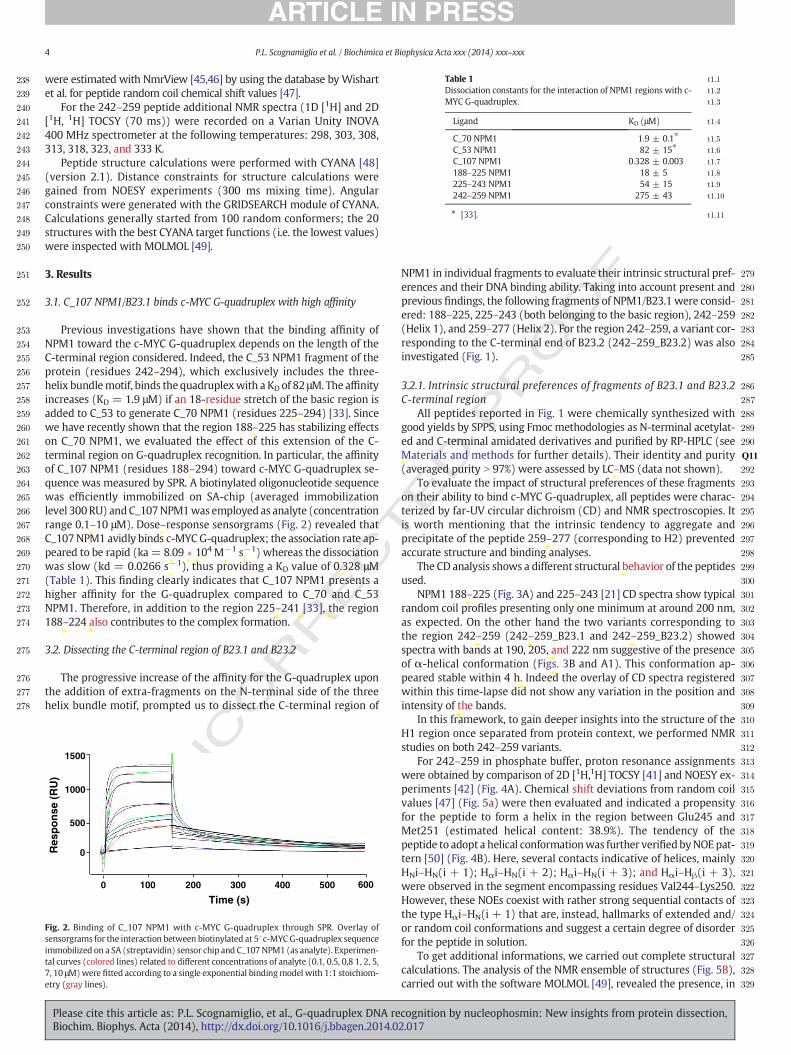
T
238
239
240
241
242
243
244
245
246
247
248
249
250
251
252
253
254
255
256
257
258
259
260
261
262
263
264
265
266
267
268
269
270
271
272
273
274
275
276
277
278
279
280
281
282
283
284
285
286
287
288
289
290
291Q11
292
293
294
295
296
297
298
299
300
301
302
303
304
305
306
307
308
309
t1:1Table 1t1:2Dissociation constants for the interaction of NPM1 regions with c-t1:3MYC G-quadruplex.
Ligand KD (μM) t1:4
C_70 NPM1 1.9 ± 0.1⁎ t1:5
C_53 NPM1 82 ± 15⁎ t1:6
C_107 NPM1 0.328 ± 0.003 t1:7
188–225 NPM1 18 ± 5 t1:8
225–243 NPM1 54 ± 15 t1:9
242–259 NPM1 275 ± 43 t1:10
t1:11⁎ [33].
4 P.L. Scognamiglio et al. / Biochimica et Biophysica Acta xxx (2014) xxx–xxx
RREC
were estimated with NmrView [45,46] by using the database byWishartet al. for peptide random coil chemical shift values [47].
For the 242–259 peptide additional NMR spectra (1D [1H] and 2D[1H, 1H] TOCSY (70 ms)) were recorded on a Varian Unity INOVA400 MHz spectrometer at the following temperatures: 298, 303, 308,313, 318, 323, and 333 K.
Peptide structure calculations were performed with CYANA [48](version 2.1). Distance constraints for structure calculations weregained from NOESY experiments (300 ms mixing time). Angularconstraints were generated with the GRIDSEARCH module of CYANA.Calculations generally started from 100 random conformers; the 20structures with the best CYANA target functions (i.e. the lowest values)were inspected with MOLMOL [49].
3. Results
3.1. C_107 NPM1/B23.1 binds c-MYC G-quadruplex with high affinity
Previous investigations have shown that the binding affinity ofNPM1 toward the c-MYC G-quadruplex depends on the length of theC-terminal region considered. Indeed, the C_53 NPM1 fragment of theprotein (residues 242–294), which exclusively includes the three-helix bundlemotif, binds thequadruplexwith a KD of 82 μM. The affinityincreases (KD = 1.9 μM) if an 18-residue stretch of the basic region isadded to C_53 to generate C_70 NPM1 (residues 225–294) [33]. Sincewe have recently shown that the region 188–225 has stabilizing effectson C_70 NPM1, we evaluated the effect of this extension of the C-terminal region on G-quadruplex recognition. In particular, the affinityof C_107 NPM1 (residues 188–294) toward c-MYC G-quadruplex se-quence was measured by SPR. A biotinylated oligonucleotide sequencewas efficiently immobilized on SA-chip (averaged immobilizationlevel 300RU) andC_107NPM1was employed as analyte (concentrationrange 0.1–10 μM). Dose–response sensorgrams (Fig. 2) revealed thatC_107NPM1 avidly binds c-MYCG-quadruplex; the association rate ap-peared to be rapid (ka= 8.09 ∗ 104 M−1 s−1) whereas the dissociationwas slow (kd = 0.0266 s−1), thus providing a KD value of 0.328 μM(Table 1). This finding clearly indicates that C_107 NPM1 presents ahigher affinity for the G-quadruplex compared to C_70 and C_53NPM1. Therefore, in addition to the region 225–241 [33], the region188–224 also contributes to the complex formation.
3.2. Dissecting the C-terminal region of B23.1 and B23.2
The progressive increase of the affinity for the G-quadruplex uponthe addition of extra-fragments on the N-terminal side of the threehelix bundle motif, prompted us to dissect the C-terminal region of
UNCO 310
311
312
313
314
315
316
317
318
319
320
321
322
323
324
325
326
327
328
329
0 100 200 300 400 500 600
Time (s)
Res
po
nse
(R
U)
0
500
1000
1500
Fig. 2. Binding of C_107 NPM1 with c-MYC G-quadruplex through SPR. Overlay ofsensorgrams for the interaction between biotinylated at 5′ c-MYC G-quadruplex sequenceimmobilized on a SA (streptavidin) sensor chip and C_107NPM1 (as analyte). Experimen-tal curves (colored lines) related to different concentrations of analyte (0.1, 0.5, 0,8 1, 2, 5,7, 10 μM)were fitted according to a single exponential bindingmodel with 1:1 stoichiom-etry (gray lines).
Please cite this article as: P.L. Scognamiglio, et al., G-quadruplex DNA reBiochim. Biophys. Acta (2014), http://dx.doi.org/10.1016/j.bbagen.2014.0
ED P
RO
OFNPM1 in individual fragments to evaluate their intrinsic structural pref-
erences and their DNA binding ability. Taking into account present andprevious findings, the following fragments of NPM1/B23.1 were consid-ered: 188–225, 225–243 (both belonging to the basic region), 242–259(Helix 1), and 259–277 (Helix 2). For the region 242–259, a variant cor-responding to the C-terminal end of B23.2 (242–259_B23.2) was alsoinvestigated (Fig. 1).
3.2.1. Intrinsic structural preferences of fragments of B23.1 and B23.2C-terminal region
All peptides reported in Fig. 1 were chemically synthesized withgood yields by SPPS, using Fmoc methodologies as N-terminal acetylat-ed and C-terminal amidated derivatives and purified by RP-HPLC (seeMaterials and methods for further details). Their identity and purity(averaged purity N 97%) were assessed by LC–MS (data not shown).
To evaluate the impact of structural preferences of these fragmentson their ability to bind c-MYC G-quadruplex, all peptides were charac-terized by far-UV circular dichroism (CD) and NMR spectroscopies. Itis worth mentioning that the intrinsic tendency to aggregate andprecipitate of the peptide 259–277 (corresponding to H2) preventedaccurate structure and binding analyses.
The CD analysis shows a different structural behavior of the peptidesused.
NPM1 188–225 (Fig. 3A) and 225–243 [21] CD spectra show typicalrandom coil profiles presenting only one minimum at around 200 nm,as expected. On the other hand the two variants corresponding tothe region 242–259 (242–259_B23.1 and 242–259_B23.2) showedspectra with bands at 190, 205, and 222 nm suggestive of the presenceof α-helical conformation (Figs. 3B and A1). This conformation ap-peared stable within 4 h. Indeed the overlay of CD spectra registeredwithin this time-lapse did not show any variation in the position andintensity of the bands.
In this framework, to gain deeper insights into the structure of theH1 region once separated from protein context, we performed NMRstudies on both 242–259 variants.
For 242–259 in phosphate buffer, proton resonance assignmentswere obtained by comparison of 2D [1H,1H] TOCSY [41] and NOESY ex-periments [42] (Fig. 4A). Chemical shift deviations from random coilvalues [47] (Fig. 5a) were then evaluated and indicated a propensityfor the peptide to form a helix in the region between Glu245 andMet251 (estimated helical content: 38.9%). The tendency of thepeptide to adopt a helical conformationwas further verifiedbyNOEpat-tern [50] (Fig. 4B). Here, several contacts indicative of helices, mainlyHNi–HN(i + 1); Hαi–HN(i + 2); Hαi–HN(i + 3); and Hαi–Hβ(i + 3),were observed in the segment encompassing residues Val244–Lys250.However, these NOEs coexist with rather strong sequential contacts ofthe type Hαi–HN(i + 1) that are, instead, hallmarks of extended and/or random coil conformations and suggest a certain degree of disorderfor the peptide in solution.
To get additional informations, we carried out complete structuralcalculations. The analysis of the NMR ensemble of structures (Fig. 5B),carried out with the software MOLMOL [49], revealed the presence, in
cognition by nucleophosmin: New insights from protein dissection,2.017
Original text:
Inserted Text
"*"
Original text:
Inserted Text
"also "
Original text:
Inserted Text
"-"
Original text:
Inserted Text
"-"
Original text:
Inserted Text
"-"
Original text:
Inserted Text
"a "
Original text:
Inserted Text
"residues "
Original text:
Inserted Text
"-"
Original text:
Inserted Text
"-"
Original text:
Inserted Text
"-"
Original text:
Inserted Text
"-"
Original text:
Inserted Text
"-"
Original text:
Inserted Text
","
Original text:
Inserted Text
"*"
Original text:
Inserted Text
"-"
Original text:
Inserted Text
"-"
Original text:
Inserted Text
"-"
Original text:
Inserted Text
","
Original text:
Inserted Text
"-"
Original text:
Inserted Text
"also "
Original text:
Inserted Text
"-"
Original text:
Inserted Text
"-"
Original text:
Inserted Text
"-"
Original text:
Inserted Text
"-"
Original text:
Inserted Text
"-"
Original text:
Inserted Text
"-"
Original text:
Inserted Text
"-"
Original text:
Inserted Text
"-"
Original text:
Inserted Text
"M"
Original text:
Inserted Text
"-"
Original text:
Inserted Text
"-"
Original text:
Inserted Text
"behaviour "
Original text:
Inserted Text
"-"
Original text:
Inserted Text
"-"
Original text:
Inserted Text
"-"
Original text:
Inserted Text
"-"
Original text:
Inserted Text
"-"
Original text:
Inserted Text
", "
Original text:
Inserted Text
", "
Original text:
Inserted Text
", "
Original text:
Inserted Text
"-"
Original text:
Inserted Text
"-"
Original text:
Inserted Text
"-"
Original text:
Inserted Text
"shifts "
Original text:
Inserted Text
"A"
Original text:
Inserted Text
"-"
Original text:
Inserted Text
"-"
Original text:
Inserted Text
"-"
Original text:
Inserted Text
"-"
Original text:
Inserted Text
"-"
Original text:
Inserted Text
"-"
Original text:
Inserted Text
"-"
Original text:
Inserted Text
" b"

T
OO
F
330
331
332
333
334
335
336
337
338
339
340
341
342
343
344
345
346
347
348
349
350
351
352
353
354
355
(deg
cm2
dm
ol-1
res-
110
3 )0
-15
-5.0
-10
200 220 240
t 0h t 1ht 2ht 2.5ht 4h
200 220 240
Wavelength (nm)Wavelength (nm)
(deg
cm2
dm
ol-1
res-
110
3 ) 7.0
-10
0
-5.0
-
-
-
-
A B
θθ
Fig. 3. Circular dichroism studies of C_107NPM1-based peptides. CD spectra of NPM1 (A) 188–225 and (B) 242–259, for the latter the overlay of spectra recorded at different times (0–4 h)is reported, and they were registered at 293 K in 10 mM phosphate buffer, pH 7.2.
5P.L. Scognamiglio et al. / Biochimica et Biophysica Acta xxx (2014) xxx–xxx
most of the conformers, of a disordered pseudo-helical segmentin between Val244 and Met251, composed of a turn of 3.10 helix(Val244–Ile247), and of a bend from Ile247 to Met251. This analysisclearly indicates that the C-terminal end is flexible.
NMR conformational analyses were carried out for 242–259_B23.2in similar experimental conditions. Recorded 2D [1H, 1H] TOCSY andNOESY spectra (Fig. A2) allowed to assign proton resonances also forthis peptide. The NMR parameters, i.e. chemical shift deviations fromrandom coil values (Fig. 5g) andNOE pattern (Fig. A3), indicate a similarhelix propensity and conformational behavior as 242–259 in the regionbetween Val244 and Ala249. Despite the two amino-acid substitutionsoccurring at the very end of the two peptides (Fig. 1), the C-terminusof both 242–259 and 242–259_B23.2 is disordered.
UNCO
RREC
1.00
2.00
3.00
4.00
8.40 8.20 8.40 8.201H ppm
1H p
pm
A
Fig. 4.NMR analysis of NPM1 242–259 B23.1 peptide. (A) 2D [1H, 1H] TOCSY (70msmixing timproton correlation region is shown. (B) NOE intensity diagrams for NPM1 242–259 B23.1. The anectivity between the Hx and Hy protons in the i and i + z residue respectively is indicated wsoftware CYANA [48] was used to generate the NOE diagram.
Please cite this article as: P.L. Scognamiglio, et al., G-quadruplex DNA reBiochim. Biophys. Acta (2014), http://dx.doi.org/10.1016/j.bbagen.2014.0
ED P
R3.2.2. The helical fragments 242–259 of B23.1 and B23.2 are endowed witha remarkable stability
The analysis of the thermal and chemical stabilities of 242–259 and242–259_B23.2 unveils some unexpected features. Spectra collectedover the temperature range 293 to 371 K indicate a marginal decreaseof the CD signal (Fig. 6A). This experiment indicates that this sequence re-tains a significant amount of the helical structure at a very high tempera-ture, even if a slight variation of ellipticity at 222 nm in the range 333–371K (inset of Fig. 6A) suggests that a decrease of total helical content occurs.This thermal stability is very unusual for short peptides and has beenoccasionally reported for peptides with unnatural constrains [51].
These observations prompted us to perform additional NMR studiesto better characterize the influence of higher temperatures on 242–259
S S V E D I K A KM Q A S I E K G GdNN(i,i+1)d N(i,i+1)d N(i,i+1)
dNN(i,i+2)
d N(i,i+2)
d N(i,i+3)
d (i,i+3)
B
αα
α
β
α
αβ
e) (left panel) and 2D [1H, 1H] NOESY (300msmixing time) (right panel), theHN-aliphaticmino acid sequence is reported on the top of diagramwith the one letter code. A NOE con-ith “dxy(i, i + z)”. The thickness of the bars reflects the corresponding NOE intensity. The
cognition by nucleophosmin: New insights from protein dissection,2.017
Original text:
Inserted Text
"-"
Original text:
Inserted Text
"-"
Original text:
Inserted Text
"Figure "
Original text:
Inserted Text
"shifts "
Original text:
Inserted Text
"G"
Original text:
Inserted Text
"Figure "
Original text:
Inserted Text
"-"
Original text:
Inserted Text
"-"
Original text:
Inserted Text
"-"
Original text:
Inserted Text
"-"
Original text:
Inserted Text
"stability "
Original text:
Inserted Text
"-"
Original text:
Inserted Text
"-"
Original text:
Inserted Text
"-"
Original text:
Inserted Text
"-"
Original text:
Inserted Text
"-"
Original text:
Inserted Text
"insert "
Original text:
Inserted Text
"-"
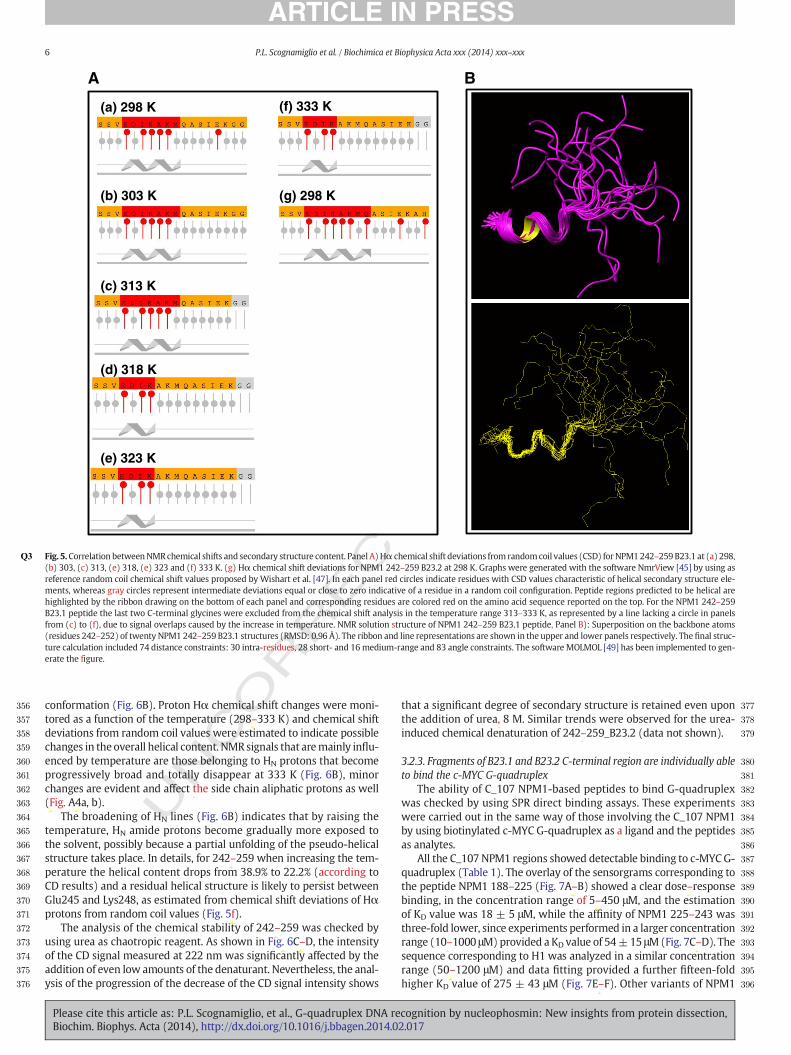
RRECTED P
RO
OF
356
357
358
359
360
361
362
363
364
365
366
367
368
369
370
371
372
373
374
375
376
377
378
379
380
381
382
383
384
385
386
387
388
389
390
391
392
393
394
395
396
(f) 333 K
(e) 323 K
(a) 298 K
(b) 303 K
(c) 313 K
(d) 318 K
(g) 298 K
A B
Q3 Fig. 5.Correlation betweenNMRchemical shifts and secondary structure content. Panel A)Hα chemical shift deviations from randomcoil values (CSD) forNPM1242–259 B23.1 at (a) 298,(b) 303, (c) 313, (e) 318, (e) 323 and (f) 333 K. (g) Hα chemical shift deviations for NPM1 242–259 B23.2 at 298 K. Graphs were generated with the software NmrView [45] by using asreference random coil chemical shift values proposed by Wishart et al. [47]. In each panel red circles indicate residues with CSD values characteristic of helical secondary structure ele-ments, whereas gray circles represent intermediate deviations equal or close to zero indicative of a residue in a random coil configuration. Peptide regions predicted to be helical arehighlighted by the ribbon drawing on the bottom of each panel and corresponding residues are colored red on the amino acid sequence reported on the top. For the NPM1 242–259B23.1 peptide the last two C-terminal glycines were excluded from the chemical shift analysis in the temperature range 313–333 K, as represented by a line lacking a circle in panelsfrom (c) to (f), due to signal overlaps caused by the increase in temperature. NMR solution structure of NPM1 242–259 B23.1 peptide, Panel B): Superposition on the backbone atoms(residues 242–252) of twenty NPM1 242–259 B23.1 structures (RMSD: 0.96 Å). The ribbon and line representations are shown in the upper and lower panels respectively. The final struc-ture calculation included 74 distance constraints: 30 intra-residues, 28 short- and 16medium-range and 83 angle constraints. The software MOLMOL [49] has been implemented to gen-erate the figure.
6 P.L. Scognamiglio et al. / Biochimica et Biophysica Acta xxx (2014) xxx–xxx
UNCOconformation (Fig. 6B). Proton Hα chemical shift changes were moni-
tored as a function of the temperature (298–333 K) and chemical shiftdeviations from random coil values were estimated to indicate possiblechanges in the overall helical content. NMRsignals that aremainly influ-enced by temperature are those belonging to HN protons that becomeprogressively broad and totally disappear at 333 K (Fig. 6B), minorchanges are evident and affect the side chain aliphatic protons as well(Fig. A4a, b).
The broadening of HN lines (Fig. 6B) indicates that by raising thetemperature, HN amide protons become gradually more exposed tothe solvent, possibly because a partial unfolding of the pseudo-helicalstructure takes place. In details, for 242–259 when increasing the tem-perature the helical content drops from 38.9% to 22.2% (according toCD results) and a residual helical structure is likely to persist betweenGlu245 and Lys248, as estimated from chemical shift deviations of Hαprotons from random coil values (Fig. 5f).
The analysis of the chemical stability of 242–259 was checked byusing urea as chaotropic reagent. As shown in Fig. 6C–D, the intensityof the CD signal measured at 222 nm was significantly affected by theaddition of even low amounts of the denaturant. Nevertheless, the anal-ysis of the progression of the decrease of the CD signal intensity shows
Please cite this article as: P.L. Scognamiglio, et al., G-quadruplex DNA reBiochim. Biophys. Acta (2014), http://dx.doi.org/10.1016/j.bbagen.2014.0
that a significant degree of secondary structure is retained even uponthe addition of urea, 8 M. Similar trends were observed for the urea-induced chemical denaturation of 242–259_B23.2 (data not shown).
3.2.3. Fragments of B23.1 and B23.2 C-terminal region are individually ableto bind the c-MYC G-quadruplex
The ability of C_107 NPM1-based peptides to bind G-quadruplexwas checked by using SPR direct binding assays. These experimentswere carried out in the same way of those involving the C_107 NPM1by using biotinylated c-MYC G-quadruplex as a ligand and the peptidesas analytes.
All the C_107 NPM1 regions showed detectable binding to c-MYCG-quadruplex (Table 1). The overlay of the sensorgrams corresponding tothe peptide NPM1 188–225 (Fig. 7A–B) showed a clear dose–responsebinding, in the concentration range of 5–450 μM, and the estimationof KD value was 18 ± 5 μM, while the affinity of NPM1 225–243 wasthree-fold lower, since experiments performed in a larger concentrationrange (10–1000 μM)provided a KD value of 54±15 μM(Fig. 7C–D). Thesequence corresponding to H1 was analyzed in a similar concentrationrange (50–1200 μM) and data fitting provided a further fifteen-foldhigher KD value of 275 ± 43 μM (Fig. 7E–F). Other variants of NPM1
cognition by nucleophosmin: New insights from protein dissection,2.017
Original text:
Inserted Text
" "
Original text:
Inserted Text
"s"
Original text:
Inserted Text
","
Original text:
Inserted Text
"-"
Original text:
Inserted Text
"Figure "
Original text:
Inserted Text
"Figure "
Original text:
Inserted Text
"-"
Original text:
Inserted Text
"-"
Original text:
Inserted Text
"accordingly "
Original text:
Inserted Text
" F"
Original text:
Inserted Text
"-"
Original text:
Inserted Text
"-"
Original text:
Inserted Text
"-"
Original text:
Inserted Text
"-"
Original text:
Inserted Text
"-"
Original text:
Inserted Text
"-"
Original text:
Inserted Text
"-"
Original text:
Inserted Text
"-"
Original text:
Inserted Text
"-"
Original text:
Inserted Text
"-"
Original text:
Inserted Text
"-"
Original text:
Inserted Text
"-"
Original text:
Inserted Text
"-"
Original text:
Inserted Text
"-"
Original text:
Inserted Text
"-"

RECTED P
RO
OF
397
398
399
400
401
402
403
404
405
406
407
408
409
410
411
412
413
414
415
416
417
418
419
420
421
422
423
424
425
426
427
428
429
430
431
432
433
434
435
436
437
438
439
440
-6.0
-4.0
-2.0
0
220 230 240 250
Wavelength (nm)
0M1M2M3M4M5M6M7M8M
298 K
308 K
318 K
323 K
333 K
8.60 8.20 7.80 7.40 7.00
1H ppm
BA
DC
293 K
371 K
7.0
-10
0
- 5.0
-
-
-
-200 220 240
Wavelength (nm)
-5.0
-7.0
-6.5
-6.0
295 371313 333 353Temperature (K)
0 2 4 6 8 10-8,0
-6,0
-4,0
-2,0
0
Urea (M)
θ(d
eg c
m2 d
mo
l-1re
s-1 1
03 )θ(
deg
cm
2 d
mo
l-1re
s-1 1
03 )
θ222
222n
m
nm (d
eg c
m2 dm
ol-1re
s-1 10
3 )
θ 220
nm22
0nm
(deg
cm
2 d
mo
l-1re
s-1 1
03 )
Fig. 6. Analysis of the stability of NPM1 242–259 B23.1 peptide. Overlay of CD spectra registered at (A) different temperatures in the range 293–371 K, asQ4 inset of the dependence of el-lipticity at 222 nm versus temperature is reported, (C) increasing urea concentrations in the range 0–8 M. In the latter experiment CD spectra were registered in the range 215–260 nmsince at lower wavelengths the presence of urea causes too high signal-to-noise ratio; (B) 1D [1H] spectra of NPM1 242–259 B23.1 recorded at different temperatures. In detail, in the re-ported expansion – from6.80 to 8.60 ppm – signals arising frombackbone and side chain HN protons can be observedQ5 in (D) interpolation of experimental ellipticity at 222 nmversus ureaconcentration.
7P.L. Scognamiglio et al. / Biochimica et Biophysica Acta xxx (2014) xxx–xxx
UNCO
R242–259 exhibited similar values of KD toward c-MYC G-quadruplex(Fig. A5). The presence of different residues in the C-terminal regiondid not substantially affect the binding capacities of H1 variants.
4. Discussion
The involvement of NPM1/B23 in a variety of distinct biological pro-cesses is a direct consequence of its ability to establish intermolecularpartnerships. The promiscuity of the protein is favored by its modularstructure, in which structured domains alternate with disordered re-gions. Despite the importance of the NPM1/B23 interactions in physio-logical and pathological conditions, their biophysical and structuralcharacterization is still incomplete. Very recently, it has been shownthat the binding of G-quadruplex DNA by NPM1/B23 is important forthe nucleolar localization of the protein [52]. The inability of leukemicNPM1/B23 variant to establish this interaction leads to the aberrant cy-toplasmic localization of the protein.
Present findings demonstrate that the C-terminal region of the pro-tein is able to bind the c-MYC G-quadruplex with a sub-micromolar af-finity. The comparison of these results with previous literature dataindicates that the progressive inclusion of portions of the basic regionincreases the binding affinity of NPM1/B23 for the G-quadruplex se-quence. Intriguingly, the inclusion of these regions had also stabilizing
Please cite this article as: P.L. Scognamiglio, et al., G-quadruplex DNA reBiochim. Biophys. Acta (2014), http://dx.doi.org/10.1016/j.bbagen.2014.0
effects on the thermal and chemical stabilities of the terminal threehelix bundle motif [21,24].
The dissection of NPM1/B23 demonstrates that several fragments ofthe protein are intrinsically able to bind the G-quadruplex with highaffinity. Present data also indicate that, in addition to the unfoldedregion 225–242, another intrinsically disordered fragment of theprotein (residues 188–225) significantly contributes to the bindingof the G-quadruplex. Therefore, the tight binding of NPM1/B23 to theG-quadruplex is achieved through the cooperation of both folded andunfolded regions that are individually able to bind it. As mentioned inthe Introduction, in the NMR structure of the complex between NPM1C_70 and the c-MYC G-quadruplex direct interactions with the DNAwere established only by the three helix bundle motif [34]. Indeed, theregion 225–242 remains flexible without forming any permanent con-tact with the G-quadruplex. On the other hand, here we show thatboth regions 188–225 and 225–243 of NPM1/B23 are indeed able to di-rectly bind the G-quadruplex. These apparently contrasting results maybe reconciled by assuming that the three helix bundlemotif and the dis-ordered basic regions recognize the same regions of the G-quadruplex.In this scenario, the intrinsically disordered regions may play a dualrole in the G-quadruplex binding. Taking advantage of the great flexibil-ity, they could efficiently anchor the DNA in the initial stage of the rec-ognition process. Subsequently, after the binding of theG-quadruplex to
cognition by nucleophosmin: New insights from protein dissection,2.017
Original text:
Inserted Text
"-"
Original text:
Inserted Text
"figure "
Original text:
Inserted Text
"stability "
Original text:
Inserted Text
"-"
Original text:
Inserted Text
"-"
Original text:
Inserted Text
"contribute "
Original text:
Inserted Text
"i"
Original text:
Inserted Text
"-"
Original text:
Inserted Text
"-"
Original text:
Inserted Text
"-"

ECTED P
RO
OF
441
442
443
444
445
446
447
448
449
450
451
452
453
454
455
456
457
458
459
460
461
462
463
464
465
466
467
468
469
470Q12
471
472
473
474
475
476
477
478
479
480
481
482
483
484
485
486
487
488
489
490
0 250 500 750 1000 12500
25
50
75
NPM1 188-225 ( M)0 100 200 300 400 500
0
50
100
150
0
50
100
150
0 100 200 300 400 500 600
0
50
100
150
0 100 200 300 400 500 600
0 250 500 750 12500
200
300
400
RU
max
RU
max
RU
max
100
10000 100 200 300 400 500
Time (s)
Time (s)
Time (s)
600
200
300
400
100
0Res
po
nse
(R
U)
Res
po
nse
(R
U)
Res
po
nse
(R
U)
NPM1 225-243 ( M)
NPM1 242-259 ( M)
A B
C D
E F
Fig. 7. Binding of C_107NPM1 derived peptides to c-MYCG-quadruplex through SPR. Overlay of sensorgrams for the interaction between biotinylated at 5′ c-MYCG-quadruplex sequenceimmobilized on a SA (streptavidin) sensor chip and C_107 NPM1 derived peptides. NPM1 (A) 188–225 (C) 225–243, and (E) 242–259 experimental curves that represent different con-centrations of analyte and were fitted according to a single exponential binding model with 1:1 stoichiometry, (B), (D), and (F), respectively.
8 P.L. Scognamiglio et al. / Biochimica et Biophysica Acta xxx (2014) xxx–xxx
UNCO
RRthe three helix binding motif, the positively charges of these basic re-
gions may generate a long-range favorable electrostatic environment,as previously suggested [34]. The ability of analyzed peptides to bindc-MYC G-quadruplex represents a solid ground for designing newmol-ecules able to interact with this DNA motif.
The present findings also suggest a reduced G-quadruplex affinityfor the two splicing variants B23.2 and B23.3. In particular, a reduced af-finity of B23.3 is likely due to the lack of the basic region 195–223 in thisisoform. Moreover, the truncation of the C-terminus of the three helixbundle motif to the helix H1 in B23.2 is also expected to strongly de-crease the affinity for the G-quadruplex, since we show here that thishelix exhibits a significantly reduced affinity, when compared to thewhole domain. These suggestions are in line with the results indepen-dently obtained by Okuwaki and coworkers, who very recently ana-lyzed the binding of the different NPM1/B23 isoforms and truncatedforms to RNA [53]. However, very recently, it has been demonstrated,through ITC analysis, that the affinity of the full-length NPM1 for thesame G-quadruplex is 3.8 μM [54] that is a value closer to that one ob-tained for the C_70 NPM1 than that for the C_107 NPM1. In the case offull-length NPM1 the electrostatic interactions established betweenthe basic domain and the preceding acidic unstructured domainscould influence G-quadruplex recognition.
The dissection of NPM1/B23 and the biophysical characterization ofthe resulting fragments also unveil that the H1 helix of both B23.1 andB23.2 is intrinsically endowed with an unusual thermal stability. A
Please cite this article as: P.L. Scognamiglio, et al., G-quadruplex DNA reBiochim. Biophys. Acta (2014), http://dx.doi.org/10.1016/j.bbagen.2014.0
combined CD and NMR analyses show that the N-terminal region ofH1 assumes a helical conformation, despite the intrinsic flexibility ofthe peptides. Indeed, the NMR analysis shows that the segment fromGlu245 to Met251 clearly exhibits a tendency to adopt an orderedhelical structure. CD and NMR data both indicate that this short helicalsegment is rather stable upon temperature increase. Evidence ofthis structure is detected in the entire temperature range (293–371 Kfor CD and 298–333 K for NMR) explored by using the two techniques.This thermal stability is very unusual for a short peptide that hasbeen derived from a protein sequence. Similar high stabilities haverather been reported, only in few cases, for peptides whose sequenceshave been ad-hoc designed [51,55]. These unexpected results hold in-teresting implications for NPM1/B23 folding/unfolding process. In thelast years, the folding pathway of several constructs of the NPM1 C-terminal region has been characterized in detail [21–24]. These studieshave highlighted the presence of residual structure in the denaturedstate of the protein [56]. In particular, biophysical characterizationshave detected a “native-like” structure that is retained in the denaturedstate of the wild-type protein, involving the helices H2–H3 of the threehelix bundle motif. It has been shown that the presence of these native-like features in the NPM1/B23 denatured state accelerates the foldingprocess. Moreover, it has been suggested that the identification andthe characterization of these native-like featuresmay be away to designnew molecules that could restore the native fold in pathological mu-tants. The intrinsic stability of the H1 helix provides support to the
cognition by nucleophosmin: New insights from protein dissection,2.017
Original text:
Inserted Text
"both "
Original text:
Inserted Text
"represent "
Original text:
Inserted Text
"-"
Original text:
Inserted Text
"-"
Original text:
Inserted Text
"Okawaki "
Original text:
Inserted Text
"unveils "
Original text:
Inserted Text
"unveils "
Original text:
Inserted Text
"analysis "
Original text:
Inserted Text
"both "
Original text:
Inserted Text
"-"
Original text:
Inserted Text
"-"

T
491
492
493
494
495
496
497
498
499
500
501
502
503
504
505
506
507
508
509
510
511
512
513
514
515
516
517
518
519
520
521522523524525526527528529530531532533534535536537538539540541542543544545546547548549550551552553554555
556557558559560561562563564565566567568569570571572573574575576577578579580581582583584585586587588589590591592593594595596597598599600601602603604605606607608609610611612613614615616617618619620621622623624625626627628629630631632633634635636637638639640641
9P.L. Scognamiglio et al. / Biochimica et Biophysica Acta xxx (2014) xxx–xxx
UNCO
RREC
view that the denatured state retains some native-like structural prop-erties, although in previous analyses this helix was considered to beunfolded. Of particular interest, in this context, is the observationthat Trp290 interacts in the native fold with the intrinsically folded N-terminal region of H1. In line with the previous suggestions, this obser-vation may be exploited in future studies for designing molecules ableto stabilize the fold of the domain and to restore the protein functionin NPM1/B23 mutants.
5. Conclusions
In conclusion this study unveils structural and functional determi-nants of the interaction between G-quadruplex DNA and NPM1, by an-alyzing the contribution of single protein regions in DNA recognitionmechanism. Through a rational fragmentation approach results clearlyindicate a crucial and direct role exerted by IDRs in DNA recognition,speculating a synergic action of IDRs along with H1 region in formationof the complexwith G-quadruplex inwhich the flexible basic fragmentsprimarily allow to accommodate DNA structures. This study shed newlight for the design of small molecules and/or peptides able to stabilizeG-quadruplex/NPM1complex which could help the research in thefield of AML disease.
Acknowledgements
This work was funded in part by the MIUR Ricerca e Competitività2007–2013 #PON 01_2388. The authors are grateful to Luca De Lucaand Dr Giuseppe Perretta for the technical assistance, and Prof A. Zagariand Dr V. Granata of CEINGE — NAPOLI for their helpful discussion onSPR experiments.
Appendix A. Supplementary data
Supplementary data to this article can be found online at http://dx.doi.org/10.1016/j.bbagen.2014.02.017.
References
[1] Y.J. Kang, M.O. Olson, C. Jones, H. Busch, Nucleolar phosphoproteins of normal ratliver and Novikoff hepatoma ascites cells, Cancer Res. 35 (1975) 1470–1475.
[2] M. Okuwaki, K.Matsumoto,M. Tsujimoto, K. Nagata, Function of nucleophosmin/B23, anucleolar acidic protein, as a histone chaperone, FEBS Lett. 506 (2001) 272–276.
[3] R.A. Borer, C.F. Lehner, H.M. Eppenberger, E.A. Nigg, Major nucleolar proteins shuttlebetween nucleus and cytoplasm, Cell 56 (1989) 379–390.
[4] M. Okuwaki, A. Iwamatsu, M. Tsujimoto, K. Nagata, Identification of nucleophosmin/B23, an acidic nucleolar protein, as a stimulatory factor for in vitro replication of adeno-virus DNA complexed with viral basic core proteins, J. Mol. Biol. 311 (2001) 41–55.
[5] M.S. Lindstrom, NPM1/B23: a multifunctional chaperone in ribosome biogenesisand chromatin remodeling, Biochem. Res. Int. (2011) 195209 (16 pp.).
[6] L.B. Maggi Jr., M. Kuchenruether, D.Y. Dadey, R.M. Schwope, S. Grisendi, R.R.Townsend, P.P. Pandolfi, J.D.Weber, Nucleophosmin serves as a rate-limiting nuclearexport chaperone for the mammalian ribosome, Mol. Cell. Biol. 28 (2008)7050–7065.
[7] E. Colombo, M. Alcalay, P.G. Pelicci, Nucleophosmin and its complex network: a pos-sible therapeutic target in hematological diseases, Oncogene 30 (2011) 2595–2609.
[8] J.P. Yun, J. Miao, G.G. Chen, Q.H. Tian, C.Q. Zhang, J. Xiang, J. Fu, P.B. Lai, Increased ex-pression of nucleophosmin/B23 in hepatocellular carcinoma and correlation withclinicopathological parameters, Br. J. Cancer 96 (2007) 477–484.
[9] S. Grisendi, C. Mecucci, B. Falini, P.P. Pandolfi, Nucleophosmin and cancer, Nat. Rev.Cancer 6 (2006) 493–505.
[10] R. Balusu, W. Fiskus, R. Rao, D.G. Chong, S. Nalluri, U. Mudunuru, H. Ma, L. Chen, S.Venkannagari, K. Ha, S. Abhyankar, C. Williams, J. McGuirk, H.J. Khoury, C. Ustun,K.N. Bhalla, Targeting levels or oligomerization of nucleophosmin 1 induces differ-entiation and loss of survival of human AML cells with mutant NPM1, Blood 118(2011) 3096–3106.
[11] B. Falini, I. Nicoletti, N. Bolli, M.P. Martelli, A. Liso, P. Gorello, F. Mandelli, C. Mecucci,M.F. Martelli, Translocations and mutations involving the nucleophosmin (NPM1)gene in lymphomas and leukemias, Haematologica 92 (2007) 519–532.
[12] B. Falini, C. Mecucci, E. Tiacci, M. Alcalay, R. Rosati, L. Pasqualucci, R. La Starza, D.Diverio, E. Colombo, A. Santucci, B. Bigerna, R. Pacini, A. Pucciarini, A. Liso, M.Vignetti, P. Fazi, N. Meani, V. Pettirossi, G. Saglio, F. Mandelli, F. Lo-Coco, P.G. Pelicci,M.F. Martelli, Cytoplasmic nucleophosmin in acute myelogenous leukemia with anormal karyotype, N. Engl. J. Med. 352 (2005) 254–266.
Please cite this article as: P.L. Scognamiglio, et al., G-quadruplex DNA reBiochim. Biophys. Acta (2014), http://dx.doi.org/10.1016/j.bbagen.2014.0
ED P
RO
OF
[13] B. Falini, N. Bolli, J. Shan, M.P. Martelli, A. Liso, A. Pucciarini, B. Bigerna, L. Pasqualucci,R. Mannucci, R. Rosati, P. Gorello, D. Diverio, G. Roti, E. Tiacci, G. Cazzaniga, A. Biondi,S. Schnittger, T. Haferlach, W. Hiddemann, M.F. Martelli, W. Gu, C. Mecucci, I.Nicoletti, Both carboxy-terminus NES motif and mutated tryptophan(s) are crucialfor aberrant nuclear export of nucleophosmin leukemic mutants in NPMc + AML,Blood 107 (2006) 4514–4523.
[14] B. Falini, I. Nicoletti, M.F. Martelli, C. Mecucci, Acute myeloid leukemia carryingcytoplasmic/mutated nucleophosmin (NPMc + AML): biologic and clinical fea-tures, Blood 109 (2007) 874–885.
[15] M. Okuwaki, A. Sumi, M. Hisaoka, A. Saotome-Nakamura, S. Akashi, Y. Nishimura, K.Nagata, Function of homo- and hetero-oligomers of human nucleoplasmin/nucleophosmin family proteins NPM1, NPM2 and NPM3 during sperm chromatinremodeling, Nucleic Acids Res. 40 (2012) 4861–4878.
[16] M. Okuwaki, The structure and functions of NPM1/nucleophosmin/B23, a multifunc-tional nucleolar acidic protein, J. Biochem. 143 (2008) 441–448.
[17] V.M. Namboodiri, I.V. Akey, M.S. Schmidt-Zachmann, J.F. Head, C.W. Akey, The struc-ture and function of Xenopus NO38-core, a histone chaperone in the nucleolus,Structure 12 (2004) 2149–2160.
[18] H.H. Lee, H.S. Kim, J.Y. Kang, B.I. Lee, J.Y. Ha, H.J. Yoon, S.O. Lim, G. Jung, S.W. Suh,Crystal structure of human nucleophosmin-core reveals plasticity of thepentamer–pentamer interface, Proteins 69 (2007) 672–678.
[19] C.G. Grummitt, F.M. Townsley, C.M. Johnson, A.J. Warren, M. Bycroft, Structural con-sequences of nucleophosmin mutations in acute myeloid leukemia, J. Biol. Chem.283 (2008) 23326–23332.
[20] L. Federici, B. Falini, Nucleophosmin mutations in acute myeloid leukemia: a tale ofprotein unfolding and mislocalization, Protein Sci. 22 (2013) 545–556.
[21] D. Marasco, A. Ruggiero, C. Vascotto, M. Poletto, P.L. Scognamiglio, G. Tell, L. Vitagliano,Role of mutual interactions in the chemical and thermal stability of nucleophosminNPM1 domains, Biochem. Biophys. Res. Commun. 430 (2013) 523–528.
[22] F. Scaloni, L. Federici, M. Brunori, S. Gianni, Deciphering the folding transition statestructure and denatured state properties of nucleophosmin C-terminal domain,Proc. Natl. Acad. Sci. U. S. A. 107 (2010) 5447–5452.
[23] F. Scaloni, S. Gianni, L. Federici, B. Falini, M. Brunori, Folding mechanism of theC-terminal domain of nucleophosmin: residual structure in the denatured stateand its pathophysiological significance, FASEB J. 23 (2009) 2360–2365.
[24] S. Chiarella, L. Federici, A. Di Matteo, M. Brunori, S. Gianni, The folding pathway of afunctionally competent C-terminal domain of nucleophosmin: protein stability and de-natured state residual structure, Biochem. Biophys. Res. Commun. 435 (2013) 64–68.
[25] D. Fantini, C. Vascotto, D. Marasco, C. D'Ambrosio, M. Romanello, L. Vitagliano, C.Pedone, M. Poletto, L. Cesaratto, F. Quadrifoglio, A. Scaloni, J.P. Radicella, G. Tell, Crit-ical lysine residues within the overlooked N-terminal domain of human APE1 regu-late its biological functions, Nucleic Acids Res. 38 (2010) 8239–8256.
[26] M. Hisaoka, S. Ueshima, K. Murano, K. Nagata, M. Okuwaki, Regulation of nucleolarchromatin by B23/nucleophosmin jointly depends upon its RNA binding activityand transcription factor UBF, Mol. Cell. Biol. 30 (2010) 4952–4964.
[27] E. Colombo, J.C. Marine, D. Danovi, B. Falini, P.G. Pelicci, Nucleophosmin regulatesthe stability and transcriptional activity of p53, Nat. Cell Biol. 4 (2002) 529–533.
[28] C. Lee, B.A. Smith, K. Bandyopadhyay, R.A. Gjerset, DNA damage disrupts thep14ARF-B23(nucleophosmin) interaction and triggers a transient subnuclear redis-tribution of p14ARF, Cancer Res. 65 (2005) 9834–9842.
[29] Y. Jian, Z. Gao, J. Sun, Q. Shen, F. Feng, Y. Jing, C. Yang, RNA aptamers interfering withnucleophosmin oligomerization induce apoptosis of cancer cells, Oncogene 28(2009) 4201–4211.
[30] S.S. Gadad, R.E. Rajan, P. Senapati, S. Chatterjee, J. Shandilya, P.K. Dash, U. Ranga, T.K.Kundu, HIV-1 infection induces acetylation of NPM1 that facilitates Tat localizationand enhances viral transactivation, J. Mol. Biol. 410 (2011) 997–1007.
[31] D. Wang, A. Baumann, A. Szebeni, M.O. Olson, The nucleic acid binding activity ofnucleolar protein B23.1 resides in its carboxyl-terminal end, J. Biol. Chem. 269(1994) 30994–30998.
[32] T.S. Dumbar, G.A. Gentry, M.O. Olson, Interaction of nucleolar phosphoprotein B23with nucleic acids, Biochemistry 28 (1989) 9495–9501.
[33] L. Federici, A. Arcovito, G.L. Scaglione, F. Scaloni, C. Lo Sterzo, A. Di Matteo, B.Falini, B. Giardina, M. Brunori, Nucleophosmin C-terminal leukemia-associateddomain interacts with G-rich quadruplex forming DNA, J. Biol. Chem. 285(2010) 37138–37149.
[34] A. Gallo, C. Lo Sterzo, M. Mori, A. Di Matteo, I. Bertini, L. Banci, M. Brunori, L. Federici,Structure of nucleophosmin DNA-binding domain and analysis of its complexwith aG-quadruplex sequence from the c-MYC promoter, J. Biol. Chem. 287 (2012)26539–26548.
[35] S. Burge, G.N. Parkinson, P. Hazel, A.K. Todd, S. Neidle, Quadruplex DNA: sequence,topology and structure, Nucleic Acids Res. 34 (2006) 5402–5415.
[36] A. Siddiqui-Jain, C.L. Grand, D.J. Bearss, L.H. Hurley, Direct evidence for aG-quadruplex in a promoter region and its targeting with a small moleculeto repress c-MYC transcription, Proc. Natl. Acad. Sci. U. S. A. 99 (2002)11593–11598.
[37] C. Vascotto, D. Fantini, M. Romanello, L. Cesaratto, M. Deganuto, A. Leonardi, J.P.Radicella, M.R. Kelley, C. D'Ambrosio, A. Scaloni, F. Quadrifoglio, G. Tell, APE1/Ref-1interacts with NPM1 within nucleoli and plays a role in the rRNA quality controlprocess, Mol. Cell. Biol. 29 (2009) 1834–1854.
[38] M. Poletto, C. Vascotto, P.L. Scognamiglio, L. Lirussi, D. Marasco, G. Tell, Role ofthe unstructured N-terminal domain of the hAPE1 (human apurinic/apyrimidinicendonuclease 1), Biochem. J. 452 (2013) 545–557.
[39] R.L. Rich, D.G. Myszka, Advances in surface Plasmon resonance biosensor analysis,Curr. Opin. Biotechnol. 11 (2000) 54–61.
[40] G.B. Fields, R.L. Noble, Solid phase peptide synthesis utilizing 9-fluorenylmethoxycarbonyl amino acids, Int. J. Pept. Protein Res. 35 (1990) 161–214.
cognition by nucleophosmin: New insights from protein dissection,2.017
Original text:
Inserted Text
"-"
Original text:
Inserted Text
"nucleophsmin"
Original text:
Inserted Text
"nucleophsmin"
Original text:
Inserted Text
"-"
642643644645646647648649650651652653654655656657658659660661662663664
665666667668669670671672673674675676677678679680681682683684685686687
10 P.L. Scognamiglio et al. / Biochimica et Biophysica Acta xxx (2014) xxx–xxx
[41] C. Griesinger, G. Otting, K. Wuthrich, R.R. Ernst, Clean TOCSY for H-1 spinsystem-identification in macromolecules, J. Am. Chem. Soc. 110 (1988)7870–7872.
[42] A. Kumar, R.R. Ernst, K. Wuthrich, A two-dimensional nuclear overhauser enhance-ment (2D NOE) experiment for the elucidation of complete proton–protoncross-relaxation networks in biological macromolecules, Biochem. Biophys. Res.Commun. 95 (1980) 1–6.
[43] C. Dalvit, Efficient multiple-solvent suppression for the study of the interactions oforganic solvents with biomolecules, J. Biomol. NMR 11 (1998) 437–444.
[44] C. Bartels, T.H. Xia, M. Billeter, P. Guntert, K. Wuthrich, The program XEASY forcomputer-supported NMR spectral analysis of biological macromolecules, J. Biomol.NMR 6 (1995) 1–10.
[45] B.A. Johnson, Using NMRView to visualize and analyze the NMR spectra of macro-molecules, Methods Mol. Biol. 278 (2004) 313–352.
[46] S. Schwarzinger, G.J. Kroon, T.R. Foss, P.E. Wright, H.J. Dyson, Random coil chemicalshifts in acidic 8 M urea: implementation of random coil shift data in NMRView, J.Biomol. NMR 18 (2000) 43–48.
[47] D.S. Wishart, C.G. Bigam, A. Holm, R.S. Hodges, B.D. Sykes, 1H, 13C and 15N randomcoil NMR chemical shifts of the common amino acids. I. Investigations ofnearest-neighbor effects, J. Biomol. NMR 5 (1995) 67–81.
[48] T. Herrmann, P. Guntert, K. Wuthrich, Protein NMR structure determination withautomated NOE assignment using the new software CANDID and the torsionangle dynamics algorithm DYANA, J. Mol. Biol. 319 (2002) 209–227.
UNCO
RRECT
Please cite this article as: P.L. Scognamiglio, et al., G-quadruplex DNA reBiochim. Biophys. Acta (2014), http://dx.doi.org/10.1016/j.bbagen.2014.0
F
[49] R. Koradi, M. Billeter, K. Wuthrich, MOLMOL: a program for display and analysis ofmacromolecular structures, J. Mol. Graph. 14 (1996) 51–55 (29–32).
[50] K. Wüthrich, NMR of Proteins and Nucleic Acids, John Wiley & Sons, New York,1986.
[51] N.E. Shepherd, H.N. Hoang, G. Abbenante, D.P. Fairlie, Single turn peptide alpha he-lices with exceptional stability in water, J. Am. Chem. Soc. 127 (2005) 2974–2983.
[52] S. Chiarella, A. De Cola, G.L. Scaglione, E. Carletti, V. Graziano, D. Barcaroli, C. LoSterzo, A. Di Matteo, C. Di Ilio, B. Falini, A. Arcovito, V. De Laurenzi, L. Federici,Nucleophosmin mutations alter its nucleolar localization by impairingG-quadruplex binding at ribosomal DNA, Nucleic Acids Res. 41 (2013) 3228–3239.
[53] M. Hisaoka, K. Nagata, M. Okuwaki, Intrinsically disordered regions ofnucleophosmin/B23 regulate its RNA binding activity through their inter- andintra-molecular association, Nucleic Acids Res. 17 (2014) 1180–1195.
[54] S. Banuelos, B. Lectez, S.G. Taneva, G.Ormaza,M.Alonso-Marino, X. Calle,M.A.Urbaneja,Recognition of intermolecular G-quadruplexes by full length nucleophosmin. Effect of aleukaemia-associated mutation, FEBS Lett. 587 (2013) 2254–2259.
[55] D. Diana, B. Ziaco, G. Colombo, G. Scarabelli, A. Romanelli, C. Pedone, R. Fattorusso,L.D. D'Andrea, Structural determinants of the unusual helix stability of a de novoengineered vascular endothelial growth factor (VEGF) mimicking peptide, Chemis-try 14 (2008) 4164–4166.
[56] I.E. Sanchez, T. Kiefhaber, Hammond behavior versus ground state effects in proteinfolding: evidence for narrow free energy barriers and residual structure in unfoldedstates, J. Mol. Biol. 327 (2003) 867–884.
ED P
RO
O
cognition by nucleophosmin: New insights from protein dissection,2.017
Original text:
Inserted Text
" "
Original text:
Inserted Text
"-"


ACKNOWLEDGEMENTS
A great deal of my gratitude goes to Prof. Gianluca Tell, who has been mentor, source of
inspiration and friend.
I am immensely grateful to the past and present members of the GT lab: Lisa, Giulia,
Arianna, Elena, Carlo, Damiano, Laura, Erika, Milly, and Marta, who have always been kind, friendly
and ready to help in any situation. In particular, I would like to express my gratitude to Matilde, for
her invaluable help in the lab and for being always ready to share a smile.
Thanks also to the people in the LMG lab, to Dr. David Wilson III and his group in particular,
because they provided a stimulating environment and they made me feel at home even thousand miles
away.
All my colleagues deserve a huge acknowledgement: Federica, on top of everyone, then
Paolo, Enrico, Andrea, and Davide, just to name a few, because they shared and breathed with me the
PhD experience, inside and especially outside the lab.
A really extraordinary acknowledgement goes to Andrea, who has been more than a friend,
more than a colleague, more than a roommate, more than anything else.
I conclude by thanking Michela. No words in any language will be enough to express how
grateful I am for her contribute to this thesis and to my life…THANK YOU!