Defective transcription initiation causes postnatal growth failure in a mouse model of nucleotide...
6
Defective transcription initiation causes postnatal growth failure in a mouse model of nucleotide excision repair (NER) progeria Irene Kamileri a,b , Ismene Karakasilioti a,b , Aria Sideri a , Theodoros Kosteas a , Antonis Tatarakis c , Iannis Talianidis c , and George A. Garinis a,b,1 a Institute of Molecular Biology and Biotechnology, Foundation for Research and Technology-Hellas, Nikolaou Plastira 100, 70013, Heraklion, Crete, Greece; b Department of Biology, University of Crete, Vassilika Vouton, GR71409, Heraklion, Crete, Greece; and c Biomedical Sciences Research Center Al. Fleming, 16672 Vari, Greece Edited by Philip C. Hanawalt, Stanford University, Stanford, CA, and approved January 17, 2012 (received for review September 10, 2011) Nucleotide excision repair (NER) defects are associated with cancer, developmental disorders and neurodegeneration. However, with the exception of cancer, the links between defects in NER and developmental abnormalities are not well understood. Here, we show that the ERCC1-XPF NER endonuclease assembles on active promoters in vivo and facilitates chromatin modifications for tran- scription during mammalian development. We find that Ercc1 -/- mice demonstrate striking physiological, metabolic and gene ex- pression parallels with Taf10 -/- animals carrying a liver-specific transcription factor II D (TFIID) defect in transcription initiation. Pro- moter occupancy studies combined with expression profiling in the liver and in vitro differentiation cell assays reveal that ERCC1-XPF interacts with TFIID and assembles with POL II and the basal tran- scription machinery on promoters in vivo. Whereas ERCC1-XPF is required for the initial activation of genes associated with growth, it is dispensable for ongoing transcription. Recruitment of ERCC1- XPF on promoters is accompanied by promoter-proximal DNA demethylation and histone marks associated with active hepatic transcription. Collectively, the data unveil a role of ERCC1/XPF endonuclease in transcription initiation establishing its causal con- tribution to NER developmental disorders. DNA damage | genetics | metabolism D evelopmental-stage and tissue-specific programs of gene expression require the action of sequence-specific DNA binding factors, the basal transcription machinery and chromatin remodeling and modification enzymes (1). Together, these fac- tors create a chromatin environment that allows the synthesis of the primary transcript (2). If the transcriptional machinery is defective or challenged due to e.g., transcription-blocking DNA lesions, the process of RNA synthesis halts. To ensure that the genetic information is preserved and that transcription is not compromised, cells use DNA repair systems aimed at counter- acting DNA damage (3). For bulky helix-distorting damage, the principal repair mechanism is the evolutionarily conserved nu- cleotide excision repair (NER) pathway. NER operates via a “cut and patch” type of mechanism involving ∼30 proteins that rec- ognize and remove helical distortions throughout the genome (global genome NER; GGR), or selectively from the transcribed strand of active genes (transcription-coupled repair; TCR) (4). In GGR, the DNA is surveyed by the XPC-hR23B complex and the UV-damaged DNA-binding protein. Instead, damage rec- ognition in TCR requires the RNA polymerase II (POL II), CSA, and CSB. Unwinding the DNA around a lesion and sta- bilization of single-stranded DNA is followed by the XPG and ERCC1-XPF endonucleases that cleave on the 3′ and 5′side of the DNA lesion, respectively followed by excision of the damage and gap-filling DNA synthesis (5). Inborn NER defects may lead to skin cancer-prone xeroderma pigmentosum (XP) (6) or to a heterogeneous group of devel- opmental disorders, including Cockayne syndrome (CS; affected genes: Csb and Csa) and trichothiodystrophy (TTD; affected genes: Xpd and Xpb) (7). CS and TTD patients are characterized by postnatal growth failure, skeletal and neuronal abnormalities, s.c. fat loss and short lifespan (collectively designated as “segmental” NER progeroid features), but not cancer (8). Mouse mutants with inborn NER defects closely mimic their human counterparts and display severe developmental abnormalities and short lifespan (9). Whereas defective NER of damaged DNA has been estab- lished as the underlying cause of mutations leading to skin cancer, the links between NER defects and the developmental abnor- malities seen in NER disorders remain unclear (10–12). Earlier studies have shown that distinct NER factors play a role in tran- scription (5, 13, 14) and that, upon stimulation, they are recruited to active promoters in vitro (15). However, the in vivo relevance of NER-mediated transcription to the NER developmental dis- orders remains elusive, primarily due to difficulties in dissecting the dual role of NER in DNA repair and transcription in an intact organism. Here, we provide evidence that key developmental abnormalities associated with a defect in NER originate from defective transcription initiation of gene expression programs. Results Ercc1 -/- Mice Demonstrate Physiologic and Metabolic Parallels with Liver-Specific Taf10 -/- Mice. To assess the contribution of NER in transcription during development, we compared the liver phe- notypes of NER-deficient Ercc1 −/− animals that closely mimic a severe form of CS (11) with transcription factor II D (TFIID)- defective Taf10 −/− mice exhibiting a liver-specific defect in transcription initiation (Taf10 −/− -Alb-Cre) (16). Ercc1 −/− mice show attenuated growth, resulting in cachectic dwarfism during the second week of life and premature death before postnatal day P35 (Fig. 1A) (11). Likewise, liver-specific disruption of Taf10 gene in Taf10 −/− animals leads to severe growth failure during the second week after birth, more than 50% reduction in body weight at day P30, and premature death at ∼P35 (Fig. 1C) (16). Oil Red O and PAS staining in Ercc1 −/− and Taf10 −/− livers revealed a uniform accumulation of triglycerides and glycogen resulting in a “fatty liver” appearance with unusually large gly- cogen depots (Fig. 1 B and D). Apoptosis was considerably higher in both animal models compared with controls (Fig. 1 A– D Lower). Thus, NER-defective Ercc1 −/− mice and Taf10 −/− animals deficient in transcription initiation share striking growth and metabolic abnormalities during postnatal development. Author contributions: G.A.G. designed research; I. Kamileri, I. Karakasilioti, A.S., T.K., and G.A.G. performed research; A.T. and I.T. contributed new reagents/analytic tools; I.T. and G.A.G. analyzed data; and G.A.G. wrote the paper. The authors declare no conflict of interest. This article is a PNAS Direct Submission. Data deposition: Microarray data have been deposited and are available at ArrayExpress (accession nos. E-MEXP-1503, E-MEXP-835, and E-MEXP-3442). 1 To whom correspondence should be addressed. E-mail: [email protected]. This article contains supporting information online at www.pnas.org/lookup/suppl/doi:10. 1073/pnas.1114941109/-/DCSupplemental. www.pnas.org/cgi/doi/10.1073/pnas.1114941109 PNAS Early Edition | 1 of 6 GENETICS
Transcript of Defective transcription initiation causes postnatal growth failure in a mouse model of nucleotide...

Defective transcription initiation causes postnatalgrowth failure in a mouse model of nucleotideexcision repair (NER) progeriaIrene Kamileria,b, Ismene Karakasiliotia,b, Aria Sideria, Theodoros Kosteasa, Antonis Tatarakisc, Iannis Talianidisc,and George A. Garinisa,b,1
aInstitute of Molecular Biology and Biotechnology, Foundation for Research and Technology-Hellas, Nikolaou Plastira 100, 70013, Heraklion, Crete, Greece;bDepartment of Biology, University of Crete, Vassilika Vouton, GR71409, Heraklion, Crete, Greece; and cBiomedical Sciences Research Center Al. Fleming,16672 Vari, Greece
Edited by Philip C. Hanawalt, Stanford University, Stanford, CA, and approved January 17, 2012 (received for review September 10, 2011)
Nucleotide excision repair (NER) defects are associated with cancer,developmental disorders and neurodegeneration. However, withthe exception of cancer, the links between defects in NER anddevelopmental abnormalities are not well understood. Here, weshow that the ERCC1-XPF NER endonuclease assembles on activepromoters in vivo and facilitates chromatin modifications for tran-scription during mammalian development. We find that Ercc1−/−
mice demonstrate striking physiological, metabolic and gene ex-pression parallels with Taf10−/− animals carrying a liver-specifictranscription factor II D (TFIID) defect in transcription initiation. Pro-moter occupancy studies combined with expression profiling in theliver and in vitro differentiation cell assays reveal that ERCC1-XPFinteracts with TFIID and assembles with POL II and the basal tran-scription machinery on promoters in vivo. Whereas ERCC1-XPF isrequired for the initial activation of genes associated with growth,it is dispensable for ongoing transcription. Recruitment of ERCC1-XPF on promoters is accompanied by promoter-proximal DNAdemethylation and histone marks associated with active hepatictranscription. Collectively, the data unveil a role of ERCC1/XPFendonuclease in transcription initiation establishing its causal con-tribution to NER developmental disorders.
DNA damage | genetics | metabolism
Developmental-stage and tissue-specific programs of geneexpression require the action of sequence-specific DNA
binding factors, the basal transcription machinery and chromatinremodeling and modification enzymes (1). Together, these fac-tors create a chromatin environment that allows the synthesis ofthe primary transcript (2). If the transcriptional machinery isdefective or challenged due to e.g., transcription-blocking DNAlesions, the process of RNA synthesis halts. To ensure that thegenetic information is preserved and that transcription is notcompromised, cells use DNA repair systems aimed at counter-acting DNA damage (3). For bulky helix-distorting damage, theprincipal repair mechanism is the evolutionarily conserved nu-cleotide excision repair (NER) pathway. NER operates via a “cutand patch” type of mechanism involving ∼30 proteins that rec-ognize and remove helical distortions throughout the genome(global genome NER; GGR), or selectively from the transcribedstrand of active genes (transcription-coupled repair; TCR) (4).In GGR, the DNA is surveyed by the XPC-hR23B complex andthe UV-damaged DNA-binding protein. Instead, damage rec-ognition in TCR requires the RNA polymerase II (POL II),CSA, and CSB. Unwinding the DNA around a lesion and sta-bilization of single-stranded DNA is followed by the XPG andERCC1-XPF endonucleases that cleave on the 3′ and 5′side ofthe DNA lesion, respectively followed by excision of the damageand gap-filling DNA synthesis (5).Inborn NER defects may lead to skin cancer-prone xeroderma
pigmentosum (XP) (6) or to a heterogeneous group of devel-opmental disorders, including Cockayne syndrome (CS; affectedgenes:Csb andCsa) and trichothiodystrophy (TTD; affected genes:
Xpd and Xpb) (7). CS and TTD patients are characterized bypostnatal growth failure, skeletal and neuronal abnormalities, s.c.fat loss and short lifespan (collectively designated as “segmental”NER progeroid features), but not cancer (8). Mouse mutants withinborn NER defects closely mimic their human counterparts anddisplay severe developmental abnormalities and short lifespan (9).Whereas defective NER of damaged DNA has been estab-
lished as the underlying cause of mutations leading to skin cancer,the links between NER defects and the developmental abnor-malities seen in NER disorders remain unclear (10–12). Earlierstudies have shown that distinct NER factors play a role in tran-scription (5, 13, 14) and that, upon stimulation, they are recruitedto active promoters in vitro (15). However, the in vivo relevance ofNER-mediated transcription to the NER developmental dis-orders remains elusive, primarily due to difficulties in dissectingthe dual role of NER in DNA repair and transcription in an intactorganism. Here, we provide evidence that key developmentalabnormalities associated with a defect in NER originate fromdefective transcription initiation of gene expression programs.
ResultsErcc1−/− Mice Demonstrate Physiologic and Metabolic Parallels withLiver-Specific Taf10−/− Mice. To assess the contribution of NER intranscription during development, we compared the liver phe-notypes of NER-deficient Ercc1−/− animals that closely mimica severe form of CS (11) with transcription factor II D (TFIID)-defective Taf10−/− mice exhibiting a liver-specific defect intranscription initiation (Taf10−/−-Alb-Cre) (16). Ercc1−/− miceshow attenuated growth, resulting in cachectic dwarfism duringthe second week of life and premature death before postnatalday P35 (Fig. 1A) (11). Likewise, liver-specific disruption ofTaf10 gene in Taf10−/− animals leads to severe growth failureduring the second week after birth, more than 50% reduction inbody weight at day P30, and premature death at ∼P35 (Fig. 1C)(16). Oil Red O and PAS staining in Ercc1−/− and Taf10−/− liversrevealed a uniform accumulation of triglycerides and glycogenresulting in a “fatty liver” appearance with unusually large gly-cogen depots (Fig. 1 B and D). Apoptosis was considerablyhigher in both animal models compared with controls (Fig. 1 A–D Lower). Thus, NER-defective Ercc1−/− mice and Taf10−/−
animals deficient in transcription initiation share striking growthand metabolic abnormalities during postnatal development.
Author contributions: G.A.G. designed research; I. Kamileri, I. Karakasilioti, A.S., T.K., andG.A.G. performed research; A.T. and I.T. contributed new reagents/analytic tools; I.T. andG.A.G. analyzed data; and G.A.G. wrote the paper.
The authors declare no conflict of interest.
This article is a PNAS Direct Submission.
Data deposition: Microarray data have been deposited and are available at ArrayExpress(accession nos. E-MEXP-1503, E-MEXP-835, and E-MEXP-3442).1To whom correspondence should be addressed. E-mail: [email protected].
This article contains supporting information online at www.pnas.org/lookup/suppl/doi:10.1073/pnas.1114941109/-/DCSupplemental.
www.pnas.org/cgi/doi/10.1073/pnas.1114941109 PNAS Early Edition | 1 of 6
GEN
ETICS
Genome-Wide Hepatic Gene Expression Similarities Between Ercc1−/−
and Taf10−/− Animals.Using independent liver gene expression data-sets and genomics approaches (SI Appendix) (17), we next evalu-ated the gene expression similarities between the livers of age-matched NER mutants (SI Appendix) displaying severe (Csbm/m/Xpa−/−, Ercc1−/−), mild (Csbm/m), or no significant (Xpa−/−) growthdefects. The Ercc1−/− transcriptome closely resembled that of theCsbm/m/Xpa−/− cachectic dwarfs but not that of the growth-pro-ficientCsbm/m orXpa−/−mice (Fig. 1E). Similarly, theTaf10−/− livertranscriptome was strikingly similar to that of Ercc1−/− or Csbm/m/Xpa−/− but not to that of Csbm/m or Xpa−/− animals (Fig. 1F). Thestrength of the Taf10−/− gene expression similarity (r = 0.72) togrowth-defective NER mutants was comparable to that previouslyshown for the functionally and phenotypically interrelatedErcc1−/−
and Csbm/m/Xpa−/− livers (r= 0. 82; Fig. 1E). Thus, the physiologicparallels between Ercc1−/− and Taf10−/− animals extend to hepaticgene expression similarities.Venn’s logic revealed that Taf10−/− livers share the great ma-
jority (77%) of Ercc1−/− gene expression changes (SI Appendix).Ercc1−/− livers shared a smaller percentage of significant gene
expression changes (40%) with Taf10−/− livers likely reflectingadditional transcriptional responses against irreparable DNAlesions not encountered byTaf10−/− livers.We then used availablealgorithms (SI Appendix) on the 1123 differentially expressedgenes that showed overlapping gene expression changes betweenthe Ercc1−/− and Taf10−/− livers. Genes related to growth, energyand detoxification metabolism (SI Appendix) were significantlyover-represented in this gene set. Unlike the P15 Csbm/m livers,analysis on growth-defective NER mutant and Taf10−/− liversrevealed, among others: a down-regulation of the somatotrophic,thyrotrophic, and lactotrophic axes, of glucose catabolism, and ofcytochrome P450s; and the up-regulation of anaerobic metabo-lism genes and of genes involved in fatty acid synthesis and anti-oxidant and detoxification responses (SI Appendix). Thus, theErcc1−/− and Taf10−/− liver transcriptomes are associated withbiological processes that closely reflect the growth defect seen inthese animals.
TFIID Is Assembled in Ercc1−/− Livers. In Ercc1−/− livers, neither themRNA or protein levels of individual Tafs nor the assembly ofTFIID or the mRNA levels of all basal transcription factorsubunits examined were affected by ERCC1 inactivation (SIAppendix). We also detected similar transcript levels when usingprimers that amplify the N and C termini of insulin-like growthfactor (Igf1), growth hormone receptor (GhR), deiodinase I(Dio1), and prolactin receptor (PrlR) RNAs (SI Appendix). Thus,the mRNA changes in Ercc1−/− livers are not generated bydefects in TFIID integrity, aberrant processing of the pre-mRNA, or compromised mRNA stability. Similarly, in Taf10−/−
livers, the XPF and ERCC1 protein levels were comparable tothose seen in wild-type (wt) controls (SI Appendix), minimizingthe possibility that the changes in Taf10−/− livers result froma constitutive defect in ERCC1-XPF.
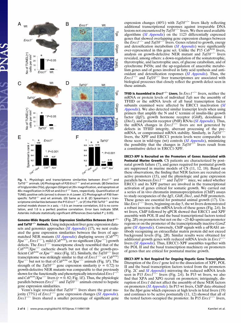
ERCC1-XPF Is Recruited on the Promoters of Genes Associated withPostnatal Murine Growth. CS patients are characterized by post-natal growth failure (7), and genes required for postnatal growthare suppressed in murine models of CS (11, 12, 18). Based onthese observations, the finding that NER factors are recruited onactive promoters (15), and the physiologic and gene expressionparallels between Ercc1−/− and Taf10−/− mice, we asked whetherERCC1 and its XPF partner are involved in the transcriptionalactivation of genes critical for somatic growth. We carried outa series of in vivo chromatin immunoprecipitation (ChIP) assaysto study occupancies of the Igf1, GhR, Dio1, and PrlR promoters.These genes are essential for postnatal animal growth (17). Un-likeErcc1−/− livers, beginning on day 5, the wt livers demonstrateda robust increase in the mRNA levels of these genes (Fig. 2A). Inwt livers, ChIP followed by qPCR showed that ERCC1 and XPFassemble with POL II and the basal transcriptional factors tested(Fig. 2B) on promoters but not on the−25-Kb upstream promoterregions or on the promoter of the transcriptionally inactiveGzmZgene (SI Appendix). Conversely, ChIP signals with a αFRAS1 an-tibody recognizing an extracellular matrix protein did not exceedbackground levels (Fig. 2B). Similar results were obtained foradditional growth genes with reduced mRNA levels in Ercc1−/−
livers (SI Appendix). Thus, ERCC1-XPF assembles together withthe POL II and the basal transcription machinery on promotersof genes that are critical for postnatal murine growth.
ERCC1-XPF Is Not Required for Ongoing Hepatic Gene Transcription.Disruption of the Ercc1 gene led to the dissociation of XPF, POLII and the basal transcription factors tested from the promoters(Fig. 2C and SI Appendix) mirroring the reduced mRNA levelsseen in P15 Ercc1−/− livers (Fig. 2A). In P15 wt livers, we alsofind that XPA and XPG recruit on promoters; intriguingly, dis-ruption of Ercc1 did not affect the assembly of these NER factorson promoters (SI Appendix). In P15 wt livers, ChIP data obtainedfor the Hprt gene which expresses at high levels in fetal livers (19)and continues to be active postnatally (11, 12) showed that all ofthe tested factors occupied the promoter. In P15 Ercc1−/− livers,
Wt
Ercc1
Wt
Wt
sg
Tn
eg/
lG
LE
NU
T0
10
20
30
Wt
*:P<0.05
0.72 0.74
0.07
0.15
0
0.2
0.4
0.6
0.8
1
Csb
-Xpa
Erc
c1 Csb Xpa
: P<0.001
0.82
0.29 0.27
0
0.2
0.4
0.6
0.8
1
Csb Xpa
Ercc1
*: P<0.001
r k
nar
s’n
amr
ae
pS
Wt
Ercc1
0
10
20
30
Ercc1 Wt
)+(
LE
NU
Tf
o o
Nm
m re
p sll
ec
2
*:P<0.05
Taf10
Taf10
Taf10
Taf10
Csb
-Xpa
)+(
LE
NU
Tf
o o
Nm
m re
p sll
ec
2
E F
Tg
sn
eg /
lG
LE
NU
T
BA
C D
Fig. 1. Physiologic and transcriptome similarities between Ercc1−/− andTaf10−/− animals. (A) Photograph of P20 Ercc1−/− andwt animals. (B) Detectionof triglycerides (TGs), glycogen (Gl/gen) at 20×magnification, and apoptosis at40×magnification in P20wt and Ercc1−/− livers, respectively. Quantification ofTUNEL-positive cells (arrow) is shown in A Lower. (C) Photograph of P30 liver-specific Taf10−/− and wt animals. (D) Same as in B. (E) Spearman’s r tran-scriptome similarities between the P15 Ercc1−/−, or (F) the P30 Taf10−/− and theanimal models shown in x axis; −1.0 is an inverse correlation, 0.0 is no corre-lation, and 1.0 is a perfect positive correlation. Error bars indicate SEM.Asterisks indicate statistically significant differences (two-tailed P ≤ 0.05).
2 of 6 | www.pnas.org/cgi/doi/10.1073/pnas.1114941109 Kamileri et al.
the Hprt mRNA levels and the occupancy of all factors tested onHprt promoter were not significantly affected; however ChIPsignals for the XPF were reduced to background levels (Fig. 2C).Similar results were obtained for other genes known to beactively transcribed long before birth (SI Appendix). Instead,ChIP signals for genes that are not expressed in P15 Ercc1−/−
livers remain close to background levels (SI Appendix). Thus,similar to TAF10, XPF and ERCC1 are required for the optimalactivation of target genes, but are dispensable for ongoinghepatic transcription.
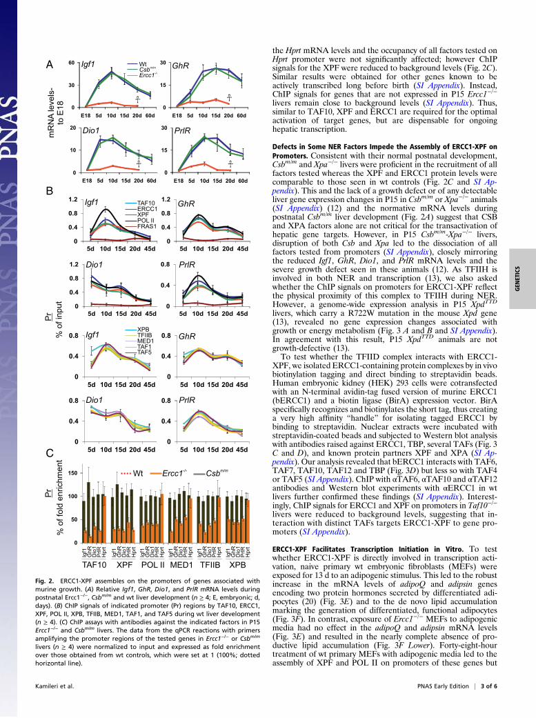
Defects in Some NER Factors Impede the Assembly of ERCC1-XPF onPromoters. Consistent with their normal postnatal development,Csbm/m and Xpa−/− livers were proficient in the recruitment of allfactors tested whereas the XPF and ERCC1 protein levels werecomparable to those seen in wt controls (Fig. 2C and SI Ap-pendix). This and the lack of a growth defect or of any detectableliver gene expression changes in P15 in Csbm/m or Xpa−/− animals(SI Appendix) (12) and the normative mRNA levels duringpostnatal Csbm/m liver development (Fig. 2A) suggest that CSBand XPA factors alone are not critical for the transactivation ofhepatic gene targets. However, in P15 Csbm/m-Xpa−/− livers,disruption of both Csb and Xpa led to the dissociation of allfactors tested from promoters (SI Appendix), closely mirroringthe reduced Igf1, GhR, Dio1, and PrlR mRNA levels and thesevere growth defect seen in these animals (12). As TFIIH isinvolved in both NER and transcription (13), we also askedwhether the ChIP signals on promoters for ERCC1-XPF reflectthe physical proximity of this complex to TFIIH during NER.However, a genome-wide expression analysis in P15 XpdTTD
livers, which carry a R722W mutation in the mouse Xpd gene(13), revealed no gene expression changes associated withgrowth or energy metabolism (Fig. 3 A and B and SI Appendix).In agreement with this result, P15 XpdTTD animals are notgrowth-defective (13).To test whether the TFIID complex interacts with ERCC1-
XPF, we isolated ERCC1-containing protein complexes by in vivobiotinylation tagging and direct binding to streptavidin beads.Human embryonic kidney (HEK) 293 cells were cotransfectedwith an N-terminal avidin-tag fused version of murine ERCC1(bERCC1) and a biotin ligase (BirA) expression vector. BirAspecifically recognizes and biotinylates the short tag, thus creatinga very high affinity “handle” for isolating tagged ERCC1 bybinding to streptavidin. Nuclear extracts were incubated withstreptavidin-coated beads and subjected to Western blot analysiswith antibodies raised against ERCC1, TBP, several TAFs (Fig. 3C and D), and known protein partners XPF and XPA (SI Ap-pendix). Our analysis revealed that bERCC1 interacts with TAF6,TAF7, TAF10, TAF12 and TBP (Fig. 3D) but less so with TAF4or TAF5 (SI Appendix). ChIP with αTAF6, αTAF10 and αTAF12antibodies and Western blot experiments with αERCC1 in wtlivers further confirmed these findings (SI Appendix). Interest-ingly, ChIP signals for ERCC1 and XPF on promoters in Taf10−/−
livers were reduced to background levels, suggesting that in-teraction with distinct TAFs targets ERCC1-XPF to gene pro-moters (SI Appendix).
ERCC1-XPF Facilitates Transcription Initiation in Vitro. To testwhether ERCC1-XPF is directly involved in transcription acti-vation, naive primary wt embryonic fibroblasts (MEFs) wereexposed for 13 d to an adipogenic stimulus. This led to the robustincrease in the mRNA levels of adipoQ and adipsin genesencoding two protein hormones secreted by differentiated adi-pocytes (20) (Fig. 3E) and to the de novo lipid accumulationmarking the generation of differentiated, functional adipocytes(Fig. 3F). In contrast, exposure of Ercc1−/− MEFs to adipogenicmedia had no effect in the adipoQ and adipsin mRNA levels(Fig. 3E) and resulted in the nearly complete absence of pro-ductive lipid accumulation (Fig. 3F Lower). Forty-eight-hourtreatment of wt primary MEFs with adipogenic media led to theassembly of XPF and POL II on promoters of these genes but
C t nemhci r ne
dl of f o%
rP
TAF10 XPF POL II MED1 TFIIB XPB
0
0.4
0.8
1.2
5d 10d 15d 20d 45d
0
0.4
0.8
5d 10d 15d 20d 45d
t upni f o%
rP
ERCC1TAF10
XPFPOL IIFRAS1
MED1TAF1TAF5
XPBTFIIB
Igf1
0
0.4
0.8
1.2
5d 10d 15d 20d 45d
GhR
0
0.4
0.8
1.2
5d 10d 15d 20d 45d0
0.4
0.8
5d 10d 15d 20d 45d
Dio1 PrlR
0
0.4
0.8
5d 10d 15d 20d 45d
0
0.4
0.8
5d 10d 15d 20d 45d0
0.4
0.8
5d 10d 15d 20d 45d
0
50
100
150
GhR
1oiD
R lrP
trpHIg
f1 R hG
1oiD
Rl rP
t rpHIg
f1
GhR
1 oiD
Rl rP
t rpHIg
f1 R hG
1oiD
Rl rP
trpHIg
f1
GhR
1 oiD
RlrP
t rpHIg
f1
GhR
1 oiD
Rl rP
t rpHIg
f1
Wt Csbm/mErcc1-/-
Igf1 GhR
Dio1 PrlR
0
30
60
E18 5d 10d 15d 20d 60d0
15
30
E18 5d 10d 15d 20d 60d
0
10
20
E18 5d 10d 15d 20d 60d
-sleve lA
NR
m81
Eot
A Igf1 GhR
Dio1
Csbm/mErcc1-/-Wt
0
15
30
E18 5d 10d 15d 20d 60d
PrlR
B
Fig. 2. ERCC1-XPF assembles on the promoters of genes associated withmurine growth. (A) Relative Igf1, GhR, Dio1, and PrlR mRNA levels duringpostnatal Ercc1−/−, Csbm/m and wt liver development (n ≥ 4; E, embryonic; d,days). (B) ChIP signals of indicated promoter (Pr) regions by TAF10, ERCC1,XPF, POL II, XPB, TFIIB, MED1, TAF1, and TAF5 during wt liver development(n ≥ 4). (C) ChIP assays with antibodies against the indicated factors in P15Ercc1−/− and Csbm/m livers. The data from the qPCR reactions with primersamplifying the promoter regions of the tested genes in Ercc1−/− or Csbm/m
livers (n ≥ 4) were normalized to input and expressed as fold enrichmentover those obtained from wt controls, which were set at 1 (100%; dottedhorizontal line).
Kamileri et al. PNAS Early Edition | 3 of 6
GEN
ETICS
not on the −25-Kb upstream promoter regions (SI Appendix)matching the onset of increase in mRNA levels (Fig. 3E). InErcc1−/− MEFs, ChIP signals for XPF, POL II, MED1, andTFIIB on promoters were markedly reduced compared with wtMEFs (Fig. 3G). Under these conditions, we also found thatXPA assembles on adipoQ and adipsin promoters; although XPA
ChIP signals for the adipoQ and adipsin promoters in Ercc1−/−
MEFs were slightly reduced compared with wt MEFs, they werenot abolished. Consistent with the high expression of the Hprtgene in Ercc1−/− MEFs (21), POL II, MED1, and TFIIB occu-pied this promoter, whereas ChIP signals for XPF were sub-stantially reduced compared with wt MEFs (Fig. 3G). Todemonstrate that the recruitment of ERCC1-XPF on promotersreflects a real association with the transcription machinery, wtMEFs were simultaneously exposed for 12 h to the transcriptioninhibitor α-amanitin and the adipogenic stimulus. This led to theinhibition of adipsin and adipoQ mRNA synthesis (SI Appendix).Neither POL II nor XPF or ERCC1 were detected on the adi-poQ and the adipsin promoters (Fig. 3H). We also monitored theresponsiveness of adipoQ and Igf1 promoters to ERCC1 andXPF by transiently cotransfecting pCMV-bErcc1 (Fig. 3C) orpCMV-bXpf (SI Appendix) together with the adipoQ or Igf1 lu-ciferase promoter plasmids in NIH 3T3 cells. In line, the bindingof ERCC1 and XPF factors on adipoQ and Igf1 promoter plas-mids significantly increased the promoter-driven luciferase ac-tivities (SI Appendix) further supporting the role of ERCC1-XPFin transcription activation.
Assembly of ERCC1-XPF on Promoters Is Accompanied by DNADemethylation and Histone Marks Associated with Active Tran-scription. Gadd45a interacts with and requires XPG to facilitatepromoter demethylation during transcription (22). In this work,we find that, Gadd45a assembles on promoters but not on the−25-Kb upstream promoter regions of growth genes or on thepromoter of the transcriptionally inactive GzmZ gene (SI Ap-pendix) during liver development. Unlike Csbm/m livers, disrup-tion of Ercc1 led to the dissociation of Gadd45a from promoters(SI Appendix). Thus, Gadd45a assembles on promoters duringhepatic development; a defect in Ercc1, but not in Csb, sub-stantially affects the recruitment of Gadd45a on promoters.Using amethylation-sensitive ChIP approach (ChIP-chop) (23),
we next sought to evaluate whether DNA methylation interfereswith the assembly of ERCC1-XPF and Gadd45a on promoters.Before qPCR, the input, ERCC1-, XPF- and Gadd45a-enrichedDNA samples derived from wt livers were digested with themethylation-sensitive HpaII and methylation-insensitive MspI re-striction enzymes. Beginning day 5, we noticed a high content ofHpaII-resistant input DNA (i.e., methylation) on promoters thatgradually decreased reaching a minimum signal at ∼P15 (i.e.,demethylation) (Fig. 4A). As shown by the “ChIP-chop” com-parative analysis of input versus ERCC1- or XPF-bound DNAfragments, ERCC1 did not bind DNA in a methylation-sensitivemanner; XPF showed a small preference toward binding non-methylated DNA (SI Appendix). Instead, Gadd45a was preferen-tially assembled on nonmethylated DNA (SI Appendix). Unlikethe P15 Csbm/m livers, disruption of the Ercc1 gene in Ercc1−/−
livers led to the aberrant DNA methylation on promoters com-pared with age-matched wt livers (Fig. 4B and SI Appendix). Thus,the presence of ERCC1-XPF on promoters is accompanied bypromoter DNA demethylation with Gadd45a protein bindingpreferentially nonmethylated DNA. A similar analysis on theHprtpromoter in all of the animal models tested revealed no differencein the content of HpaII-resistant DNA of input and ChIP samples(SI Appendix).Examination of the chromatin status in P15 Ercc1−/− livers
revealed a loss of activating acetylated histone H3Ac and H3K4trimethylation and a concomitant increase of repressive histoneH3K9 dimethylation and H3K27 trimethylation marks withpromoter-specific requirements (Fig. 5A). At the Dio1 and PrlRpromoters, a decrease in the acetylation of histone 3 and H3K4trimethylation was accompanied by a significant increase inH3K27 trimethylation and H3K9 dimethylation; at the Igf1 andGhR promoters, a decrease of H3Ac and H3K4me3 ChIP signalscoincided with an increase of H3K27me3 but not H3K9me2 ChIPsignals (Fig. 5A). Thus, ERCC1-XPF assembly on promoters isaccompanied by active DNA demethylation and histone post-translational modifications associated with active transcription.
D0 D13
1c
cr
E-/
-
Wt
E
0
10
20
30
0d 2d 4d 7d 13d
0
10
20
30
0d 2d 4d 7d 13d
Adipsin
Adipo Q
Ercc1-/-
Wt
A
0
50
100
150
Ghr
Igf1
Ig�
p4 slafgI1fgF1oiD1r3 kiP
Prlr
tw
fo%
-s lev elA
NR
m
Wt log norm. intensity2
Igf1
GhR
Dio1
PrlR
-slevelA
NR
m to 0
day
s (d
)
Input Pull-down Flow-thr.
B
F
dp
X
)ytisne tni.mron
goL(2
bERCC1en. ERCC1bERCC1/stp-HRPunsp./stp-HRP
BirA Bi
rA+b
ERC
C1
BirA Bi
rA+b
ERC
C1
BirA Bi
rA+b
ERC
C1
Input Pull-down Flow-thr.
TAF10
TBPTAF12
TAF6TAF7
CD
38kDa43kDa
AriB
AriB
1C
CR
Eb+
AriB
A riB
1C
CR
Eb+
Ar iB
AriB
1C
CR
Eb+
0
0.5
1
0
0.5
1
IIL
OP
FP
X
1C
CR
E
rP
tupnifo%
Adipsin
Adipo Q
A.c.A.c. + α-amanitinH
0
50
100
150G
Adipsin Adipo Q
1D
EM TF
IIBFP
XII
LO
P
Hprt
1D
EM TF
IIBFP
XII
LO
P
1D
EM TF
IIBFP
XII
LO
P
Ercc1-/-
Wt
rP
tnemhcirne
dloffo%
AP
X
AP
X
AP
X
DT
T/
DT
T
Fig. 3. Recruitment of ERCC1-XPF on promoters during adipogenesis. (A)Scatter plot of normalized (norm.) microarray hybridization signals obtainedfrom P15 XpdTTD liver RNA samples versus wt controls (SI Appendix). (B) qPCRevaluation of mRNA levels of genes representing the GH/IGF1 axis and mito-genic signals in P15 XpdTTD livers. For each gene, expression levels in the XpdTTD
livers are plotted relative to those of age-matched controls (red dotted line).Error bars indicate SEMbetween replicates (n≥ 4). (C) Nuclear extracts fromHEK293 cells expressing both bERCC1 and BirA biotin ligase were tested byWesternblot. The blotwas probedwith αERCC1 antibody revealing bands correspondingto the biotin-tagged (b)ERCC1 (top band) and the endogenous ERCC1 (en.ERCC1) as well as with streptavidin-HRP (stp-HRP) probe, which confirms bio-tinylation of ERCC1 (bERCC1). Endogenously unspecific (unsp.) biotinylatedproteins detected by streptavidin-HRP probe were used as loading control(lower lane). (D) bERCC1 pull downs analyzed by Western blotting for TBP andthe indicated TAFs. The input and flow-through are 1/15 and 1/20 of the extractused, respectively. (E)Adipsin and AdipoQmRNA levels in wt and Ercc1−/−MEFsafter exposure to adipogenic stimulus compared with day 0 (d: days; arrowindicates the 48-h time point for ChIP assays shown in Fig. 2 E–G). (F) Oil Red Ostaining (arrow) ofwt and Ercc1−/−MEFs subjected to adipogenic stimulation (D:days). (G) ChIP signals for promoters (as shown) with antibodies against the in-dicated factors in Ercc1−/− and wt MEFs exposed to 48 h of adipogenic stimulus.The data are presented as in Fig. 2C. Scale bars showmean values and SDs fromat least four independent experiments. (H) POLII, XPF, and ERCC1 ChIP signals onpromoters (as shown) inwtMEFs exposed for 12 h to adipogenicmixture (Ac) inthe absence (dark blue) or presence (light blue) of α-amanitin (10 μg/mL).
4 of 6 | www.pnas.org/cgi/doi/10.1073/pnas.1114941109 Kamileri et al.

DiscussionWhy some, but not all, defects in NER lead to developmental ab-normalities and how such pathological outcomes manifest in some,but not all, organs remains an intriguing question that argues fordistinct NER factors having tissue-specific functions beyond NER(15). Here, we provide in vivo evidence that the severe growth re-tardation seen in Ercc1−/− animals originates from defective tran-scription initiation of developmental gene expression programs.
Defect in Transcription Initiation Recapitulates the Growth Defect inErcc1−/− Animals. Both the NER progeroid Ercc1−/− and TFIID-defective Taf10−/− mice are growth defective, show noticeableliver apoptosis, have amarked propensity to store rather than burnglycogen and fat, die before weaning, and share unusually similarhepatic gene expression profiles. Strikingly, the latter reflectsmorethe severity of growth retardation rather than the DNA-repairdefect in NER. The extensive parallels between these otherwisedistinct mouse strains occur in the absence of a TFIID deficiencyin Ercc1−/− livers or of an ERCC1-XPF defect in Taf10−/− livers.Instead, we find that ERCC1-XPF assembles with the basaltranscription machinery on promoters during liver development.
Failure of ERCC1-XPF to do so results in defective transcriptioninitiation of genes critical for postnatal growth.Given the known role of CSB and XPD in transcription (24,
25) and the fact that XPA is upstream of ERCC1 in the ca-nonical NER pathway, it is surprising that none of these proteinsalone are required for the initial activation of hepatic genes;recruitment of ERCC1-XPF on promoters requires the concur-rent availability of CSB and XPA factors (as shown in Csbm/m-Xpa−/− livers). However, TFIIH and likely also CSB may only bepartially defective in XpdTTD and Csbm/m livers, respectively. Wealso find that ERCC1-XPF is dispensable for ongoing tran-scription; unlike POL II or any of the factors tested, ChIP signalsfor genes whose high expression levels remain unaltered inErcc1−/− livers revealed a complete dissociation of ERCC1-XPFfrom promoters (Fig. 2C and SI Appendix). Our finding thatdistinct TAFs and TBP physically interact with ERCC1 mayexplain how ERCC1-XPF is specifically recruited to the pro-moters of active genes. Interestingly, the contribution ofERCC1-XPF to the de novo initiation of hepatic transcriptionextends to genes promoting adipogenesis; assembly of ERCC1-XPF on promoters was also sensitive to the transcription in-hibitor α-amanitin. Finally, ERCC1 and XPF significantly in-crease the activity of adipoQ and Igf1 promoter-driven luciferaseactivities in transiently transfected NIH 3T3 cells (SI Appendix).
ERCC1-XPF Complex Functions in Transcription Initiation Beyond NER.In view of theNERdefect, one could envision that at least some ofthe Ercc1−/− liver gene expression changes originate from thepresence of irreparable DNA lesions. Indeed, DNA lesions wouldfurther aggravate the transcriptional defect inErcc1−/− animals or,in certain instances, increased DNA damage levels could com-promise the availability of ERCC1-XPF aimed for transcriptioninitiation during hepatic development (Fig. 5B). Moreover, theNER defect alone could account for the numerous progressivehepatic symptoms associated with aging in Ercc1-/Δ mice thatare healthy into adulthood and live substantially longer than the
0
40
80
120
0
40
80
120
0
40
80
120
Uncut HPAII MSPI
0
40
80
120
Uncut HPAII MSPI
Igf1 Ghr
Dio1 PrlR
Ercc1
Wt*
*
**
*: P<0.05
tucnu fo %
0
40
80
120
5d 10d 15d 20d 45d
0
40
80
120
5d 10d 15d 20d 45d
0
40
80
120
5d 10d 15d 20d 45d
0
40
80
120
5d 10d 15d 20d 45d
Ghr
Dio1 PrlR
MSP IHPA IIUncut
Igf1
1C
CR
E-PIh
CA
B
Fig. 4. Assembly of ERCC1-XPF on promoters is accompanied by DNAdemethylation. (A) Methylation-sensitive ChIP-qPCR signals of input (solidline) and ERCC1-enriched DNA samples (dotted line) digested with HpaII,MspI, or left undigested during wt liver development. For each promoter,the percentage of HpaII or MspI digestion resistance is represented asa percentage of mock digested DNA (y axis). (B) Methylation-sensitive qPCRsignals of DNA samples in P15 Ercc1−/− livers compared to controls.
NER defects
Defective ti
ranscriptionnitiation
DNAdamage
Mutations
Cancer
DNA
lesions
Alteredgene expression
3
21
Developmental defects
tw-t
ne
mh
c irn
edl
off
o%
A
B
0
50
100
150
Igf1
GhR 1oiD
RlrP
Igf1
GhR 1oiD
RlrP
0
50
100
150
Igf1
GhR 1oiD
RlrP
Igf1
GhR 1oiD
RlrP
0
100
200
300
400
500
Igf1
GhR 1oiD
RlrP
Igf1
GhR 1oiD
RlrP
0
100
200
300
400
500
Igf1
GhR 1oiD
RlrP
Igf1
GhR 1oiD
RlrP
Ercc1
Csb
3eM4K3HcA3H
3eM72K3H2eM9K3H
tw-t
ne
mh
cirn
ed l
off
o%
Fig. 5. Histone posttranslational modifications in Ercc1−/− and Csbm/m livers.(A) ChIP signals for activating (H3Ac, H3K4Me3) and repressive (H3K9Me2,H3K27Me3) histone modifications at the indicated promoter regions in P15Ercc1−/− and Csbm/m livers (n ≥ 4). The data are presented as shown in Fig. 2C.(B) A model integrating the dual function of NER in DNA repair and tran-scription initiation: ERCC1-XPF together with other NER factors functionsbeyond NER in the transcriptional activation of developmental stage- andtissue-specific gene expression programs. The presence of irreparable DNAlesions (due to, for example, defective NER) may further aggravate thepathological outcome of NER abnormalities.
Kamileri et al. PNAS Early Edition | 5 of 6
GEN
ETICS
growth-defective, short-lived Ercc1−/− and Taf10−/− animals (26).Mutations in ERCC1, XPF, and XPG represent the only single-gene defects known in NER that are associated with growth at-tenuation and death before weaning in mice (27). However, sev-eral double NER mutant mice are also associated with cachecticdwarfism and, for the animal models tested so far, their P15hepatic gene expression changes are exceptionally similar tothose seen in Taf10−/− or Ercc1−/− livers (11, 12, 18). This sur-prising requirement of certain, but not all, NER factors in thetranscription initiation of hepatic genes during developmentcontrasts with the UV sensitivity seen in XP, CS, and TTDpatients; whereas the latter depends on theDNArepair defect, theformer likely reflects a transcriptional defect during development.
Recruitment of ERCC1-XPF on Promoters Is Accompanied by DNADemethylation and Histone Posttranslational Modifications. Despitethe controversial involvement of Gadd45a in active promoterDNA demethylation (22, 28), we found that Gadd45a assemblestogether with ERCC1-XPF on promoters. Recruitment ofERCC1-XPF on promoters was followed by DNA demethylationon promoter-proximal DNA closely mirroring the peak ofmRNA levels during development. Instead, a defect in Ercc1, butnot in Csb, led to aberrant promoter DNA methylation. TAF12was recently shown to recruit Gadd45a and the NER complex tothe promoter of rRNA genes leading to active DNA demethy-lation (29). These findings point to a similar role for TAFs andERCC1-XPF during POL II-mediated transcription. Consis-tently, the aberrant promoter-proximal DNA methylation in P15Ercc1−/− livers was associated with a decrease of activating H3Acand H3K4me3 histone marks and a concomitant enrichmentin trimethylation of H3K27 more than H3K9 dimethylation.With the exception of a mild increase in H3K9 dimethylationon Dio1 promoter, the P15 Csbm/m livers showed none of theErcc1−/−-associated histone marks on promoters.
ConclusionsIn summary, we find that, upon gene activation, ERCC1-XPFrecruits together with the RNA POL II and the basal transcriptionmachinery at the promoters of hepatic genes. Assembly ofERCC1-XPF on promoters is followed by histone marks and
promoter proximal DNA demethylation associated with activetranscription. Whereas the role of XPG, TFIIH and CSB in POLII-mediated transcription has been well documented (30), it hasbeen difficult to envisage a role for ERCC1-XPF in transcriptioninitiation in vivo. Our data are in line with previous findings onXPG (22), supporting a similar role for ERCC1-XPF in facilitatingrepair-mediated active DNA demethylation on promoters. In-teraction of ERCC1-XPF complex with specific TAFs during geneactivation also suggests that this complex acts as a coactivatorduring the transcription process. This is in line with the fact thatTBP/TAF complexes often recruit various classes of coactivatorsto execute specific transcriptional programs (1, 31) as well as therecently proposed function of the XPC/RAD23B/CETN2 NERcomplex in the maintenance and re-establishment of stem cellpluripotency (14). Thus, although ERCC1/XPF may not be es-sential for initiating basal transcription itself, it is required for thefine-tuning of optimal transactivation of target genes. The periodat which NER factors optimize transcription during postnataldevelopment may well explain the heterogeneous and tissue-spe-cific pathology of NER syndromes. It is, therefore, attractive tospeculate that the so called “segmental” NER progeroid featuresmay also reflect the “segmental” transcriptional requirements forcertain NER factors during mammalian development.
MethodsInformation on the animal models used is shown in SI Appendix. Cell cul-turing, the Periodic Acid Schiff, Oil Red O, and TdT-mediated dUTP Nick-EndLabeling were performed as previously described (12, 21). Microarrays,qPCRs, and data analysis were performed as previously described (21).Detailed information on in vivo biotinylation tagging approach and thereporter gene assays is shown in SI Appendix. Westerns blots, ChIP, coim-munoprecipitation assays, and the methylation-sensitive ChIP assay wereperformed as previously described (16, 23).
ACKNOWLEDGMENTS.WethankProf. J.H. J.HoeijmakersandDr.B.Schumacherfor critical discussions on the manuscript, Dr. G. T. J. van der Horst for Ercc1−/−
and Csbm/m animals, Dr L. Tora for Taf10lox/lox mice, Dr. G. Chalepakis forαFRAS1 antibody, and Dr. J. Strouboulis for the in vivo biotinylation taggingconstructs. This work was supported by Capacities-FP7-REGPOT-2008-1“ProFI”, the National Strategic Reference Framework 2007-2013 “HerakleitosII (KA3396)”, and “Cooperation” No. EDGE 901-13/11/2009 programs.
1. Lemon B, Tjian R (2000) Orchestrated response: A symphony of transcription factorsfor gene control. Genes Dev 14:2551–2569.
2. Ohler U, Wassarman DA (2010) Promoting developmental transcription. Development137:15–26.
3. Hoeijmakers JH (2001) Genome maintenance mechanisms for preventing cancer.Nature 411:366–374.
4. Hanawalt PC (2002) Subpathways of nucleotide excision repair and their regulation.Oncogene 21:8949–8956.
5. Lainé JP, Egly JM (2006) When transcription and repair meet: A complex system.Trends Genet 22:430–436.
6. Cleaver JE (2005) Cancer in xeroderma pigmentosum and related disorders of DNArepair. Nat Rev Cancer 5:564–573.
7. Bootsma D, Kraemer KH, Cleaver JE, Hoeijmakers JHJ (1998) Nucleotide excision re-pair syndromes: Xeroderma pigmentosum, Cockayne syndrome and trichothiodys-trophy. The genetic basis of human cancer, eds Vogelstein B, Kinzler KW (McGraw-Hill, New York), pp 245–274.
8. Hoeijmakers JH (2009) DNA damage, aging, and cancer. N Engl J Med 361:1475–1485.9. Schumacher B, Garinis GA, Hoeijmakers JH (2008) Age to survive: DNA damage and
aging. Trends Genet 24:77–85.10. Garinis GA, van der Horst GT, Vijg J, Hoeijmakers JH (2008) DNA damage and ageing:
New-age ideas for an age-old problem. Nat Cell Biol 10:1241–1247.11. Niedernhofer LJ, et al. (2006) A new progeroid syndrome reveals that genotoxic stress
suppresses the somatotroph axis. Nature 444:1038–1043.12. van der Pluijm I, et al. (2007) Impaired genome maintenance suppresses the growth
hormone—insulin-like growth factor 1 axis in mice with Cockayne syndrome. PLoSBiol 5:e2.
13. de Boer J, et al. (2002) Premature aging in mice deficient in DNA repair and tran-scription. Science 296:1276–1279.
14. Fong YW, et al. (2011) A DNA repair complex functions as an Oct4/Sox2 coactivator inembryonic stem cells. Cell 147:120–131.
15. Le May N, et al. (2010) NER factors are recruited to active promoters and facilitatechromatin modification for transcription in the absence of exogenous genotoxicattack. Mol Cell 38:54–66.
16. Tatarakis A, et al. (2008) Dominant and redundant functions of TFIID involved in theregulation of hepatic genes. Mol Cell 31:531–543.
17. Schumacher B, et al. (2008) Delayed and accelerated aging share common longevityassurance mechanisms. PLoS Genet 4:e1000161.
18. van de Ven M, et al. (2006) Adaptive stress response in segmental progeria resembleslong-lived dwarfism and calorie restriction in mice. PLoS Genet 2:e192.
19. Duncan SA (2000) Transcriptional regulation of liver development. Dev Dyn 219:131–142.
20. Gupta RK, et al. (2010) Transcriptional control of preadipocyte determination byZfp423. Nature 464:619–623.
21. Garinis GA, et al. (2009) Persistent transcription-blocking DNA lesions trigger somaticgrowth attenuation associated with longevity. Nat Cell Biol 11:604–615.
22. Barreto G, et al. (2007) Gadd45a promotes epigenetic gene activation by repair-mediated DNA demethylation. Nature 445:671–675.
23. van de Nobelen S, et al. (2010) CTCF regulates the local epigenetic state of ribosomalDNA repeats. Epigenetics Chromatin 3:19.
24. Balajee AS, May A, Dianov GL, Friedberg EC, Bohr VA (1997) Reduced RNA polymeraseII transcription in intact and permeabilized Cockayne syndrome group B cells. ProcNatl Acad Sci USA 94:4306–4311.
25. Iyer N, Reagan MS, Wu KJ, Canagarajah B, Friedberg EC (1996) Interactions involvingthe human RNA polymerase II transcription/nucleotide excision repair complex TFIIH,the nucleotide excision repair protein XPG, and Cockayne syndrome group B (CSB)protein. Biochemistry 35:2157–2167.
26. Gregg SQ, et al. (2011) A mouse model of accelerated liver aging due to a defect inDNA repair. Hepatology, 10.1002/hep.24713.
27. Niedernhofer LJ (2008) Nucleotide excision repair deficient mouse models and neu-rological disease. DNA Repair (Amst) 7:1180–1189.
28. Jin SG, Guo C, Pfeifer GP (2008) GADD45A does not promote DNA demethylation.PLoS Genet 4:e1000013.
29. Schmitz KM, et al. (2009) TAF12 recruits Gadd45a and the nucleotide excision repaircomplex to the promoter of rRNA genes leading to active DNA demethylation. MolCell 33:344–353.
30. Le May N, Egly JM, Coin F (2010) True lies: The double life of the nucleotide excisionrepair factors in transcription and DNA repair. J Nucleic Acids 2010:2010.
31. Näär AM, et al. (1998) Chromatin, TAFs, and a novel multiprotein coactivator arerequired for synergistic activation by Sp1 and SREBP-1a in vitro. Genes Dev 12:3020–3031.
6 of 6 | www.pnas.org/cgi/doi/10.1073/pnas.1114941109 Kamileri et al.