ADP ribose is an endogenous ligand for the purinergic P2Y1 receptor

Mutation Research 704 (2010) 12–20
Review
Multiple roles of the cell cycle inhibitor p21CDKN1A in the DNA damage response
Ornella Cazzalini a, A. Ivana Scovassi b, Monica Savio a, Lucia A. Stivala a, Ennio Prosperi b,c,*a Dipartimento di Medicina Sperimentale, sez. Patologia Generale ‘‘C. Golgi’’, Universita di Pavia, 27100 Pavia, Italyb Istituto di Genetica Molecolare del CNR (IGM-CNR), 27100 Pavia, Italyc Dipartimento di Biologia Animale, Universita di Pavia, 27100 Pavia, Italy
Contents
1. Introduction . . . . . . . . . . . . . . . . . . . . . . . . . . . . . . . . . . . . . . . . . . . . . . . . . . . . . . . . . . . . . . . . . . . . . . . . . . . . . . . . . . . . . . . . . . . . . . . . . . . . . . 12
2. Roles of p21 in DNA damage response . . . . . . . . . . . . . . . . . . . . . . . . . . . . . . . . . . . . . . . . . . . . . . . . . . . . . . . . . . . . . . . . . . . . . . . . . . . . . . . . . 13
2.1. Cell-cycle regulation . . . . . . . . . . . . . . . . . . . . . . . . . . . . . . . . . . . . . . . . . . . . . . . . . . . . . . . . . . . . . . . . . . . . . . . . . . . . . . . . . . . . . . . . . . 13
2.2. Apoptosis. . . . . . . . . . . . . . . . . . . . . . . . . . . . . . . . . . . . . . . . . . . . . . . . . . . . . . . . . . . . . . . . . . . . . . . . . . . . . . . . . . . . . . . . . . . . . . . . . . . 14
2.3. Transcription regulation . . . . . . . . . . . . . . . . . . . . . . . . . . . . . . . . . . . . . . . . . . . . . . . . . . . . . . . . . . . . . . . . . . . . . . . . . . . . . . . . . . . . . . . 15
2.4. DNA repair . . . . . . . . . . . . . . . . . . . . . . . . . . . . . . . . . . . . . . . . . . . . . . . . . . . . . . . . . . . . . . . . . . . . . . . . . . . . . . . . . . . . . . . . . . . . . . . . . 15
3. Post-translational regulation of p21 in the DNA damage response. . . . . . . . . . . . . . . . . . . . . . . . . . . . . . . . . . . . . . . . . . . . . . . . . . . . . . . . . . . 16
4. Conclusions and perspectives . . . . . . . . . . . . . . . . . . . . . . . . . . . . . . . . . . . . . . . . . . . . . . . . . . . . . . . . . . . . . . . . . . . . . . . . . . . . . . . . . . . . . . . . 17
Acknowledgements . . . . . . . . . . . . . . . . . . . . . . . . . . . . . . . . . . . . . . . . . . . . . . . . . . . . . . . . . . . . . . . . . . . . . . . . . . . . . . . . . . . . . . . . . . . . . . . . 17
References . . . . . . . . . . . . . . . . . . . . . . . . . . . . . . . . . . . . . . . . . . . . . . . . . . . . . . . . . . . . . . . . . . . . . . . . . . . . . . . . . . . . . . . . . . . . . . . . . . . . . . . 17
A R T I C L E I N F O
Article history:
Received 14 September 2009
Received in revised form 29 December 2009
Accepted 13 January 2010
Available online 22 January 2010
Keywords:
p21CDKN1A
DNA repair
Cell cycle checkpoints
DNA damage response
PCNA interaction
A B S T R A C T
Among cell cycle regulatory proteins that are activated following DNA damage, the cyclin-dependent
kinase inhibitor p21CDKN1A plays essential roles in the DNA damage response, by inducing cell cycle arrest,
direct inhibition of DNA replication, as well as by regulating fundamental processes, like apoptosis and
transcription. These functions are performed through the ability of p21 to interact with a number of
proteins involved in these processes. Despite an initial controversy, during the last years several lines of
evidence have also indicated that p21 may be directly involved in DNA repair. In particular, the
participation of p21 in nucleotide excision repair (NER), base excision repair (BER), and DNA translesion
synthesis (TLS), has been suggested to occur thanks to its interaction with proliferating cell nuclear antigen
(PCNA), a crucial protein involved in several aspects of DNA metabolism, and cell-cycle regulation.
In this review, the multiple roles of p21 in the DNA damage response, including regulation of cell
cycle, apoptosis and gene transcription, are discussed together with the most recent findings supporting
the direct participation of p21 protein in DNA repair processes. In particular, spatio-temporal dynamics
of p21 recruitment to sites of DNA damage will be considered together with several lines of evidence
indicating a regulatory role for p21. In addition, the relevance of post-translational regulation in the fate
(e.g. degradation) of p21 protein after cell exposure to DNA damaging agents will be analyzed. Both sets
of evidence will be discussed in terms of the overall DNA damage response.
� 2010 Elsevier B.V. All rights reserved.
Contents lists available at ScienceDirect
Mutation Research/Reviews in Mutation Research
journal homepage: www.e lsev ier .com/ locate / rev iewsmrCommuni ty address : www.elsev ier .com/ locate /mutres
1. Introduction
Exposure to different environmental stress conditions, includ-ing radiation (like ionizing and UV radiation), may induce theformation of a variety of DNA genotoxic lesions that cells must
* Corresponding author at: IGM-CNR, sez. Istochimica e Citometria, Via Ferrata 1,
27100 Pavia, Italy. Tel.: +39 0382 986267; fax: +39 0382 986430.
E-mail address: [email protected] (E. Prosperi).
1383-5742/$ – see front matter � 2010 Elsevier B.V. All rights reserved.
doi:10.1016/j.mrrev.2010.01.009
remove in order to avoid genomic instability, and to prevent cancerformation. To this end, virtually every organism has developedhighly conserved genome surveillance and signaling mechanisms,collectively known as the DNA damage response (DDR). Thispathway consists of DNA damage signaling cascade (cell cyclecheckpoints), and of DNA repair processes able to recognize andremove a great number of DNA lesions [1].
Among the factors involved in this pathway, p21CDKN1A proteincontributes to the cell response to DNA damage, by regulatingfundamental processes, like cell cycle progression, apoptosis, and
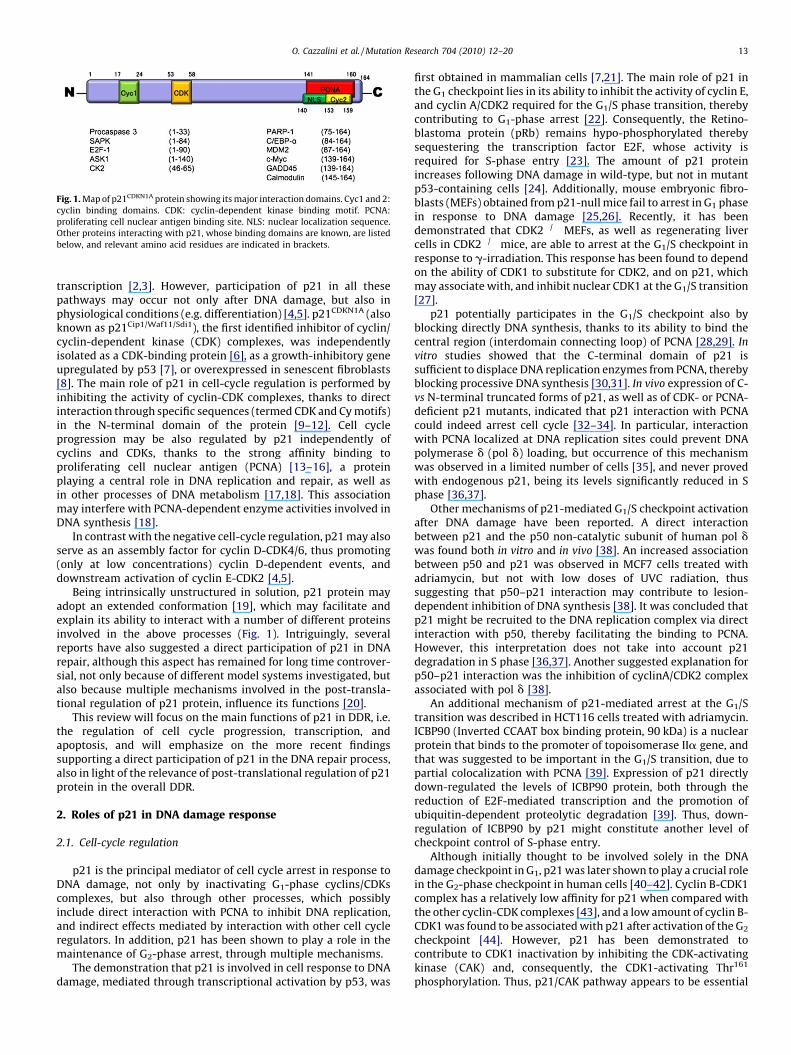
Fig. 1. Map of p21CDKN1A protein showing its major interaction domains. Cyc1 and 2:
cyclin binding domains. CDK: cyclin-dependent kinase binding motif. PCNA:
proliferating cell nuclear antigen binding site. NLS: nuclear localization sequence.
Other proteins interacting with p21, whose binding domains are known, are listed
below, and relevant amino acid residues are indicated in brackets.
O. Cazzalini et al. / Mutation Research 704 (2010) 12–20 13
transcription [2,3]. However, participation of p21 in all thesepathways may occur not only after DNA damage, but also inphysiological conditions (e.g. differentiation) [4,5]. p21CDKN1A (alsoknown as p21Cip1/Waf11/Sdi1), the first identified inhibitor of cyclin/cyclin-dependent kinase (CDK) complexes, was independentlyisolated as a CDK-binding protein [6], as a growth-inhibitory geneupregulated by p53 [7], or overexpressed in senescent fibroblasts[8]. The main role of p21 in cell-cycle regulation is performed byinhibiting the activity of cyclin-CDK complexes, thanks to directinteraction through specific sequences (termed CDK and Cy motifs)in the N-terminal domain of the protein [9–12]. Cell cycleprogression may be also regulated by p21 independently ofcyclins and CDKs, thanks to the strong affinity binding toproliferating cell nuclear antigen (PCNA) [13–16], a proteinplaying a central role in DNA replication and repair, as well asin other processes of DNA metabolism [17,18]. This associationmay interfere with PCNA-dependent enzyme activities involved inDNA synthesis [18].
In contrast with the negative cell-cycle regulation, p21 may alsoserve as an assembly factor for cyclin D-CDK4/6, thus promoting(only at low concentrations) cyclin D-dependent events, anddownstream activation of cyclin E-CDK2 [4,5].
Being intrinsically unstructured in solution, p21 protein mayadopt an extended conformation [19], which may facilitate andexplain its ability to interact with a number of different proteinsinvolved in the above processes (Fig. 1). Intriguingly, severalreports have also suggested a direct participation of p21 in DNArepair, although this aspect has remained for long time controver-sial, not only because of different model systems investigated, butalso because multiple mechanisms involved in the post-transla-tional regulation of p21 protein, influence its functions [20].
This review will focus on the main functions of p21 in DDR, i.e.the regulation of cell cycle progression, transcription, andapoptosis, and will emphasize on the more recent findingssupporting a direct participation of p21 in the DNA repair process,also in light of the relevance of post-translational regulation of p21protein in the overall DDR.
2. Roles of p21 in DNA damage response
2.1. Cell-cycle regulation
p21 is the principal mediator of cell cycle arrest in response toDNA damage, not only by inactivating G1-phase cyclins/CDKscomplexes, but also through other processes, which possiblyinclude direct interaction with PCNA to inhibit DNA replication,and indirect effects mediated by interaction with other cell cycleregulators. In addition, p21 has been shown to play a role in themaintenance of G2-phase arrest, through multiple mechanisms.
The demonstration that p21 is involved in cell response to DNAdamage, mediated through transcriptional activation by p53, was
first obtained in mammalian cells [7,21]. The main role of p21 inthe G1 checkpoint lies in its ability to inhibit the activity of cyclin E,and cyclin A/CDK2 required for the G1/S phase transition, therebycontributing to G1-phase arrest [22]. Consequently, the Retino-blastoma protein (pRb) remains hypo-phosphorylated therebysequestering the transcription factor E2F, whose activity isrequired for S-phase entry [23]. The amount of p21 proteinincreases following DNA damage in wild-type, but not in mutantp53-containing cells [24]. Additionally, mouse embryonic fibro-blasts (MEFs) obtained from p21-null mice fail to arrest in G1 phasein response to DNA damage [25,26]. Recently, it has beendemonstrated that CDK2�/� MEFs, as well as regenerating livercells in CDK2�/� mice, are able to arrest at the G1/S checkpoint inresponse to g-irradiation. This response has been found to dependon the ability of CDK1 to substitute for CDK2, and on p21, whichmay associate with, and inhibit nuclear CDK1 at the G1/S transition[27].
p21 potentially participates in the G1/S checkpoint also byblocking directly DNA synthesis, thanks to its ability to bind thecentral region (interdomain connecting loop) of PCNA [28,29]. In
vitro studies showed that the C-terminal domain of p21 issufficient to displace DNA replication enzymes from PCNA, therebyblocking processive DNA synthesis [30,31]. In vivo expression of C-vs N-terminal truncated forms of p21, as well as of CDK- or PCNA-deficient p21 mutants, indicated that p21 interaction with PCNAcould indeed arrest cell cycle [32–34]. In particular, interactionwith PCNA localized at DNA replication sites could prevent DNApolymerase d (pol d) loading, but occurrence of this mechanismwas observed in a limited number of cells [35], and never provedwith endogenous p21, being its levels significantly reduced in Sphase [36,37].
Other mechanisms of p21-mediated G1/S checkpoint activationafter DNA damage have been reported. A direct interactionbetween p21 and the p50 non-catalytic subunit of human pol dwas found both in vitro and in vivo [38]. An increased associationbetween p50 and p21 was observed in MCF7 cells treated withadriamycin, but not with low doses of UVC radiation, thussuggesting that p50–p21 interaction may contribute to lesion-dependent inhibition of DNA synthesis [38]. It was concluded thatp21 might be recruited to the DNA replication complex via directinteraction with p50, thereby facilitating the binding to PCNA.However, this interpretation does not take into account p21degradation in S phase [36,37]. Another suggested explanation forp50–p21 interaction was the inhibition of cyclinA/CDK2 complexassociated with pol d [38].
An additional mechanism of p21-mediated arrest at the G1/Stransition was described in HCT116 cells treated with adriamycin.ICBP90 (Inverted CCAAT box binding protein, 90 kDa) is a nuclearprotein that binds to the promoter of topoisomerase IIa gene, andthat was suggested to be important in the G1/S transition, due topartial colocalization with PCNA [39]. Expression of p21 directlydown-regulated the levels of ICBP90 protein, both through thereduction of E2F-mediated transcription and the promotion ofubiquitin-dependent proteolytic degradation [39]. Thus, down-regulation of ICBP90 by p21 might constitute another level ofcheckpoint control of S-phase entry.
Although initially thought to be involved solely in the DNAdamage checkpoint in G1, p21 was later shown to play a crucial rolein the G2-phase checkpoint in human cells [40–42]. Cyclin B-CDK1complex has a relatively low affinity for p21 when compared withthe other cyclin-CDK complexes [43], and a low amount of cyclin B-CDK1 was found to be associated with p21 after activation of the G2
checkpoint [44]. However, p21 has been demonstrated tocontribute to CDK1 inactivation by inhibiting the CDK-activatingkinase (CAK) and, consequently, the CDK1-activating Thr161
phosphorylation. Thus, p21/CAK pathway appears to be essential

O. Cazzalini et al. / Mutation Research 704 (2010) 12–2014
in sustaining the G2 arrest in response to DNA damage [44]. Otherlikely targets of p21 in G2 phase are cyclin A-CDK1/2 complexes[45,46]. As an additional mechanism of G2 arrest, p21 was alsosuggested to mediate nuclear retention of cyclin B1-CDK1 complexin response to genotoxic stress, thus preventing its activation byCdc25 and CAK [47]. Recently, it has been also proposed that p21contributes to G2 arrest by mediating cyclin B degradation inresponse to DNA damage [48]. Furthermore, a new p21-dependentmechanism to maintain G2 arrest after DNA damage has beenshown to involve Emi1 protein, an inhibitor of the AnaphasePromoting Complex (APC) whose destruction controls progressionthrough mitosis to G1 phase [49]. It has been reported that p21down-regulates Emi1 in cells arrested in G2 by DNA damage,thereby contributing to APC activation and degradation of keysubstrates, including cyclins A2 and B1. Thus, p21 controlspositively this checkpoint preventing G2-arrested cells fromentering mitosis [49].
Beyond the function of CDK inhibitor, and its ability to interactwith different cell cycle regulatory proteins, p21 has been recentlyshown to contribute to cell cycle arrest after DNA damage, throughtranscriptional repression of cell cycle regulatory genes (seeSection 2.3).
2.2. Apoptosis
An active role for p21 in inhibiting apoptosis has been described[2,3,50–53]; however, this function is not absolute since, undersome circumstances (e.g. enforced overexpression), p21 maypromote (and not impair) the signaling apoptotic pathway thatultimately determines cell death [50].
Pioneering work providing the evidence that in the absence ofp21, DNA-damaged cells undergo cell cycle arrest followed bydeath characterized by typical apoptotic hallmarks [42,54],suggested that p21 could exert an anti-apoptotic function inresponse to DNA damage. The mechanism by which p21 negativelyregulates DNA damage-induced death machinery relies on itsbinding to key apoptotic regulatory proteins (e.g. proteaseprecursors and specific kinases) [50]. Indeed, p21 physicallyinteracts, through its first N-terminal 33 aminoacids, with pro-caspase 3, i.e. the inactive precursor of the apoptotic executionercaspase 3 [55,56]; when bound to p21, the inactive pro-caspasecannot be converted into the active protease and apoptosis isimpeded [56]. Caspase 2, which acts upstream caspase 3, is alsokept in a repressed status by p21 [57]. The strict interactionbetween p21 and caspases is supported also by the observationthat p21 itself is cleaved by caspases early during DNA damage-induced apoptosis; proteolysis involves p21 NLS region, andimpairs p21 translocation into the nucleus [58–60]. Within thecytoplasm, p21 also forms a complex with the apoptosis signal-regulating kinase 1 (ASK1), inhibits stress-activated MAP kinasecascade and acts as an inhibitor of apoptosis [61]. In this respect, ithas been reported that p21 promotes the inhibition of a number ofkinases implicated in the cell response to stress, e.g. MAPKs, andJNK/SAPKs [reviewed in 5], thus ensuring a way to disable cellularpro-apoptotic signaling. The most convincing demonstration for arole of p21 in the modulation of kinase activity came from theevidence that p21 suppresses rapamycin-induced apoptosis byassociating with and inhibiting ASK1, thereby blocking anothercrucial step in the apoptotic pathway, that is c-Jun phosphorylation[62–64]. Moreover, p21 seems to have an anti-apoptotic activitythrough the inhibition of CDK activity required for activation of thecaspase cascade downstream of mitochondria [65–67].
An important consequence of the inhibitory activity ofapoptosis in a variety of systems is that p21 could dramaticallyimpair the effectiveness of chemotherapeutic agents acting bydamaging DNA. In this respect, an innovative strategy to kill cancer
cells is based on the direct or indirect attenuation of p21 (obtainedby different approaches) before chemotherapy [68–72]. Forinstance, it has been reported that the overexpression of c-Mycprevents the transcriptional activation of p21 in cancer cells, thusrestoring the apoptotic response to DNA damage inducers such asradiation and topoisomerases II inhibitors [73]. On the whole,these observations suggest that the anti-apoptotic function of p21could be counteracted by the administration of p21 attenuators toimprove the effectiveness of conventional chemo- and radio-therapy [74].
The opposite effect of promoting apoptosis has been describedto be mainly associated with the enforced overexpression of p21. Infact, this was found to enhance the apoptotic response to radiationor chemicals in some cancer cell types, mostly expressing mutantor non-functional p53 [reviewed in 50,75]. In addition, thymocytesfrom mice carrying a p21 transgene targeted for restrictedexpression in the T cell lineage, were found to be hypersensitiveto radiation-induced programmed cell death [76]. A pro-apoptoticeffect of p21 was also observed in MCF-7 breast cancer cells treatedwith sodium butyrate, which is an inducer of p21 expression;interestingly, in these cells the pro-apoptotic effect required theinteraction of p21 with PCNA [77].
The anti- or pro-apoptotic role of p21 could depend on thenature of the apoptotic stimulus. A recent survey of geneexpression in human laryngeal squamous carcinoma cells withenforced expression of p21, revealed a dual role of p21 inmodulating the response to drug treatment. In fact, apoptosis wasenhanced or inhibited by p21, according to whether cells weretreated with cisplatin, or with methotrexate [78]. The conditionspromoting the shift from the anti- to the pro-apoptotic role of p21are often undistinguishable from those connected to p53; thus, acareful analysis is required to better elucidate the specificmechanisms by which p21 cooperates with apoptotic factors[10,75].
Among other factors influencing the impact of p21 activity onthe apoptotic process, the subcellular localization has been shownto be crucial [79]: in the cytoplasm, p21 may block the activation ofpro-caspase 3 and stress-induced kinases (see above), therebyinhibiting apoptosis. In contrast, the nuclear localization of p21will favor the occurrence of apoptosis after DNA damage [3,79],given that such localization is required for p21 degradation [80].
Functions of p21 in response to DNA damage could be alsomodulated by the extent of genotoxic lesions, through eitherstabilization or degradation of the protein: low levels of DNAlesions will allow p21 stabilization, and induce cell cycle arrest(thus having anti-apoptotic activity); in contrast, after extensiveDNA damage, p21 down-modulation will allow cells to go toapoptosis [81,82].
Recently, it has been observed that p21 plays a pivotal role inregulating another type of cell death, i.e. autophagy, a processwhere membrane-enclosed vesicles engulf and consume cellularcomponents, and which has been shown to engage in a complexinterplay with apoptosis [83]. The treatment of p21-null MEFs withC2-ceramide leads to the occurrence of autophagy, thus suggestingthat the presence of p21 contributes to the maintenance ofautophagic proteins in an inactive status [84]. Similarly, ceramideproduction in primary hepatocytes promotes a balance betweenapoptosis and autophagy, depending on p53 and p21 levels [85]. Ithas been shown that the cytoplasmic fraction of p53 triggersapoptosis and inhibits autophagy, while in the nucleus p53 acts asan autophagy-inducing transcription factor [86,87]. The precisefunction of p21 in p53-independent pathways of death is stillunder investigation.
In conclusion, p21 is involved in the regulation of cell death(s),even if playing opposite roles: blocking crucial apoptotic (as wellautophagic) factors, thus inhibiting death occurrence, or partici-

O. Cazzalini et al. / Mutation Research 704 (2010) 12–20 15
pating in the signal transduction pathway of apoptosis, as aneffector of this process. Thus, the activity of p21 towards stressconditions, including DNA damage, appears to be finely tuned,although the signals required to direct the p21 response have notbeen clearly identified [53].
2.3. Transcription regulation
Another activity of p21 in the cell response to DNA damage istranscription regulation, either positive or negative [2,3,88]. Thisfunction may occur via different mechanisms: (i) p21 can represstranscription by inhibiting cyclin-CDK complex; (ii) p21 maydirectly bind, and consequently inhibit the activity of severaltranscription factors; (iii), p21 may also function by inhibiting thetranscriptional repression domain of transcriptional co-activators,like p300/CBP.
According to the first mechanism, CDK inhibition will preventthe phosphorylation of Rb-family proteins, thereby inactivatingE2F-dependent transcription [2,88]. Through the second mecha-nism, p21 functions as a transcriptional cofactor regulating theactivity of different DNA-binding proteins such as NF-kB, Myc, E2F,p300, STAT3, and estrogen receptors [2,3,88,89]. It has been alsoreported that p21 acts as a negative regulator of the cellular levelsof p53 and its stability in different cell lines [90]. Interaction of p21with STAT3 proteins inhibits the transcriptional activity of thesefactors: overexpression of p21 was shown to reduce thetranscriptional activity of STAT3 proteins, without modifying theirDNA binding activity [91]. In addition, it was shown that p21 mayspecifically repress E2F-dependent transcription [92], not onlythrough its effects on cyclin/CDK activity and substrate association,but also through its direct interaction with E2F factor [93], whichcould function as an anchor for p21 [2]. Binding of p21 to the N-terminus of c-Myc, will result in the interference of c-Myc-Maxassociation, suppressing c-Myc-dependent transcription. At thesame time, the interaction between c-Myc and p21 may directlycounteract p21-dependent inhibition of DNA synthesis, as c-Mycbinds p21 in competition with PCNA [94].
A general correlation has been observed between p21inhibitory effects and cell cycle-dependent element (CDE) andcell cycle gene homology regions (CHR), which are DNA sequencesinvolved in cell cycle-dependent transcriptional regulation [95].These sequences have been found in the promoter of some genesthat are inhibited by p21, such as PLK1, cyclin B1 or TopoIIa.Mutations in the CDE-CHR sequences prevent the transcriptionalinhibition of PLK1 and Cdk1 by p21 [95]. The topoisomerase IIagene promoter is also regulated by p21 via down-regulation of thetranscription factor ICBP90, as described above [39].
The transcriptional functions of p21 also play an important rolein DDR, e.g. by down-regulating transcription of the glucocorti-coid-induced tumor necrosis factor receptor. Since this receptorconfers resistance to UVB-induced apoptosis in keratinocytes, p21will enhance the apoptotic response to DNA damage [96]. Recently,it has been shown that p21 functions as transcriptional repressor ofthe myc and cdc25A genes upon DNA damage [97]. Ectopicexpression of the cell cycle inhibitor down-modulated myc andcdc25A transcription: p21 was recruited to the promoters of thesetwo genes together with the STAT3 and E2F1 transcription factors.Its presence on DNA was associated with inhibition of p300recruitment, and down-regulation of histone H4 acetylation, thatwere observed also after DNA damage induced by topoisomeraseinhibitors, such as sn38 or doxorubicin [97]. Down-regulation ofthese genes following treatment with DNA-damaging drugsindicated that p21 binds not only to cyclin-CDK complexes, butalso to the promoter of cell cycle genes upon DNA damage [97].
p21 may bind to other transcription factors and modulatepositively their function. Estrogen receptor (ERa)-dependent
transcription may be enhanced by p21 through two mechanisms.First, it alleviates the block on CBP function mediated by CDK2[98,99]; second, the binding of p21 to CBP and ERa can facilitatethe recruitment of CBP to the receptor, or regulate protein–proteininteractions responsible for CBP-mediated transcriptional activa-tion by ERa [89]. The cooperation with CBP and p21 may alsoconcern transcription factors other than ERa. For instance, it hasbeen reported that p300/CBP HAT activity is critical for retinoicacid-induced differentiation of F9 cells [100], myogenic terminaldifferentiation downstream of the expression of p21 [101], andp21-dependent differentiation of keratinocytes [102,103].
The third mechanism occurs via interaction with transcription-al co-activators, such as p300 or CBP [98]. These proteins areessential co-activators that stimulate gene expression throughtheir acetyl transferase activity, or through their ability to interactwith components of the transcriptional machinery [104,105]. Ithas been shown that p21 prevents the recruitment of p300 on theWnt4 gene promoter, causing histone hypoacetylation andtranscriptional repression [106]. The ability of p300 to interactwith NF-kB, and to promote NF-kB-dependent transcription, isnegatively controlled by the association of p300 with active cyclin/CDK complexes. In this context, inhibition of cyclin-CDK complexby p21 could explain its ability to activate p300-dependenttranscription [98].
p21 was also shown to regulate the transcriptional activity ofp300 by modulation of a repression domain [107]. The reduction inp300 protein levels following p21 up-regulation also maycontribute to the down-regulation of other genes, such as DNAmethyltransferase 1 (DNMT1) [108]. This novel p21–p300–DNMT1pathway may play a pivotal role to ensure regulated DNMT1expression and DNA methylation in mammalian cell division[108]. However, whether this novel regulatory function of p21plays also a role in DDR, deserves future investigations.
After UV-induced DNA damage, p21 has been shown to directlyinteract and to regulate the HAT activity of p300 [109], whichprovides accessibility of NER machinery to DNA damage sitesthrough histone acetylation [110]. For this activity, full-length p21protein is required and its binding to p300 is not dependent oninteraction with PCNA [109]. It is known that both p21 and PCNAmay bind p300 at basal levels, and that PCNA inhibits thetranscriptional activity of p300 [111]. After DNA damage, p21 mayrestore p300-HAT activity by disrupting the inhibitory interactionwith PCNA, thereby allowing p300 to participate in NER [109].
Interestingly, p21 also up regulates multiple genes that havebeen associated with senescence or implicated in age-relateddiseases, in which DDR could be involved [112]. For example, p21stimulates the expression of amyloid precursor protein and tissuetransglutaminase 2, implicated in Alzheimer’s disease, and that ofp66Shc, a mediator of oxidative stress [113].
Further elucidation of the mechanisms of transcriptionalregulation by p21 should help identifying news targets involvedin DDR.
2.4. DNA repair
Although a long debate on the negative or absent effects of p21in DNA repair, recent lines of evidence, obtained with and withoutoverexpression systems, and especially in untransformed cells,support a positive role for p21 in DNA repair. The idea that p21could play a role in DNA repair was first suggested by the evidenceshowing that p21 interacts with PCNA [9,10,13–16]. Since thisbinding results in competition and displacement of PCNA-interacting proteins thereby inhibiting DNA synthesis [13–15,29], it was proposed that p21 could inhibit DNA repair, in asimilar way as it affects DNA replication. In fact, the firstbiochemical studies showed that high p21 levels could inhibit

O. Cazzalini et al. / Mutation Research 704 (2010) 12–2016
nucleotide excision repair (NER) in a reconstituted in vitro system[114,115]. A similar effect was observed when purified p21 proteinwas introduced into cells by electroporation [116]. Other studiesperformed on p21-null murine fibroblasts, or on p21�/� HCT116tumor cell line, reported that the NER process was not significantlyaffected in the absence of the protein, thus implying that p21 wasnot involved in NER [117–120].
In contrast with these findings, a careful in vitro analysisshowed that a reconstituted NER reaction was insensitive to p21,given the non-processive DNA synthesis of NER [121,122]. Inaddition, early studies using ectopic expression of the proteinshowed that p21 did not inhibit NER [123,124]. In particular, cellsexpressing a p21 mutant form unable to bind PCNA were deficientin NER, but when the wild-type protein was expressed, cellsbecame proficient for repair [123]. A positive role for p21 in NER,was also suggested by the co-localization and interaction of p21with PCNA in actively repairing normal fibroblasts [125,126], andby increased DNA repair in cells treated with DNA-damagingdrugs, after p21 overexpression [127]. Accordingly, deletion of p21gene in primary human fibroblasts resulted in increased sensitivityto UV radiation, together with reduced DNA repair efficiency,namely in the global genome excision repair sub-pathway [128].Overall, the discrepancy of these results may be attributed to thedifferent experimental conditions in biochemical assays (e.g. lowvs high concentrations of p21 in in vitro reactions), and to thedifferent cell model systems utilized (e.g. tumor vs normal cells,murine vs human cells), that could have introduced biasing factors,such as reduced NER efficiency in tumor cells, and the reducedglobal genome repair pathway in rodent cells [129].
More recently, results showing the in vivo behavior offluorescently-tagged p21 protein in living cells challenged withDNA damaging radiation, have shed more light on the role of p21 inthe DNA repair. In fact, spatio-temporal analysis of GFP-tagged p21showed that the protein was rapidly recruited to local DNA damageregions [130]. Interestingly, the dynamics of the process wasdependent on, and closely followed with a little delay, that ofPCNA, suggesting that p21 was required at a later step after PCNArecruitment. Interestingly, p21 was recruited to DNA damage sitesin a DNA repair-dependent process, since it did not occur in NER-deficient XPA fibroblasts. In addition, endogenous p21 in normalfibroblasts, as well as ectopic p21 protein expressed in HeLa cells,were found to co-localize with NER factors interacting with PCNA(e.g. XPG, DNA polymerase d, and CAF-1), and to be present incomplexes containing these NER factors. Finally, conditionsinducing an increase in endogenous p21 protein, or its ectopicexpression, did not result in inhibition of NER [130]. Anindependent confirmation that p21 does not affect NER, and thatthe protein co-localizes with NER factors, like XPB, has beenobtained with a different cell line and expression system [131].Very recently, nuclear translocation of p21 has been reported toallow PCNA interaction, and to enhance DNA repair [132]. Takentogether, these lines of evidence suggest that p21 accumulates atsites of DNA damage similarly to DNA repair factors [133].Interestingly, co-localization of p21 with proteins involved indouble-strand break repair (i.e. Mre11, Rad50 and PCNA) was alsoobserved in human fibroblasts irradiated with heavy-ions [134],thus lending further support to the accumulation of p21 at sites ofDNA damage.
A further step in clarifying what could be the role of p21 in DNArepair has been recently obtained by investigating commoninteractors of p21 and PCNA. One such protein was found to bethe histone acetyltransferase (HAT) p300 [109], which wassuggested to have a role in DNA repair synthesis [135], probablyacting as a p53-dependent regulator of chromatin accessibility toNER machinery [110]. p21 has been found to regulate HAT activityrequired during DNA repair, by dissociating the p300-PCNA
interaction [109], thereby removing the inhibitory effect of PCNAon HAT activity [111]. Taken together, these findings delineate adirect participation of p21 in DNA repair processes through one ofits primary functions, i.e. regulation of multiple PCNA interactions,necessary to orchestrate the repair process.
The same conclusion emerges from other researches investi-gating the translesion DNA synthesis (TLS), a process that takesplace at arrested replication forks, in a PCNA-dependent manner[18]. In this process, p21 is required to limit within low levelsmutations arising from the error-prone lesion bypass; interesting-ly, the interaction with PCNA was shown to be important for theregulatory role of p21 in TLS [136]. This function of p21 has beensuggested to control the loading of DNA polymerase h to PCNA,thereby contributing to limit TLS activity and the associatedmutagenesis effect [131,137]. In addition, p21 was shown tomodulate the level of PCNA ubiquitination occurring during TLS.Impaired PCNA ubiquitination was observed when p21 wasknocked-down by RNA interference [136], but also when a non-degradable form of p21 was expressed [138]. Again, these oppositeresults may be explained by the different experimental approachand model system, yet they indicate that p21 protein must befinely tuned in order to fulfill its functions in DDR.
Further pieces of evidence suggesting that p21 is involved inDNA repair pathways by regulating PCNA-interacting proteins,were obtained by investigating the effect of p21 in the BER process.In vitro experiments showed that p21, in the presence of APendonuclease 1 and PCNA, inhibited DNA polymerase d, but notpolymerase b-dependent long-patch BER, indicating a regulatoryrole in BER [139]. The requirement of p21 in BER is furthersupported by findings showing that p21-null human fibroblastsare more sensitive to DNA damage, and deficient in DNA repairinduced by alkylating agents [161]. A direct physical associationbetween p21 and poly(ADP-ribose) polymerase 1 (PARP-1),another important player in BER, was described. In particular,p21 was shown to compete with PARP-1 for binding to PCNA in
vitro, and an association between p21 and PARP-1 was also foundin normal fibroblasts treated with alkylating agents [140]. Werecently observed that p21 might regulate interaction of BERfactors with PARP-1: the recruitment of PARP-1 and PCNA todamaged DNA is greater in p21�/� fibroblasts than in p21+/+
parental cells. This accumulation results in persistent interactionof PARP-1 with BER factors, such as XRCC1 and DNA polymerase b,suggesting that prolonged association reduces the DNA repairefficiency [161]. These results indicate that p21 regulates theinteraction between PARP-1 and BER factors, to promote efficientDNA repair.
In conclusion, all the above findings indicate a regulatory role ofp21 in DNA repair, which is based on its ability to control, perhapsboth spatially and temporally, the interaction of repair factors withPCNA. However, the active role of p21 in DNA repair has beendebated by findings indicating that ubiquitin-dependent protea-somal degradation of p21 is necessary for DNA repair [141]. Therelevance of post-translational regulation of p21 in DDR will bediscussed in the next section.
3. Post-translational regulation of p21 in the DNA damageresponse
Most of the mechanisms regulating p21 expression at thetranscriptional level, have been extensively described elsewhere[2,5,53], and they will be not further discussed. Instead, therelevance of post-translational modifications of p21 (i.e. phos-phorylation and ubiquitination) in the regulation of proteinstability will be approached here, in light of the outcome ofDDR. Several studies have been devoted to explaining mechanisms,both at basal level and after DNA damage, regulating p21 protein

Fig. 2. Schematic representation of the model proposed in which the balance
between the amount of p21 protein and the extent of DNA damage, will
alternatively favor DNA repair, or apoptotic cell death.
O. Cazzalini et al. / Mutation Research 704 (2010) 12–20 17
levels. Phosphorylation of p21 may occur at different serine orthreonine residues, by the activity of various protein kinases, asrecently reviewed [10]. The importance of this modification relieson its influence on both protein localization and stability, likelyaffecting the DDR in terms of apoptosis (as discussed earlier), butalso of DNA repair. As an example, phosphorylation of p21 at serine114 by glycogen synthase kinase 3b was found to be important forproteasomal degradation after UV irradiation [142]. The otherimportant post-translational modification of p21, is ubiquitina-tion, that triggers the well-known mechanism driving proteindegradation via the proteasome complex [143]. At basal level,however, both ubiquitin-dependent and -independent mechan-isms have been reported, underscoring the importance of p21regulation in the normal cell cycle [36,144–146]. The directassociation of p21 with C8a-subunit of the 20S proteasomecomplex has been shown to induce p21 degradation [147].However, a similar mechanism was also suggested to occur afterDNA damage, thanks to the interaction of p21 with MDM2, butindependently of its E3 ligase activity [148,149]. The ubiquitin-dependent mechanisms have been described to occur via differentE3 ubiquitin ligases, namely SCFSkp2, APC/CCdc20 and CRLCdt2, bothin basal conditions [36,150,151], and after DNA damage induced byUV or ionizing radiation, at least for SCFSkp2 and CRLCdt2 complexes[152–154]. Interestingly, the ubiquitin-independent degradationof p21, through interaction with C8 a-subunit, is protected byinteraction of p21 with PCNA [147]. However, a similar phenome-non was also observed with ubiquitin-dependent degradation[155]. In contrast, CRLCdt2-mediated ubiquitin-dependent degra-dation of p21 has been reported to require the interaction withPCNA [152–154]. The reasons for these different mechanisms maybe again dependent on the different model systems investigated,since p21 degradation was more pronounced in transformed celllines lacking functional p53 and Rb proteins [154], as well as on theoverexpression system that may result in reduced degradation[80,155].
It was previously suggested that p21 destruction was requiredfor efficient DNA repair, implying an adverse effect, in particular forthe NER process [141]. However, as discussed in the previoussection, other studies have shown that p21 does not inhibit NER[80,130,131], and that p21 is required for efficient NER in normaluntransformed cells [109,128]. More recently, it has been shownthat degradation of p21 after DNA damage is triggered by theextent of DNA damage rather than the type of lesion and is notrequired for DNA repair [80]. Importantly, a recent report showedthat inhibition of p21 degradation by deletion of Cul4A ubiquitinligase, resulted in NER stimulation [156]. Similarly, translocationto the nucleus and interaction with PCNA after UV-damage, havebeen found to save p21 from degradation, and to enhance DNArepair [132].
Accumulation of p21 at DNA damage sites has been shown tooccur rapidly [130], and also persistently, in normal untrans-formed cells [125,126], while it is late and distinct in NER-deficientcells [125,130]. In contrast, p21 degradation is a phenomenonindependent of DNA repair, since it occurs also in NER-deficientfibroblasts [157].
The question of why p21 must be degraded after DNA damageappears to be answered by the evidence that this protein is anefficient inhibitor of apoptosis. Therefore, the removal of p21should be required to allow efficient clearance of those cells thathave sustained irreparable, or at least dangerous levels of DNAlesions. It must be noted that in most studies investigating p21degradation, cells were exposed to experimental conditions likelyinducing extensive DNA damage [141,149,152,153]. Remarkably,cell exposure to sub-lethal DNA damaging conditions, do not leadto evident p21 degradation [80,130,158], while under stressconditions induced by DNA damaging agents, p21 degradation, as
stimulated by E3 ligases associated with MKRN1 or DDB2, has beenshown to facilitate the apoptotic cell death pathway, as opposed tothe cell cycle arrest [82,159].
4. Conclusions and perspectives
The protective effects of p21 against DNA damage and genomeinstability are supported not only by the role played by p21 inmultiple processes evoked by DNA damage, but also by theevidence that lack of p21 may induce tumorigenesis [160]. Directinhibition of cell cycle progression, together with indirect effectson cell-cycle related genes, are important but not unique functionsof p21 helping cells to deal with DNA damage, as well as othertypes of stress (e.g. oncogene activation). The direct participationof p21 in DNA repair has emerged in the more recent period,although protein degradation has been considered a need toovercome repair inhibition. In contrast, we suggest a model inwhich p21 protein will help DNA repair, in the presence of lowlevels of DNA damage, while its removal will favor apoptotic celldeath, when high levels of genotoxic lesions are induced (Fig. 2).Thus, the duality of p21 (i.e. anti- vs pro-apoptotic functions) maybe reconciled by the pathway that will be chosen in DDR (cell cyclearrest and DNA repair vs cell death).
Future studies are needed to further explore the interaction ofp21 with DNA repair proteins, and to establish the relation with theprotein turnover. The involvement in DNA repair supports themultifaceted functions of p21 in the DDR, and highlights its role ofbarrier protein against genome instability.
Conflict of interest statement
The authors declare that they do not have any conflict ofinterest in this manuscript and in the work described therein.
Acknowledgements
We apologize to those colleagues whose work could not be citedowing to space limitation. Research in the author laboratories issupported in part by MIUR grants, and by the AIRC IG grant no.5126.
References
[1] J. Bartek, J. Lukas, DNA damage checkpoints: from initiation to recovery oradaptation, Curr. Opin. Cell Biol. 19 (2007) 238–245.
[2] G.P. Dotto, p21(WAF1/Cip1): more than a break to the cell cycle? Biochim.Biophys. Acta 1471 (2000) M43–56.
[3] O. Coqueret, New roles for p21 and p27 cell-cycle inhibitors: a function for eachcell compartment? Trends Cell. Biol. 13 (2003) 65–70.
[4] C.J. Sherr, J.M. Roberts, CDK inhibitors: positive and negative regulators of G1phase progression, Genes Dev. 13 (1999) 1501–1512.
[5] A. Besson, S.F. Dowdy, J.M. Roberts, CDK inhibitors: cell cycle regulators andbeyond, Dev. Cell 14 (2008) 159–169.
[6] J.W. Harper, G. Adami, N. Wei, K. Keyomarsi, S. Elledge, The p21 cdk-interactingprotein Cip1 is a potent inhibitor of G1 cyclin-dependent kinases, Cell 75 (1993)805–816.
[7] W. el-Deiry, T. Tokino, V.E. Velculescu, D.B. Levy, R. Parsons, J.M. Trent, D. Lin,W.E. Mercer, K.W. Kinzier, B. Volgestein, WAF1, a potential mediator of p53tumor suppressor, Cell 75 (1993) 817–825.

O. Cazzalini et al. / Mutation Research 704 (2010) 12–2018
[8] A. Noda, Y. Ning, S.F. Venalle, O.M. Pereira-Smith, J.R. Smith, Cloning of senescentcell-derived inhibitors of DNA synthesis using an expression screen, Exp. CellRes. 211 (1994) 90–98.
[9] J. Chen, P.K. Jackson, M.W. Kirschner, A. Dutta, Separate domains of p21 involvedin the inhibition of Cdk kinase and PCNA, Nature 374 (1995) 386–388.
[10] F. Goubin, B. Ducommun, Identification of binding domains on the p21Cip1
cyclin-dependent kinase inhibitor, Oncogene 10 (1995) 2281–2287.[11] J. Chen, P. Saha, S. Kornbluth, B.D. Dynlacht, A. Dutta, Cyclin-binding motifs are
essential for the function of p21Cip1, Mol. Cell. Biol. 16 (1996) 4673–4682.[12] R. Fotedar, P. Fitzgerald, T. Rousselle, D. Cannella, M. Doree, H. Messier, A.
Fotedar, p21 contains independent binding sites for cyclin and cdk2: both sitesare required to inhibit cdk2 kinase activity, Oncogene 12 (1996) 2155–2164.
[13] H. Flores-Rozas, Z. Kelman, F.B. Dean, Z.Q. Pan, J.W. Harper, S.J. Elledge, M.O’Donnell, J. Hurwitz, Cdk-interacting protein 1 directly binds with proliferatingcell nuclear antigen and inhibits DNA replication catalyzed by the DNA poly-merase delta holoenzyme, Proc. Natl. Acad. Sci. U.S.A. 91 (1994) 8655–8659.
[14] S. Waga, G.J. Hannon, D. Beach, B. Stillman, The p21 inhibitor of cyclin-depen-dent kinases controls DNA replication by interaction with PCNA, Nature 369(1994) 574–578.
[15] Y. Luo, J. Hurwitz, J. Massague, Cell-cycle inhibition by independent CDK andPCNA binding domains in p21, Nature 375 (1995) 159–161.
[16] L.T. Chen, M. Akamatsu, M.L. Smith, F.D.T. Lung, D. Duba, P.P. Roller, A.J. FornaceJr., P.M. O’Connor, Characterization of p21Cip1/Waf1 peptide domains required forcyclin E/cdk2 and PCNA interactions, Oncogene 12 (1996) 595–607.
[17] E. Prosperi, The fellowship of the rings: distinct pools of proliferating cell nuclearantigen (PCNA) trimer at work, FASEB J. 20 (2006) 833–837.
[18] G.L. Moldovan, B. Pfander, S. Jentsch, PCNA, the maestro of replication fork, Cell129 (2007) 665–679.
[19] V Esteve, N. Canela, A. Rodriguez-Vilarrupla, R. Aligue, N. Agell, I. Mingarro, O.Bachs, E. Perz-Paya, The structural plasticity of the C terminus of p21Cip1 is adeterminant for target protein recognition, ChemBioChem. 4 (2003) 863–869.
[20] E.S. Child, D.J. Mann, The intricacies of p21 phosphorylation: protein/proteininteractions, subcellular localization and stability, Cell Cycle 12 (2006) 1313–1319.
[21] T Waldman, K.W. Kinzler, B. Vogelstein, p21 is necessary for the p53-mediatedG1 arrest in human cancer cells, Cancer Res. 55 (1995) 5187–5190.
[22] J. Brugarolas, K. Moberg, S.D. Boyd, Y. Taya, T. Jacks, J.A. Lees, Inhibition of cyclin-dependent kinase 2 by p21 is necessary for retinoblastoma protein-mediated G1arrest after gamma-irradiation, Proc. Natl. Acad. Sci. U.S.A. 96 (1999) 1002–1007.
[23] M.E. Ewen, H.K. Sluss, C.J. Sherr, H. Matsushime, J. Kato, D.M. Livingston,Functional interactions of the retinoblastoma protein with mammalian D-typecyclins, Cell 73 (1993) 487–497.
[24] W.S. el-Deiry, J.W. Harper, P.M. O’Connor, V.E. Velculescu, C.E. Canman, J. Jack-man, J.A. Pietenpol, M. Burrell, D.E. Hill, Y. Wang, K.G. Wiman, W.E. Mercer, M.B.Kastan, K.W. Kohn, S.J. Elledge, K.W. Kinzler, B. Vogelstein, WAF1/CIP1 is inducedin p53-mediated G1 arrest and apoptosis, Cancer Res. 54 (1994) 1169–1174.
[25] C. Deng, P. Zhang, J.W. Harper, S.J. Elledge, P. Leder, Mice lacking p21CIP1/WAF1
undergo normal development, but are defective in G1 checkpoint control, Cell 82(1995) 675–684.
[26] J Brugarolas, C. Chandrasekaran, J.I. Gordon, D. Beach, I. Jacks, G.J. Hannon,Radiation-induced cell cycle arrest compromised by p21 deficiency, Nature377 (1995) 552–557.
[27] A. Satyanarayana, M.B. Hilton, P. Kaldis, p21 inhibits CDK1 in the absence of Cdk2to maintain the G1/S phase DNA damage checkpoint, Mol. Biol. Cell 19 (2008)65–77.
[28] J.M. Gulbis, Z. Kelman, J. Hurtwitz, M. O’Donnel, J. Kuriyan, Structure of the C-terminal region of p21waf1/cip1 complexed with human PCNA, Cell 87 (1996)297–306.
[29] T. Oku, S. Ikeda, H. Sasaki, K. Fukuda, H. Morioka, E. Ohtsuka, H. Yoshikawa, T.Tsurimoto, Functional sites of human PCNA which interact with p21 (Cip1/Waf1), DNA polymerase delta and replication factor C, Gene Cells 3 (1998) 357–369.
[30] E. Warbrick, D.P. Lane, D.M. Glover, L.S. Cox, A small peptide inhibitor of DNAreplication defines the site of interaction between the cyclin-dependent kinaseinhibitor p21WAF1 and proliferating cell nuclear antigen, Curr. Biol. 5 (1995)275–282.
[31] J. Chen, R. Peters, P. Saha, P. Lee, A. Theodoras, M. Pagano, G. Wagner, A. Dutta, A39 amino acid fragment of the cell cycle regulator p21 is sufficient to bind PCNAand partially inhibit DNA replication in vivo, Nucleic Acids Res. 24 (1996) 1727–1733.
[32] C. Cayrol, M. Knibiehler, B. Ducommun, p21 binding to PCNA causes G1 and G2cell cycle arrest in p53-deficient cells, Oncogene 16 (1998) 311–320.
[33] D. Rousseau, D. Cannella, J. Boulaire, P. Fitzgerald, A. Fotedar, R. Fotedar, Growthinhibition by CDK-cyclin and PCNA binding domains of p21 occurs by distinctmechanisms and is regulated by ubiquitin-proteasome pathway, Oncogene 18(1999) 4313–4325.
[34] H. Mattock, D.P. Lane, E. Warbrick, Inhibition of cell proliferation by the PCNA-binding protein region of p21 expressed as a GFP miniprotein, Exp. Cell Res. 265(2001) 234–241.
[35] O. Cazzalini, P. Perucca, F. Riva, L.A. Stivala, L. Bianchi, V. Vannini, B. Ducommun,E. Prosperi, p21CDKN1A does not interfere with loading of PCNA at DNA replicationsites, but inhibits subsequent binding of DNA polymerase d at the G1/S phasetransition, Cell Cycle 2 (2003) 596–603.
[36] G. Bornstein, J. Bloom, D. Sitry-Ahevah, K. Nakayama, M. Pagano, A. Hershko, Roleof the SCFSkp2 ubiquitin ligase in the degradation of p21Cip1 in S phase, J. Biol.Chem. 278 (2003) 25752–25757.
[37] V. Gottifredi, K. McKinney, M.V. Poyurovsky, C. Prives, Decreased p21 levels arerequired for efficient restart of DNA synthesis after S phase block, J. Biol. Chem.279 (2004) 5802–5810.
[38] H. Li, B. Xie, A. Rahmeh, Y. Zhou, M.Y.W.T. Lee, Direct interaction of p21 withp50, the small subunit of human DNA polymerase delta, Cell Cycle 5 (2006)428–436.
[39] Y. Arima, T. Hirota, C. Bronner, M. Mousli, T. Fujiwara, S. Niwa, H. Ishikawa, H.Saya, Down-regulation of nuclear protein ICBP90 by p53/p21cip1/waf1-dependentDNA-damage checkpoint signals contributes to cell cycle arrest at G1/S transi-tion, Genes Cells 9 (2004) 131–142.
[40] F. Bunz, A. Dutriaux, C. Lengauer, T. Waldman, S. Zhou, J.P. Brown, J.M. Sedivy,K.W. Kinzler, B. Vogelstein, Requirement for p53 and p21 to sustain G2 arrestafter DNA damage, Science 282 (1998) 1497–1501.
[41] A.B. Niculescu, X. Chen, M. Smeets, L. Hengst, C. Prives, S.I. Reed, Effects ofp21Cip1/Waf1 at both the G1/S and the G2/M cell cycle transitions: pRb is a criticaldeterminant in blocking DNA replication and in preventing endoreduplication,Mol. Cell. Biol. 18 (1998) 629–643.
[42] T. Waldman, C. Lengauer, K.W. Kinzler, B. Vogelstein, Uncoupling of S phase andmitosis induced by anticancer agents in cells lacking p21, Nature 381 (1996)713–716.
[43] J.W. Harper, S.J. Elledge, K. Keyomarsi, B. Dynlacht, L.H. Tsai, P. Zhang, S.Dobrowolski, C. Bai, L. Connell-Crowley, E. Swindell, M.P. Fox, N. Wei, Inhibitionof cyclin-dependent kinases by p21, Mol. Biol. Cell 6 (1995) 387–400.
[44] V.A. Smits, R. Klompmaker, T. Vallenius, G. Rijksen, T.P. Makela, R.H. Medema,p21 inhibits Thr161 phosphorylation of Cdc2 to enforce the G2 DNA damagecheckpoint, J. Biol. Chem. 275 (2000) 30638–30643.
[45] V. Dulic, G.H. Stein, D.F. Far, S.I. Reed, Nuclear accumulation of p21Cip1 at theonset of mitosis: a role at the G2/M-phase transition, Mol. Cell. Biol. 18 (1998)546–557.
[46] F. Baus, V. Gire, D. Fisher, J. Piette, V. Dulic, Permanent cell cycle exit in G2phase after DNA damage in normal human fibroblasts, EMBO J. 22 (2003)3992–4002.
[47] F.B. Charrier-Savournin, M.T. Chateau, V. Gire, J. Sedivy, J. Piette, V. Dulic, p21-Mediated nuclear retention of cyclin B1-Cdk1 in response to genotoxic stress,Mol. Biol. Cell 15 (2004) 3965–3976.
[48] L.D. Gillis, A.M. Leidal, R. Hill, P.W.K. Lee, p21waf1/cip1 mediates cyclin B1degradation in response to DNA damage, Cell Cycle 8 (2009) 253–256.
[49] J. Lee, A.K. Kim, V. Barbier, A. Fotedar, R. Fotedar, DNA damage triggersp21WAF1-dependent Emi1 down-regulation that maintains G2 arrest, Mol. Biol.Cell 20 (2009) 1891–1902.
[50] S. Liu, W.R. Bishop, M. Liu, Differential effects of cell cycle regulatory proteinp21(WAF1/Cip1) on apoptosis and sensitivity to cancer chemotherapy, DrugResist. Update 6 (2003) 183–195.
[51] R.A. Blundell, The biology of p21Waf1\Cip1, Am. J. Biochem. Biotechnol. 2 (2006)33–40.
[52] E. Garner, K. Raj, Protective mechanisms of p53–p21–pRb proteins against DNAdamage-induced cell death, Cell Cycle 7 (2008) 277–282.
[53] T. Abbas, A. Dutta, p21 in cancer: intricate networks and multiple activities, Nat.Rev. Cancer 9 (2009) 400–414.
[54] T. Waldman, Y. Zhang, L. Dillehay, J. Yu, K. Kinzler, B. Vogelstein, J. Williams, Cell-cycle arrest versus cell death in cancer therapy, Nat. Med. 3 (1997) 1034–1036.
[55] A. Suzuki, Y. Tsutomi, K. Akahane, T. Araki, M. Miura, Resistance to Fas-mediatedapoptosis: activation of caspase 3 is regulated by cell cycle regulator p21WAF1and IAP gene family ILP, Oncogene 17 (1998) 931–939.
[56] A. Suzuki, Y. Tsutomi, M. Miura, K. Akahane, Caspase 3 inactivation to suppressFas-mediated apoptosis: identification of binding domain with p21 and ILP andinactivation machinery by p21, Oncogene 18 (1999) 1239–1244.
[57] N. Baptiste-Okoh, A.M. Barsotti, C. Prives, Caspase 2 is both required for p53-mediated apoptosis and downregulated by p53 in a p21-dependent manner, CellCycle 7 (2008) 1133–1138.
[58] J.L. Gervais, P. Seth, H. Zhang, Cleavage of CDK inhibitor p21(Cip1/Waf1) bycaspases is an early event during DNA damage-induced apoptosis, J. Biol. Chem.273 (1998) 19207–19212.
[59] B. Levkau, H. Koyama, E.W. Raines, B.E. Clurman, B. Herren, K. Orth, J.M. Roberts,R. Ross, Cleavage of p21Cip1/Waf1 and p27Kip1 mediates apoptosis in endothe-lial cells through activation of Cdk2: role of a caspase cascade, Mol. Cell 1 (1998)553–563.
[60] Y.H. Jin, K.J. Yoo, Y.H. Lee, S.K. Lee, Caspase 3-mediated cleavage of p21WAF1/CIP1 associated with the cyclin A-cyclin-dependent kinase 2 complex is aprerequisite for apoptosis in SK-HEP-1 cells, J. Biol. Chem. 275 (2000) 30256–30263.
[61] M. Asada, T. Yamada, H. Ichijo, D. Delia, K. Miyazono, K. Fukumuro, S. Mizutani,Apoptosis inhibitory activity of cytoplasmic p21(Cip1/WAF1) in monocyticdifferentiation, EMBO J. 18 (1999) 1223–1234.
[62] S. Huang, L. Shu, M.B. Dilling, J. Easton, F.C. Harwood, H. Ichijo, P.J. Houghton,Sustained activation of the JNK cascade and rapamycin-induced apoptosis aresuppressed by p53/p21(Cip1), Mol. Cell 11 (2003) 1491–1501.
[63] Y. Xue, N.T. Ramaswamy, X. Hong, J.C. Pelling, Association of JNK1 with p21waf1and p53: modulation of JNK1 activity, Mol. Carcinog. 36 (2003) 38–44.
[64] J. Zhan, J.B. Easton, S. Huang, A. Mishra, L. Xiao, E.R. Lacy, R.W. Kriwacki, P.J.Houghton, Negative regulation of ASK1 by p21Cip1 involves a small domain thatincludes Serine 98 that is phosphorylated by ASK1 in vivo, Mol. Cell. Biol. 27(2007) 3530–3541.
[65] H.V. Le, A.J. Minn, J. Massague, Cyclin-dependent kinase inhibitors uncouple cellcycle progression from mitochondrial apoptotic functions in DNA-damagedcancer cells, J. Biol. Chem. 280 (2005) 32018–32025.

O. Cazzalini et al. / Mutation Research 704 (2010) 12–20 19
[66] D. Sohn, F. Essmann, K. Schulze-Osthoff, R.U. Janicke, p21 blocks irradiation-induced apoptosis downstream of mitochondria by inhibition of cyclin-de-pendent kinase-mediated caspase-9 activation, Cancer Res. 66 (2006) 11254–11262.
[67] R.U. Janicke, F. Essmann, K. Schulze-Osthoff, The multiple battles fought by anti-apoptotic p21, Cell Cycle 6 (2007) 407–413.
[68] Y. Fan, A.D. Borowsky, R.H. Weiss, An antisense oligodeoxynucleotide top21(Waf1/Cip1) causes apoptosis in human breast cancer cells, Mol. CancerTher. 2 (2003) 773–782.
[69] R.H. Weiss, p21Waf1/Cip1 as a therapeutic target in breast and other cancers,Cancer Cell 4 (2003) 425–429.
[70] R.H. Weiss, D. Marshall, L. Howard, A.M. Corbacho, A.T. Cheung, E.T. Sawai,Suppression of breast cancer growth and angiogenesis by an antisense oligo-deoxynucleotide to p21(Waf1/Cip1), Cancer Lett. 189 (2003) 39–48.
[71] S.H. Park, J.Y. Park, R.H. Weiss, Antisense attenuation of p21 sensitizes kidneycancer to apoptosis in response to conventional DNA damaging chemotherapyassociated with enhancement of phospho-p53, J. Urol. 180 (2008) 352–360.
[72] P.Y. Lin, S.P. Fosmire, S.H. Park, J.Y. Park, S. Baksh, J.F. Modiano, R.H. Weiss,Attenuation of PTEN increases p21 stability and cytosolic localization in kidneycancer cells: a potential mechanism of apoptosis resistance, Mol. Cancer 14(2007) 6–16.
[73] J. Seoane, H.V. Le, J. Massague, Myc suppression of the p21(Cip1) Cdk inhibitorinfluences the outcome of the p53 response to DNA damage, Nature 419 (2002)729–734.
[74] H. Tian, E.K. Wittmack, T.J. Jorgensen, p21WAF1/CIP1 antisense therapy radio-sensitizes human colon cancer by converting growth arrest to apoptosis, CancerRes. 60 (2000) 679–684.
[75] A.L. Gartel, The conflicting roles of the cdk inhibitor p21CIP1/WAF1 in apoptosis,Leuk. Res. 29 (2005) 1237–1238.
[76] R. Fotedar, H. Brickner, N. Saadatmandi, T. Rousselle, L. Diederich, A. Munshi, B.Jung, J.C. Reed, A. Fotedar, Effect of p21waf1/cip1 transgene on radiation inducedapoptosis in T cells, Oncogene 18 (1999) 3652–3658.
[77] V. Chopin, R.A. Toillon, N. Jouy, X. Le Bourhis, P21(WAF1/CIP1) is dispensable forG1 arrest, but indispensable for apoptosis induced by sodium butyrate in MCF-7breast cancer cells, Oncogene 23 (2004) 21–29.
[78] S. Kraljevic Pavelic, T. Cacev, M. Kralj, A dual role of p21waf1/cip1 gene inapoptosis of HEp-2 treated with cisplatin or methotrexate, Cancer Gene Ther. 15(2008) 576–590.
[79] M.V. Blagosklonny, Are p27 and p21 cytoplasmic oncoproteins? Cell Cycle 1(2002) 391–393.
[80] M. Savio, T. Coppa, O. Cazzalini, P. Perucca, D. Necchi, T. Nardo, L.A. Stivala, E.Prosperi, Degradation of p21CDKN1A after DNA damage is independent of typeof lesion, and is not required for DNA repair, DNA Repair 8 (2009) 778–785.
[81] L.A Martinez, J. Yang, E.S. Vazquez, M.del.C. Rodriguez-Vargas, M. Olive, J.T.Hsieh, C.J. Logothetis, N.M. Navone, p21 modulates threshold of apoptosisinduced by DNA-damage and growth factor withdrawal in prostate cancer cells,Carcinogenesis 23 (2002) 1289–1296.
[82] E.W. Lee, M.S. Lee, S. Camus, J. Ghim, M.R. Yang, W. Oh, N.C. Ha, D.P. Lane, J. Song,Differential regulation of p53 and p21 by MKRN1 E3 ligase controls cell cyclearrest and apoptosis, EMBO J. 28 (2009) 2100–2113.
[83] A. Eisenberg-Lerner, S. Bialik, U.K. Simon, A. Kimchi, Life and death partners:apoptosis, autophagy and the cross-talk between them, Cell Death Differ. 16(2009) 966–975.
[84] K Fujiwara, S. Daido, A. Yamamoto, R. Kobayashi, T. Yokoyama, H. Aoki, E. Iwado,N. Shinojima, Y. Kondo, S. Kondo, Pivotal role of the cyclin-dependent kinaseinhibitor p21WAF1/CIP1 in apoptosis and autophagy, J. Biol. Chem. 283 (2008)388–397.
[85] G. Zhang, M.A. Park, C. Mitchell, T. Walker, H. Hamed, E. Studer, M. Graf, M.Rahmani, S. Gupta, P.B. Hylemon, P.B. Fisher, S. Grant, P. Dent, Multiple cyclinkinase inhibitors promote bile acid-induced apoptosis and autophagy in primaryhepatocytes via p53-CD95-dependent signaling, J. Biol. Chem. 283 (2008)24343–24358.
[86] D.R. Green, G. Kroemer, Cytoplasmic functions of the tumour suppressor p53,Nature 458 (2009) 1127–1130.
[87] M.C. Maiuri, E. Tasdemir, A. Criollo, E. Morselli, J.M. Vicencio, R. Carnuccio, G.Kroemer, Control of autophagy by oncogenes and tumor suppressor genes, CellDeath Differ. 16 (2009) 87–93.
[88] N.D. Perkins, Not just a CDK inhibitor: regulation of transcription by p21(WAF1/
CIP1/SDI1), Cell Cycle 1 (2002) 39–41.[89] A. Fritah, C. Saucier, J. Mester, G. Redeuilh, M. Sabbah, p21WAF1/CIP1 selectively
controls the transcriptional activity of estrogen receptor alpha, Mol. Cell. Biol. 25(2005) 2419–2430.
[90] E.V. Broude, Z.N. Demidenko, C. Vivo, M.E. Swift, B.M. Davis, M.V. Blagosklonny,I.B. Roninson, p21 (CDKN1A) is a negative regulator of p53 stability, Cell Cycle 6(2007) 1468–1471.
[91] O. Coqueret, H. Gascan, Functional interaction of STAT3 transcription factor withthe cell inhibitor p21WAF1/CIP1/SDI1, J. Biol. Chem. 275 (2000) 18794–18800.
[92] P. Shiyanov, S. Bagchi, G. Adami, J. Kokontis, N. Hay, M. Arroyo, A. Morozov, P.Raychaudhuri, p21 disrupts the interaction between cdk2 and the E2F-p130complex, Mol. Cell. Biol. 16 (1996) 737–744.
[93] L. Delavaine, N.B. La Thangue, Control of E2F activity by p21Waf1/Cip1, Onco-gene 18 (1999) 5381–5392.
[94] H. Kitaura, M. Shinshi, Y. Uchikoshi, T. Ono, S.M. Iguchi-Ariga, H. Ariga, Reciprocalregulation via protein–protein interaction between c-Myc and p21(cip1/waf1/sdi1) in DNA replication and transcription, J. Biol. Chem. 275 (2000) 10477–10483.
[95] H. Zhu, B.D. Chang, T. Uchiumi, I.B. Roninson, Identification of promoter elementsresponsible for transcriptional inhibition of polo-like kinase 1 and topoisomer-ase II alpha genes by p21(WAF1/CIP1/SDI1), Cell Cycle 1 (2002) 59–66.
[96] J. Wang, V. Devgan, M. Corrado, N. Prabhu, W.S. El-Deiry, C. Riccardi, P.P. Pandolfi,C. Missero, G.P. Dotto, Glucocorticoid-induced tumor necrosis factor receptor is ap21Cip1/WAF1 transcriptional target conferring resistance of keratinocytes to UVlight-induced apoptosis, J. Biol. Chem. 280 (2005) 37725–37731.
[97] A. Vigneron, J. Cherier, B. Barre, E. Gamelin, O. Coqueret, The cell cycle inhibitorp21waf1 binds to the myc and cdc25A promoters upon DNA damage and inducestranscriptional repression, J. Biol. Chem. 281 (2006) 34742–34750.
[98] N.D. Perkins, L.K. Felzien, J.C. Betts, K. Leung, D.H. Beach, G.J. Nabel, Regulation ofNF-kappaB by cyclin-dependent kinases associated with the p300 coactivator,Science 275 (1997) 523–527.
[99] G. Redeuilh, A. Attia, J. Mester, M. Sabbah, Transcriptional activation by theoestrogen receptor alpha is modulated through inhibition of cyclin-dependentkinases, Oncogene 21 (2002) 5773–5782.
[100] F. Brouillard, C.E. Cremisi, Concomitant increase of histone acetyltransferaseactivity and degradation of p300 during retinoic acid-induced differentiation ofF9 cells, J. Biol. Chem. 278 (2003) 39509–39516.
[101] A. Polesskaya, I. Naguibneva, L. Fritsch, A. Duquet, S. Ait-Si-Ali, P. Robin, A.Vervisch, L.L. Pritchard, P. Cole, A. Harel-Bellan, CBP/p300 and muscle differen-tiation: no HAT, no muscle, EMBO J. 20 (2001) 6816–6825.
[102] M.B. Datto, P.P. Hu, T.F. Kowalik, J. Yingling, X.F. Wang, The viral oncoprotein E1Ablocks transforming growth factor beta-mediated induction of p21/WAF1/Cip1and p15/INK4B, Mol. Cell. Biol. 17 (1997) 2030–2037.
[103] C. Missero, E. Calautti, R. Eckner, J. Chin, L.H. Tsai, D.M. Livingston, G.P. Dotto,Involvement of the cell-cycle inhibitor Cip1/WAF1 and the E1A-associated p300protein in terminal differentiation, Proc. Natl. Acad. Sci. U.S.A. 92 (1995) 5451–5455.
[104] M.A. Martinez-Balbas, A.J. Bannister, K. Martin, P. Haus-Seuffert, M. Meister-ernst, T. Kouzarides, The acetyltransferase activity of CBP stimulates transcrip-tion, EMBO J. 17 (1998) 2886–2893.
[105] T. Nakajima, C. Uchida, S.F. Anderson, C.G. Lee, J. Hurwitz, J.D. Parvin, M.Montminy, RNA helicase A mediates association of CBP with RNA polymeraseII, Cell 90 (1997) 1107–1112.
[106] V. Devgan, C. Mammucari, S.E. Millar, C. Brisken, G.P. Dotto, p21WAF1/Cip1 is anegative transcriptional regulator of Wnt4 expression downstream of Notch1activation, Genes Dev. 19 (2005) 1485–1495.
[107] D.J. Gregory, E. Garcia-Wilson, J.C. Poole, A.W. Snowden, I.B. Roninson, N.D.Perkins, Induction of transcription through the CRD1 motif by p21WAF1/CIP1 iscore promoter specific and cyclin dependent kinase independent, Cell Cycle 1(2002) 343–350.
[108] H.H. Tan, A.G. Porter, p21(WAF1) negatively regulates DNMT1 expression inmammalian cells, Biochem. Biophys. Res. Commun. 382 (2009) 171–176.
[109] O. Cazzalini, P. Perucca, M. Savio, D. Necchi, L. Bianchi, L.A. Stivala, B. Ducommun,A.I. Scovassi, E. Prosperi, Interaction of p21(CDKN1A) with PCNA regulates thehistone acetyltransferase activity of p300 in nucleotide excision repair, NucleicAcids Res. 36 (2008) 1713–1722.
[110] C.P. Rubbi, J. Milner, p53 is a chromatin accessibility factor for nucleotideexcision repair of DNA damage, EMBO J. 22 (2003) 975–986.
[111] R. Hong, D. Chakravarti, The human proliferating cell nuclear antigen regulatestranscriptional coactivator p300 activity and promotes transcriptional repres-sion, J. Biol. Chem. 278 (2003) 44505–44513.
[112] B.D. Chang, K. Watanabe, E.V. Broude, J. Fang, J.C. Poole, T.V. Kalinichenko, I.B.Roninson, Effects of p21Waf1/Cip1/Sdi1 on cellular gene expression: implica-tions for carcinogenesis, senescence, and age-related diseases, Proc. Natl. Acad.Sci. U.S.A. 97 (2000) 4291–4296.
[113] J.C. Poole, A. Thain, N.D. Perkins, I.B. Roninson, Induction of transcription byp21Waf1/Cip1/Sdi1: role of NFkappaB and effect of non-steroidal anti-inflam-matory drugs, Cell Cycle 3 (2004) 931–940.
[114] Z.-Q. Pan, J.T. Reardon, L. Li, H. Flores-Rozas, R. Legerski, A. Sancar, J. Hurwitz,Inhibition of nucleotide excision repair by cyclin-dependent kinase inhibitorp21, J. Biol. Chem. 270 (1995) 22008–22016.
[115] V.N. Podust, L. Podust, F. Goubin, B. Ducommun, H. Hubscher, Mechanism ofinhibition of proliferating cell nuclear antigen-dependent DNA synthesis by thecyclin-dependent kinase inhibitor p21, Biochemistry 34 (1995) 8869–8875.
[116] M.P. Cooper, A.S. Balajee, V.A. Bohr, The C-terminal domain of p21 inhibitsnucleotide excision repair in vitro and in vivo, Mol. Biol. Cell 10 (1999) 2119–2129.
[117] L. Smith, J.M. Ford, M.C. Hollander, R.A. Bortnick, S.A. Amounson, Y.R. Seo, C.Deng, P.C. Hanawalt, A.J. Fornace, p53-mediated DNA repair responses to UVradiation: studies of mouse cells lacking p53, p21, and/or gadd45 genes, Mol. Cell.Biol. 20 (2000) 3705–3714.
[118] S. Adimoolam, C.X. Lin, J.M. Ford, The p53 regulated cyclin-dependent kinaseinhibitor, p21 (cip1,waf1,sdi1), is not required for global genomic and transcrip-tional coupled nucleotide excision repair of UV-induced DNA photoproducts, J.Biol. Chem. 28 (2001) 25813–25822.
[119] J.P. Therrien, M. Loignon, R. Drouin, E.A. Drobetsky, Ablation of p21waf1cip1expression enhances the capacity of p53-deficient human tumor cells to repairUVB-induced DNA damage, Cancer Res. 61 (2001) 3781–3786.
[120] M.A. Wani, G. Wani, J. Yao, Q. Zhu, A. Wani, Human cells deficient in p53regulated p21waf/cip1 expression exhibit normal nucleotide excision repair ofUV-induced DNA damage, Carcinogenesis 3 (2002) 403–410.
[121] M.K.K. Shivji, S.J. Grey, U.P. Strausfeld, R.D. Wood, J.J. Blow, Cip1 inhibits DNAreplication but not PCNA-dependent nucleotide excision repair, Curr. Biol. 4(1994) 1062–1068.

O. Cazzalini et al. / Mutation Research 704 (2010) 12–2020
[122] M.K.K. Shivji, E. Ferrari, K. Ball, U. Hubscher, R.D. Wood, Resistance of humannucleotide excision repair synthesis in vitro to p21CDKN1, Oncogene 17 (1998)2827–2838.
[123] E.R. McDonald III, G.S. Wu, T. Waldman, W.S. El-Deiry, Repair defect of p21waf1/
cip1�/� human cancer cells, Cancer Res. 56 (1996) 2250–2255.[124] M.S. Sheikh, Y.Q. Chen, M.L. Smith, A.J. Fornace Jr., Role of p21waf/cip1/sdi1 in
cell death and DNA repair as studied using a tetracycline-inducible system inp53-deficient cells, Oncogene 14 (1997) 1875–1882.
[125] R. Li, G.J. Hannon, D. Beach, B. Stillman, Subcellular distribution of p21 and PCNAin normal and repair-deficient cells following DNA damage, Curr. Biol. 6 (1996)189–199.
[126] M. Savio, L.A. Stivala, A.I. Scovassi, L. Bianchi, E. Prosperi, p21waf1/cip1 proteinassociates with the detergent-insoluble form of PCNA concomitantly withdisassembly of PCNA at nucleotide excision repair sites, Oncogene 13 (1996)1591–1598.
[127] S. Ruan, M.F. Okcu, J.P. Ren, P. Chiao, M. Andreeff, V. Levin, W. Zhang, Over-expressed WAF1/Cip1 renders glioblastoma cells resistant to chemotherapyagents 1,3-bis(2-chloroethyl)-1-nitrosourea and cisplatin, Cancer Res. 58(1998) 1538–1543.
[128] L.A. Stivala, F. Riva, O. Cazzalini, M. Savio, E. Prosperi, p21waf1/cip1-null humanfibroblasts are deficient in nucleotide excision repair downstream the recruit-ment of PCNA to DNA repair sites, Oncogene 20 (2001) 563–570.
[129] P.C. Hanawalt, Revisiting the rodent repairadox, Environ. Mol. Mutagen. 38(2001) 89–96.
[130] P. Perucca, O. Cazzalini, O. Mortusewicz, D. Necchi, M. Savio, T. Nardo, L.A.Stivala, H. Leonhardt, M.C. Cardoso, E. Prosperi, Spatiotemporal dynamics ofp21CDKN1A protein recruitment to DNA-damage sites and interaction withproliferating cell nuclear antigen, J. Cell Sci. 119 (2006) 1517–1527.
[131] G. Soria, J. Speroni, O.L. Podhajcer, C. Prives, V. Gottifredi, p21 differentiallyregulates DNA replication and DNA-repair-associated processes after UV irradi-ation, J. Cell Sci. 121 (2008) 3271–3282.
[132] J.Y. Lee, H.K. Kim, J.Y. Kim, J. Sohn, Nuclear translocation of p21WAF1/CIP1 proteinprior to its cytosolic degradation by UV enhances DNA repair and survival,Biochem. Biophys. Res. Commun. 390 (2009) 1361–1366.
[133] V. Mocquet, J.P. Laine, T. Riedl, Z. Yajin, M.Y. Lee, J.M. Egly, Sequential recruit-ment of the repair factors during NER: the role of XPG in initiating the resyn-thesis step, EMBO J. 27 (2007) 155–167.
[134] B. Jakob, M. Scholz, G. Taucher-Scholz, Characterization of CDKN1A (p21)binding to sites of heavy-ion-induced damage: colocalization with proteinsinvolved in DNA repair, Int. J. Radiat. Biol. 78 (2002) 75–88.
[135] S. Hasan, P.O. Hassa, R. Imhof, M.O. Hottiger, Transcription coactivator p300binds PCNA and may have a role in DNA repair synthesis, Nature 410 (2001)387–391.
[136] S. Avkin, Z. Sevilya, L. Toube, N. Geacintov, S.G. Chaney, M. Oren, Z. Livneh, p53and p21 regulate error-prone DNA repair to yield a lower mutation load, Mol.Cell 22 (2006) 407–413.
[137] C. Prives, V. Gottifredi, The p21 and PCNA partnership. A new twist for an oldplot, Cell Cycle 7 (2008) 3840–3846.
[138] G. Soria, O. Podhajcer, C. Prives, V. Gottifredi, p21Cip1/WAF1 downregulation isrequired for efficient PCNA ubiquitination after UV irradiation, Oncogene 25(2006) 2829–2838.
[139] S. Tom, T.A. Ranalli, V.N. Podust, R.A. Bambara, Regulatory roles of p21 andapurinic/apyrimidinic endonuclease 1 in base excision repair, J. Biol. Chem. 276(2001) 48781–48789.
[140] I. Frouin, G. Maga, M. Denegri, F. Riva, M. Savio, S. Spadari, E. Prosperi, A.I.Scovassi, Human proliferating cell nuclear antigen, poly(ADP-ribose) polymer-ase 1, and p21waf1/cip1. A dynamic exchange of partners, J. Biol. Chem. 278 (2003)39265–39268.
[141] M. Bendjennat, J. Boulaire, T. Jascur, H. Brickner, V. Barbier, A. Sarasin, A. Fotedar,R. Fotedar, UV irradiation triggers ubiquitin-dependent degradation of p21WAF1
to promote DNA repair, Cell 114 (2003) 599–610.[142] J.Y. Lee, S.J. Yu, Y.G. Park, J. Kim, J. Sohn, Glycogen synthase kinase 3b phosphor-
ylates p21WAF1/CIP1 for proteasomal degradation after UV irradiation, Mol. Cell.Biol. 27 (2007) 3187–3198.
[143] M.V. Blagosklonny, G.S. Wu, S. Omura, W.S. el-Deiry, Proteasome-dependentdegradation of p21WAF1/CIP1 expression, Biochem. Biophys. Res. Commun. 227(1996) 564–569.
[144] R.J. Sheaff, J.D. Singer, J. Swanger, M. Smitherman, J.M. Roberts, B.E. Clurman,Proteasomal turnover of p21Cip1 dose not require p2Cip1 ubiquitination, Mol. Cell5 (2000) 403–410.
[145] J. Bloom, V. Amador, F. Bartolini, G. DeMartino, M. Pagano, Proteasome-mediateddegradation of p21 via N-terminal ubiquitinylation, Cell 115 (2003) 71–82.
[146] M.L. Coleman, C.J. Marshall, M.F. Olson, Ras promotes p21Waf1/Cip1 proteinstability via cyclin-D1-imposed block in proteasome-mediated degradation,EMBO J. 22 (2003) 2036–2046.
[147] R. Touitou, J. Richardson, S. Bose, M. Nakanishi, J. Rivett, M.J. Allday, A degrada-tion signal located in the C-terminus of p21WAF1/CIP1 is a binding site for the C8a-subunit of the 20S proteasome, EMBO J. 20 (2001) 2367–2375.
[148] Z. Zhang, H. Wang, M. Li, S. Agrawal, X. Chen, R. Zhang, MDM2 is a negativeregulator of p21WAF1/CIP1, independent of p53, J. Biol. Chem. 279 (2004)16000–16006.
[149] H. Lee, S.X. Zeng, H. Lu, UV induces p21 rapid turnover independently ofubiquitin and Skp2, J. Biol. Chem. 281 (2006) 26876–26883.
[150] V. Amador, S. Ge, P.G. Santamaria, D. Guardavaccaro, M. Pagano, APC/CCdc20
controls the ubiquitin-mediated degradation of p21 in prometaphase, Mol. Cell27 (2007) 462–473.
[151] Y. Kim, N.G. Starostina, E.T. Kipreos, The CRL4Ctdt2 ubiquitin ligase targets thedegradation of p21Cip1 to control replication licensing, Genes Dev. 22 (2008)2507–2519.
[152] T. Abbas, U. Sivaprasad, K. Terai, V. Amador, M. Pagano, A. Dutta, PCNA-depen-dent regulation of p21 ubiquitylation and degradation via the CRL4Cdt2 ubiquitinligase complex, Genes Dev. 22 (2008) 2496–2506.
[153] H. Nishitani, Y. Shiomi, H. Iida, M. Michishita, T. Takami, T. Tsurimoto, CDKinhibitor p21 is degraded by a PCNA coupled Cul4-DDB1Cdt2 pathway during Sphase and after UV irradiation, J. Biol. Chem. 283 (2008) 29045–29052.
[154] S.A. Stuart, J.Y.J. Wang, Ionizing radiation induces ATM-independent degrada-tion of p21Cip1 in transformed cells, J. Biol. Chem. 284 (2009) 15061–15070.
[155] C. Cayrol, B. Ducommun, Interaction with cyclin-dependent kinases and PCNAmodulates proteasome-dependent degradation of p21, Oncogene 17 (1998)2437–2444.
[156] L. Liu, S. Lee, J. Zhang, S.B. Peters, J. Hannah, Y. Zhang, Y. Yin, A. Koff, L. Ma, P. Zhou,Cul4A abrogation augments DNA damage response and protection against skincarcinogenesis, Mol. Cell 34 (2009) 451–460.
[157] B.C. McKay, M. Ljungman, A.J. Rainbow, Persistent DNA damage induced byultraviolet light inhibits p21waf1 and bax expression: implications for DNA repair,UV sensitivity and the induction of apoptosis, Oncogene 17 (1998) 545–555.
[158] T. Itoh, S. Linn, The fate of p21CDKN1A in cells surviving UV-irradiation, DNARepair 4 (2005) 1457–1462.
[159] T. Stoyanova, N. Roy, D. Kopanja, S. Bagchi, P. Raychaudhuri, DDB2 decides cellfate following DNA damage, Proc. Natl. Acad. Sci. U.S.A. 106 (2009) 10690–10695.
[160] J. Martin-Caballero, J.M. Flores, P. Garcıa-Palencia, M. Serrano, Tumour suscep-tibility of p21waf1/cip1-deficient mice, Cancer Res. 61 (2001) 6234–6238.
[161] O. Cazzalini, F. Dona, M. Savio, M. Tillhon, C. Maccario, P. Perucca, L.A. Stivala, A.I.Scovassi, E., Prosperi, p21CDKN1A participates in base excision repair by regu-lating the activity of poly(ADP-ribose) polymerase 1. DNA Repair.
Copyright © 2022 FDOKUMEN