
Action of multiple base excision repair enzymes on the 2′-deoxyribonolactone
8
Action of multiple base excision repair enzymes on the 2 0 -deoxyribonolactone q Virginie Faure a , Murat Saparbaev b, * , Pascal Dumy a , Jean-Franc ¸ois Constant a, * a LEDSS-UMR 5616, ICMG-FR 2607, BP 53, Universite ´ Joseph Fourier, 38041 Grenoble Cedex 9, France b Group Re ´paration de l’ADN, UMR 8126 C.N.R.S., Institut Gustave Roussy, 39, rue Camille Desmoulins, 94805 Villejuif Cedex, France Received 6 January 2005 Available online 28 January 2005 Abstract Free radical attack on the sugar-phosphate backbone generates oxidized apurinic/apyrimidinic (AP) residues in DNA. 2 0 -deoxy- ribonolactone (dL) is a C1 0 -oxidized AP site damage generated by UV and c-irradiation, and certain anticancer drugs. If not repaired dL produces G fi A transitions in Escherichia coli. In the base excision repair (BER) pathway, AP endonucleases are the major enzymes responsible for 5 0 -incision of the regular AP site (dR) and dL. DNA glycosylases with associated AP lyase activ- ity can also efficiently cleave regular AP sites. Here, we report that dL is a substrate for AP endonucleases but not for DNA gly- cosylases/AP lyases. The kinetic parameters of the dL-incision were similar to those of the dR. DNA glycosylases such as E. coli formamidopyrimidine-DNA glycosylase, mismatch-specific uracil-DNA glycosylase, and human alkylpurine-DNA N-glycosylase bind strongly to dL without cleaving it. We show that dL cross-links with the human proteins 8-oxoguanine-DNA (hOGG1) and thymine glycol-DNA glycosylases (hNth1), and dR cross-links with Nth and hNth1. These results suggest that dL and dR induced genotoxicity might be strengthened by BER pathway in vivo. Ó 2005 Elsevier Inc. All rights reserved. Keywords: Reactive oxygen species; Ionizing radiation; Oxidative DNA damage; Protein/DNA cross-links; Apurinic/apyrimidinic site; Oxidized abasic site; Base excision repair; AP endonuclease; DNA glycosylase; AP lyase DNA damage can be generated in cells by various mechanisms that involve radical oxygen species (ROS) attack and other chemical and/or physical agents. In addition to base damage, ROS are also able to generate oxidized abasic sites (OAS) including 2 0 - deoxyribonolactone (dL), which structurally corre- sponds to the 1 0 -oxidized form of the 2 0 -deoxyribose (dR) [1,2]. dL is produced after a radical abstraction of the C1 0 hydrogen induced by UV and c-radiations or antibiotics such as neocarzinostatin or organic compounds containing a metallic cation. Previously, we reported an efficient synthesis of dL using the quan- titative photochemical conversion of a nitro-indole nucleoside precursor selectively incorporated in an oli- gonucleotide [3,4]. Using specifically modified DNA duplexes, we have shown that despite structural anal- ogy between dL and dR, their repair and mutagenesis are different [5]. In Escherichia coli, dL codes mainly for thymidine, at variance to regular abasic site, which codes for adenosine. The dL is mainly processed by the 0006-291X/$ - see front matter Ó 2005 Elsevier Inc. All rights reserved. doi:10.1016/j.bbrc.2005.01.082 q Abbreviations: ROS, reactive oxygen species; AP site, apurinic/ apyrimidinic site; dR, 2 0 -deoxyribose; dL, 2 0 -deoxyribonolactone; OAS, oxidized abasic sites; BER, base excision repair; TagI, E. coli 3-methyladenine-DNA-glycosylase I; AlkA, E. coli 3-methyladenine- DNA-glycosylase II; UNG, E. coli uracil-DNA-glycosylase; Fpg, E. coli formamidopyrimidine-DNA glycosylase; Nth, E. coli endonu- clease III; Nfo, E. coli endonuclease IV; MUG, E. coli mismatch- specific uracil-DNA glycosylase; ANPG, human alkylpurine-DNA N-glycosylase; APDG, rat ANPG; hOGG1, human 7,8-dihydro-8- oxoguanine-DNA glycosylase; hNth1, human endonuclease III; Ape1, human AP-endonuclease; hTDG, human thymine-DNA glycosylase. * Corresponding authors. Fax: +33 4 76514649 (J.-F. Constant), +33 1 42115494 (M. Saparbaev). E-mail addresses: [email protected] (M. Saparbaev), jean-francois. [email protected] (J.-F. Constant). www.elsevier.com/locate/ybbrc Biochemical and Biophysical Research Communications 328 (2005) 1188–1195 BBRC
-
Upload
independent -
Category
Documents
-
view
1 -
download
0
Transcript of Action of multiple base excision repair enzymes on the 2′-deoxyribonolactone

www.elsevier.com/locate/ybbrc
Biochemical and Biophysical Research Communications 328 (2005) 1188–1195
BBRC
Action of multiple base excision repair enzymeson the 2 0-deoxyribonolactoneq
Virginie Faurea, Murat Saparbaevb,*, Pascal Dumya, Jean-Francois Constanta,*
a LEDSS-UMR 5616, ICMG-FR 2607, BP 53, Universite Joseph Fourier, 38041 Grenoble Cedex 9, Franceb Group Reparation de l’ADN, UMR 8126 C.N.R.S., Institut Gustave Roussy, 39, rue Camille Desmoulins, 94805 Villejuif Cedex, France
Received 6 January 2005Available online 28 January 2005
Abstract
Free radical attack on the sugar-phosphate backbone generates oxidized apurinic/apyrimidinic (AP) residues in DNA. 2 0-deoxy-ribonolactone (dL) is a C1 0-oxidized AP site damage generated by UV and c-irradiation, and certain anticancer drugs. If notrepaired dL produces G fi A transitions in Escherichia coli. In the base excision repair (BER) pathway, AP endonucleases arethe major enzymes responsible for 5 0-incision of the regular AP site (dR) and dL. DNA glycosylases with associated AP lyase activ-ity can also efficiently cleave regular AP sites. Here, we report that dL is a substrate for AP endonucleases but not for DNA gly-cosylases/AP lyases. The kinetic parameters of the dL-incision were similar to those of the dR. DNA glycosylases such as E. coliformamidopyrimidine-DNA glycosylase, mismatch-specific uracil-DNA glycosylase, and human alkylpurine-DNA N-glycosylasebind strongly to dL without cleaving it. We show that dL cross-links with the human proteins 8-oxoguanine-DNA (hOGG1)and thymine glycol-DNA glycosylases (hNth1), and dR cross-links with Nth and hNth1. These results suggest that dL and dRinduced genotoxicity might be strengthened by BER pathway in vivo.� 2005 Elsevier Inc. All rights reserved.
Keywords: Reactive oxygen species; Ionizing radiation; Oxidative DNA damage; Protein/DNA cross-links; Apurinic/apyrimidinic site; Oxidizedabasic site; Base excision repair; AP endonuclease; DNA glycosylase; AP lyase
DNA damage can be generated in cells by variousmechanisms that involve radical oxygen species(ROS) attack and other chemical and/or physical
0006-291X/$ - see front matter � 2005 Elsevier Inc. All rights reserved.
doi:10.1016/j.bbrc.2005.01.082
q Abbreviations: ROS, reactive oxygen species; AP site, apurinic/apyrimidinic site; dR, 2 0-deoxyribose; dL, 20-deoxyribonolactone;OAS, oxidized abasic sites; BER, base excision repair; TagI, E. coli3-methyladenine-DNA-glycosylase I; AlkA, E. coli 3-methyladenine-DNA-glycosylase II; UNG, E. coli uracil-DNA-glycosylase; Fpg,E. coli formamidopyrimidine-DNA glycosylase; Nth, E. coli endonu-clease III; Nfo, E. coli endonuclease IV; MUG, E. coli mismatch-specific uracil-DNA glycosylase; ANPG, human alkylpurine-DNAN-glycosylase; APDG, rat ANPG; hOGG1, human 7,8-dihydro-8-oxoguanine-DNA glycosylase; hNth1, human endonuclease III; Ape1,human AP-endonuclease; hTDG, human thymine-DNA glycosylase.
* Corresponding authors. Fax: +33 4 76514649 (J.-F. Constant),+33 1 42115494 (M. Saparbaev).
E-mail addresses: [email protected] (M. Saparbaev), [email protected] (J.-F. Constant).
agents. In addition to base damage, ROS are also ableto generate oxidized abasic sites (OAS) including 2 0-deoxyribonolactone (dL), which structurally corre-sponds to the 1 0-oxidized form of the 2 0-deoxyribose(dR) [1,2]. dL is produced after a radical abstractionof the C1 0 hydrogen induced by UV and c-radiationsor antibiotics such as neocarzinostatin or organiccompounds containing a metallic cation. Previously,we reported an efficient synthesis of dL using the quan-titative photochemical conversion of a nitro-indolenucleoside precursor selectively incorporated in an oli-gonucleotide [3,4]. Using specifically modified DNAduplexes, we have shown that despite structural anal-ogy between dL and dR, their repair and mutagenesisare different [5]. In Escherichia coli, dL codes mainlyfor thymidine, at variance to regular abasic site, whichcodes for adenosine. The dL is mainly processed by the
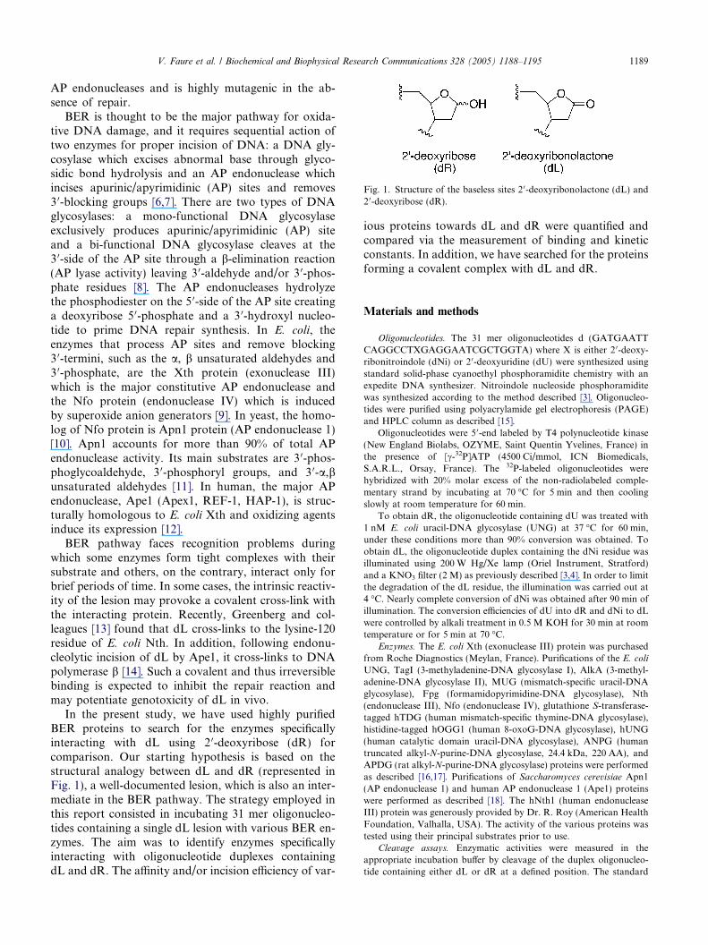
Fig. 1. Structure of the baseless sites 2 0-deoxyribonolactone (dL) and2 0-deoxyribose (dR).
V. Faure et al. / Biochemical and Biophysical Research Communications 328 (2005) 1188–1195 1189
AP endonucleases and is highly mutagenic in the ab-sence of repair.
BER is thought to be the major pathway for oxida-tive DNA damage, and it requires sequential action oftwo enzymes for proper incision of DNA: a DNA gly-cosylase which excises abnormal base through glyco-sidic bond hydrolysis and an AP endonuclease whichincises apurinic/apyrimidinic (AP) sites and removes3 0-blocking groups [6,7]. There are two types of DNAglycosylases: a mono-functional DNA glycosylaseexclusively produces apurinic/apyrimidinic (AP) siteand a bi-functional DNA glycosylase cleaves at the3 0-side of the AP site through a b-elimination reaction(AP lyase activity) leaving 3 0-aldehyde and/or 3 0-phos-phate residues [8]. The AP endonucleases hydrolyzethe phosphodiester on the 5 0-side of the AP site creatinga deoxyribose 5 0-phosphate and a 3 0-hydroxyl nucleo-tide to prime DNA repair synthesis. In E. coli, theenzymes that process AP sites and remove blocking3 0-termini, such as the a, b unsaturated aldehydes and3 0-phosphate, are the Xth protein (exonuclease III)which is the major constitutive AP endonuclease andthe Nfo protein (endonuclease IV) which is inducedby superoxide anion generators [9]. In yeast, the homo-log of Nfo protein is Apn1 protein (AP endonuclease 1)[10]. Apn1 accounts for more than 90% of total APendonuclease activity. Its main substrates are 3 0-phos-phoglycoaldehyde, 3 0-phosphoryl groups, and 3 0-a,bunsaturated aldehydes [11]. In human, the major APendonuclease, Ape1 (Apex1, REF-1, HAP-1), is struc-turally homologous to E. coli Xth and oxidizing agentsinduce its expression [12].
BER pathway faces recognition problems duringwhich some enzymes form tight complexes with theirsubstrate and others, on the contrary, interact only forbrief periods of time. In some cases, the intrinsic reactiv-ity of the lesion may provoke a covalent cross-link withthe interacting protein. Recently, Greenberg and col-leagues [13] found that dL cross-links to the lysine-120residue of E. coli Nth. In addition, following endonu-cleolytic incision of dL by Ape1, it cross-links to DNApolymerase b [14]. Such a covalent and thus irreversiblebinding is expected to inhibit the repair reaction andmay potentiate genotoxicity of dL in vivo.
In the present study, we have used highly purifiedBER proteins to search for the enzymes specificallyinteracting with dL using 2 0-deoxyribose (dR) forcomparison. Our starting hypothesis is based on thestructural analogy between dL and dR (represented inFig. 1), a well-documented lesion, which is also an inter-mediate in the BER pathway. The strategy employed inthis report consisted in incubating 31 mer oligonucleo-tides containing a single dL lesion with various BER en-zymes. The aim was to identify enzymes specificallyinteracting with oligonucleotide duplexes containingdL and dR. The affinity and/or incision efficiency of var-
ious proteins towards dL and dR were quantified andcompared via the measurement of binding and kineticconstants. In addition, we have searched for the proteinsforming a covalent complex with dL and dR.
Materials and methods
Oligonucleotides. The 31 mer oligonucleotides d (GATGAATTCAGGCCTXGAGGAATCGCTGGTA) where X is either 2 0-deoxy-ribonitroindole (dNi) or 2 0-deoxyuridine (dU) were synthesized usingstandard solid-phase cyanoethyl phosphoramidite chemistry with anexpedite DNA synthesizer. Nitroindole nucleoside phosphoramiditewas synthesized according to the method described [3]. Oligonucleo-tides were purified using polyacrylamide gel electrophoresis (PAGE)and HPLC column as described [15].
Oligonucleotides were 5 0-end labeled by T4 polynucleotide kinase(New England Biolabs, OZYME, Saint Quentin Yvelines, France) inthe presence of [c-32P]ATP (4500 Ci/mmol, ICN Biomedicals,S.A.R.L., Orsay, France). The 32P-labeled oligonucleotides werehybridized with 20% molar excess of the non-radiolabeled comple-mentary strand by incubating at 70 �C for 5 min and then coolingslowly at room temperature for 60 min.
To obtain dR, the oligonucleotide containing dU was treated with1 nM E. coli uracil-DNA glycosylase (UNG) at 37 �C for 60 min,under these conditions more than 90% conversion was obtained. Toobtain dL, the oligonucleotide duplex containing the dNi residue wasilluminated using 200 W Hg/Xe lamp (Oriel Instrument, Stratford)and a KNO3 filter (2 M) as previously described [3,4]. In order to limitthe degradation of the dL residue, the illumination was carried out at4 �C. Nearly complete conversion of dNi was obtained after 90 min ofillumination. The conversion efficiencies of dU into dR and dNi to dLwere controlled by alkali treatment in 0.5 M KOH for 30 min at roomtemperature or for 5 min at 70 �C.
Enzymes. The E. coli Xth (exonuclease III) protein was purchasedfrom Roche Diagnostics (Meylan, France). Purifications of the E. coliUNG, TagI (3-methyladenine-DNA glycosylase I), AlkA (3-methyl-adenine-DNA glycosylase II), MUG (mismatch-specific uracil-DNAglycosylase), Fpg (formamidopyrimidine-DNA glycosylase), Nth(endonuclease III), Nfo (endonuclease IV), glutathione S-transferase-tagged hTDG (human mismatch-specific thymine-DNA glycosylase),histidine-tagged hOGG1 (human 8-oxoG-DNA glycosylase), hUNG(human catalytic domain uracil-DNA glycosylase), ANPG (humantruncated alkyl-N-purine-DNA glycosylase, 24.4 kDa, 220 AA), andAPDG (rat alkyl-N-purine-DNA glycosylase) proteins were performedas described [16,17]. Purifications of Saccharomyces cerevisiae Apn1(AP endonuclease 1) and human AP endonuclease 1 (Ape1) proteinswere performed as described [18]. The hNth1 (human endonucleaseIII) protein was generously provided by Dr. R. Roy (American HealthFoundation, Valhalla, USA). The activity of the various proteins wastested using their principal substrates prior to use.
Cleavage assays. Enzymatic activities were measured in theappropriate incubation buffer by cleavage of the duplex oligonucleo-tide containing either dL or dR at a defined position. The standard

Fig. 2. Cleavage of dL and dR duplex oligonucleotides by variousBER proteins. Ten nanomolar 5 0-32P-labeled duplex oligonucleotidescontaining either dL (A) or dA (B) were incubated with 10 nM DNArepair enzyme at 37 �C for 30 min. Lanes: 1, no enzyme; 2, treated withNaOH; 3, incubated in the presence of Ape1; 4, UNG; 5, hOGG1; 6,MUG; 7, Nth; 8, Apn1; 9, hNth1; 10, hUNG; 11, ANPG; 12, AlkA;13, TagI; 14, Fpg; 15, APDG; 16, hTDG; 17, Nfo; and 18, Xth (in thepresence of 5 mM CaCl2). The products of the reaction were analyzedas described under Materials and methods.
1190 V. Faure et al. / Biochemical and Biophysical Research Communications 328 (2005) 1188–1195
assay (20 ll) for Ape1, Apn1, Nfo, and Xth contained 0.2 pmol of the5 0-[32P]-end labeled oligonucleotide duplex, 50 mM Hepes/KOH, pH7.6, 50 mM KCl, 5 mM of 2-mercaptoethanol, 100 lg/mL BSA, and5 mM MgCl2 (or 0.1 mM EDTA for Nfo). The standard assay (20 ll)for UNG; hOGG1; Nth, hNth1, ANPG, TagI, Fpg, and APDGcontained 0.2 pmol of the 5 0-[32P]-end labeled oligonucleotide duplex,70 mM Hepes/KOH, pH 7.8, 100 mM KCl, 5 mM of 2-mercap-toethanol, 100 lg/mL BSA, and 1 mM EDTA. The standard assay(20 ll) for MUG, AlkA, and hTDG was the same as for UNG butwithout 100 mMKCl. The standard assay (20 ll) for hUNG contained0.2 pmol of the 5 0-[32P]-end labeled oligonucleotide duplex, 20 mMTris/HCl, pH 7.5, 60 mM NaCl, 1 mM DTT, 100 lg/mL BSA, and1 mM EDTA. Incubations were carried out at 37 �C, except forhTDG, which was at 30 �C, for 30 min and quenched by adding 10 llof 1.5% SDS/0.3 mg/ml proteinase K followed by incubation for15 min at 37 �C. The mixture was desalted by hand-made spin-downcolumns filled with Sephadex G25 (Amersham Biosciences) equili-brated in 5 M urea. Purified reaction products were separated byelectrophoresis in denaturing 20% (w/v) polyacrylamide gels (20:1, 7 Murea, 0.5· TBE). The gels were exposed to a Fuji FLA-3000 PhosphorScreen and analyzed using Image Jauge V3.12 software.
Electrophoretic mobility shift assay (EMSA). The standard bindingreaction mixture (20ll) contained 1 nM of 5 0-[32P]-labeled oligonu-cleotide duplex, 100 nM pure protein, 100 nM non-labeled regularoligonucleotide duplex, and respective incubation buffer, unlessotherwise stated. The mixture was incubated for 20 min on ice andthen an aliquot was analyzed by electrophoresis on a 10% non-dena-turing polyacrylamide gel (37.5:1 acrylamide/bisacrylamide) using 0.5·TBE buffer (45 mM Tris–borate, pH 8, 1 mM EDTA) at 4 mA/100 Vfor 14 h at +4 �C. The gels were analyzed as described above.
Formation of enzyme–DNA covalent complexes. To evidence theDNA-protein cross-links, 0.2 pmol of 5 0-32P-labeled oligonucleotideduplex was incubated with 100 nM protein at 37 �C for 20 min in itsincubation buffer (20 lL). As a positive control for complex formationan aliquot of the reaction was incubated in the presence of 0.2 MNaBH4 for 30 min at 37 �C. The reaction was stopped by addition of1 lL of 10% SDS and heating for 10 min at 60 �C. The reactionproducts were separated in 15% SDS–PAGE. The gels were analyzedas described above.
Results
Activity of various BER proteins on oligonucleotides
containing dL and dR
The incision activity of various DNA glycosylases andAP endonucleases was evaluated on oligonucleotide du-plexes dL Æ dA and dR Æ dA. Despite the fact that it is wellknown that UNG, MUG, AlkA, hTDG, and ANPG arenot able to cleave an AP site, their effect on dL lesion wasinvestigated since this lesion was shown to be more sen-sitive to b-elimination reaction than an AP site [4]. Asshown in Fig. 2A, dL Æ dA was a good substrate for allAP endonucleases tested (lanes 3, 8, 17, and 18); in con-trast, none of the DNA glycosylases tested were able tocleave the oligonucleotide even at high enzyme concen-tration. As expected, dR Æ dA was efficiently cleaved byall AP endonucleases and DNA glycosylases/AP lyasestested, except hOGG1, (Fig. 2B, lanes 3, 7–9, 14, 17,and 18). Interestingly, a non-specific 3 0 fi 5 0 exonucleaseactivity with excess amount of Apn1 was observed for
both 5 0-labeled duplex oligonucleotides with dR anddL lesions. Previously, it was shown that Apn1 containsexonuclease activity at nicks [19].
The kinetic constants for AP endonuclease-catalyzedincision of dL Æ dA and dR Æ dA and the influence of thebase opposite were measured. As shown in Table 1 theapparent kcat/KM values measured for dL incision byXth, Apn1, andApe1 were 2- to 4-fold lower as comparedto the values measured for dR incision. Interestingly, Nfoshowsmore than 10-fold lowerKMand kcat/KMvalues fordL than for dR incision. These data are in agreement withprevious studies by Xu et al. [20] on Ape1. Although, theauthors obtained very similar KM values they reported a5-fold higher kcat/KM for dR. Fig. 3 presents the initialvelocity of the four enzymes when acting upon the fourdifferent dL Æ dN pairs. All four duplexes were substratesfor the AP endonucleases, which preferentially incise dLwhen it is opposite dA. The relative order of dL incisionfor Xth, Apn1, and Ape1 was dA > dG > T > dC andfor Nfo was dA > dG > dC P T. Interestingly, the initialrates of cleavage of dL Æ dC by Ape1 and Xth were 3- and6-fold lower, respectively, as compared to dL Æ dA. Incontrast, the incision efficiency of Nfo and Apn1 on

Fig. 4. Electrophoretic mobility shift assay of various DNA repairproteins with dL Æ dA (A) and dR Æ dA (B) containing duplex oligo-nucleotides. 0.1 nM of 5 0-[32P]-labeled duplex oligonucleotide wasincubated at 0 �C for 20 min in the absence (lane 1) or presence of100 nM purified repair protein (lanes 2–17 or 2–16). Free probe, non-bound oligonucleotide; bound, enzyme-bound oligonucleotide. Theenzyme-bound fraction was analyzed as described under Materials andmethods.
Table 1Kinetic parameters of Xth, Nfo, Apn1, and Ape1 for duplex DNA substrates containing dL or dR residues
Lesion Parameters Xth Nfo Apn1 Ape1
KM (nM) 20 ± 0.5 85 ± 1.3 27.7 ± 1.2 70 ± 2dL kcat (min�1) 8.14 ± 0.2 9.2 ± 0.4 7.5 ± 0.3 26.4 ± 0.7
kcat/KM (lM�1 min�1) 407 108 270 377
KM (nM) 16 ± 0.7 21.5 ± 1 5.0 ± 2.1 17dR kcat (min�1) 13.6 ± 0.6 9.9 ± 0.15 3.1 ± 0.1 1.2 ± 0.03
kcat/KM (lM�1 min�1) 848 217 560 1456
Reactions contained 1–400 nM DNA duplex. Values of the apparent KM and kcat were determined from Lineweaver–Burk plots.
Fig. 3. Relative initial velocity of incision of the dL Æ dN oligonucleo-tide duplexes by Xth, Nfo, Apn1, and Ape1.
V. Faure et al. / Biochemical and Biophysical Research Communications 328 (2005) 1188–1195 1191
dL Æ dN duplexes varied only slightly depending on thepaired nucleotides.
Binding of various DNA repair proteins to dL Æ dA and
dR Æ dA oligonucleotide duplexes
The interaction of 5 0-[32P]-labeled dL Æ dA with vari-ous BER proteins was examined using an electropho-retic mobility shift assay (EMSA). For comparison, weused regular AP site containing duplex oligonucleotidedR Æ dA. To avoid non-specific interactions, the stan-dard binding mixture contained a 17-fold excess ofBSA and a 100-fold excess of cold, non-modified 31mer T Æ dA duplex oligonucleotide over labeled dL Æ dAand dR Æ dA. As shown in Fig. 4A, Nfo, hTDG, Fpg,ANPG, Apn1, and MUG interact in a specific mannerwith dL Æ dA, with the apparition of specific slowmigrating bands on the gel (lanes 5, 7, 10, 13, 15, and16), whereas, the dR Æ dA oligonucleotide formed stablecomplexes with Nfo, Fpg, ANPG, AlkA, Apn1, Nth,and MUG (Fig. 4B, lanes 4, 6, 10, 11, and 12). Interest-ingly, Fpg, Apn1, and MUG bind significantly to bothdL and dR, whereas, ANPG and AlkA bind moretightly to dL and dR, respectively, and Nth binds onlyto dR. To demonstrate binding specificity of the Fpg,ANPG, Apn1, and MUG proteins to dL, increasingamounts of either unlabeled non-specific T Æ dA or spe-cific dL Æ dA duplex oligonucleotides were added intobinding reactions with the 32P-labeled dL Æ dA. Molarexcess of the cold dL Æ dA duplex oligonucleotide effi-

Table 2Apparent dissociation constants Kd (in nM) of protein/oligo complexes
Protein Kd (nM)
Apn1 MUG Fpg AlkA ANPG
dL 0.25 ± 0.04 8.56 ± 0.55 3.74 ± 0.18 ND 5.44 ± 0.47dR 0.96 ± 0.24 2.99 ± 0.73 0.4 ± 0.08 1.76 ± 0.37 ND
ND, not determined.
Fig. 5. Cross-linking of dL Æ dA (A) and dR Æ dA (B) containingduplex oligonucleotides with various BER proteins. Ten nanomolar 5 0-[32P]-labeled duplex oligonucleotide was incubated at 37 �C for 20 minin the absence (lane 1) or presence of 100 nM purified repair protein(lanes 2–18). Free probe, non-bound oligonucleotide; bound, enzyme-trapped oligonucleotide. The covalent-trapped products were analyzedas described under Materials and methods.
1192 V. Faure et al. / Biochemical and Biophysical Research Communications 328 (2005) 1188–1195
ciently inhibited complex formation for all proteinstested whereas little inhibition was observed with thesame amount of cold T Æ dA DNA (data not shown).Similar results were obtained when checking bindingspecificity towards dR. Thus, the results show thatApn1, MUG, Fpg, AlkA, and ANPG proteins bind todL and/or dR in a specific manner.
To compare the affinity of these proteins towards dLand/or dR, we measured apparent dissociation equilib-rium constants (Kd) using EMSA for each protein exceptfor hTDG and Nfo because of low specificity andcomplex instability. The assay was performed underconditions where [DNA] = 0.1 nM > Kd. As shown inTable 2 the apparent Kd values were lower than 10 nMindicating high affinity of the proteins tested towardsbaseless residues in DNA. Comparison of the Kd valuesfor dL and dR shows that in contrast to MUG andFpg, the Apn1 protein has a higher affinity for dL thanfor dR site.
Covalent-trapping between BER proteins and dL
and/or dR
It was shown that dL efficiently cross-links with aDNA glycosylase/AP lyase [13]. Here, the formationof covalent complexes at 37 �C between BER proteinsand dL or dR was examined. SDS–PAGE revealedcross-links. As control for DNA–protein covalent com-plex formation, dL and/or dR were incubated with Fpgin the presence of NaBH4. In these conditions the Fpg-oligonucleotide covalent complex was generated byreducing the Schiff base intermediate formed betweenthe sugar residue and an amino residue of the protein.As shown in Fig. 5A, among BER proteins tested onlytwo hOGG1 and hNth1 cross-linked to dL-containingoligonucleotide (lanes 5 and 9). Surprisingly, we didnot observe a covalent complex with Nth and dL underthe conditions used (Fig. 5A, lane 7). However, whenhigher oligonucleotide concentration was used theNth–dL complex was detected (data not shown). Unex-pectedly, we observed highly efficient covalent-trappingof the Nth protein by dR containing oligonucleotide(Fig. 5B, lane 7). In addition, very small amount ofdR cross-link to hNth1 was detected (Fig. 5B, lane 9).Although, the Apn1, MUG, Fpg, AlkA, and ANPGproteins bind to dL and/or dR in a specific manner wedid not detect any cross-links to these proteins, underthe conditions used.
Discussion
About 20% of total DNA damage induced by ioniz-ing radiation are oxidized abasic sites which suggests amost likely biological role for this type of lesion [21].It was proposed that OAS might be poorly handled bythe BER pathway [22]. Indeed, it has been demonstratedthat OAS and their incision products cross-link DNA

V. Faure et al. / Biochemical and Biophysical Research Communications 328 (2005) 1188–1195 1193
glycosylases and DNA polymerase b in vitro, therebydisrupting normal repair process [21]. The goal of thisstudy was to determine the ability of various DNA gly-cosylases and AP endonucleases to interact with dL.Regular AP site, dR, was used to differentiate proteins�specificity to dL.
In the present study, we showed that dL is cleavedefficiently by all AP endonucleases tested but not byDNA glycosylases/AP lyases. This is the major differ-ence with dR residue, which is cleaved also by DNA gly-cosylases/AP lyases through a b-elimination reaction.Formation of a Schiff base is not possible in the caseof dL and could explain the resistance of dL to theb-elimination reaction. Comparison of kinetic parame-ters measured using dL and dR substrates indicates thatboth lesions are repaired with similar efficiency by all APendonucleases tested. This is in agreement with our invivo data showing that, in E. coli, dL is repaired mainlyby Xth and Nfo, and not by DNA glycosylases/APlyases. However, we observed a 2- to 5-fold preferenceof all AP endonucleases to dR compared to dL. Thispreference is mainly due to higher KM values for thedL incision suggesting that the AP endonucleases havelower affinity towards oxidized AP site. It was demon-strated that AP endonucleases incise efficiently at diverseabasic lesions such as tetrahydrofuran, propanyl, etha-nyl, and urea [23–25]. Similar to other baseless modifica-tions, dL in a DNA duplex induces only localizedperturbations at the lesion site, suggesting that an over-all duplex conformation is important for specific recog-nition by this type of enzymes [26]. Consistent with thefact that, when generated by ionizing radiations, thedL sites are expected to arise at any position in DNA,all of the duplexes containing each of the four basesopposite to the lesion were substrates for the AP endo-nucleases tested. The relative order of dL incision wassimilar for all enzymes tested with dL Æ dA as the mostpreferred pair. After our experiments were completed,Greenberg et al. [27] reported similar results on the re-pair of dL by the E. coli Xth and Nfo proteins.
Previously, it was shown that duplex DNA contain-ing a pyrrolidine abasic site analog specifically interactsand inhibits a variety of DNA glycosylases [28]. There-fore, we examined whether dL form a non-covalentDNA–protein complex. Indeed, Fpg, Apn1, and MUGbind in a highly specific manner to dL and dR contain-ing DNA, albeit with varied Kd values. It was knownthat the E. coli Fpg and MUG have high affinity forAP sites [29,30] and our results suggest that these pro-teins do not discriminate dR and dL residues in DNA.All AP endonucleases tested can recognize and incisedL but only Apn1 could form stable complex with dLat 4 �C. Little is known about AP site structural deter-minants for Apn1 specific recognition due to the absenceof crystallographic data. However, we may propose thatApn1 could bind dL in a non-catalytic manner without
incision, which leads to the formation of a stable sub-strate–enzyme complex at low temperature. We haveshown that E. coli AlkA and human ANPG preferen-tially bind to dR and dL, respectively, thus discriminat-ing the subtle structural difference between these twolesions. Although AlkA and ANPG have common sub-strate specificities, the kinetic parameters for excision ofcertain damaged bases and three-dimensional structuresof these two DNA glycosylases are very different sug-gesting that binding specificity can vary tremendously.Recently, we have shown that hijacking of a humanDNA glycosylase by an exocyclic DNA adduct might in-crease genotoxicity of this adduct to cells [31]. Similarly,binding of dL by Fpg, MUG, and ANPG may interferewith the repair of this lesion in vivo.
BER pathway eliminates non-bulky oxidized residuesgenerated by ionizing radiations and endogenous ROS.Although, unrepaired DNA damage can result in muta-tions, processing of the closely opposed lesions throughBER pathway may actually result in double-strandbreaks [32,33]. Furthermore, dL can efficiently cross-link to Nth, and very weakly to Fpg, hOGG1, andhNth1 present in high molar excess [34]. Attempted re-pair of dL in mammalian cells can lead to covalent trap-ping of DNA polymerase b [14]. These observationsimply that excision repair may not be appropriate to cer-tain forms of oxidative DNA damage. Our work extendsthe latter studies by examining the formation of covalentcomplexes between baseless residues and various puri-fied BER enzymes. We have shown that dL forms cova-lent complexes only with the human proteins hNth1 andhOGG1, suggesting that dL is highly toxic in mammals.Under conditions used, Nth did not cross-link to dL,however upon increase of the lesion concentration thecovalent binding appeared confirming previous observa-tions. Covalent complex formation was also studiedusing dR residue. Surprisingly, among the proteinstested we have found that Nth cross-links with good effi-ciency to dR. Furthermore, a small amount of covalentcomplex was detected also with hNth1. Thus, the E. coliNth and its human structural homolog, and to some ex-tent hOGG1, could perturb repair of regular and oxi-dized AP sites, and sensitize cells to genotoxic agents.Interestingly, E. coli and mice mutants lacking theseDNA glycosylases are not sensitive to ionizing radiation[35,36]. We may speculate that increased level of theunrepaired thymine glycols and fragmented purines inthe mutants might be compensated by the absence ofcovalently trapped proteins at the AP sites. Interest-ingly, no cross-linking was detected with the proteinshaving high affinity for dR and dL, suggesting that theseproteins and others might protect the AP sites from acci-dental covalent trapping with other cellular proteins.
In conclusion, the dL and dR lesions have structuralsimilarity and their repair is performed with similar effi-ciencies by the major type II endonucleases. This

1194 V. Faure et al. / Biochemical and Biophysical Research Communications 328 (2005) 1188–1195
strongly suggests that Nfo and Xth in E. coli, Apn1 inbudding yeast, and Ape1 in humans are the first playersin the defense of these organisms against dL-inducedgenotoxicity. Quantitative examination of dL incisionby these four enzymes reveals that the lesion is repairedwith an efficiency similar to that of dR. We have demon-strated for the first time that several BER proteins bindto dL in a highly specific and non-covalent manner. Asalready reported by others, we showed that dL lesioncross-links to human oxidative DNA glycosylases. Suchan enzyme trapping is expected to disturb the repairmachinery and sensitize cells to killing effect of ionizingradiation and certain drugs.
Acknowledgments
We wish to thank Drs. Alexander Ishchenko andNathalie Berthet for helpful discussion, Dr MitsuharuKotera for the dNi phosphoramidite, and ElisabethLoire and Gesa Tietjens for technical assistance. Thiswork was supported (M.S. and J.-F.C.) by Electricitede France Contrat Radioprotection RB 2003-28(J.-F.C and P.D.) and RB2004-02 (M.S.), grant fromEuropean Community RISC-RAD FI6R-CT-2003-508842 (M.S.), Association pour la Recherche sur leCancer (M.S.).
References
[1] C. von Sonntag, The Chemical Basis of Radiation Biology, Taylor& Francis, London, 1987.
[2] P.C. Dedon, I.H. Goldberg, Free-radical mechanisms involved inthe formation of sequence-dependent bistranded DNA lesions bythe antitumor antibiotics bleomycin, neocarzinostatin, and cali-cheamicin, Chem. Res. Toxicol. 5 (1992) 311–332.
[3] M. Kotera, A.-G. Bourdat, E. Defrancq, J. Lhomme, A highlyefficient synthesis of oligodeoxyribonucleotides containing the 2 0-deoxyribonolactone lesion, J. Am. Chem. Soc. 120 (1998) 11810–11811.
[4] Y. Roupioz, M. Kotera, J. Lhomme, Chemistry of the 2-deoxyribonolactone lesion in oligonucleotides: Cleavage kineticsand products analysis, J. Am. Chem. Soc. 124 (2002) 9129–9135.
[5] V. Faure, J.F. Constant, P. Dumy, M. Saparbaev, 2 0-Deoxy-ribonolactone lesion produces G to A transitions in Escherichia
coli, Nucleic Acids Res. 32 (2004) 2937–2946.[6] T. Lindahl, Keynote: Past, present, and future aspects of base
excision repair, Prog. Nucleic Acid Res. Mol. Biol. 68 (2001) xvii–xxx.
[7] L. Gros, M.K. Saparbaev, J. Laval, Enzymology of the repair offree radicals-induced DNA damage, Oncogene 21 (2002) 8905–8925.
[8] M.L. Dodson, M.L. Michaels, R.S. Lloyd, Unified catalyticmechanism for DNA glycosylases, J. Biol. Chem. 269 (1994)32709–32712.
[9] B. Demple, L. Harrison, Repair of oxidative damage to DNA:Enzymology and biology, Annu. Rev. Biochem. 63 (1994) 915–948.
[10] S.C. Popoff, A.I. Spira, A.W. Johnson, B. Demple, Yeaststructural gene (APN1) for the major apurinic endonuclease:
Homology to Escherichia coli endonuclease IV, Proc. Natl. Acad.Sci. USA 87 (1990) 4193–4197.
[11] D. Ramotar, S.C. Popoff, E.B. Gralla, B. Demple, Cellular role ofyeast Apn1 apurinic endonuclease/3 0-diesterase: Repair of oxida-tive and alkylation DNA damage and control of spontaneousmutation, Mol. Cell. Biol. 11 (1991) 4537–4544.
[12] D.M. Wilson III, D. Barsky, The major human abasic endonu-clease: Formation, consequences and repair of abasic lesions inDNA, Mutat. Res. 485 (2001) 283–307.
[13] M. Hashimoto, M.M. Greenberg, Y.W. Kow, J.T. Hwang, R.P.Cunningham, The 2-deoxyribonolactone lesion produced in DNAby neocarzinostatin and other damaging agents forms cross-linkswith the base-excision repair enzyme endonuclease III, J. Am.Chem. Soc. 123 (2001) 3161–3162.
[14] M.S. DeMott, E. Beyret, D. Wong, B.C. Bales, J.T. Hwang,M.M. Greenberg, B. Demple, Covalent trapping of human DNApolymerase beta by the oxidative DNA lesion 2-deoxyribonolac-tone, J. Biol. Chem. 277 (2002) 7637–7640.
[15] J. Sambrook, E.F. Fitsch, T. Maniatis, Molecular Cloning: ALaboratory Manual, Cold Spring Harbor Laboratory, ColdSpring Harbor, NY, 1989.
[16] M. Saparbaev, S. Langouet, C.V. Privezentzev, F.P. Guengerich,H. Cai, R.H. Elder, J. Laval, 1,N(2)-ethenoguanine, a mutagenicDNA adduct, is a primary substrate of Escherichia coli mismatch-specific uracil-DNA glycosylase and human alkylpurine-DNA-N-glycosylase, J. Biol. Chem. 277 (2002) 26987–26993.
[17] N. Scaramozzino, G. Sanz, J.M. Crance, M. Saparbaev, R.Drillien, J. Laval, B. Kavli, D. Garin, Characterisation of thesubstrate specificity of homogeneous vaccinia virus uracil-DNAglycosylase, Nucleic Acids Res. 31 (2003) 4950–4957.
[18] A.A. Ishchenko, G. Sanz, C.V. Privezentzev, A.V. Maksimenko,M. Saparbaev, Characterisation of new substrate specificities ofEscherichia coli and Saccharomyces cerevisiae AP endonucleases,Nucleic Acids Res. 31 (2003) 6344–6353.
[19] J.R. Vance, T.E. Wilson, Repair of DNA strand breaks by theoverlapping functions of lesion-specific and non-lesion-specificDNA 3 0 phosphatases, Mol. Cell. Biol. 21 (2001) 7191–7198.
[20] Y.J. Xu, M.S. DeMott, J.T. Hwang, M.M. Greenberg, B. Demple,Action of human apurinic endonuclease (Ape1) on C10-oxidizeddeoxyribose damage in DNA, DNA Repair (Amst) 2 (2003) 175–185.
[21] B. Demple, M.S. DeMott, Dynamics and diversions in baseexcision DNA repair of oxidized abasic lesions, Oncogene 21(2002) 8926–8934.
[22] J. Nakamura, D.K. La, J.A. Swenberg, 5 0-nicked apurinic/apyrimidinic sites are resistant to beta-elimination by beta-polymerase and are persistent in human cultured cells afteroxidative stress, J. Biol. Chem. 275 (2000) 5323–5328.
[23] M. Takeuchi, R. Lillis, B. Demple, M. Takeshita, Interactions ofEscherichia coli endonuclease IV and exonuclease III with abasicsites in DNA, J. Biol. Chem. 269 (1994) 21907–21914.
[24] D.M. Wilson III, M. Takeshita, A.P. Grollman, B. Demple,Incision activity of human apurinic endonuclease (Ape) at abasicsite analogs in DNA, J. Biol. Chem. 270 (1995) 16002–16007.
[25] Y.W. Kow, S.S. Wallace, Exonuclease III recognizes urea residuesin oxidized DNA, Proc. Natl. Acad. Sci. USA 82 (1985) 8354–8358.
[26] M. Jourdan, J. Garcia, E. Defrancq, M. Kotera, J. Lhomme, 2 0-Deoxyribonolactone lesion in DNA: Refined solution structuredetermined by nuclear magnetic resonance and molecular mod-eling, Biochemistry 38 (1999) 3985–3995.
[27] M.M. Greenberg, Y.N. Weledji, J. Kim, B.C. Bales, Repair ofoxidized abasic sites by exonuclease III, endonuclease IV, andendonuclease III, Biochemistry 43 (2004) 8178–8183.
[28] O.D. Scharer, H.M. Nash, J. Jiricny, J. Laval, G.L. Verdine,Specific binding of a designed pyrrolidine abasic site analog tomultiple DNA glycosylases, J. Biol. Chem. 273 (1998) 8592–8597.

V. Faure et al. / Biochemical and Biophysical Research Communications 328 (2005) 1188–1195 1195
[29] B. Castaing, J.L. Fourrey, N. Hervouet, M. Thomas, S. Boiteux,C. Zelwer, AP site structural determinants for Fpg specificrecognition, Nucleic Acids Res. 27 (1999) 608–615.
[30] R.J. O�Neill, O.V. Vorob�eva, H. Shahbakhti, E. Zmuda, A.S.Bhagwat, G.S. Baldwin, Mismatch uracil glycosylase from Esch-
erichia coli: A general mismatch or a specific dna glycosylase? J.Biol. Chem. 278 (2003) 20526–20532.
[31] L. Gros, A.V. Maksimenko, C.V. Privezentzev, J. Laval, M.K.Saparbaev, Hijacking of the human alkyl-N-purine-DNA glyco-sylase by 3,N4-ethenocytosine, a lipid peroxidation-induced DNAadduct, J. Biol. Chem. 279 (2004) 17723–17730.
[32] J.O. Blaisdell, S.S. Wallace, Abortive base-excision repair ofradiation-induced clustered DNA lesions in Escherichia coli, Proc.Natl. Acad. Sci. USA 98 (2001) 7426–7430.
[33] I. D�Souza, D.L. Harrison, Repair of clustered uracil DNAdamages in Escherichia coli, Nucleic Acids Res. 31 (2003) 4573–4581.
[34] K.M. Kroeger, M. Hashimoto, Y.W. Kow, M.M. Greenberg,Cross-linking of 2-deoxyribonolactone and its beta-eliminationproduct by base excision repair enzymes, Biochemistry 42 (2003)2449–2455.
[35] R.P. Cunningham, B. Weiss, Endonuclease III (nth) mutantsof Escherichia coli, Proc. Natl. Acad. Sci. USA 82 (1985) 474–478.
[36] M.T. Ocampo, W. Chaung, D.R. Marenstein, M.K. Chan, A.Altamirano, A.K. Basu, R.J. Boorstein, R.P. Cunningham, G.W.Teebor, Targeted deletion of mNth1 reveals a novel DNA repairenzyme activity, Mol. Cell. Biol. 22 (2002) 6111–6121.


![The Sequence Dependence of Human Nucleotide Excision Repair Efficiencies of Benzo[ a]pyrene-derived DNA Lesions: Insights into the Structural Factors that Favor Dual Incisions](https://static.fdokumen.com/doc/165x107/6313e5adc32ab5e46f0ca10d/the-sequence-dependence-of-human-nucleotide-excision-repair-efficiencies-of-benzo.jpg)

