Expressing Thermal Comfort in the Urban Climate of Athens, Greece
Upload
independentCategory
view
4download
0
Somatotopic Organization andFunctional Properties of
Mechanosensory Neurons ExpressingSensorin-A mRNA in Aplysia californica
EDGAR T. WALTERS,1* MICHAELA BODNAROVA,2 ALLEN J. BILLY,1,3
MICHAEL F. DULIN,1,4 MANUEL DIAZ-RIOS,5,6 MARK W. MILLER,5
AND LEONID L. MOROZ2
1Department of Integrative Biology and Pharmacology, University of Texas-HoustonMedical School, Houston, Texas 77030
2The Whitney Laboratory, Department of Neuroscience, University of Florida,St. Augustine, Florida 32080
3Program in Basic Health Sciences, School of Health Sciences, British Columbia Instituteof Technology, Burnaby, British Columbia V5G 3H2, Canada
4Harrisburg Family Physicians, Harrisburg, North Carolina 280755Department of Anatomy and Institute of Neurobiology, University of Puerto Rico, Medical
Sciences Campus, San Juan, Puerto Rico 009016Neurobiology and Behavior, Cornell University, Ithaca, New York 14853
ABSTRACTA previous study reported that a peptide, sensorin-A, is expressed exclusively in mech-
anosensory neurons having somata in central ganglia of Aplysia. The present study utilized insitu hybridization, staining by nerve back-fill and soma injection, and electrophysiological meth-ods to characterize the locations, numbers, and functions of sensorin-A-expressing neurons andto define the relationships between soma locations and the locations of peripheral axons andreceptive fields. Approximately 1,000 cells express sensorin-A mRNA in young adult animals(10–30 g) and 1,200 cells in larger adults (100–300 g). All of the labeled somata are in the CNS,primarily in the abdominal LE, rLE, RE and RF, pleural VC, cerebral J and K, and buccal Sclusters. Expression also occurs in a few sparsely distributed cells in most ganglia. Together,receptive fields of all these mechanosensory clusters cover the entire body surface. Each VCcluster forms a somatotopic map of the ipsilateral body, a “sensory aplunculus.” Cells in thepleural and cerebral clusters have partially overlapping sensory fields and synaptic targets.Buccal S cells have receptive fields on the buccal mass and lips and display notable differences inelectrophysiological properties from other sensorin-A-expressing neurons. Neurons in all of theclusters have relatively high mechanosensory thresholds, responding preferentially to threaten-ing or noxious stimuli. Synaptic outputs to target cells having defensive functions support anociceptive role, as does peripheral sensitization following noxious stimulation, although addi-tional functions are likely in some clusters. Interesting questions arise from observations thatmRNA for sensorin-A is present not only in the somata but also in synaptic regions, connectives,and peripheral fibers. J. Comp. Neurol. 471:219–240, 2004. © 2004 Wiley-Liss, Inc.
Indexing terms: nociceptor; receptive field; axon; neuropeptide; defensive reflex; sensory map
Grant sponsor: National Institutes of Health (NIH); Grant number:NS35979 (E.T.W.); Grant number: NS35882 (E.T.W.); Grant sponsor: NIH;Grant number: NS39103; Grant number: MH60261 (L.L.M.); Grant number:P50G002806 (L.L.M.); Grant sponsor: the David and Lucile Packard Founda-tions (L.L.M.); Grant sponsor: the Evelyn F. McKnight Brain Research GrantProgram (L.L.M.); Grant sponsor: NIH; Grant number: NS07464 (M.W.M.);Grant number: MH048190 M-RISP (M.W.M.); Grant number: GM08224MBRS (M.W.M.); Grant sponsor: NIH; Grant number: RR10294.
*Correspondence to: Edgar T. Walters, Department of Integrative Biologyand Pharmacology, University of Texas-Houston Medical School, 6431 FanninBlvd. MSB 4.116, Houston, TX 77030. E-mail: [email protected]
Received 18 August 2003; Revised 19 November 2003; Accepted 5 De-cember 2003
DOI 10.1002/cne.20042Published online the week of February 16, 2004 in Wiley InterScience
(www.interscience.wiley.com).
THE JOURNAL OF COMPARATIVE NEUROLOGY 471:219–240 (2004)
© 2004 WILEY-LISS, INC.

The gastropod mollusc Aplysia californica has proved tobe a useful species for exploring mechanisms of neuralplasticity related to learning and memory (see, e.g., Kan-del, 2001) and injury (see, e.g., Ambron and Walters,1996). Much of this utility comes from experimental ad-vantages offered by mechanosensory neurons, whosereadily identifiable somata are clustered in the animal’scentral ganglia. The sensory clusters that have been uti-lized most extensively by neurobiologists are the left E(LE) cluster in the abdominal ganglion (Castellucci et al.,1970; Byrne et al., 1974), the ventrocaudal (VC) clusters inthe pleural ganglia (Walters et al., 1983), and the J and Kclusters in the cerebral ganglia (Rosen et al., 1979; see Fig.1). These sensory populations were shown to innervate,respectively, the siphon, the tail, and the head, althoughnone of these studies systematically examined the entiresurface of the body for receptive fields. Mechanosensoryneurons displaying morphological and response propertiessimilar to those of these three groups of cells were alsofound in the abdominal ganglion right E (RE), right F(RF), and rostral LE (rLE) clusters (Byrne et al., 1974;Byrne, 1980; Dubuc and Castellucci, 1991) and in thebuccal ganglion S1 and S2 clusters (Fiore and Geppeti,1981). All of the abdominal ganglion clusters innervatestructures in the mantle cavity, whereas the buccal gan-glion S clusters appear to innervate the buccal mass.
By using a differential screening approach, Brunet andcolleagues (1991) identified an mRNA encoding a potentialneuropeptide precursor that was expressed in pleural VCclusters. Physiological experiments demonstrated thatsensorin-A, a nonapeptide (ARYRVGYMF-NH2) cleavedfrom this precursor, could act as an inhibitory cotransmit-ter at VC cell synapses. In situ hybridization with anantisense probe to the sensorin mRNA and immunohisto-chemistry with an antibody that recognized sensorin-Aand its propeptide indicated that sensorin-A is selectivelyexpressed in all of the known mechanosensory clusters inthe central ganglia of Aplysia. Moreover, sensorin-A ex-pression appeared to be absent from all other cells of theCNS (Brunet et al., 1991). Selective probes for sensorin-Aand its mRNA offer powerful tools that have been ex-ploited to study axonal regeneration (Steffensen et al.,1995a), changes in gene expression after nerve injury(Noel et al., 1995), and expression and stability of neu-ropeptide mRNA during synapse formation and synapticplasticity (Santarelli et al., 1996; Schacher et al., 1999;Sun et al., 2001; Hu et al., 2002). However, except for thebrief, preliminary description of sensorin-A staining inpreviously defined mechanosensory clusters by Brunet etal. (1991), probes for sensorin-A have not yet been used toexamine systematically the topographic organization ofthe mechanosensory systems expressing this neuropep-tide.
The discrete but distributed population of neurons de-fined by the expression of sensorin-A raises basic ques-tions that are addressed herein. How many sensorin-A-expressing neurons are there in Aplysia and what is theirdetailed central distribution? How are the locations of thecentral sensorin-A-expressing somata related to the dis-tribution of their peripheral axons and the locations oftheir mechanosensory receptive fields? Are all of thesensorin-A-expressing clusters actually mechanosensoryin function? In this paper, we describe the central andperipheral organization of these cells and show that thereceptive fields of the sensorin-A-expressing clusters cover
the entire surface of the animal’s body. Furthermore, bydocumenting receptive fields of the buccal S cells, we con-firm that all of the sensorin-A-expressing clusters in Aply-sia contain mechanosensory neurons and show that thesecells, like neurons in the cerebral, pleural, and abdominalclusters, have relatively high mechanosensory thresholds.
Materials and Methods
Animals
Aplysia californica (10–300 g) were supplied by AlacrityMarine Biological Services (Redondo Beach, CA), Marinus(Long Beach, CA), and the NIH Aplysia Resource Facility(Miami, FL). Animals were housed in aquaria containingartificial seawater (ASW; Instant Ocean, Burlington, NC)at 15–18°C on a 12:12-hour light:dark cycle for up to 3weeks before use. Constant body weight was maintainedon a diet of live Gracilaria seaweed or dried seaweedlaver. Animals weighed 10–30 or 100–300 g. The smalleranimals were probably young adults, as indicated by thepresence of bag cell clusters, but they may have includedsome animals transitioning from the last juvenile stage toadulthood (Kriegstein, 1977a,b; Cash and Carew, 1989).Prior to dissection, animals were anesthetized by injectinga volume of isotonic MgCl2 (337 mM) equivalent to 50–60% of their weight.
In situ hybridization protocol
All in situ hybridization experiments were performedusing whole-mount preparations of the CNS of A. califor-nica. Ganglia (Fig. 1) were isolated, and before fixationthey were pinned to a Sylgard dish in artificial seawater(ASW: 460 mM NaCl, 10 mM KCl, 55 mM MgCl2, 11 mMCaCl2, 10 mM HEPES, pH 7.6). Plasmids containing thesequence of full-length A. californica sensorin (GeneBankaccession number gi:5589) subcloned in pGEM-T vector(Promega, Madison, WI) were obtained during EST se-quencing from the CNS and sensory cells of A. californica(Moroz et al., 2002). The plasmid (JM-109 in Escherichiacoli) was extracted, purified, and sequenced to obtain theorientation of the gene. The plasmid was linearized withspecific endonucleases (NotI for the antisense probe usingT7 polymerase, NcoI for the sense probe with Sp6 poly-merase) and used as a template for the preparation ofspecific antisense and sense digoxigenin (DIG)-labeledsensorin RNA probes following the Roche protocol forprobe preparation with a DIG RNA labeling kit (Sp6/T7).Control experiments (six preparations) using sense probesand identical labeling protocols and conditions did notproduce specific staining in the CNS.
Our in situ hybridization protocol was based on previ-ously published reports (Bogdanov Yu et al., 1996), withseveral modifications. Briefly, dissected central gangliafrom Aplysia were treated with 1% trypsin (Sigma, St.Louis, MO) in filtered sea water (FSW) for 45–60 minutesat 34°C, rinsed for 5 minutes in FSW, and incubated with1% trypsin inhibitor for 15 minutes in FSW at room tem-perature. The ganglia were fixed overnight in 4% parafor-maldehyde in 0.2 M phosphate-buffered saline (PBS; pH7.2) at 4°C. Preparations were washed in PBS, and con-nective tissues of the ganglionic sheath were carefullyremoved from all ganglia. The ganglia were washed inPTW (0.1% Tween 20 in PBS) three times for 5 minuteseach, followed by a 10-minute incubation in increasing
220 E.T. WALTERS ET AL.

amounts of methanol (3:1 PTW:MetOH, 1:1, 1:3, and 100%MetOH), in which the ganglia were preserved for up to 1week. In most experiments, ganglia were transferred backto PTW in the same fashion by repetitive substitution ofmethanol by PTW (10-minute washes with 1:3 PTW/MetOH, 1:1, 3:1, PTW, followed by a 10-minute wash with0.3% Triton in PBS, PTW for 5 minutes). After incubationwith proteinase K (10 �g/ml) in PTW at room temperaturefor 1 hour, the ganglia were postfixed with 4% parafor-maldehyde in PBS for 20 minutes at 4°C and washed (2 �5 minutes) in each of the following solutions: glycine (2mg/ml PTW), PTW, 0.1 M triethanolamine hydrochloride(TEA HCl; pH 8.0), and anhydrous acetate (2.5 �l/ml) inTEA HCl. Next, the ganglia were repeatedly washed inPTW and incubated at –20°C in hybridization buffer (50%formamide, 5 mM EDTA, 5� SSC, 1� Denhardt solution[0.02% ficoll, 0.02% polyvinylpyrrolidone, 0.02% bovineserum albumin (BSA)], 0.1% Tween 20, 0.5 mg/ml yeasttRNA (Gibco BRL) overnight. All subsequent steps untildetection (including the 6–8-hour prehybridization at60°C) were peformed with moderate shaking. DIG-labeledRNA probes (2 �l/ml) were added, and ganglia were incu-bated for a further 6–8 hours. Hybridization was followedby 30 minutes of washing at 60°C in 50% formamide/5�SSC/1% sodium dodecyl sulfate (SDS), then 50%formamide/2� SSC/1% SDS, then 0.2� SSC, twice for 30minutes at 55°C. After three washes in PBT (1� PBS,
0.1% Triton X-100, 2 mg/ml BSA), ganglia were treatedwith 10% normal goat serum in PBT at 4°C for up to 90minutes and left overnight with a 1:1,350 dilution of al-kaline phosphatase-conjugated DIG antibodies (Boehring,Indianapolis, IN) in 1% goat serum in PBT at 4°C. Un-bound antibodies were washed out in several PBT incuba-tions at 4°C (at least 2–3 hours total). After two 5-minuteincubations in detection buffer (100 mM NaCl, 50 mMMgCl2, 100 mM NaCl, 100 mM Tris Cl, pH 9.5, 0.01%Tween 20, 1 mM levamisol), NBT/BCIP color substrates(DIG Nucleic Acid Detection Kit; Boehringer) were addedto detect the hybridized probes. At this stage, ganglia werekept at 4°C in the dark with periodic visual assessment ofthe staining intensity. After the detection procedure (usu-ally 30–60 minutes), ganglia were postfixed in 4% para-formaldehyde in methanol for up to 1 hour and washedtwice in 100% ethanol. Permanent preparations were pro-duced by incubating ganglia in methylsalycilate and em-bedding in Permount (Fisher, Fair Lawn, NJ) on micro-scopic slides. Examination and analysis of staining wereconducted on an upright Nikon Optiphot-2 or an OlympusSZX 12 dissecting microscope. Images were acquired witha Nikon Coolpix 4500 digital camera and initially saved asJPEG files.
Electrophysiological properties andreceptive field mapping
Receptive fields and peripheral axon distributions ofindividual cells in sensorin-A-expressing clusters werestudied in semiintact or excised ganglia preparationswhile recording intracellularly from sensory neuron so-mata. All recordings were made at room temperature (18–22°C). Semiintact preparations used to map receptivefields were modified from those shown to provide access tomost of the body surface (Hening et al., 1979; Walters etal., 1983; Billy and Walters, 1989a) or to optimize access toeither the mantle organs (Dubuc and Castellucci, 1991;Illich and Walters, 1997) or the head and buccal mass(Rosen et al., 1979; Teyke et al., 1989; Miller et al., 1994).Most commonly, the body remained intact, except for dor-sal and ventral incisions to expose the circumesophagealganglia. The esophagus was cut, tied off, pulled out fromthe circumesophageal ganglia, and pinned to the Sylgardsubstrate out of the way. In other cases, all of the visceraand different parts of the body were removed. Margins ofthe foot and parapodia, or other body parts (depending onthe preparation), were stretched taut with numerous re-straining pins. Selected ganglia were then pinned tightlyto the substrate. During surgical procedures, the gangliawere bathed in a solution containing equal amounts ofASW and isotonic MgCl2. After desheathing, the MgCl2was washed from the bath with large volumes of bufferedASW at least 1 hour before recording. MgCl2 was washedout of peripheral tissues by injecting 60–120 ml of buff-ered ASW into the sinus between the skin and the under-lying muscle layer. The effectiveness of washout was in-dicated by the return of spontaneous movements and oflocal and centrally mediated withdrawal reflexes in theinjected region (see, e.g., Walters et al., 1983; Billy andWalters, 1989b; Krontiris-Litowitz et al., 1989; Illich andWalters, 1997). The buffered ASW contained (in mM)NaCl, 460; CaCl2, 11; KCl, 10; MgCl2, 30; MgSO4, 25; andTris buffer, 10 (pH 7.6). Neuronal clusters in the ganglia ofinterest were sketched in detail, and a life-sized diagramof the available body surface was made on graph paper by
Fig. 1. Schematic diagram of the ventral side of the Aplysia CNS,with the sensorin-A-expressing mechanosensory clusters indicated byblack shading (bilateral buccal S; bilateral cerebral J and K; bilateralpleural VC; and abdominal rLE, LE, RF, and RE clusters). The fol-lowing nerves are labeled: c1 (superior labial), c2 (anterior tentacu-lar), p1 (anterior pedal), p7 (middle parapodial), p8 (middle pedal), p9(posterior pedal), si (siphon), pc (pericardial), gn (genital), br(branchial).
221SOMATOTOPIC ORGANIZATION OF APLYSIA SENSORY NEURONS
using dividers for precise transfer of each measurement(Billy and Walters, 1989a).
Intracellular recordings from neuronal somata weremade with glass microelectrodes filled with 3 M potassiumacetate and 6 mM fast green dye (Sigma; electrode resis-tance 10–30 M�). Electrophysiological evidence for thepresence of an axon in a peripheral nerve was obtainedwhen electrical stimulation of a nerve (2–5-msec pulsesthrough suction electrodes placed 1–3 cm from the appro-priate ganglion) reliably evoked all-or-none depolarizingpotentials when the ganglion was bathed in ASW contain-ing 1% normal [Ca2�] (0.1 mM, substituting MgCl2 for 11mM CaCl2) to block synaptic transmission. Injection of thesame solution into peripheral tissue blocked spontaneouscontractions and local contractions evoked by local or dis-tant tactile stimulation. Evidence for an axon was usuallythe elicitation of overshooting action potentials, but all-or-none blocked spikes were occasionally elicited (see Fig.9D; see also Clatworthy and Walters, 1993a). Becausespikes are not evoked by synaptic inputs to VC cells, mostof the receptive field mapping of this cluster was per-formed in ASW, although a few receptive fields of VCcluster and S cluster neurons were tested with the periph-ery bathed in 1% [Ca2�] solution in order to block periph-eral synaptic transmission. Excitatory receptive fieldswere mapped by systematically tapping peripheral tissuewith a stiff von Frey hair (bending pressure 60 g/mm2;Stoelting, Chicago, IL) and transferring the perimetermeasurements to graph paper with dividers. Mechanicalthreshold was defined as the lowest pressure from a stan-dard, ascending series of von Frey hairs that activated thecell when the von Frey hair bent. Mechanosensory thresh-old and receptive field area were determined using thesame methods and von Frey hairs utilized in previousstudies (Billy and Walters, 1989a; Illich and Walters,1997). In a few experiments, synaptic potentials frompleural and cerebral sensory neurons to cerebral motorneurons were tested using conventional methods in a bathcontaining elevated divalent cation concentrations (121mM Mg2� and 14 mM Ca2�) to reduce polysynaptic con-tributions (Liao and Walters, 2002). In some cases, somaexcitability was assessed by injecting a 2-second, 2-nAdepolarizing pulse and counting the evoked spikes.
Nerve back-fills and dye injections
The biotin-avidin protocol followed the methods of Xinet al. (1999), with modifications based on Dıaz-Rıos et al.(1999). The ganglion of interest was excised from a smallanimal (20–30 g) and pinned out near a small vaselinewell that was formed on the Sylgard surface. The nerve ofinterest was cut near the periphery and drawn into thewell. Care was taken to avoid contact between the end ofthe nerve and the vaseline. The tip of the nerve was cutone more time, and then the ASW inside the well waswithdrawn and replaced with a saturated aqueous solu-tion (1.6 mg/30 �l) of biocytin (Sigma). The walls of thewell were then built up with successive layers of vaseline,forming an “igloo” that effectively isolated the biocytinpool from the ASW surrounding the ganglion. The prepa-ration was covered and incubated overnight at 14°C. Thewell was then removed, and ganglia were washed three tofive times in ASW, repinned, and fixed in paraformalde-hyde as described above. The fixed ganglia were trans-ferred to microcentrifuge tubes, washed five times (30minutes each) with PTA solution, and incubated overnight
(room temperature, with shaking) in rhodamine600 avidinD (Vector, Burlingame, CA) diluted 1:3,000 in PTA (24–48hours, room temperature).
Carboxyfluorescein injections were achieved by ionto-phoresis from microelectrode tips (10–30 M�) filled with4% carboxyfluorescein dissolved in 0.5 M KCl, 50 mM Tris(pH 7.6). The electrode shafts were filled with 2 M KCl.Depolarizing current pulses (1–2 nA, 0.5 seconds, 1 Hz,10–30 minutes) were used to eject the dye. This proceduredid not alter the resting potential of the injected neuron.Preparations were left at room temperature for 5–10 min-utes to allow material to diffuse from the injection site inthe soma into small and distant processes. They were thenrepinned if necessary and photographed under a Nikon(Optiphot) fluorescent microscope.
Pressure injection was used for staining of individualneurons with horseradish peroxidase (HRP). A solution of4% HRP in 1 mM KCl and 6 mM fast green was ejectedfrom micropipettes pulled to a resistance of 3–5 M� (Nazifet al., 1991; Steffensen et al., 1995a). A Picospritzer II(General Valve) delivered two or three pulses at 20–25 psito the back-filled electrode while the injected cell wasmonitored visually. Ganglia were left in culture mediumat 4°C for 20–24 hours, fixed in a solution containing 2.5%glutaraldehyde and 30% sucrose in 0.1 M PBS (pH 7.3) for1 hour, washed with PBS, and placed in a solution con-taining diaminobenzidine (Vector). Tissue was then re-acted using 0.003% peroxide for 15 minutes, dehydrated ina graded series of EtOH concentrations (25%, 50%, 75%,and 95%), and cleared in methyl salicylate. Whole-mountpreparations in Permount were viewed on an OlympusBH-2 microscope and photographed with Tmax 100(Kodak) black-and-white film.
Statistical analysis
Statistical comparisons of receptive field area and mech-anosensory threshold were made with one-way analysis ofvariance (ANOVA), followed by Newman-Keuls post-hoctests. Other comparisons utilized unpaired t-tests. A prob-ability of �0.05 was considered significant.
Results
Central distribution and number ofsensorin-A-expressing somata
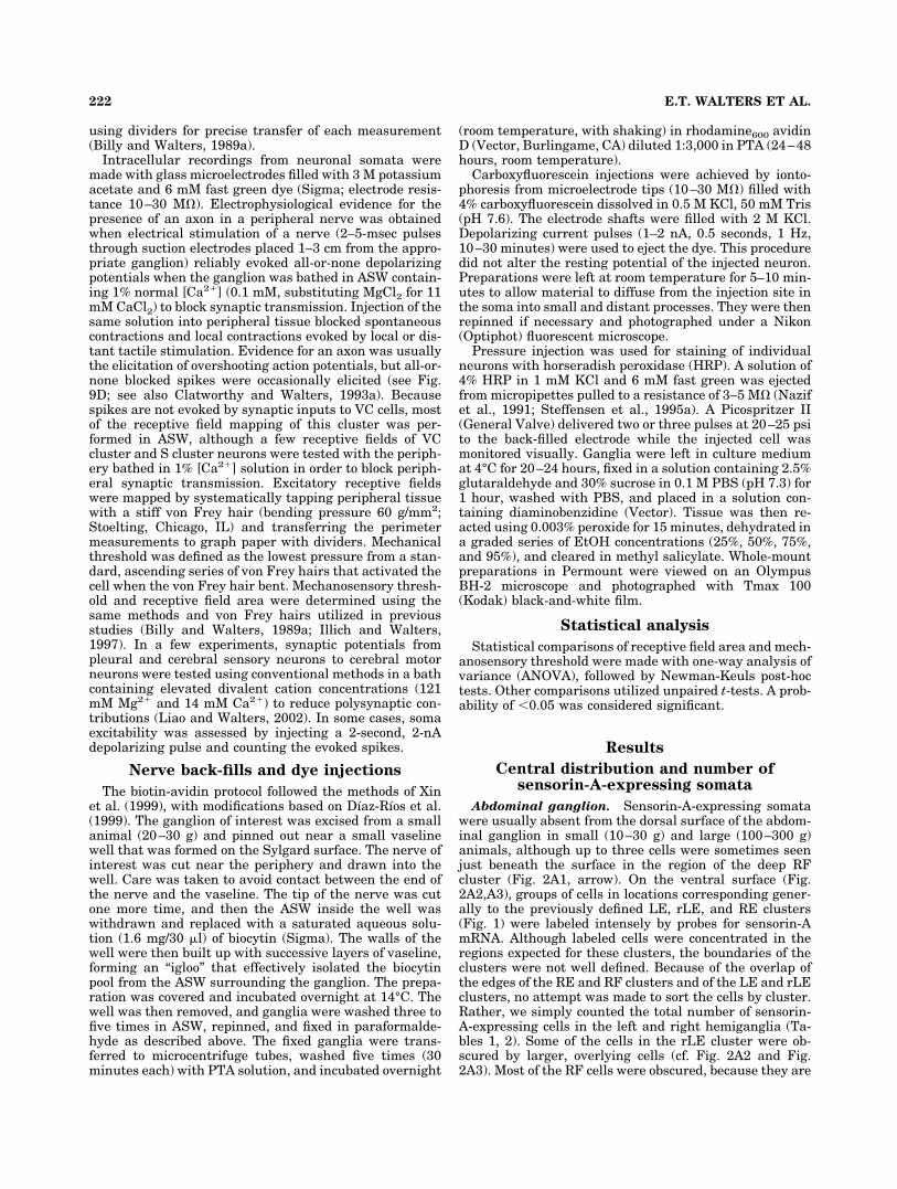
Abdominal ganglion. Sensorin-A-expressing somatawere usually absent from the dorsal surface of the abdom-inal ganglion in small (10–30 g) and large (100–300 g)animals, although up to three cells were sometimes seenjust beneath the surface in the region of the deep RFcluster (Fig. 2A1, arrow). On the ventral surface (Fig.2A2,A3), groups of cells in locations corresponding gener-ally to the previously defined LE, rLE, and RE clusters(Fig. 1) were labeled intensely by probes for sensorin-AmRNA. Although labeled cells were concentrated in theregions expected for these clusters, the boundaries of theclusters were not well defined. Because of the overlap ofthe edges of the RE and RF clusters and of the LE and rLEclusters, no attempt was made to sort the cells by cluster.Rather, we simply counted the total number of sensorin-A-expressing cells in the left and right hemiganglia (Ta-bles 1, 2). Some of the cells in the rLE cluster were ob-scured by larger, overlying cells (cf. Fig. 2A2 and Fig.2A3). Most of the RF cells were obscured, because they are
222 E.T. WALTERS ET AL.

located deep in the right hemiganglion, close to both theventral and the dorsal sides of the commissure (Byrne,1980). The deep cells were revealed by pushing the largeroverlying somata aside or by adjusting the plane of focusin cleared preparations (not shown). Because of the possi-
bilities that some of the deep cells were missed and that afew cells could have been lost during desheathing or pro-cessing of the ganglia, a conservative estimate of the num-ber of sensorin-A-expressing cells in the ganglion is thegreatest number of cells counted in any of the prepara-
Fig. 2. Somata in abdominal, pleural, and pedal ganglia labeled byin situ hybridization with antisense probe to sensorin-A mRNA.A1: Dorsal surface of abdominal ganglion from a 100-g animal. Thearrow indicates labeling of three neurons in the RF cluster. These arepartially obscured by overlying unlabeled cells. The giant cell, R2, bagcells (bc), and the branchial nerve (br) are shown for reference.A2: Ventral surface of the same ganglion. The siphon nerve (si) is alsoshown for reference. Although many superficial cells are labeled withthe antisense probe, several are obscured by unlabeled cells. A3: Thesame view of the same ganglion after clearing and embedding toreveal the rLE cluster. The RE, RF, LE, and rLE mechanosensoryclusters are labeled, and several previously obscured cells, includingmost of the rLE cluster, can now be seen. B1: Relatively deep focus on
a whole-mount preparation of the right pleural and pedal gangliafrom a 30-g animal after clearing and embedding. This revealedlabeled cells throughout the VC cluster and a smaller, more lightlylabeled cell in the pedal ganglion (arrow). Note the intense labeling ofneurites in the pleural–pedal connective (pl-p) and the neuropilthroughout the pedal ganglion. B2: Right pleural and pedal ganglia ina 100-g animal. The cerebral–pleural connective (c-pl) is shown forreference. In addition to the VC cluster, 11 small cells are labeled inthe pedal ganglion (arrows). B3: Two views of the left pleural gan-glion, showing opposite edges of the VC cluster, which curves aroundthe ganglion. Scale bar in A1 � 200 �m (applies to A1–A3); 200 �m inB1–B3.
223SOMATOTOPIC ORGANIZATION OF APLYSIA SENSORY NEURONS

tions examined (Table 1, N � 6–12 for different ganglia;see also Table 2). No qualitative differences between smalland large animals were noted in the positions or arrange-ment of any of the sensorin-A-expressing cells in the ab-dominal ganglion (or in the other ganglia). The averagenumber of sensorin-A-expressing somata in the abdominalganglion was not significantly different in small and largeanimals (Table 2). Soma size, as measured by maximumdiameter, showed no apparent differences among sensoryclusters in the abdominal ganglion. Soma size rangedbetween 20 and 50 �m in small animals and between 30and 60 �m in larger animals. In cleared preparations,sensorin-A labeling could be seen in the neuropil and rootsof the siphon and branchial nerves (not shown), indicatingthat sensorin mRNA is present in axons as well as somata(see also Brunet et al., 1991).
Pedal ganglia. We failed to find sizable clusters ofsensorin-A-expressing somata in pedal ganglia from largeor small animals (Fig. 2B), which is consistent with thelack of previous reports of mechanosensory neurons in thepedal ganglia. Labeled fibers were found, however,throughout much of the pedal ganglion. These fibers ap-peared to come largely from cells in the pleural ganglion(see below). We observed, in 20 of 22 pedal ganglia exam-ined, a small and highly variable number (2–11) of labeledsomata (arrows in Fig. 2B1,B2; see Tables 1, 2). No sig-nificant difference in the number of sensorin-A-expressingneurons was seen between smaller and larger animals.Most of these cells were found near the root of the pleural–pedal (pl-p) connective (Fig. 2B2) and were about half thediameter (20–40 �m) of the nearby sensory neuron so-mata in the pleural VC cluster. We recorded intracellu-larly from three small cells in this region of the pedalganglion, in a 150-g animal, that had the same visualappearance as the VC sensory neurons. The somata ofthese cells displayed electrophysiological properties char-acteristic of those of the VC neurons on the other side of
the connective, i.e., lack of spontaneous action potentialsor postsynaptic potentials and clear spike accommodationin response to long depolarizing current pulses. Althoughwe have not yet looked for peripheral axons or receptivefields of these cells, the general similarity of the smallsensorin-A-expressing neurons in each pedal ganglion toother sensorin-A-expressing neurons suggests that theymay also be mechanosensory neurons.
Pleural ganglia. As predicted by earlier descriptionsof the pleural VC clusters, we found that these bilateralclusters have the largest number of sensorin-A-expressingcells and that these cells are tightly packed into a strik-ingly uniform cluster in each pleural ganglion (Fig. 2B),which appeared quite similar in small and large animals.Nevertheless, every ganglion had a few cells separatedfrom the main cluster. Because each VC cluster curvesaround the pleural ganglion, two images were needed todisplay the entire cluster (Fig. 2B3). The average numberof sensorin-A-expressing somata in each pleural ganglion(Table 1) was similar to the estimate of about 200 mech-anosensory neurons that was originally made by countingcells that had been defined as VC cells on the basis ofshared visual appearance and electrophysiological proper-ties (Walters et al., 1983). Interestingly, the number ofsensorin-A-expressing somata increased significantly inlarger animals (Table 2). Soma size ranged between 30and 60 �m in small animals and between 40 and 80 �m inlarger animals. The smallest somata tended to be in theanterolateral region of the cluster, close to the cerebral–pleural connective. Because of the distinctive size anddense packing of cells in the VC cluster, we were able toobserve that all of the cells in the cluster exhibited intenselabeling with the antisense probe for sensorin-A. We ob-served extensive labeling of fibers in the pl-p connective,as was reported by Brunet et al. (1991), and these ex-tended into the pedal ganglion (Fig. 2B1,B2).
Cerebral ganglion. Most of the cells expressingsensorin-A mRNA were located in the previously de-scribed J and K clusters on the ventral side of each hemi-ganglion (Fig. 3A1). As with cells in the pleural VC sen-sory clusters, the J and K cells were relatively small(30–60 �m soma diameter) and packed uniformly withineach cluster, which appeared quite similar in small andlarge animals. In addition, a number of very small cells(�20 �m diameter) were distributed on the dorsal andventral surfaces, near the middle of each cerebral hemi-ganglion. On the dorsal surface (Fig. 3A2), these cellsappeared to be part of the D clusters (Jahan-Parwar andFredman, 1976). Caudal to these cells on the dorsal sur-face of each hemiganglion was a single stained cell (60–70�m diameter) with a prominently stained initial axonsegment. The cells within the J and K clusters consis-tently exhibited more intense labeling than the smallercells located outside of these clusters. As found in thepleural ganglia, the number of sensorin-A-expressing so-mata in the cerebral ganglion increased significantly inlarger animals (Table 2). Average soma diameter was alsoabout 50% larger in larger animals. Labeling of sensorin-AmRNA was also found in the neuropil and roots of theconnectives (Fig. 3A1) and roots of cerebral nerves (notshown).
Buccal ganglia. Large clusters of sensorin-A-expressing somata, corresponding to the S clusters ofFiore and Meunier (1975), were found to curve over thedorsal surface of each hemiganglion, extending from the
TABLE 1. Highest Counts of Sensorin-A-Expressing Somata
Ganglion Side Number
Abdominal L 85R 66
Pedal L 11R 9
Pleural L 231R 223
Cerebral L 126R 122
Buccal L 176R 152
Total 1,201
TABLE 2. Increase in Number of Sensorin-A-Expressing Somata in LargerAnimals
Ganglia
Small animals Large animals1
P3Mean SEM N2 Mean SEM N
Abdominal 94 8 7 79 4 5 0.190Pedal (L � R) 9 1 7 9 2 6 0.870Pleural (L � R) 355 9 3 438 4 3 0.001*Cerebral 203 8 6 234 8 3 0.045*Buccal 246 7 6 294 14 4 0.008*
1Small animals weighed 10–30 g and large animals 100–300 g.2N indicates number of animals examined for each type of ganglion.3P indicates probability of a significant difference between soma numbers from largeand small animals (unpaired t-test).*P � 0.05.
224 E.T. WALTERS ET AL.

caudal to the rostral side (Fig. 3B1). Compared with clus-ters in the other ganglia, the buccal clusters showed con-siderable heterogeneity in soma size and sensorin mRNAlabeling intensity (Fig. 3B2). We failed to see in manyanimals a distinct division between subclusters thatwould correspond to the S1 and S2 dichotomy, so we simplyuse the term S cluster to describe the core group of small(20–30 �m diameter in small animals), intensely labeledsomata. Ventral to each S cluster was a group of 15–20larger (40–60 �m) sensorin-A-expressing neurons, addi-tional small neurons with a sparser distribution, and adistinctive larger ventral cell (arrow in Fig. 3B1) that wasseparated from the others. As found in the pleural andcerebral ganglia, the number of sensorin-A-expressing so-mata in the buccal ganglia increased significantly inlarger animals (Table 2). Average soma size was also
�50% larger in larger animals. In cleared preparations,labeling for sensorin-A could be seen in fibers in the neu-ropil, buccal commissure, and roots of the buccal nerves(see the buccal commissure and radula nerve in Fig. 3B1).
Periphery. No sensorin-A-expressing neuronal so-mata were found outside the central ganglia in adult an-imals. In juvenile animals (�2 g), two or three sensorin-A-expressing neurons have been observed in mantle tissue(Moroz and Bodnarova, unpublished observations), whichmight be a site of neurogenesis (McAllister et al., 1983;Hickmott and Carew, 1991).
Peripheral targets of the sensorin-A-expressing clusters
Detailed descriptions of the receptive field propertiesand peripheral axon distributions of sensorin-A-
Fig. 3. Sensorin-A-expressing neurons in the cerebral and buccalganglia. A1: Ventral view of cerebral ganglion in a 100-g animal,showing intense labeling of J and K mechanosensory clusters. Land-marks are the anterior tentacular nerve (c2), and cerebral–pedal (c-p)and c-pl connectives. Note the labeled fibers in the left c-pl connective(red arrows). A2: Dorsal view of cerebral ganglion. Additional land-marks are the giant metacerebral cells (MCC). The red arrows indi-cate a pair of large labeled neurons with prominent axons that areinvariably found in these locations. Anterior to these are small cellslabeled within the D clusters (yellow arrows). Labeled cells near the
roots of the c-pl connectives are from the J clusters. B1: Rostral andcaudal views of the buccal ganglia from a 25-g animal, showinglabeling of the S clusters. Red arrows (bottom) indicate larger cellsreliably labeled on the ventral-caudal surface of the ganglia. Noticethe variation in labeling intensity and cell size. Yellow arrow (top)indicates labeled axons in the radula nerve. B2: Nomarski high-resolution images from buccal ganglion illustrating the variation inlabeling intensity and cell size in a 30-g animal. Scale bars � 500 �min A1,A2; 200 �m in B1; 50 �m in B2 (left); 25 �m in B2 (right).
225SOMATOTOPIC ORGANIZATION OF APLYSIA SENSORY NEURONS

expressing sensory clusters in the abdominal ganglion andcerebral ganglia have already been published (Byrne etal., 1974; Rosen et al., 1979, 1982; Byrne, 1980; Dubuc andCastellucci, 1991; Illich and Walters, 1997). For the ab-dominal LE, RE, and rLE clusters, we have confirmed thegeneral locations and typical sizes of receptive fields thatwere previously reported on the siphon and mantle (datanot shown). The receptive field locations, peripheral axondistributions, and newly described physiological proper-ties found in some of the other sensorin-A-expressing clus-ters are described below.
Receptive fields of pleural VC clusters. When thepresent studies began, it was known that the mantle or-gans, head, and buccal mass are innervated by sensorin-A-expressing sensory clusters in the abdominal, cerebral,and buccal ganglia, respectively. The remaining sensorin-A-expressing clusters, the two pleural VC clusters, hadbeen shown to have receptive fields on the tail and poste-rior parapodia (Walters et al., 1983), but only a small partof each VC cluster had been mapped. When combined, allthe documented receptive fields of sensorin-A-expressingneurons left much of the foot, parapodia, midbody, andposterior neck uncovered. We predicted that the remain-ing parts of the body surface are covered by receptivefields of VC mechanosensory neurons whose receptivefields had not yet been examined.
To test this hypothesis, we investigated the receptivefields of 287 VC cluster neurons in 25 semiintact prepa-rations from animals weighing 115–275 g (mean 185 g).Figure 4 shows the receptive fields found in six represen-tative experiments. Cells throughout the VC clustershowed electrophysiological properties that appeared in-distinguishable from those in the previously describedsubset of VC sensory neurons that innervates the tail(Walters et al., 1983): resting potentials between –40 and–55 mV, lack of spontaneous activity or synaptic input,bursts of action potentials lacking prepotentials duringreceptive field stimulation (Fig. 4, inset), action potentialamplitudes between –85 and –105 mV (during good im-palements), lack of excitatory postsynaptic potentials (EP-SPs) during receptive field stimulation, and (sometimes)slow hyperpolarizing responses to stimulation outside theexcitatory receptive field. Excitatory receptive fields werefound over the entire surface of the body ipsilateral to eachVC cluster (Fig. 4). As noted previously for receptive fieldson the tail (Billy and Walters, 1989a), very few receptivefields in other regions extended across the midline. Thehighest incidence of crossover was on the ventral surfaceof the foot (Fig. 4B2), where 16 of 110 midline fields (15%)extended over to the contralateral side. In contrast, only 1of 24 (4%) crossed over on the dorsal surface of the head,and 0 of 26 crossed over on the neck.
Receptive field sizes in this study ranged from 0.1 to 12cm2. Although receptive field size varied considerablywithin each part of the body, receptive fields on the ten-tacles, head, and anterior foot (propodium) were signifi-cantly smaller than those on more posterior parts of thebody (Fig. 5A; one-way ANOVA, P � 0.0001, with P � 0.05for each post-hoc comparison using the Newman-Keulstest). No significant differences among regions were foundin mechanosensory thresholds (Fig. 5B); the medianthreshold in all regions was 35 g/mm2, except for thetentacles, where the median threshold was 45 g/mm2.
Somatotopic organization of the pleural VC clusters.
It was originally noted by Walters et al. (1983) that the VCcluster is organized at least partially in a somatotopicfashion, because all the somata of neurons with receptivefields on the tail are grouped on the medial side of thecluster. In the receptive field study just described (N �287 VC cells in 25 animals), we confirmed this localizationof the somata of tail sensory neurons to the medial side ofthe cluster and found that cells with receptive fields on thehead were restricted to the opposite (anterolateral) side ofthe cluster. Cells with receptive fields in the middle of thebody were generally found in the central part of the VCcluster. Thus, each VC cluster (Fig. 6B) forms a crudesomatotopic map of the ipsilateral surface of the body (Fig.6A) or, in effect, a “sensory aplunculus” (see Discussion).
Additional information about the somatotopic organiza-tion of the VC sensory neurons came from investigatingthe distribution of these cells’ axons in peripheral nerves.Previous studies have shown the approximate sensory andmotor fields (which are largely coextensive) of peripheralnerves in Aplysia (Jahan-Parwar and Fredman, 1976,1978; Kandel, 1979; Walters et al., 1983; Dulin et al.,1995), and these provide a guide to the general regionswhere receptive fields of sensory neurons with axons inspecific nerves would be expected (Fig. 7A). Note that thissimplified map does not show the marked overlap thatexists at the boundaries of the fields, which is most exten-sive in the head and neck.
We used electrical stimulation of peripheral nerves,combined with intracellular recording from VC somata(Fig. 7, inset), to relate peripheral axon distribution tosoma location. Figure 7B shows the position of the VCcluster when the left pleural ganglion is pinned out in ourstandard experimental conformation. In this position, themedial side (containing tail sensory neurons) is completelyvisible. However, because the VC cluster winds aroundthe lateral side of the ganglion (Fig. 2B3), the anterolat-eral edge of the cluster is hidden when the medial edge isvisible. The inset shows the complete cluster as it wouldappear if it were unwound from the ganglion. Results froma single experiment in which 102 sensory neurons weresampled while brief stimuli were delivered to ipsilateralpedal nerves p9, p8, p1, and the cerebral–pleural connec-tive (c-pl) revealed that somata with apparent axons innerve p9 were largely on the medial side of the VC cluster,that somata with axons in nerve p1 or the c-pl connectivewere largely in the anterolateral region, and that somatawith axons in nerve p8 were primarily in the centralregion of the cluster (Fig. 7C1). Figure 7C2 summarizes 22experiments, showing the approximate locations of all thesomata responding with action potentials to stimulation ofpedal nerves p1, p4, p5, p6, p7, p8, p9 (excluding theanterior branch, p9a, which innervates the posteriorparapodium), p9a, or the c-pl connective. Somata in theVC cluster are organized so that cells with axons project-ing toward the anterior of the animal are primarily in theanterolateral region of the cluster, cells with axons pro-jecting to the middle of the animal are primarily in thecentral region of the cluster, and cells with axons project-ing to the posterior of the animal are primarily in themedial region of the cluster. Part of this study has beendescribed in abstract form (Billy and Walters, 1987).
The maximum numbers of VC cells found in a singlepreparation having apparent axons in specific nerves areshown in Table 3. As indicated by action potentials evoked
226 E.T. WALTERS ET AL.

by stimulating different nerves, dual axons were uncom-mon, being found in only 3% (17 of 550) of cells tested. Thehighest incidence of dual axons occurred in the posteriorand anterior branches of p9 (p9p and p9a). In the twoexperiments in which the two major branches of this nerve
were stimulated together, 5 of 63 cells (8%) had axons inboth branches. The next highest incidence was in p6 andp7, where 6 of 114 cells tested in seven experiments haddual axons (5%). Dual axons were also implicated in p8and p9 (twice), p7 and p8 (twice), p6 and p8 (once), and,
Fig. 4. Examples of receptive fields of mechanosensory neurons inthe pleural VC and cerebral J clusters. For reference, schematicdrawings of dorsal and ventral views of intact Aplysia are presentedin the left-most panels. The inset shows an example of action poten-tial discharge evoked by a brief (�0.5-second) poke of the left parapo-dium by the stiff (60 g/mm2) von Frey hair used to map the receptivefields. A1: Dorsal fields of cells in left VC cluster. The anterior half ofthe animal was discarded and Lp7, Lp8, and Lp9 nerves were intact.A2: Dorsal fields of cells in right and left VC clusters. Except for theexcised parapodia and incisions above and below the circumesopha-
geal ring ganglia, all of the body was intact. No fields were found onthe mantle or siphon. Tonic contractions of the base of each cutparapodium obscured the mantle floor, which was not tested in thisanimal. A3: Dorsal fields of cells in the left VC cluster on the head andneck. B1: Ventral fields of cells in the left and right VC clusters thatfail to cross the midline on the foot. B2: Ventral fields (red) that crossthe midline of the foot. No VC fields in any animal were completelycontralateral. B3: Ventral fields of VC and J cells (see red lines) fromleft pleural and cerebral ganglia. Scale bar � 4 cm.
227SOMATOTOPIC ORGANIZATION OF APLYSIA SENSORY NEURONS

surprisingly, p1 and p9 (once). We found in separate ex-periments (see below) that VC cells with axons in nerve p1often have an axon in the c-pl connective as well.
Back-fill experiments provided complementary evidencefor the somatotopic organization of the VC cluster (Fig. 8).These experiments and the nerve shock experimentsyielded similar estimates for the numbers of VC neuronswith axons in the nerves that were tested with both meth-ods: p9, p8, p1, and the c-pl connective (Table 3). Nerveback-fills also provided clear documentation of the group-ing of somata having axons in different major nerves intodifferent regions of the VC cluster. Figure 8A shows cellswith axons in the posterior pedal nerve (p9) concentratedin the medial portion of the VC cluster, whereas cells withaxons in the anterior pedal nerve (p1) or in the c-pl con-nective or anterior tentacular nerve (c2) are concentratedin the anterolateral part of the cluster (Fig. 8C–E), andcells with axons in the middle pedal nerve (p8) are con-centrated in the central region of the cluster (Fig. 8B).Similar findings were described in a preliminary report onnickel-lysine and cobalt back-fills of nerves p7, p8, and p9and the c-pl connective (Zhang et al., 1993).
Receptive fields of the cerebral sensory clusters.
We have extended previous studies by showing that re-ceptive fields of some J cells are located on the ipsilateralrhinophore and propodium, in addition to the previouslyreported fields on the anterior tentacle, lip region, anddorsal surface of the head (Rosen et al., 1979, 1982). In asample of 11 J cells (in three preparations), five receptivefields were on the tentacle, two were on the lips near themidline, two were on the propodium, and two were on therhinophore (Fig. 4B3). All receptive fields were ipsilateral
to the sampled cells. Because the preparations werepinned ventral side up, we were not able to sample recep-tive fields on the dorsal surface of the head. No receptivefields were found in the middle of the body, even thoughthe innervation of the parapodia and entire foot remainedintact. We did not quantify receptive field sizes, but thesizes appeared similar to those previously described for Jand K cells on the tentacles and lips (Rosen et al., 1979,1982). The median mechanosensory threshold of the sam-pled J cells was 35 g/mm2, with a range of 15–45 g/mm2.This median threshold was the same as that found previ-ously with the same calibrated von Frey hairs for tail(Billy and Walters, 1989a) and siphon (Illich and Walters,1997) sensory neurons.
Intersecting functions of cerebral J/K cells and pleu-
ral VC cells. Back-filling the anterior tentacular nerve(c2) stains somata in both the J/K and the VC clusters(Fig. 8E), and the excitatory receptive fields of cells in theJ/K clusters and cells in the anterolateral region of theipsilateral VC cluster show extensive overlap on the ten-tacles and anterior foot (Fig. 4). Moreover, receptive fieldsof J/K cells and VC cells in this general region display thesame range of mechanosensory thresholds. These obser-vations suggest that many of the cerebral and pleuralsensory neurons share one or more common functions. Alikely function is to sense threatening or noxious stimuliand trigger appropriate defensive responses, such as thehead-withdrawal reflex (Walters and Erickson, 1986;Teyke et al., 1989). This function is supported by ourobservation that VC cells with receptive fields on theanterior part of the body, such as the anterior foot (Fig.
Fig. 5. Areas and thresholds of VC cell receptive fields in differentregions of the body. A: Significantly smaller receptive fields in thehead/propodium region and anterior tentacles than in more posteriorregions (*P � 0.05). B: Lack of significant differences in mechanosen-sory threshold in different regions. In each case the central lineindicates the median, the box the interquartile range (i.e., betweenthe 25th and 75th percentiles), and the bars the total range.
Fig. 6. Somatotopic representation of mechanosensory receptivefields in the VC cluster. A: Drawing of the right side of an Aplysiafacing the right. B: A micrograph of the right VC cluster as revealedby in situ hybridization of a probe for sensorin-A mRNA. The regionswithin the cluster having somata with receptive fields on the head,midbody, and tail are indicated. The anterior side of the cluster is atthe top of the image, and the medial and lateral sides are indicated(see Figs. 2, 7). Scale bar � 100 �m.
228 E.T. WALTERS ET AL.

Fig. 7. Relationship between positions of the somata in the VCcluster and axonal projections into peripheral nerves. A: Simplifiedmap of the major sensory fields of left pedal nerves and a left cerebralnerve, c2 (anterior tentacular nerve). The extensive overlap of differ-ent fields is not indicated on this map. B: Schematic drawing of VCcluster in left pleural ganglion. The entire cluster is representedtwo-dimensionally by unwinding it (left) from the cylindrical tractextending from the pl-p to the c-pl connectives. The right VC cluster
is very similar (not shown). C: The inset shows a spike evoked by a2-msec shock delivered to nerve p9. C1: Positions and relative sizesrecorded in a single experiment of cells in left VC cluster that re-sponded with spikes to stimulation of axons in the indicated nerves.C2: Summary of results from all nerve stimulation experiments (N �22). Dots represent locations (not size of soma) of all cells respondingto stimulation of the indicated nerves in all the experiments.
229SOMATOTOPIC ORGANIZATION OF APLYSIA SENSORY NEURONS

9A) and tentacle (Fig. 9B), evoke EPSPs in the Bn motorneurons, which mediate the head-withdrawal reflex(Teyke et al., 1989). Figure 9C shows an example of anEPSP in a cerebral Bn motor neuron evoked by a singleaction potential in a pleural VC cell having a receptivefield on the propodium. The EPSP appeared to be mono-synaptic, because it was evoked in a solution containing2.2 � normal [Mg2�] and 1.25 � normal [Ca2�]. We foundthat 9 of 10 sensory neurons (in two animals) sampled inthe region of the VC cluster innervating the anterior partof the body had connections to Bn motor neurons, with themean amplitude being 5 mV and the range 2–9 mV. In onecase, three Bn neurons were sampled, and all receivedEPSPs from the same VC cell. This small sample suggeststhat most VC cells with receptive fields on the head con-nect to most Bn neurons, as is true for the J/K cells (Teykeet al., 1989).
Back-filling the c-pl connective in smaller animals(20–30 g) revealed about 20 VC cells with axons projectingto the cerebral ganglion (Fig. 8D). About 12 cells projectedan axon into c2, the anterior tentacular nerve, as revealedby separate back-fills of this nerve (Fig. 8E). Dye fills ofindividual VC cells demonstrated that at least some of theVC cells project axons into both the cerebral ganglion andthe pedal ganglion (Fig. 10). In the three filled cells havingdual axons, the axon projecting into the c-pl connectivewas substantially thinner than the axon projecting intothe pleural–pedal connective. This morphology was con-sistent with electrophysiological responses of the VC cellsthat responded to electrical stimulation of both nerve p1and the c-pl connective. In all five cases, single, brief p1stimuli invariably evoked normal overshooting spikes,whereas, in three of five cases, single, brief stimuli to thec-pl connective evoked small, all-or-none depolarizing po-tentials that had the properties of blocked spikes. Thesmall spikes summed at high frequency to initiate a fullaction potential in the VC cell soma (Fig. 9D). In this samestudy, two VC cells responded to p1 stimulation but not toc-pl connective stimulation, and one VC cell responded totentacle stimulation but not to p1 stimulation. Extendingearlier reports (Rosen et al., 1979; Clatworthy andWalters, 1994), 6 of 10 sampled J/K cells responded withall-or-none action potentials to electrical stimulation ofthe c-pl connective, and three of these also responded to p1stimulation, indicating that some of the J/K cells inner-vate the anterior foot via axons that project via this pedalnerve.
Receptive fields of the buccal S clusters. Of all thesensorin-A-expressing clusters in Aplysia, the least stud-ied have been the S clusters in the buccal ganglia. To ourknowledge, there has only been a single report about thereceptive fields of buccal S cells, in a review article thatstated only that these cells can respond to mechanicalstimulation of “specific muscular or epithelial regions inthe buccal mass” even when synaptic transmission in thetissue is reduced by quadrupling extracellular [Mg2�](Fiore and Geppeti, 1981). Although a comprehensive in-vestigation of the S cells is beyond the scope of this paper,we wished to extend this earlier report by identifyinggeneral regions of the body that have S cell receptive fieldsand by describing basic electrophysiological properties ofthe S cells.
Dye fills of S cell somata (not shown) and back-fills ofnerves and connectives indicated that many of these cellsproject axons into buccal nerves, but only a few projectinto the esophageal nerve (approximately six cells) andnone into the cerebral–buccal connective. Back-fills of theventral buccal nerve (b1; Fig. 11A) revealed 14–16 S cellsomata in the ipsilateral cluster and two in the contralat-eral cluster. Back-fills of the lateral buccal nerve (b2; Fig.11B) revealed 45–55 S cell somata in the ipsilateral clus-ter and six to eight in the contralateral cluster. In con-trast, the dorsal buccal nerve (b3; Fig. 11C) revealed apreponderance of S cell axons from contralateral somata:there were 5 to 10 filled cells in the rostral half of thecontralateral S cluster vs. two cells in the rostral half ofthe ipsilateral cluster and 25–30 cells in the caudal half ofthe contralateral cluster vs. five cells in the caudal half ofthe ipsilateral cluster. Back-fills of the radula nerve re-vealed the largest number of S cell axons, filling approxi-mately 120 S cell somata, evenly divided between the leftand the right hemiganglia, but with a larger number ofsomata on the rostral than caudal side of the cluster. Inour clearest back-fill of the radula nerve (Fig. 11D), therewere 36 filled cells in the rostral half of the left S cluster,38 cells in the rostral half of the right cluster, 24 cells inthe caudal half of the left cluster, and 20 cells in thecaudal half of the right cluster. These observations agreegenerally with previous results obtained with cobalt back-fills (Scott et al., 1991) and with the original claim that theS cells “send axons to the periphery through all the buccalnerves” (Fiore and Geppeti, 1981). This suggests that theS cells have receptive fields on or in most of the buccalmass and perhaps in the perioral area and part of theesophagus.
All S cells sampled with intracellular electrodes (N � 21cells in three animals) were silent in the absence of pe-ripheral stimulation and showed no evidence of spontane-ous synaptic input or of evoked EPSPs during stimulationof peripheral receptive fields (Fig. 12A,B). Basic electro-physiological properties were assessed in the nine cellsshowing the best impalements, as judged by the mostnegative resting potentials and largest action potentialamplitudes. Mean resting potential was –51.1 � 1.8 mV.Mean action potential amplitude was 98.6 � 1.6 mV. Bothof these values are similar to those of other sensorin-A-expressing sensory neurons. However, three propertieswere different. Compared with VC, J/K, or LE cells (see,e.g., Walters et al., 1991; Clatworthy et al., 1994; Clatwor-thy and Walters, 1994; Illich and Walters, 1997), meanspike afterhyperpolarization (15.9 � 0.7 mV) was aboutthree times larger, mean spike duration (6.4 � 0.4 msec at
TABLE 3. Axon Distribution of Pleural VC Cluster
Nerve Target
Maximum number of cells with axons1
Shock N (preps) Back-fill N (preps)
p9 Tail 28 (14) 30 (4)p9a Posterior PP2 15 (3)p8 Midfoot 30 (13) 30 (2)p7 Mid-PP 20 (11)p6 Anterior PP 15 (12)p5 Posterior neck 7 (3)p4 Anterior neck 9 (2)p1 Anterior foot 25 (10) 35 (4)c-pl Head 22 (2) 20 (2)c2 Head 12 (1)
1Presence of an axon is defined by the occurrence of an action potential in the somaevoked by electric shock delivered to the indicated nerve, or by filling of the soma afterdelivery of dye to the indicated nerve. The maximum number of cells corresponds to thegreatest number with axons observed in any one of the indicated number of prepara-tions (preps).2Parapodia.
230 E.T. WALTERS ET AL.
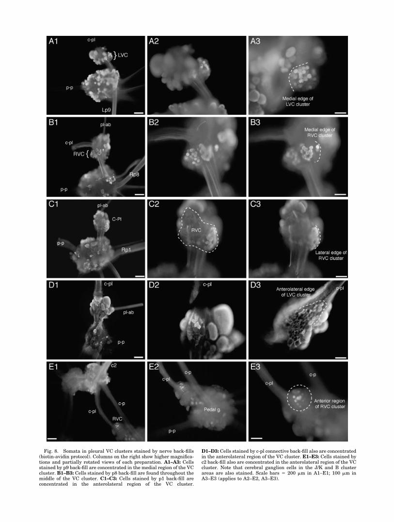
Fig. 8. Somata in pleural VC clusters stained by nerve back-fills(biotin-avidin protocol). Columns on the right show higher magnifica-tions and partially rotated views of each preparation. A1–A3: Cellsstained by p9 back-fill are concentrated in the medial region of the VCcluster. B1–B3: Cells stained by p8 back-fill are found throughout themiddle of the VC cluster. C1–C3: Cells stained by p1 back-fill areconcentrated in the anterolateral region of the VC cluster.
D1–D3: Cells stained by c-pl connective back-fill also are concentratedin the anterolateral region of the VC cluster. E1–E3: Cells stained byc2 back-fill also are concentrated in the anterolateral region of the VCcluster. Note that cerebral ganglion cells in the J/K and B clusterareas are also stained. Scale bars � 200 �m in A1–E1; 100 �m inA3–E3 (applies to A2–E2, A3–E3).

Fig. 9. Evidence for shared functions of cerebral J cells and an-terolateral pleural VC cells. A: Simultaneous activation of a J sensoryneuron (SN) and VC sensory neuron by brief von Frey hair stimula-tion of the ipsilateral tentacle. B: Activation of an anterolateral VCcell by von Frey stimulation of the ipsilateral anterior foot (propo-dium). C: Probable monosynaptic EPSP produced in ipsilateral Bmotor neuron (MN) by intracellular activation of anterolateral VCcell. Elevation of divalent cation concentrations minimized possible
activation of interneurons. D: Evidence for dual axons with differentconduction properties in anterolateral VC cell. Brief shock of anteriorpedal nerve (p1) activates an overshooting spike. Brief shock to c-plconnective evokes an 8-mV, all-or-none depolarization. Longer shockto the connective evokes two small depolarizations, which sum toactivate an overshooting spike in the soma (see text). Recordings weremade in saline containing elevated divalent cation concentrations.
Fig. 10. Morphological evidence for dual axons in anterolateral VCsensory neurons. A: Low-magnification view of cells injected withhorseradish peroxidase (HRP) in the middle and anterolateral regionsof the left VC cluster. Most axons project to the right, into the pl-pconnective. Arrowheads indicate two axons projecting to the left,toward the c-pl connective. B: Close-up view showing a thin process(arrowhead) projecting toward the c-pl connective, whereas the major
axon projects toward the pl-p connective. Direct inspection throughthe microscope (adjusting the focal plane) confirmed that the thinprocess came from the large axon and traveled into the c-pl connec-tive. C: Example from another preparation showing a thin processprojecting toward the c-pl connective and a thick process (out of thefocal plane) projecting toward the pl-p connective. Scale bar � 100 �min A; 25 �m in C (applies to B,C).
232 E.T. WALTERS ET AL.
the base of the spike) was 50–100% longer, and the cellsshowed remarkably little spike accommodation duringprolonged depolarizations (Fig. 12A,C). Our standard2-second, 2-nA test pulse evoked 38.0 � 3.9 spikes, and, inevery case, spikes were generated until the end of the2-second pulse. No afterdischarge was observed followingthe intracellular test pulse.
Receptive fields were sampled using three semiintactpreparations, each of which maintained most buccal, ce-rebral, and anterior pedal nerve connections to the buccalmass, esophagus, oral veil/tentacle region, and anteriorfoot. In the first preparation, the buccal mass was leftintact (precluding stimulation of internal structures, suchas the radula and odontophore), and the radula nerve wascut. Six of nine S cells recorded were found to have recep-
tive fields, all ipsilateral: five on the muscular outer sur-face of the buccal mass and one on the perioral zone andlip, near the propodium. The mechanosensory thresholdsof the cells innervating the buccal mass ranged between30 and 65 g/mm2. The threshold of the cell innervating theperioral zone/lip (Fig. 12A–C) was 20 g/mm2. No responseswere evoked by concentrated NaCl solutions or NaCl crys-tals dropped on the skin. In the second preparation, amidline incision was made in the caudal/dorsal surface ofthe buccal mass to expose its internal structures, and theradula nerve remained intact. Three of five recorded cellshad receptive fields: two on the external surface of theradula (with thresholds of 45 and 60 g/mm2), and one onthe perioral zone/lip (threshold 20 g/mm2). All S cellsdisplayed responses that were graded with the intensity of
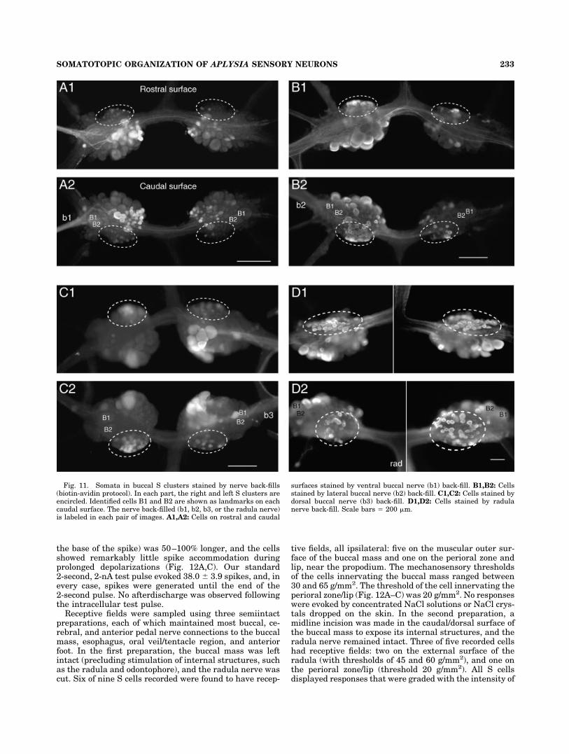
Fig. 11. Somata in buccal S clusters stained by nerve back-fills(biotin-avidin protocol). In each part, the right and left S clusters areencircled. Identified cells B1 and B2 are shown as landmarks on eachcaudal surface. The nerve back-filled (b1, b2, b3, or the radula nerve)is labeled in each pair of images. A1,A2: Cells on rostral and caudal
surfaces stained by ventral buccal nerve (b1) back-fill. B1,B2: Cellsstained by lateral buccal nerve (b2) back-fill. C1,C2: Cells stained bydorsal buccal nerve (b3) back-fill. D1,D2: Cells stained by radulanerve back-fill. Scale bars � 200 �m.
233SOMATOTOPIC ORGANIZATION OF APLYSIA SENSORY NEURONS
the mechanical stimulus, and strong mechanical stimulioften evoked an afterdischarge that could persist for sev-eral seconds after stimulus offset (Fig. 12B). A given pres-sure evoked more spikes in the cells innervating the peri-oral zone/lip than in cells innervating the radula (Fig.12B,D). Except at the lowest stimulus pressures, mech-anosensory responses (like the responses to soma stimu-lation) showed very little accommodation (Fig. 12B). Inthe third preparation, a midline incision was made in theventral surface of the buccal mass. Two receptive fieldswere found on the lumenal surface of the wall of the buccalcavity adjacent to the radula (thresholds 35 g/mm2). Onewas found in the perioral zone. This cell initially had athreshold between 25 and 30 g/mm2, but, after repeatedtesting, the threshold fell to 15 g/mm2, and the cell thenfired sporadically for several minutes in response to brieftaps at this lower pressure. This suggests that the S cells,like the VC cells and LE cells (Billy et al., 1989; Illich andWalters, 1997), can exhibit peripheral sensitization. Re-
ceptive field sizes were not measured systematically, but,in both preparations, receptive fields appeared smallerthan the average size of those of cerebral J/K cells orpleural VC cells innervating the head. The responses toperioral zone/lip stimulation persisted after the cerebral–buccal connectives were transected, indicating that the Scell axons traveled in a buccal nerve. Recordings from twoof these cells showed that mechanosensory responses per-sisted after the receptive field was injected with salinecontaining 2 � normal [Mg2�] and 0.1 � normal [Ca2�],indicating that the responses do not require peripheralchemical synaptic transmission. Although this blocked allbehavioral indications of local synaptic transmission (seeMaterials and Methods), we cannot exclude the possibilitythat mechanical stimulation activates the peripheral ter-minals of the S cells indirectly, by electrical synapses orhighly encapsulated chemical synapses from other cells.
Discussion
Number and distribution of sensorin-A-expressing neurons
In adult Aplysia, all of the somata labeled by sensorin-Aantisense probes in our whole-mount preparations were incentral ganglia; none was in peripheral ganglia or othertissues. The labeled somata correspond to previously de-scribed mechanosensory clusters in the abdominal, pleu-ral, cerebral, and buccal ganglia. This agrees with obser-vations of sensorin-A expression in serial sections of theganglia by Brunet et al. (1991). Our counts indicate thatthe total number of sensorin-A-expressing neurons in thebody is about 1,200 in reproductively mature animals(100–300 g) and 1,000 in small animals (10–30 g). Thus,sensorin-A-expressing neurons represent 5–10% of all theneurons in the CNS (Cash and Carew, 1989; Kandel,2001). The small animals we examined probably includedyoung adults and individuals transitioning from the lastjuvenile stage to adulthood (Kriegstein, 1977b; Cash andCarew, 1989). Significant increases in the number ofsensorin-A-expressing cells with animal size were seen inthe pleural, cerebral, and buccal ganglia but not in theabdominal or pedal ganglia (Table 2).
An interesting question concerns the source of the ad-ditional sensorin-A-expressing neurons counted in thelarger animals. One possibility is that the numbers insmall and large animals are actually the same but that wefound fewer sensorin-A-expressing cells in smaller ani-mals because accidental cell loss during surgicaldesheathing was greater in these animals or because thelabeled cells were more likely to be obscured by overlyingcells in smaller animals. Neither of these explanationsseems likely to us, but we cannot rule them out. A secondpossibility is that some cells in these mechanosensoryclusters begin to express sensorin-A at detectable levelsonly after the animal has reached a relatively large size oradvanced stage of adult development. This would be con-sistent with indirect observations suggesting that theadult complement of sensory neurons may be largely com-plete by the early adult stage (Marcus et al., 1988; Cashand Carew, 1989). The wide range of labeling intensitiesobserved in some clusters (especially the buccal clusters)might reflect different stages of development of sensorin-Aexpression in different neurons. A third possibility is thatnew sensorin-A-expressing neurons continue to be gener-
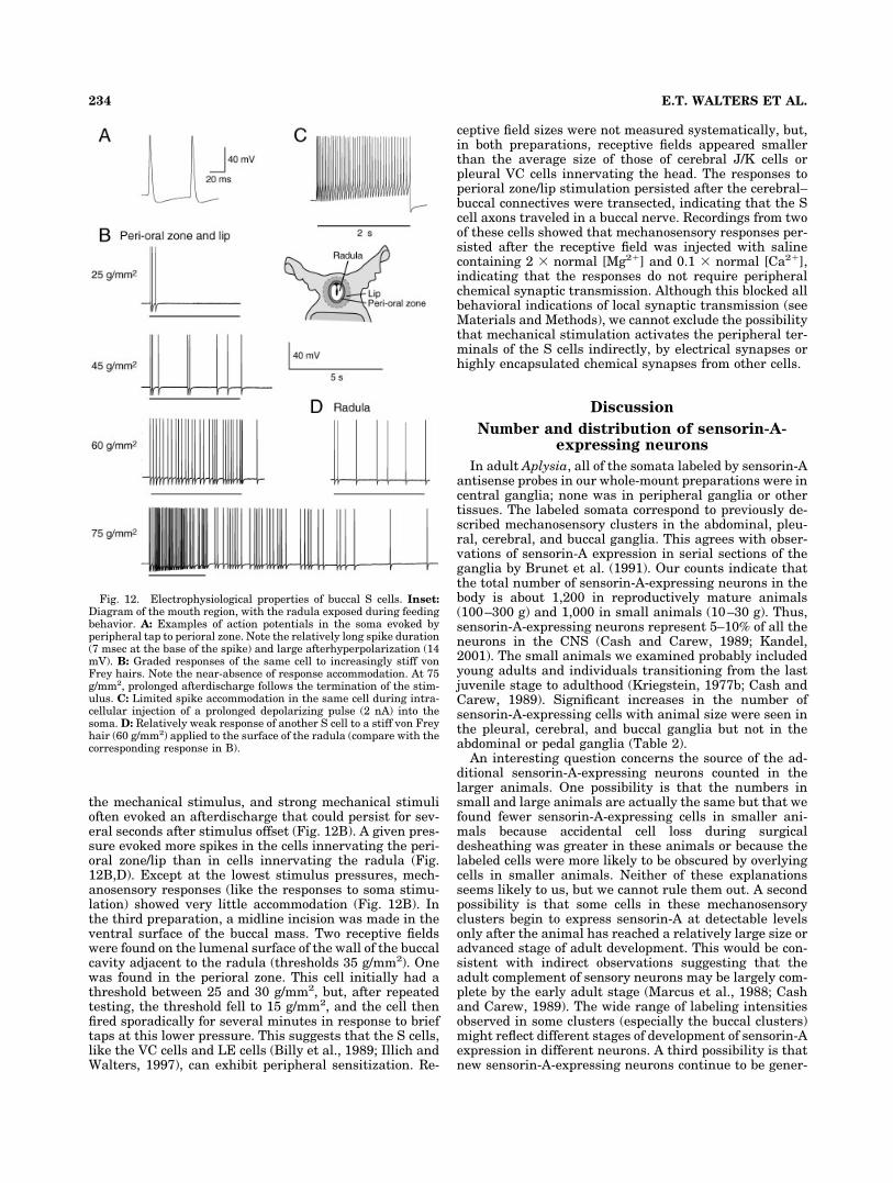
Fig. 12. Electrophysiological properties of buccal S cells. Inset:Diagram of the mouth region, with the radula exposed during feedingbehavior. A: Examples of action potentials in the soma evoked byperipheral tap to perioral zone. Note the relatively long spike duration(7 msec at the base of the spike) and large afterhyperpolarization (14mV). B: Graded responses of the same cell to increasingly stiff vonFrey hairs. Note the near-absence of response accommodation. At 75g/mm2, prolonged afterdischarge follows the termination of the stim-ulus. C: Limited spike accommodation in the same cell during intra-cellular injection of a prolonged depolarizing pulse (2 nA) into thesoma. D: Relatively weak response of another S cell to a stiff von Freyhair (60 g/mm2) applied to the surface of the radula (compare with thecorresponding response in B).
234 E.T. WALTERS ET AL.

ated in adulthood. Although addition of new neurons inadult mollusks has not, to our knowledge, been reported,such a mechanism might be useful to maintain completecoverage of the body surface by receptive fields of centralmechanosensory neurons during the extraordinarygrowth of these animals. Adult Aplysia exhibit over a100-fold increase in weight and volume, growing from �30g to 3,000 g within several months (Carefoot, 1987). Thesubstantial variation in numbers of sensorin-A-expressingcells observed in animals within the same size range (Ta-ble 2) might also reflect accidental cell loss during micro-surgery, differences in developmental onset of sensorin-Aexpression, and/or differences in late generation ofsensorin-A-expressing neurons across animals.
Somatotopic organization of sensorin-A-expressing neurons
The present report completes the macroscopic map ofthe peripheral receptive fields of sensorin-A-expressingcell clusters. Figure 13 shows the combined receptivefields of cells in each of the abdominal clusters (LE, rLE,RE, and RF), the cerebral J and K clusters, the buccal Sclusters, and the pleural VC clusters. Our study confirmsearlier descriptions of receptive fields in Aplysia (Byrne etal., 1974; Rosen et al., 1979, 1982; Byrne, 1980; Fiore andGeppeti, 1981; Walters et al., 1983; Billy and Walters,1989a; Dubuc and Castellucci, 1991; Illich and Walters,1997) and provides new information about coverage of themidbody and anterior body by the pleural VC clusters, ofthe rhinophores and propodium by the cerebral J clusters,and of the lip region and buccal mass by the buccal Sclusters. When all of this information is combined,sensorin-A-expressing cells are seen to provide a centrallydistributed map of the entire surface of the body (Fig. 13).
The receptive fields of the sensorin-A-expressing clus-ters exhibit modest cephalization, with the head havingthe smallest, most densely packed fields. Receptive fieldsof pleural VC cells are significantly smaller on the headthan on other parts of the body (Fig. 5B). In the perioral/lip region, the receptive fields of the J/K cells (Rosen et al.,1979, 1982) and the buccal S cells (see Results) appear tobe particularly small and numerous. The head is unique inshowing overlap of fields from clusters in three differentganglia: the buccal S, cerebral J/K, and pleural VC clus-ters, and of course there is also extensive overlap of recep-tive fields among cells within each cluster. The other partof the body showing extensive overlap of receptive fieldsfrom different clusters is the mantle cavity, where all fourof the clusters in the abdominal ganglion (LE, rLE, RE,and RF) innervate some parts of the mantle and branchialcavity (Dubuc and Castellucci, 1991). Perhaps reflectingevolutionary pressures similar to those responsible forcephalization, the anterior part of the mantle and siphontip display the smallest receptive fields found in the man-tle region (Dubuc and Castellucci, 1991).
Because of their large number, straightforward identi-fication, easy accessibility, and apparently uniform prop-erties, the pleural VC neurons have been utilized exten-sively for studies of neuronal plasticity in Aplysia.However, little was known about the locations of the re-ceptive fields of the VC neurons. We have now shown thataxons from each VC cluster project into all of the pedalnerves (after passing through the pl-p connective) and intoat least one of the cerebral nerves (c2, after passingthrough the c-pl connective) and that body regions sup-
plied by each pedal nerve contain receptive fields of VCneurons. Only the mantle organs and the floor of themantle cavity were found to lack VC cell receptive fields,and no VC cell axons were found in the pl-ab connective.Although in most regions of the body the VC cell receptivefields were restricted to the ipsilateral side (see also Billy
Fig. 13. Sensorin-A-expressing clusters form a macroscopic map ofthe entire surface of Aplysia. The dorsal views show the parapodia(PP) closed (left) and open (right), to reveal the mantle organs. Theventral views show the foot and perioral/lip area. Clusters aregrouped by ganglion and their fields color coded as indicated. When allthe clusters’ fields are combined (bottom row), the fields cover thewhole body. Overlap of the cerebral J/K fields (yellow) and pleural VCfields (blue) is indicated by green.
235SOMATOTOPIC ORGANIZATION OF APLYSIA SENSORY NEURONS

and Walters, 1989a), about 15% of the receptive fields onthe foot crossed the midline (Fig. 4B2). Several VC cellswere found to have dual axons, with the greatest incidenceof dual axons in cells in the anterolateral region of thecluster, where cells often send one axon to the anteriorfoot through nerve p1 and the pl-p connective and a secondthrough the c-pl connective to synapse with B motor neu-rons in the cerebral ganglion and/or to innervate the ipsi-lateral tentacle (Figs. 4, 8, 9). These anterolateral VC cellsare probably a major source of sensory neurons with dualaxons that are used in studies of synapse-specific plastic-ity in dissociated cell culture (see, e.g., Martin et al., 1997;Schacher et al., 1999).
A clear (albeit rough) topographic relationship was dem-onstrated between the location of each sensory neuronsoma in the VC cluster and both the location of its periph-eral receptive field (Fig. 6) and the nerve carrying itsprimary axon (Fig. 7; see also Zhang et al., 1993). Thus,each VC cluster forms a somatotopic map of most of theipsilateral body surface. Although somatotopic mapping ofthe body surface onto topographically organized popula-tions of central neurons (e.g., the sensory homunculus insensory cortex) is a common feature of mammalian sen-sory systems, and somatotopic organization of afferentterminals is well known in arthropods (see, e.g., Murphey,1981; Peterson and Weeks, 1988), this remains the onlysomatotopic mapping of peripheral receptive fields onto adiscrete cluster of central neuronal somata of which weknow in an invertebrate (Walters et al., 1983). Interestingquestions arise about the functional significance, if any, ofthis “sensory aplunculus” in each pleural ganglion and thedevelopmental mechanisms responsible for its formation.
None of the other sensorin-A-expressing clusters hasrevealed distinct somatotopic maps of their peripheraltargets. However, more investigation is needed of the or-ganization of the receptive fields of the buccal S clusters.Our nerve back-fills (Fig. 12) revealed intriguing differ-ences in the mapping of soma position to axon locationamong the major buccal nerves. One surprise was thatsomata filled from the dorsal buccal nerve (b3) were con-tralateral to the back-filled nerve, in contrast to the ipsi-lateral patterns produced by filling the ventral and lateralbuccal nerves (b1 and b2; see also Scott et al., 1991). Thesignificance of these observations must await detailedstudies on the locations, sizes, and overlap of the S cellreceptive fields. Our preliminary observations on S cellreceptive fields were intended only to establish that S cellsare mechanosensory in function and that they have fieldson the buccal mass and perioral region. The fact thatseparate back-fills of the major buccal nerves (b1, b2, b3,and the radula nerve) appeared to stain all of the cells inthe S clusters (Fig. 12; see also Scott et al., 1991) suggeststhat all of the S cells may have receptive fields in theregions supplied by these nerves. The observation thatdifferent back-fills stained nearly all the cells in someoverlapping regions of the buccal clusters suggests thatsome S cells send at least two axons to the peripherythrough different nerves.
Functions of sensorin-A-expressing neurons
Is the expression of sensorin-A associated with uniqueneuronal functions? Little light is shed on this question bythe initial physiological effect reported for sensorin-A; ap-plication of the peptide hyperpolarized a subset of thepostsynaptic target neurons, suggesting that it can func-
tion as an inhibitory cotransmitter (Brunet et al., 1991).However, the most prominent synaptic effect of sensorin-A-expressing neurons is rapid excitation, probably medi-ated by release of glutamate (Dale and Kandel, 1993;Trudeau and Castellucci, 1993; Gapon and Kupfermann,1996; Levenson et al., 2000). On the other hand, the de-gree of sensorin-A expression in dissociated cell cultureshas correlated well with the presence and strength ofexcitatory synapses onto motor neuron targets (Santarelliet al., 1996; Schacher et al., 1999; Sun et al., 2001; Hu etal., 2002). This suggests that sensorin-A has functionsrelated to synaptic transmission or synaptic plasticity,although the paucity of hyperpolarizing responses re-ported for sensory neuron stimulation and the lack ofdepolarizing responses to exogenous sensorin-A applica-tion means that sensorin-A is unlikely to function like aclassical neurotransmitter. Axotomized sensory neurons,cut off from peripheral receptive fields but still maintain-ing central synapses, display a decrease in sensorin-AmRNA expression, whereas contralateral, intact sensoryneurons display a simultaneous increase in expression(Noel et al., 1995). If sensorin had an immediate inhibitoryeffect, the observed facilitation of EPSPs from regenerat-ing tail sensory neurons (Walters et al., 1991) might bedue to disinhibition resulting from a decrease in tonicrelease of sensorin-A from sensory neurons after axotomy.However, exogenous sensorin-A failed to inhibit thesesame synapses onto tail motor neurons (Brunet et al.,1991). Recent evidence from dissociated cell culture indi-cates that sensorin may act as a growth factor, contribut-ing to synapse formation and long-term synaptic facilita-tion (Hu et al., 2003). It is interesting that sensorin-A isalso present in coiled sensory terminals (Steffensen andMorris, 1996) located in muscle layers distant from knownsynapses of these cells. This suggests additional, nonsyn-aptic functions of sensorin-A, perhaps related to mechano-reception or nociception.
The most notable feature of sensorin-A is its close asso-ciation with cells known to be mechanosensory neurons.Our studies have strengthened and extended this associ-ation by confirming the original patterns of expressionreported by Brunet et al. (1991) and by demonstrating asensory function of the buccal S cells (Fig. 12), the onlyprevious evidence for which had been largely undocu-mented (Fiore and Geppeti, 1981). Although all thesensorin-A-expressing clusters in Aplysia have demon-strated sensory functions, we do not know whether allsensorin-A-expressing neurons do. Indeed, the presentstudy has revealed small numbers of sparsely distributedsensorin-A-expressing neurons in novel locations (pedalganglia, dorsal surface of the cerebral ganglion, ventral-caudal region of the buccal ganglia). These are oftensmaller than the cells in the sensorin-A-expressing sen-sory clusters, although a few are somewhat larger thancells in the clusters. Possible sensory functions of thesenewly discovered cells remain to be tested. On the otherhand, extensive investigations of the LE clusters (Byrne etal., 1974, 1978a; Dubuc and Castellucci, 1991; Illich andWalters, 1997) and VC clusters (this paper; see alsoWalters et al., 1983; Walters, 1987; Billy and Walters,1989a,b; Clatworthy and Walters, 1993b) indicatestrongly that all of the cells in the LE and VC clustershave peripheral mechanosensory fields.
What types of mechanosensory function are associatedwith sensorin-A expression? Mechanosensory function in
236 E.T. WALTERS ET AL.

Aplysia does not require sensorin-A, insofar as other cen-trally located mechanosensory neurons have been de-scribed in Aplysia that fail to express this peptide, includ-ing giant neurons R1 and L1 in the abdominal ganglion(Cobbs and Pinsker, 1978), proprioceptive multiactionneurons B4 and B5 in the buccal ganglion (Jahan-Parwaret al., 1983), the proprioceptive modulatory neuron C2 inthe cerebral ganglion (Weiss et al., 1986a–c), and rostralclusters of radula mechanoafferents in the buccal gan-glion. The latter are distinguished from adjacent S cells bythe presence of peptides SCPA and SCPB and a lack ofsensorin-A (Miller et al., 1994; Borovikov et al., 2000;Rosen et al., 2000). In addition, indirect evidence suggeststhe existence of unidentified mechanosensory populationshaving peripherally located somata (Kupfermann et al.,1974; Peretz et al., 1976; Walters et al., 1983; Xin et al.,1995; Frost et al., 1997; Hickie et al., 1997; Walters andCohen, 1997; Croll, 2001). We found no evidence ofsensorin-A expression in peripheral somata of adult ani-mals. One difference between the other central mech-anosensory neurons and the sensorin-A-expressing neu-rons is that the former are much more likely to show
substantial fast excitation from synaptic inputs and thusappear to have interneuronal (and sometimes motor) aswell as sensory functions. In contrast, fast PSPs fromother neurons have not been reported for most of thesensorin-A-expressing clusters (Table 4). The major excep-tion is the buccal S cells, which receive fast EPSPs (�1mV) from many other S cells (Fiore and Meunier, 1975). Inaddition, �4% of cells in the cerebral J cluster evoke verysmall EPSPs (�1 mV) in pleural VC cells (Raymond andByrne, 1994). Another difference is that most of the othercentral and at least some of the peripheral mechanosen-sory neurons are sensitive to very weak, innocuous stim-uli. In contrast, we have now shown that sensorin-A-expressing neurons in all of the ganglia have relativelyhigh thresholds to mechanical stimulation. J/K cells, VCcells, and S cells tested with the same set of von Frey hairsall showed mean response thresholds of 30–35 g/mm2, asdid LE cells and VC cells in previous studies (Billy andWalters, 1989a; Illich and Walters, 1997), and the thresh-olds ranged between 15 and 65 g/mm2. At pressures of25–35 g/mm2, the siphon skin begins to be perforated(Illich and Walters, 1997). If the threshold for tissue in-
TABLE 4. Synaptic Targets of Sensorin-A-Expressing Clusters
Ganglionand cluster Target1 Cell type2
Cellnumber3
PSPtypes4 Mono-test5 Target functions6 Original references
AbdominalLE L7 MN, gill 1 fE TEA GWR, SWR Castellucci et al., 1970; Castellucci
and Kandel, 1974L16 IN, inh 1 fE GWR, SWR Castellucci and Kandel, 1974;
Hawkins et al., 1981aLDG MN, gill 2 fE hi-di GWR Byrne et al., 1978b; Frost et al., 1997L9 MN, gill 2 fE GWR Byrne et al., 1978bRDG MN, gill 1 fE GWR Byrne et al., 1978bL24 IN, multi 1 fE, sI ? Byrne and Koester, 1978L14 MN, ink 3 fE hi-di Ink Byrne, 1980R18 IN, exc 1 fE Ink Byrne, 1980L22 IN, exc 3 fE GWR Hawkins et al., 1981aL23 IN, exc 1 fE GWR Hawkins et al., 1981aL28 IN, multi 1 fE, sI GWR Hawkins et al., 1981a,bL29 IN, multi 5 fE GWR Hawkins et al., 1981a,bL30 IN, inhib 3 fE GWR Hawkins et al., 1981aPeriph MN, siphon 50 fE hi-di SWR Bailey et al., 1979LFS MN, siphon 7 fE SWR Carew et al. 1981; Frost et al. 1988L34 IN, exc 2 fE hi-di SWR Trudeau and Castellucci, 1992; Frost
and Kandel, 1995LDS MN, siphon 1 fE SWR Frost and Kandel, 1995
rLE L7 MN, gill 1 fE GWR Dubuc and Castellucci, 1991LFS MN, siphon 7 fE SWR Dubuc and Castellucci, 1991
RE L14 MN, ink 3 fE hi-di Ink Byrne, 1980RF L14 MN, ink 3 fE hi-di Ink Byrne, 1980
L7 MN, gill 1 fE hi-di GWR Byrne, 1981Pleural
VC P5/6/7 MN, tail 6 fE hi-di TWR Walters et al., 1983LP117 IN, exc 2 fE TWR, SWR, GWR Cleary and Byrne, 1993RP14 IN, inh 2 fE TWR Xu et al., 1994Bn MN, head 60–70 fE hi-di HWR This paper
CerebralJ/K Bn MN, head 60–70 fE hi-di HWR Rosen et al., 1979, 1982; Teyke et al.,
1989B4/5 SN, IN,
MN, multi4 fE hi-di Ingestion, egestion Rosen et al., 1982
MCC IN, multi 2 fE hi-di Feeding Rosen et al., 1982VC SN 440 sE, sI,
fE, fIDefense Raymond and Byrne, 1994
LP117 In, exc 2 fE, sE TWR, SWR, GWR Raymond and Byrne, 1994Buccal
S B4/5 SN, IN,MN, multi
4 fE hi-di Ingestion, egestion Fiore and Meunier, 1975; Sussweinand Byrne, 1988
S SN 320 fE TEA ? Fiore and Meunier, 1975
1Periph, peripheral siphon motor neurons; MCC, metacerebral giant cell. Some uniquely identified cells have been combined with highly similar individuals (P5/6/7 � P5, P6, andP7; B4/5 � B4 and B5).2MN, motor neuron; IN, interneuron; SN, sensory neuron; exc, excitatory interneuron; inh, inhibitory interneuron; multi, multiaction (with at least two of the following actionson the same or different follower neurons: excitation, inhibition, or modulation with little or no change in potential).3Number is for the entire animal (i.e., double the number for individual paired ganglia).4fE, fast EPSP; sE, slow EPSP; fI, fast IPSP; sI, slow IPSP.5Test for monosynapticity; hi-di, high divalent cation solution; TEA, presynaptic TEA injection; blanks indicate no reported tests.6Behavioral functions documented; GWR, gill-withdrawal reflex; SWR, siphon-withdrawal reflex; TWR, tail-withdrawal reflex; HWR, head-withdrawal reflex.
237SOMATOTOPIC ORGANIZATION OF APLYSIA SENSORY NEURONS

jury in other regions is also within this range, then cells inthese clusters are tuned to mechanical stimuli that pro-duce or threaten tissue damage; i.e., sensorin-A expres-sion is likely to be associated with a nociceptive function.
Further insight into the functions of sensorin-A-neurons comes from the known synaptic connections ofthese cells. The connections reported to be direct are listedin Table 4, which reveals that neurons in every sensorin-A-expressing cluster produce fast EPSPs in specific motorneurons (e.g., Fig. 9C) as well as other types of neurons. Inmost cases, the identified target neurons have defensivefunctions, which include driving the release of ink andwithdrawal of the gill, siphon, tail, or head (Table 4).Subsets of both the pleural VC cells and the cerebral J/Kcells contribute jointly to head- and tentacle-withdrawalreflexes, because some of their receptive fields overlap andthey often excite the same Bn motor neurons (Fig. 9C; seealso Raymond and Byrne, 1994). In addition, the VC cells(Billy and Walters, 1989a,b; Clatworthy and Walters,1993b), LE cells (Illich and Walters, 1997), and perhaps Scells (see Results) display nociceptive sensitization of theirperipheral receptive fields. Peripheral sensitization fol-lowing intense activation appears to be a sensory propertythat is unique to nociceptors; other sensory neurons usu-ally show adaptation instead (Light, 1992). Taken to-gether, the synaptic outputs of the sensorin-A-expressingneurons and their capacity for peripheral sensitizationprovide additional evidence for defensive and nociceptivefunctions of many of these cells.
The functions of the buccal S cells, however, remainmysterious. These cells not only excite each other (Table 4)but excite and sometimes inhibit many other buccal gan-glion neurons (Fiore and Meunier, 1975, 1979), many ofwhich participate in controlling ingestive and egestivebehaviors of the buccal mass. In addition to their unusualsynaptic interconnections, the S cells exhibit several otherfeatures that distinguish them from most of the othersensorin-A-expressing neurons, suggesting that the S cellsmay have some unique functions in the animal. For exam-ple, whereas the somata of most sensorin-A-expressingneurons show rapidly accommodating responses to pro-longed depolarizing pulses (see, e.g., Klein et al., 1986;Walters et al., 1991; Clatworthy and Walters, 1994), thebuccal S cells show very little spike accommodation in thesoma (Fig. 12C) as well as little accommodation duringmechanosensory responses (Fig. 12B). Compared withother sensorin-A-expressing neurons, the S cells also havespike durations that are about twice as long, and afterhy-perpolarizations that are three times the amplitude ofthose in the VC, J/K, and LE neurons (cf. Hochner et al.,1986; Baxter and Byrne, 1990; Walters et al., 1991; Clat-worthy and Walters, 1994; Gunstream et al., 1995; Illichand Walters, 1997; Ungless et al., 2002). These unusualfeatures of the S cells may reflect unique biomechanicalproperties or functions of the highly complex buccal mass,or its location inside the body (the other clusters innervatetissues near the surface of the body). We have recordedfrom too few S cells to know whether the properties foundin this preliminary study are characteristic of the entirecluster or whether subpopulations exist with differentproperties and sensory functions. Given the diversity oftissues in the buccal mass/lip region that can evoke spikesin S cells (including buccal muscle, lip/perioral tissue,hard radula, and soft tissues lining the buccal cavity), itwould be surprising if all functional properties were ex-
pressed uniformly across all the sensorin-expressing neu-rons in the buccal ganglia. Moreover, although the othersensorin-A-expressing clusters share many physiologicalproperties, all of their properties are not identical. Forexample, subsets of the sensory cells in the cerebral andabdominal clusters show different responses to the sameneuromodulators, including serotonin and FMRFamide(Rosen et al., 1989; Storozhuk and Castellucci, 1998), andno systematic studies have yet been performed on pleuralVC clusters to examine patterns of response to endoge-nous neuromodulators across the entire VC cell popula-tion.
Although much remains to be learned about the func-tions of sensorin-A and the neurons that express thispeptide, the availability of well-defined populations ofmechanosensory neurons in Aplysia that expresssensorin-A along with other mechanosensory populationsthat fail to express the peptide means that powerful com-parative methods can be used to elucidate such functions.This power will be increased if the comparisons can beextended to mechanosensory neurons in other molluscanspecies. Sensorin-A-expressing neurons have been foundin the snail Lymnaea stagnalis (Steffensen et al., 1995b),and it will be interesting to see whether some of themechanosensory populations described in other gastropodmolluscs also express sensorin-A (see, e.g., Getting, 1976;Spray et al., 1980; Elliott and Benjamin, 1989; Inoue et al.,1996; Malyshev et al., 2002). Among the functional ques-tions that will be important to address is why the mRNAfor sensorin-A is expressed so widely within the cell, beingfound not only in the soma but in dendritic and axonalregions within ganglia and connectives (Figs. 2B1,3A1,B1; Brunet et al., 1991), in neurites and synapticterminals in dissociated cell culture (Santarelli et al.,1996; Schacher et al., 1999), and in peripheral axons (Fig.3B1).
ACKNOWLEDGMENTS
Some of the animals were supplied by the NationalCenter for Research Resources, National Resource forAplysia, at the University of Miami.
LITERATURE CITED
Ambron RT, Walters ET. 1996. Priming events and retrograde injurysignals: a new perspective on the cellular and molecular biology ofnerve regeneration. Mol Neurobiol 13:61–79.
Bailey CH, Castellucci VF, Koester J, Kandel ER. 1979. Cellular studies ofperipheral neurons in siphon skin of Aplysia californica. J Neuro-physiol 42:530–557.
Baxter DA, Byrne JH. 1990. Differential effects of cAMP and serotonin onmembrane current, action-potential duration, and excitability in so-mata of pleural sensory neurons of Aplysia. J Neurophysiol 64:978–990.
Billy AJ, Walters ET. 1989a. Long-term expansion and sensitization ofmechanosensory receptive fields in Aplysia support an activity-dependent model of whole-cell sensory plasticity. J Neurosci 9:1254–1262.
Billy AJ, Walters ET. 1989b. Modulation of mechanosensory threshold inAplysia by serotonin, small cardioactive peptideB (SCPB), FMRF-amide, acetylcholine, and dopamine. Neurosci Lett 105:200–204.
Bogdanov Yu D, Balaban PM, Zakharov IS, Poteryaev DA, Belyavsky AV.1996. Identification of two novel genes specifically expressed in theD-group neurons of the terrestrial snail CNS. Invert Neurosci 2:61–69.
Borovikov D, Evans CG, Jing J, Rosen SC, Cropper EC. 2000. A proprio-ceptive role for an exteroceptive mechanoafferent neuron in Aplysia.J Neurosci 20:1990–2002.
238 E.T. WALTERS ET AL.

Brunet JF, Shapiro E, Foster SA, Kandel ER, Iino Y. 1991. Identification ofa peptide specific for Aplysia sensory neurons by PCR-based differen-tial screening. Science 252:856–859.
Byrne JH. 1980. Neural circuit for inking behavior in Aplysia californica.J Neurophysiol 43:896–911.
Byrne JH. 1981. Comparative aspects of neural circuits for inking behaviorand gill withdrawal in Aplysia californica. J Neurophysiol 45:98–106.
Byrne JH, Koester J. 1978. Respiratory pumping: neuronal control of acentrally commanded behavior in Aplysia. Brain Res 143:878–105.
Byrne JH, Castellucci V, Kandel ER. 1974. Receptive fields and responseproperties of mechanoreceptor neurons innervating siphon skin andmantle shelf in Aplysia. J Neurophysiol 37:1041–1064.
Byrne JH, Castellucci VF, Carew TJ, Kandel ER. 1978a. Stimulus-response relations and stability of mechanoreceptor and motor neuronsmediating defensive gill-withdrawal reflex in Aplysia. J Neurophysiol41:402–417.
Byrne JH, Castellucci VF, Kandel ER. 1978b. Contribution of individualmechanoreceptor sensory neurons to defensive gill-withdrawal reflex inAplysia. J Neurophysiol 41:418–431.
Carefoot T. 1987. Aplysia: its biology and ecology. Oceanogr Mar Biol AnnuRev 25:167–284.
Carew TJ, Walters ET, Kandel ER. 1981. Associative learning in Aplysia:cellular corelates supporting a conditioned fear hypothesis. Science211:501–504.
Cash D, Carew TJ. 1989. A quantitative analysis of the development of thecentral nervous system in juvenile Aplysia californica. J Neurobiol20:25–47.
Castellucci VF, Kandel ER. 1974. A quantal analysis of the synapticdepression underlying habituation of the gill-withdrawal reflex inAplysia. Proc Natl Acad Sci U S A 71:5004–5008.
Castellucci V, Pinsker H, Kupfermann I, Kandel ER. 1970. Neuronalmechanisms of habituation and dishabituation of the gill-withdrawalreflex in Aplysia. Science 167:1745–1748.
Clatworthy AL, Walters ET. 1993a. Activity-dependent depression ofmechanosensory discharge and excitability in Aplysia. J Neurophysiol70:1195–1209.
Clatworthy AL, Walters ET. 1993b. Rapid amplification and facilitation ofmechanosensory discharge in Aplysia by noxious stimulation. J Neu-rophysiol 70:1181–1194.
Clatworthy AL, Walters ET. 1994. Comparative analysis of hyperexcitabil-ity and synaptic facilitation induced by nerve injury in two populationsof mechanosensory neurones of Aplysia californica. J Exp Biol 190:217–238.
Cleary LJ, Byrne JH. 1993. Identification and characterization of a mul-tifunctional neuron contributing to defensive arousal in Aplysia. J Neu-rophysiol 70:1767–1776.
Cobbs JS, Pinsker HM. 1978. In vivo responses of paired giant mechano-receptor neurons in Aplysia abdominal ganglion. J Neurobiol 9:121–141.
Croll RP. 2001. Catecholamine-containing cells in the central nervoussystem and periphery of Aplysia californica. J Comp Neurol 441:91–105.
Dale N, Kandel ER. 1993. L-glutamate may be the fast excitatory trans-mitter of Aplysia sensory neurons. Proc Natl Acad Sci U S A 90:7163–7167.
Dıaz-Rıos M, Suess E, Miller MW. 1999. Localization of GABA-like immu-noreactivity in the central nervous system of Aplysia californica.J Comp Neurol 413:255–270.
Dubuc B, Castellucci VF. 1991. Receptive fields and properties of a newcluster of mechanoreceptor neurons innervating the mantle region andthe branchial cavity of the marine mollusk Aplysia californica. J ExpBiol 156:315–334.
Dulin MF, Steffensen I, Morris CE, Walters ET. 1995. Recovery of function,peripheral sensitization, and sensory neurone activation by novel path-ways following axonal injury in Aplysia. J Exp Biol 198:2055–2066.
Elliott CJ, Benjamin PR. 1989. Esophageal mechanoreceptors in the feed-ing system of the pond snail, Lymnaea stagnalis. J Neurophysiol 61:727–736.
Fiore L, Geppeti L. 1981. Neural control of buccal mass activity of Aplysia.In: Salanki J, editor. Neurobiology of invertebrates. London: PergamonPress. p 201–223.
Fiore L, Meunier JM. 1975. A network of synaptic relations in the buccalganglia of Aplysia. Brain Res 92:336–340.
Fiore L, Meunier JM. 1979. Synaptic connections and functional organiza-tion in Aplysia buccal ganglia. J Neurobiol 10:13–29.
Frost WN, Clark GA, Kandel ER. 1988. Parallel processing of short-termmemory for sensitization in Aplysia. J Neurobiol 19:297–334.
Frost L, Kaplan SW, Cohen TE, Henzi V, Kandel ER, Hawkins RD. 1997.A simplified preparation for relating cellular events to behavior: con-tribution of LE and unidentified siphon sensory neurons to mediationand habituation of the Aplysia gill- and siphon-withdrawal reflex.J Neurosci 17:2900–2913.
Frost WN, Kandel ER. 1995. Structure of the network mediating siphon-elicited siphon withdrawal in Aplysia. J Neurophysiol 73:2413–2427.
Gapon S, Kupfermann I. 1996. Evidence for transmitter similarity of twoclasses of mechanoreceptor neurons in the cerebral ganglion of Aplysia.Neurosci Lett 205:157–160.
Getting PA. 1976. Afferent neurons mediating escape swimming of themarine mollusc, Tritonia. J Comp Physiol 110:271–286.
Gunstream JD, Castro GA, Walters ET. 1995. Retrograde transport ofplasticity signals in Aplysia sensory neurons following axonal injury.J Neurosci 15:439–448.
Hawkins RD, Castellucci VF, Kandel ER. 1981a. Interneurons involved inmediation and modulation of gill-withdrawal reflex in Aplysia. I. Iden-tification and characterization. J Neurophysiol 45:304–314.
Hawkins RD, Castellucci VF, Kandel ER. 1981b. Interneurons involved inmediation and modulation of gill-withdrawal reflex in Aplysia. II. Iden-tified neurons produce heterosynaptic facilitation contributing to be-havioral sensitization. J Neurophysiol 45:315–328.
Hawkins RD, Abrams TW, Carew TJ, Kandel ER. 1983. A cellular mech-anism of classical conditioning in Aplysia: activity-dependent amplifi-cation of presynaptic facilitation. Science 219:400–405.
Hening WA, Walters ET, Carew TJ, Kandel ER. 1979. Motorneuronalcontrol of locomotion in Aplysia. Brain Res 179:231–253.
Hickie C, Cohen LB, Balaban PM. 1997. The synapse between LE sensoryneurons and gill motoneurons makes only a small contribution to theAplysia gill-withdrawal reflex. Eur J Neurosci 9:627–636.
Hickmott PW, Carew TJ. 1991. An autoradiographic analysis of neurogen-esis in juvenile Aplysia californica. J Neurobiol 22:313–326.
Hochner B, Klein M, Schacher S, Kandel ER. 1986. Action-potential dura-tion and the modulation of transmitter release from the sensory neu-rons of Aplysia in presynaptic facilitation and behavioral sensitization.Proc Natl Acad Sci U S A 83:8410–8414.
Hu JY, Meng X, Schacher S. 2002. Target interaction regulates distribu-tion and stability of specific mRNAs. J Neurosci 22:2669–2678.
Hu JY, Wu F, Meng X, Wang D, Schacher S. 2003. Release of sensorinpeptide is required for synapse formation and long-term synaptic fa-cilitation in Aplysia. Soc Neurosci Abstr 478.4.
Illich PA, Walters ET. 1997. Mechanosensory neurons innervating Aplysiasiphon encode noxious stimuli and display nociceptive sensitization.J Neurosci 17:459–469.
Inoue T, Takasaki M, Lukowiak K, Syed NI. 1996. Identification of aputative mechanosensory neuron in Lymnaea: characterization of itssynaptic and functional connections with the whole-body withdrawalinterneuron. J Neurophysiol 76:3230–3238.
Jahan-Parwar B, Fredman SM. 1976. Cerebral ganglion of Aplysia: cellu-lar organization and origin of nerves. Comp Biochem Physiol A54:347–357.
Jahan-Parwar B, Fredman SM. 1978. Pedal locomotion in Aplysiacalifornica—I. Sensory and motor fields of pedal nerves. Comp BiochemPhysiol 60A:459–465.
Jahan-Parwar B, Wilson AH Jr, Fredman SM. 1983. Role of proprioceptivereflexes in control of feeding muscles of Aplysia. J Neurophysiol 49:1469–1480.
Kandel ER. 1979. Behavioral biology of Aplysia. San Francisco: W.H.Freeman & Co.
Kandel ER. 2001. The molecular biology of memory storage: a dialoguebetween genes and synapses. Science 294:1030–1038.
Klein M, Hochner B, Kandel ER. 1986. Facilitatory transmitters and cAMPcan modulate accommodation as well as transmitter release in Aplysiasensory neurons: evidence for parallel processing in a single cell. ProcNatl Acad Sci USA 83:7994–7998.
Kriegstein AR. 1977a. Development of the nervous system of Aplysiacalifornica. Proc Natl Acad Sci U S A 74:375–378.
Kriegstein AR. 1977b. Stages in the post-hatching development of Aplysiacalifornica. J Exp Zool 199:275–288.
Krontiris-Litowitz JK, Cooper BF, Walters ET. 1989. Humoral factors
239SOMATOTOPIC ORGANIZATION OF APLYSIA SENSORY NEURONS

released during trauma of Aplysia. I. Body wall contraction, cardiacmodulation, and central reflex suppression. J Comp Physiol B159:211–223.
Kupfermann I, Carew TJ, Kandel ER. 1974. Local, reflex, and centralcommands controlling gill and siphon movements in Aplysia. J Neuro-physiol 37:996–1019.
Levenson J, Sherry DM, Dryer L, Chin J, Byrne JH, Eskin A. 2000.Localization of glutamate and glutamate transporters in the sensoryneurons of Aplysia. J Comp Neurol 423:121–131.
Liao X, Walters ET. 2002. The use of elevated divalent cation solutions toisolate monosynaptic components of sensorimotor connections in Aply-sia. J Neurosci Methods 120:45.
Light AR. 1992. The initial processing of pain and its descending control:spinal and trigeminal systems. Basel: Karger.
Malyshev AY, Balaban PM. 2002. Identification of mechanoafferent neu-rons in terrestrial snail: response properties and synaptic connections.J Neurophysiol 87:2364–2371.
Marcus EA, Schuerman G, Stopfer M, Carew TJ. 1988. Development of tailsensory neurons in the pleural ganglia of Aplysia. Soc Neurosci Abstr14:481.
Martin KC, Casadio A, Zhu H, Yaping E, Rose JC, Chen M, Bailey CH,Kandel ER. 1997. Synapse-specific, long-term facilitation of Aplysiasensory to motor synapses: a function for local protein synthesis inmemory storage. Cell 91:927–938.
McAllister LB, Scheller RH, Kandel ER, Axel R. 1983. In situ hybridizationto study the origin and fate of identified neurons. Science 222:800–808.
Miller MW, Rosen SC, Schissel SL, Cropper EC, Kupfermann I, Weiss KR.1994. A population of SCP-containing neurons in the buccal ganglion ofAplysia are radula mechanoafferents and receive excitation of centralorigin. J Neurosci 14:7008–7023.
Moroz LL, Iannuccilli B, Si K, Sathyanarayanan PV, Sadreyev R, EdwardsJ, Panchin Y, Meleshkevich E, Nguyen T, Farmerie W, Ju J, KandelER. 2002. Aplysia EST project: from the CNS to neuronal and synaptictranscriptome. Soc Neurosci Abstr 376.13.
Murphey RK. 1981. The structure and development of a somatotopic mapin crickets: the cercal afferent projection. Dev Biol 88:236–246.
Nazif FA, Byrne JH, Cleary LJ. 1991. cAMP induces long-term morpho-logical changes in sensory neurons of Aplysia. Brain Res 539:324–327.
Noel F, Frost WN, Tian L-M, Colicos MA, Dash PK. 1995. Recovery of thetail-elicited siphon-withdrawal reflex following unilateral axonal in-jury is associated with ipsi- and contralateral changes in gene expres-sion in Aplysia californica. J Neurosci 15:6926–6938.
Peretz B, Jacklet JW, Lukowi K. 1976. Habituation of reflexes in Aplysia:contribution of the peripheral and central nervous systems. Science191:396–399.
Peterson BA, Weeks JC. 1988. Somatotopic mapping of sensory neuronsinnervating mechanosensory hairs on the larval prolegs of Manducasexta. J Comp Neurol 275:128–144.
Raymond JL, Byrne JH. 1994. Distributed input to the tail-siphon with-drawal circuit in Aplysia from neurons in the J cluster of the cerebralganglion. J Neurosci 14:2444–2454.
Rosen SC, Weiss KR, Kupfermann I. 1979. Response properties and syn-aptic connections of mechanoafferent neurons in cerebral ganglion ofAplysia. J Neurophysiol. 42:954–974.
Rosen SC, Weiss KR, Cohen JL, Kupfermann I. 1982. Interganglioniccerebral-buccal mechanoafferents of Aplysia: receptive fields and syn-aptic connections to different classes of neurons involved in feedingbehavior. J Neurophysiol 48:271–288.
Rosen SC, Susswein AJ, Cropper EC, Weiss KR, Kupfermann I. 1989.Selective modulation of spike duration by serotonin and the neuropep-tides, FMRFamide, SCPB, buccalin and myomodulin in differenntclasses of mechanoafferent neurons in the cerebral ganglion of Aplysia.J Neurosci 9:390–402.
Rosen SC, Miller MW, Cropper EC, Kupfermann I. 2000. Outputs of radulamechanoafferent neurons in Aplysia are modulated by motor neurons,interneurons, and sensory neurons. J Neurophysiol 83:1621–1636.
Santarelli L, Montarolo P, Schacher S. 1996. Neuropeptide localization invaricosities of Aplysia sensory neurons is regulated by target andneuromodulators evoking long-term synaptic plasticity. J Neurobiol31:297–308.
Schacher S, Wu F, Panyko JD, Sun ZY, Wang D. 1999. Expression andbranch-specific export of mRNA are regulated by synapse formationand interaction with specific postsynaptic targets. J Neurosci 19:6338–6347.
Scott ML, Govind CK, Kirk MD. 1991. Neuromuscular organization of thebuccal system in Aplysia californica. J Comp Neurol 312:207–222.
Spray DC, Spira ME, Bennett MV. 1980. Peripheral fields and branchingpatterns of buccal mechanosensory neurons in the opisthobranch mol-lusc, Navanax inermis. Brain Res 182:253–270.
Steffensen I, Morris CE. 1996. Coiled mechanoreceptors in Aplysia re-vealed by sensorin immunofluorescence and confocal microscopy. In-vert Neurosci 2:129–134.
Steffensen I, Dulin MF, Walters ET, Morris CE. 1995a. Peripheral regen-eration and central sprouting of sensory neurone axons in Aplysiacalifornica following nerve injury. J Exp Biol 198:2067–2078.
Steffensen I, Syed NI, Lukowiak K, Bulloch AGM, Morris CE. 1995b.Sensorin-A immunocytochemistry reveals putative mechanosensoryneurons in Lymnaea CNS. Invert Neurosci 1:207–213.
Storozhuk MV, Castellucci VF. 1998. The synaptic junctions of LE and RFcluster sensory neurones of Aplysia californica are differentially mod-ulated by serotonin. J Exp Biol 202:115–120.
Sun ZY, Wu F, Schacher S. 2001. Rapid bidirectional modulation of mRNAexpression and export accompany long-term facilitation and depressionof Aplysia synapses. J Neurobiol 46:41–47.
Susswein AJ, Byrne JH. 1988. Identification and characterization of neu-rons initiating patterned neural activity in the buccal ganglia of Aply-sia. J Neurosci 8:2049–2061.
Teyke T, Weiss KR, Kupfermann I. 1989. A subpopulation of cerebral Bcluster neurones of Aplysia californica is involved in defensive headwithdrawal but not appetitive head movements. J Exp Biol 147:1–20.
Trudeau LE, Castellucci VF. 1992. Contribution of polysynaptic pathwaysin the mediation and plasticity of Aplysia gill and siphon withdrawalreflex: evidence for differential modulation. J Neurosci 12:3838–3848.
Trudeau LE, Castellucci VF. 1993. Excitatory amino acid neurotransmis-sion at sensory-motor and interneuronal synapses of Aplysia califor-nica. J Neurophysiol 70:1221–1230.
Ungless MA, Gasull X, Walters ET. 2002. Long-term alteration of S-typepotassium current and passive membrane properties in Aplysia sen-sory neurons following axotomy. J Neurophysiol 87:2408–2420.
Walters ET. 1987. Multiple sensory neuronal correlates of site-specificsensitization in Aplysia. J Neurosci 7:408–417.
Walters ET, Cohen LB. 1997. Functions of the LE sensory neurons inAplysia. Invert Neurosci 3:15–25.
Walters ET, Erickson MT. 1986. Directional control and the functionalorganization of defensive responses in Aplysia. J Comp Physiol A159:339–351.
Walters ET, Byrne JH, Carew TJ, Kandel ER. 1983. Mechanoafferentneurons innervating tail of Aplysia. I. Response properties and synap-tic connections. J Neurophysiol 50:1522–1542.
Walters ET, Alizadeh H, Castro GA. 1991. Similar neuronal alterationsinduced by axonal injury and learning in Aplysia. Science 253:797–799.
Weiss KR, Chiel HJ, Koch U, Kupfermann I. 1986a. Activity of an identi-fied histaminergic neuron, and its possible role in arousal of feedingbehavior in semi-intact Aplysia. J Neurosci 6:2403–2415.
Weiss KR, Chiel HJ, Kupfermann I. 1986b. Sensory function and gating ofhistaminergic neuron C2 in Aplysia. J Neurosci 6:2416–2426.
Weiss KR, Shapiro E, Kupfermann I. 1986c. Modulatory synaptic actions ofan identified histaminergic neuron on the serotonergic metacerebralcell of Aplysia. J Neurosci 6:2393–2402.
Xin Y, Weiss KR, Kupfermann I. 1995. Distribution in the central nervoussystem of Aplysia of afferent fibers arising from cell bodies located inthe periphery. J Comp Neurol 359:627–643.
Xin Y, Hurwitz I, Perrins R, Evans CG, Alexeeva V, Weiss KR, Kupfer-mann I. 1999. Actions of a pair of identified cerebral–buccal interneu-rons (CBI-8/9) in Aplysia that contain the peptide myomodulin. J Neu-rophysiol 81:507–520.
Xu Y, Cleary LJ, Byrne JH. 1994. Identification and characterization ofpleural neurons that inhibit tail sensory neurons and motor neurons inAplysia: correlation with FMRFamide immunoreactivity. J Neurosci14:3565–3577.
240 E.T. WALTERS ET AL.
Copyright © 2022 FDOKUMEN