Interferon Beta and Long-term Disability in Multiple Sclerosis
Upload
independentCategory
view
0download
0
Preferential Recruitment of Interferon-�–Expressing TH17 Cells in Multiple Sclerosis
Hania Kebir, MSc,1 Igal Ifergan, MSc,1 Jorge Ivan Alvarez, PhD,1 Monique Bernard, MSc,1
Josee Poirier, BScN,2 Nathalie Arbour, PhD,1 Pierre Duquette, MD,2 and Alexandre Prat, MD, PhD1,2
Objective: There is substantial evidence supporting the role of interferon (IFN)-�–producing T helper (TH) 1 and interleukin(IL)-17–expressing TH17 lymphocytes in multiple sclerosis (MS) and its animal model, experimental allergic encephalomyelitis(EAE). However, to date little is known about the potential cooperative interplay between these 2 cytokines. In the currentstudy, we sought to evaluate the frequency of IFN-�–expressing TH17 lymphocytes in MS and EAE, and study their recruitmentinto the central nervous system (CNS).Methods: Human TH17 lymphocytes were expanded in vitro from the blood of healthy controls and relapsing MS patientsusing IL-23. Immune cell migration to the CNS was assessed in vitro with primary cultures of human blood–brain barrier(BBB)-derived endothelial cells, and in vivo in EAE mice.Results: We demonstrate that in response to IL-23, human memory lymphocytes expand into a TH17 phenotype, with asubpopulation of cells simultaneously expressing IFN-� and IL-17. We note that lymphocytes obtained from the blood ofrelapsing MS patients have an increased propensity to expand into IFN-�–producing TH17 cells and identify numerous Tlymphocytes coexpressing IL-17 and IFN-� in brain tissue of MS patients. We also find lymphocytes expressing both the TH1-and the TH17-associated transcription factors ROR�t and T-bet, in situ and in vitro. We further provide in vitro and in vivoevidence that IFN-�� TH17 lymphocytes preferentially cross the human BBB and accumulate in the CNS of mice during theeffector phase of EAE.Interpretation: Our data underscore the involvement of IFN-�� TH17 lymphocytes in the pathology of MS and EAE and theirpreferential recruitment into the CNS during inflammatory events.
Ann Neurol 2009;66:390–402
Multiple sclerosis (MS) is an immune-mediated disor-der of the central nervous system (CNS) characterizedby multifocal areas of leukocyte infiltration, demyelina-tion, and axonal damage. Abundance of immune cellsand their products (cytokines, chemokines, and immu-noglobulins) in MS plaques, and their accumulation inthe cerebrospinal fluid of affected individuals, supportthe notion that MS is a CNS-targeted inflammatorydisorder.1,2 Typically, demyelination is associated withan infiltration of memory CD4�CD45RO� T lym-phocytes, effector memory CD8� T lymphocytes,macrophages, and dendritic cells that arise from migra-tion of peripheral blood immune cells across CNS mi-crovascular endothelial cells (ECs) forming the blood–brain barrier (BBB).3–9
Myelin-specific T helper (TH) 1 lymphocytes (ie,CD4� cells driven by interleukin [IL]-12 to secrete in-terferon [IFN]-�) were long thought to be the onlydisease-inducing cells in MS and its animal model, ex-
perimental allergic encephalomyelitis (EAE).10,11 How-ever, a large body of evidence now points to the addi-tional involvement, along with TH1 cells, of myelin-reactive TH17 lymphocytes (ie, CD4� lymphocytesexpressing IL-17 and IL-22) as important coeffectors ofdisease in EAE12,13 and MS.4,14 Naive recipient ani-mals, when transferred with TH17 lymphocytes, showclinical signs and some pathological features of EAE.12
However, IL-17 neutralization only confers partial pro-tection against EAE,15 and new evidence based onstudies on genetic deficiency of IL-17 suggests that thehallmark TH17 cytokine would only play a modest rolein the development of EAE pathology.16 In addition,there are now compelling and undisputed data sup-porting the notion that both myelin-specific IL-12–and IL-23–driven T lymphocytes are able to induceEAE.17 Although the current literature does not con-vincingly determine whether single cytokines such asIL-17 or IFN-� are sufficient to induce disease, it is
From the 1Neuroimmunology Research Unit, Center for Excellencein Neuromics, and 2Multiple Sclerosis Clinic, University of MontrealHospital Center–Notre Dame Hospital, Montreal, Quebec, Canada.
Address correspondence to Dr Prat, Neuroimmunology Laboratoryand MS Clinic, Y-3608, CHUM-Notre-Dame Hospital, 1560 Sher-brooke Street East, Montreal, Quebec, H2L 4M1 Canada; E-mail:[email protected]
Potential conflict of interest: Nothing to report.
Additional Supporting Information may be found in the online ver-sion of this article.
Received Jan 24, 2009, and in revised form May 7. Accepted forpublication May 8, 2009. Published online in Wiley InterScience(www.interscience.wiley.com). DOI: 10.1002/ana.21748
390 © 2009 American Neurological Association

likely that any combination of factors produced by IL-12–driven TH1 or IL-23–driven TH17 cells will par-ticipate in the myelin-specific CNS-targeted inflamma-tory process. Although simultaneous expression ofIL-17 and IFN-� at the single cell level was recentlydocumented by Lee et al,18–20 coexpression of thesecytokines by a specific lymphocyte population remainsa topic of debate, and their relevance to MS and EAEis unknown.
We and others have previously demonstrated thatIFN-� can activate brain endothelium and upregulatethe expression of specific adhesion molecules on thesurface of BBB-ECs.21 On the other hand, IL-17 andIL-22, molecules secreted by TH17 lymphocytes, bothhave the capacity to disrupt the BBB and promote leu-kocyte recruitment to the CNS.4 In the current report,we identified using the peripheral blood of acutely re-lapsing MS patients, a subpopulation of TH17 lym-phocytes that, when stimulated with IL-23, coexpressIFN-� and IL-17. We provide evidence that these cellsmigrate avidly across the BBB, by the use of intercel-lular adhesion molecule (ICAM)-1 expressed on theendothelium. We further demonstrate a selective accu-mulation of these IFN-�–IL-17 coexpressing cells inthe CNS of EAE animals, during the effector phase ofthe disease.
Materials and MethodsPatient CharacteristicsTwelve patients with clinically definite MS characterized by arelapse–remitting disease course were recruited from theMultiple Sclerosis Clinic of Notre Dame Hospital (Montreal,Canada). The age of the patients ranged from 26 to 47 years,with a mean of 37.8 � 2.2 years. Eight of the MS patientswere in the acute phase of disease (relapse), defined by theoccurrence of a new neurological symptom lasting at least 24hours. The remaining 4 patients were clinically stable andshowed no sign of disease activity. None of the patients re-ceived immunosuppressive, immunomodulatory, or steroidtherapy for at least 6 months prior to blood collection. Eighthealthy women volunteers (mean age, 34.3 � 2.5 years) wereincluded as controls. Informed written consent was obtainedfrom all participating subjects, in accordance with the localethics committee (BH 07.001).
Human TH Cell Expansion and CultureHuman TH1 and TH17 lymphocyte expansion and culturewere performed as described.4 In brief, peripheral bloodmononuclear cell suspensions of healthy donors or MS pa-tients were obtained by density gradient centrifugation onFicoll-Paque (GE Healthcare, Oakville, ON, Canada). Hu-man CD14� monocytes, CD4� T lymphocytes, and naive(CD4�CD45RA�) and memory (CD4�CD45RO�) T cellswere purified by magnetic cell sorting (Miltenyi Biotec, Au-burn, CA) according to the manufacturer’s instructions. Cellpurity was consistently �97%, as determined by flow cytom-etry. T cells (1 � 106 cells/ml) were cultured with autolo-gous monocytes at a 2:1 ratio, and stimulated with anti-CD3
(2.5�g/ml, clone OKT3, eBioscience, San Diego, CA) inRPMI 1640 medium supplemented with 5% human serum,2mM L-glutamine, 100U/ml penicillin, and 100�g/ml strep-tomycin (Sigma, Oakville, ON, Canada). For TH1 polariza-tion, recombinant human (rh) IL-12 (10ng/ml) and anti-human IL-4 antibody (5�g/ml, clone 3007) were added,whereas for TH17 lymphocyte expansion, T cells were cul-tured in the presence of rhIL-23 (10ng/ml), as well as withneutralizing antibodies against IFN-� (5�g/ml, clone K3.53)and against IL-4. Recombinant cytokines and antibodieswere purchased from R&D Systems (Minneapolis, MN).Cells were harvested on day 4–6 for cytokine determinationusing commercially available enzyme-linked immunosorbentassay kits for IFN-� (BD Biosciences, Mississauga, ON,Canada), IL-17 (Biosource, Carlsbad, CA), and IL-22 (R&DSystems).
MiceSix- to 8-week-old female wild-type C57BL/6 mice werepurchased from Charles River Laboratories (Montreal, QC,Canada). All animal protocols were approved by the Institu-tional Animal Protection Committee of the University ofMontreal Hospital Center (N07027PAs).
Active Induction of EAEMice were immunized subcutaneously with 200�g of myelinoligodendrocyte glycoprotein (MOG)35–55 peptide (AlphaDiagnostic International, San Antonio, TX) emulsified incomplete Freund’s adjuvant supplemented with 600�g ofMycobacterium tuberculosis (both from Difco, Detroit, MI).Pertussis toxin (500ng) was injected intraperitoneally (i.p.)on the day of immunization and 48 hours later. The severityof the disease was monitored daily and graded on a scale of0–5 as follows: 0, no clinical symptoms; 1, limp tail; 2, hindlimb weakness and/or ataxia; 3, hind limb paralysis; 4, hindand forelimb paralysis; 5, moribund.
Adoptive Transfer of EAESpleen and inguinal lymph nodes were extracted on day 8after immunization and passed through a 70�m cell strainer(BD Biosciences). The cell suspension was treated witherythrocyte lysing solution (0.83% ammonium chloride) andresuspended in RPMI 1640 supplemented with 10% fetalcalf serum, 2mM L-glutamine, 100U/ml penicillin, and100�g/ml streptomycin. Cells were cultured in vitro for 2days with 15�g/ml MOG35–55 and 20ng/ml recombinantmouse IL-23 to favor the expansion of TH17 cells. Cells wereharvested, washed in phosphate-buffered saline (PBS), andinjected i.p. into naive recipient mice (25 � 106 cells/mouse). Animals received 500ng of pertussis toxin i.p. at thetime of transfer and 48 hours later.
Intracellular Cytokine Staining and Flow CytometryAfter 4–6 days of polarization, TH cells of control subjectsand MS patients were characterized for the expression of IL-
Kebir et al: IFN-�–Expressing TH17 Cells in MS 391

17, IL-22, and IFN-� by intracellular cytokine staining andflow cytometry, according to a previously published proto-col.4 In brief, cells were stimulated for 6 hours with phorbol12-myristate 13-acetate (PMA, 20ng/ml) and ionomycin(1�g/ml) in the presence of brefeldin A (2�g/ml) (all fromSigma). Cells were first stained for the surface antigens lym-phocyte function-associated antigen (LFA)-1, CD3 (BD Bio-sciences), and CD45RO (Invitrogen, Burlington, ON, Can-ada), then fixed in 4% (w/v) paraformaldehyde andpermeabilized with 0.1% saponin. Intracellular cytokinestaining was performed using antibodies specific for humanIL-17 (eBioscience), IL-22 (R&D Systems), and IFN-� (BDBiosciences). Intracellular staining with antibodies raisedagainst ROR�t (clone AFKJS-9, eBioscience) and T-bet(clone 4B10, eBioscience) were also performed according tothe manufacturer’s protocol. Appropriate fluorochrome-matched isotype antibodies were used to determine nonspe-cific background staining. Samples were acquired on a BDBiosciences LSR II flow cytometer and analyzed using BDFACSDiva Software. In mice, spleen, lymph node, andCNS cells were labeled with surface markers CD4, CD3,CD44, and CD62L. Intracellular staining included anti-bodies specific for mouse IL-17 and IFN-� (BD Bio-sciences). Flow cytometric analysis was conducted by gatingon CD4�CD3� cells.
BBB-EC Isolation and CultureBBB-ECs were isolated from CNS tissue specimens of tem-poral lobe resections from young adults undergoing surgeryfor the treatment of intractable epilepsy, as previously de-scribed.22 Informed consent and ethical approval were givenprior to surgery (ethical approval number HD04.046). Cul-tures expressed endothelial markers factor VIII, Ulex Agglu-tenens Europaensis-1 binding sites, and antigen HT-7 untilpassage 7–8. No immune reactivity with �-tubulin,�-myosin, or glial fibrillary acidic protein could be detected,confirming the absence of contaminating neurons, smoothmuscle cells, and astrocytes.
Migration AssayBBB-ECs grown in primary cultures were used to generatean in vitro model of the human BBB, as published previ-ously.22 Migration assays were performed on a 24-well platemodified Boyden chamber. In brief, 3 � 104 human brainECs were seeded on top of a gelatin-coated 3�m pore sizemembrane in EC culture medium supplemented with 40%(v/v) astrocyte-conditioned media, shown to induce andmaintain BBB characteristics in vitro.23 After 3 days of cul-ture, the ECs had formed a confluent monolayer, as con-firmed by hematoxylin–eosin staining. At that point, whenapplicable, ECs were treated with antibodies specific forICAM-1, vascular cell adhesion molecule (VCAM)-1, ormouse IgG1 (10�g/ml, all from R&D Systems) for 1 hourprior to immune cell migration. A suspension of 1 � 106 Tlymphocytes/ml was loaded in the upper chamber. Theability of TH17 lymphocyte subsets to cross the monolayerwas assessed by counting the absolute number of cells thattransmigrated to the lower chamber after 18 hours. All mi-
gration data shown represent at least 3 independent exper-iments performed in triplicate.
Immunofluorescent Staining for IL-17, IFN-�,ROR�t, and T-betPostmortem frozen brain sections from MS patients werefixed in –20°C acetone for 10 minutes, hydrated in PBS,and blocked with the avidin/biotin blocking kit (Invitro-gen). Nonspecific immunoglobulin binding was blockedwith 10% goat serum for 30 minutes at room temperature(rt). Sections were incubated for 40 minutes with mouseanti–IL-17 (1/50, R&D Systems) diluted in 3% goat serumand washed 7 times with PBS and 0.05% Tween 20 aftereach incubation. A goat anti-mouse Alexa 488 (1/300, In-vitrogen) was incubated for 30 minutes at rt. Then, amouse anti–IFN-� biotinylated antibody (1/100, R&DSystems) was incubated overnight at 4°C, followed by a30-minute incubation at rt with streptavidin-Cy3 (1/500,Jackson ImmunoResearch, West Grove, PA). Sections weremounted using Gelvatol containing Hoechst 33258 penta-hydrate (10�g/ml, Molecular Probes, Eugene, OR). Nega-tive controls were performed omitting the primary anti-body. Expression of transcription factors T-bet and ROR�twas also assessed in situ in human CNS specimens and onTH1 and TH17 lymphocytes cultured in vitro. For thatpurpose, cytospined TH1 or TH17 cells were permeabilizedand fixed with cold acetone (for 10 minutes at –20°C) andimmunostained with anti–T-bet (1/10) and anti-ROR�t(1/20) antibodies for 60 minutes at rt. Then, a goat anti-ratCy3 antibody (1/200, Jackson ImmunoResearch) wasadded for 30 minutes at rt. Fluorescence was visualized ona Leica DM6000 B epifluorescent microscope equippedwith a DFC480 digital camera (Leica Microsystems, Wet-zlar, Germany) or on a Leica SP5 confocal microscope. Im-ages were acquired using Openlab 4.0.4 (Improvision,Waltham, MA) and processed and analyzed with AdobePhotoshop CS2 (Adobe, Mountain View, CA). Colocaliza-tion studies of IL-17 and IFN-�, as well as ROR�t andT-bet, were done on images acquired with a Leica SP5 con-focal microscope. The degree of colocalization was esti-mated in IL-17–IFN-� double-positive cells with the LeicaLAS AF software, where the overlap coefficient was calcu-lated. One represents the maximum degree of colocaliza-tion, and zero indicates no colocalization.
Statistical AnalysesStatistical analyses were performed using PRISM 4 Graph-pad Software (San Diego, CA) and are given as themean � the standard error of the mean. One-way analysisof variance (ANOVA) was performed followed by Bonfer-roni multiple comparison post-test for all experiments ex-cept for the in vitro cytokine profile comparison before andafter migration across the BBB, which was done using2-way ANOVA. Only p values 0.05 were considered sig-nificant. The data reported are either from 1 representativeexperiment of 3 independent experiments or pooled from3–5 experiments.
392 Annals of Neurology Vol 66 No 3 September 2009
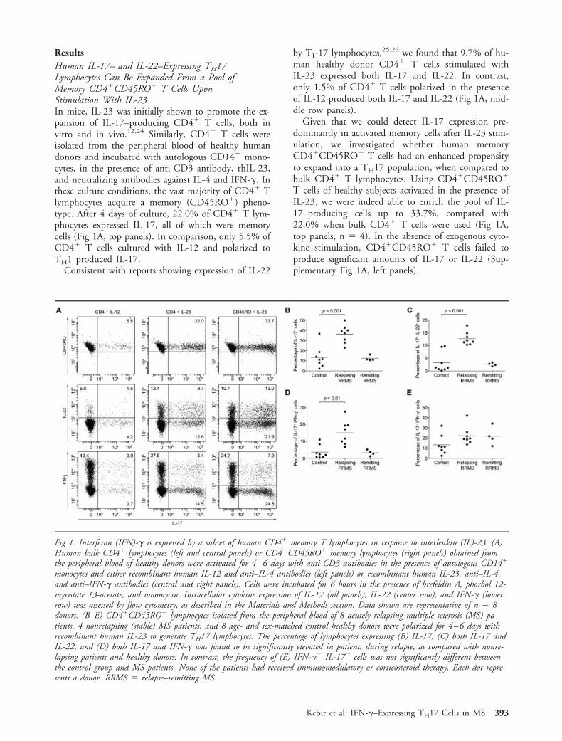
ResultsHuman IL-17– and IL-22–Expressing TH17Lymphocytes Can Be Expanded From a Pool ofMemory CD4�CD45RO� T Cells UponStimulation With IL-23In mice, IL-23 was initially shown to promote the ex-pansion of IL-17–producing CD4� T cells, both invitro and in vivo.12,24 Similarly, CD4� T cells wereisolated from the peripheral blood of healthy humandonors and incubated with autologous CD14� mono-cytes, in the presence of anti-CD3 antibody, rhIL-23,and neutralizing antibodies against IL-4 and IFN-�. Inthese culture conditions, the vast majority of CD4� Tlymphocytes acquire a memory (CD45RO�) pheno-type. After 4 days of culture, 22.0% of CD4� T lym-phocytes expressed IL-17, all of which were memorycells (Fig 1A, top panels). In comparison, only 5.5% ofCD4� T cells cultured with IL-12 and polarized toTH1 produced IL-17.
Consistent with reports showing expression of IL-22
by TH17 lymphocytes,25,26 we found that 9.7% of hu-man healthy donor CD4� T cells stimulated withIL-23 expressed both IL-17 and IL-22. In contrast,only 1.5% of CD4� T cells polarized in the presenceof IL-12 produced both IL-17 and IL-22 (Fig 1A, mid-dle row panels).
Given that we could detect IL-17 expression pre-dominantly in activated memory cells after IL-23 stim-ulation, we investigated whether human memoryCD4�CD45RO� T cells had an enhanced propensityto expand into a TH17 population, when compared tobulk CD4� T lymphocytes. Using CD4�CD45RO�
T cells of healthy subjects activated in the presence ofIL-23, we were indeed able to enrich the pool of IL-17–producing cells up to 33.7%, compared with22.0% when bulk CD4� T cells were used (Fig 1A,top panels, n 4). In the absence of exogenous cyto-kine stimulation, CD4�CD45RO� T cells failed toproduce significant amounts of IL-17 or IL-22 (Sup-plementary Fig 1A, left panels).
Fig 1. Interferon (IFN)-� is expressed by a subset of human CD4� memory T lymphocytes in response to interleukin (IL)-23. (A)Human bulk CD4� lymphocytes (left and central panels) or CD4�CD45RO� memory lymphocytes (right panels) obtained fromthe peripheral blood of healthy donors were activated for 4–6 days with anti-CD3 antibodies in the presence of autologous CD14�
monocytes and either recombinant human IL-12 and anti–IL-4 antibodies (left panels) or recombinant human IL-23, anti–IL-4,and anti–IFN-� antibodies (central and right panels). Cells were incubated for 6 hours in the presence of brefeldin A, phorbol 12-myristate 13-acetate, and ionomycin. Intracellular cytokine expression of IL-17 (all panels), IL-22 (center row), and IFN-� (lowerrow) was assessed by flow cytometry, as described in the Materials and Methods section. Data shown are representative of n 8donors. (B–E) CD4�CD45RO� lymphocytes isolated from the peripheral blood of 8 acutely relapsing multiple sclerosis (MS) pa-tients, 4 nonrelapsing (stable) MS patients, and 8 age- and sex-matched control healthy donors were polarized for 4–6 days withrecombinant human IL-23 to generate TH17 lymphocytes. The percentage of lymphocytes expressing (B) IL-17, (C) both IL-17 andIL-22, and (D) both IL-17 and IFN-� was found to be significantly elevated in patients during relapse, as compared with nonre-lapsing patients and healthy donors. In contrast, the frequency of (E) IFN-�� IL-17� cells was not significantly different betweenthe control group and MS patients. None of the patients had received immunomodulatory or corticosteroid therapy. Each dot repre-sents a donor. RRMS relapse–remitting MS.
Kebir et al: IFN-�–Expressing TH17 Cells in MS 393

In our culture system, IL-17– or IL-22–expressinglymphocytes could not be expanded from human naiveCD4�CD45RA� T cells sorted and cultured in thepresence of autologous monocytes, in response toIL-23 (Supplementary Fig 1B). Surprisingly, even after4–6 days in culture under these conditions, the major-ity of naive T cells remained CD45RA�.
In parallel with the intracellular cytokine produc-tion, we measured secretion of IL-17, IL-22, andIFN-� in the supernatants of the TH1- and TH17-polarized human CD4� or CD4�CD45RO� T cells(Supplementary Fig 2A). As expected, without IL-23stimulation, activated CD4�CD45RO� T cells didnot secrete significant amounts of IL-17 or IL-22. Inthe presence of IL-23, memory CD4�CD45RO� lym-phocytes secreted high levels of IL-17 and IL-22.
A Subpopulation of Human MemoryCD4�CD45RO� T Cells Concomitantly ExpressesIL-17 and IFN-� Upon Stimulation With IL-23In addition to IL-17– and IL-22–producing lympho-cytes, we also noted in the IL-23–driven TH17 culturesthe presence of cells coexpressing IL-17 and IFN-�(Fig 1A, lower panels), a phenomenon rarely seen inmice, at least not to this magnitude, but previously re-ported in the gut of patients with inflammatory boweldisease27 and in coronary artery infiltrates.19 Notably,although a relatively high amount of TH17-polarizedcells expressed IFN-� by intracellular cytokine staining(7.9% IL-17� IFN-��, Fig 1A), IFN-� secretion byTH17 cells remained low compared with TH1-polarized cells (Supplementary Fig 2A). The propor-tion of CD4�CD45RO� T lymphocytes expressingIL-17, IL-22, IFN-�, or a combination of these cyto-kines increased over time, at least until 6 days afterIL-23 stimulation (Supplementary Fig 2B). Collec-tively, our data demonstrate that a subpopulation ofhuman IL-17–expressing CD4� T lymphocytes alsoproduces IFN-�. This unique population can be ex-panded from a subset of CD4�CD45RO� T cells inresponse to IL-23 stimulation.
Increased Frequency of Lymphocytes Coexpressing IL-17 and IFN-� in TH17 Cell Lines of MS PatientsTo establish the relevance and relative contribution ofthese T cell subsets in MS, we analyzed the intracellu-lar expression of IL-17, IL-22, and IFN-� in peripheralblood CD4�CD45RO� memory T lymphocytes ofMS patients and healthy donors by flow cytometry.Twelve female patients with clinically definite relapse–remitting MS (RRMS) were included in the study.Their main characteristics are summarized in the Ma-terials and Methods section. As in healthy controls,freshly isolated peripheral blood CD4�CD45RO�
memory T lymphocytes from MS patients did not ex-press high levels of proinflammatory cytokines even
following 6 hour activation with PMA and ionomycin.Both in MS and controls, IL-17–producing cells ac-counted for 2% of the total CD4�CD45RO� Tlymphocyte population (data not shown). However,when memory T cells were stimulated in vitro for 6days with IL-23 to induce a TH17 profile, according tothe protocol described above, we observed significantdifferences in the intracellular cytokine expression pat-tern between TH17 cell lines of MS patients and thoseof age-matched controls. The percentages of IL-17�
(Fig 1B) and of IL-22� (data not shown) lymphocytes,as well as the percentage of cells simultaneously ex-pressing IL-17 and IL-22 (Fig 1C), were elevated inTH17 cell lines derived from the blood of RRMS pa-tients during a clinical exacerbation as compared withpatients in remission and healthy individuals. Likewise,the frequency of CD4� lymphocytes coexpressingIL-17 and IFN-� was greatly increased in RRMS pa-tients with active disease relative to controls (Fig 1D).Interestingly, although the overall production of IFN-�increased in MS patients, the percentages of IFN-� sin-gle producers were not significantly different betweenthe control group and MS patients, whether in relapseor in remission (Fig 1E). Surprisingly, in men, no dif-ferences were observed in the cytokine expression pro-file between controls and MS patients (data notshown). The reason for this gender discrepancy is notclear at present, and could also be due to the relativelysmall sample size.
TH17 Lymphocyte Migration Across the Human BBBIs Mediated by ICAM-1Leukocyte infiltration into the CNS is known to be animportant event in the development of MS lesions.2 Inour previous study, we showed that TH17 lympho-cytes, like TH1 cells, migrate efficiently across the BBB,and we reported an abundance of IL-17– and IL-22–expressing CD45RO� memory T lymphocytes inhighly infiltrated MS lesions.4 However, the exact ad-hesion molecules that direct the trafficking of TH17lymphocytes into the CNS remain largely unknown.ICAM-1 and VCAM-1, 2 members of the immuno-globulin superfamily expressed by the brain endothe-lium, are thought to participate in this highly regulatedprocess. To address the specific contribution of theseadhesion molecules to the TH17 transmigration pro-cess, we performed an in vitro transendothelial migra-tion assay, using primary cultures of human BBB-ECs,in the presence of blocking antibodies to ICAM-1 andVCAM-1. The addition of anti–ICAM-1 antibody tohuman BBB-ECs led to a significant decrease in TH17migration across the endothelium (p 0.001; n 6),whereas no inhibition was observed in the presence ofanti–VCAM-1 antibody, as compared with the isotypecontrol (Fig 2A). The anti–VCAM-1 antibody was pre-viously shown to be effective in blocking the migration
394 Annals of Neurology Vol 66 No 3 September 2009

Fig 2. Adhesion molecules involved in the recruitment of human TH17 lymphocytes across the blood–brain barrier (BBB).CD4�CD45RO� lymphocytes were isolated from the peripheral blood of healthy human donors and cultured for 4 days with autol-ogous CD14� monocytes, anti-CD3, anti-interleukin (IL)-4, and anti-interferon (IFN)-� antibodies in the presence of IL-23 togenerate TH17 lymphocytes. (A) TH17 lymphocytes (1 � 106) were allowed to migrate for 18 hours across a confluent monolayerof primary culture of human BBB endothelial cells (ECs) in a Boyden chamber migration assay, in the presence of blocking anti-bodies against intercellular adhesion molecule (ICAM)-1 or vascular cell adhesion molecule (VCAM)-1. The bar chart is representa-tive of 5 independent experiments using 4 different BBB-EC preparations. (B) Intracellular cytokine expression of IL-17 and IFN-�and surface expression of lymphocyte function-associated antigen (LFA)-1 was assessed in human TH1 or TH17 lymphocytes by flowcytometry. Data are representative of n 6 independent experiments. (C) Confluent monolayers of primary cultures of humanBBB-ECs were treated with IFN-� (100U/ml), IL-17 (100ng/ml); or IL-22 (100ng/ml) for 24 hours, and surface expression of theadhesion molecule ICAM-1 was assessed by flow cytometry. Data are representative of n 3 independent experiments using 3 dis-tinct BBB-EC preparations. (D) TH17 lymphocytes (1 � 106) were allowed to migrate for 18 hours across confluent monolayers ofprimary cultures of human BBB-ECs in Boyden chamber migration assays. A total of 2.5 � 105 TH17 cells migrated and werecollected from the lower chamber. IFN-� and IL-17 expression by TH17 lymphocytes was assessed prior to (black bars) and follow-ing (white bars) migration. Data demonstrate a significant enrichment of IL-17� IFN-�� TH17 lymphocytes on trans-BBB migra-tion. The bar chart represents the mean � standard error of the mean (right axis) and percentage (left axis) of migrated and non-migrated cells. There were 3 independent experiments using 6 lymphocyte donors and 3 distinct BBB-EC cultures.
Kebir et al: IFN-�–Expressing TH17 Cells in MS 395
of monocytes across the BBB in a similar migrationassay system.7 Like TH1 lymphocytes, the vast majorityof TH17 lymphocytes expressed LFA-1, the bindingpartner of ICAM-1 (Fig 2B). These results indicatethat the transmigration of TH17 lymphocytes acrossthe human BBB is mediated mainly by ICAM-1, al-though the involvement of other adhesion moleculescannot be ruled out.
It is well established that the expression of severaladhesion molecules is upregulated on the surface ofECs in response to inflammatory stimuli.28,29 Wetherefore sought to determine whether any of the
proinflammatory cytokines produced by TH17 lym-phocytes could induce a change in the EC expressionof ICAM-1, and in turn favor their recruitment intothe CNS. Confluent monolayers of BBB-ECs weretreated for 24 hours with IL-17, IL-22, or IFN-�,and the level of surface-expressed ICAM-1 was deter-mined by flow cytometric analysis. A representativehistogram is shown in Figure 2C. Whereas bothIL-17 and IL-22 failed to modulate the expression ofICAM-1 on the endothelium, exposure to IFN-� in-duced a strong upregulation of the adhesion moleculeon human BBB-ECs (from 38.8% in resting condi-
396 Annals of Neurology Vol 66 No 3 September 2009

tions to 71.8% upon activation with IFN-�). Cotreat-ment of BBB-derived ECs with combinations of IL-17, IL-22, and IFN-�, did not affect or potentiate theeffect seen with IFN-� alone (data not shown). Col-lectively, these data suggest that IFN-� may play aninstrumental role in facilitating the recruitment ofTH17 lymphocytes across the BBB by enhancing thelocal expression of ICAM-1, an effect that seems tobe independent of IL-17 or IL-22.
IFN-�–Producing TH17 Lymphocytes Migrate MoreEfficiently Across Human BBB ECsWe next sought to determine whether the productionof IFN-� by TH17 lymphocytes could influence theirability to migrate across the BBB, and would conferIL-17–IFN-� double-producing cells an advantageover IL-17 single producers in the in vitro transmi-gration assay system. To do so, we compared the cy-tokine profile of TH17-expanded cells before and af-ter transendothelial migration, with respect to theirIL-17 and IFN-� expression. The subpopulation ofTH17 lymphocytes that concomitantly express IL-17and IFN-� was markedly enriched following migra-tion across the human EC monolayer, doubling from16.3% � 2.2% of the input cells to 33.3% � 2.6%of the transmigrated fraction ( p 0.01; Fig 2D). Incontrast, the frequency of IL-17 single producers re-mained similar in the 2 fractions (14.9% � 2.2% be-fore migration and 21.7% � 1.3% after migration,p � 0.05). Likewise, we did not observe a significantchange in the percentage of single IFN-�–expressingcells following transmigration across the human BBB.Together, these findings suggest that the subpopula-
tion of TH17 lymphocytes coproducing IL-17 andIFN-� has an increased propensity to migrate acrossthe brain endothelium and may display an advantageto be selectively recruited to sites of inflammation.Although unlikely, we cannot exclude the possibilitythat the brain endothelium induces the production ofIL-17 or IFN-� during the transmigration process.
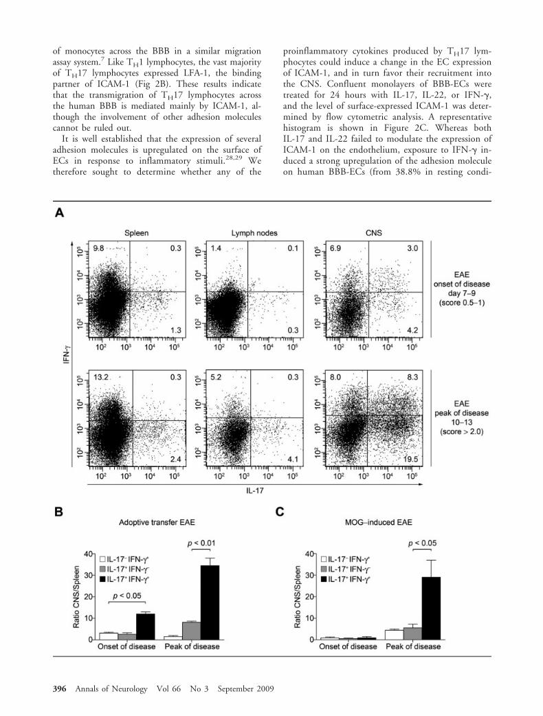
IFN-�–Secreting TH17 Lymphocytes Are Recruited tothe CNS of Mice During the Course of EAETo confirm the in vivo significance of our findings, weelected to study the kinetics of TH17 lymphocyte entryinto the CNS by transferring IL-23–driven MOG–spe-cific TH17 lymphocytes into naive animals. For thatpurpose, C57BL/6 mice immunized with MOG35–55
were sacrificed during the presymptomatic phase of thedisease. Spleen and inguinal lymph nodes were col-lected, dissociated, and cultured with MOG peptide inthe presence of IL-23. In the initial in vitro IL-23–driven TH17 cultures, 8.4% of the CD3�CD4� Tcells were IL-17�, and merely 1.9% coexpressed IL-17and IFN-� (data not shown). The in vitro–generatedTH17 lymphocyte cultures were reinjected intoC57BL/6 recipient animals, which were sacrificed atthe onset of disease (day 7–9, EAE score 0.5–1.0) or atthe peak of disease (day 10–13, EAE score �2.5). Asshown in Figure 3A, we found that a significant pro-portion of IL-17–producing CD4� T lymphocytes re-covered from the CNS of recipient animals also coex-pressed IFN-�. The proportion of IFN-�� IL-17�
CD4� T cells in the CNS was markedly enriched ascompared with the injected population (8.3% vs1.9%), as well as with those found in the secondary
Š Fig 3. Preferential recruitment of interferon (IFN)-�–expressing TH17 lymphocytes in the central nervous system (CNS) ofC57BL/6 experimental allergic encephalomyelitis (EAE) mice. (A) Immune cells were isolated from the spleen and lymph nodes ofmyelin oligodendrocyte glycoprotein (MOG)35–55-induced EAE animals at day 8 and restimulated in vitro for 48 hours with anti-CD3 and recombinant mouse interleukin (IL)-23 to induce a TH17 phenotype. TH17 lymphocytes (25 � 106) were transferredintraperitoneally to naive C57BL/6 animals. Adoptively transferred EAE mice were sacrificed at the onset of symptoms (days 7–9,score 0.5–1.0, top panels) or at the peak of disease (days 10–13, EAE score �2.5, bottom panels). IFN-� and IL-17 expression byCD3�CD4� lymphocytes recovered from the spleen (left panels), the inguinal lymph nodes (center panels), and the CNS (rightpanels) of recipient animals were assessed by flow cytometry. Data reveal a substantial enrichment of IFN-�–expressing TH17 lym-phocytes in the CNS of EAE mice at the peak of the disease, as compared with the spleen or the lymph nodes of the same animals.Data are representative of 2 independent experiments with n 3 animals per group. Dot plots were gated on CD3�CD4� Tcells. (B) EAE was induced by adoptive transfer of TH17 lymphocytes as described above. Animals were sacrificed at the onset ofsymptoms (EAE score 0.5–1.0, days 7–9) or at the peak of disease (score �2.5, days 10–13). IFN-� and IL-17 expression byCD3�CD4� lymphocytes recovered from the spleen and the CNS of recipient animals was assessed by flow cytometry. Data arepresented as the ratio (CNS over spleen) of the absolute number of CD4� T cells recovered from individual animals expressing IL-17� IFN-�� (white bars), IL-17� IFN-�� (grey bars), and IL-17� IFN-�� (black bars). Data demonstrate a significant enrich-ment of IFN-�–expressing CD4� TH17 lymphocytes in the CNS of adoptively transferred EAE animals, as compared with IFN-��
TH17 lymphocytes, especially at the peak of disease. Bar chart represents the mean � standard error of the mean (SEM) from n 2 independent experiments using 3 animals per group. (C) EAE was induced in C57BL/6 mice by active immunization withMOG35–55 peptide. Animals were sacrificed at the onset of symptoms (EAE score 0.5–1.0, days 10–12) or at the peak of disease(EAE score �2.5, days 16–18). IFN-� and IL-17 expression by CD3�CD4� lymphocytes recovered from the spleen and the CNSwas assessed by flow cytometry, as previously described. Again, data demonstrate that IFN-�–expressing TH17 lymphocytes accumu-late specifically in the CNS and do so more than IFN-�� TH17 lymphocytes. Bar charts represent the mean � SEM from n 3independent experiments using 3 animals per group.
Kebir et al: IFN-�–Expressing TH17 Cells in MS 397

lymphoid organs (Fig 3A). During early disease, wemeasured a 12-fold increase in the frequency of IFN-�–IL-17 double producers in the CNS relative toIL-17 or IFN-� single-producing cells (Fig 3B, p 0.05). This specific CNS enrichment of IL-17�
IFN-�� CD4 T cells was even more pronounced atthe peak of disease, with a nearly 35-fold increase (Fig3B, p 0.01).
Likewise, when EAE was induced in C57BL/6 ani-mals by active immunization with MOG35–55, we mea-sured an increase of double-positive cells in the CNS,albeit only at the peak of disease (Fig 3C, 29-fold, p 0.05). Together with the results obtained in the invitro migration assay, these in vivo data confirm thepreferential ability of IFN-�–expressing TH17 lympho-cytes over IFN-�� TH17 cells to access the CNS com-partment.
IFN-�–Expressing TH17 Lymphocytes Are Present inActive MS Perivascular InfiltratesWe and others have previously reported the presence ofIL-17� perivascular lymphocytes in active lesions ofMS patients.4,14 Using our collection of frozen MSbrains, we sought to evaluate whether IL-17–expressinglymphocytes were also immunopositive for IFN-�within MS perivascular infiltrates. Histological exami-nation (Luxol Fast Blue and hematoxylin–eosin stain-ing) of 15–20 MS frozen tissue blocks revealed thepresence of perivascular infiltrates within areas of lim-ited demyelination (Fig 4A) in 8 blocks from 2 pa-tients, suggestive of early lesion formation. In thesesamples, MS tissue infiltrates consisted mainly ofCD3� T cells (data not shown) but also comprisedmyelin debris, as depicted in the corner insets of Figure4A. We found that a subpopulation of perivascular IL-17–expressing cells was also immunopositive forIFN-�, in 12 out of 12 active lesions (Fig 4B, n 2patients). In our hands, most of the IL-17–expressinglymphocytes found within MS tissue were immunopo-sitive for IFN-�, corroborating the findings by Annun-ziato et al. in human Crohn’s disease.27 In addition,IFN-�–expressing TH17 lymphocytes were detected inparenchymal tissue (Fig 4C), indicating that these Tcells are not restricted to perivascular infiltrates.
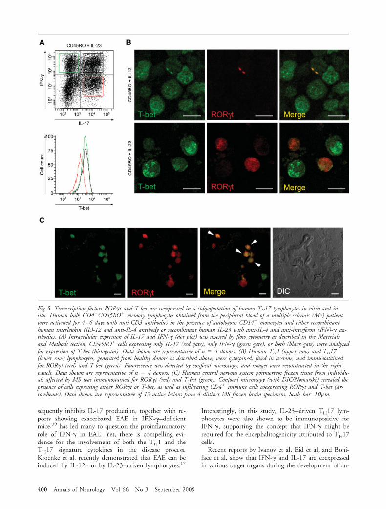
The Transcriptional Regulators ROR�t and T-betAre Coexpressed in Subsets of TH17 LymphocytesIn Vitro and In SituROR�t and T-bet are well-known transcriptional reg-ulators of the cytokine phenotype of lymphocytes.Whereas ROR�t is strongly associated with productionof IL-17 and the TH17 lineage,30,31 expression ofT-bet controls IFN-� production and defines the TH1phenotype.32 We thus elected to study the expressionof these 2 transcriptional markers to validate our intra-cellular cytokine staining and in situ immunohistofluo-
rescence findings. Human CD4�CD45RO� lympho-cytes were polarized for 3–5 days in the presence ofIL-23 with autologous CD14� monocytes, and T-betwas analyzed in cells expressing IFN-� alone (Fig 5A,green gate), IL-17 alone (red gate), or both IFN-� andIL-17 (black gate). We found that IFN-� single-producing lymphocytes expressed higher levels of T-betthan IL-17–producing cells. Cells coproducing IL-17and IFN-� expressed similar levels of T-bet as IFN-�single producers.
To evaluate whether ROR�t and T-bet could be co-expressed in individual lymphocytes, we next per-formed cytospin of human CD4�CD45RO� TH1 andTH17 lymphocyte cultures followed by immunofluo-rescent staining and confocal microscopy. In TH1 lym-phocyte cultures, most cells were strongly immunore-active for T-bet, whereas ROR�t signal was rare (Fig5B). Conversely, in TH17 cultures, we detectedROR�t� and T-bet� lymphocytes, as well as numer-ous T lymphocytes coexpressing T-bet and ROR�t. Fi-nally, we looked for the presence and number of lym-phocytes coexpressing T-bet and ROR�t within activeMS lesions. We found cells expressing T-bet, ROR�t,or both regulators of transcription in the perivascularspace of active MS lesions (Fig 5C). T-bet– andROR�t–expressing cells were seen in equal proportion;30% of the infiltrating lymphocytes coexpressed T-betand ROR�t (Fig 5C). Interestingly, whereas T-bet ex-pression was found to be stable, we detected variablelevels of ROR�t expression in individual lymphocytes(Fig 5C, arrowheads), suggesting that IL-17 regulationand TH17 plasticity can probably occur within the hu-man CNS.
DiscussionBoth TH1 and TH17 lymphocytes have been impli-cated in the pathology of MS and EAE. Their postu-lated role in driving CNS inflammation has been as-cribed essentially to IFN-� and IL-17, the effectorcytokines that respectively define these T-cell subsets.Originally, TH1 lymphocytes were recognized as themajor pathogenic population in MS and more partic-ularly in EAE. But the discovery of the TH17 lineageuncovered a pivotal role for IL-17–producing CD4� Tcells in mediating the disease in mice.33,34 Subsequentreports, including our own, extended these findings tohumans by showing the presence of IL-17–expressingmemory T cells in active MS lesions.4,7,14
Unlike the TH1–driving cytokine IL-12, IL-23 issaid to be unequivocally required for the induction ofEAE.12 A positive correlation was also established inhumans between levels of IL-23 and MS.35 In apparentcontrast, the precise role of the TH17 effector cytokineIL-17 in the initiation of EAE and MS remains con-troversial. The observation that blockade of IL-17 onlyhas a minor impact on the development of the disease
398 Annals of Neurology Vol 66 No 3 September 2009

in mice15,16 raises the possibility of additional effectorcytokines, or combination of cytokines, being essentialfor the induction of the CNS inflammatory process. Inthe current study, we demonstrate that upon stim-ulation with IL-23, human peripheral bloodCD4�CD45RO� memory lymphocytes adopt a TH17phenotype characterized by the production of IL-17and IL-22. We confirm that human memoryCD4�CD45RO� cells are more prone to expandinto TH17 lymphocytes as compared with naiveCD4�CD45RA� or unfractionated CD4� T cells. Wepostulate that these peripheral blood memory lympho-cytes probably enclose a larger pool of already predif-ferentiated TH17 cells, as previously hypothesized byStockinger.36 More importantly, we detect in the IL-
23–driven human TH17 cell lines a small proportionof CD4� lymphocytes expressing IFN-�, with a sub-population of cells simultaneously producing IL-17and IFN-�, despite the fact that cells were expanded inthe presence of IFN-�–neutralizing antibodies. Weshow that the proportion of IFN-�� IL-17� dual pro-ducers is significantly increased in MS patients in acuterelapse but not during remission, and that these cellscoexpress the transcription factors T-bet and ROR�t,known to be required for IFN-� and IL-17 produc-tion, respectively. Our data further suggest that theseIFN-�� IL-17� double producers have an advantageover IL-17 or IFN-� single-expressing cells to cross theBBB and access the CNS, both in vitro and in vivo.Moreover, we demonstrate that these cells represent animportant population of T lymphocytes within perivas-cular infiltrates in MS brain, supporting the biologicalrelevance of IFN-�–expressing TH17 lymphocytes. Thedemonstration that ROR�t and T-bet are also coex-pressed in situ within individual cells in the perivascu-lar space of MS lesions supports the pathological rele-vance of IFN-�� TH17 lymphocytes in CNSinflammatory events. Thus, our data expand and areconsistent with the report by Annunziato et al. show-ing coexpression of IFN-� and IL-17 by CD4� T cellsin the gut of patients with Crohn’s disease,27 and putforward a potential important role for IFN-�� IL-17�
memory CD4� T lymphocytes in the pathology of MSand the development of MS lesions.
The initial observation by Harrington et al37 andPark et al38 that IFN-� impedes the differentiation ofnaive CD4� T cells into TH17 lymphocytes, and con-
Š Fig 4. Abundance of interferon (IFN)-�–interleukin (IL)-17coexpressing CD4 lymphocytes in active multiple sclerosis (MS)lesions. (A) Luxol Fast Blue and hematoxylin–eosin staining offrozen human central nervous system (CNS) postmortem mate-rial from MS-affected individuals. Corner insets display ahigher magnification (2.5�) of the area indicated with thearrowhead and show myelin debris. Perivascular infiltrates aresurrounded by a small area of demyelination, suggestive ofearly lesions. Scale bar: 20�m. (B) Human CNS postmortemfrozen tissue from individuals affected by MS were immuno-stained for IL-17 (green), IFN-� (red), and nuclear stain(Hoechst). Fluorescent microscopy revealed the presence of nu-merous cells coexpressing IFN-� and IL-17 (merged image) in12 of 12 active lesions from 2 distinct MS frozen brain speci-mens. Scale bar: 10�m. (C) Confocal microscopy confirmedcoexpression of IL-17 (green) and IFN-� (red) at the single-cell level within MS tissues. The scatter graph in the bottomright panel shows the distribution of pixels for both fluoro-chromes, where high degree of colocalization is seen in pixelsmaking up the cell indicated with the arrowhead (white dot).Colocalization analysis of cells coexpressing IL-17 and IFN-�resulted in an overlap coefficient ranging from 0.9 to 1. Datashown are representative of n 20 fields from 2 distinct MSbrain specimens. Scale bar: 10�m.
Kebir et al: IFN-�–Expressing TH17 Cells in MS 399
sequently inhibits IL-17 production, together with re-ports showing exacerbated EAE in IFN-�–deficientmice,39 has led many to question the proinflammatoryrole of IFN-� in EAE. Yet, there is compelling evi-dence for the involvement of both the TH1 and theTH17 signature cytokines in the disease process.Kroenke et al. recently demonstrated that EAE can beinduced by IL-12– or by IL-23–driven lymphocytes.17
Interestingly, in this study, IL-23–driven TH17 lym-phocytes were also shown to be immunopositive forIFN-�, supporting the concept that IFN-� might berequired for the encephalitogenicity attributed to TH17cells.
Recent reports by Ivanov et al, Eid et al, and Boni-face et al. show that IFN-� and IL-17 are coexpressedin various target organs during the development of au-
Fig 5. Transcription factors ROR�t and T-bet are coexpressed in a subpopulation of human TH17 lymphocytes in vitro and insitu. Human bulk CD4�CD45RO� memory lymphocytes obtained from the peripheral blood of a multiple sclerosis (MS) patientwere activated for 4–6 days with anti-CD3 antibodies in the presence of autologous CD14� monocytes and either recombinanthuman interleukin (IL)-12 and anti-IL-4 antibody or recombinant human IL-23 with anti-IL-4 and anti-interferon (IFN)-� an-tibodies. (A) Intracellular expression of IL-17 and IFN-� (dot plot) was assessed by flow cytometry as described in the Materialsand Methods section. CD45RO� cells expressing only IL-17 (red gate), only IFN-� (green gate), or both (black gate) were analyzedfor expression of T-bet (histogram). Data shown are representative of n 4 donors. (B) Human TH1 (upper row) and TH17(lower row) lymphocytes, generated from healthy donors as described above, were cytospined, fixed in acetone, and immunostainedfor ROR�t (red) and T-bet (green). Fluorescence was detected by confocal microscopy, and images were reconstructed in the rightpanels. Data shown are representative of n 4 donors. (C) Human central nervous system postmortem frozen tissue from individu-als affected by MS was immunostained for ROR�t (red) and T-bet (green). Confocal microscopy (with DIC/Nomarski) revealed thepresence of cells expressing either ROR�t or T-bet, as well as infiltrating CD4� immune cells coexpressing ROR�t and T-bet (ar-rowheads). Data shown are representative of 12 active lesions from 4 distinct MS frozen brain specimens. Scale bar: 10�m.
400 Annals of Neurology Vol 66 No 3 September 2009

toimmune experimental inflammation, includingEAE.19,20,31 Studies by our group further support therelevance of these 2 cytokines to pathogenic processesoccurring at the level of the BBB. IFN-� modulates theexpression of several adhesion molecules on the endo-thelium surface, whereas IL-17 contributes to thebreakdown of the BBB by disrupting tight junc-tions.4,21 But in spite of the extensive amount of stud-ies investigating the specific roles of IFN-� and IL-17in CNS inflammation, only scarce data are availableregarding the possible collaborative interplay betweenthese 2 cytokines, especially at the single-cell level.Here, we provide evidence that unlike IL-17 and IL-22, IFN-� upregulates the expression of ICAM-1 onthe surface of BBB-ECs. We further identify ICAM-1,but not VCAM-1, as an important adhesion moleculethat controls TH17 lymphocyte migration across theBBB. The ability of IFN-� to induce expression of theadhesion molecule ICAM-1 on the brain endotheliummay therefore positively affect the capacity of TH17lymphocytes to migrate across the BBB. Combinedwith the barrier disrupting effect conferred by IL-17and IL-22,4 expression of IFN-� by TH17 lymphocytesthus represents an additional means by which TH17cells could promote their own recruitment to the CNS,as well as that of bystander leukocytes. As VCAM-1blockade failed to impact on TH17 migration, our dataalso suggest that the clinical efficacy of anti-very lateantigen (VLA)-4 antibody natalizumab (Tysabri) is de-pendent on the ability of VLA-4 to bind a yet uniden-tified ligand, in addition to VCAM-1.
Although it is difficult at this point to establishwhether the concomitant production of IFN-� andIL-17 by CD4� lymphocytes represents a transitionalstage of commitment of TH17 cells, we demonstratehere that coexpression of IL-17 and IFN-� confers asignificant migratory advantage over IL-17 or IFN-�single producers. In a recent report, Lee et al. put for-ward the concept of late developmental plasticity in theTH17 lineage.18 In their study, they show that TGF-�is required to sustain IL-17 expression. Conversely, theabsence of transforming growth factor (TGF)-� pro-motes IFN-� production by CD4� lymphocytes in anIL-23– and IL-12–dependent way. We thus speculatethat following migration into the CNS, IL-17–IFN-�double producers (possible “transitional” TH17) foundin the blood of relapsing MS patients could be influ-enced by the CNS environment, presumably throughTGF-�, to fully differentiate into TH17 cells. In thisrespect, we and others have recently shown that humanand mouse CNS-derived ECs, astrocytes, and dendriticcells secrete TGF-� and promote a TH17, rather thana TH1 phenotype.7
Overall, our data underline the existence and regu-lation of IFN-�–expressing TH17 lymphocytes in MS.Our study further sheds light on the possible synergis-
tic effect of these proinflammatory cytokines, especiallyin favoring the migration of double-producing cellsacross the BBB. This particular subpopulation of cells,characterized by the simultaneous production of IL-17and IFN-� is preferentially recruited to the CNSmainly through the IFN-�–inducible ICAM-1/LFA-1adhesion pathway. Given that IL-23 is important fordevelopment of EAE and of IL-17� IFN-�� doubleproducers, and that both T-bet and ROR�t play a cru-cial role in CNS inflammation, we speculate that co-expression of ROR�t, T-bet and IFN-� by TH17lymphocytes could contribute to their pathogenic po-tential.
This study was supported by funding from the Canadian Institutesof Health Research (CIHR) and the Multiple Sclerosis Society ofCanada (MSSC). A.P. and N.A. are Research Scholars from theFonds de la Recherche en Sante du Quebec and hold the DonaldPaty Career Development Award of the MSSC. H.K. holds a schol-arship research award from the CIHR. J.I.A. holds a fellowship fromthe CIHR Strategic Training Initiative in Health Research Neuroin-flammation Training Program and the MSSC.
We thank K. Kreymborg for helpful advice on theadoptive transfer protocol.
References1. Weiner HL. Multiple sclerosis is an inflammatory T-cell-
mediated autoimmune disease. Arch Neurol 2004;61:1613–1615.
2. Sospedra M, Martin R. Immunology of multiple sclerosis.Annu Rev Immunol 2005;23:683–747.
3. Burns J, Bartholomew B, Lobo S. Isolation of myelin basicprotein-specific T cells predominantly from the memory T-cellcompartment in multiple sclerosis. Ann Neurol 1999;45:33–39.
4. Kebir H, Kreymborg K, Ifergan I, et al. Human TH17 lym-phocytes promote blood-brain barrier disruption and centralnervous system inflammation. Nat Med 2007;13:1173–1175.
5. Lucchinetti CF, Bruck W, Rodriguez M, et al. Distinct patternsof multiple sclerosis pathology indicates heterogeneity onpathogenesis. Brain Pathol 1996;6:259–274.
6. Greter M, Heppner FL, Lemos MP, et al. Dendritic cells per-mit immune invasion of the CNS in an animal model of mul-tiple sclerosis. Nat Med 2005;11:328–334.
7. Ifergan I, Kebir H, Bernard M, et al. The blood-brain barrierinduces differentiation of migrating monocytes into Th17-polarizing dendritic cells. Brain 2008;131:785–799.
8. Friese MA, Jakobsen KB, Friis L, et al. Opposing effects ofHLA class I molecules in tuning autoreactive CD8� T cells inmultiple sclerosis. Nat Med 2008;14:1227–1235.
9. Babbe H, Roers A, Waisman A, et al. Clonal expansions ofCD8(�) T cells dominate the T cell infiltrate in active multiplesclerosis lesions as shown by micromanipulation and single cellpolymerase chain reaction. J Exp Med 2000;192:393–404.
10. Renno T, Zeine R, Girard JM, et al. Selective enrichment ofTh1 CD45RBlow CD4� T cells in autoimmune infiltrates inexperimental allergic encephalomyelitis. Int Immunol 1994;6:347–354.
11. Merrill JE, Kono DH, Clayton J, et al. Inflammatory leuko-cytes and cytokines in the peptide-induced disease of experi-mental allergic encephalomyelitis in SJL and B10.PL mice. ProcNatl Acad Sci U S A 1992;89:574–578.
Kebir et al: IFN-�–Expressing TH17 Cells in MS 401

12. Langrish CL, Chen Y, Blumenschein WM, et al. IL-23 drives apathogenic T cell population that induces autoimmune inflam-mation. J Exp Med 2005;201:233–240.
13. Cua DJ, Sherlock J, Chen Y, et al. Interleukin-23 rather thaninterleukin-12 is the critical cytokine for autoimmune inflam-mation of the brain. Nature 2003;421:744–748.
14. Tzartos JS, Friese MA, Craner MJ, et al. Interleukin-17 pro-duction in central nervous system-infiltrating T cells and glialcells is associated with active disease in multiple sclerosis. Am JPathol 2008;172:146–155.
15. Komiyama Y, Nakae S, Matsuki T, et al. IL-17 plays an im-portant role in the development of experimental autoimmuneencephalomyelitis. J Immunol 2006;177:566–573.
16. Haak S, Croxford AL, Kreymborg K, et al. IL-17A and IL-17Fdo not contribute vitally to autoimmune neuro-inflammation inmice. J Clin Invest 2009;119:61–69.
17. Kroenke MA, Carlson TJ, Andjelkovic AV, et al. IL-12- andIL-23-modulated T cells induce distinct types of EAE based onhistology, CNS chemokine profile, and response to cytokine in-hibition. J Exp Med 2008;205:1535–1541.
18. Lee YK, Turner H, Maynard CL, et al. Late developmentalplasticity in the T helper 17 lineage. Immunity 2009;30:92–107.
19. Eid RE, Rao DA, Zhou J, et al. Interleukin-17 and interferon-gamma are produced concomitantly by human coronary artery-infiltrating T cells and act synergistically on vascular smoothmuscle cells. Circulation 2009;119:1424–1432.
20. Boniface K, Bak-Jensen KS, Li Y, et al. Prostaglandin E2 reg-ulates Th17 cell differentiation and function through cyclicAMP and EP2/EP4 receptor signaling. J Exp Med 2009;206:535–548.
21. Cayrol R, Wosik K, Berard JL, et al. Activated leukocyte celladhesion molecule promotes leukocyte trafficking into the cen-tral nervous system. Nat Immunol 2008;9:137–145.
22. Biernacki K, Prat A, Blain M, et al. Regulation of Th1 andTh2 lymphocyte migration by human adult brain endothelialcells. J Neuropathol Exp Neurol 2001;60:1127–1136.
23. Prat A, Biernacki K, Wosik K, et al. Glial cell influence on thehuman blood-brain barrier. Glia 2001;36:145–155.
24. Veldhoen M, Hocking RJ, Atkins CJ, et al. TGFbeta in thecontext of an inflammatory cytokine milieu supports de novodifferentiation of IL-17-producing T cells. Immunity 2006;24:179–189.
25. Liang SC, Tan XY, Luxenberg DP, et al. Interleukin (IL)-22and IL-17 are coexpressed by Th17 cells and cooperatively en-hance expression of antimicrobial peptides. J Exp Med 2006;203:2271–2279.
26. Kreymborg K, Etzensperger R, Dumoutier L, et al. IL-22 isexpressed by Th17 cells in an IL-23-dependent fashion, but notrequired for the development of autoimmune encephalomyeli-tis. J Immunol 2007;179:8098–8104.
27. Annunziato F, Cosmi L, Santarlasci V, et al. Phenotypic andfunctional features of human Th17 cells. J Exp Med 2007;204:1849–1861.
28. Bo L, Peterson JW, Mork S, et al. Distribution of immuno-globulin superfamily members ICAM-1, -2, -3, and the beta 2integrin LFA-1 in multiple sclerosis lesions. J Neuropathol ExpNeurol 1996;55:1060–1072.
29. Luster AD, Alon R, von Andrian UH. Immune cell migrationin inflammation: present and future therapeutic targets. NatImmunol 2005;6:1182–1190.
30. Manel N, Unutmaz D, Littman DR. The differentiation of hu-man T(H)-17 cells requires transforming growth factor-betaand induction of the nuclear receptor RORgammat. Nat Im-munol 2008;9:641–649.
31. Ivanov II, McKenzie BS, Zhou L, et al. The orphan nuclearreceptor RORgammat directs the differentiation program ofproinflammatory IL-17� T helper cells. Cell 2006;126:1121–1133.
32. Lovett-Racke AE, Rocchini AE, Choy J, et al. Silencing T-betdefines a critical role in the differentiation of autoreactive Tlymphocytes. Immunity 2004;21:719–731.
33. McKenzie BS, Kastelein RA, Cua DJ. Understanding the IL-23-IL-17 immune pathway. Trends Immunol 2006;27:17–23.
34. Iwakura Y, Ishigame H. The IL-23/IL-17 axis in inflammation.J Clin Invest 2006;116:1218–1222.
35. Vaknin-Dembinsky A, Balashov K, Weiner HL. IL-23 is in-creased in dendritic cells in multiple sclerosis and down-regulation of IL-23 by antisense oligos increases dendritic cellIL-10 production. J Immunol 2006;176:7768–7774.
36. Stockinger B. Good for goose, but not for gander: IL-2 inter-feres with Th17 differentiation. Immunity 2007;26:278–279.
37. Harrington LE, Hatton RD, Mangan PR, et al. Interleukin 17-producing CD4(�) effector T cells develop via a lineage dis-tinct from the T helper type 1 and 2 lineages. Nat Immunol2005;6:1123–1132.
38. Park H, Li ZX, Yang XO, et al. A distinct lineage of CD4 Tcells regulates tissue inflammation by producing interleukin 17.Nat Immunol 2005;6:1133–1141.
39. Ferber IA, Brocke S, Taylor-Edwards C, et al. Mice with a dis-rupted IFN-gamma gene are susceptible to the induction of ex-perimental autoimmune encephalomyelitis (EAE). J Immunol1996;156:5–7.
402 Annals of Neurology Vol 66 No 3 September 2009
Copyright © 2022 FDOKUMEN