Architectures of the unique domains associated with the DEAD-box helicase motif
Upload
fountain-universityCategory
view
4download
0
Structure of the DNA Repair Helicase Hel308 Reveals DNABinding and Autoinhibitory Domains*
Received for publication, September 10, 2007, and in revised form, November 2, 2007 Published, JBC Papers in Press, December 4, 2007, DOI 10.1074/jbc.M707548200
Jodi D. Richards1, Kenneth A. Johnson1, Huanting Liu, Anne-Marie McRobbie, Stephen McMahon, Muse Oke,Lester Carter, James H. Naismith2, and Malcolm F. White3
From the Centre for Biomolecular Sciences, University of St. Andrews, North Haugh, St. Andrews, Fife KY16 9ST, Scotland
Hel308 is a superfamily 2 helicase conserved in eukaryotes andarchaea. It is thought to function in the early stages of recombina-tion following replication fork arrest and has a specificity forremoval of the lagging strand inmodel replication forks. A homol-ogous helicase constitutes the N-terminal domain of humanDNApolymeraseQ.TheDrosophilahomologuemus301 is implicated indouble strand break repair and meiotic recombination. We havesolved thehigh resolution crystal structure ofHel308 fromthe cre-narchaeon Sulfolobus solfataricus, revealing a five-domain struc-turewith a central pore linedwith essentialDNAbinding residues.The fifth domain is shown to act as an autoinhibitory domain ormolecular brake, clamping the single-stranded DNA extrudedthrough the central pore of the helicase structure to limit the heli-case activity of the enzyme.This provides an elegantmechanism totune the processivity of the enzyme to its functional role. Hel308can displace streptavidin from a biotinylated DNA molecule, andthis activity is only partially inhibited when theDNA is pre-boundwith abundantDNA-binding proteins RPAorAlba1, whereas pre-bindingwith therecombinaseRadAhasnoeffectonactivity.Thesedata suggest thatone functionof theenzymemaybe in the removalof bound proteins at stalled replication forks and recombinationintermediates.
DNA helicases unwind duplex DNA and are essential com-ponents of the DNA replication, recombination, and repairmachinery in all cellular organisms and many viruses. DNAhelicases utilize the energy released by ATP hydrolysis toundergo conformational cycling and translocate along single-stranded DNA (ssDNA),4 displacing a duplex DNA strand in
the process. Many helicases belong to one of three superfami-lies (SF1, 2, and 3), classified according to the conservation ofspecific sequence motifs (1). SF1 and SF2 helicases possess twomotor domains with RecA-like folds that couple ATP hydroly-sis to DNA translocation (2). SF2 DNA helicases include RecGin bacteria, hepatitis C virus NS3, and the RecQ family heli-cases, which all translocate along ssDNAwith a 3� to 5� polarity(3). The RecQ helicases play a key role in maintaining genomicintegrity by stabilizing stalled replication forks and removingintermediates ofDNA recombination (4). Previous studies haveshown that RecQ proteins target specialized DNA structures,specifically branched substrates that mimic replication forksand Holliday junctions. In humans, RecQ family helicasesinclude the BLM and WRN proteins, mutated in certain rareinherited diseases in humans (5).TheHel308 family SF2 helicases, like RecQ, are implicated in
DNA repair, recombination, and genome stability. The found-ingmember, Mus308 fromDrosophila melanogaster, was iden-tified in a screen for mutations conferring hypersensitivity toDNA cross-linking reagents (6). Mus308 consists of an N-ter-minal SF2 helicase fused to a C-terminal DNApolymerase. Thehuman ortholog, PolQ, has the same arrangement (7), and thepolymerase domain has been shown to function efficiently inthe bypass of damagedDNA templates, consistentwith a role inDNA repair (8, 9). In addition to this helicase-polymerasefusion protein, metazoans also encode an ortholog of the heli-case alone. This protein, known asHel308 inHomo sapiens, hasbeen characterized biochemically and shown to function as atypical SF2, 3� to 5�DNAhelicasewith limited processivity (10).The ortholog from D. melanogaster, Mus301, has been shownto function in double strand break repair andmeiotic recombi-nation (11). The Hel308 family helicases therefore have RecQ-like properties.Bacteria and fungi lack orthologs of the Hel308 family, but
clear homologs are present in archaea. Hel308 proteins fromPyrococcus furiosus (also known asHjmhelicase) andMethano-thermobacter thermautotrophicum have been cloned and stud-ied biochemically (12, 13). Both proteins have RecQ-like activ-ities in vitro, targeting branched DNA substrates that aremodels for stalled replication forks and unwinding laggingstrands. Hel308 from M. thermautotrophicum functions likeRecQ in a genetic screen for synthetic lethality in Escherichiacoli, reinforcing the impression that the two proteins may haverelated functions (13).Here we report the high resolution crystal structure of
Hel308 from the crenarchaeote Sulfolobus solfataricus strainPBL2025. The structure reveals a five-domain organization
* This work was supported by the Biotechnology and Biological SciencesResearch Council (BBSRC). The native structure of Hel308 was determinedby the Scottish Structural Proteomics Facility, which is funded by theBBSRC, Scottish Funding Council, and The University of St. Andrews. Thecosts of publication of this article were defrayed in part by the payment ofpage charges. This article must therefore be hereby marked “advertise-ment” in accordance with 18 U.S.C. Section 1734 solely to indicate this fact.
The atomic coordinates and structure factors (code 2va8) have been deposited inthe Protein Data Bank, Research Collaboratory for Structural Bioinformatics,Rutgers University, New Brunswick, NJ (http://www.rcsb.org/).
The nucleotide sequence(s) reported in this paper has been submitted to theDDBJ/GenBankTM/EBI Data Bank with accession number(s) AM778123.
1 Both authors contributed equally to this work.2 To whom correspondence may be addressed. Tel.: 44-1334-463792; E-mail:
[email protected] To whom correspondence may be addressed. Tel.: 44-1334-463432; E-mail:
[email protected] The abbreviations used are: ssDNA, single-stranded DNA; dsDNA, double-
stranded DNA; PEG, polyethylene glycol; rmsd, root mean square devia-tion; MES, 4-morpholineethanesulfonic acid.
THE JOURNAL OF BIOLOGICAL CHEMISTRY VOL. 283, NO. 8, pp. 5118 –5126, February 22, 2008© 2008 by The American Society for Biochemistry and Molecular Biology, Inc. Printed in the U.S.A.
5118 JOURNAL OF BIOLOGICAL CHEMISTRY VOLUME 283 • NUMBER 8 • FEBRUARY 22, 2008
by guest on September 5, 2016
http://ww
w.jbc.org/
Dow
nloaded from
with the first four domains, including the twomotor domains, awinged-helix domain 3, and domain 4, forming a ring structurewith a central cavity for ssDNA. The fifth domain adopts ahelix-loop-helix structure known to function in DNA bindinginmany other proteins. Site-directedmutagenesis of conservedarginine residues confirms the path of ssDNA through the cen-tral cavity, and mutant helicases with domain 5 removed ormutated have significantly higher processivity than the wild-type enzyme. This suggests that domain 5 acts as an autoinhibi-tory domain to control the helicase activity of the enzyme invivo. Hel308 displaces streptavidin from a biotinylated oligonu-cleotide efficiently, consistent with a role in the removal ofbound proteins from stalled replication forks or recombinationintermediates. Together with the recent report of the crystalstructure of Hel308 from Archaeoglobus fulgidus bound to aDNA substrate (14), these observations provide considerablenew information on the molecular basis for the function of thisimportant class of DNA helicases.
EXPERIMENTAL PROCEDURES
Overexpression and Purification of Recombinant Hel308—The S. solfataricus strain PBL2025 Hel308 gene was clonedinto a pDEST14 vector using the Gateway� cloning system(Invitrogen) and provided by Scottish Structural ProteomicsFacility, St. Andrews University. The gene was sequenced andsubmitted to the EMBL database with accession numberAM778123. For expression of domain 5 of Hel308, the domainwas amplified by PCR using the primers 5�-CACCGGTATA-AAGGAAGAGCTATTG and 5�-CTAATGAAATCTATTA-AGTAATCTTGC and cloned into the pET151/D TOPO vec-tor using the Companion pET directional TOPO expression kit(Invitrogen) according to the manufacturer’s instructions.Recombinant Hel308 was expressed with a His tag in C43
cells; the cultures were grown at 37 °C to an A600 of �0.8 inLuria Bertani broth containing ampicillin at a final concentra-tion of 100 �g/ml. Protein expression was induced by additionof 0.1 mM isopropyl-1-thio-�-D-galactopyranoside, and thecells were incubated for a further 3 h at 30 °C before beingharvested. Selenomethionine-labeled Hel308 protein was pro-duced in BL21 (DE3) cells using a published procedure thatemploys a simplified selenomethionine medium (15). An over-night culture in 100 ml of Luria broth supplemented with 100�g/ml of ampicillin was harvested and the pellet washed gentlythree times in the simple selenomethionine medium, resultingin a final volume of 10 ml of cells that was used to inoculate 1liter of the same medium supplemented with 100 �g/ml ofampicillin. The cells were grown in shaker flasks at 200 rpm and37 °C to an optical density of 0.6, whereupon the temperaturewas reduced to 25 °C and expression was induced with 0.2 mMisopropyl-1-thio-�-D-galactopyranoside. The cells were har-vested the following day.For purification of native and seleno-labeled protein, cell pel-
lets with overexpressed PBL2025 Hel308 or domain 5 proteinwere resuspended in lysis buffer (50mM sodium phosphate, pH7.5, 500 mM NaCl, 1 mg ml�1 lysozyme) with appropriate pro-tease inhibitors and lysed on ice using a Constant Systems celldisrupter at 207mega paseals. The crude lysate was centrifuged(15,000 � g, 30 min, 277 K), and the cleared lysate was filtered
through a 0.22-�m filter. Protein was purified from clearedlysate by two-step nickel-affinity chromatography. The lysatewas batch bound to nickel-Sepharose 6 fast flow medium (GEHealthcare), poured into a column, andwashedwith 50 columnvolumes of lysis buffer plus 20 mM imidazole. The His-taggedprotein was eluted in ten column volumes of lysis buffer plus500 mM imidazole and immediately desalted into 50 mM Tris-HCl, 500mMNaCl, pH 7.5, on aHiPrep 26/10 desalting column(GE Healthcare) to remove imidazole. His-tagged tobacco etchvirus protease was added to the protein at a 1:10 mass ratio toremove the N-terminal His tag, leaving the native protein. Theprotease/target protein mixture was incubated at room tem-perature for 15 h. Cleaved Hel308 was separated from His-tagged tobacco etch virus by difference purification on nickelresin and polished by a Hiload superdex 16/60 S-200 gel filtra-tion column (GE Healthcare) equilibrated and eluted with 10mM Tris-chloride, pH 7.5, 150 mM sodium chloride. The puri-fied protein was characterized by SDS-PAGE and mass spec-trometry. For crystallization trials, the protein was concen-trated to 10 mg�ml�1.Crystallization—Initial hits for crystallization were found by
performing sitting drop experiments of 1 �l of protein solution(10 mg�ml�1 in 10 mM Tris-Cl, pH 7.5, 150 mM NaCl) plus 1 �lof well solution against the following four 96 condition crystal-lization screens: the Classics, PEGs and JCSG� suites (Qiagen)and JMAC, a homemade PEG-based screen.5 The crystals usedfor data collectionwere grown fromhanging drops containing 1�l of the 10 mg�ml�1 protein solution mixed with 0.5 �l of the0.5 ml of well solution containing, for native protein crystals,15.3% PEG8000, 0.1 M sodium cacodylate, pH 6.5, 0.13 Mammonium sulfate, and 0.03 M magnesium chloride and forselenomethionine-labeled protein, 13.6% PEG 8000, 0.1 Msodium cacodylate, pH 6.5, 0.12 M ammonium sulfate, and 0.03M magnesium chloride. Crystals were prepared for data collec-tion by soaking in a cryoprotecting solution containing 16%PEG 8000 (w/v), 0.1 M sodium cacodylate, pH 6.5, 0.1 M ammo-nium sulfate, 0.05 M magnesium chloride, and 20% PEG 400(w/v). The cryoprotected crystals were flash-frozen in liquidnitrogen and stored at �80 °C for shipment to the EuropeanSynchrotron Radiation Facility.Data Collection, Structure Solution, and Refinement—Data
from the crystal of the native protein for refinement were col-lected to 2.3 Å in 0.5° slices at x-ray wavelength of 0.934 Å onbeamline ID14-1 at the European Synchrotron Radiation Facil-ity using an ADSC Q210 detector. The native data were pro-cessed using Mosflm and scaled using Scala from the CCP4suite (16) (CCP no. 4, 1994; Table 1). Data from the crystal ofthe selenomethionine-labeled proteinwere collected to 2.6Å in0.5° slices at the selenium peak (0.979 Å) on beamline BM14 atthe European Synchrotron Radiation Facility using a MAR225detector. The selenium peak data were processed using theHKL2000 interface (17) to Denzo and scaled using Scalepackwith the “nomerge original index” flag set. Scaling statistics forTable 1 were recalculated using Scala.The 24 selenium sites were readily located and confirmed
using the ShelxC, ShelxD, and ShelxE programs (18) as imple-
5 S. McMahon and J. Maclean, unpublished.
Hel308 Helicase Structure and Autoinhibition
FEBRUARY 22, 2008 • VOLUME 283 • NUMBER 8 JOURNAL OF BIOLOGICAL CHEMISTRY 5119
by guest on September 5, 2016
http://ww
w.jbc.org/
Dow
nloaded from

mented in theHKL2MAP interface (17). Phases were improvedby 2-fold averaging density improvement routines in the Solve/Resolve (19, 20) software package. The structure was partiallybuilt automatically using Buccaneer (21) from the CCP4 suiteand completed by building manually into the experimentallyphased electron density map using Xfit (22).The structure was refined against the 2.3 Å native data using
Refmac5 (23) from the CCP4 suite and iteratively rebuilt usingXfit. Simulated annealing was performed (24) and analyzed bysuperposition using the program Sequoia (25) to help with thebuilding of less ordered loops. Transport layer security param-eters were refined for each domain, giving ten in total. Sevenlarge difference peaks near positively charged side chains weremodeled as sulfate ions. The final structure has two monomersof Hel308 comprising 1388 amino acid residues, 7 sulfate ions,and 146 water molecules (Table 1). Only two gaps occur, one of14 residues (358–371) in monomer A and one of 13 residues(358–370) in monomer B, where should be found the “plow-share” motif that physically separates the two DNA strands(14). The structure was analyzed using theMolprobity software(26). Side chain rotamers, main chain bond angles, and C�-C�
bond distances were corrected and the structure refined in aniterative manner resulting in a final structure with overall geome-try scores in the97thpercentileof structureswith resolutions from2.1 to 2.5Å (Table 1). The coordinates have been submitted to theProtein Data Bank data base with accession number 2va8.
Site-directed Mutagenesis—Site-directed mutant forms ofthe Hel308 protein were prepared using the QuikChange pro-tocol (Stratagene) followed by sequencing of the entire gene toensure that no spurious mutations had been introduced. Theoligonucleotide sequences used for mutagenesis are availablefrom the corresponding author on request.Assembly and Purification of DNA Substrates—Oligonucleo-
tideswere 5� [�-32P]ATP end-labeled and the substrates assem-bled by slow cooling, followed by purification on 12% nativeacrylamide: Tris borate-EDTA (TBE) gels, as described previ-ously (27). The oligonucleotide sequences were as describedpreviously (28). The shorter 3�-overhang substrate with a 25-bpduplexwasmade by annealing oligo x50with oligo r26–50. The3�-overhangwith the 50-bpduplexwasmade by annealing oligob50 with a complementary sequence that included a 25T tail (asingle-stranded DNA sequence of 25 deoxythymidine nucleo-tides at the 3�-end. The stalled replication fork model substratewas made by annealing oligos b50, x50, and r26–50.Fluorescence Anisotropy—The DNA binding affinity of
Hel308 was investigated using fluorescence anisotropy using aVarian Cary Eclipse fluorometer with automatic polarizers. Allexperiments were carried out under temperature control at20 °C. For direct titration, a 15-mer oligonucleotide (5�-TCG-GAGTACAGTGGG) with a 5�-fluorescein label was used at afinal concentration of 20 nM in 150 �l of anisotropy buffer (20mM HEPES, pH 7.6, 100 mM NaCl, 1 mM dithiothreitol, 0.01%Triton X-100). The first measurement was taken prior to theaddition of protein; thiswas subtracted from the data as a blank.The protein concentration was increased cumulatively and fur-ther readings were taken after each addition, with correctionsmade for dilution. Changes in fluorescence intensity were alsorecorded; to avoid anisotropy effects on fluorescence intensity,“magic angle” conditions were used (29). Decreases in fluores-cence intensity of up to 30% were observed, which is not unusualgiven the pH-sensitive nature of the fluorophore (30). Measure-ments were also taken for a double-stranded 15 mer (15-mer oli-gonucleotide described above, annealed to its complement). Datawere fitted, using Kaleidagraph, to the following equation, A �Amin � [(D � E � KD) � {(D � E � KD)2 � (4DE)}1/2](Amax �Amin)/(2D), whereA is themeasured anisotropy,E is the total pro-teinconcentration,andD represents the totalDNAconcentration.Amin (minimum anisotropy) is the anisotropy of free DNA, Amax(maximumanisotropy) is theanisotropyof theDNA-proteincom-plex, andKD is the dissociation constant (29).For the competition assays, Hel308 was added to 100 nM
fluorescently labeled 15-mer DNA in anisotropy buffer toobtain �80% saturation, and then unlabeled 15-mer oligonu-cleotide was added progressively, with measurements taken asdescribed above. The titration curveswere fitted as described inReid et al. (29) assuming a 1:1 interaction between the proteinand the DNA. The KD values calculated from direct and com-petitive titrations were in good agreement.Helicase Assay—The helicase reactions were set up contain-
ing 1� helicase buffer (100 mM MES, pH 6.0, 5 mM dithiothre-itol, 100 mM NaCl, 0.1 mg/ml bovine serum albumin), 10 nM32P-labeled DNA substrates, and 0.5 �M protein. The assay wascarried out at either 60 or 45 °C, as stated, in a final volume of 60�l. Reactions were equilibrated at this temperature for 5 min
TABLE 1Crystallographic statistics for data collected on native andselenomethionine Hel308
Native Selenium peakData collectionSpace group P21 P21Unit cell (Å, °) a � 61.7 a � 61.8
b � 138.1 b � 138.0c � 107.6 c � 107.5� � 94.7 � � 95.2
Resolution (Å) 30-2.3 (2.42-2.3) 30-2.6 (2.74-2.6)Wavelength (Å) 0.934 0.979Unique reflections 79222 (11421) 54876 (7481)Multiplicity 6.0 (3.6) 3.7 (3.7)Completeness (%) 99.8 (99.3) 99.5 (98.3)Rmerge (%) 7.8 (30.9) 8.3 (35.8)I/�(I) 14.7 (4.2) 11.2 (4.3)
Structure solutionMonomers 2 2Selenium sites 24
RefinementProtein atoms 11116Sulfate ions 7Waters 146
Average B-factors (Å2)Chain A 45.76Chain B 45.92Sulfate ions 48.75Waters 39.58
Monomer superposition rmsd (Å)Domains 1–5 0.874 (690 residues)Domains 1–4 0.446 (630 residues)Domain 5 0.389 (61 residues)
r.m.s.d. bond lengths (Å) 0.011Angles (°) 1.18Rfactor/Rfree (%) 21.2 (25.9)
ValidationRamachandran anglesFavored (%) 97.5Disallowed (%) 0.07
Hel308 Helicase Structure and Autoinhibition
5120 JOURNAL OF BIOLOGICAL CHEMISTRY VOLUME 283 • NUMBER 8 • FEBRUARY 22, 2008
by guest on September 5, 2016
http://ww
w.jbc.org/
Dow
nloaded from
and initiated by the addition of 1 mM Mg�ATP. 10-�l sampleswere taken at relevant time points and added to 20 �l of chilledSTOP Solution (100 mM Tris, pH 8.0, 50 mM EDTA, 0.5% SDS,1 mg/ml Proteinase K, 300 mMNaCl, and 5 �M of a competitorDNA designed to bind to the displaced strand to prevent rean-nealing). Gel loading dyewas added, and sampleswere analyzedon 12% native acrylamide:TBE gels. The gels were phosphor-imaged and quantified as described previously (32).Streptavidin Displacement Assay—To monitor the displace-
ment of streptavidin from a biotinylated oligonucleotide, a 32P-labeled 50-mer DNA oligonucleotide (oligo b50) (28), with abiotin label on the 5�-endwas incubated at a final concentrationof 10 nMwith 300 nM streptavidin (Sigma) in 1� helicase bufferwith 1 mM Mg�ATP at 45 °C for 5 min to allow the streptavidin
to bind to the DNA. 6�M free biotinwas then added as a streptavidintrap, and the reaction was initiatedby the addition of 0.5 �M Hel308(final volume 60 �l). At the indi-cated time points, 10-�l aliquots ofthe reaction mixture were removedand added to 10�l of STOP solution(1 M NaCl, 100 mM Tris-HCl, pH8.0, 200mMEDTA, pH8.0) togetherwith 10 �M non-biotinylated b50oligonucleotide to bind to the pro-tein and reduce band shifting. Inexperiments with competitor pro-teins, recombinant S. solfataricusAlba1, RPA and RadA purified asdescribed (33–35) were added at afinal concentration of 10 �M andincubated with the DNA for 5 minprior to addition of 0.5 �M Hel308 asdescribed above. Samples were col-lected at 0.5, 1, 2, 3, 5, 10, and 15 minand separated on a 12% native acryl-amide:TBE gel as described for thehelicase assays above.
RESULTS
Gene Cloning, Site-directed Mu-tagenesis, and Protein Expression—The hel308 gene from the crenar-chaeote S. solfataricus strainPBL2025 (36)was amplified by PCR,cloned into the expression vectorpDEST14, and expressed in E. coliwith an N-terminal His tag. Therecombinant protein was purifiedusing immobilized metal affinitychromatography and gel filtration asdescribed under “Experimental Pro-cedures.” For crystallization, the tagwas removed by cleavage with thetobacco etch virus protease duringthe purification process. A K52A var-iant corresponding to a mutation in
the canonicalWalkerAmotifwas constructed as a control lackingATPase and helicase activity. Two conserved arginine residuesexpected to have a role in DNA binding weremutated to producethe R255A and R320A mutants. A further two mutants targetedthe C-terminal domain 5 of ssHel308. The first arginine of theconserved RAR motif was mutated to alanine (R662A), and theentire domain 5 was removed by truncation of the protein inanothermutant (K646-stop). Site-directedmutant versions of theprotein were expressed and purified as for the wild type.Structure of Hel308—The asymmetric unit contains two
monomers of the protein. Analysis using PISA (37) indicatesthe protein is a monomer, consistent with gel filtration results.The monomer can be decomposed into five domains, asreported previously (14) (Fig. 1). Domain 1 (residues 1–197)
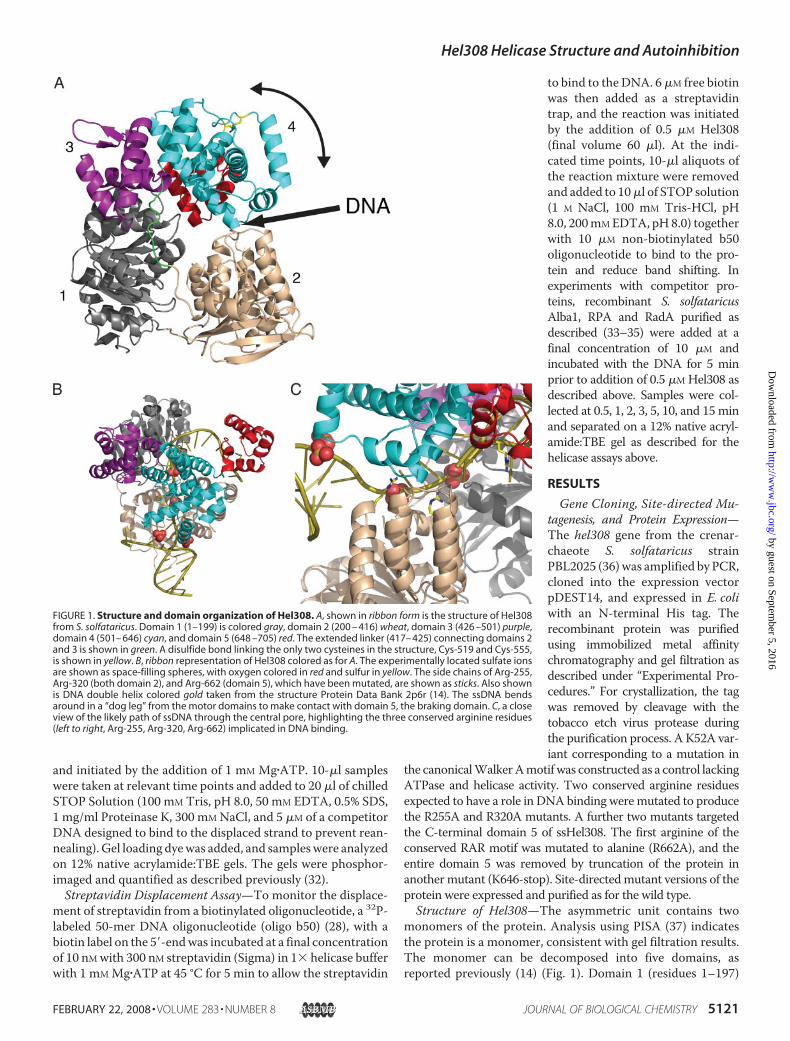
FIGURE 1. Structure and domain organization of Hel308. A, shown in ribbon form is the structure of Hel308from S. solfataricus. Domain 1 (1–199) is colored gray, domain 2 (200 – 416) wheat, domain 3 (426 –501) purple,domain 4 (501– 646) cyan, and domain 5 (648 –705) red. The extended linker (417– 425) connecting domains 2and 3 is shown in green. A disulfide bond linking the only two cysteines in the structure, Cys-519 and Cys-555,is shown in yellow. B, ribbon representation of Hel308 colored as for A. The experimentally located sulfate ionsare shown as space-filling spheres, with oxygen colored in red and sulfur in yellow. The side chains of Arg-255,Arg-320 (both domain 2), and Arg-662 (domain 5), which have been mutated, are shown as sticks. Also shownis DNA double helix colored gold taken from the structure Protein Data Bank 2p6r (14). The ssDNA bendsaround in a “dog leg” from the motor domains to make contact with domain 5, the braking domain. C, a closeview of the likely path of ssDNA through the central pore, highlighting the three conserved arginine residues(left to right, Arg-255, Arg-320, Arg-662) implicated in DNA binding.
Hel308 Helicase Structure and Autoinhibition
FEBRUARY 22, 2008 • VOLUME 283 • NUMBER 8 JOURNAL OF BIOLOGICAL CHEMISTRY 5121
by guest on September 5, 2016
http://ww
w.jbc.org/
Dow
nloaded from
and domain 2 (200–416) are the classical ATP binding motordomains seen in all SF1 and SF2 helicase structures. These twodomains share the same core �/� fold; the rmsd of 106 super-imposable C� atoms is 2.6 Å. The interface between these twodomains forms the ATP binding site, and the conformationchanges induced during the complete cycle of ATP hydrolysisare thought to drive duplex unwinding (reviewed in Ref. (3)).Domain 3 (426–501) is a winged helix domain commonly seenin nucleic acid-binding proteins and closely matches HistoneH5 (1.5 Å rmsd for 62 overlapping carbons). A simple search ofstructural similarity using SSM reveals over 400 such domainshavebeenstructurally characterized.Domain4 (502–644) is a sev-en-helical bundle that is found relatively rarely in proteins, allow-ing little meaningful functional assignment of its role a priori.Domain 5 (647–705) is a simple helix hairpin helixmotif found invery many proteins and is commonly associated with single-stranded nucleic acid binding. Domain 5 hangs down like a capbehind the central hole. This domain contains theRARmotif (res-idues Arg-662, Arg-664) conserved in all Hel308 family helicasesin an �-helix. The positively charged N terminus of this �-helixpoints toward thecentralpore.Thering formedbydomains1–4 is
not covalently closed, and opening ofa gapbetweendomains 2 and4wouldbe necessary to allow the helicase toload onto substrates lacking a3�-ssDNAend, suchas stalled replica-tion forks. An analysis of the interfacebetween domains 2 and 4 reveals onlyaweak interaction.Thedomain:dom-ain contact buries a total of 650 Å2,and many of the interactions are saltbridgesorpolar contacts. Suchaweakinteraction would allow the twodomains to separate to allow the pas-sage of ssDNA into the central pore.The hinge for this movement has notbeen identified, although we notethere is a long loop (416–426) con-necting domain 2 and domain 3 thatcould act as a hinge (Fig. 1A).Overall, the five-domain arrange-
ment is very similar to that reportedfor the structure of Hel308 from A.fulgidus (14). For 600 superimposableC� atoms the rmsd is 2.0 Å; themaindifferences are in the positions ofsome secondary structure elementsand loops. Our apo structure super-imposes equally well on the apo andDNA-bound forms of A. fulgidusHel308, suggesting that this helicaseadopts quite a rigid structure that isnot perturbed significantly by DNAbinding. This has allowedus tomodelthe DNA from the A. fulgidus com-plex into our apo S. solfataricus struc-ture (Fig. 1B). The DNA threadsthrough the central pore, forming
interactions with domains 3 and 4, and engages domain 5 on theopposite side. The interactions with domain 3 and domain 4 areextensive. Loss of these domains uncouples ATP hydrolysis fromhelicase activity, and domain 4 has been proposed as the ratchetpowered byATPhydrolysis that unwinds theDNA (14). Four sul-fates present in our crystal structure coincide with the likely posi-tions of phosphate residues in the DNA backbone. In the Hel308-DNAco-crystal structure, theDNAduplex is split bya loopalmostat the entrance to the central hole that forms a small two-stranded� sheet (residues 347–359). In our apo structure this loop is disor-dered, as was observed in the apo form ofA. fulgidusHel308 (14).Three conserved arginine residues (Arg-255, Arg-320, and Arg-662)mutated in this studyclearly adoptpositions consistentwitharole in DNA binding (Fig. 1C).DNA Binding by Wild-type and Mutant Hel308 Proteins—
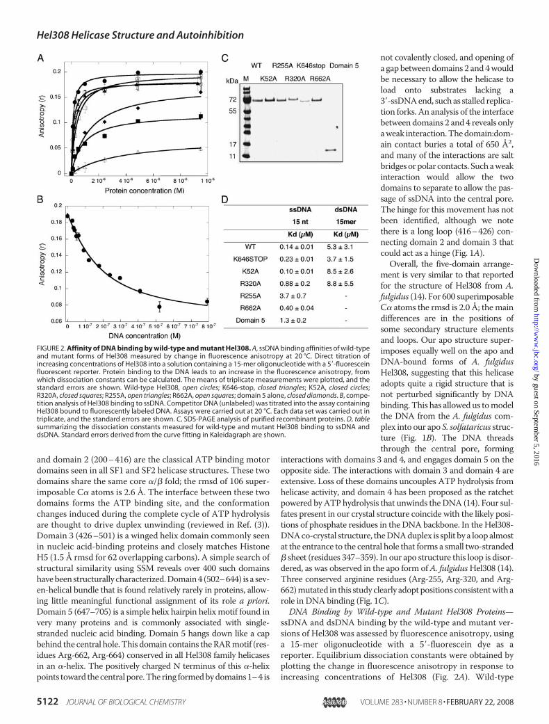
ssDNA and dsDNA binding by the wild-type and mutant ver-sions of Hel308 was assessed by fluorescence anisotropy, usinga 15-mer oligonucleotide with a 5�-fluorescein dye as areporter. Equilibrium dissociation constants were obtained byplotting the change in fluorescence anisotropy in response toincreasing concentrations of Hel308 (Fig. 2A). Wild-type
FIGURE 2. Affinity of DNA binding by wild-type and mutant Hel308. A, ssDNA binding affinities of wild-typeand mutant forms of Hel308 measured by change in fluorescence anisotropy at 20 °C. Direct titration ofincreasing concentrations of Hel308 into a solution containing a 15-mer oligonucleotide with a 5�-fluoresceinfluorescent reporter. Protein binding to the DNA leads to an increase in the fluorescence anisotropy, fromwhich dissociation constants can be calculated. The means of triplicate measurements were plotted, and thestandard errors are shown. Wild-type Hel308, open circles; K646-stop, closed triangles; K52A, closed circles;R320A, closed squares; R255A, open triangles; R662A, open squares; domain 5 alone, closed diamonds. B, compe-tition analysis of Hel308 binding to ssDNA. Competitor DNA (unlabeled) was titrated into the assay containingHel308 bound to fluorescently labeled DNA. Assays were carried out at 20 °C. Each data set was carried out intriplicate, and the standard errors are shown. C, SDS-PAGE analysis of purified recombinant proteins. D, tablesummarizing the dissociation constants measured for wild-type and mutant Hel308 binding to ssDNA anddsDNA. Standard errors derived from the curve fitting in Kaleidagraph are shown.
Hel308 Helicase Structure and Autoinhibition
5122 JOURNAL OF BIOLOGICAL CHEMISTRY VOLUME 283 • NUMBER 8 • FEBRUARY 22, 2008
by guest on September 5, 2016
http://ww
w.jbc.org/
Dow
nloaded from

Hel308 bound the single-stranded and duplex DNA ligandswithKDs of 0.14 and 5.3�M, respectively. The strong preferencefor binding to ssDNA is consistent with the function of thehelicase, which must track along ssDNA and displace a duplexDNA strand. To ensure that the DNA binding affinity was notaffected by protein-dye interactions, competition assays wereperformed by first forming a complex of Hel308 with the fluo-rescent oligo and then titrating an unlabeled competitorssDNA of the same sequence (29). The decrease in anisotropyobserved in Fig. 2B yielded a KD of 0.13 �M, in good agreementwith that calculated from the forward titration, suggesting thatthe influence of the fluorescein was minimal. As expected, theK52Amutant bound ssDNA and dsDNA with similar affinitiesto the wild-type protein. Both the R255A and R320A mutantshad significantly reduced binding affinities, with the R255Amutant showing the largest effect, a 25-fold increase in the KDfor ssDNA. These data confirm an important role in ssDNAbinding for Arg-320 and particularly Arg-255, which line thecentral pore predicted as the path for ssDNA (Fig. 1). The trun-cated protein lacking domain 5 and theR662Amutant both hadssDNA binding affinities comparable with or only slightlyweaker than the wild-type enzyme for this short oligonucleo-tide. A longer oligonucleotide might be expected to engagemore fully with domain 5. A role for ssDNA binding by domain5 was confirmed by expressing this domain independently andmeasuring its affinity for ssDNA. Domain 5 adopts a stable,autonomous folded structure in isolation and binds ssDNAwith a dissociation constant of 1.3 �M, �10-fold weaker thanthe intact protein but more tightly than the R255A mutant.Helicase Activity of Wild-type and Mutant Hel308 Proteins—
The 3�-5� helicase activity of Hel308 was assayed using a mini-mal substrate: a 3�-overhang with a 25-nt ssDNA region and a25-bp duplex region. Helicase assays were carried out at 60 °Cover a 3-min time course and analyzed by gel electrophoresis(Fig. 3). The wild-type protein displaced the duplex strand effi-ciently, as observed previously for the homologues fromarchaea andH. sapiens (10, 13). As expected, the K52Amutantwas unable to function as a helicase and the R255A and R320Amutants had significantly reduced helicase activity, consistentwith a defect in ssDNA binding. Surprisingly, the K646-stopmutant lacking domain 5 and the R662A mutant lacking thefirst arginine of the RARmotif in domain 5 both showed signif-icantly faster rates of DNA unwinding than the wild-type pro-tein. To follow up this observation, we tested the helicase activ-ity of the wild-type and truncated proteins using a DNAsubstrate with a longer duplex region of 50 bp (Fig. 3). Hel308has been shown previously to have a limited processivity and alimited ability to displace longer DNA duplex strands (13).Consistent with these observations, the wild-type ssHel308protein showed only a very weak helicase activity against thelarger substrate. However, significantly faster unwinding wasobserved with the truncatedmutant, consistent with the effectsobserved for the shorter substrate.Previously, biochemical studies have identified branched
DNA structures, and specifically substrates resembling a stalledDNA replication fork, as the preferred substrates of Hel308(13). Accordingly, we compared the activities of the wild-typeand truncationmutant using amodel replication fork substrate
with a 25-bp duplex region corresponding to the lagging strandof a replication fork (Fig. 4). These assayswere carried out at thereduced temperature of 45 °C to allow more accurate determi-nation of reaction progress. The transient production and thendisappearance of the 3�-overhang intermediate at early timepoints suggested that unwindingmay proceed via displacementof the top strand, yielding a 3�-overhang substrate intermediatethat is then unwound to yield the final products. This conclu-sionwas strengthened by the observation that a simple 3�-over-hang substrate was unwound at the same rate as the fork sub-strate (Fig. 4). Once again, the truncation mutant lackingdomain 5 unwound the fork structure muchmore quickly thanthe wild-type protein, disassembling �80% of the substratewithin 30 s, whereas the wild-type enzyme unwound only�15% in the same period. As for the wild-type protein, thetruncationmutant showednodifference in the rates of unwind-ing of the fork and a simple 3�-overhang (Fig. 4).Taken together, these observations suggest that domain 5,
which is conserved in all Hel308 proteins, functions as an auto-inhibitory domain, limiting the activity of the helicase, possiblyby binding the emergent ssDNA product and functioning as a“molecular brake.”Deletion of this domain does not perturb therelative rates of unwinding of fork versus overhang substrates,
FIGURE 3. Helicase activity of wild-type and mutant Hel308. The helicaseactivity was measured using 10 nM
32P-labeled 3�-overhang substrate and 0.5�M protein at 60 °C as described under “Experimental Procedures.” The wild-type and all mutants were assayed using DNA substrates with 25-bp duplexregions (left). The wild-type and K646-stop mutant were also assayed using aDNA substrate with a 50-bp duplex region (right). Each experiment was car-ried out in triplicate, and a representative gel is shown. The plot shows aquantification of the time course of unwinding for selected proteins for the25- and 50-bp duplex substrates: wild-type (open circles), K646-stop (closedtriangles), R662A (open triangles). All data points are the means obtained fromtriplicate experiments, and standard errors are shown.
Hel308 Helicase Structure and Autoinhibition
FEBRUARY 22, 2008 • VOLUME 283 • NUMBER 8 JOURNAL OF BIOLOGICAL CHEMISTRY 5123
by guest on September 5, 2016
http://ww
w.jbc.org/
Dow
nloaded from

as both rates are enhanced to the same extent in the truncatedspecies.Hel308 Displaces Streptavidin and DNA-binding Proteins
from a Biotinylated Oligonucleotide—The translocation ofHel308 along ssDNA was investigated by its ability to displace
streptavidin bound to a biotinylatedoligonucleotide. Wild-type andmutant Hel308 proteins were incu-bated with the DNA substrate in thepresence of ATP at 45 °C followedby electrophoresis to separate thefree biotinylated probes from thosebound by streptavidin (38). Wild-type Hel308 was able to displace�80% of the streptavidin in 10 min(Fig. 5). The ability of Hel308 totranslocate along ssDNA with aforce sufficient to dissociate atightly bound biotin:streptavidinlinkage suggests that Hel308 couldact to displace proteins bound tossDNA in vivo (39). We thereforeinvestigated the effect of adding theS. solfataricus DNA-binding pro-teins RPA (the canonical ssDNA-binding protein), Alba1 (the majorchromatin protein that binds bothdsDNA and ssDNA), and RadA (thearchaeal Rad51 family recombinase)to the streptavidin-bound DNA at aconcentration of 10 �M before addi-tion of the helicase at 0.5�M (Fig. 5).Streptavidin displacement byHel308 was not inhibited by RadA,whereas reduced activity wasobserved in the presence of Alba1and RPA. By comparison, the repli-cative helicase MCM from S. solfa-taricus was completely inhibited bya 2-fold molar excess of Alba1 (40).These data are consistentwith a rolefor Hel308 in remodeling stalledreplication forks by protein as wellas DNA displacement.The truncation mutant retained
the ability to displace streptavidin(Fig. 5) with activity comparablewith the wild-type protein. Thefinding that the truncated enzyme isa better helicase but no better at dis-placing bound proteins than thewild-type protein probably relatesto the difference between the twotypes of assay, as the streptavidindisplacement assay reflects theforce exerted by the helicase as ittranslocates along the ssDNA andallows helicase binding anywhere
on the ssDNA molecule.
DISCUSSION
The high resolution crystal structure of Hel308 presentedhere, togetherwith the site-directedmutagenesis and biochem-
FIGURE 4. Wild-type and truncated Hel308 helicase activity with a stalled replication fork model sub-strate. The activity of wild-type and K646-stop mutant of Hel308 was compared using a model substrateresembling a stalled replication fork at 45 °C. Unwinding was observed to proceed as a two-step process, withinitial unwinding of the top strand followed by displacement of the labeled oligonucleotide from the resulting3�-overhang. Quantification of the reactions showed that the truncated mutant (open squares) unwound thissubstrate much faster than the wild-type protein (open circles). The rate of unwinding of the fork substrate wasessentially identical to the rate of unwinding of a 3�-overhang substrate for both enzymes (closed symbols). Themeans of triplicate experiments are shown along with standard errors.
FIGURE 5. Hel308 displaces streptavidin and DNA-binding proteins from a biotinylated oligonucleotide.Wild-type and K646-stop Hel308 were incubated with an oligonucleotide bound to streptavidin via a 5�-biotinmodification. The black circle indicates the 5�-32P-labeled DNA end, with the larger circle representing the streptavi-din. Both the wild-type and K646-stop mutant lacking domain 5 displaced streptavidin efficiently at 45 °C, wellbelow the normal operating temperature of the enzyme. When the assay was carried out with a 20-fold molar excessof RadA pre-bound to the oligo, no reduction in streptavidin displacement was observed. In contrast, both enzymeswere partially inhibited by the same concentration of RPA or Alba1. The plot on the right shows a quantification oftriplicate experiments, with means and standard errors shown, for the wild-type enzyme on its own (open circles) andin the presence of RadA (open squares), RPA (closed circles), and Alba1 (closed triangles). Control c1 shows the labeledoligonucleotide in the absence of streptavidin, c2 shows the oligonucleotide bound to streptavidin, and c3 showsproducts after incubation for 15 min in the presence of Hel308 and absence of ATP.
Hel308 Helicase Structure and Autoinhibition
5124 JOURNAL OF BIOLOGICAL CHEMISTRY VOLUME 283 • NUMBER 8 • FEBRUARY 22, 2008
by guest on September 5, 2016
http://ww
w.jbc.org/
Dow
nloaded from

ical data, provide several new insights into themolecularmech-anismof this conserved SF2DNAhelicase. The helicase is orga-nized into five domains. The first four form a ringwith a centralpore large enough for the passage of single-stranded, but notdouble-stranded, DNA, consistent with the recently publishedstructure of Hel308 from A. fulgidus in the presence andabsence of a DNA substrate (14). The crystal structure ofHel308 suggests an ancillary role for domain 5, which sits per-pendicular to the ring formed by the first four domains and hasbeen shown to engage the 3�-DNA end that extrudes throughthe central pore as the helicase translocates DNA. Hopfner andcoworkers (14) have suggested a role as a specificity domain fordomain 5, predicting that it confers binding specificity for thebranched DNA substrates preferred by the enzyme. However,our data show clearly that either deletion of domain 5 or abro-gation of its DNA binding affinity in the R662A mutant resultsin an increase in activity of the helicase. Importantly, the trun-catedmutant retains a faster helicase activity than thewild-typeenzyme and can displace longer DNA strands more effectively.The effect is equally pronouncedwith theminimal 3�-overhangsubstrate and the preferred lagging strand substrate, which areunwound with equal rates, arguing against a role for domain 5in determining substrate specificity. Altogether, our data sug-gest strongly that the function of domain 5 in vivo is to act as amolecular brake, limiting the extent of DNA unwinding byHel308, rather than a specificity domain.Autoinhibitory domains have been observed in other heli-
cases. The SF1 helicase Rep, which functions in replicationrestart, in inhibited by its 2B subdomain, which is thought toblock theRephelicase from invading theDNAduplex (41). Thisinhibition is alleviated by dimerization of the Rep protein.Recently, the C-terminal D7 domain of the transcription-cou-pled repair protein Mfd from E. coli has also been shown toinhibit the helicase activity of Mfd in vitro (42). In this case, theinteraction of Mfd with a stalled RNA polymerase molecule isthought to result in a conformational change in the D7 domain,alleviating the repression of the helicase. Finally, domain 4 ofthe bacterial DNA repair helicase UvrB has also been shown tofunction as an autoinhibitory domain in vitro, where it is pro-posed to regulate the repair activity of the UvrABC system andprevent spurious incision events (43). For all these examples,inhibition is thought to be “on” by default and to be relieved bythe formation of cognate protein-protein interactions. Hel308domain 5 differs from all these examples in binding ssDNAdirectly, thus binding the product of theDNAhelicase reaction.This represents an elegant mechanism with the potential tolimit or tune helicase activity in vivo. It remains to be deter-minedwhether the autoinhibitory function ofHel308 domain 5is alleviated by interactions with other proteins.The limited helicase activity of Hel308 is consistent with the
proposed function in vivo in the removal of short sections of thelagging strand next to a stalled replication fork to clear anssDNA binding site for proteins that can restart replication orinitiate recombination (13) (Fig. 6). In E. coli, the 3�-5� helicaseactivities of PriA or Rep carry out this role (reviewed in Ref.(44)).Wehave also shown thatHel308 can displace streptavidinfrom biotinylated ssDNA, as has been demonstrated previouslyfor a number of helicases, such as the replicative helicase Dda
(45). The ability of helicases to disrupt protein-nucleic acidcomplexesmay be relevant in vivo, where abundantDNA-bind-ing proteins are associatedwith dsDNAand ssDNAundermostconditions. In the case of Hel308, we see no significant reduc-tion of streptavidin displacement activity when using a DNAsubstrate pre-boundwith RadA and significant residual activityin the presence of 20-fold molar excess of the abundant DNA-binding proteins RPA and Alba1. These assays were all carriedout at 45 °C, well below the in vivo operating temperature of80 °C where one would expect significantly more robust heli-case and protein displacement activity. A plausible additionalfunction of Hel308 may therefore be to displace DNA-bindingproteins from the site of a stalled replication fork, allowingbinding of Rad51 (RadA in archaea) and the initiation of recom-bination, or the binding of other proteins for DNA replication(Fig. 6). This is analogous to the role proposed for RecFOR inthe displacement of single-stranded DNA-binding proteinfrom stalled forks to allow RecA-mediated recombination (46).Equally, Hel308 could function like bacterial UvrD, another3�-5� helicase that has been shown to disrupt RecA nucleopro-tein filaments and thus limit recombination (47). In this con-text, it is intriguing to note that Ishino and coworkers (12)report a direct interaction between Hel308 and RadA from P.furiosus.
Acknowledgment—We thank Georg Lipps for the kind donation ofPBL2025 chromosomal DNA.
REFERENCES1. Gorbalenya, A. E., and Koonin, E. V. (1993) Curr. Opin. Struct. Biol. 3,
419–4292. Singleton, M. R., and Wigley, D. B. (2002) J. Bacteriol. 184, 1819–18263. Singleton, M. R., Dillingham, M. S., and Wigley, D. B. (2007) Annu. Rev.
FIGURE 6. Schematic model for possible roles of Hel308 at stalled replica-tion forks. Hel308 can translocate in a 3� to 5� direction along ssDNA anddisplace either bound protein or a duplex DNA strand. In vivo, the proteincould function to generate an area of ssDNA on the lagging strand to allowreplication restart or the initiation of recombination. This could involve theremoval of bound proteins such as RPA or RadA (archaeal Rad51), as shownon the left, or the displacement of a short section of the lagging strand, asshown on the right.
Hel308 Helicase Structure and Autoinhibition
FEBRUARY 22, 2008 • VOLUME 283 • NUMBER 8 JOURNAL OF BIOLOGICAL CHEMISTRY 5125
by guest on September 5, 2016
http://ww
w.jbc.org/
Dow
nloaded from
Biochem. 76, 23–504. Sharma, S., Doherty, K. M., and Brosh, R. M., Jr. (2006) Biochem. J. 398,
319–3375. Hanada, K., and Hickson, I. D. (2007) Cell Mol. Life Sci. 64, 2306–23336. Boyd, J. B., Golino,M.D., Nguyen, T. D., andGreen,M.M. (1976)Genetics
84, 485–5067. Seki, M., Marini, F., and Wood, R. D. (2003) Nucleic Acids Res. 31,
6117–61268. Yoshimura, M., Kohzaki, M., Nakamura, J., Asagoshi, K., Sonoda, E., Hou,
E., Prasad, R., Wilson, S. H., Tano, K., Yasui, A., Lan, L., Seki, M., Wood,R. D., Arakawa, H., Buerstedde, J. M., Hochegger, H., Okada, T., Hiraoka,M., and Takeda, S. (2006)Mol. Cell 24, 115–125
9. Takata, K., Shimizu, T., Iwai, S., andWood, R. D. (2006) J. Biol. Chem. 281,23445–23455
10. Marini, F., and Wood, R. D. (2002) J. Biol. Chem. 277, 8716–872311. McCaffrey, R., St. Johnston, D., and Gonzalez-Reyes, A. (2006) Genetics
174, 1273–128512. Fujikane, R., Shinagawa, H., and Ishino, Y. (2006) Genes Cells 11, 99–11013. Guy, C. P., and Bolt, E. L. (2005) Nucleic Acids Res. 33, 3678–369014. Buttner, K., Nehring, S., and Hopfner, K. P. (2007) Nat. Struct. Mol. Biol.
14, 647–65215. Guerrero, S. A., Hecht, H. J., Hofmann, B., Biebl, H., and Singh, M. (2001)
Appl. Microbiol. Biotechnol. 56, 718–72316. Potterton, L., McNicholas, S., Krissinel, E., Gruber, J., Cowtan, K., Emsley,
P., Murshudov, G. N., Cohen, S., Perrakis, A., and Noble, M. (2004) ActaCrystallogr. Sect. D Biol. Crystallogr. 60, Pt. 12 Pt. 1, 2288–2294
17. Pape, T., and Schneider, T. R. (2004) J. Appl. Crystallogr. 37, 843–84418. Schneider, T. R., and Sheldrick, G.M. (2002)Acta Crystallogr. Sect. D Biol.
Crystallogr. 58, Pt. 10 Pt. 2, 1772–177919. Terwilliger, T. C., and Berendzen, J. (1999) Acta Crystallogr. Sect. D Biol.
Crystallogr. 55, Pt. 4, 849–86120. Terwilliger, T. C. (2000) Acta Crystallogr. Sect. D Biol. Crystallogr. 56, Pt.
8, 965–97221. Cowtan, K. (2001) Acta Crystallogr. Sect. D Biol. Crystallogr. 57, Pt. 10,
1435–144422. McRee, D. E. (1999) J. Struct. Biol. 125, 156–16523. Murshudov, G. N., Vagin, A. A., and Dodson, E. J. (1997)Acta Crystallogr.
Sect. D Biol. Crystallogr. 53, Pt. 3, 240–25524. Brunger, A. T., Adams, P. D., Clore, G. M., DeLano, W. L., Gros, P.,
Grosse-Kunstleve, R.W., Jiang, J. S., Kuszewski, J., Nilges,M., Pannu,N. S.,Read, R. J., Rice, L. M., Simonson, T., and Warren, G. L. (1998) Acta
Crystallogr. Sect. D Biol. Crystallogr. 54, Pt. 5, 905–92125. Bruns, C. M., Hubatsch, I., Ridderstrom, M., Mannervik, B., and Tainer,
J. A. (1999) J. Mol. Biol. 288, 427–43926. Davis, I. W., Leaver-Fay, A., Chen, V. B., Block, J. N., Kapral, G. J., Wang,
X., Murray, L. W., Arendall, W. B., III, Snoeyink, J., Richardson, J. S., andRichardson, D. C. (2007) Nucleic Acids Res. 35,W375–W383
27. Kvaratskhelia, M., and White, M. F. (2000) J. Mol. Biol. 295, 193–20228. Roberts, J., and White, M. F. (2005) J. Biol. Chem. 280, 5924–592829. Reid, S. L., Parry, D., Liu, H. H., and Connolly, B. A. (2001) Biochemistry
40, 2484–249430. Lundblad, J. R., Laurance,M., andGoodman, R.H. (1996)Mol. Endocrinol.
10, 607–61231. Deleted in proof32. Rudolf, J., Makrantoni, V., Ingledew, W. J., Stark, M. J., and White, M. F.
(2006)Mol. Cell 23, 801–80833. Ariza, A., Richard, D. J.,White,M. F., and Bond, C. S. (2005)Nucleic Acids
Res. 33, 1465–147334. Jelinska, C., Conroy, M. J., Craven, C. J., Hounslow, A. M., Bullough, P. A.,
Waltho, J. P., Taylor, G. L., andWhite, M. F. (2005) Structure 13, 963–97135. Wadsworth, R. I., andWhite, M. F. (2001)Nucleic Acids Res. 29, 914–92036. Schelert, J., Dixit, V., Hoang, V., Simbahan, J., Drozda, M., and Blum, P.
(2004) J. Bacteriol. 186, 427–43737. Krissinel, E., and Henrick, K. (2007) J. Mol. Biol. 372, 774–79738. Morris, P. D., and Raney, K. D. (1999) Biochemistry 38, 5164–517139. Byrd, A. K., and Raney, K. D. (2004) Nat. Struct. Mol. Biol. 11, 531–53840. Marsh, V. L., McGeoch, A. T., and Bell, S. D. (2006) J. Mol. Biol. 357,
1345–135041. Brendza, K.M., Cheng,W., Fischer, C. J., Chesnik,M.A., Niedziela-Majka,
A., and Lohman, T. M. (2005) Proc. Natl. Acad. Sci. U. S. A. 102,10076–10081
42. Smith, A. J., Szczelkun, M. D., and Savery, N. J. (2007) Nucleic Acids Res.35, 1802–1811
43. Wang, H., DellaVecchia, M. J., Skorvaga, M., Croteau, D. L., Erie, D. A.,and Van Houten, B. (2006) J. Biol. Chem. 281, 15227–15237
44. Heller, R. C., andMarians, K. J. (2006)Nat. Rev.Mol. Cell. Biol. 7, 932–94345. Byrd, A. K., and Raney, K. D. (2006) Nucleic Acids Res. 34, 3020–302946. Michel, B., Grompone, G., Flores, M. J., and Bidnenko, V. (2004) Proc.
Natl. Acad. Sci. U. S. A. 101, 12783–1278847. Veaute, X., Delmas, S., Selva, M., Jeusset, J., Le Cam, E., Matic, I., Fabre, F.,
and Petit, M. A. (2005) EMBO J. 24, 180–189
Hel308 Helicase Structure and Autoinhibition
5126 JOURNAL OF BIOLOGICAL CHEMISTRY VOLUME 283 • NUMBER 8 • FEBRUARY 22, 2008
by guest on September 5, 2016
http://ww
w.jbc.org/
Dow
nloaded from

McMahon, Muse Oke, Lester Carter, James H. Naismith and Malcolm F. WhiteJodi D. Richards, Kenneth A. Johnson, Huanting Liu, Anne-Marie McRobbie, Stephen
Autoinhibitory DomainsStructure of the DNA Repair Helicase Hel308 Reveals DNA Binding and
doi: 10.1074/jbc.M707548200 originally published online December 4, 20072008, 283:5118-5126.J. Biol. Chem.
10.1074/jbc.M707548200Access the most updated version of this article at doi:
Alerts:
When a correction for this article is posted•
When this article is cited•
to choose from all of JBC's e-mail alertsClick here
http://www.jbc.org/content/283/8/5118.full.html#ref-list-1
This article cites 46 references, 18 of which can be accessed free at
by guest on September 5, 2016
http://ww
w.jbc.org/
Dow
nloaded from
Copyright © 2022 FDOKUMEN