The S. cerevisiae Rrm3p DNA helicase moves with the replication fork and affects replication of all...
14
The S. cerevisiae Rrm3p DNA helicase moves with the replication fork and affects replication of all yeast chromosomes Anna Azvolinsky, 1 Stephen Dunaway, 1 Jorge Z. Torres, Jessica B. Bessler, and Virginia A. Zakian 2 Department of Molecular Biology, Princeton University, Princeton, New Jersey 08544, USA The Saccharomyces cerevisiae DNA helicase Rrm3p is needed for normal fork progression through >1000 discrete sites scattered throughout the genome. Here we show that replication of all yeast chromosomes was markedly delayed in rrm3 cells. Delayed replication was seen even in a region that lacks any predicted Rrm3p-dependent sites. Based on the pattern of replication intermediates in two-dimensional gels, the rate of fork movement in rrm3 cells appeared similar to wild-type except at known Rrm3p-dependent sites. These data suggest that although Rrm3p has a global role in DNA replication, its activity is needed only or primarily at specific, difficult-to-replicate sites. By the criterion of chromatin immunoprecipitation, Rrm3p was associated with both Rrm3p-dependent and -independent sites, and moved with the replication fork through both. In addition, Rrm3p interacted with Pol2p, the catalytic subunit of DNA polymerase , in vivo. Thus, rather than being recruited to its sites of action when replication forks stall at these sites, Rrm3p is likely a component of the replication fork apparatus. [Keywords: Rrm3p; Mrc1p; DNA replication; helicase; yeast; chromatin] Supplemental material is available at http://www.genesdev.org. Received August 2, 2006; revised version accepted September 19, 2006. DNA helicases have fundamental roles in all processes involving DNA, including replication, recombination, and repair (Caruthers and McKay 2002; Tuteja and Tu- teja 2004; Eoff and Raney 2005). Rrm3p is a member of the Pif1 family of DNA helicases, a family that is highly conserved from yeasts to humans (Boule and Zakian 2006). Saccharomyces cerevisiae RRM3 was first discov- ered because its mutation causes increased recombina- tion in the ribosomal DNA (rDNA) (Keil and McWil- liams 1993). As assayed by two-dimensional (2D) gel electrophoresis, the absence of Rrm3p results in replica- tion fork pausing at multiple sites in the ribosomal DNA (rDNA) (Ivessa et al. 2000), in telomeres and subtelo- meric DNA (Ivessa et al. 2000, 2002), as well as at tRNA genes, inactive replication origins, centromeres, and the silent mating-type loci (Ivessa et al. 2003). By the same assay, the absence of Rrm3p results in increased replica- tion fork pausing within plasmid borne polymerase II transcribed genes when replication and transcription move in opposite directions through the gene (Prado and Aguilera 2005). All Rrm3p-dependent sites that have been identified to date are assembled into stable, non- nucleosomal protein–DNA complexes. Disruption of these complexes makes replication through them Rrm3p independent (Ivessa et al. 2003; Torres et al. 2004a). Rrm3p acts catalytically to promote fork progression as mutations that eliminate its ability to hydrolyze ATP have the same phenotypes as an RRM3 deletion (Ivessa et al. 2000, 2002). The role of Rrm3p in replication fork progression con- tributes to genomic integrity. Broken replication forks are detected in rrm3 cells within rDNA, subtelomeric DNA, and tRNA genes (Ivessa et al. 2000, 2002, 2003). These breaks trigger a checkpoint response as the effec- tor checkpoint kinase Rad53p is constitutively phos- phorylated in rrm3 cells (Ivessa et al. 2003). The DNA breaks also promote recombination, which is elevated in rrm3 cells. Cells lacking Rrm3p exhibit increased recom- bination in the rDNA (Keil and McWilliams 1993), which generates rDNA circles (Ivessa et al. 2000; Torres et al. 2004a) that are associated with premature aging (Sinclair and Guarente 1997). Recombination is also in- creased between other tandem repeats such as CUP1 genes (Keil and McWilliams 1993) and subtelomeric Y elements (Ivessa et al. 2002), within a 184-kb tRNA-rich region on chromosome VII (Ivessa et al. 2003; Admire 1 These authors contributed equally to this work. 2 Corresponding author. E-MAIL [email protected]; FAX (609) 258-1701. Article is online at http://www.genesdev.org/cgi/doi/10.1101/gad.1478906. 3104 GENES & DEVELOPMENT 20:3104–3116 © 2006 by Cold Spring Harbor Laboratory Press ISSN 0890-9369/06; www.genesdev.org Cold Spring Harbor Laboratory Press on June 18, 2016 - Published by genesdev.cshlp.org Downloaded from
-
Upload
independent -
Category
Documents
-
view
5 -
download
0
Transcript of The S. cerevisiae Rrm3p DNA helicase moves with the replication fork and affects replication of all...

The S. cerevisiae Rrm3p DNA helicasemoves with the replication forkand affects replication of allyeast chromosomesAnna Azvolinsky,1 Stephen Dunaway,1 Jorge Z. Torres, Jessica B. Bessler, and Virginia A. Zakian2
Department of Molecular Biology, Princeton University, Princeton, New Jersey 08544, USA
The Saccharomyces cerevisiae DNA helicase Rrm3p is needed for normal fork progression through >1000discrete sites scattered throughout the genome. Here we show that replication of all yeast chromosomes wasmarkedly delayed in rrm3 cells. Delayed replication was seen even in a region that lacks any predictedRrm3p-dependent sites. Based on the pattern of replication intermediates in two-dimensional gels, the rate offork movement in rrm3 cells appeared similar to wild-type except at known Rrm3p-dependent sites. Thesedata suggest that although Rrm3p has a global role in DNA replication, its activity is needed only or primarilyat specific, difficult-to-replicate sites. By the criterion of chromatin immunoprecipitation, Rrm3p wasassociated with both Rrm3p-dependent and -independent sites, and moved with the replication fork throughboth. In addition, Rrm3p interacted with Pol2p, the catalytic subunit of DNA polymerase �, in vivo. Thus,rather than being recruited to its sites of action when replication forks stall at these sites, Rrm3p is likely acomponent of the replication fork apparatus.
[Keywords: Rrm3p; Mrc1p; DNA replication; helicase; yeast; chromatin]
Supplemental material is available at http://www.genesdev.org.
Received August 2, 2006; revised version accepted September 19, 2006.
DNA helicases have fundamental roles in all processesinvolving DNA, including replication, recombination,and repair (Caruthers and McKay 2002; Tuteja and Tu-teja 2004; Eoff and Raney 2005). Rrm3p is a member ofthe Pif1 family of DNA helicases, a family that is highlyconserved from yeasts to humans (Boule and Zakian2006). Saccharomyces cerevisiae RRM3 was first discov-ered because its mutation causes increased recombina-tion in the ribosomal DNA (rDNA) (Keil and McWil-liams 1993). As assayed by two-dimensional (2D) gelelectrophoresis, the absence of Rrm3p results in replica-tion fork pausing at multiple sites in the ribosomal DNA(rDNA) (Ivessa et al. 2000), in telomeres and subtelo-meric DNA (Ivessa et al. 2000, 2002), as well as at tRNAgenes, inactive replication origins, centromeres, and thesilent mating-type loci (Ivessa et al. 2003). By the sameassay, the absence of Rrm3p results in increased replica-tion fork pausing within plasmid borne polymerase IItranscribed genes when replication and transcriptionmove in opposite directions through the gene (Prado and
Aguilera 2005). All Rrm3p-dependent sites that havebeen identified to date are assembled into stable, non-nucleosomal protein–DNA complexes. Disruption ofthese complexes makes replication through them Rrm3pindependent (Ivessa et al. 2003; Torres et al. 2004a).Rrm3p acts catalytically to promote fork progression asmutations that eliminate its ability to hydrolyze ATPhave the same phenotypes as an RRM3 deletion (Ivessaet al. 2000, 2002).
The role of Rrm3p in replication fork progression con-tributes to genomic integrity. Broken replication forksare detected in rrm3 cells within rDNA, subtelomericDNA, and tRNA genes (Ivessa et al. 2000, 2002, 2003).These breaks trigger a checkpoint response as the effec-tor checkpoint kinase Rad53p is constitutively phos-phorylated in rrm3 cells (Ivessa et al. 2003). The DNAbreaks also promote recombination, which is elevated inrrm3 cells. Cells lacking Rrm3p exhibit increased recom-bination in the rDNA (Keil and McWilliams 1993),which generates rDNA circles (Ivessa et al. 2000; Torreset al. 2004a) that are associated with premature aging(Sinclair and Guarente 1997). Recombination is also in-creased between other tandem repeats such as CUP1genes (Keil and McWilliams 1993) and subtelomeric Y�elements (Ivessa et al. 2002), within a 184-kb tRNA-richregion on chromosome VII (Ivessa et al. 2003; Admire
1These authors contributed equally to this work.2Corresponding author.E-MAIL [email protected]; FAX (609) 258-1701.Article is online at http://www.genesdev.org/cgi/doi/10.1101/gad.1478906.
3104 GENES & DEVELOPMENT 20:3104–3116 © 2006 by Cold Spring Harbor Laboratory Press ISSN 0890-9369/06; www.genesdev.org
Cold Spring Harbor Laboratory Press on June 18, 2016 - Published by genesdev.cshlp.orgDownloaded from

et al. 2006), and between plasmid-borne RNA polymer-ase II-transcribed genes (Prado and Aguilera 2005).
Although rrm3 cells are viable, they require check-point, replication, and repair genes for normal growth(Schmidt and Kolodner 2004; Tong et al. 2004; Torres etal. 2004b). For example, rrm3 cells that lack Mec1p orRad53p, which are, respectively, sensor and effector ki-nases for both the intra-S-phase and DNA damage check-points, are either dead or very slow growing (Ivessa et al.2003; Torres et al. 2004b). Likewise, a rrm3 mrc1 strainis not viable nor is the defect in this doubly mutantstrain rescued by deletion of RAD51, suggesting that theevents that impose the lethality are upstream of recom-bination (Torres et al. 2004b). Although Mrc1p has bothreplication and intra-S-phase checkpoint functions (Al-casabas et al. 2001; Osborn and Elledge 2003), only itsreplication function is essential in rrm3 cells (Szyjka etal. 2005). Mrc1p travels with the replication fork (Alca-sabas et al. 2001; Katou et al. 2003; Osborn and Elledge2003), and in its absence, replication forks move at ∼50%of the wild-type rate (Szyjka et al. 2005; Tourriere et al.2005).
Taken together, the data suggest that the catalytic ac-tivity of Rrm3p is required for efficient replication pastspecific, particularly stable chromatin-associated com-plexes. In the absence of Rrm3p, replication forks stalland break at these sites, and this damage is repaired byrecombination. Rrm3p is unlikely to be necessary to re-start or to repair stalled or broken replication forks asrrm3 cells are not sensitive to DNA damaging agentssuch as ultraviolet (UV), hydroxyurea, or X-rays (Torreset al. 2004b; S. Dunaway and V.A. Zakian, unpubl.).Rather, Rrm3p likely promotes DNA replication di-rectly. Rrm3p could do so by being recruited to its sitesof action when replication forks stall at these sites or itcould be a constitutive part of the replication machinerybut act only at discrete sites. Alternatively, Rrm3p couldaffect replication of the entire genome but be particu-larly important for replication through stable protein–DNA complexes.
Here we show that Rrm3p has a more global role inDNA replication than predicted from previous data. Rep-lication of all yeast chromosomes was delayed in its ab-sence, including a chromosomal region that contains noknown Rrm3p-dependent sites. Rrm3p is probably partof the replisome as it moved with the replication forkthrough both Rrm3p-dependent and -independent sitesand interacted with the catalytic subunit of DNA poly-merase �. These results suggest that by virtue of its rolein promoting fork progression through difficult-to-repli-cate sites, Rrm3p is needed for the timely replication ofthe entire genome.
Results
The Rrm3p helicase affects replication of every yeastchromosome
Although 2D gel analysis of DNA replication in rrm3cells indicates that replication forks stall at ∼1400 dis-
crete sites, by the same assay, replication fork progres-sion through a large portion of chromosome VI is notRrm3p sensitive (Ivessa et al. 2003). Since a large fractionof the pause sites (∼900) are in the rDNA on chromosomeXII (Ivessa et al. 2000), it is not clear if timely replicationof all chromosomes is Rrm3p dependent.
To determine if Rrm3p has global effects on DNA rep-lication, we synchronized cells and examined the repli-cation timing of individual chromosomes by pulsed fieldgel (PFG) electrophoresis. A replicating chromosomedoes not enter a PFG but instead is enriched in the well(Hennessy et al. 1991). We examined DNA replication inwild-type, rrm3, and mrc1 cultures. The mrc1 strain waschosen for comparison because it proceeds more slowlythrough S phase than wild type and because the replica-tion function of Mrc1p is essential in rrm3 cells (seeabove). To obtain synchronous cultures, cells were ar-rested in late G1 phase by incubation with � factor. Afterremoval from � factor (0 time point), nocodazole wasadded to the media in order to prevent entry into a sec-ond S phase. Cells proceeded synchronously through Sphase at 24°C as monitored by flow cytometry (Fig. 1D).Samples were removed at 15-min (and later 30-min) in-tervals and analyzed by PFG electrophoresis. Ethidiumbromide staining of the gels allowed visualization of the16 yeast chromosomes (Fig. 1A). In wild-type cells, allchromosomes were underrepresented in the gel 30 and45 min after �-factor release but were back to normallevels by 60 min, indicating that replication was mostlycomplete by this time (Fig. 1A, left panel). In the rrm3strain, chromosomes were depleted from the gel at the30-, 45-, 60-, and 75-min time points (Fig. 1A, middlepanel), indicating a delay in replication as compared withwild-type cells. As expected, replication was also delayedin mrc1 cells (Fig. 1A, right panel).
To investigate the replication behavior of individualchromosomes, the gels were transferred to membranesand analyzed by Southern hybridization. For each timepoint, we estimated the replication timing of individualchromosomes by monitoring the enrichment of signal inthe well and the diminished signal of the chromosome inthe gel, as compared with the signal at the 0 time point,which represents one copy of each chromosome per cell.
By the criterion of 2D gels, chromosome VI, which at270 kb is the second-smallest yeast chromosome, con-tains multiple Rrm3p-dependent sites as well as longstretches that lack detectable pauses in rrm3 cells (Ivessaet al. 2003). In wild-type cells, chromosome VI replica-tion occurred mainly in the 30- and 45-min time points(Fig. 1B, left panel). In the 45- and 60-min samples, somechromosome VI hybridizing DNA was found at a size ofabout twice that of chromosome VI (Fig. 1B, marked byarrow). This transient structure likely represents an al-most linear dimer of chromosome VI in which the sisterchromatids are held together by a replication fork nearone telomere. In rrm3 cells, chromosome VI was de-pleted in the gel in the 30–75-min time points (Fig. 1B,middle panel). The putative chromosome VI dimer wasvisible in the 75- and 90-min time points. Moreover, en-richment of chromosome VI DNA in the well occurred
Rrm3p DNA helicase has global replication effects
GENES & DEVELOPMENT 3105
Cold Spring Harbor Laboratory Press on June 18, 2016 - Published by genesdev.cshlp.orgDownloaded from

as early as 15 min and extended up to the 90-min timepoint. Thus, chromosome VI replication took consider-ably longer in rrm3 compared with wild-type cells. Chro-mosome VI replication was also delayed in mrc1 cells,with most replication occurring between 30 and 75 min(Fig. 1B, right panel). A similar replication pattern wasseen for a medium-sized chromosome (785 kb), chromo-some XIV (Supplementary Fig. 1). Compared with wild-type cells, replication of chromosome XIV was markedlydelayed in both rrm3 and mrc1 cells.
Finally, we examined replication of chromosome XII,the largest yeast chromosome. Chromosome XII con-tains ∼100–200 tandem copies of the 9.1-kb rDNA repeat(Skryabin et al. 1984) as well as ∼1 Mb of the non-rDNAsequence. Each rDNA repeat has six Rrm3p-dependentsites; resolution of forks converged at the replication
fork barrier (RFB) is particularly delayed in rrm3 cells(Ivessa et al. 2000). In wild-type cells, chromosome XIIreplication was most evident in the 30- and 45-min timepoints but was still ongoing at 60 min (Fig. 1C, leftpanel). In rrm3 cells, chromosome XII replication hadbegun by 15 min and was not complete even at 120 min(Fig. 1C, middle panel). Chromosome XII replication wasalso delayed in the mrc1 strain but to a lesser extent thanin rrm3 cells (complete at 120 min) (Fig. 1C, right panel).
Considering both the ethidium bromide-stained pro-file and the Southern analysis of chromosomes VI, XIV,and XII, we conclude that Rrm3p is needed for the timelyreplication of most, and probably all, yeast chromo-somes. By PFG analysis, replication in rrm3 cells is ini-tiated slightly earlier than in wild-type or mrc1 cells, andtakes somewhat longer than in the mrc1 strain. The rep-
Figure 1. Replication of all chromosomes is delayed in rrm3 cells. All time points are indicated below the ethidium bromide-stainedwild-type (WT) gel. For ease of viewing, only time points where chromosomes are replicating are indicated below all subsequent gelsand Southern blots. (A) Ethidium bromide-stained PFGs of chromosomal-sized DNA molecules from wild-type, rrm3, and mrc1strains. Chromosomal markers (M) are indicated on the left in kilobases. (B) PFGs were visualized by Southern hybridization with achromosome VI probe (HIS2). Arrows indicate a form of chromosome VI detected in late S phase that behaves like a linear dimer. Themembrane in B was stripped and reprobed sequentially for chromosome XIV (ARS1414 probe; Supplementary Fig. 1) and chromosomeXII (rDNA probe; C). (D) Flow-cytometric profiles of synchronized cultures.
Azvolinsky et al.
3106 GENES & DEVELOPMENT
Cold Spring Harbor Laboratory Press on June 18, 2016 - Published by genesdev.cshlp.orgDownloaded from
lication of chromosome XII is particularly prolonged inrrm3 compared with wild-type or even mrc1 cells.
Slow replication of chromosome XII is due largelyto delayed replication of the rDNA
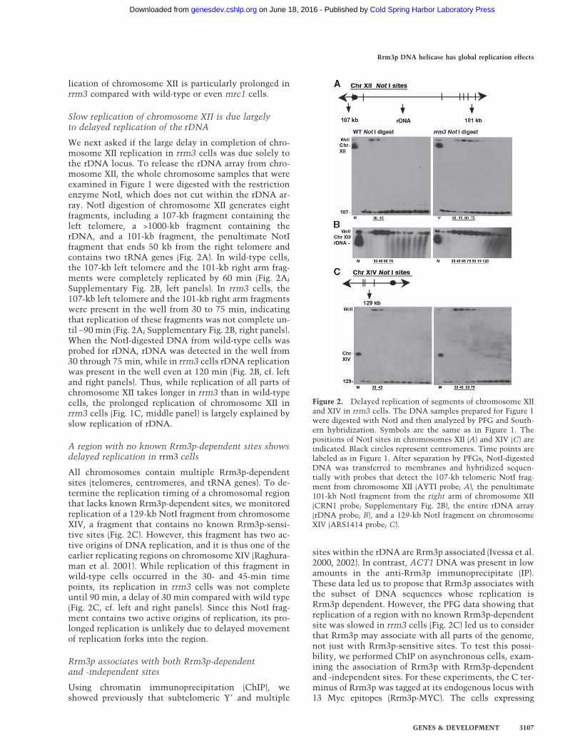
We next asked if the large delay in completion of chro-mosome XII replication in rrm3 cells was due solely tothe rDNA locus. To release the rDNA array from chro-mosome XII, the whole chromosome samples that wereexamined in Figure 1 were digested with the restrictionenzyme NotI, which does not cut within the rDNA ar-ray. NotI digestion of chromosome XII generates eightfragments, including a 107-kb fragment containing theleft telomere, a >1000-kb fragment containing therDNA, and a 101-kb fragment, the penultimate NotIfragment that ends 50 kb from the right telomere andcontains two tRNA genes (Fig. 2A). In wild-type cells,the 107-kb left telomere and the 101-kb right arm frag-ments were completely replicated by 60 min (Fig. 2A;Supplementary Fig. 2B, left panels). In rrm3 cells, the107-kb left telomere and the 101-kb right arm fragmentswere present in the well from 30 to 75 min, indicatingthat replication of these fragments was not complete un-til ∼90 min (Fig. 2A; Supplementary Fig. 2B, right panels).When the NotI-digested DNA from wild-type cells wasprobed for rDNA, rDNA was detected in the well from30 through 75 min, while in rrm3 cells rDNA replicationwas present in the well even at 120 min (Fig. 2B, cf. leftand right panels). Thus, while replication of all parts ofchromosome XII takes longer in rrm3 than in wild-typecells, the prolonged replication of chromosome XII inrrm3 cells (Fig. 1C, middle panel) is largely explained byslow replication of rDNA.
A region with no known Rrm3p-dependent sites showsdelayed replication in rrm3 cells
All chromosomes contain multiple Rrm3p-dependentsites (telomeres, centromeres, and tRNA genes). To de-termine the replication timing of a chromosomal regionthat lacks known Rrm3p-dependent sites, we monitoredreplication of a 129-kb NotI fragment from chromosomeXIV, a fragment that contains no known Rrm3p-sensi-tive sites (Fig. 2C). However, this fragment has two ac-tive origins of DNA replication, and it is thus one of theearlier replicating regions on chromosome XIV (Raghura-man et al. 2001). While replication of this fragment inwild-type cells occurred in the 30- and 45-min timepoints, its replication in rrm3 cells was not completeuntil 90 min, a delay of 30 min compared with wild type(Fig. 2C, cf. left and right panels). Since this NotI frag-ment contains two active origins of replication, its pro-longed replication is unlikely due to delayed movementof replication forks into the region.
Rrm3p associates with both Rrm3p-dependentand -independent sites
Using chromatin immunoprecipitation (ChIP), weshowed previously that subtelomeric Y� and multiple
sites within the rDNA are Rrm3p associated (Ivessa et al.2000, 2002). In contrast, ACT1 DNA was present in lowamounts in the anti-Rrm3p immunoprecipitate (IP).These data led us to propose that Rrm3p associates withthe subset of DNA sequences whose replication isRrm3p dependent. However, the PFG data showing thatreplication of a region with no known Rrm3p-dependentsite was slowed in rrm3 cells (Fig. 2C) led us to considerthat Rrm3p may associate with all parts of the genome,not just with Rrm3p-sensitive sites. To test this possi-bility, we performed ChIP on asynchronous cells, exam-ining the association of Rrm3p with Rrm3p-dependentand -independent sites. For these experiments, the C ter-minus of Rrm3p was tagged at its endogenous locus with13 Myc epitopes (Rrm3p-MYC). The cells expressing
Figure 2. Delayed replication of segments of chromosome XIIand XIV in rrm3 cells. The DNA samples prepared for Figure 1were digested with NotI and then analyzed by PFG and South-ern hybridization. Symbols are the same as in Figure 1. Thepositions of NotI sites in chromosomes XII (A) and XIV (C) areindicated. Black circles represent centromeres. Time points arelabeled as in Figure 1. After separation by PFGs, NotI-digestedDNA was transferred to membranes and hybridized sequen-tially with probes that detect the 107-kb telomeric NotI frag-ment from chromosome XII (AYTI probe; A), the penultimate101-kb NotI fragment from the right arm of chromosome XII(CRN1 probe; Supplementary Fig. 2B), the entire rDNA array(rDNA probe; B), and a 129-kb NotI fragment on chromosomeXIV (ARS1414 probe; C).
Rrm3p DNA helicase has global replication effects
GENES & DEVELOPMENT 3107
Cold Spring Harbor Laboratory Press on June 18, 2016 - Published by genesdev.cshlp.orgDownloaded from

Rrm3p-MYC and the isogenic no-tag control strain lackthe distal portion of the ADH4 gene (a sequence called A+).
If Rrm3p does indeed associate with all sites in thegenome, there is no DNA sequence in the same cell thatcan serve as an appropriate negative control. To circum-vent this problem, we performed ChIP on a mixture oftwo strains (Fig. 3A). Prior to cross-linking, asynchro-nous cells from the test strains (strains expressingRrm3p-MYC or the isogenic no-tag control) were mixedwith an equal number of asynchronous cells from astrain that contains the entire ADH4 gene (A+ strain).The cell mixtures were then cross-linked with formal-dehyde and processed for ChIP. The amount of A+ DNAin the IP served as a negative control for normalization.
We monitored the presence of four test sequences aswell as the A+ sequence in the anti-MYC IP. Two of thesequences, the RFB within the rDNA (Fig. 3B) and asingle-copy sequence near the left telomere of chromo-some VII (TEL), are sites of replication pausing in rrm3cells (Ivessa et al. 2000, 2002). The other two sites are asegment within the ADH4 gene that is present in theRrm3p-MYC, no-tag, and A+ strains and a segment of theARO1 gene. Neither the ADH4 nor the ARO1 segmentscontain a known Rrm3p-dependent site. PCR reactionswere multiplexed with each reaction containing a set of
primers for the A+ sequence and one of the four testsites. Compared with the amount of A+ DNA in the IPs,the ARO1 and ADH4 fragments were enriched, respec-tively, 11 ± 0.6 (average ± standard deviation) and 9 ± 1.5(Fig. 3C). The enrichment of the single-copy TEL se-quence was four- to fivefold higher (48 ± 3.5-fold).Thelevel of enrichment at the RFB (29 ± 2.7) was not directlycomparable to the three single-copy sequences, as its am-plification was accomplished with fewer PCR cycles dueto its being multicopy. At all four sites, association ofRrm3p-MYC was dependent on both in vivo cross-link-ing and the presence of the MYC tag (data not shown).
If Rrm3p associates with DNA during DNA replica-tion, we expect its association to be higher in asynchro-nous cells than in G1- or G2/M-arrested cells. Indeed,the enrichment of Rrm3p at the RFB was higher in asyn-chronous cultures (29 ± 2.6 fold) than in either �-factor-arrested (3 ± 0.6-fold) or nocodazole-arrested cells(2.7 ± 0.6) (Fig. 3D). In contrast, the RFB-binding Fob1pprotein showed robust association with the RFB in asyn-chronous, G1, and G2/M cells (Fig. 3E). Taken together,these data suggest that Rrm3p associates with many ge-nomic sites, including those that are not known to beRrm3p dependent, and this association likely occurs dur-ing S phase.
Figure 3. Rrm3p associates with Rrm3p-dependent and -independent sites in a cell cycle-dependent manner. (A) Schematic ofcell-mixing experiment used for C and D. The A+ strain contains a portion of ADH4 that is absent from the no-tag control andRrm3p-MYC strains. (B) Schematic of a single 9.1-kb rDNA repeat. Bars indicate positions of PCR-amplified sequences used for D(RFB) and E (RFB, 35S). (C) DNA immunoprecipitated from the Rrm3p-MYC strain was PCR-amplified for 23 cycles using primers forthe RFB and for 28 cycles using primers for the single-copy TEL, ARO, or ADH sequences. Intensities of amplified fragments werequantified by densitometric analysis. The A+ sequence, which is absent from experimental strains, was used for background normal-ization. Fold enrichment ± standard deviations are indicated here and in D and E. (D) DNA was immunoprecipitated from asynchro-nous, G1 phase, and G2/M phase Rrm3p-MYC cells and amplified using 23 cycles of multiplex PCR with primers specific to the RFB.The amount of RFB sequence in the immunoprecipitate was normalized to the background intensity of the A+ sequence. (E) DNA wasimmunoprecipitated as described for D from a Fob1p-MYC strain except that the amount of RFB sequence in the immunoprecipitatewas normalized to a sequence from within the 35S-encoding rDNA. (F) Rrm3p-MYC abundance is constant throughout the cell cycle.Synchronized Rrm3p-MYC cells were collected at the indicated time points and analyzed for Rrm3p-MYC abundance after separationby SDS-PAGE and Western blotting with MYC antibody and subsequently with Tpd3 antibody. (G) Flow-cytometric profile ofsynchronized cells used in F.
Azvolinsky et al.
3108 GENES & DEVELOPMENT
Cold Spring Harbor Laboratory Press on June 18, 2016 - Published by genesdev.cshlp.orgDownloaded from

To determine if Rrm3 protein levels fluctuate through-out the cell cycle, �-factor-arrested Rrm3p-MYC-ex-pressing cells were released into the cell cycle at 24°Cand time points were processed for flow cytometry (Fig.3G) and protein analysis (Fig. 3F). We find that Rrm3p-MYC abundance was similar from late G1 phase throughthe end of the cell cycle. Likewise, RRM3 transcriptionis not cell cycle regulated (Spellman et al. 1998).
Rrm3p loads onto origins at the beginning of S phaseand migrates with Mcm4p and Pol2p through adjacentDNA, even in regions that contain no knownRrm3p-dependent sites
The observation that Rrm3p is associated with Rrm3p-dependent and -independent sites in a cell cycle-depen-dent manner (Fig. 3) suggested that Rrm3p may be a com-ponent of the replication fork machinery. To address thisquestion, we used the ChIP assay in synchronized cells.For these experiments, we constructed two strains,SDY2 and SDY1. Both strains expressed Rrm3p-MYC
and either Mcm4p-HA (SDY2) or Pol2p-HA (SDY1)tagged at their C termini with three HA epitopes (Apari-cio et al. 1997). Mcm4p, a component of the prereplica-tion complex (pre-RC) (Labib et al. 2001), associates withorigins in G1 phase and then moves with the replicationfork upon origin firing (Aparicio et al. 1997). Pol2p, thecatalytic subunit of DNA polymerase �, which isthought to be the leading-strand polymerase, loads ontoreplication origins at the time of origin firing and thenmoves with the replication fork (Aparicio et al. 1997;Kawasaki and Sugino 2001). These doubly tagged strainsallowed us to compare in the same cells the spatial andtemporal chromatin association of Rrm3p with that ofknown components of the replication complex.
Cells released from an �-factor-induced G1-phase ar-rest (0 time point) at 18°C proceeded synchronouslythrough the cell cycle as monitored by flow cytometry(Fig. 4B–D). Samples were taken throughout the cellcycle and assayed by ChIP. In strain SDY2, we examinedprotein association at ARS305 on chromosome III and attwo sites 8 and 17 kb to the right of ARS305 (Fig. 4A,
Figure 4. Rrm3p loads onto origins at thebeginning of S phase and migrates with thereplication fork through both Rrm3p-de-pendent and -independent sites. (A) Dia-gram of the ARS305 region on chromo-some III (left) and the ARS607 region onchromosome VI (right). The oval indicatesthe origin of DNA replication. Verticalbars indicate the positions of two tRNAgenes to the right of ARS607. Horizontalbars indicate the positions of the segmentsamplified by PCR. (B-D) Flow-cytometricprofiles for strains used for E and F (SDY2cells, express Mcm4p-HA and Rrm3p-MYC), G and H (SDY1 cells, expressPol2p-HA and Rrm3p-MYC), and I and Jand Supplementary Figure 3 (SDY1 cells,express Pol2p-HA and Rrm3p-MYC). (E,F)SDY2 cells were arrested in late G1 phaseand released synchronously into the cellcycle at 18°C. Samples were collected forflow cytometry, ChIP, and Western blotanalysis at 12-min intervals. Formalde-hyde-fixed cells were immunoprecipitatedwith anti-HA (Mcm4p, E) or anti-MYC(Rrm3p, F) antibodies. DNA was purified,PCR-amplified, and resolved on a 2.8%ethidium bromide agarose gel. Eachprimer set was used to amplify both inputand immunoprecipitated DNA but onlyone representative input reaction per siteis shown. (G,H). Synchronized SDY1 cellswere processed for ChIP and analyzed forassociation of Pol2p-HA (G) and Rrm3p-MYC (H) with the ARS305 region as de-scribed for E and F. (I,J) SynchronizedSDY1 cells were processed for ChIP andanalyzed for association of Pol2p-HA (I)and Rrm3p-MYC (J) with the ARS607 re-gion as described for E and F.
Rrm3p DNA helicase has global replication effects
GENES & DEVELOPMENT 3109
Cold Spring Harbor Laboratory Press on June 18, 2016 - Published by genesdev.cshlp.orgDownloaded from

left). This region was chosen because the movement ofMcm4p and Pol2p through these sites has been studiedpreviously (Aparicio et al. 1997). This region has no pre-dicted Rrm3p-dependent sites and no detectable replica-tion pauses in rrm3 cells (data not shown).
As reported previously (Aparicio et al. 1997), Mcm4p-HA was associated with ARS305 in G1-arrested cells(Fig. 4E, row one, 0-min time point) and moved along thechromosome as S phase progressed. Mcm4p-HA was as-sociated with ARS305 + 8 kb at 36 and 48 min after re-lease from � factor (Fig. 4E, second row), and withARS305 + 17 kb at 48 and 60 min (Fig. 4E, third row). Inthe same cells, Rrm3p-MYC was not origin associated inG1-arrested cells (Fig. 4F, row one, 0-min time point),indicating that Rrm3p is not part of the pre-RC. Rrm3p-MYC was ARS305 associated at 36 min, which by flowcytometry (Fig. 4B) corresponds to the beginning of Sphase. The timing and pattern of Rrm3p-MYC associa-tion with sites ARS305 + 8 kb and ARS305 + 17 kb wasindistinguishable from that of Mcm4p-HA.
Next, we used strain SDY1 to compare the associationof Pol2p-HA (Fig. 4G) and Rrm3p-MYC (Fig. 4H) throughthe ARS305 region. Pol2p-HA and Rrm3p-MYC showedsimilar patterns of association: Neither was ARS305bound in G1-arrested cells, but both loaded onto ARS305at the beginning of S phase (36 min) (Fig. 4C). As S phaseprogressed, both proteins dissociated from origin DNA.Both proteins were maximally associated withARS305 + 8 kb at 48 min and with ARS305 + 17 kb at 60min. As a negative control, we used a strain expressingEst2p-MYC, the catalytic subunit of telomerase, whichis not expected to be associated with the replication fork.We saw no association of Est2p-MYC with the ARS305region at any time in the cell cycle (data not shown). Inaddition, Pol2p-HA and Rrm3p-MYC associated simi-larly with the ARS1 region on chromosome IV, which,like the ARS305 region, lacks any known Rrm3p-sensi-tive sites (Supplementary Fig. 3). Neither protein wasARS1 associated in G1 phase (0 min), both were ARS1associated at the start of S phase (36 min) (Fig. 4D) andboth moved with similar kinetics through sites that are3 and 12 kb to the left of ARS1.
We also examined Pol2p-HA and Rrm3p-MYC asso-ciation with ARS607 and sites that are 5 and 10 kb to theright of ARS607 (Fig. 4A, right). The ARS607 + 5-kb seg-ment contains a tRNA gene, tA(AGC)F, that is a site ofdramatic replication pausing in rrm3 cells (see also Fig.7B, below; Ivessa et al. 2003). There is a second tRNAgene, tY(GUA)F2, ∼70 base pairs (bp) to the right of theARS607 + 10-kb site that is surely Rrm3p dependent astranscription and replication proceed in opposite direc-tions through this gene. As at the other sites, Pol2p-HA(Fig. 4I) and Rrm3p-MYC (Fig. 4J) bound to ARS607 atthe beginning of S phase (Fig. 4D), and the two proteinscomigrated through ARS607 + 5 kb and ARS607 + 10 kb.
Thus, Rrm3p-MYC was associated with three differentorigins at the beginning of S phase but not in G1 phase.Rrm3p-MYC comigrated with Pol2p-HA through threedifferent regions, including two regions that do not con-tain Rrm3p-dependent sites. We conclude that Rrm3p
moves with the replication fork machinery through bothRrm3p-dependent and -independent sites.
Rrm3p associates with Pol2p in vivo
To provide further support for the interpretation thatRrm3p is part of the replication machinery, we asked ifRrm3p-MYC is associated in vivo with Pol2p-HA (Fig.5A). Strain SDY1, which expresses Rrm3p-MYC andPol2p-HA, or control strains expressing only one of thetwo tagged proteins were arrested with � factor, releasedinto the cell cycle at 24°C, and collected at 40 min whencells were in mid-S phase as determined by flow cytom-etry (data not shown). Lysates were prepared from eachstrain and precipitated with anti-MYC antibody (Fig. 5A,left panel) or with anti-HA antibody (Fig. 5A, rightpanel). The proteins in the IP were separated by SDS-PAGE and analyzed by Western blotting with both anti-HA and anti-MYC antibodies.
In strain SDY1, an interaction of Pol2p-HA andRrm3p-MYC was readily detected in the S-phase extracts(Fig. 5A). This interaction was seen when extracts wereprecipitated with either anti-MYC (Fig. 5A, lanes 3,4) oranti-HA (Fig. 5A, lanes 7,8), and was maintained evenwhen the lysate was treated with DNase I prior to im-munoprecipitation (Fig. 5A, lanes 4,8). In the controlstrain expressing Rrm3p-MYC or Pol2p-HA alone, therewere no cross-reacting proteins in either the anti-MYCor anti-HA IPs (Supplementary Fig. 4A,B)
The same experiment was performed using strainSDY2, which expresses both Rrm3p-MYC and Mcm4p-HA. Although an interaction between Rrm3p-MYC andMcm4-HA was seen in the absence of DNase I treatment(Fig. 5B, lanes 4,8), this interaction was lost when thelysates were pretreated with DNase I (Fig. 5B, lanes 5,9).The coimmunoprecipitation (co-IP) in a DNA-dependentmanner of Rrm3p-MYC with Mcm4p-HA probably re-
Figure 5. Rrm3p associates with Pol2p during S phase. Proteinextracts were prepared from mid-S-phase cultures of strainsSDY1 (A) and SDY2 (B), treated with DNase I (+) or not (−), andimmunoprecipitated (IP) with either anti-C-MYC Agarose-con-jugated beads or anti-HA Agarose-conjugated beads. Input andIP samples were separated on SDS-PAGE gels, and analyzed byWestern blotting. Blots were probed with anti-HA (top) or anti-MYC (bottom).
Azvolinsky et al.
3110 GENES & DEVELOPMENT
Cold Spring Harbor Laboratory Press on June 18, 2016 - Published by genesdev.cshlp.orgDownloaded from

flects the fact that although both proteins are at the forkin mid-S phase, they are not in close proximity.
In the absence of Rrm3p, replication forks slowat Rrm3p-dependent sites
To determine if Pol2p progression through DNA is af-fected by the absence of Rrm3p, we constructed twostrains, SDY5 and SDY8, both of which express Pol2ptagged at its C terminus with 13 MYC epitopes (Pol2p-MYC). The two strains were isogenic except that RRM3was deleted from strain SDY8. Both strains were arrestedwith � factor and released synchronously into the cellcycle at 18°C, and samples were processed for flow cy-tometry (Fig. 6A) and ChIP. In the same synchrony, weexamined replication through the ARS305 region, whichcontains no known Rrm3p-dependent sites (Fig. 4A, left)and the ARS607 region that contains two Rrm3p-depen-dent sites, one at 5 kb and the other at 10 kb to the rightof ARS607 (Fig. 4A, right). If replication forks move moreslowly through a given site in rrm3 compared with wild-type cells, there should be more of that sequence in theanti-MYC IP from the mutant cells.
By flow-cytometric analysis, both strains were in earlyS phase at the 36-min time point (Fig. 6A). At bothARS305 and ARS607, Pol2p-MYC was detected at theorigin even at 24 min in rrm3 cells (Fig. 6B,C), consistentwith evidence from PFG experiments for earlier initia-tion in rrm3 cells. However, this earlier association ofPol2p-MYC with both origins was seen in only one oftwo experiments. The levels of Pol2p-MYC associationwith ARS305 + 8 kb were similar in wild-type and rrm3cells (Fig. 6B, middle panel) in two of two experiments.The relatively modest increase in Pol2p-MYC associa-
tion with ARS305 + 17 kb in rrm3 compared with wild-type cells was also seen in both experiments (Fig. 6B,right panel).
More dramatic differences in Pol2p-MYC progressionbetween the wild-type and rrm3 strains were seen in theARS607 region (Fig. 6C), and these differences were ob-served in two of two experiments. There was morePol2p-MYC associated at ARS607 + 5 kb in rrm3 cellscompared with wild type (Fig. 6C, middle panel). Theassociation of Pol2p-MYC with ARS607 + 10 kb waseven higher and persisted for longer in rrm3 than in wild-type cells (Fig. 6C, right panel). Indeed, in rrm3 cells,association of Pol2p-MYC was readily detected at thissite at 84 and even 96 min, when flow-cytometric analy-sis shows that replication of most of the genome wascomplete (Fig. 6A). These data suggest that in the ab-sence of Rrm3p, the replication fork moves more slowlyat sites that are Rrm3p dependent.
In the absence of Rrm3p, replication fork progressionis similar in wild-type and rrm3 cellsat Rrm3p-independent sites
The rate of fork progression can also be estimated fromthe abundance of replication intermediates in 2D gels.Slower fork movement causes the fraction of DNA inreplication structures to increase in vivo. We used 2Dgels to examine replication intermediates at multipleloci in wild-type, rrm3, and mrc1 cells (Fig. 7; data notshown). Of the four loci examined in Figure 7, only onecontains a known Rrm3p-dependent site (Fig. 7B, tRNA-Ala). At each of the four loci, the intensity of the arc offorked replication intermediates was similar in wild-type and rrm3 cells. As shown previously (Szyjka et al.
Figure 6. Pol2p migrates more slowly inthe absence of Rrm3p through Rrm3p-de-pendent sites. Strains SDY5 (expressesPol2p-MYC) and SDY8 (rrm3 expressesPol2p-MYC) were synchronized and pro-cessed for ChIP as described in Figure 4.(A) Flow-cytometric profiles for synchro-nized SDY5 and SDY8. (B) The associationof Pol2p-MYC with the ARS305 region(diagrammed in Fig. 4A) with (white bars)and without (gray bars) Rrm3p was exam-ined. (C) DNA from the same samplesused in B was analyzed for the associationof Pol2p-HA with the ARS607 region,which contains two Rrm3p-dependentsites (diagrammed in Fig. 4A, right), with(white bars) and without (gray bars)Rrm3p. In B and C, the intensities of am-plified fragments were quantified by den-sitometric analysis. The percentage of in-put DNA precipitated (% IP’ed) is graphedfor one of the two independent experi-ments performed with each strain.
Rrm3p DNA helicase has global replication effects
GENES & DEVELOPMENT 3111
Cold Spring Harbor Laboratory Press on June 18, 2016 - Published by genesdev.cshlp.orgDownloaded from

2005), there were more replication intermediates inmrc1 cells at each site. Whereas increased replicationpausing at tRNA-Ala was readily detected in rrm3 cells,there was no detectable pausing at this site in mrc1 cells(Fig. 7B). Thus, although replication of all chromosomesis slowed in both rrm3 and mrc1 cells (Figs. 1, 2), themechanism leading to this effect is different in the twomutants.
Discussion
Here we show that lack of Rrm3p delays replication ofall yeast chromosomes (Fig. 1). The extent of this delaywas particularly dramatic at the rDNA locus on chromo-some XII (Fig. 2B). However, the impact on other chro-mosomes such as chromosomes VI and XIV was alsostriking, as complete replication of these chromosomestook roughly twice as long in rrm3 compared with wild-type cells (Fig. 1B; Supplementary Fig. 1). The extent ofthis replication delay was even greater than for mrc1cells (Fig. 1) where replication forks are estimated tomove at 50% of wild-type rates. We cannot rule out thatsome of the delay in chromosomes entering the PFGs inthe rrm3 strain was due to the presence of recombinationintermediates rather than to replication intermediates.However, even though recombination is increased inrrm3 cells, the recombination rate is still sufficientlylow that recombination should affect the behavior of
only a small number of chromosomes. For example, therate of recombination in rrm3 cells in a tRNA-rich 184-kb interval on chromosome VII is only 1.5 × 10⁻⁵ eventsper cell division (Ivessa et al. 2003).
Not only was replication of all chromosomes Rrm3pdependent, replication of a region without knownRrm3p-dependent sites, a 129-kb NotI fragment on chro-mosome XIV, was dramatically slowed in rrm3 cells (Fig.2C). These results suggest that there are additionalRrm3p-dependent sites that have not yet been identified.By the criterion of 2D gels, we did not see strong repli-cation pausing at the ∼50 RNA polymerase II-transcribedgenes within a 105-kb region of chromosome VI nor dur-ing galactose-induced transcription of the GAL1, GAL7,or GAL10 genes (Ivessa et al. 2003). However, using 2Dgels, others detect replication pauses within genes tran-scribed from the GAL1 promoter when these genes arecarried on plasmids, and this pausing is increased inrrm3 cells (Prado and Aguilera 2005). Regions like the129-kb NotI fragment may contain one or more geneswhose replication is Rrm3p sensitive, even if this sensi-tivity cannot be detected with 2D gels when the genesare in their normal chromosomal context. In addition,Rrm3p may be needed for efficient resolution of con-verged replication forks throughout the genome, as it isin the rDNA (Ivessa et al. 2000). Consistent with thispossibility, X-shaped molecules are more abundantwithin subtelomeric Y� elements, the silent mating type
Figure 7. Abundance of replication intermediatesin wild-type (WT), rrm3, and mrc1 strains. (A) Sche-matics of replication intermediates as visualized in2D gels. (1N) Nonreplicating fragment; (2N) almostfully replicated fragment right before sister chroma-tids separate; (P) replication pause; (BU) bubble-shaped replication intermediates. Dotted lines indi-cate arc formed by linear DNA molecules. (B–E)DNA from glucose-grown asynchronous cells fromthe indicated strains was restriction enzyme-di-gested, separated on 2D gels, and analyzed by South-ern blotting with the indicated probes (restrictionenzymes and hybridization probes are noted in pa-rentheses). (B) tRNAA (BglII, HIS2); arrows indicatethe position of the tRNA gene on the arc of replica-tion intermediates; a lower exposure of the samplefrom the mrc1 strain is shown to provide better vi-sualization of fork movement through the tRNAgene. (C) GAL10 (PvuII, GAL10). (D) ARS305(EcoRV, ARS305). (E) YCK2 (BglII, YCK2).
Azvolinsky et al.
3112 GENES & DEVELOPMENT
Cold Spring Harbor Laboratory Press on June 18, 2016 - Published by genesdev.cshlp.orgDownloaded from

loci, and on plasmid-borne RNA polymerase II-tran-scribed genes in rrm3 cells (Ivessa et al. 2000, 2002, 2003;Prado and Aguilera 2005). Since the 129-kb NotI frag-ment contains two early-firing origins, its delayed repli-cation in rrm3 cells may be due in part to decreasedefficiency in resolution of converged forks. A role forRrm3p in the resolution of converged forks may alsoexplain the modest increase in Pol2-MYC atARS305 + 17 kb in the absence of Rrm3p (Fig. 6B). Thissite is midway between ARS305 and ARS306, a regionwhere replication forks are expected to converge.
One model for the mechanism of Rrm3p action is thatit is recruited to the fork when the fork stalls at a diffi-cult-to-replicate site. However, in asynchronous cells,Rrm3p was associated with both Rrm3p-dependent andRrm3p-independent sites (Fig. 3C). Moreover, in syn-chronized cells, Rrm3p loaded onto three different ori-gins, ARS305, ARS1, and ARS607 at the same time asPol2p. Rrm3p movement through the DNA adjacent tothe three ARSs paralleled that of both Pol2p and Mcm4p,even though only one of these regions, ARS607, con-tained Rrm3p-sensitive sites (Fig. 4; Supplementary Fig.3). In addition, using co-IP, we found that Rrm3p wasassociated with Pol2p in vivo (Fig. 5). Thus, our dataargue strongly against a model in which Rrm3p is re-cruited to specific sites upon stalling of the replicationfork. Rather, our data suggest that Rrm3p is a constitu-tive part of the replication apparatus. Alternatively,Rrm3p could be part of a chromatin remodeling complexthat moves immediately ahead of the replication forkand makes chromatin more accessible to the replicationmachinery.
Our results disagree with another study that examinedreplication through the ARS305 region into which twoectopic RFB sites were inserted (Calzada et al. 2005). Be-cause this group detected Rrm3p at the RFB but not atARS305 nor at a site distal to the ectopic RFB, they con-cluded that Rrm3p is not part of the replisome but ratheris recruited to stalled forks. In contrast, in the unmodi-fied ARS305 region, as well as at two additional genomicloci, we saw Rrm3p moving with the fork (Fig. 4; Supple-mentary Fig. 3). We think a possible explanation for thediscrepancy between the data from the two labs is thatthere is more Rrm3p associated with the RFB as com-pared with the number of Rrm3p molecules that travelwith the replication machinery. We speculate that moreRrm3p molecules may be recruited to particularly stableprotein–DNA complexes such as the complexes at theRFB and telomeres (Fig. 3). The number of Rrm3p mol-ecules required to facilitate replication past specific pro-tein–DNA complexes may depend on the stability of thecomplex in front of the fork.
Is replication fork stalling at stable chromatin struc-tures and difficulty in resolving converged forks suffi-cient to explain the large replication delay seen in rrm3cells? A decrease in the number of active origins can alsoprolong S phase. However, origin usage is similar inwild-type and rrm3 cells in the rDNA (Ivessa et al. 2000),at ARS605 (Ivessa et al. 2003), and at ARS305 (Fig. 7D),while normally inactive origins are actually more active
in rrm3 cells (Ivessa et al. 2002, 2003). A decrease in thespeed of replication forks can also prolong S phase.Slower fork movement in mrc1 cells results in more rep-lication intermediates, as assayed by 2D gels, a resultconfirmed here (Fig. 7). However, increased fork pausingat Rrm3p-dependent sites such as tRNA genes was notseen in mrc1 cells (Fig. 7B; data not shown). In contrastto the results in mrc1 cells, the fraction of DNA in rep-lication intermediates in rrm3 and wild-type cells wassimilar (Fig. 7), except at Rrm3p-dependent sites such asat a tRNA gene (Fig. 7B).
We conclude that neither reduced origin usage nor agenome-wide slowing of replication forks explains thelonger S phase in rrm3 cells. Rather, the global delay inDNA replication observed in this strain (Figs. 1, 2) is duelargely or entirely to site-specific delays in replication,including compromised resolution of converged forks.Others have speculated that the lethality of mrc1 rrm3cells reflects a requirement for Rrm3p in the restart offorks that are damaged as a result of replication in theabsence of Mrc1p (Szyjka et al. 2005). However, we findthat while replication takes longer in both mrc1 andrrm3 cells (Fig. 1), the mechanism causing slow replica-tion was different in the two strains (Fig. 7). We thinkthat the most likely explanation for the lethality of mrc1rrm3 doubly mutant cells is the additive effects of twodifferent types of defects in DNA replication.
Although there are several examples of prokaryotic he-licases that help replication forks move past protein–DNA complexes (Bedinger et al. 1983; Yancey-Wronaand Matson 1992; Barry and Alberts 1994), Rrm3p is thefirst eukaryotic enzyme shown to function in this way.Since these difficult-to-replicate protein–DNA com-plexes are encountered in every S phase, they may posean even greater threat to genome stability than exog-enous DNA damage. Genetic assays using mutants inreplication and repair genes have found high frequenciesof genomic instability at Ty1 elements, tRNA genes, andconverging replication forks, sites that include Rrm3p-dependent loci (Scholes et al. 2001; Cha and Kleckner2002; Lemoine et al. 2005; Admire et al. 2006). Some ofthese sites overlap with Rrm3-dependent sites, suggest-ing that forks stall and collapse at preferred locationswithin the genome that are difficult to replicate. Therrm3 system provides a unique opportunity to study themechanism of replication fork stalling and breakage atsites that are natural impediments to fork progression.Analysis of Rrm3p may provide insights into the fate ofreplication fork arrest and breakage at difficult-to-repli-cate loci in human chromosomes, which become fragilesites in response to replication stress and whose break-age can initiate the types of chromosomal aberrationsfound in human tumors.
Materials and methods
Strains
Yeast strains are described in Supplementary Table 1. Precisedeletions of the RRM3 and MRC1 ORFs were created using the
Rrm3p DNA helicase has global replication effects
GENES & DEVELOPMENT 3113
Cold Spring Harbor Laboratory Press on June 18, 2016 - Published by genesdev.cshlp.orgDownloaded from

pRS303-HIS3 plasmid (Sikorski and Hieter 1989) as a templateto generate HIS3 PCR products with overhangs that targeted theproduct to either RRM3 or MRC1 (Ivessa et al. 2000). Similarly,the BAR1 ORF was precisely deleted using pRS304-TRP1 (Si-korski and Hieter 1989). The bar1��TRP1 wild-type, rrm3�,and mrc1� strains were constructed by B. Lenzmeier. The bar1�
allele was introduced into rrm3� and mrc1� strains by matingfollowed by sporulation and identification of doubly mutantspore clones. Rrm3p, Fob1p, and Pol2p were tagged at their Ctermini with 13 MYC epitopes using the plasmid pFA6-TRP-13MYC as a template (Longtine et al. 1998). The Rrm3p-MYCand Fob1p-MYC had wild-type function as assayed by 2D gelanalysis of rDNA replication intermediates (data not shown).The growth rate of the Pol2p-MYC strain was indistinguishablefrom wild type, suggesting that the Pol2p-MYC also had wild-type function (data not shown). To generate VPS106-UT,VPS106 was transformed with the 2.4-kb SalI–EcoR1 fragmentof pADH4UCAIV. Integration of this fragment results in dele-tion of the terminal ∼15 kb of chromosome VII-L and its replace-ment with URA3 adjacent to a new telomere (Gottschling et al.1990). The strains for the fork migration studies wererrm3��HIS3 or Rrm3p-MYC13 derivatives of strains gener-ously provided by O. Aparicio and S. Bell that express Pol2p-3HA (strain OAy618) or Mcm4p-3HA (strain OAy535) (Aparicioet al. 1997). In all cases, tagged proteins were expressed fromtheir endogenous promoters, and expression was verified byWestern analysis.
Cell synchronies
For PFG, ChIP, and co-IP experiments, single colonies were in-oculated in 5 mL of YEPD, grown for 6 h at 30°C, diluted intoYEPD, and grown overnight at 30°C to an OD660 of 0.15. �
Factor (Princeton University) was added to cultures at a con-centration of 0.015 ng/mL, and cultures were incubated for 3 hat 24°C until microscopic examination indicated that ∼95% ofcells were unbudded. Cells were washed in YEPD; released intofresh YEPD containing 70 µg/mL Pronase (Sigma) at 24°C forco-IP, PFG, and asynchronous ChIP experiments, and at 18°Cfor synchronous ChIP; and samples were collected at the indi-cated time points. For PFG experiments, 15 µg/mL nocodazole(Sigma) was added to the releasing media to arrest cells in G2/Mand prevent their entry into a second cell cycle. The quality ofeach synchrony was monitored by flow cytometry. Briefly, 500µL of cells from each time point were resuspended in water,sonicated, and fixed in 70% ethanol at room temperature for 3h. Cells were resuspended in 50 mM sodium citrate and incu-bated with 0.125 mg of RNase A overnight at 50°C. One milli-gram of Proteinase K (Roche) was added and cells were incu-bated for 2 h at 50°C. Cells were sonicated and incubated in thedark with Sytox Green (2 µM) (Invitrogen) for 45 min at roomtemperature. Samples were analyzed in a FACScan single-laserfixed-alignment benchtop analyzer with an emission at 488 nm.A 530/30 bandpass filter was used to collect emissions in frontof the detector.
Gel methods
To process samples for PFGs, cultures were killed in 0.1% so-dium azide, pelleted, and washed in ddH2O and then in SPE (0.5M Sorbitol, 100 mM NaPO4 at pH 7.5, 10 mM EDTA). Cellswere resuspended in 1.5 mL of 1 M sorbitol, pelleted, and re-suspended in SPEM (SPE containing 30 mM �-mercapthenol)and Zymolyase (100 µg/mL; ICN Biomedicals). After cells wereincubated for 10 min at 30°C, an equal volume of InCert agarose(BMA) was added to each cell suspension, and the mixture was
pipetted into disposable CHEF plug molds (Bio-Rad) and allowedto solidify on ice. Plugs were incubated in SPEM containing 0.05mg/mL Zymolyase for 3 h at 37°C, rinsed in 0.5 M EDTA (pH9.0), resuspended in lysis solution (10 mM Tris-HCl at pH 8.0,50 mM EDTA at pH 9.0, 1% w/v Sarkosyl) containing protein-ase K (Roche) at a final concentration of 1 mg/mL, and incu-bated overnight at 55°C. Plugs were washed twice with 2.5 mL0.5× TBE (1 mM EDTA at pH 8.0, 0.044 mM boric acid, 0.045mM Tris Base), resuspended in 2.5 mL 0.5× TBE Buffer, andstored at 4°C. Chromosomes were separated using 1% gels (Bio-Rad Pulsed Field Certified Agarose) in 0.5× TBE buffer using theBio-Rad CHEF-DR III System. Running conditions for PFGswere 60–120-sec switch time, 6 V/cm voltage gradient, and 120°angle for 26 h at 14°C. NE Biolaboratories yeast chromosomePFG Marker was run as a reference. Gels were stained in 0.5µg/mL ethidium bromide for 30 min, destained in deionizedwater for 20 min, and photographed using the AlphaImager sys-tem (Alpha Innotech). Chromosomes were UV-cross-linked andtransferred to nylon Hybond membranes (Amersham) for South-ern analysis. All probes, with the exception of the BglII-B rDNAprobe, were PCR products amplified from yeast genomic DNA(Supplementary Table 2). PCR products were gel-purified usingZymoclean DNA column (Zymo Research), and 10 ng of prod-uct was used as a template for a second round of PCR. Thesecond PCR products were gel-purified and labeled using theRediPrime II Random Prime Labeling System (Amersham) and10 µL of 6000 µCi �-P32 dCTP (IDT). Membranes were subse-quently reprobed after stripping with boiling 0.25% SDS solu-tion. For NotI-digested samples, DNA agarose plugs were di-gested with 10,000 U of NotI (NE Biolaboratories) for 12 h at37°C and analyzed as described above. Methods of DNA prepa-ration and conditions for running 2D gels to detect replicationintermediates are as described (Ivessa et al. 2000).
ChIP
ChIP analyses with synchronized cells were performed essen-tially as described (Taggart et al. 2002) except samples were notmultiplexed. Monoclonal anti-HA (12CA5; Roche or Santa CruzBiotechnology) or anti-Myc (BD Biosciences) antibodies wereused. The number of PCR cycles varied from 25 to 30, depend-ing on the sequence being amplified and on the protein that wasanalyzed. PCR products were resolved in 2.8% ethidium bro-mide agarose gels. Amplified fragment intensities were visual-ized using the Alpha Imager system. Ethidium bromide gels ofPCR products were quantified by densitometric analysis usingScion Image 4.0.3.2 software (http://www.scioncorp.com). Thepercent immunoprecipitated is the relative recovery of immu-noprecipitated DNA relative to the amount of input DNA basedon the quantification of the respective PCR products and thedilution factor of immunoprecipitated and input DNA. EachChIP experiment was done at least twice, with similar results.
For ChIPs on asynchronous cultures, cells were grown to anOD660 of 0.7. The Rrm3p-MYC (JZT200) or the untagged controlstrain (VPS106UT) was mixed with an equal number of VPS106cells. Strains VPS106UT, JZT200, and JZT515 lack a portion ofthe ADH4 gene, called the A+ sequence, that is present inVPS106. The amount of A+ DNA in the IP served as a negativecontrol for a sequence that is not Rrm3p-MYC associated.Cross-linking and ChIP were carried out as described exceptthat magnetic protein G Dynabeads (Dynal Biotech) were used.DNA immunoprecipitated from Rrm3p-MYC was amplified us-ing 23 cycles of PCR with primers that amplify the RFB and 28cycles with primers that amplify the ARO1, ADH4, TEL, andA+ sequences. PCR products were resolved on 2.8% ethidiumbromide agarose gels and bands were quantified by densitomet-
Azvolinsky et al.
3114 GENES & DEVELOPMENT
Cold Spring Harbor Laboratory Press on June 18, 2016 - Published by genesdev.cshlp.orgDownloaded from

ric analysis (NIH Image1.60). Fold enrichment of Rrm3p-boundDNA was determined as follows: (MYC-tagged B/no-tagB) × (no-tag A+/MYC-tagged A+), where B denotes tested se-quence. For G1 (�-factor-arrested) and G2/M (nocodazole-ar-rested) cells, cultures were grown to an OD660 of 0.3 and ar-rested with a final concentration of 2 µg/mL � factor for 2 h at30°C or with a final concentration of 15 µg/mL nocodazole for3 h at 30°C prior to mixing and in vivo cross-linking. To deter-mine Fob1p-MYC association with the RFB, DNA was ampli-fied using 23 cycles of PCR with primers that amplify the RFBand a 407-bp sequence internal to the 35S rDNA (35S). Foldenrichment of Fob1p-MYC with the RFB was determined asfollows: (MYC-tagged RFB/no-tag RFB) × (no-tag 35S/MYC-tagged 35S).
Co-IP of Rrm3p with replication proteins
Cell cultures were synchronized with � factor at 24°C as de-scribed, collected at the 40-min S-phase time point, killed with0.1% sodium azide on ice, pelleted, resuspended in cell lysisbuffer (50 mM HEPES at pH 7.5, 10% v/v glycerol, 140 mMNaCl, 1 mM EDTA, 0.5% IGEPAL CA-630, 1 mM EDTA, 1 mMPMSF, Roche Complete EDTA-free Protease inhibitors), andfrozen as pellets in liquid nitrogen. Cell pellets were processedusing a Freezer Mill (SPEX CertiPrep) to >95% lysis efficiency.An aliquot was taken and resuspended in protein sample buffer(Fig. 5, Input). Cell lysates were spun down at 4°C to clear ly-sates, which were divided in half for control and DNaseI-treated samples. Twenty millimolar MgCl2 and 1 mM PMSFwere added to all lysates, and 150 U of DNase I (Sigma) wereadded to DNase I samples. Lysates were incubated on ice for 15min. Aliquots of cell extracts were taken after DNase I incuba-tion and resuspended in protein sample buffer as protein con-trols and for phenol-chloroform extraction of DNA to assay ef-ficient digestion by DNase I. Fifty microliters of anti-C-MYCAgarose-conjugated beads (Sigma) for anti-MYC immunopre-cipitation or 50 µL of rabbit anti-HA Agarose-conjugated beads(Abcam) were added to both samples and the mixtures wererotated for 1.5 h at 4°C. Beads were washed seven times in 1 mLof cell lysis buffer, spun down, resuspended in protein samplebuffer, and boiled. Equal portions of each IP were separated on7% SDS-PAGE gels and transferred to PVDF membranes (Mil-lipore). Membranes were blocked in TBST (10 mM Tris-HCl atpH 8, 150 mM NaCl, 0.07% Tween-20) containing 5% milk,incubated in a 1:400 dilution of HA monoclonal antibody (SantaCruz Biotechology) for anti-MYC IPs or 1:1000 dilution of MYCmonoclonal antibody (BD Biosciences) for anti-HA IPs, and thenincubated in a 1:3000 dilution of goat antimouse horseradishperoxidase-conjugated secondary antibody (Bio-Rad). Mem-branes were developed using ECL chemiluminescence system(Amersham) and exposed to Kodak Biomax XAR autoradiogra-phy film. Membranes were subsequently incubated in strippingbuffer (100 mM �-mercapthenol, 2% SDS, 64 mM Tris-HCl atpH 6.8) for 30 min at 50°C, blocked in TBST containing 5%milk, and reprobed with an antibody corresponding to the oneused for the IP (MYC or HA) to evaluate efficiency of the IP.
Levels of Rrm3p over the cell cycle
To determine Rrm3p levels throughout the cell cycle, cultureswere synchronized with � factor and released into the cell cycleat 24°C as described above. Cells were collected at the indicatedtime points and crude extract was prepared using the trichlor-acetic acid (TCA) method described in Pellicioli et al. (1999).Protein concentration in TCA protein preparations were deter-mined using RC DC colorimetric assay (Bio-Rad). For each time
point, equal protein amounts were processed for Western analy-sis. Proteins were separated on 7% SDS-PAGE gels and trans-ferred to PVDF membranes (Millipore). After probing with anti-MYC antibody, membranes were stripped and probed with1:1000 dilution of anti-Tpd3 antibody (gift of J. Broach), andthen incubated in a 1:3000 dilution of goat anti-rabbit secondaryantibody (Bio-Rad). Membranes were then stained with 0.2%Ponceau Stain for 30 min, and destained in water as a secondcontrol for protein loading.
ACKNOWLEDGMENTS
We thank J. Broach for the anti-Tpd3 antibody, S. Bell and O.Aparicio for providing strains, R. Ricke and A.K. Bielinsky foradvice on co-IP, A. Ivessa for assistance with 2D gels, C. Tuzonfor assistance with PFGs, M. Sabourin for advice on ChIP, and S.Aubert, M. Mateyak, S. Pinter, and C. Webb for suggestions onthis manuscript. This work was supported by National Insti-tutes of Health grants R37 GM26938, T32CA09328 (to J.Z.T.,A.A., and S.D.) and a predoctoral fellowship from the NJ Com-mission of Cancer Research (J.B.B.).
REFERENCES
Admire, A., Shanks, L., Danzl, N., Wang, M., Weier, U., Stevens,W., Hunt, E., and Weinert, T. 2006. Cycles of chromosomeinstability are associated with a fragile site and are increasedby defects in DNA replication and checkpoint controls inyeast. Genes & Dev. 20: 159–173.
Alcasabas, A., Osborn, A., Bachant, J., Hu, F., Werler, P., Bous-set, K., Furuya, K., Diffley, J., Carr, A., and Elledge, S. 2001.Mrc1 transduces signals of DNA replication stress to acti-vate Rad53. Nat. Cell Biol. 3: 958–965.
Aparicio, O.M., Weinstein, D.M., and Bell, S.P. 1997. Compo-nents and dynamics of DNA replication complexes in S. ce-revisiae: Redistribution of MCM proteins and Cdc45p dur-ing S phase. Cell 91: 59–69.
Barry, J. and Alberts, B. 1994. A role for two DNA helicases inthe replication of T4 bacteriophage DNA. J. Biol. Chem. 269:33063–33068.
Bedinger, P., Hochstrasser, M., Jongeneel, C., and Alberts, B.1983. Properties of the T4 bacteriophage DNA replicationapparatus: The T4 dda DNA helicase is required to pass abound RNA polymerase molecule. Cell 34: 115–123.
Boule, J.B. and Zakian, V.A. 2006. Roles of Pif1-like helicases inthe maintenance of genomic stability. Nucleic Acids Res. 4:4147–4153..
Calzada, A., Hodgson, B., Kanemaki, M., Bueno, A., and Labib,K. 2005. Molecular anatomy and regulation of a stable repli-some at a paused eukaryotic DNA replication fork. Genes &Dev. 19: 1905–1919.
Caruthers, J.M. and McKay, D.B. 2002. Helicase structure andmechanism. Curr. Opin. Struct. Biol. 12: 123–133.
Cha, R.S. and Kleckner, N. 2002. ATR homolog Mec1 promotesfork progression, thus averting breaks in replication slowzones. Science 297: 602–606.
Eoff, R.L. and Raney, K.D. 2005. Helicase-catalysed transloca-tion and strand separation. Biochem. Soc. Trans. 33: 1474–1478.
Gottschling, D.E., Aparicio, O.M., Billington, B.L., and Zakian,V.A. 1990. Position effect at S. cerevisiae telomeres: Revers-ible repression of Pol II transcription. Cell 63: 751–762.
Hennessy, K.M., Lee, A., Chen, E., and Botstein, D. 1991. Agroup of interacting yeast DNA replication genes. Genes &
Rrm3p DNA helicase has global replication effects
GENES & DEVELOPMENT 3115
Cold Spring Harbor Laboratory Press on June 18, 2016 - Published by genesdev.cshlp.orgDownloaded from

Dev. 5: 958–969.Ivessa, A.S., Zhou, J.-Q., and Zakian, V.A. 2000. The Saccharo-
myces Pif1p DNA helicase and the highly related Rrm3phave opposite effects on replication fork progression in ribo-somal DNA. Cell 100: 479–489.
Ivessa, A.S., Zhou, J.-Q., Schulz, V.P., Monson, E.M., and Za-kian, V.A. 2002. Saccharomyces Rrm3p, a 5� to 3� DNA he-licase that promotes replication fork progression throughtelomeric and sub-telomeric DNA. Genes & Dev. 16: 1383–1396.
Ivessa, A.S., Lenzmeier, B.A., Bessler, J.B., Goudsouzian, L.K.,Schnakenberg, S.L., and Zakian, V.A. 2003. The Saccharo-myces cerevisiae helicase Rrm3p facilitates replication pastnonhistone protein–DNA complexes. Mol. Cell 12: 1525–1536.
Katou, Y., Kanoh, Y., Bando, M., Noguchi, H., Tanaka, H., Ashi-kari, T., Sugimoto, K., and Shirahige, K. 2003. S-phase check-point proteins Tof1 and Mrc1 form a stable replication-paus-ing complex. Nature 424: 1078–1083.
Kawasaki, Y. and Sugino, A. 2001. Yeast replicative DNA poly-merases and their role at the replication fork. Mol. Cells 12:277–285.
Keil, R.L. and McWilliams, A.D. 1993. A gene with specific andglobal effects on recombination of sequences from tandemlyrepeated genes in Saccharomyces cerevisiae. Genetics 135:711–718.
Labib, K., Kearsey, S.E., and Diffley, J.F. 2001. MCM2-7 proteinsare essential components of prereplicative complexes thataccumulate cooperatively in the nucleus during G1-phaseand are required to establish, but not maintain, the S-phasecheckpoint. Mol. Biol. Cell 12: 3658–3667.
Lemoine, F.J., Degtyareva, N.P., Lobachev, K., and Petes, T.D.2005. Chromosomal translocations in yeast induced by lowlevels of DNA polymerase a model for chromosome fragilesites. Cell 120: 587–598.
Longtine, M.S., McKenzie III, A., Demarini, D.J., Shah, N.G.,Wach, A., Brachat, A., Philippsen, P., and Pringle, J.R. 1998.Additional modules for versatile and economical PCR-basedgene deletion and modification in Saccharomyces cerevi-siae. Yeast 14: 953–961.
Osborn, A.J. and Elledge, S.J. 2003. Mrc1 is a replication forkcomponent whose phosphorylation in response to DNA rep-lication stress activates Rad53. Genes & Dev. 17:1755–1767.
Pellicioli, A., Lucca, C., Liberi, G., Marini, F., Lopes, M., Plev-ani, P., Romano, A., Di Fiore, P.P., and Foiani, M. 1999.Activation of Rad53 kinase in response to DNA damage andits effect in modulating phosphorylation of the laggingstrand DNA polymerase. EMBO J. 18: 6561–6572.
Prado, F. and Aguilera, A. 2005. Impairment of replication forkprogression mediates RNA polII transcription-associated re-combination. EMBO J. 24: 1267–1276.
Raghuraman, M.K., Winzeler, E.A., Collingwood, D., Hunt, S.,Wodicka, L., Conway, A., Lockhart, D.J., Davis, R.W.,Brewer, B.J., and Fangman, W.L. 2001. Replication dynamicsof the yeast genome. Science 294: 115–121.
Ricke, R.M. and Bielinsky, A.K. 2004. Mcm10 regulates thestability and chromatin association of DNA polymerase-�.Mol. Cell 16: 173–185.
Schmidt, K.H. and Kolodner, R.D. 2004. Requirement of Rrm3helicase for repair of spontaneous DNA lesions in cells lack-ing Srs2 or Sgs1 helicase. Mol. Cell. Biol. 24: 3213–3226.
Scholes, D.T., Banerjee, M., Bowen, B., and Curcio, M.J. 2001.Multiple regulators of Ty1 transposition in Saccharomycescerevisiae have conserved roles in genome maintenance. Ge-netics 159: 1449–1465.
Schulz, V.P. and Zakian, V.A. 1994. The saccharomyces PIF1DNA helicase inhibits telomere elongation and de novo telo-mere formation. Cell 76: 145–155.
Sikorski, R.S. and Hieter, P. 1989. A system of shuttle vectorsand yeast host strains designed for efficient manipulation ofDNA in Saccharomyces cerevisiae. Genetics 122: 19–27.
Sinclair, D.A. and Guarente, L. 1997. Extrachromosomal rDNAcircles—A cause of aging in yeast. Cell 91: 1033–1042.
Skryabin, K.G., Eldarov, M.A., Larionov, V.L., Bayev, A.A.,Klootwijk, J., de Regt, V.C., Veldman, G.M., Planta, R.J.,Georgiev, O.I., and Hadjiolov, A.A. 1984. Structure and func-tion of the nontranscribed spacer regions of yeast rDNA.Nucleic Acids Res. 12: 2955–2968.
Spellman, P.T., Sherlock, G., Zhang, M.Q., Iyer, V.R., Anders,K., Eisen, M.B., Brown, P.O., Botstein, D., and Futcher, B.1998. Comprehensive identification of cell cycle-regulatedgenes of the yeast Saccharomyces cerevisiae by microarrayhybridization. Mol. Biol. Cell 9: 3273–3297.
Szyjka, S.J., Viggiani, C.J., and Aparicio, O.M. 2005. Mrc1 isrequired for normal progression of replication forks through-out chromatin in S. cerevisiae. Mol. Cell 19: 691–697.
Taggart, A.K.P., Teng, S.-C., and Zakian, V.A. 2002. Est1p as acell cycle-regulated activator of telomere-bound telomerase.Science 297: 1023–1026.
Tong, A.H., Lesage, G., Bader, G.D., Ding, H., Xu, H., Xin, X.,Young, J., Berriz, G.F., Brost, R.L., Chang, M., et al. 2004.Global mapping of the yeast genetic interaction network.Science 303: 808–813.
Torres, J.Z., Bessler, J.B., and Zakian, V.A. 2004a. Local chro-matin structure at the ribosomal DNA causes replicationfork pausing and genome instability in the absence of the S.cerevisiae DNA helicase Rrm3p. Genes & Dev. 18: 498–503.
Torres, J.Z., Schnakenberg, S.L., and Zakian, V.A. 2004b. TheSaccharomyces cerevisiae Rrm3p DNA helicase promotesgenome integrity by preventing replication fork stalling: Vi-ability of rrm3 cells requires the intra S phase checkpointand fork restart activities. Mol. Cell. Biol. 24: 3198–3212.
Tourriere, H., Versini, G., Cordon-Preciado, V., Alabert, C., andPasero, P. 2005. Mrc1 and Tof1 promote replication fork pro-gression and recovery independently of Rad53. Mol. Cell 19:699–706.
Tuteja, N. and Tuteja, R. 2004. Prokaryotic and eukaryoticDNA helicases. Essential molecular motor proteins for cel-lular machinery. Eur. J. Biochem. 271: 1835–1848.
Yancey-Wrona, J.E. and Matson, S.W. 1992. Bound Lac repressorprotein differentially inhibits the unwinding reactions cata-lyzed by DNA helicases. Nucleic Acids Res. 20: 6713–6721.
Azvolinsky et al.
3116 GENES & DEVELOPMENT
Cold Spring Harbor Laboratory Press on June 18, 2016 - Published by genesdev.cshlp.orgDownloaded from

10.1101/gad.1478906Access the most recent version at doi: 2006 20: 3104-3116 Genes Dev.
Anna Azvolinsky, Stephen Dunaway, Jorge Z. Torres, et al. and affects replication of all yeast chromosomes
Rrm3p DNA helicase moves with the replication forkS. cerevisiaeThe
Material
Supplemental
http://genesdev.cshlp.org/content/suppl/2006/10/26/20.22.3104.DC1.html
References
http://genesdev.cshlp.org/content/20/22/3104.full.html#ref-list-1
This article cites 41 articles, 21 of which can be accessed free at:
ServiceEmail Alerting
click here.right corner of the article orReceive free email alerts when new articles cite this article - sign up in the box at the top
http://genesdev.cshlp.org/subscriptionsgo to: Genes & Development To subscribe to
Copyright © 2006, Cold Spring Harbor Laboratory Press
Cold Spring Harbor Laboratory Press on June 18, 2016 - Published by genesdev.cshlp.orgDownloaded from