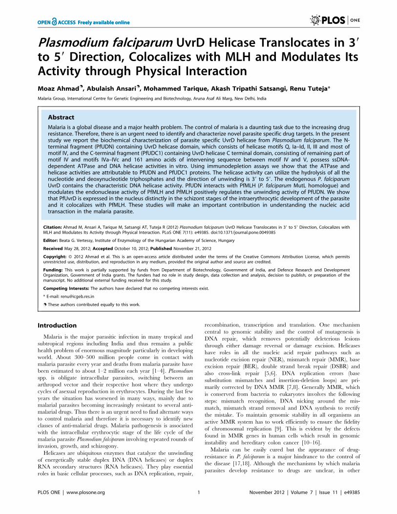
Plasmodium falciparum UvrD Helicase Translocates in 3′ to 5′ Direction, Colocalizes with MLH and...
21
Plasmodium falciparum UvrD Helicase Translocates in 39 to 59 Direction, Colocalizes with MLH and Modulates Its Activity through Physical Interaction Moaz Ahmad . , Abulaish Ansari . , Mohammed Tarique, Akash Tripathi Satsangi, Renu Tuteja* Malaria Group, International Centre for Genetic Engineering and Biotechnology, Aruna Asaf Ali Marg, New Delhi, India Abstract Malaria is a global disease and a major health problem. The control of malaria is a daunting task due to the increasing drug resistance. Therefore, there is an urgent need to identify and characterize novel parasite specific drug targets. In the present study we report the biochemical characterization of parasite specific UvrD helicase from Plasmodium falciparum. The N- terminal fragment (PfUDN) containing UvrD helicase domain, which consists of helicase motifs Q, Ia–Id, II, III and most of motif IV, and the C-terminal fragment (PfUDC1) containing UvrD helicase C terminal domain, consisting of remaining part of motif IV and motifs IVa–IVc and 161 amino acids of intervening sequence between motif IV and V, possess ssDNA- dependent ATPase and DNA helicase activities in vitro. Using immunodepletion assays we show that the ATPase and helicase activities are attributable to PfUDN and PfUDC1 proteins. The helicase activity can utilize the hydrolysis of all the nucleotide and deoxynucleotide triphosphates and the direction of unwinding is 39 to 59. The endogenous P. falciparum UvrD contains the characteristic DNA helicase activity. PfUDN interacts with PfMLH (P. falciparum MutL homologue) and modulates the endonuclease activity of PfMLH and PfMLH positively regulates the unwinding activity of PfUDN. We show that PfUvrD is expressed in the nucleus distinctly in the schizont stages of the intraerythrocytic development of the parasite and it colocalizes with PfMLH. These studies will make an important contribution in understanding the nucleic acid transaction in the malaria parasite. Citation: Ahmad M, Ansari A, Tarique M, Satsangi AT, Tuteja R (2012) Plasmodium falciparum UvrD Helicase Translocates in 39 to 59 Direction, Colocalizes with MLH and Modulates Its Activity through Physical Interaction. PLoS ONE 7(11): e49385. doi:10.1371/journal.pone.0049385 Editor: Beata G. Vertessy, Institute of Enzymology of the Hungarian Academy of Science, Hungary Received May 28, 2012; Accepted October 10, 2012; Published November 21, 2012 Copyright: ß 2012 Ahmad et al. This is an open-access article distributed under the terms of the Creative Commons Attribution License, which permits unrestricted use, distribution, and reproduction in any medium, provided the original author and source are credited. Funding: This work is partially supported by funds from Department of Biotechnology, Government of India, and Defence Research and Development Organization, Government of India grants. The funders had no role in study design, data collection and analysis, decision to publish, or preparation of the manuscript. No additional external funding received for this study. Competing Interests: The authors have declared that no competing interests exist. * E-mail: [email protected] . These authors contributed equally to this work. Introduction Malaria is the major parasitic infection in many tropical and subtropical regions including India and thus remains a public health problem of enormous magnitude particularly in developing world. About 300–500 million people come in contact with malaria parasite every year and deaths from malaria parasite have been estimated to about 1–2 million each year [1–4]. Plasmodium spp. is obligate intracellular parasites, switching between an arthropod vector and their respective host where they undergo cycles of asexual reproduction in erythrocytes. During the last few years the situation has worsened in many ways, mainly due to malarial parasites becoming increasingly resistant to several anti- malarial drugs. Thus there is an urgent need to find alternate ways to control malaria and therefore it is necessary to identify new classes of anti-malarial drugs. Malaria pathogenesis is associated with the intracellular erythrocytic stage of the life cycle of the malaria parasite Plasmodium falciparum involving repeated rounds of invasion, growth, and schizogony. Helicases are ubiquitous enzymes that catalyze the unwinding of energetically stable duplex DNA (DNA helicases) or duplex RNA secondary structures (RNA helicases). They play essential roles in basic cellular processes, such as DNA replication, repair, recombination, transcription and translation. One mechanism central to genomic stability and the control of mutagenesis is DNA repair, which removes potentially deleterious lesions through either damage reversal or damage excision. Helicases have roles in all the nucleic acid repair pathways such as nucleotide excision repair (NER), mismatch repair (MMR), base excision repair (BER), double strand break repair (DSBR) and also cross-link repair [5,6]. DNA replication errors (base substitution mismatches and insertion-deletion loops) are pri- marily corrected by DNA MMR [7,8]. Generally MMR, which is conserved from bacteria to eukaryotes involves the following steps: mismatch recognition, DNA nicking around the mis- match, mismatch strand removal and DNA synthesis to rectify the mistake. To maintain genomic stability in all organisms an active MMR system has to work efficiently to ensure the fidelity of chromosomal replication [9]. This is evident by the defects found in MMR genes in human cells which result in genomic instability and hereditary colon cancer [10–16]. Malaria can be easily cured but the appearance of drug- resistance in P. falciparum is a major hindrance to the control of the disease [17,18]. Although the mechanisms by which malaria parasites develop resistance to drugs are unclear, in other PLOS ONE | www.plosone.org 1 November 2012 | Volume 7 | Issue 11 | e49385
-
Upload
independent -
Category
Documents
-
view
2 -
download
0
Transcript of Plasmodium falciparum UvrD Helicase Translocates in 3′ to 5′ Direction, Colocalizes with MLH and...
Plasmodium falciparum UvrD Helicase Translocates in 39to 59 Direction, Colocalizes with MLH and Modulates ItsActivity through Physical InteractionMoaz Ahmad., Abulaish Ansari., Mohammed Tarique, Akash Tripathi Satsangi, Renu Tuteja*
Malaria Group, International Centre for Genetic Engineering and Biotechnology, Aruna Asaf Ali Marg, New Delhi, India
Abstract
Malaria is a global disease and a major health problem. The control of malaria is a daunting task due to the increasing drugresistance. Therefore, there is an urgent need to identify and characterize novel parasite specific drug targets. In the presentstudy we report the biochemical characterization of parasite specific UvrD helicase from Plasmodium falciparum. The N-terminal fragment (PfUDN) containing UvrD helicase domain, which consists of helicase motifs Q, Ia–Id, II, III and most ofmotif IV, and the C-terminal fragment (PfUDC1) containing UvrD helicase C terminal domain, consisting of remaining part ofmotif IV and motifs IVa–IVc and 161 amino acids of intervening sequence between motif IV and V, possess ssDNA-dependent ATPase and DNA helicase activities in vitro. Using immunodepletion assays we show that the ATPase andhelicase activities are attributable to PfUDN and PfUDC1 proteins. The helicase activity can utilize the hydrolysis of all thenucleotide and deoxynucleotide triphosphates and the direction of unwinding is 39 to 59. The endogenous P. falciparumUvrD contains the characteristic DNA helicase activity. PfUDN interacts with PfMLH (P. falciparum MutL homologue) andmodulates the endonuclease activity of PfMLH and PfMLH positively regulates the unwinding activity of PfUDN. We showthat PfUvrD is expressed in the nucleus distinctly in the schizont stages of the intraerythrocytic development of the parasiteand it colocalizes with PfMLH. These studies will make an important contribution in understanding the nucleic acidtransaction in the malaria parasite.
Citation: Ahmad M, Ansari A, Tarique M, Satsangi AT, Tuteja R (2012) Plasmodium falciparum UvrD Helicase Translocates in 39 to 59 Direction, Colocalizes withMLH and Modulates Its Activity through Physical Interaction. PLoS ONE 7(11): e49385. doi:10.1371/journal.pone.0049385
Editor: Beata G. Vertessy, Institute of Enzymology of the Hungarian Academy of Science, Hungary
Received May 28, 2012; Accepted October 10, 2012; Published November 21, 2012
Copyright: � 2012 Ahmad et al. This is an open-access article distributed under the terms of the Creative Commons Attribution License, which permitsunrestricted use, distribution, and reproduction in any medium, provided the original author and source are credited.
Funding: This work is partially supported by funds from Department of Biotechnology, Government of India, and Defence Research and DevelopmentOrganization, Government of India grants. The funders had no role in study design, data collection and analysis, decision to publish, or preparation of themanuscript. No additional external funding received for this study.
Competing Interests: The authors have declared that no competing interests exist.
* E-mail: [email protected]
. These authors contributed equally to this work.
Introduction
Malaria is the major parasitic infection in many tropical and
subtropical regions including India and thus remains a public
health problem of enormous magnitude particularly in developing
world. About 300–500 million people come in contact with
malaria parasite every year and deaths from malaria parasite have
been estimated to about 1–2 million each year [1–4]. Plasmodium
spp. is obligate intracellular parasites, switching between an
arthropod vector and their respective host where they undergo
cycles of asexual reproduction in erythrocytes. During the last few
years the situation has worsened in many ways, mainly due to
malarial parasites becoming increasingly resistant to several anti-
malarial drugs. Thus there is an urgent need to find alternate ways
to control malaria and therefore it is necessary to identify new
classes of anti-malarial drugs. Malaria pathogenesis is associated
with the intracellular erythrocytic stage of the life cycle of the
malaria parasite Plasmodium falciparum involving repeated rounds of
invasion, growth, and schizogony.
Helicases are ubiquitous enzymes that catalyze the unwinding
of energetically stable duplex DNA (DNA helicases) or duplex
RNA secondary structures (RNA helicases). They play essential
roles in basic cellular processes, such as DNA replication, repair,
recombination, transcription and translation. One mechanism
central to genomic stability and the control of mutagenesis is
DNA repair, which removes potentially deleterious lesions
through either damage reversal or damage excision. Helicases
have roles in all the nucleic acid repair pathways such as
nucleotide excision repair (NER), mismatch repair (MMR), base
excision repair (BER), double strand break repair (DSBR) and
also cross-link repair [5,6]. DNA replication errors (base
substitution mismatches and insertion-deletion loops) are pri-
marily corrected by DNA MMR [7,8]. Generally MMR, which
is conserved from bacteria to eukaryotes involves the following
steps: mismatch recognition, DNA nicking around the mis-
match, mismatch strand removal and DNA synthesis to rectify
the mistake. To maintain genomic stability in all organisms an
active MMR system has to work efficiently to ensure the fidelity
of chromosomal replication [9]. This is evident by the defects
found in MMR genes in human cells which result in genomic
instability and hereditary colon cancer [10–16].
Malaria can be easily cured but the appearance of drug-
resistance in P. falciparum is a major hindrance to the control of
the disease [17,18]. Although the mechanisms by which malaria
parasites develop resistance to drugs are unclear, in other
PLOS ONE | www.plosone.org 1 November 2012 | Volume 7 | Issue 11 | e49385
Characterization of P. falciparum UvrD Helicase
PLOS ONE | www.plosone.org 2 November 2012 | Volume 7 | Issue 11 | e49385
organisms, defects in DNA MMR have been linked to increased
mutation rates and drug resistance. It is well established that the
underlying cause of drug resistance in malaria is the de-
velopment of specific genetic mutations. There are several
sequences identified in PlasmoDB, that are homologous to genes
involved in repair pathways from other organisms, indicating
that this pathway is likely present in the parasite [19]. The most
well characterized MMR pathway is of Escherichia coli. In E. coli
UvrD is known to play an essential role in both the forms of
DNA repair such as MMR [20] and the NER [21].
UvrD or DNA helicase II is a superfamily 1A helicase
universally distributed across bacteria and extensively character-
ized [22]. It has also been reported that UvrD and its homologues
such as PcrA and Rep represent one family known as PUR family
and are targets for drug discovery because the deletion of PcrA is
lethal in Staphylococcal species and Bacillus subtilis [23]. It has
been shown that Mycobacterial UvrD2 is a DNA-dependent
ATPase with 39 to 59 helicase activity [24]. In a recent work the
UvrD from Haemophilus influenza (HiUvrD) and Helicobacter pylori
(HpUvrD) have been shown to exhibit strong single-stranded
DNA-specific ATPase and 39–59 helicase activities [25]. It is well
known that the three helicases Bacillus stearothermophilus PcrA, E. coli
Rep and E. coli UvrD are structurally similar and contain a two
domain (1 and 2) structure with each domain made of two sub-
domains (1A, 1B, 2A and 2B) and a C-terminal extension [26–28].
It has been shown that a truncated form of E. coli UvrD that lacks
the C-terminal extension retains helicase activity on a variety of
substrates [29].
The repair of misincorporated bases and damaged DNA is
very important for maintenance of genomic integrity. It has
been proposed recently that P. falciparum drug resistant parasites
have defective MMR and this is the underlying mechanism in
the development of antimalarial drug resistance [30]. Very little
is known about DNA repair mechanisms in P. falciparum but due
to the availability of its genome sequence direct comparison of
potential DNA repair genes to their E. coli counterpart can be
done. Previously we have reported that the parasite P. falciparum
genome contains the homologues of major components of
MMR complex such as UvrD helicase and MutL homologue
(MLH) [22]. In a recent study we have reported the isolation
and characterization of MLH from P. falciparum [31].
In the present study we describe the expression, purification
and biochemical characterization of another main component of
MMR complex, UvrD from P. falciparum 3D7 in detail. We
found that the N-terminal (PfUDN, consisting of domain 1A
and 1B) and the C-terminal (PfUDC1, consisting of first half of
domain 2A and 2B) fragments of PfUvrD contain ssDNA-
dependent ATPase and helicase activities. The Km values for
helicase activity are 1.260.1 nM for PfUDN and 3.260.4 nM
for PfUDC1 respectively. PfUDN is capable of unwinding the
blunt end duplex DNA substrate also. The helicase activity is
almost equal in all the NTPs and dNTPs tested and the polarity
of unwinding is 39 to 59. The endogenous P. falciparum UvrD of
,170 kDa contains the characteristic DNA helicase activity. We
further show that PfUDN and PfMLH interact and positively
regulate each other’s activity. Using immunofluorescence assays
we report that both PfUvrD and PfMLH co-localize in the
parasite P. falciparum 3D7 strain and are expressed in the
schizont stages of intraerythrocytic development. These studies
will advance our knowledge in the field of nucleic acid
metabolism in the parasite.
Materials and Methods
Ethics StatementThe animal studies described in this study were approved by the
ICGEB Institutional Animal Ethics Committee (IAEC Reference
No. MAL-55). ICGEB is licensed to conduct animal studies for
research purposes under the registration number 18/1999/
CPCSEA (dated 10/1/99).
Identification and Cloning of P. falciparum UvrD GeneIn order to clone the UvrD helicase from P. falciparum, the
sequence was downloaded and analyzed in detail. The nucleotide
sequence of PfUvrD is 4326 bases and it codes for a protein of
1441 amino acids. PCR amplification was done using genomic
DNA as the gene is not interrupted by introns. Accordingly the
primers PfUF1 (BamHI site) and PfUR1 (Xho I site), PfUF2
(BamHI site) and PfUR2 (Xho I site) and PfUF3 (BamHI site) and
PfUR3 (Xho I site) were synthesized to clone the N-terminal and
the C-terminal fragments C1 and C2. The P. falciparum UvrD
helicase gene was amplified in three fragments using the following
forward and the reverse primers.
1. PfUF1: 59- GGGATCCAACTTTTCTGAGGAAC- 39
2. PfUR1: 59-GCTCGAGTGTACTTCGAAAATTATT- 39
3. PfUF2: 59-GGGATCCGAAATTGTTAGAGTTTC- 39
4. PfUR2: 59-GCTCGAGATTTTGAGGTGCATGT- 39
5. PfUF3: 59- GGGATCCCAAAATAAATGTAGCG 39
6. PfUR3: 59- GCTCGAGTATATTCATTTCATTAAT 39
The N-terminal fragment is 2079 bases and codes for PfUDN
fragment from amino acid 35–727, the C1 fragment is 1128
bases and codes for PfUDC1 fragment from amino acid 729–
1104 and the C2 fragment is 1020 bases and codes for PfUDC2
fragment from amino acid 1103–1441 respectively. The PCR
conditions used were 95uC for 1 minute, 54uC for 1 min and
72uC for 2 min for the amplification of N terminal fragment.
For the amplification of the C terminal fragments the extension
was done only for 1.5 minutes. This was repeated for a total of
35 cycles and at the end one elongation was done at 72uC for
12 min. The PCR products were gel purified using Qiagen gel
extraction kit and cloned into the pGEM-T easy vector from
Promega using T-A cloning (Madison, WI, USA) and the clones
were sequenced by dideoxy sequencing reactions (Macrogen,
Korea). The nucleotide sequence was submitted to GenBank
and the accession number for the PfUDN fragment is
FJ588849, for the PfUDC1 fragment is FJ455133 and for the
PfUDC2 fragment is JN016528.
Using BamHI and XhoI enzymes (New England Biolabs,
Beverly, MA, USA) all the fragments were excised from
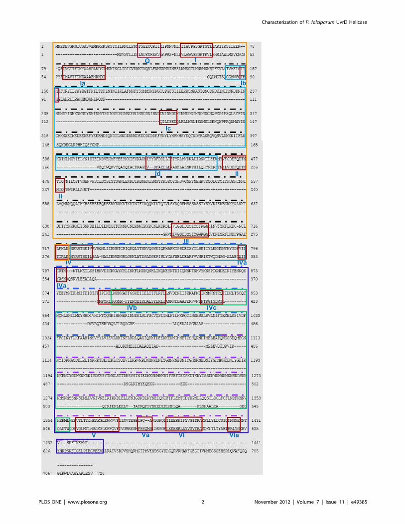
Figure 1. Comparison of amino acid sequence of Plasmodium falciparum (Pf) UvrD (1–1441) and Escherichia coli (E. coli) UvrD (1–720).The alignment was done using BLAST program (http://blast.ncbi.nlm.nih.gov/Blast). The conserved motifs are boxed in red color and the name ofeach motif (from Q to VIa) is written in roman numerals. The PlasmoDB number for P. falciparum UvrD sequence is PFE0705c and the accessionnumber for E. coli UvrD sequence is BAA00048.1. The black, blue and purple dotted lines indicate the PfUDN, PfUDC1 and PfUDC2 fragmentsrespectively. The orange box indicates the domain 1A (from amino acid 1–722) and the blue box inside it indicates the domain 1B (from amino acid150–464). The purple box denotes the domain 2A (from amino acid 723–1441) and the green box inside it indicates the domain 2B (from amino acid896–1359).doi:10.1371/journal.pone.0049385.g001
Characterization of P. falciparum UvrD Helicase
PLOS ONE | www.plosone.org 3 November 2012 | Volume 7 | Issue 11 | e49385
pGEMT easy clones, gel purified and subsequently cloned into
pET28a+ expression vector (Novagen, Madison, WI, USA) at
the appropriate sites. For protein expression, the clones were
transformed into BL21 (DE3) pLysS cells. 1% of the overnight
grown primary culture was inoculated in 500 ml LB (Luria
Broth) and allowed to grow at 37uC. At OD 0.6 the culture was
induced with 1 mM IPTG and then again allowed to grow for
another 4–6 hours. The harvested cells were lysed by using lysis
buffer of pH 7.8 (20 mM Tris-HCl, 250 mM NaCl, 0.1%
Tween 20, 0.1% Triton 100 and the protease inhibitor cocktail
from Sigma, St. Louis, MO, USA) and subsequently the cells
were sonicated to lyse maximum number of cells. After
centrifugation the soluble fraction was allowed to bind to pre-
equilibrated Ni-NTA (Qiagen, GmbH, Germany) resin for one
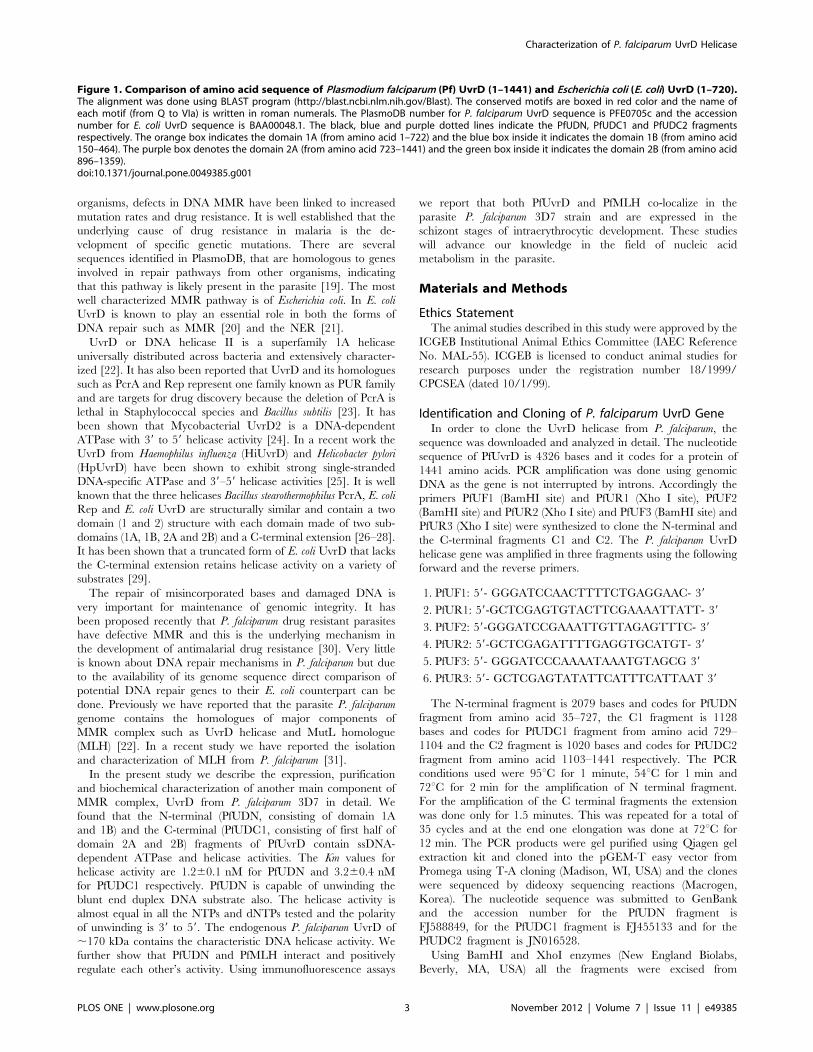
Figure 2. Schematic diagrams showing the domain organization. A, E. coli and B, P. falciparum UvrD helicases. Domain analysis was doneusing Scan Prosite at (http://expasy.org). The domain structure was taken from the results and used in the figures. UvrD helicase ATP binding andUvrD C-terminal domains are shown. The numbers show the amino acids spanning these motifs. C, The detailed domain organization of P. falciparumUvrD helicase. The conserved sequences of each domain are written inside the boxes. The text in blue refers to the names of various conserveddomains and the numbers refer to the amino acids separating the various domains and the length of N- and C-terminal extensions. This figure is notdrawn to scale. D–F, The detailed domain structure of PfUDN, PfUDC1 and PfUDC2 fragments of P. falciparum UvrD. The details are as in C. Thecolored lines are same as in Figure 1 and correspond to domain 1A, 1B, 2A and 2B present in PfUDN, PfUDC1 and PfUDC2 respectively. (G–I) Structuremodeling. The PfUvrD full-length sequence was submitted to Swissmodel server and the structure was obtained. The molecular graphic images wereproduced using the UCSF Chimera package from the resource for Biocomputing, Visualization, and Informatics (http://www.cgl.ucsf.edu/chimera) atthe University of California, San Francisco (supported by NIH P41 RR-01081). G. Template; H. full-length PfUvrD; I. superimposed image.doi:10.1371/journal.pone.0049385.g002
Characterization of P. falciparum UvrD Helicase
PLOS ONE | www.plosone.org 4 November 2012 | Volume 7 | Issue 11 | e49385
hour at 4uC. The column was first washed with the wash buffer
(lysis buffer without detergent with 25 mM imidazole). The
bound His-tagged proteins were eluted with varying (100–
150 mM) concentration of imidazole in the protein buffer
(20 mM Tris–HCl pH 8.0, 250 mM NaCl, 10% (v/v) glycerol
and protease inhibitor cocktail from Sigma, St. Louis, MO,
USA) and was checked for purity by SDS-PAGE (10% (w/v)
polyacrylamide gel) and silver staining using slight modifications
of the standard protocol [32]. The slight modification included
extensive washing of the gel after fixation and this step reduces
the background and increases the sensitivity of the stain.
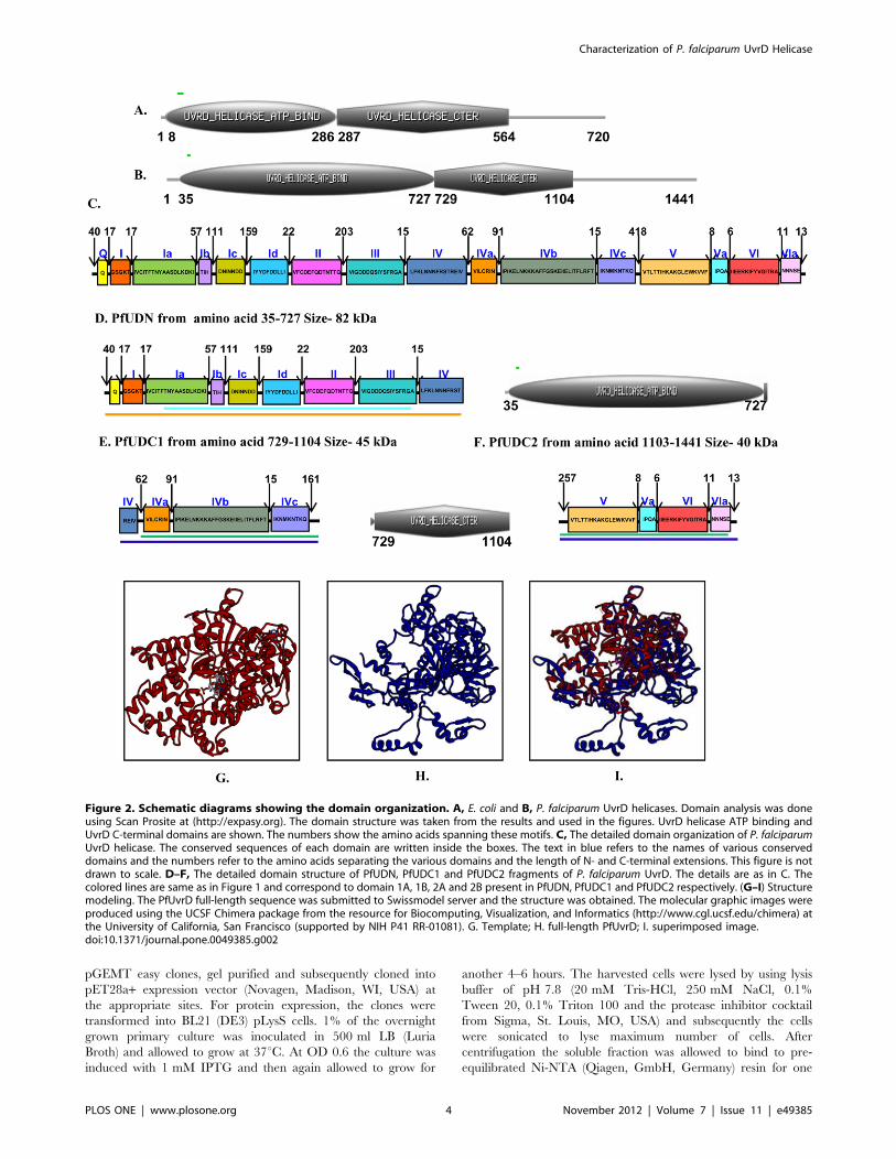
Figure 3. Purification and ATPase activity analysis. A, Silver-stained gel of purified PfUDN. Lane M contains the protein molecular weightmarker and lane 1 and 2 contain 0.3 and 0.2 mg of the purified PfUDN. B, Silver-stained gel of purified PfUDC1. Lane M contains the protein molecularweight marker and lane 1 contains 0.2 mg of the purified PfUDC1. C, Silver-stained gel of purified PfUDC2. Lane M contains the protein molecularweight marker and lane 1 contains 0.2 mg of the purified PfUDC2. D, Western blot of purified PfUDN. Lane M contains the protein molecular weightmarker and lane 1 and 2 contain 0.3 and 0.2 mg of the purified PfUDN. E, Western blot of purified PfUDC1. Lane M contains the protein molecularweight marker and lane 1 contains 0.2 mg of the purified PfUDC1. F, Western blot of purified PfUDC2. Lane M contains the protein molecular weightmarker and lane 1 contains 0.2 mg of the purified PfUDC2.G, ATPase activity of purified PfUDN. Lane C, reaction without enzyme, Lanes 1–4, reactionswith enzyme in the presence of ssDNA and Mg2+. H, The quantitative data of the autoradiogram in G. I, Time dependence of ATPase activity ofPfUDN. The time of incubation in minutes is mentioned at the top of the autoradiogram and C is the control reaction without enzyme. J, Thequantitative data of the autoradiogram in I. K, ATPase activity of purified PfUDC1. Lane C, reaction without enzyme, Lanes 1–4, reactions with enzymein the presence of ssDNA and Mg2+. L, The quantitative data of the autoradiogram in K. M, Time dependence of ATPase activity of PfUDC1. The timeof incubation in minutes is mentioned at the top of the autoradiogram and C is the control reaction without enzyme. N, The quantitative data of theautoradiogram in M.doi:10.1371/journal.pone.0049385.g003
Characterization of P. falciparum UvrD Helicase
PLOS ONE | www.plosone.org 5 November 2012 | Volume 7 | Issue 11 | e49385
Western Blot AnalysisFor western blotting, the proteins were separated by SDS-
PAGE and transferred electrophoretically to nitrocellulose mem-
brane as described [32]. After blocking with 3% skimmed milk in
TBST (Tris buffered saline with 0.05% Tween 20), the membrane
was incubated with the appropriate primary antibody (Penta-His
from Qiagen, GmbH, Germany) for 3 h at room temperature.
After washing, the blot was incubated with the appropriate
secondary antibody coupled to alkaline phosphatase (Sigma, St.
Louis, MO, USA) and developed using 5-Bromo-4-Chloro-3-
Indolyl Phosphate and Nitro Blue Tetrazolium obtained from
Sigma.
Generation of Polyclonal AntiseraPurified PfUDN was used for the preparation of antibodies in
mice using the standard protocols [32]. The polyclonal antibodies
were purified as IgG fractions using protein A-Sepharose as
described [32].
ATPase AssayThe ATPase reaction was performed in the buffer (20 mM
Tris-HCl, pH 8.0, 8 mM DTT, 1.0 mM MgCl2, 20 mM KCl
and 16 mg/ml BSA) for 1 hour at 37uC in the presence of
purified PfUDN, PfUDC1 or PfUDC2 and 10 ng of M13 mp19
ssDNA and a mixture of [c-32P] ATP (,17 nM) and 1 mM
cold ATP. The products were separated by thin layer
chromatography (TLC) [33–35] and the plate was exposed to
hyper film for autoradiography or scanned on phosphoimager.
The quantitation was done using IMAGE j/geldoc software
(http://rsbweb.nih.gov/ij/). For the concentration curve analysis
different concentrations of PfUDN (from 6 to 72 nM) and
PfUDC1 (from 20 to 260 nM) proteins were used. The time
course analysis was performed with a fixed concentration of
PfUDN or PfUDC1 and time duration ranging from 10 to 90
minutes. The quantitation was done using IMAGE j/geldoc
software (http://rsbweb.nih.gov/ij/) and percentage of ATP
hydrolysis was plotted as the bar diagram.
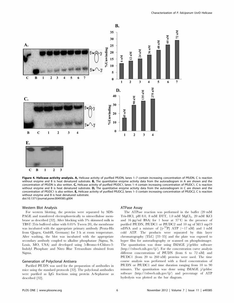
Figure 4. Helicase activity analysis. A, Helicase activity of purified PfUDN, lanes 1–7 contain increasing concentration of PfUDN, C is reactionwithout enzyme and B is heat denatured substrate. B, The quantitative enzyme activity data from the autoradiogram in A are shown and theconcentration of PfUDN is also written. C, Helicase activity of purified PfUDC1, lanes 1–4 contain increasing concentration of PfUDC1, C is reactionwithout enzyme and B is heat denatured substrate. D, The quantitative enzyme activity data from the autoradiogram in C are shown and theconcentration of PfUDC1 is also written. E, Helicase activity of purified PfUDC2, lanes 1–3 contain increasing concentration of PfUDC2, C is reactionwithout enzyme and B is heat denatured substrate.doi:10.1371/journal.pone.0049385.g004
Characterization of P. falciparum UvrD Helicase
PLOS ONE | www.plosone.org 6 November 2012 | Volume 7 | Issue 11 | e49385
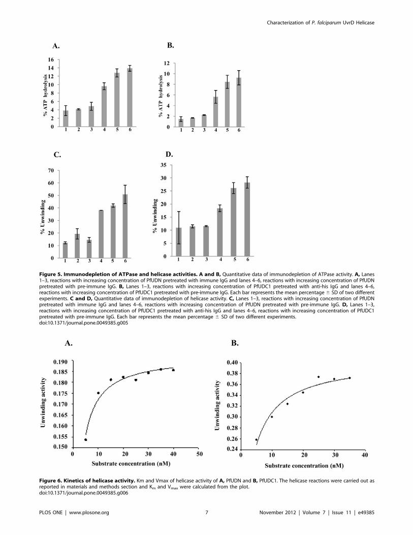
Figure 5. Immunodepletion of ATPase and helicase activities. A and B, Quantitative data of immunodepletion of ATPase activity. A, Lanes1–3, reactions with increasing concentration of PfUDN pretreated with immune IgG and lanes 4–6, reactions with increasing concentration of PfUDNpretreated with pre-immune IgG. B, Lanes 1–3, reactions with increasing concentration of PfUDC1 pretreated with anti-his IgG and lanes 4–6,reactions with increasing concentration of PfUDC1 pretreated with pre-immune IgG. Each bar represents the mean percentage6 SD of two differentexperiments. C and D, Quantitative data of immunodepletion of helicase activity. C, Lanes 1–3, reactions with increasing concentration of PfUDNpretreated with immune IgG and lanes 4–6, reactions with increasing concentration of PfUDN pretreated with pre-immune IgG. D, Lanes 1–3,reactions with increasing concentration of PfUDC1 pretreated with anti-his IgG and lanes 4–6, reactions with increasing concentration of PfUDC1pretreated with pre-immune IgG. Each bar represents the mean percentage 6 SD of two different experiments.doi:10.1371/journal.pone.0049385.g005
Figure 6. Kinetics of helicase activity. Km and Vmax of helicase activity of A, PfUDN and B, PfUDC1. The helicase reactions were carried out asreported in materials and methods section and Km and Vmax were calculated from the plot.doi:10.1371/journal.pone.0049385.g006
Characterization of P. falciparum UvrD Helicase
PLOS ONE | www.plosone.org 7 November 2012 | Volume 7 | Issue 11 | e49385
Preparation of DNA Helicase Substrate and HelicaseAssayThe helicase activity of PfUDN, PfUDC1 and PfUDC2 was
determined by the standard strand displacement assay using the
partially duplex substrate and the method described previously
[33–35]. The normal substrate (substrate 1, Table S1) used in this
study consisted of a 32P-labelled 47-mer DNA oligodeoxynucleo-
tide annealed to M13mp19 phage ssDNA to create a partial
duplex. At both the 59 and 39 ends, this oligodeoxynucleotide
contains 15 base-pairs of non-complementary region. 10 ng of the
oligodeoxynucleotide was labelled at 59-end with T4 polynucleo-
tide kinase (PNK) (5 U) (New England Biolabs) and 1.85 MBq of
[c-32P]ATP (specific activity 222 TBq/mmol) using the standard
PNK buffer (New England Biolabs) at 37uC for one hour. The
labeled oligodeoxynucleotide was then annealed with 0.5 mg of
single-stranded circular M13mp19 (+) phage DNA using standard
annealing buffer (20 mM Tris-HCl, pH 7.5, 10 mM MgCl2,
100 mM NaCl, 1 mM DTT) by heating at 95uC for 1 min,
transferring immediately to 65uC for 2 min and then cooling
slowly to room temperature. The non-hybridized oligodeoxynu-
cleotide was removed using gel filtration through a Sepharose 4B
column (Pharmacia, Sweden). The reaction mixture (10 ml)containing appropriate buffer (20 mM Tris-HCl, pH 8.0, 8 mM
DTT, 1.0 mM MgCl2, 20 mM KCl and 16 mg/ml BSA), the 32P-
labeled helicase substrate (1000 cpm/10 ml) (Table S1) and the
purified protein fractions to be assayed was incubated at 37uC for
60 min. The substrate and products were separated by electro-
phoresis on a nondenaturing 12% or 15% (for the blunt end
substrate) PAGE, dried, and the gel was exposed to hyper film for
autoradiography or scanned on phosphoimager and both the
substrate and unwound DNA bands were quantified. The
quantitation was done using IMAGE j/geldoc software (http://
rsbweb.nih.gov/ij/) and the percent unwinding was plotted as the
bar diagram.
Immunodepletion AssayFor this assay aliquots of the purified PfUDN and PfUDC1 were
incubated with IgG purified from anti-preimmune and/or anti-
PfUDN or anti-His antisera (for PfUDC1) respectively at 0uC for
60 min. The antigen–antibody complexes were removed by the
addition of protein A Sepharose beads. The supernatants were
used for the ATPase and helicase activity analysis using substrate 1
(Table S1) in the same way as described above. The quantitation
was done using IMAGE j/geldoc software (http://rsbweb.nih.
gov/ij/) and the percentage of ATP hydrolysis and percent
unwinding was plotted as the bar diagram and each bar indicates
the mean percentage 6 SD (standard deviation).
Determination of Km and VmaxHelicase assay reactions for PfUDN and PfUDC1 were
performed using the normal substrate (substrate 1, Table S1) of
different concentrations (5–40 nM) in a standard reaction buffer
(20 mM Tris-HCl, pH 8.0, 8 mM DTT, 1.0 mM MgCl2, 20 mM
KCl and 16 mg/ml BSA). The amount of dsDNA and unwound
ssDNA was quantified from the autoradiogram using ImageJ
software (http://rsbweb.nih.gov/ij/) and used for the Km and
Vmax calculations.
Preparation of Blunt end DNA Helicase SubstrateThe sequence of 17 mer oligodeoxynucleotide used for making
the blunt end duplex substrate (substrate 2, Table S1) is as follows
59-GTTTTCCCAGTCACGAC-39. This was labeled at 59 end
using the method described above and was annealed to its
complementary oligodeoxynucleotide with the sequence 59-
GTCGTGACTGGGAAAAC-39. The substrate was purified
and used for the assay using the method described above. The
amount of dsDNA and unwound ssDNA was quantified from the
autoradiogram using ImageJ software (http://rsbweb.nih.gov/ij/)
and the percent unwinding was plotted as the bar diagram.
Preparation of Direction Specific SubstratesFor constructing a 59 to 39 direction-specific substrate (substrate
3A, Table S1), the oligodeoxynucleotide 32-mer (59-
TTCGAGCTCGGTACCCGGGGATCCTCTAGAGT-39) was
first annealed to M13mp19 ssDNA using annealing buffer (20 mM
Tris-HCl, pH 7.5, 10 mM MgCl2, 100 mM NaCl, 1 mM DTT)
and then labeled at 39-OH end in appropriate buffer with
50 mCurie [a-32P]dCTP and 5 units of DNA polymerase I (large
fragment) at 23uC for 20 min. The incubation was continued for
an additional 20 min at 23uC after increasing the dCTP to 50 mM
using unlabelled dCTP. This resulting duplex substrate was
digested with SmaI and purified by gel filtration through 1 ml
Sepharose 4B. The substrate consisting of long linear M13mp19
ssDNA with short duplex ends for 39 to 59 unwinding (substrate
3B, Table S1) was prepared by first 59-end labeling of 32-mer
oligodeoxynucleotide and then annealing with M13mp19 ssDNA
as described above. The annealed substrate was digested with
SmaI and purified by gel filtration through 1 ml of Sepharose 4B.
Purification of Endogenous UvrD Protein from ParasiteLysateThe endogenous UvrD protein was recovered by immunoaffi-
nity purification from P. falciparum 3D7 strain. P. falciparum 3D7
strain was cultured with human erythrocytes (4% hematocrit) in
RPMI media supplemented with 10% O+ human serum using
standard protocol [36]. The basic protocol of the kit was slightly
modified to minimize the nonspecific binding and elution of
protein in the active state (as per recommendation of the Pierce kit
protocol). P. falciparum 3D7 parasite pellet was suspended in the
lysis buffer (Pierce, Thermo Scientific) containing 25 mM Tris-
HCl pH 7.2, 150 mM NaCl, 1 mM EDTA, 1% NP-40 and 5%
glycerol. After repeated freeze thaw, the lysate was centrifuged at
,13000 6 g for 10 minute to pellet the cell debris. Before
performing the immunoprecipitation, soluble fraction of the
parasite lysate was first precleared using control agarose resin
(Pierce, Thermo Scientific) then with preimmune IgG-protein A
agarose column to minimize the non specific protein binding. The
purified IgGs from preimmune serum or anti-PfUDN mouse
antiserum were diluted 1:1 with 1x coupling buffer and were
incubated in the column containing protein A/G+ agarose. After
one hour of binding, the beads were washed with 1x coupling
buffer containing 10 mM sodium phosphate pH 7.2 and 150 mM
NaCl. The bound IgG-protein A/G+ agarose was cross linked in
the coupling buffer containing 450 mM DSS (disuccinimidyl
substrate) following the crosslinked immunoprecipitation kit pro-
tocol (Pierce, Thermo Scientific). The cross-linked beads were
washed twice with elution buffer to remove non-crosslinked IgGs
and quenching the cross linking reaction. Before incubating the
cross linked beads with precleared parasite lysate, the beads were
washed twice with lysis buffer as per instructions in PierceHcrosslink immunoprecipitation kit protocol. Equal amounts of the
precleared parasite lysate was incubated overnight (4uC) with both
preimmune and anti-PfUDN columns separately. The column was
washed twice with lysis buffer then with 1X TBS (Tris buffered
saline) to remove the detergent from column. The beads were
subsequently washed with conditioning buffer (neutral pH buffer).
The bound proteins were eluted in neutral pH buffer ((Pierce,
Characterization of P. falciparum UvrD Helicase
PLOS ONE | www.plosone.org 8 November 2012 | Volume 7 | Issue 11 | e49385
Thermo Scientific) and quickly buffer was exchanged with Tris
buffer (20 mM Tris-HCl, 150 mM NaCl with protease inhibitor
cocktail) by using 10 kDa cutoff amicon filters (Pall Life Sciences).
All the column elutes (preimmune column and anti-PfUDN
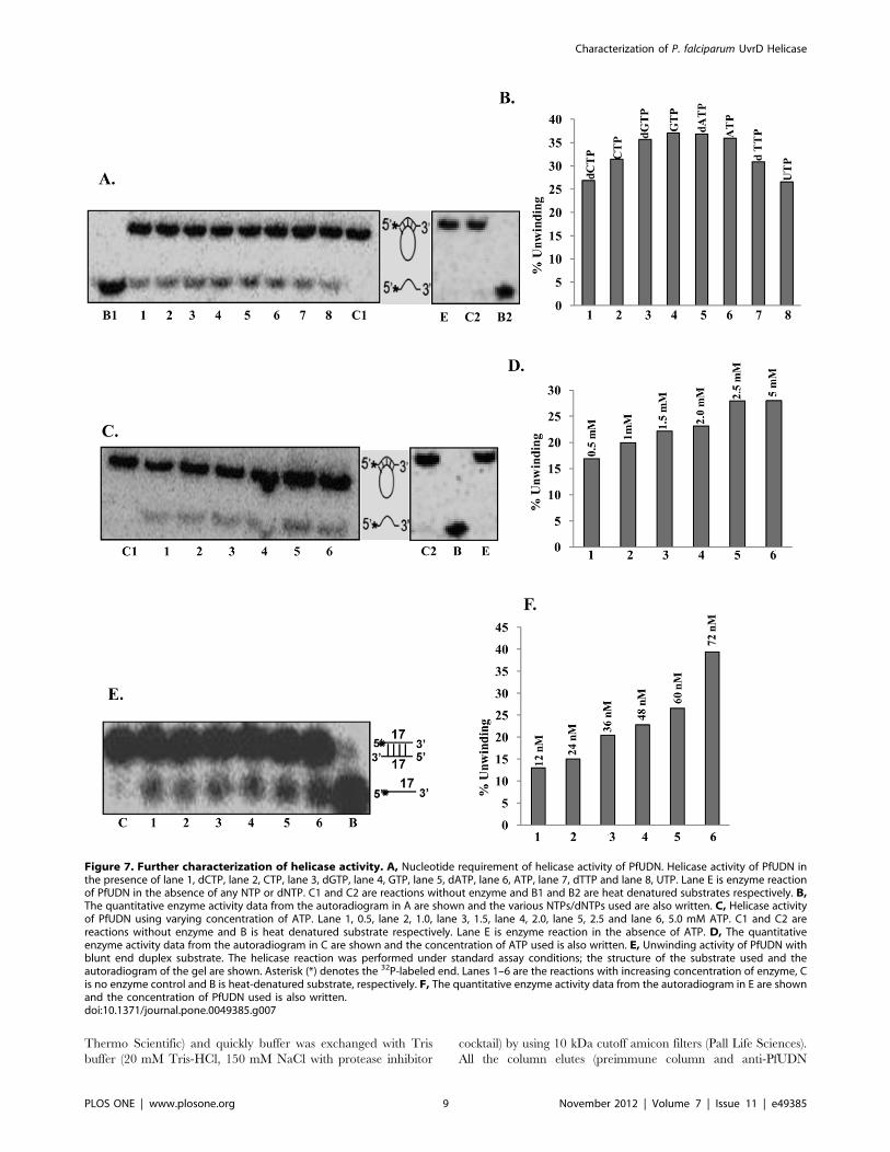
Figure 7. Further characterization of helicase activity. A, Nucleotide requirement of helicase activity of PfUDN. Helicase activity of PfUDN inthe presence of lane 1, dCTP, lane 2, CTP, lane 3, dGTP, lane 4, GTP, lane 5, dATP, lane 6, ATP, lane 7, dTTP and lane 8, UTP. Lane E is enzyme reactionof PfUDN in the absence of any NTP or dNTP. C1 and C2 are reactions without enzyme and B1 and B2 are heat denatured substrates respectively. B,The quantitative enzyme activity data from the autoradiogram in A are shown and the various NTPs/dNTPs used are also written. C, Helicase activityof PfUDN using varying concentration of ATP. Lane 1, 0.5, lane 2, 1.0, lane 3, 1.5, lane 4, 2.0, lane 5, 2.5 and lane 6, 5.0 mM ATP. C1 and C2 arereactions without enzyme and B is heat denatured substrate respectively. Lane E is enzyme reaction in the absence of ATP. D, The quantitativeenzyme activity data from the autoradiogram in C are shown and the concentration of ATP used is also written. E, Unwinding activity of PfUDN withblunt end duplex substrate. The helicase reaction was performed under standard assay conditions; the structure of the substrate used and theautoradiogram of the gel are shown. Asterisk (*) denotes the 32P-labeled end. Lanes 1–6 are the reactions with increasing concentration of enzyme, Cis no enzyme control and B is heat-denatured substrate, respectively. F, The quantitative enzyme activity data from the autoradiogram in E are shownand the concentration of PfUDN used is also written.doi:10.1371/journal.pone.0049385.g007
Characterization of P. falciparum UvrD Helicase
PLOS ONE | www.plosone.org 9 November 2012 | Volume 7 | Issue 11 | e49385
column) were checked with SDS-PAGE coupled western blot
analysis and used for further biochemical assays. Before enzymatic
assay the elutes were passed through protein A agarose spin
columns to trap the IgG and the flowthrough was used for the
assay. For western blot analysis the proteins were transferred onto
nitrocellulose membrane. After overnight blocking in 5% skimmed
milk in TBST (Tris buffered saline with.05% Tween 20), the
membrane was first incubated overnight at 4uC with a 1:500
dilution of the purified IgG of anti-PfUDN and then incubated
with the anti-mouse secondary antibodies (Sigma, St. Louis, MO,
USA) coupled to horse radish peroxidase. The blot was developed
using Sigma FastTM DAB (3, 3-diaminobenzidine tetrahydrochlor-
ide) with urea enhancer tablets (St. Louis, MO, USA) according to
the manufacturer’s instructions.
Helicase Assays with the Endogenous PfUvrDThe helicase assays were performed with increasing volume of
the elutes obtained from the pre-immune and anti-PfUDN
columns described in the previous section. The normal substrate
(substrate 1, Table S1) and the method described in the earlier
section was used.
Helicase Assay in the Presence of PfMLHHelicase assay was performed using the previously characterized
purified recombinant synthetic PfMLH [31], catalytically inactive
N-terminal (PfMLHN), C-terminal (PfMLHC) [31] and the heat
inactivated PfMLH and PfUDN. The strand displacement assay
was used with both normal (substrate 1, Table S1) and direction-
specific substrates (substrate 3B, Table S1). Both substrate and
unwound DNA bands were quantified from the autoradiogram
using ImageJ software (http://rsbweb.nih.gov/ij/) and the mean
percentage of unwinding was plotted in the bar diagram and each
bar indicates the mean percentage 6 SD (standard deviation).
Endonuclease Assay in the Presence of PfUDN andPfUDC1The endonuclease assay reaction (10 ml) was performed as
described earlier [31] by incubating 20 nM of PfMLH with 50 ng
of pBR322 DNA in a buffer (buffer 4, New England Biolabs)
containing 50 mM potassium acetate, 20 mM Tris–acetate,
pH 7.9, 10 mM magnesium acetate and 1 mM dithiothreitol at
37uC for 1 hour. The reactions were stopped with loading dye
(0.5% SDS, 50 mM EDTA, 40% glycerol, 0.1% bromophenol
blue, 0.1% xylene cyanol) and the products were analyzed by 1%
agarose gel electrophoresis and the bands were quantitated by
ImageJ software (http://rsbweb.nih.gov/ij/). The percentage of
nicked DNA was plotted in the bar diagram. Each bar indicates
the mean percentage 6 SEM (standard error mean). To
investigate the effect of PfUDN and PfUDC1 on the endonuclease
activity of PfMLH, the assay was performed with 20 nM PfMLH
along with 15 nM PfUDN or PfUDC1. The negative control
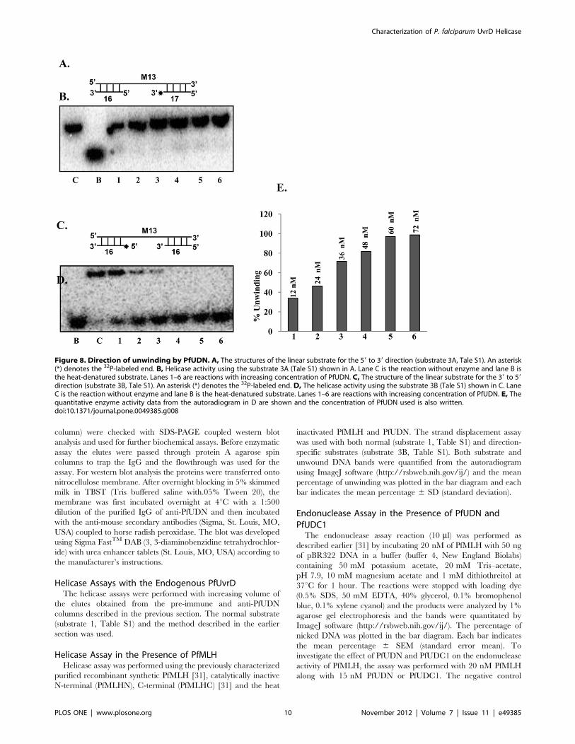
Figure 8. Direction of unwinding by PfUDN. A, The structures of the linear substrate for the 59 to 39 direction (substrate 3A, Tale S1). An asterisk(*) denotes the 32P-labeled end. B, Helicase activity using the substrate 3A (Tale S1) shown in A. Lane C is the reaction without enzyme and lane B isthe heat-denatured substrate. Lanes 1–6 are reactions with increasing concentration of PfUDN. C, The structure of the linear substrate for the 39 to 59direction (substrate 3B, Tale S1). An asterisk (*) denotes the 32P-labeled end. D, The helicase activity using the substrate 3B (Tale S1) shown in C. LaneC is the reaction without enzyme and lane B is the heat-denatured substrate. Lanes 1–6 are reactions with increasing concentration of PfUDN. E, Thequantitative enzyme activity data from the autoradiogram in D are shown and the concentration of PfUDN used is also written.doi:10.1371/journal.pone.0049385.g008
Characterization of P. falciparum UvrD Helicase
PLOS ONE | www.plosone.org 10 November 2012 | Volume 7 | Issue 11 | e49385
reactions without protein were also performed to compare the
results.
Enzyme-linked Immunosorbent Assay (ELISA) for Protein-protein Interaction StudyThe 96 well ELISA plates were coated with fixed concentration
(10 ng) of purified PfMLH diluted in bicarbonate/carbonate
coating buffer (100 mM NaHCO3 and Na2CO3, pH 9.6), over-
night at 4uC in 100 ml volume. The plate was emptied and washed
three times with washing buffer phosphate buffered saline (PBS
containing 0.05% Tween 20) and blocked for 2 hour with 5% non-
fat milk in PBS. The blocking buffer was removed and the wells
were washed 3 times with same washing buffer. Varying
concentrations of the interacting proteins (from 2.5 ng to 30 ng)
were added and the incubation was continued at 37uC for further
2 hour. The plate was emptied and washed three times with
washing buffer (PBS containing 0.05% Tween 20). The assembly
of the protein complex in the wells was then assessed through the
use of polyclonal antibodies at 1:15000 dilutions. The incubation
with antibodies was done for 2 hour at 37uC. The plate was
washed three times with washing buffer to remove the unbound
antibodies. The plate was further incubated with 1:3000 dilution
of horse radish peroxidase (HRP) conjugated secondary antibody
for 2 hours at 37uC. The unbound antibody was removed by
washing the wells with washing buffer. The binding of the
secondary antibody to the protein complex was then detected by
applying the o-Phenylenediamine (OPD) substrate (Sigma, St.
Louis, MO, USA) in appropriate buffer. The reaction was stopped
and the plate was read at 490 nm using an ELISA reader and each
bar indicates the mean percentage 6 SD (standard deviation).
In vitro Protein-protein Interaction Using ImmunoaffinityColumnProtein A sepharose columns were prepared using anti-PfUDN
and anti-preimmune IgGs. Purified anti-PfUDN IgGs and
preimmune IgGs were allowed separately to bind onto protein A
Sepharose column and both the columns were washed 3–4 times
with buffer (20 mM Tris-HCl, pH 8.0, 135 mM NaCl and
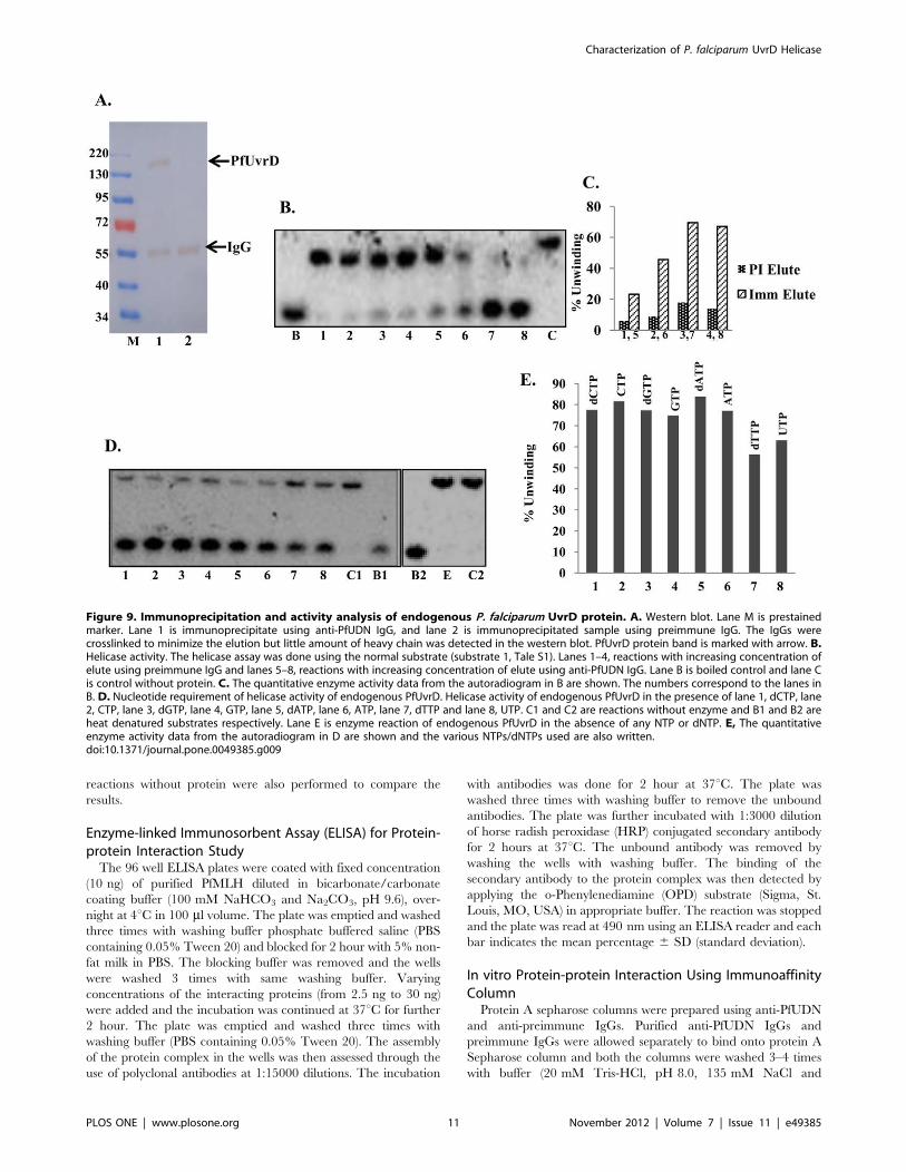
Figure 9. Immunoprecipitation and activity analysis of endogenous P. falciparum UvrD protein. A. Western blot. Lane M is prestainedmarker. Lane 1 is immunoprecipitate using anti-PfUDN IgG, and lane 2 is immunoprecipitated sample using preimmune IgG. The IgGs werecrosslinked to minimize the elution but little amount of heavy chain was detected in the western blot. PfUvrD protein band is marked with arrow. B.Helicase activity. The helicase assay was done using the normal substrate (substrate 1, Tale S1). Lanes 1–4, reactions with increasing concentration ofelute using preimmune IgG and lanes 5–8, reactions with increasing concentration of elute using anti-PfUDN IgG. Lane B is boiled control and lane Cis control without protein. C. The quantitative enzyme activity data from the autoradiogram in B are shown. The numbers correspond to the lanes inB. D. Nucleotide requirement of helicase activity of endogenous PfUvrD. Helicase activity of endogenous PfUvrD in the presence of lane 1, dCTP, lane2, CTP, lane 3, dGTP, lane 4, GTP, lane 5, dATP, lane 6, ATP, lane 7, dTTP and lane 8, UTP. C1 and C2 are reactions without enzyme and B1 and B2 areheat denatured substrates respectively. Lane E is enzyme reaction of endogenous PfUvrD in the absence of any NTP or dNTP. E, The quantitativeenzyme activity data from the autoradiogram in D are shown and the various NTPs/dNTPs used are also written.doi:10.1371/journal.pone.0049385.g009
Characterization of P. falciparum UvrD Helicase
PLOS ONE | www.plosone.org 11 November 2012 | Volume 7 | Issue 11 | e49385
protease inhibitor cocktail) to remove the unbound antibody. For
the assay 10 mg of each protein i.e. PfUDN and PfMLH in buffer
(20 mM Tris-HCl, pH 8.0, 135 mM NaCl and protease inhibitor
cocktail from Sigma, St. Louis, MO, USA) was mixed and allowed
to interact for 1 hour at 4uC. Equal amount of this mixture was
incubated with protein A Sepharose coupled to preimmune or
anti-PfUDN IgG column for 1 hour at 4uC on shaker. After four
washes with the buffer (20 mM Tris-HCl, pH 8.0, 135 mM NaCl
and protease inhibitor cocktail from Sigma, St. Louis, MO, USA),
the bound protein was eluted separately from both the columns
with 100 mM glycine pH 3.0 and each fraction of both the elutes
was run on SDS/PAGE and western blot analysis was performed.
The anti-PfUDN IgG protein A Sepharose column elute was run
in duplicate in order to probe with both the antibodies separately
(anti-PfUDN and anti-PfMLH). The blots were further probed
with appropriate secondary antibodies coupled with horse radish
peroxidase (HRP) and were developed by Sigma fastH DAB tablets
according to manufacturer’s instructions.
Immunofluorescence Assay and Western BlottingP. falciparum 3D7 strain was cultured with human erythrocytes
(4% hematocrit) in RPMI media supplemented with 10% O+human serum using standard protocol [36]. Thin smears of
parasitized red blood cells (RBC) of different developmental stages
were prepared and fixed in acetone for 5 minutes followed by
chilled methanol for 40 seconds at room temperature. The fixed
slides were dried and incubated in 10% fetal calf serum (FCS) in
PBS in a humid chamber at 37uC for 2 hour for blocking. The
slides were washed with PBS and incubated with purified IgG of
anti-PfUDN antibodies (raised in mice) and purified IgG of anti-
PfMLH antibodies (raised in rabbit) both at 1:100 dilutions in PBS
containing 10% FCS for 1 hour at 37uC. The slides were then
washed four times with PBS for 15 min each and then incubated
for 1 hour at 37uC with secondary antibodies (ALEXA 488-green-
conjugated- anti-mouse IgG from Invitrogen, USA) and (ALEXA
594-red- conjugated- anti-rabbit IgG from Invitrogen, USA) both
diluted 1:500 in PBS containing 10% FCS. After washing, the
slides were incubated in 49, 69- di-amidino-2-phenylindole-
dihydrochloride (DAPI) for nuclear staining. The slides were
washed thrice with PBST (PBS, 0.5% Tween 20) for 10 minutes
each and twice with PBS for 10 min each and mounted with
antifade reagent Fluroguard purchased from BioRad (Hercules,
CA, USA) and viewed under oil immersion. The images were
collected using a Bio-Rad 2100 laser-scanning microscope
attached to a Nikon 2000U microscope.
For the purpose of western blotting parasites were released from
mixed infected erythrocytes cultures by treatment with 0.1% (w/v)
saponin. The cell-free protein extracts from early schizont stage
parasite cultures were prepared by repeated liquid nitrogen freeze-
thaw in the lysis buffer (Pierce, Rockford, IL USA) containing
10 mM Tris–HCl, pH 7.4, 150 mM NaCl, 10 mM EDTA, 1%
NP-40, 5% glycerol and protease inhibitor cocktail (Sigma, St.
Louis, MO, USA). The extracted proteins were resolved by SDS-
PAGE and transferred onto nitrocellulose membrane for the
purpose of western blotting using the purified IgG of anti-PfUDN
antibodies. The membrane was first incubated overnight at 4uCwith a 1:500 dilution of the purified IgG of anti-PfUDN antibodies
and then incubated with the HRP conjugated secondary antibody
(Sigma, St. Louis, MO, USA). The blot was developed using
Sigma FastH DAB (3,39-diaminobenzidine tetrahydrochloride)
tablet with urea enhancer tablets (Sigma, St. Louis, MO, USA)
according to the manufacturer’s instructions.
Results
Identification and Sequence Analysis of PfUvrDUvrD is a member of superfamily 1 of helicases. An alignment
of the complete amino-acid sequence of UvrD homologue from P.
falciparum with E. coli UvrD using BLAST (http://blast.ncbi.nlm.
nih.gov/Blast) revealed that PfUvrD aligned contiguously with its
E. coli counterpart (Figure 1). Further detailed analysis of the
protein sequence at Expasy (http://prosite.expasy.org) indicated
that similar to E. coli UvrD, PfUvrD also contains two distinct
domains: an UvrD like DNA helicase ATP-binding domain and
an UvrD like DNA helicase C-terminal domain (Figure 2A and
2B). It contains Q motif at the extreme N-terminal and all the
conserved motifs from Ia-Id, II, III, IV, IVa-IVc, V, Va, VI and
VIa (Figure 1, Figure 2C) [28]. Similar to UvrD from other
sources PfUvrD also contains all the motifs clustered in its N-
terminal region (Figure 2C) [37]. A detailed analysis of PfUvrD
amino acid sequence indicated that the distance between each
motif is variable between E. coli and PfUvrD proteins [22]
(Figure 1, Figure 2C). As reported earlier E. coli UvrD contains
four domains (1A, 1B, 2A and 2B) and a C-terminal extension
[29]. A comparison of PfUvrD sequence with E. coli UvrD shows
that it also contains all these domains but no C-terminal extension
(Figure 1). The 1A domain in PfUvrD is from amino acid 1–722
and the 1B domain is from amino acid 150–464 (Figure 1, orange
and light blue boxes respectively). The 2A domain in PfUvrD is
from amino acid 723–1441 and the 2B domain is from amino acid
896–1359 (Figure 1, purple and green boxes respectively). The
PlasmoDB number for PfUvrD is PFE0705c. The blast analysis of
PfUvrD against PlasmoDB database (www.plasmodb.org) revealed
that this gene is located on chromosome 5 of P. falciparum and it
contains no introns.
The nucleotide sequence of PfUvrD is 4326 bases and it codes
for a protein of 1441 amino acids. For the amplification of full-
length PfUvrD, genomic DNA from P. falciparum 3D7 strain was
used with the primer pair PfUF1 and PfUR3. On repeated trials,
we were not able to obtain the amplification of the full-length
PfUvrD from genomic DNA or cDNA preparations. Therefore
the sequence was divided according to the presence of different
domains as shown into N terminal, PfUDN (2079 bases,,82 kDa)
containing UvrD helicase domain (domain 1A and 1B) [29], which
consists of motifs Q, Ia-Id, II, III and part of motif IV (Figure 2D),
and C terminal 1, PfUDC1 (1128 bases, ,45 kDa) containing
UvrD helicase C terminal domain (first half of domain 2A and 2B),
which consists of remaining part of motif IV and motifs IVa–IVc
and 161 amino acids of intervening sequence between motif IV
and V (Figure 2E). The C terminal 2, PfUDC2 (1020 bases,
,40 kDa, consisting of second half of domain 2A and 2B) contains
remaining part (261 amino acids) of intervening sequence between
motif IV and V and motifs V, Va, VI and VIa (Figure 2F). Each
fragment i.e. PfUDN, PfUDC1 and PfUDC2 was amplified and
cloned as described in materials and methods.
Molecular Modeling of PfUvrD Structure2For structural modeling the sequence of full-length PfUvrD was
submitted to the Swissmodel homology-modeling server (http://
swissmodel.expasy.org/) [38]. A total of five models were obtained
and four models covered the areas ranging from 57 to 168 amino
acids of PfUvrD only but only one model covered a larger range
(amino acid 32–815) of the PfUvrD sequence (Data S1). Therefore
this model which was built using PcrA DNA helicase from B.
stearothermophilus as template was studied in detail [27]. PfUvrD
primary sequence residues 32 to 815 showed ,13% identity to the
PcrA DNA helicase from B. stearothermophilus [27]. The structural
Characterization of P. falciparum UvrD Helicase
PLOS ONE | www.plosone.org 12 November 2012 | Volume 7 | Issue 11 | e49385
modeling of the PfUvrD was therefore done using the known
crystal structure of this homologue as the template (PDB number
3pjrA at http://www.ncbi.nlm.nih.gov/Structure/mmdb). The
ribbon diagram of the template is shown in Figure 2G and the
predicted structure of PfUvrD is shown in Figure 2H. When the
modeled structure of PfUvrD and the template were super-
imposed, it is clear that these structures superimpose partially
(Figure 2I). Molecular graphic images were produced using the
UCSF Chimera package (http://www.cgl.ucsf.edu/chimera) from
the Resource for Biocomputing, Visualization, and Informatics at
the University of California, San Francisco (supported by NIH
P41 RR-01081) [39]. Further structure analysis with chimera
using matchmaker structure comparison tool revealed that the
RMSD between 303 atom pairs is 0.295 angstroms [40]. The
RMSD value corresponds to the amino acid residue pairs which
align perfectly in the pairwise alignment (Data S1).
Purification of PfUvrD Fragments and Characterization ofATPase and DNA Helicase ActivitiesThe expression clones corresponding to each fragment such as
PfUDN, PfUDC1 and PfUDC2 were transformed into E. coli
strain BL21 (DE3) pLysS and the recombinant proteins were
purified using method described in materials and methods section.
The SDS–PAGE analysis followed by silver staining of the purified
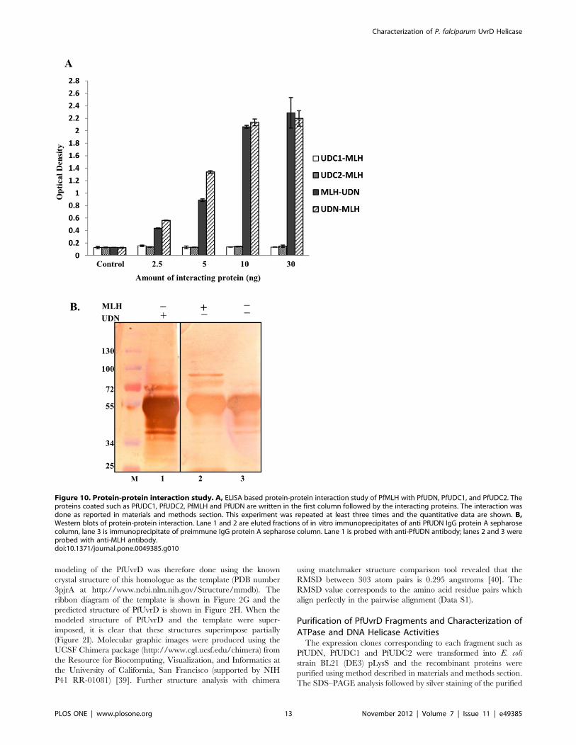
Figure 10. Protein-protein interaction study. A, ELISA based protein-protein interaction study of PfMLH with PfUDN, PfUDC1, and PfUDC2. Theproteins coated such as PfUDC1, PfUDC2, PfMLH and PfUDN are written in the first column followed by the interacting proteins. The interaction wasdone as reported in materials and methods section. This experiment was repeated at least three times and the quantitative data are shown. B,Western blots of protein-protein interaction. Lane 1 and 2 are eluted fractions of in vitro immunoprecipitates of anti PfUDN IgG protein A sepharosecolumn, lane 3 is immunoprecipitate of preimmune IgG protein A sepharose column. Lane 1 is probed with anti-PfUDN antibody; lanes 2 and 3 wereprobed with anti-MLH antibody.doi:10.1371/journal.pone.0049385.g010
Characterization of P. falciparum UvrD Helicase
PLOS ONE | www.plosone.org 13 November 2012 | Volume 7 | Issue 11 | e49385
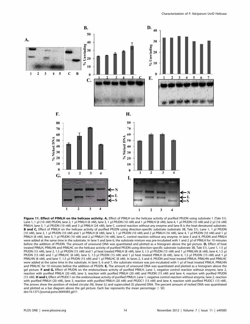
Figure 11. Effect of PfMLH on the helicase activity. A, Effect of PfMLH on the helicase activity of purified PfUDN using substrate 1 (Tale S1).Lane 1, 1 ml (10 nM) PfUDN, lane 2, 1 ml PfMLH (8 nM), lane 3, 1 ml PfUDN (10 nM) and 1 ml PfMLH (8 nM), lane 4, 1 ml PfUDN (10 nM) and 2 ml (16 nM)PfMLH, lane 5, 1 ml PfUDN (10 nM) and 3 ml PfMLH (24 nM), lane C, control reaction without any enzyme and lane B is the heat-denatured substrate.B and C, Effect of PfMLH on the helicase activity of purified PfUDN using direction-specific substrate (substrate 3B, Tale S1). Lane 1, 1 ml PfUDN(10 nM), lane 2, 1 ml PfUDN (10 nM) and 1 ml PfMLH (8 nM), lane 3, 1 ml PfUDN (10 nM) and 2 ml PfMLH (16 nM), lane 4, 1 ml PfUDN (10 nM) and 1 mlPfMLH (8 nM), lane 5, 1 ml PfUDN (10 nM) and 2 ml PfMLH (16 nM), lane C, control reaction without any enzyme. In lane 3 and 4, PfUDN and PfMLHwere added at the same time in the substrate. In lane 5 and lane 6, the substrate mixture was pre-incubated with 1 and 2 ml of PfMLH for 10 minutesbefore the addition of PfUDN. The amount of unwound DNA was quantitated and plotted as a histogram above the gel picture. D, Effect of heattreated PfMLH, PfMLHN and PfMLHC on the helicase activity of purified PfUDN using direction-specific substrate (substrate 3B, Tale S1). Lane 1, 1.5 mlPfUDN (15 nM), lane 2, 1.5 ml PfUDN (15 nM) and 1 ml heat treated PfMLH (8 nM), lane 3, 1.5 ml PfUDN (15 nM) and 1 ml PfMLHN (8 nM), lane 4, 1.5 mlPfUDN (15 nM) and 1 ml PfMLHC (8 nM), lane 5, 1.5 ml PfUDN (15 nM) and 1 ml heat treated PfMLH (8 nM), lane 6, 1.5 ml PfUDN (15 nM) and 1 mlPfMLHN (8 nM), and lane 7, 1.5 ml PfUDN (15 nM) and 1 ml PfMLHC (8 nM). In lanes 2, 3 and 4, PfUDN and heat treated PfMLH, PfMLHN and PfMLHCwere added at the same time in the substrate. In lane 5, 6 and 7, the substrate mixture was pre-incubated with 1 ml of heat treated PfMLH, PfMLHNand PfMLHC for 10 minutes before the addition of PfUDN. E, The amount of unwound DNA was quantitated and plotted as a histogram above thegel picture. F and G, Effect of PfUDN on the endonuclease activity of purified PfMLH. Lane 1, negative control reaction without enzyme, lane 2,reaction with purified PfMLH (20 nM), lane 3, reaction with purified PfMLH (20 nM) and PfUDN (15 nM) and lane 4, reaction with purified PfUDN(15 nM). H and I, Effect of PfUDC1 on the endonuclease activity of purified PfMLH. Lane 1, negative control reaction without enzyme, lane 2, reactionwith purified PfMLH (20 nM), lane 3, reaction with purified PfMLH (20 nM) and PfUDC1 (15 nM) and lane 4, reaction with purified PfUDC1 (15 nM).The arrows show the position of nicked circular (N), linear (L) and supercoiled (S) plasmid DNA. The percent amount of nicked DNA was quantitatedand plotted as a bar diagram above the gel picture. Each bar represents the mean percentage 6 SD.doi:10.1371/journal.pone.0049385.g011
Characterization of P. falciparum UvrD Helicase
PLOS ONE | www.plosone.org 14 November 2012 | Volume 7 | Issue 11 | e49385
Characterization of P. falciparum UvrD Helicase
PLOS ONE | www.plosone.org 15 November 2012 | Volume 7 | Issue 11 | e49385
proteins showed that all the fragments PfUDN (Figure 3A, lanes 1
and 2), PfUDC1 (Figure 3B, lane 1) and PfUDC2 (Figure 3C, lane
1) contain almost no contaminating protein and are homogeneous
preparations. The purified fractions were further checked by
western blot analysis using anti-His antibodies and only a single
band in each of the purified fraction was detected for PfUDN
(Figure 3D, lanes 1 and 2), PfUDC1 (Figure 3E, lane 1) and
PfUDC2 (Figure 3F, lane 1) respectively. These purified prepara-
tions were used for all of the assays described in the following
sections. The purified PfUDN protein was also used for the
production of polyclonal antibodies in mice.
The ssDNA-dependent ATPase activity of PfUDN, PfUDC1
and PfUDC2 was checked using standard assay conditions as
described in materials and methods in the presence of traces of
radiolabelled ATP with 1 mM cold ATP and purified enzymes.
The concentration-dependence of ATPase activity was checked by
using 6 to 72 nM of PfUDN and 20 to 260 nM of PfUDC1
proteins. The percent release of radioactive inorganic phosphate
(Pi) from [c32P] ATP was measured. The results clearly showed
that PfUDN (Figure 3G and 3H, lanes 1–4) and PfUDC1 contain
concentration and ssDNA dependent ATPase activity (Figure 3K
and 3L, lanes 1–4). The ATPase reaction using 72 nM of purified
PfUDN and 40 nM of purified PfUDC1 at different time points
was carried out in order to study the time dependence of ATPase
activity. The percent release of radioactive Pi from [c-32P] ATPshowed linearity up to 60 minutes in both PfUDN and PfUDC1
(Figure 3I and 3J, lanes 1–5 and Figure 3M and 3N, lanes 1–5).
On repeated trials we were unable to detect any ATPase activity in
PfUDC2 (data not shown).
The standard helicase strand-displacement assay measures the
unwinding of 32P-labelled DNA fragment from a partially duplex
nucleic acid. In order to characterize the DNA unwinding activity
of PfUDN, PfUDC1 and PfUDC2 the standard strand-displace-
ment assay was used. The DNA unwinding activity using different
concentration of purified PfUDN (6 to 72 nM) and PfUDC1 (45 to
275 nM) and optimal assay conditions as described in materials
and methods with ,1000 cpm of the substrate 1 (Table S1) in
buffer having 1 mM ATP, 1 mM MgCl2 and 75 mM KCl was
tested. It is interesting to note that PfUDN (Figure 4A and 4B,
lanes 1–7) and PfUDC1 (Figure 4C and 4D, lanes 1–4) both
showed the concentration-dependent helicase activity. On re-
peated trials we were unable to detect any helicase activity in
PfUDC2 (Figure 4E, lanes 1–3).
Immunodepletion of ATPase and Helicase Activities ofPfUDN and PfUDC1Purified PfUDN was allowed to react separately with IgGs
purified from the pre-immune sera and from the sera of the mice
immunized with PfUDN using the method described in materials
and methods section. We were unable to generate antibodies
against purified PfUDC1 therefore for this assay purified PfUDC1
was allowed to react with anti-His antibodies. The immunode-
pleted supernatants were checked for various activities. The results
showed that the ATPase activity of PfUDN (Figure 5A, lanes 1–3)
and PfUDC1 (Figure 5B, lanes 1–3) was depleted with the specific
or anti-His antibodies. On the contrary the samples treated with
pre-immune IgG of both PfUDN and PfUDC1 showed concen-
tration-dependent ATPase activity (Figure 5A, lanes 4–6 and
Figure 5B, lanes 4–6 respectively). Similar results were obtained
with helicase activity also. The results showed that the helicase
activity of PfUDN using substrate 1 (Table S1) (Figure 5C, lanes
1–3) and PfUDC1 (Figure 5D, lanes 1–3) was also depleted with
the specific or anti-His antibodies. But the samples treated with
pre-immune IgG of both PfUDN and PfUDC1 showed concen-
tration-dependent helicase activity (Figure 5C, lanes 4–6 and
Figure 5D, lanes 4–6 respectively). These data further confirm that
the ATPase and helicase activities are due to the purified PfUDN
and PfUDC1 proteins and not due to any contamination in the
purified preparations. This experiment was repeated with two
different preparations of PfUDN and PfUDC1 proteins and each
bar in Figure 5A–D represents the mean percentage 6 SD of two
different experiments.
Determination of Km and Vmax for the Helicase Activity ofPfUDN and PfUDC1Helicase assay reactions were performed using the substrate 1
(Table S1) of different concentrations (5–40 nM) in a standard
reaction buffer. The amount of dsDNA and unwound ssDNA was
quantified as described in materials and methods section. A
conventional hyperbolic dependence of the rate of reaction on
substrate concentration was obtained, such that the rate of
substrate unwinding was initially linear and later saturated with
increasing substrate concentrations that gave best-fit to the
Michaelis–Menten equation. The Km and Vmax of helicase activity
for PfUDN and PfUDC1 was measured by using Sigma plot
software (http://www.sigmaplot.com/). Nonlinear regression
analysis of this data yielded a Km value of 1.260.1 nM and
3.260.4 nM for PfUDN (Figure 6A) and PfUDC1 respectively
(Figure 6B). The Vmax value is 0.1920 nM/min/ng and
0.4048 nM/min/ng for PfUDN and PfUDC1 respectively.
Further Characterization of Unwinding ActivityFurther characterization of unwinding activity was done using
PfUDN only. It is well established that helicases have specific
nucleotide requirement to couple the hydrolysis of nucleotide to
unwinding activity. Therefore the helicase activity of PfUDN was
measured with different deoxynucleotide triphosphates and
nucleotide triphosphates using substrate 1 (Tale S1). It is
interesting to note that PfUDN showed the unwinding activity in
the presence of all the dNTPs and NTPs such as dCTP, CTP,
dGTP, GTP, dATP, ATP, dTTP and UTP used for the reaction
(Figure 7A and 7B, lanes 1–8 respectively). On the other hand
there was no unwinding activity of PfUDN in the absence of any
NTP or dNTP (Figure 7A, lane E). The concentration re-
quirement using ATP showed that the unwinding activity of
PfUDN was maximal at 2.5 mM ATP concentration and it did
not increase further on increasing the ATP concentration to
5.0 mM (Figure 7C and 7D, lane 5 and 6 respectively). It is
interesting to note that PfUDN did not show any unwinding
activity in the absence of ATP (Figure 7C, lane E).
In order to determine the specificity of PfUDN, its DNA
unwinding activity was tested with blunt end duplex substrate also
(substrate 2, Tale S1). This substrate had blunt ends but contained
identical core sequence and same duplex length (17 basepair) so
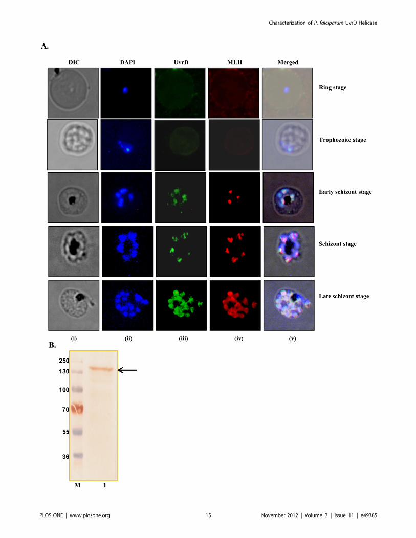
Figure 12. Localization of PfUvrD. A, Immunofluorescence staining. The cells were fixed and immunostained. Panel (i) phase (ii) image of cellstained with DAPI (iii) immunofluorescently stained cell (green, P. falciparum UvrD) (iv) immunofluorescently stained cell (red, P. falciparum MLH) and(v) super-imposed images. Control normal mouse sera produced no fluorescence (data not shown). B, Western blot analysis. Lane M is the proteinmolecular weight marker and lane 1 is protein from early schizont stages of the intraerythrocytic developmental of the parasite. The arrow shows thePfUvrD band.doi:10.1371/journal.pone.0049385.g012
Characterization of P. falciparum UvrD Helicase
PLOS ONE | www.plosone.org 16 November 2012 | Volume 7 | Issue 11 | e49385
that as far as possible, any differences in efficiency of unwinding
due to sequence differences could be eliminated. The assay was
performed using the method described in the previous section.
The results clearly indicate that PfUDN unwinds the blunt end
duplex substrate (substrate 2, Tale S1) also in concentration-
dependent manner (Figure 7E and 7F, lanes 1–6).
Determination of Direction of Unwinding by PfUDNIt is well established that all the helicases preferentially unwind
nucleic acids in a polar fashion by moving unidirectionally on the
bound strand in a duplex. The direction of unwinding by a helicase
is defined by the strand to which the enzyme binds and moves.
The unwinding activity of purified PfUDN was tested by using two
different direction-specific substrates, one specific for the 59 to 39
(substrate 3A, Tale S1) and the other for the 39 to 59 (substrate 3B,
Tale S1) direction, prepared as described in materials and
methods section. The DNA unwinding activity, using both the
direction-specific substrates with different concentrations of
PfUDN enzyme was determined. The release of radiolabelled
DNA from the substrates of Figure 8A and 8C by PfUDN enzyme
will indicate the movement in 59 to 39 and 39 to 59 directions,
respectively. The results show that PfUDN was unable to show the
activity with the 59 to 39 direction-specific substrate (substrate 3A,
Tale S1) (Figure 8A, lanes 1–6). On the other hand it is evident
from the results that PfUDN could unwind the 39 to 59 direction-
specific duplex substrate (substrate 3B, Tale S1) very efficiently
(Figure 8D, lanes 1–6) indicating that it contains unidirectional
DNA unwinding activity. The helicase activity with this substrate
was also directly proportional to the concentration of PfUDN used
in the reaction (Figure 8D and 8E, lanes 1–6). It should be noted
here that the 72 nM of PfUDN resulted in complete unwinding of
the direction-specific substrate (substrate 3B, Tale S1) (Figure 8D,
lane 6) as opposed to ,30% unwinding observed with the normal
substrate (substrate 1, Tale S1) (Figure 4A, lane 7). This difference
in activity may be due to the difference in the processivity of the
PfUDN.
Purification of Endogenous UvrD Protein from P.falciparum and its Activity AnalysisThe purification of endogenous P. falciparum 3D7 UvrD protein
was performed as described in materials and methods section. The
standard protocol was used with slight modification mentioned in
the material and methods section. Before performing the
immunoprecipitation, the parasite lysate was first precleared with
control agarose resin then with pre-immune IgG-protein A-
Sepharose column to reduce non specific protein binding. The
purified IgGs from pre-immune serum and anti-PfUDN serum
were cross-linked successfully and to minimize any non specific
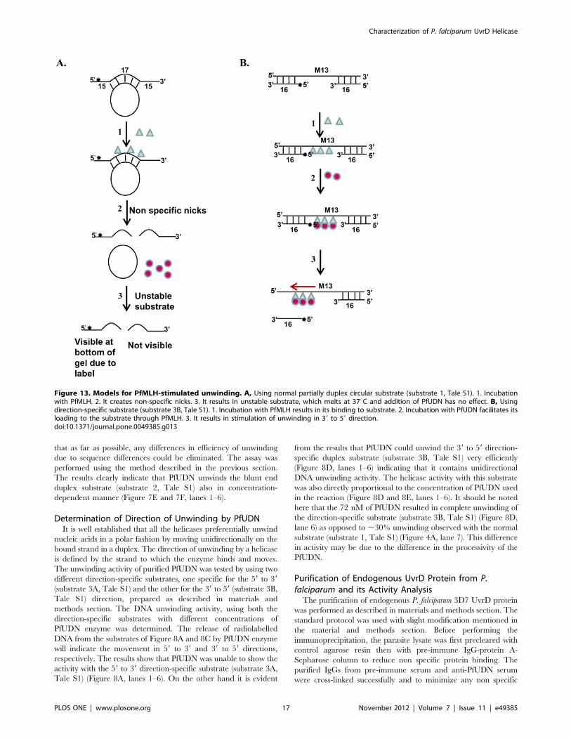
Figure 13. Models for PfMLH-stimulated unwinding. A, Using normal partially duplex circular substrate (substrate 1, Tale S1). 1. Incubationwith PfMLH. 2. It creates non-specific nicks. 3. It results in unstable substrate, which melts at 37uC and addition of PfUDN has no effect. B, Usingdirection-specific substrate (substrate 3B, Tale S1). 1. Incubation with PfMLH results in its binding to substrate. 2. Incubation with PfUDN facilitates itsloading to the substrate through PfMLH. 3. It results in stimulation of unwinding in 39 to 59 direction.doi:10.1371/journal.pone.0049385.g013
Characterization of P. falciparum UvrD Helicase
PLOS ONE | www.plosone.org 17 November 2012 | Volume 7 | Issue 11 | e49385
binding, both the columns were washed stringently with the lysis
buffer. To remove the detergent, the columns were washed three
times with the wash buffer (1X TBS). Both column elutes
(preimmune column and anti-PfUDN column elute) were checked
with SDS-PAGE coupled western blot analysis and the results
clearly indicate the presence of PfUvrD of ,170 kDa in the anti-
PfUDN column elute (Figure 9A, lane 1). Traces of heavy chain of
IgG of secondary anti mouse antibody were also coeluted/detected
in both the anti-PfUDN column elute as well as the preimmune
column elute (Figure 9A, lanes 1 and 2 respectively).
Different concentrations of the recovered endogenous PfUvrD
protein were used for the helicase activity assay using the substrate
1 (Tale S1) and the method described. We also used the pre
immune elute as a control for the enzymatic assays. The results
reveal that the P. falciparum endogenous UvrD protein contains
concentration-dependent DNA helicase activity (Figure 9B and
9C, lanes 5–8). The elute obtained with pre-immune serum
contained some background activity (Figure 9B and 9C, lanes 1–
4).
Further Characterization of Unwinding Activity ofEndogenous PfUvrDFurther characterization of unwinding activity of endogenous
PfUvrD was also done. The helicase activity of endogenous
PfUvrD was also measured with different deoxynucleotide tripho-
sphates and nucleotide triphosphates using substrate 1 (Tale S1). It
is interesting to note that similar to PfUDN, endogenous PfUvrD
also showed the unwinding activity in the presence of all the
dNTPs and NTPs such as dCTP, CTP, dGTP, GTP, dATP, ATP,
dTTP and UTP used for the reaction (Figure 9D and 9E, lanes 1–
8 respectively). Similar to PfUDN, endogenous PfUvrD also did
not show any unwinding activity in the absence of any NTP or
dNTP (Figure 9D, lane E).
Interaction of PfUvrD Fragments with PfMLHThe functions of most of the proteins are dependent upon their
direct physical interactions with other polypeptides within a cell. It
has been shown previously that E. coli MutL and UvrD proteins
interact [41]. In a recent study from our laboratory we have
reported the identification and functional characterization of
PfMLH from P. falciparum where we reported that PfMLH is
a component of MMR and it contains weak ATPase and
endonuclease activity [31]. In the present study using ELISA
based assays we performed the interaction analysis as described in
materials and methods section. PfUDN, PfUDC1, PfUDC2 and
PfMLH were used for the interaction study. The results show that
only PfUDN interacts with PfMLH (Figure 10A) and PfUDC1 and
PfUDC2 had no detectable interaction with PfMLH (Figure 10A).
Furthermore the results suggest that this interaction is concentra-
tion-dependent (Figure 10A).
In vitro Protein-protein Interaction Study UsingImmunoaffinity ColumnThe in vitro protein-protein interaction study was done using
preimmune and anti-PfUDN immunoaffinity column as described
in materials and methods section. Equal amount of (PfUDN and
PfMLH) mixture was incubated with protein A sepharose coupled
to preimmune or anti-PfUDN IgGs. Both the column elutes were
separated by SDS PAGE coupled western blot analysis. The
results clearly show that in addition to PfUDN (Figure 10B, lane
1), PfMLH (Figure 10B, lane 2) was also detected in the eluted
fractions of the anti-PfUDN column because it interacted with
PfUDN. On the other hand PfMLH was not detected in the eluted
fractions of the anti-preimmune column (Figure 10B, lane 3).
These results further confirm the interaction between PfUDN and
PfMLH proteins.
Helicase Assays in the Presence of PfMLHWe have reported previously that PfMLH contains no helicase
activity [31]. To investigate the effect of PfMLH on the activity of
PfUDN, the helicase activity of PfUDN was assayed in the
presence of differing amounts of purified PfMLH protein [31].
Using the substrate 1 (Tale S1) we were able to show that PfUDN
contains unwinding activity (Figure 11A, lane 1). But using this
substrate we were unable to observe the stimulation of unwinding
activity. Because it is most likely that before the helicase PfUDN
could reach the duplex area in this normal circular substrate for
unwinding from the 39 end, the substrate was nicked and degraded
due to the non-specific nicking endonuclease activity of PfMLH.
Due to this degradation it was not possible to observe the effect of
PfMLH on the unwinding activity as is evident from the results
shown (Figure 11A, lanes 3–5, increasing concentration of PfMLH
with constant concentration of PfUDN).
Therefore in order to check the effect of PfMLH on unwinding
activity we decided to use the 39 to 59 direction-specific substrate
(substrate 3B, Tale S1). The assay conditions were selected so that
the PfUDN (10 nM) alone unwound ,25% of the helicase
substrate in 60 minutes as described under materials and methods
section. After 60 minutes of incubation the reactions were stopped
and the products were analyzed by gel electrophoresis and
visualized by autoradiography. It is interesting to note that PfMLH
stimulated the unwinding activity of PfUDN in a concentration-
dependent manner only when the substrate mixture was pre-
incubated with PfMLH prior to the addition of PfUDN. This
stimulation was concentration-dependent and ,22–36% stimula-
tion was observed at the two concentrations of PfMLH added as
compared to the activity obtained by PfUDN alone (Figure 11B
and 11C, lane 1 versus lane 4 and 5 respectively). The minimum
concentration of PfMLH for the stimulatory effect of PfUDN
helicase activity was 8 nM. We were unable to detect the
stimulation of the helicase activity below this concentration. On
the other hand when PfUDN and two concentrations of PfMLH
were added together in the substrate mixture the stimulation of
activity was almost negligible (Figure 11B and 11C, lane 1 versus
lanes 2 and 3). As reported earlier also PfMLH alone had no
detectable helicase activity [31]. This experiment was also
repeated at least three times and the results were reproducible.
To further confirm that the stimulation of helicase activity is due to
the PfMLH, we also used the catalytically inactive N-terminal
(PfMLHN), C-terminal (PfMLHC) [31] and the heat inactivated
PfMLH (which loses its activity) in the helicase reaction. This
experiment was also done using the direction-specific substrate
(substrate 3B, Tale S1). The results clearly indicate that there was
no significant effect on the helicase activity of PfUDN (12 nM)
(Figure 11D and 11E, lane 1) when the heat treated PfMLH
(8 nM), PfMLHN (8 nM) or PfMLHC (8 nM) were used
(Figure 11D and 11E, lanes 2, 3 and 4 respectively). Furthermore
when the heat treated PfMLH (8 nM), PfMLHN (8 nM) or
PfMLHC (8 nM) were pre-incubated prior to the addition of
PfUDN (12 nM), there was still no effect but a slight inhibition of
unwinding activity was observed (Figure 11D and 11E, lane 1
versus lanes 5, 6 and 7 respectively).
Endonuclease Assays in the Presence of PfUDNAs reported in previous sections PfUDN and PfMLH interact
and there is no detectable interaction between PfMLH, PfUDC1
and PfUDC2. In a previous study we have reported that PfMLH
Characterization of P. falciparum UvrD Helicase
PLOS ONE | www.plosone.org 18 November 2012 | Volume 7 | Issue 11 | e49385
contains endonuclease activity [31]. Therefore we investigated the
effect of PfUDN and PfUDC1 on the endonuclease activity of
PfMLH. To check this effect of PfUDN and PfUDC1 on
endonuclease activity, the endonuclease assay was performed in
the presence and absence of PfMLH using the method described
in the material and methods section. The endonuclease activity
was checked by using supercoiled pBR322 DNA, PfMLH
(20 nM), PfUDN (15 nM) and/or PfUDC1 (15 nM) proteins.
The assay of PfMLH with supercoiled DNA resulted into the
nicking of supercoiled pBR322 (Figure 11F and 11G, lane 2) as
compared to no protein control (Figure 11F and 11G, lane 1). The
results further indicated that the endonuclease activity of PfMLH
(20 nM) is modulated significantly in the presence of PfUDN
(15 nM) (Figure 11F and 11G, lane 3). On the other hand PfUDN
(15 nM) alone had no detectable endonuclease activity (Figure 11F
and 11G, lane 4). Further interpretations of this result is interesting
that in the presence of PfUDN (15 nM), the percentage of the
nicked DNA increased significantly from ,56% to ,75% while
percentage of linear DNA decreased in the same ratio. On the
other hand PfUDC1 had no effect on the endonuclease activity of
PfMLH. The reaction of PfMLH (20 nM) with supercoiled DNA
resulted in nicking of supercoiled DNA (Figure 11H and 11I, lane
2) as compared to control without protein (Figure 11H and 11I,
lane 1) but the addition of PfUDC1 (15 nM) had almost no effect
on the endonuclease activity of PfMLH (Figure 11H and 11I, lane
3). PfUDC1 (15 nM) alone contains no detectable endonuclease
activity (Figure 11H and 11I, lane 4).
Localization of PfUvrD by Immunofluorescence Assayand Western BlottingThe purified antibodies to PfMLH and PfUDN were used to
study the localization of these proteins in P. falciparum by
immunohistochemical methods. To localize PfMLH and PfUvrD,
we also performed co-localization studies. We have reported in
a recent study that there was no detectable expression of PfMLH
in the ring and trophozoite stages and the expression was mainly
observed in the schizont stages of the development of the parasite
[31]. In the case of PfUvrD also, there was no detectable
expression in the ring and trophozoite stages (Figure 12A).
Therefore the co-localization studies were done using three
different stages of intraerythrocytic development such as early
schizont, schizont and late schizont stages of the parasite. The
results suggest that PfUvrD is mainly localized in the nucleus and
further indicate that most likely PfUvrD is also expressed in a cell-
cycle dependent manner with peak expression in the schizont
stages of the development (Figure 12A). The nuclei were identified
by DAPI staining and the overlay of all the images is shown in
Figure 12A (v). The results further show that both of these proteins
co-localize in all the stages of the development of the parasite
(Figure 12A). The purified antibodies were also used to detect the
level of expression of PfUvrD in the 3D7 parasite lysate. The
purified IgG recognized only a single protein of right size
(,170 kDa) in the lysate prepared from early schizont stages of
intraerythrocytic developmental cycle of malaria parasite-infected
RBCs (Figure 12B, lane 1).
Discussion
In this study we have characterized the P. falciparum UvrD
homologue in detail. The size of PfUvrD (1441 amino acid,
,170 kDa) is almost two times as compared to E. coli UvrD (720
amino acid, ,82 kDa). The activity analysis showed that the
helicase and ssDNA-dependent activities are present in the N-
terminal PfUDN and the C-terminal first half PfUDC1 fragments.
It is interesting to note that PfUDN, termed as UvrD helicase ATP
binding region (containing domain 1A and 1B) [28], consists of
motifs Q, Ia-Id, II, III and part of motif IV only but it shows the
ATP hydrolyzing and unwinding activities. PfUDC1 termed as
UvrD helicase C terminal region (containing first half of domain
2A and 2B) [28], which consists of remaining part of motif IV and
motifs IVa-IVc and 161 amino acids of intervening sequence
between motif IV and V also contains both ATP hydrolyzing and
unwinding activities. To the best of our knowledge this is the first
report showing both the enzyme activities individually in the N
and C terminal fragments of the protein UvrD. On the other hand
the PfUDC2 domain, which contains the second half of domain
2A and 2B and remaining 257 amino acids of the intervening
sequence between motif IV and V in addition to motifs V, Va, VI
and VIa did not show any enzyme activity. In previous studies it
has been reported that UvrD contains four structural domains 1A,
1B, 2A and 2B and domains 1A and 2A form the core of the
helicase [28,29]. Our results presented here suggest that PfUDN
which has only domain 1A and 1B contains ATPase activity and is
capable of unwinding the duplex DNA. Similarly PfUDC2 which
has only first half of domain 2A and 2B also contains the ATPase
and helicase activities. The E. coli Rep protein which is structurally
homologous to E. coli UvrD protein has been characterized and it
was reported that the 2B subdomain of the E. coli Rep protein is
not required for DNA helicase activity in vitro [42]. The results
presented here support these findings and further suggest that in
PfUDN, only domain 1A and 1B are enough for helicase activity.
On the other hand in PfUDC1, only the first half of domain 2A
and 2B are sufficient for helicase activity. The characterization of
E. coli UvrD1–647, in which the entire C-terminal extension was
removed but domain 2A was not disrupted showed that UvrD1–647
is an active ATPase and retains helicase activity on a wide variety
of substrates [29].
Our results suggest that the optimum concentration require-
ment of ATP for the unwinding activity of PfUDN is 2.5 mM. It
has been reported previously that Mycobacterium tuberculosis UvrD
helicase showed ,80% unwinding activity at 1 mM ATP
concentration [37]. It is noteworthy that PfUDN utilized all the
NTPs or dNTPs as cofactor for the unwinding activity. These
results are contrary to the nucleotide requirement reported for M.
tuberculosis UvrD helicase, which can use only ATP and dATP as
cofactor for its unwinding activity [37]. The SRS2 DNA helicase
of yeast is a homologue of bacterial UvrD helicase and it can also
use only ATP or dATP as cofactor for its unwinding activity [43].
It has been reported previously that Thermus thermophilus UvrD
helicase activity was supported by ATP and dATP and up to
40%–50% by GTP and dGTP but other nucleotides like CTP,
dCTP, UTP and dTTP supported the activity weakly or not at all
[44]. In a recent study it has been reported that H. pylori (Hp)
UvrD was able to hydrolyze and utilize GTP for its helicase
activity although not as effectively as ATP [25]. Our results suggest
that PfUDN can efficiently unwind the blunt end duplex substrate
also. These results are in contrast to the Haemophilus influenza and
HpUvrD, which required a minimum of 12 and 20 or more
nucleotides of 39-single-stranded DNA tail respectively for efficient
unwinding of duplex DNA [25]. M. tuberculosis UvrD helicase also
required a 39 single-stranded DNA tail of 18 nucleotides for
efficient unwinding [37]. On the other hand it was reported that E.
coli UvrD could unwind fully duplex blunt ended DNA substrates
[45]. The unwinding of blunt end duplex by PfUDN might help it
to work on wide spectra of nucleic acid targets in the parasite.
Most of the helicases belonging to PUR (Pcr/UvrD/Rep) family
contain 39 to 59 polarity of unwinding. The direction of unwinding
by PfUDN is 39 to 59, which is similar to the polarity reported for
Characterization of P. falciparum UvrD Helicase
PLOS ONE | www.plosone.org 19 November 2012 | Volume 7 | Issue 11 | e49385
M. tuberculosis UvrD helicase [37]. Thermoanaerobacter tengcongensis
UvrD also showed a 39 to 59 polarity of unwinding [46]. On the
other hand PcrA from Bacillus anthracis showed robust bidirectional
39 to 59 as well as 59 to 39 helicase activities [47]. Our results show
that the endogenous PfUvrD contains DNA helicase activity and
its characteristics are similar to the helicase activity observed by
PfUDN. To the best of our knowledge this is the second report
demonstrating the helicase activity of an endogenous P. falciparum
protein. Recently using similar approaches we reported the
enzymatic activities of an endogenous PfRuvB3 family of protein
[48].
Using two different assays we have shown that PfUDN and
PfMLH interact with each other, while PfUDC1 and PfUDC2
show no detectable interaction with PfMLH. As a result of this
interaction the unwinding activity of PfUDN was stimulated in the
presence of PfMLH to some extent. These data are in agreement
with the previously reported data for T. tengcongensis UvrD helicase
[46]. The extent of stimulation of helicase activity by MutL varies
significantly in different systems. The stimulation of unwinding
activity of T. tengcongensis UvrD by MutL was reported to be,30%
[46]. On the other hand the stimulation of helicase activity of E.
coli UvrD by MutL is about 10 fold [20,49]. It is interesting to note
that we were able to observe the effect of PfMLH on the activity of
PfUDN when the direction specific substrate was used because
PfUDN has to bind and move in single direction only. Similar
results have been reported previously also where in the case of Aae
(Aquifex aeolicus) MutL and the putative A. aeolicus DNA helicase
(Aq793) the enhancement was observed by using 39 overhang
substrate only [50]. Based on our observation we have proposed
a model for the detection of stimulation of unwinding using the
direction-specific substrate only (Figure 13). Our experimental
data suggest that PfMLH binds and nicks a normal partially
duplex circular substrate resulting in formation of shorter duplex,
which is unstable at 37uC and melts (Figure 11A, lane 2,
Figure 13A, steps 2 and 3). Further addition of PfUDN results in
no unwinding due to depletion of substrate (Figure 11A, lanes 3–5,
and Figure 13A, step 3). On the other hand using the direction-
specific substrate, most likely PfMLH first binds to the substrate
and helps in the loading of PfUDN on its addition, which results in
stimulation of unwinding (Figure 11B and 11C, lane 1, lanes 4–5,
Figure 13B, steps 1–3).
Overall in the present study we have reported that the helicase
and ssDNA-dependent ATPase activities are present in the N-
terminal and first half of the C terminal of P. falciparum UvrD
proteins. We have also demonstrated that the endogenous PfUvrD
contains the characteristic DNA helicase activity. PfMLH interacts
with PfUDN and positively regulates its helicase activity and
PfUDN stimulates the endonuclease activity of PfMLH. PfUvrD
protein is expressed in the schizont stages of intraerythrocytic
development and it colocalizes with PfMLH, a protein involved in
mismatch repair. We have reported the schizont stage specific
expression of PfDH60 helicase in a previous study [35] and
PfMLH is also expressed in schizont stages [31]. The expression of
PfUvrD in schizont stages further suggests that during this stage
the parasite needs UvrD in order to correct the mismatches which
might have ensued during the course of replication.
Supporting Information
Table S1 Substrates used.
(DOCX)
Data S1 Supplementary data and figures. Figure S1:
Structure modelling of PfUvrD using the Swiss Model program.
Figure S2: The comparison of the structure with the template.
(PDF)
Acknowledgments
The authors sincerely thank Sujata Bhoi with help in initial stages of this
work.
Author Contributions
Conceived and designed the experiments: RT. Performed the experiments:
MA AA MT ATS. Analyzed the data: RT MA AA. Contributed reagents/
materials/analysis tools: RT. Wrote the paper: RT AA.
References
1. Ockenhouse CF, Hu WC, Kester KE, Cummings JF, Stewart A, et al. (2006)
Common and divergent immune response signaling pathways discovered in
peripheral blood mononuclear cell gene expression patterns in presymptomatic
and clinically apparent malaria. Infect Immun 74: 5561–5573.
2. Greenwood BM, Fidock DA, Kyle DE, Kappe SHI, Alonso PA, et al. (2008)
Malaria: progress, perils and prospects for eradication. J Clin Invest 118: 1266–
1276.
3. Tuteja R (2007) Introduction: malaria-the global disease. FEBS J 274: 4669.
4. Tuteja R (2007) Malaria: an overview. FEBS J 274: 4670–4679.
5. Hall MC, Matson SW (1999) Helicase motifs: the engine that powers DNA
unwinding. Mol Microbiol 34: 867–877.
6. Tuteja N, Tuteja R (2001) Unraveling DNA repair in human: molecular
mechanisms and consequences of repair defect. Crit Rev Biochem Mol Biol 36:
261–290.
7. Rayssiguier C, Thaler DS, Radman M (1989) The barrier to recombination
between Escherichia coli and Salmonella typhimurium is disrupted in mismatch-repair
mutants. Nature 342: 396–401.
8. Worth L, Clark S, Radman M, Modrich P (1994) Mismatch repair proteins
MutS and MutL inhibit RecA-catalyzed strand transfer between diverged
DNAs. Proc Natl Acad Sci USA 91: 3238–3241.
9. Kunkel TA, Erie DA (2005) DNA mismatch repair. Annu Rev Biochem 74:
681–710.
10. Fishel R, Lescoe MK, Rao MRS, Copeland NG, Jenkins NA, et al. (1993) The
human mutator gene homolog MSH2 and its association with hereditary
nonpolyposis colon cancer. Cell 75: 1027–1038.
11. Ionov Y, Peinado MA, Malkhosyan S, Shibata S, Perucho M (1993) Ubiquitous
somatic mutations in simple repeated sequences reveal a new mechanism for
colonic carcinogenesis. Nature 363: 558–561.
12. Karran P, Bignami M (1994) DNA damage tolerance, mismatch repair and
genome instability. Bioessays 16: 833–839.
13. Eshleman JR, Markowitz SD (1995) Microsatellite instability in inherited and
sporadic neoplasms. Curr Opin Oncol 7: 83–89.
14. Jiricny J (1996) Mismatch repair and cancer. Cancer Surv 28: 47–68.
15. Umar A, Kunkel TA (1996) DNA-replication fidelity, mismatch repair and
genome instability in cancer cells. Eur J Biochem 238: 297–307.
16. Loeb LA, Loeb KR, Anderson JP (2003) Multiple mutations and cancer. Proc
Natl Acad Sci USA 100: 776–781.
17. Tuteja R (2007) Helicases: feasible antimalarial drug target for Plasmodium
falciparum. FEBS J 274: 4699–4704.
18. Hyde JE (2007) Drug-resistant malaria: an insight. FEBS J 274: 4688–4698.
19. Gardner MJ, Shallom SJ, Carlton JM, Salzberg SL, Nene V, et al. (2002)
Sequence of Plasmodium falciparum chromosomes 2, 10, 11 and 14. Nature 419:
531–534.
20. Yamaguchi M, Dao V, Modrich P (1998) MutS and MutL activate DNA
helicase II in a mismatch-dependent manner. J Biol Chem 273: 9197–9201.
21. Caron PR, Kushner SR, Grossman L (1985) Involvement of helicase II (uvrD
gene product) and DNA polymerase I in excision mediated by the UvrABC
protein complex. Proc Natl Acad Sci USA 82: 4925–4929.
22. Shankar J, Tuteja R (2008) UvrD helicase of Plasmodium falciparum. Gene 410:
223–233.
23. Chene P (2008) PcrA/UvrD/Rep DNA helicases in bacterial genomes. Biochem
Syst Ecol 36: 101–109.
24. Sinha KM, Stephanou NC, Unciuleac MC, Glickman MS, Shuman S (2008)
Domain requirements for DNA unwinding by mycobacterial UvrD2, an
essential DNA helicase. Biochemistry 47: 9355–9364.
25. Sharma R, Rao DN (2012) Functional characterization of UvrD helicases from
Haemophilus influenzae and Helicobacter pylori. FEBS J 279: 2134–2155.
26. Subramanya HS, Bird LE, Brannigan JA, Wigley DB (1996) Crystal structure of
a DExx box DNA helicase. Nature 384: 379–383.
Characterization of P. falciparum UvrD Helicase
PLOS ONE | www.plosone.org 20 November 2012 | Volume 7 | Issue 11 | e49385
27. Velankar SS, Soultanas P, Dillingham MS, Subramanya HS, Wigley DB (1999)
Crystal structures of complexes of PcrA DNA helicase with a DNA substrate
indicate an inchworm mechanism. Cell 97: 75–84.
28. Lee JY, Yang W (2006) UvrD helicase unwinds DNA one base pair at a time by
a two-part power stroke. Cell 127: 1349–1360.
29. Manelyte L, Guy CP, Smith RM, Dillingham MS, McGlynn P, et al. (2009) The
unstructured C-terminal extension of UvrD interacts with UvrB, but is
dispensable for nucleotide excision repair. DNA Repair (Amst) 8: 1300–1310.
30. Castellini MA, Buguliskisa JS, Castaa LJ, Butza CE, Clark AB, et al. (2011)
Malaria drug resistance is associated with defective DNA mismatch repair. Mol
Biochem Parasitol 177: 143–147.
31. Tarique M, Satsangi AT, Ahmad M, Singh S, Tuteja R (2012) Plasmodium
falciparum MLH is schizont stage specific endonuclease. Mol Biochem Parasitol
181: 153–161.
32. Sambrook J, Fritsch E, Maniatis TI (1989) Molecular cloning: a laboratory
manual. Cold spring harbour, New York: Cold Spring Harbour Laboratory
Press.
33. Pradhan A, Tuteja R (2007) Bipolar, dual Plasmodium falciparum helicase 45
expressed in the intraerythrocytic developmental cycle is required for parasite
growth. J Mol Biol 373: 268–281.
34. Tuteja N, Rahman K, Tuteja R, Falaschi A (1993) Human DNA helicase V,
a novel DNA unwinding enzyme from HeLa cells. Nucleic Acids Res 21: 2323–
2329.
35. Pradhan A, Chauhan VS, Tuteja R (2005) Plasmodium falciparum DNA helicase
60 is a schizont stage specific, bipolar and dual helicase stimulated by PKC
phosphorylation. Mol Biochem Parasitol 144: 133–141.
36. Trager W, Jensen JB (1976) Human malaria parasites in continuous culture.
Science 193: 673–675.
37. Curti E, Smerdon SJ, Davis EO (2007) Characterization of the helicase activity
and substrate specificity of Mycobacterium tuberculosis UvrD. J Bacteriol 189: 1542–
1555.
38. Arnold K, Bordoli L, Kopp J, Schwede T (2006) The SWISS-MODEL
Workspace: A web-based environment for protein structure homologymodelling. Bioinformatics 22: 195–201.
39. Pettersen EF, Goddard TD, Huang CC, Couch GS, Greenblatt DM, et al.
(2004) UCSF chimera - a visualization system for exploratory research andanalysis. J Comput Chem 25: 16005–16012.
40. Meng EC, Pettersen EF, Couch GS, Huang CC, Ferrin TE (2006) Tools forintegrated sequence-structure analysis with UCSF Chimera. BMC Bioinfor-
matics, 7: 339 doi:10.1186/1471-2105-7-339.
41. Matson SW, Robertson AB (2006) The UvrD helicase and its modulation by themismatch repair protein MutL. Nucleic Acids Research 34: 4089–4097.
42. Cheng W, Brendza KM, Gauss GH, Korolev S, Waksman G, et al. (2002) The2B domain of the Escherichia coli Rep protein is not required for DNA helicase
activity. Proc Natl Acad Sci U S A 99: 16006–160011.43. Rong L, Klein HL (1993) Purification and characterization of the SRS2 DNA
helicase of the yeast Saccharomyces cerevisiae. J Biol Chem 268: 1252–1259.
44. Collins R, McCarthy TV (2003) Purification and characterization of Thermusthermophilus UvrD. Extremophiles 7: 35–41.
45. Runyon GT, Lohman TM (1989) Escherichia coli Helicase II (UvrD) protein cancompletely unwind fully duplex linear and nicked circular DNA. J Biol Chem
264: 17502–17512.
46. An L, Tang W, Ranalli TA, Kim H-J, Wytiaz J, et al. (2005) Characterization ofa thermostable UvrD helicase and its participation in helicase-dependent
amplification. J Biol Chem 280: 28952–28958.47. Naqvi A, Tinsley E, Khan SA (2003) Purification and characterization of the
PcrA helicase of Bacillus anthracis. J Bacteriol 185: 6633–6639.48. Ahmad M, Singh S, Afrin F, Tuteja R (2012) Novel RuvB nuclear ATPase is
specific to intraerythrocytic mitosis during schizogony of Plasmodium falciparum.
Mol Biochem Parasitol 181: 153–161.49. Mechanic LE, Frankel BA, Matson SW (2000) Escherichia coli MutL loads DNA
helicase II onto DNA. J Biol Chem 275: 38337–38346.50. Mauris J, Evans TC Jr (2010) A human PMS2 homologue from Aquifex aeolicus
stimulates an ATP-dependent DNA helicase. J Biol Chem 285: 11087–11092.
Characterization of P. falciparum UvrD Helicase
PLOS ONE | www.plosone.org 21 November 2012 | Volume 7 | Issue 11 | e49385