Accumulation of aluminum in rat brain
14
Accumulation of Aluminum in Rat Brain Does It Lead to Behavioral and Electrophysiological Changes? TERKEN BAYDAR, 1 ANDRÁS PAPP, 2 AHMET AYDIN, 3 LASZLO NAGYMAJTENYI, 2 HORST SCHULZ, 2 ASKIN ISIMER, 3 AND GONUL SAHIN* ,1 1 Department of Toxicology, University of Hacettepe, Faculty of Pharmacy, Ankara, Turkey; 2 Department of Public Health, University of Szeged, Faculty of Medicine, Szeged, Hungary; and 3 Department of Toxicology, Gulhane Military Medicine Academy, Ankara, Turkey Received March 7, 2002; Revised April 29, 2002; Accepted June 6, 2002 ABSTRACT The present study was undertaken to examine possible aluminum (Al) accumulation in the brain of rats and to investigate whether subchronic exposure to the metal leads to behavioral and neurophysiological changes in both treated and control groups. Each of the groups was consisted of 10 animals. Aluminum chloride (AlCl 3 ) at a low (50 mg/kg/d) or high (200 mg/kg/d) dose was applied to male Wistar rats by gavage for 8 wk. Al-free water by gavage was given to the control group throughout the experiment. Behavioral effects were evaluated by open-field (OF) motor activity and by acoustic startle response (ASR). Electrophysiological examination was done by recording spontaneous activity and sensory-evoked potentials from the visual, somatosensory, as well as auditory cortex. The Al content of each whole brain was determined by electrothermal atomic absorption spec- trophotometry. Subchronic Al exposure slightly caused some changes in the evoked potentials and electrocorticograms and in the OF and ASR per- formance, but these results were not statistically significant. The brain Al levels of the control and the low and high dose of Al-exposed groups were measured as 0.717±0.208 µg/g (wet weight), 0.963±0.491 µg/g (wet weight) and 1.816±1.157 µg/g (wet weight), respectively. Index Entries: Aluminum; subchronic intake; rat; behavior; electro- physiology. Biological Trace Element Research 1 Vol. 91, 2002 © Copyright 2002 by Humana Press Inc. All rights of any nature, whatsoever, reserved. 0163-4984/02/0000–0000 $20.00 *Author to whom all correspondence and reprint requests should be addressed.
Transcript of Accumulation of aluminum in rat brain

Accumulation of Aluminum in Rat Brain
Does It Lead to Behavioral and Electrophysiological Changes?
TERKEN BAYDAR,1 ANDRÁS PAPP,2 AHMET AYDIN,3
LASZLO NAGYMAJTENYI,2 HORST SCHULZ,2 ASKIN ISIMER,3
AND GONUL SAHIN*,1
1Department of Toxicology, University of Hacettepe, Faculty of Pharmacy, Ankara, Turkey; 2Department of Public Health,University of Szeged, Faculty of Medicine, Szeged, Hungary;and 3Department of Toxicology, Gulhane Military Medicine
Academy, Ankara, Turkey
Received March 7, 2002; Revised April 29, 2002; Accepted June 6, 2002
ABSTRACT
The present study was undertaken to examine possible aluminum (Al)accumulation in the brain of rats and to investigate whether subchronicexposure to the metal leads to behavioral and neurophysiological changesin both treated and control groups. Each of the groups was consisted of 10animals. Aluminum chloride (AlCl3) at a low (50 mg/kg/d) or high (200mg/kg/d) dose was applied to male Wistar rats by gavage for 8 wk. Al-freewater by gavage was given to the control group throughout the experiment.Behavioral effects were evaluated by open-field (OF) motor activity and byacoustic startle response (ASR). Electrophysiological examination was doneby recording spontaneous activity and sensory-evoked potentials from thevisual, somatosensory, as well as auditory cortex. The Al content of eachwhole brain was determined by electrothermal atomic absorption spec-trophotometry. Subchronic Al exposure slightly caused some changes in theevoked potentials and electrocorticograms and in the OF and ASR per-formance, but these results were not statistically significant. The brain Allevels of the control and the low and high dose of Al-exposed groups weremeasured as 0.717±0.208 µg/g (wet weight), 0.963±0.491 µg/g (wet weight)and 1.816±1.157 µg/g (wet weight), respectively.
Index Entries: Aluminum; subchronic intake; rat; behavior; electro-physiology.
Biological Trace Element Research 1 Vol. 91, 2002
© Copyright 2002 by Humana Press Inc.All rights of any nature, whatsoever, reserved.0163-4984/02/0000–0000 $20.00
*Author to whom all correspondence and reprint requests should be addressed.

INTRODUCTION
Aluminum (Al) is an element ubiquitously found in the Earth’s crust.It is present in numerous sources, including water, air, food, drugs, cos-metics (including deodorants), and household materials (includingkitchen tools, paper towels, etc.). Human exposure to Al is thus inevitablebut neither cases of Al deficiency nor any physiological function for Alhave been described as yet (1–3).
Aluminum toxicity was first recognized in 1972 and its associationwith a neurological syndrome in patients on prolonged hemodialysisreported. This syndrome included progressive dementia, speech difficul-ties, facial grimacing, motor abnormalities, and electroencephalographicchanges. Since then, further neurological syndromes have been attributedto aluminum (4).
There is much evidence on mechanisms by which aluminum affectsbrain tissue, including protein synthesis, axonal transport, and neuro-transmitter-related events, such as disruption in intraneuronal calciumhomeostasis, inhibition of catechol-O-methyl transferase, ceruloplasmin,cholinesterase, choline acetyltransferase, glycerokinase, Mg-adenosinetriphosphatase, and calmodulin, and activation of adenylate cyclase and d-aminolevulinic acid dehydratase. Al combines with adenosine triphos-phate (ATP) to form an Al–ATP complex, which is a competitive inhibitorof hexokinase and dihydropteridine reductase (4–11).
Aluminum can be eliminated extremely slow from the brain and,therefore, it easily accumulates in the brain. It was shown that, in rats, Alalso causes an increase in the permeability of the blood–brain barrier tocertain small peptides like endorphin (7). An increase in the blood–brainbarrier permeability could thus lead to alterations in brain biochemistry,resulting in functional and behavioral abnormalities ultimately manifest-ing in dementia or other central nervous system (CNS) disorders. As aresult of increasing permeability, the CNS also becomes vulnerable to thehazardous effects of other xenobiotics (7,11,12).
It is suggested that the distribution of Al depends on the animalspecies in question, the route of administration, and the chemical form ofaluminum administered (3,9,13). There is much evidence that Al is neuro-toxic in experimental animals, although there is a wide variation amongspecies (10,13–15). Rabbits, cats, and mice are generally accepted as moresusceptible species to Al. Its toxicity has been characterized by progressiveneurological impairment, resulting in death with status epilepticus inthese species. It was observed that oral and parenteral, including intracra-nial, Al administration to the animals can result in impaired learning,memory retention, and motor coordination (3,10,14,15). Behavioral impair-ment has been observed in the absence of overt encephalopathy or neuro-histopathology in experimental animals exposed to soluble aluminumsalts in the diet or drinking water at doses of 50 mg Al/kg body weight perday or more (3,16).
2 Baydar et al.
Biological Trace Element Research Vol. 91, 2002

Despite using aluminum exposure as an animal model for the studyof epilepsy, information processing, cognitive dysfunction, and motor neu-ron disease, there is no extensive study on the evaluation of its effects onelectrophysiological parameters in less susceptible animals to Al such asmonkey, rats, and so forth (3,17,18). Therefore, the main goal of the pres-ent study was to investigate the accumulation of Al in rat brain by meansof graphite furnace–atomic absorption spectrophotometry and to assessthe neurotoxicity of Al by examining behavioral and electrophysiologicalmethods at the same time.
MATERIALS AND METHODS
Experimental Design
Aluminum chloride (AlCl3, purity 99.5%; Reanal, Hungary) was madeup to treatment solution by dissolving in distilled water. Thirty male Wis-tar rats (body weight ranged from 240 to 270 g) were obtained fromResearch Institute of Laboratory Animals (Gödöllö, Hungary). During thetreatment period, the rats were housed in an animal room, five in a cage,at 22–24°C and 65% humidity with a 12 : 12-h light–dark cycle, and all teststook place during the light period. The rats had access to food and waterad libitum. The animals were divided into three groups: group I (n=10)was treated with distilled water as a control group, whereas group II(n=10) and group III (n=10) were treated with aluminum at 50 mg/kgbody wt. and 200 mg/kg body wt. concentration of AlCl3, respectively.Treatments were applied by gavage for 8 wk on a 5 d per week schedule.Changes in the body weights of the animals were observed once a week.
Behavioral Tests
Motility and spontaneous exploration was investigated in an auto-matic open field (OF, 40×40×40 cm; ACTIFRAME [Gerb Electronic, Berlin,Germany]). The rats were put one by one into the OF for a 10-min sessionin the second and eighth weeks of the treatment. Horizontal, vertical, aswell as stationary movements and speed of the movements were detectedby infrared sensors. Illumination of the OF floor was 10 lx and 30 dB whitebackground noise was applied.
Auditory startle response (ASR) and the “prepulse inhibition” weremeasured subsequently by using of the Responder (Columbus Instruments,Ohio, USA) equipment. The parameters were 5000 Hz, 110 dB, and 200 msfor the eliciting stimulus, and 1000 Hz, 50 dB, and 500 ms for the prestimu-lus. Ten stimuli were applied per session in a random sequence with inter-vals between 10 and 15 s. Prestimulus was applied 200 ms before theeliciting stimulus. The whole-body motor response was detected by a force-measuring platform and crossing of the 50-g threshold was accepted as pos-itive response. Latency, peak time, and peak amplitude were measured.
Accumulation of Al in Rat Brain 3
Biological Trace Element Research Vol. 91, 2002

Evaluation of Electrophysiological Test Parameters and Relative Tissue Weights
After the behavioral investigations, electrophysiological tests weredone. Spontaneous and stimulus-evoked cortical activity was recordedfrom the animals in acute preparation. The animal was anesthetized with1000 mg/kg, ip urethane and placed in a stereotaxic frame. The left hemi-sphere was exposed by opening the skull and ball-tipped silver wire elec-trodes were placed on the primary somatosensory (Part1), visual (OclB),and auditory (Tel) cortical areas. Following a recovery of approx 30 min,simultaneous electrocorticogram (ECoG) was recorded from these pointsfor 5 min. From the same locations, sensory-evoked potentials were sub-sequently recorded. Somatosensory stimulation was done by a pair of nee-dles delivering electric shocks (1 Hz, 3–4 V, 0.2 ms) to the whiskery part ofthe skin. For visual stimulation, flashes (1 Hz, 60 lx) of a flashbulb weredirected to the contralateral eye via an optical conductor.
The ECoG analysis yielded the main frequency, mean amplitude, andthe frequency band power spectrum. The “ECoG index,” the ratio of thespectral power of slow and fast bands (delta+theta/betal+beta2), was alsocalculated (19). As for evoked activity, 50 potentials of each modality wereaveraged, and latency, duration, and amplitude of the main waves weremeasured manually on the screen. Functioning of the peripheral nervoussystem was tested on the tail verve. From the data, relative and absoluterefractory period was calculated. All electrophysiological stimulation andrecording of the activities was performed by a PC-based system (Neu-rosys, Experimetria Ltd, UK).
Just after the electrophysiological recordings, the rats were killed withan overdose of urethane. The brain, thymus, spleen, kidney, lung, heart,and liver were removed and weighed for the calculation of relative tissueweights. The brain samples were stored at –20°C until assay day for themetal determination.
Determination of Aluminum
In the whole study, deionized and distilled water was used for wash-ing, dilutions, and standard preparations. An 1000-ppm certified Al atomicabsorption standard solution (Sigma) was used to prepare working stan-dards. The standard solutions of Al were prepared in 1% nitric acid at var-ious concentrations such as 0.0, 15.0, 30.0, 45.0, and 60.0 ng/mL. Allstandard solutions were prepared daily. Determinations of Al were per-formed by means of a Varian 30/40 atomic absorption spectrophotometer,a Varian GTA 96 graphite tube atomizer, and a Varian DS-15 data station.The graphite furnace was purged with prepurified argon gas during oper-ation. The “gas stop” condition was used only during the atomizationstep. A Varian auto-sampler was used to inject 10-µL aliquots of the sam-ple solution. Pyrolytically coated graphite tubes (inner diameter, 5.8 mm;outer diameter, 80 mm; length, 28.0 mm) were used. The mixtures were
4 Baydar et al.
Biological Trace Element Research Vol. 91, 2002

transferred to the sampler of the graphite furnace and the results weretaken from the printer after starting the furnace program. Table 1 showsthe instrument parameters and furnace program for Al measurements.
The detection limit was 1 ng/mL. The analytical linear range wasbetween 5 and 80 ng/mL. Accuracy was determined by using a certifiedstandard solution. The recovery was 95%.
Each whole-brain sample was digested with a 6 : 1 mixture of nitricacid (65%, Merck) and hydrogen peroxide (30%, Merck) in a high-per-formance microwave digestion unit (Milestone MLS-1200 MEGA) beforethe determination of Al. The microwave oven parameters are shown inTable 2. After that, each sample was diluted with distilled water in theproper ratio and put into the auto-sampler of the atomic absorption spec-trophotometer unit.
The principles of the Ethical Committee for the Protection of Animalsin Research of the University were strictly followed during the wholestudy.
Accumulation of Al in Rat Brain 5
Biological Trace Element Research Vol. 91, 2002
Table 1Instrument Parameters and Furnace Programs
for Aluminum Determination

Statistical Analysis
The Kolmogorov–Smirnov test was used first to check the normality ofthe data. Behavioral effects were analyzed by multivariate analysis of vari-ance (ANOVA) following square root transformation of the data by a 3×3(time×doses) design for equal cell content or by nonparametric Kruskal–Wallis ANOVA. The electrophysiological and Al analysis data were analyzedby one-way ANOVA. The LSD test was used for a post hoc analysis of groupdifferences. A probability level of p<0.05 was accepted as significant.
RESULTS
During the treatment period, no visible abnormal sign was observedin any group. Both treatment dosages (200 mg/kg and 50 mg/kg) causedconsiderable decrease in body weight gain of the Al-exposed groups(p<0.05; see Fig. 1). This was observed from the first week of treatment onand lasted throughout the study.
Figures 2 and 3 show the open-field and acoustic startle response find-ings, respectively. Al affected none of the behavioral parameters measured
6 Baydar et al.
Biological Trace Element Research Vol. 91, 2002
Table 2Microwave Oven Parameters for Tissue Digestion
AU:Pls spell out
LSDat first use.
Fig. 1. Changes in body weights of the groups during the experiment(mean±SD).

Accumulation of Al in Rat Brain 7
Fig. 2. Open-field findings in all groups. Group I: control; group II: Al, 50mg/kg/d; group III: Al, 200 mg/kg/d. (Data are presented as mean±SEM, n=10.)

Fig. 3. Acoustic startle response findings in all groups. Group I: control;group II: Al, 50 mg/kg/d; group III: Al, 200 mg/kg/d. (Data are presented asmean±SEM, n=10.)

at either dose levels. No significant difference among the study groupswas observed in horizontal and vertical motor activities and in the startleresponses on treatment wk 2 or wk 8 (p>0.05).
The changes of latency and duration of the sensory-evoked potentialswere treatment dependent. In case of the somatosensory-evoked poten-tials, the latency of the waves increased in the treated groups. There weresignificant differences among the study groups in lengthening of thelatency of the N1 and N2 waves (F[2,27]=4.012, p=0.030; F[2,27]=3.828,p=0.034, respectively; see Fig. 4A). The interpeak durations also becamelonger, but the differences were not significant (p>0.05). In case of thevisual-evoked potential, the latency of waves decreased. Although therewas a significant difference in the latency of N1 (F[2,26]=4.662, p=0.019; seeFig. 2B), the difference in the latency of N2 was not significant (p>0.05; seeFig. 4B). Longer interpeak durations were seen in all treated groups, butthe changes were not significant (p>0.05).
There were similar trends of change of the ECoG power spectra. Thefast-wave bands (beta1 and beta2) were decreased to a small extent in theAl-exposed groups, but the differences among the all study groups werenot significant (p>0.05). The delta band was increased, whereas the otherslow-wave band, theta, was decreased in all treatment groups, but none ofthese group-to-group differences was significant (p>0.05). The otherparameters such as amplitude, frequency, and index of ECoGs revealed nosignificant differences (p>0.05).
The conduction velocity of the tail nerve was decreased in the treatedgroups, but the differences between the groups were insignificant (p>0.05).The relative and absolute refractory periods were also not significantlylonger. No significant differences of corresponding parameters from dif-ferent groups were found (p>0.05).
Accumulation of Al in Rat Brain 9
Biological Trace Element Research Vol. 91, 2002
Fig. 4. Changes of the latency of certain evoked potential waves (A:somatosensory, B: visual).

Tabl
e 3
Rel
ativ
e O
rgan
Wei
ghts
of
the
Gro
ups
(mea
n±SD
)
aF(
2,27
)=0.
523,
p=
0.59
9.b
F(2,
27)=
0.10
5, p
=0.
900.
cF(
2,27
)=1.
872,
p=
0.17
3.d
F(2,
27)=
1.71
9, p
=0.
198.
eF(
2,27
)=1.
344,
p=
0.27
8.fF(
2,27
)=1.
547,
p=
0.23
1.g
F(2,
27)=
3.87
0, p
=0.
033.
*p<
0.05
ver
sus
grou
ps I
and
II.
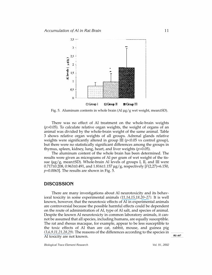
There was no effect of Al treatment on the whole-brain weights(p>0.05). To calculate relative organ weights, the weight of organs of ananimal was divided by the whole-brain weight of the same animal. Table3 shows relative organ weights of all groups. Adrenal glands relativeweights were significantly altered in group III (p<0.05 vs control group),but there were no statistically significant differences among the groups inthymus, spleen, kidney, lung, heart, and liver weights (p>0.05).
The aluminum content of the whole brain has been determined. Theresults were given as micrograms of Al per gram of wet weight of the tis-sue (µg/g, mean±SD). Whole-brain Al levels of groups I, II, and III were0.717±0.208, 0.963±0.491, and 1.816±1.157 µg/g, respectively [F(2,27)=6.150,p=0.0063]. The results are shown in Fig. 5.
DISCUSSION
There are many investigations about Al neurotoxicity and its behav-ioral toxicity in some experimental animals (11,14,15,18,20–27). It is wellknown, however, that the neurotoxic effects of Al in experimental animalsare controversial because the possible harmful effects could be dependenton the route of administration of Al, type of Al salt, and species of animal.Despite the known Al neurotoxicty in common laboratory animals, it can-not be assumed that all species, including humans, are equally susceptible.The rat and rhesus macaque, for example, appear to be less susceptible tothe toxic effects of Al than are cat, rabbit, mouse, and guinea pig(3,6,9,11,21,28,29). The reasons of the differences according to the species inAl toxicity are not known.
Accumulation of Al in Rat Brain 11
Biological Trace Element Research Vol. 91, 2002
Fig. 5. Aluminum contents in whole brain (Al µg/g wet weight, mean±SD).
AU: ok?

The present study was performed to evaluate Al brain accumulationand also certain behavioral and electrophysiological effects of the metal inrats. The doses of Al were calculated as one-fifth and one-twentieth of itsoral LD50 (3,16) and applied by gavage for 8 wk. Al did not affect bodyweight and relative organ weights except adrenal glands. However,weight gain in both treated groups was slower than in the controls over thetreatment period. Decreased weight gain rate may indicate an untowardeffect of Al on general health status. This situation might have resultedfrom interference of Al with the hormonal status and/or protein synthesis.It is noteworthy that adult rats were used in the present study. Our obser-vation pointed out that a similar experiment should be designed in new-born rats to clear up whether there is any untoward effects of Al on growthand whether it is a developmental toxic agent. The dose-related relativeweight change of adrenal glands may have originated from interactions ofAl with, most probably, the immune system or renal system. In order toanswer clearly, further investigations are needed on these points.
Aluminum did not induce gross signs of neurotoxity (paralysis,seizures). Several parameters of the behavioral tests, including horizontaland vertical activities and startle responses, remained unaffected by Alduring the test period, and the group means were quite similar. It has beensuggested that the clinical symptoms and localization of Al in the brainmost probably cause alteration of catecholamine balance. Moreover, theeffects of elevated brain Al upon the endogenous steady-stage levels ofnorepinephrine and dopamine in the frontal cortex, hippocampus, andcerebellum have been reported (30). Even though the relation betweendopamine and behavior is clearly known, there was not any change inbehavioral findings in the present study. This situation might have origi-nated from the insufficient duration of the treatment.
We observed slight changes in electrophysiological parameters such asECoGs, but the total results did not confirm the differences in electrophysi-ological findings. Similarly, Al hydroxide-treated dogs have been shownthat considerable high Al content, whereas the electroencephalogram (EEG)results were normal (18). In our early study, we showed that AlCl3, depend-ing on the duration of treatment, produced an impairment of the motorcoordination ability in mice (15). Additionally, Cutrufo et al. showed that asingle oral dose of Al hydroxide in mice induces some slow-wave EEG alter-ations and produces an increase in the Al level of the brain, depending onthe dose. This finding may suggest the possible role of low-level Al ions ingeneration of encephalopathy without any morphological changes in neu-rons (20). The discrepancies of the studies may be the result of the species ofthe animal used, the experiment period, and Al doses applied.
In the study, the rats treated with both the low and high doses had sig-nificantly higher Al levels in brains than those of the control group. Thepresent result showed that the ingestion of subchronic Al caused consider-able accumulation in brain, even though rats are accepted as less susceptibleto Al. Our previous study was indicated that the brain, bone, and kidney Al
12 Baydar et al.
Biological Trace Element Research Vol. 91, 2002

levels of mice fed with Al compounds were significantly greater than in thecontrol group (13). Similar results have been also observed in many otherinvestigations in different experimental animals (16,18,23,24,27).
Aluminum may cause or contribute to some specific diseases. It is atoxicant indeed; the increased tissue burdens can be presumed to impairbiological functions, including those of the CNS. It is proposed that cellu-lar nuclei may be an important target of potential Al accumulation becauseof the high density of phosphates in RNA and DNA. This accumulationleads most likely to various untoward effects of Al by affecting importantbiochemical pathways, including enzymes. It is known that Al has a veryhigh affinity to phosphate groups; thus, it may interact with phosphate-containing molecules and enzymes in the body (4,6,10,16,17,24,26,29).
In conclusion, further but chronic studies are apparently needed toanswers these subtle points; also, results should be supported by deter-mining some biochemical parameters that play key roles in the brain bio-chemistry in various species, including rats, at various dosage levels andfor various exposure times.
REFERENCES
1. P. O. Ganrot, Metabolism and possible health-effects of aluminium, Environ. Health Per-spect. 65, 363–441 (1986).
2. R. Massey and D. Taylor, Aluminium in Food and the Environment, Special PublicationNo. 73, Royal Society of Chemistry, London (1988).
3. IPCS, Environmental Health Criteria 194, Aluminium, WHO, Geneva (1997).4. K. A. Winship, Toxicity of aluminium: a historical review, Part 2, Adv. Drug React. Toxi-
cal Rev. 12, 177–211 (1993).5. R. J. Boegman and L. A. Bates, Neurotoxicity of aluminum, Can. J. Physiol. Pharmacol.
62, 1010–1014 (1984).6. D. R. C. McLachlan, Aluminum and Alzheimer’s disease, Neurobiol. Aging 7, 525–532
(1986).7. W. A. Banks and A. J. Kastin, Aluminum-induced neurotoxicity: alterations in mem-
brane function at the blood–brain barrier, Neurosci. Biobehav. Rev. 13, 47–53 (1989).8. J. C. Murray, C. M. Tanner, and S. M. Sprague, Aluminum neurotoxicity: a reevaluation,
Clin. Neuropharmacol. 14, 179–185 (1991).9. R. A. Amstrong, J. Anderson, J. D. Cowburn, et al., Aluminium administered in drink-
ing water but not in the diet influences biopterin metabolism in the rodent, Biol. Chem.Hoppe–Seyler 373, 1075–1078 (1992).
10. R. T. Erasmus, J. Savory, M. R. Wills, et al., Aluminum neurotoxicity in experimentalanimals, Ther. Drug Monit. 15, 588–592 (1993).
11. U. De Boni, A. Otvos, J. W. Scott, et al., Neurofibrillary degeneration induced by sys-temic aluminum, Acta Neuropath. (Berl.) 35, 285–294 (1976).
12. W. A. Banks and A. J. Kastin, Aluminium increases permeability of the blood–brain bar-rier to labelled dsip and β-endorphin: possible implications for senile and dialysisdementia, Lancet ii, 1227–1229 (1983).
13. G. Sahin, I. Varol, A. Temizer, et al., Determination of aluminum levels in the kidney,liver, and brain of mice treated with aluminum hydroxide, Biol. Trace Element Res. 41,129–135 (1994).
Accumulation of Al in Rat Brain 13
Biological Trace Element Research Vol. 91, 2002

14. R. L. Commissaris, J. J. Cordon, S. Sprague, et al., Behavioral changes in rats afterchronic aluminum and parathyroid hormone administration, Neurobehav. Toxicol. Tera-tol. 4, 403–410 (1982).
15. G. Sahin, T. Taskin, K. Benli, et al., Impairment of motor coordination in mice afteringestion of aluminum chloride, Biol. Trace Element Res. 50, 79–85 (1995).
16. G. M. Berlyne, R. Yagil, J. Ben Ari, et al., Aluminium toxicity in rats, Lancet i, 564–567(1972).
17. A. C. Alfrey, G. R. LeGendre, and W. D. Kaehny, The dialysis encephalopathy syn-drome, New Engl. J. Med. 294, 184–188 (1976).
18. A. I. Arieff, J. D. Cooper, D. Amstrong, et al., Dementia, renal failure, and brain alu-minum, Ann. Intern. Med. 90, 741–747 (1979).
19. I. Dési, Neurological investigation of pesticides in animal experiments, Neurobehav. Tox-icol. Teratol. 5, 503–517 (1983).
20. C. Cutrufo, S. Caroli, P. Delle Femmine, et al., Experimental aluminium encephalopa-thy quantitative EEG analysis of aluminium bioavailability, J. Neurol. Neurosurg. Psy-chiatry 47, 204–206 (1984).
21. T. L. Petit, G. B. Biederman, and P. A. McMullan, Neurofibrillary degeneration, dentricdying back, and learning-memory deficits after aluminum administration: implicationsfor brain aging, Exp. Neurol. 67, 152–162 (1980).
22. V. Bernuzzi, D. Desor, and P. R. Lehr, Effects of prenatal aluminum exposure on neuro-motor maturation in the rat, Neurobehav. Toxicol. Teratol. 8, 115–119 (1989).
23. B. M. Thorne, T. Donohoe, K. Lin, et al., Aluminum ingestion and behavior in the long-evans rat, Physiol. Behav. 36, 63–67 (1986).
24. P. I. Otezia, C. L. Keen, B. Han, et al., Aluminum accumulation and neurotoxicity inSwiss-Webster mice after long-term dietary exposure to aluminum and citrate, Metabo-lism 42, 1296–1300 (1993).
25. G. S. Zubenko and I. Hanin, Cholinergic and noradrenergic toxicity of intraventricularaluminum chloride in the rat hippocampus, Brain Res. 498, 381–384 (1989).
26. S. Gaytan-Garcia, H. Kim, and M. J. Strong, Spinal motor neuron neuroaxonal sphe-roids in chronic aluminum neurotoxicity contain phosphatase-resistant high molecularweight neurofilament, Toxicology 108, 17–24 (1996).
27. S. Kumar, Biophasic effects of aluminium on cholinergic enzyme of rat brain, Neurosci.Lett. 248, 121–123 (1998).
28. J. R. McDermott, A. I. Smith, M. K. Ward, et al., Brain-aluminium concentration in dial-ysis encephalopathy, Lancet i, 901–904 (1978).
29. D. R. C. McLachlan, W. J. Lukiw, and T. P. A. Kruck, New evidence for an active role ofaluminum in Alzheimer’s disease, Can. J. Neurol. Sci. 16, 490–497 (1989).
30. G. L. Wenk and K. L. Stemmer, The influence of ingested aluminum upon neorepi-nephrine and dopamine levels in the rat brain, Neurotoxicology 2, 347–353 (1981).
14 Baydar et al.
Biological Trace Element Research Vol. 91, 2002