Synthesis of tubulin and actin during the preimplantation development of the mouse
7
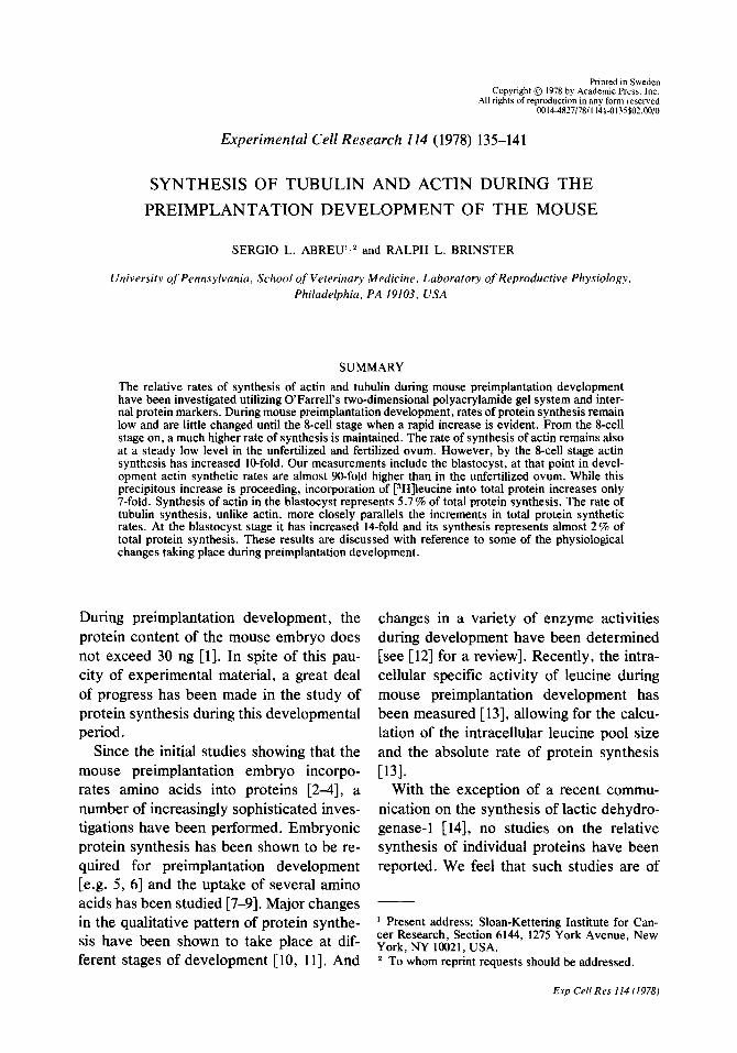
Printed in Sweden Copyright @ 1978 by Academic Press, Inc. All rights of reproduction in any form reserved 00144827/78/1141-0135$02.00/O Experimental Cell Research 114 (1978) 135-141 SYNTHESIS OF TUBULIN AND ACTIN DURING THE PREIMPLANTATION DEVELOPMENT OF THE MOUSE SERGIO L. ABREU’,* and RALPH L. BRINSTER University of Pennsylvania, School of Veterinary Medicine, Laboratory of Reproductive Physiology, Philadelphia, PA 19103, USA SUMMARY The relative rates of synthesis of actin and tubulin during mouse preimplantation development have been investigated utilizing O’Farrell’s two-dimensional polyacrylamide gel system and inter- nal protein markers. During mouse preimplantation development, rates of protein synthesis remain low and are little changed until the 8-cell stage when a rapid increase is evident. From the &cell stage on, a much higher rate of synthesis is maintained. The rate of synthesis of actin remains also at a steady low level in the unfertilized and fertilized ovum. However, by the g-cell stage actin synthesis has increased IO-fold. Our measurements include the blastocyst, at that point in devel- opment actin synthetic rates are almost 90-fold higher than in the unfertilized ovum. While this precipitous increase is proceeding, incorporation of [3H]leucine into total protein increases only 7-fold. Synthesis of actin in the blastocyst represents 5.7% of total protein synthesis. The rate of tubulin synthesis, unlike actin, more closely parallels the increments in total protein synthetic rates. At the blastocyst stage it has increased 14-fold and its synthesis represents almost 2% of total protein synthesis. These results are discussed with reference to some of the physiological changes taking place during preimplantation development. During preimplantation development, the protein content of the mouse embryo does not exceed 30 ng [l]. In spite of this pau- city of experimental material, a great deal of progress has been made in the study of protein synthesis during this developmental period. Since the initial studies showing that the mouse preimplantation embryo incorpo- rates amino acids into proteins [2-4], a number of increasingly sophisticated inves- tigations have been performed. Embryonic protein synthesis has been shown to be re- quired for preimplantation development [e.g. 5, 61 and the uptake of several amino acids has been studied [7-91. Major changes in the qualitative pattern of protein synthe- sis have been shown to take place at dif- ferent stages of development [ 10, 111. And changes in a variety of enzyme activities during development have been determined [see [12] for a review]. Recently, the intra- cellular specific activity of leucine during mouse preimplantation development has been measured [ 131,allowing for the calcu- lation of the intracellular leucine pool size and the absolute rate of protein synthesis [131- With the exception of a recent commu- nication on the synthesis of lactic dehydro- genase-1 [ 141, no studies on the relative synthesis of individual proteins have been reported. We feel that such studies are of ’ Present address: Sloan-Kettering Institute for Can- cer Research, Section 6144, 1275 York Avenue, New York, NY 10021, USA. 2 To whom reprint requests should be addressed. Exp Cell Res 114 (I 978)
-
Upload
independent -
Category
Documents
-
view
5 -
download
0
Transcript of Synthesis of tubulin and actin during the preimplantation development of the mouse
Printed in Sweden Copyright @ 1978 by Academic Press, Inc.
All rights of reproduction in any form reserved 00144827/78/1141-0135$02.00/O
Experimental Cell Research 114 (1978) 135-141
SYNTHESIS OF TUBULIN AND ACTIN DURING THE
PREIMPLANTATION DEVELOPMENT OF THE MOUSE
SERGIO L. ABREU’,* and RALPH L. BRINSTER
University of Pennsylvania, School of Veterinary Medicine, Laboratory of Reproductive Physiology, Philadelphia, PA 19103, USA
SUMMARY
The relative rates of synthesis of actin and tubulin during mouse preimplantation development have been investigated utilizing O’Farrell’s two-dimensional polyacrylamide gel system and inter- nal protein markers. During mouse preimplantation development, rates of protein synthesis remain low and are little changed until the 8-cell stage when a rapid increase is evident. From the &cell stage on, a much higher rate of synthesis is maintained. The rate of synthesis of actin remains also at a steady low level in the unfertilized and fertilized ovum. However, by the g-cell stage actin synthesis has increased IO-fold. Our measurements include the blastocyst, at that point in devel- opment actin synthetic rates are almost 90-fold higher than in the unfertilized ovum. While this precipitous increase is proceeding, incorporation of [3H]leucine into total protein increases only 7-fold. Synthesis of actin in the blastocyst represents 5.7% of total protein synthesis. The rate of tubulin synthesis, unlike actin, more closely parallels the increments in total protein synthetic rates. At the blastocyst stage it has increased 14-fold and its synthesis represents almost 2% of total protein synthesis. These results are discussed with reference to some of the physiological changes taking place during preimplantation development.
During preimplantation development, the protein content of the mouse embryo does not exceed 30 ng [l]. In spite of this pau- city of experimental material, a great deal of progress has been made in the study of protein synthesis during this developmental period.
Since the initial studies showing that the mouse preimplantation embryo incorpo- rates amino acids into proteins [2-4], a number of increasingly sophisticated inves- tigations have been performed. Embryonic protein synthesis has been shown to be re- quired for preimplantation development [e.g. 5, 61 and the uptake of several amino acids has been studied [7-91. Major changes in the qualitative pattern of protein synthe- sis have been shown to take place at dif- ferent stages of development [ 10, 111. And
changes in a variety of enzyme activities during development have been determined [see [12] for a review]. Recently, the intra- cellular specific activity of leucine during mouse preimplantation development has been measured [ 131, allowing for the calcu- lation of the intracellular leucine pool size and the absolute rate of protein synthesis [131-
With the exception of a recent commu- nication on the synthesis of lactic dehydro- genase-1 [ 141, no studies on the relative synthesis of individual proteins have been reported. We feel that such studies are of
’ Present address: Sloan-Kettering Institute for Can- cer Research, Section 6144, 1275 York Avenue, New York, NY 10021, USA. 2 To whom reprint requests should be addressed.
Exp Cell Res 114 (I 978)

136 Abreu and Brinster
importance in understanding the extent and nature of macromolecular changes taking place during mammalian embryonic differ- entiation.
The problem of how the mammalian embryo regulates the pattern and rate of protein synthesis during differentiation and growth can best be approached by the study of individual proteins. With this in mind we have studied the synthesis of tubulin and actin. These were chosen because (a) they are important proteins which have a cyto- skeletal role and are involved in a variety of essential cellular functions including cel- lular motility and contractility; (6) they are abundant proteins; and (c) both radioactive and non-radioactive markers can be pre- pared.
Here we report on the changes in tubulin and actin synthesis taking place during mouse preimplantation development.
MATERIALS AND METHODS
Handling of embryos Embryos were obtained from 6-8week-old Swiss mice and were collected and cultured as previously de- scribed [IS]. The embryos were washed three times by sequential transfer, in a vol of 5 ~1, through 3 ml of culture medium, BMOCJ [15]. The bovine serum al- bumin of this medium was replaced with 0.1% Ficoll (Pharmacia). All experiments were performed on ova that had developed in vivo.
Amino acid incorporation Groups of 45 embryos, at each developmental stage, were incubated in 50 ul drons of culture medium con- taining [3H]leucine (55 Cilmmole, Amersham Searle) at a final concentration of 54x 10-O M. The embryos were pulsed for 2 h and then chased for a total of 20 min with modified BMOC-3 containing unlabeled leu- tine at a concentration of 6~ 10m4 M. The chase con- sisted of two sequential transfers of the embryos through 3 ml of the unlabeled medium; incubating each time for 10 min. After transferring to small tubes (6x50 MM), 200 ~1 of acetone were added to each and these were stored overnight at 0”-4”C, centrifuged and dried in a Speed-Vat Concentrator (Savant Inst.). The dried samples were dissolved in 5 ~1 of buffer A: 2.3 % sodium dodecyl sulfate (SDS); 10% glycerol;
5% /3-mercaptoethanol; 62.5 mM Tris-HCI, pH 6.8 [16], and stored at -20°C for later analysis.
Two-dimensional electrophoresis The two-dimensional polyacrylamide system of 0’ Far- rell [17] was used with the following minor modifica- tions. It was not found necessary to treat the samples with RNAse or DNAse. To each of the samples, pre- viously dissolved in 5 ~1 of buffer A, 20 ~1 of O’Far- rell’s lysis buffer [17] were added before isoelectric focusintz. The lvsis buffer was therefore diluted l/5 by buff& A, and the SDS concentration in buffer A reduced to 0.46 %. Equilibration after isoelectric focusing was done for only 45 min. A 10% polyacryl- amide gel was used in the second dimension.
Assay of total protein synthesis To measure total incorporation of radioactivity, 1 ~1 of the 25 ~1 to be applied to the gel was withdrawn, placed in a test tube and after the addition of 0.5 ml of a bovine serum albumin solution (2 mg/ml), precipi- tated and analysed as described [ 181.
Preparation of markers Tubulin was prepared from adult mouse brain bv three cycles of re-polymerization according to the methods of Shelanski et al. f191. Hiahlv mire actin from rabbit muscle was obtained -from $o&hington Biochemical Corp. Radioactive actin and tubulin were prepared in a cell-free system as follows. Twenty to twenty-five neonatal mouse (24 days old) brains were homo- genized at 0”-4”C in 4 ml of buffer B (30 mM Hepes- KOH, pH 7.6; 120 mM KCI; 8 mM MgCI,; 7 mM P-mercaptoethanol and 0.25 M sucrose) with 5 strokes of a loose fitting Dounce homogenizer followed by 5 strokes with a tight fitting pestle. The homogenate was passed through a G-25 (coarse) Sephadex (Pharmacia) column and the A,., oeak fractions were collected. -“” _
A cell-free translational system was prepared which contained in a total vol of 2.0 ml the following: 20 mM Hepes-KOH, pH 7.6; 72 mM KCI; 4 mM MgCI,; 5 mM P-mercaptoethanol; I mM ATP; 5 mM phos- phoenol pyruvate; 10 IU of pyruvate kinase; 230 A,., units of the peak Sephadex fraction; 50 &i of a -“” [Ylprotein hydrolysate- (Amersham Searle) previ- ously dried, by evacuation, in the reaction vessel; and 50 PM each of five supplemental unlabeled amino- acids: met, cys, trp, gin and asn. Incubation was for 1 h at 37°C. At the end of the incubation, the reac- tion vessel was placed in ice for 1 h and centrifuned at 100000 g for 1-h at 4°C. The supematant was drawn off and precipitated with vinblastine sulfate 1201. The precipitate was washed and stored under cold acetone overnight, dried under vacuum and resuspended in buffer A.
Double label techniques The radioactive markers co-migrated in the one- dimensional gel system of Laemmli [ 161 and in the two- dimensional system with non-radioactive markers.

Embryonic synthesis of tubulin and actin 137
Table 1. Total protein synthesis and synthesis of actin and tubulin during mouse preim- plantation development
Leu. incorporated into total Leu. incorporated Leu. incorporated Actin” TubuW protein into actin into tubulin (rxlhl @g/h/
Embryonic stage (fm/h/embryo) (fm/h/embryo) (fm/h/embryo) embryo) embryo)
Unfertilized 50.86? 1.22 (10) 0.25kO.02 (6) 0.51f0.03 (6) 1.51 3.79 Fertilized 46.99k1.84 ‘(9j 0.21+0.03 (6) 0.50+0.05 (6j 1.26 3.71 2-cell 40.93+ 1.64 (8) 0.28kO.06 (5) 0.58kO.05 (5) 1.69 4.30 8-cell Blastocyst
119.98f2.95 (9j 2.41kO.47 (6) 2.07~!~0.76 (6j 14.56 15.38 375.18k7.58 (9) 21.66kO.90 (7) 7.17kO.44 (7) 130.89 53.29
All experiments were performed with embryos grown in vivo as described in Methods. Blastocysts were recovered from the uterus 3 days after detection of the vaginal plug. Values obtained are expressed in femto- moles per hour per embryo f S:E.M. Figures in parentheses indicate the number of determinations performed. a pglhlembryo. For these calculations the mol. wt of actin was assumed to be 42000 D [26] and the leucine content 6.95% [26]. b The tubulin calculations were based on a subunit mol. wt of 55000 D and an average leucine content of 7.4%; a, 7.2%; /3, 7.5% [27].
The “C-labeled tubulin and actin markers were there- fore co-electroohoresed with embryos labeled with r3H]leucine. The gels were prepared- for fluorography f211. and X-rav film (kodak Roval X-Omat) exnosed io ihe dried gels for’l-3 weeks. The characteristic spot patterns and positions of tubulin and actin in the X-ray tilm were outlined on tracing paper, placed over the dried gel and cut out with a tine scalpel. These were digested in NCS (Amersham Searle) and, after the addition of liquid scintillation fluid, counted using a SL 30 Intertechnique counter programmed for sepa- ration of 3H and r4C disintegrations.
This technique worked quite well: however, a sec- ond, simpler and faster method was utilized for over half of the gels analysed as follows. To each of the [3H]embryo samples, 2 pg each of unlabeled actin and tubulin (in lysis buffer as above) were added. After the two-dimensional run, the gel slab was stained with Coomassie blue and destained. The tubulin and actin spots were cut out, rinsed very briefly in water and then treated as described above for liquid scintillation counting. The two techniques gave comparable re- sults. A similar methodology has been successfully used by others [22, 231 for the analysis of these two proteins.
RESULTS
Two-dimensional electrophoresis The spot pattern of the cy and p chains of tubulin as well as that of actin can be seen in fig. 1 a. The tubulin pattern corresponds well with that recently described [23, 241 using the O’Farrell system. Feit et al. [24] reported microheterogeneity of the (Y and p
subunits of tubulin. We have also observed this, which is more evident in the isoelec- tric focusing dimension (data not shown). Actin heterogeneity was observed in both the neonatal mouse brain and in the preim- plantation embryo. The presence of three actin variants have been previously re- ported for calf muscle cells in culture [25]. In both the neonatal mouse brain (2-4 days old) and the preimplantation embryo the /3 form seems to be predominant with only small amounts of the cx and y forms evident.
Synthesis of total protein Incorporation of [3H]leucine by preimplan- tation embryos was measured as described in Methods. Eight to ten determinations were performed for each developmental stage. The values obtained were quite re- producible from experiment to experiment (table 1). As previously observed [3,4,7,8, 131, the incorporation of leucine into total protein in the mouse preimplantation em- bryo remains low with little change in rate up to about the g-cell stage of develop- ment. The rate of protein synthesis in- creased from 50.86 fmoles of leucine incor-
Exp Cell Res II4 (I 978)

138 Abreu and Brinster
Fig. 1. Identification and quantitation of actin and 6000 dpm of the *4C-labeled cell-free marker; (c) tubulin by two-dimensional polyacrylamide gel elec- fluorogram of blastocysts labeled with [3H]leucine plus trophoresis. (a) Fluorogram of the W-labeled cell- 6 000 dpm of the W-labeled cell-free marker. a and p free products, of a neonatal mouse brain extract, pre- in the middle of each figure correspond to the a! and cipitated by vinblastine sulfate (one precipitation p chains of tubulin. The symbols to the left and below cycle); 6000 dpm were added to the gel; (b) fluoro- the tubulin, indicate the putative a, p and y species gram of 8-cell embryos labeled with [3H]leucine plus of actin.
porated/h/ embryo for the fertilized ovum to 375.18 fmoles for the blastocyst (table 1, fig. 2a, b). This seven-fold increase falls within the range of increments reported in the literature cited above.
Synthesis of actin and tubulin The unfertilized ovum incorporated 0.25 fmole of leucine per hour into actin, repre- senting about 0.5 % of total protein synthe-
thesis of actin constituted 2.0% of total pro- tein synthesis in the 8-cell embryo (fig. 3); this represented a lo-fold increase while total protein synthesis only doubled. By the blastocyst stage actin synthesis reached 130.89 pg/h/embryo or 21.66 fmoles of leu- tine incorporated/h/embryo (table 1, fig. 26).
The rate of tubulin synthesis, like that of total protein synthesis, changed very little
sis and constituting the synthesis of 1 Sl pg during the first three stages studied here of actin/h/ovum (table 1, fig. 3). These rates (table 1, fig. 2a, b). And like total protein were maintained until the 2-cell stage when and actin it increased substantially at the 8- a slight increase was observed. The syn- cell stage, peaking at 7.17 fmole of leucine
Exp Cell Rrs 114 (1978)

Embryonic synthesis of tub&in and actin 139
i
0 450
225 i
Fig. 2. Abscissa: embryonic stage (unfertilized ovum, U; fertilized ovum, F; 2-cell embryo, 2; 8-cell embryo, 8; blastocyst, B); ordinate: (a) femtomoles of r3H]- leucine incornorated ner hour uer embrvo into total protein, O-6; (b) femtomoles of [3H]leucine incorpo- rated per hour per embryo into actin, O-O; into tubulin, A-A.
incorporated/h/embryo at the blastocyst stage. At this point tubulin synthesis was almost 2 % of the total (fig. 3). The increase in tubulin synthetic rates from the l-cell embryo to the blastocyst was 1Cfold which must be compared to a total protein syn- thetic rate increase of 7-fold and to an 86- fold increase in actin synthesis during the same developmental period.
DISCUSSION
Synthesis of actin Our results clearly demonstrate a marked increase in actin synthesis by the 8-cell stage. High relative amounts of synthesis of actin have been reported for non-muscle systems. The proportion of actin synthe-
‘I I
O---rFrT ” Fig. 3. Abscissa: embryonic stage (unfertilized ovum, U; fertilized ovum, F; 2-cell embryo, 2; 8-cell embryo, 8; blastocyst, B); ordinate: % of total [3H]leucine in- corporation going into actin, O-O; into tubulin, A-A.
sized in the nauplius stage of Artemia salina is about 3-fold higher than in the dormant cyst [23]. It represents 21% of the newly synthesized protein in the IO-day old neona- tal rat brain and 7 % when the neonate is 30 days old [28]. In growing 3T3 cells actin synthesis is 7.4% of total protein synthesis P21.
The increase in both rate and relative amounts of actin synthesis from the 8-cell stage on is of particular interest in terms of the functional role of actin and the mor- phogenetic events which are taking place. There is increasing evidence that actin and actin-containing structures in non-muscle cells are responsible for the contractile movements involved in a variety of phe- nomena such as cell movement and attach- ment, cytokinesis, membrane ruffling and cell-surface changes in general [29-3 11.
As the mouse zygote reaches the 8-cell stage, morphogenetic processes are at work which result in the compaction of the blas- tomeres into a solid embryonic mass. It is safe to assume that this process involves movement of blastomeres and changes in
Exp Cd Re\ 114 (19781

140 Abreu and Brinster
the blastomeric surface. Rhythmical con- tractions and expansion of the late morula as it becomes a blastocyst are well docu- mented, these contractions are maintained and increased during blastocyst develop- ment [32]. In view of the major role of actin in contractile processes, it can be suggested that the increase in actin synthesis de- scribed here is in reponse to morphoge- netic demands taking place as the 8-cell em- bryo continues to develop into a blastocyst. Indeed, Granholm & Brenner [33] have re- ported that the anti-microfilament agent cytochalasin B inhibits the morula to blas- tocyst transformation.
Another role of actin during this develop- mental period may be as an anti-DNAse agent. It has been shown that actin is a naturally occurring inhibitor of deoxyribo- nuclease I [34]. During this period when the embryo is rapidly increasing in cell number there is a necessary increase in DNA syn- thesis. Gene activity or derepression in- creases during development and the syn- thesis of the DNAse inhibitor could be of importance; especially in view of a recent report that showed DNAse-I to be specific for active regions of the genome [35].
Synthesis of tub&in We have shown the synthesis of tubulin to increase from about 1 .O % of total protein synthesis in the l-cell embryo to 1.9% in the blastocyst. This is a significant increase in synthesis which is accompanied by a 14- fold increase in the relative synthetic rates of tubulin.
The synthesis of tubulin has been studied in a wide variety of organisms and in many instances it represents a sizable percent of total protein synthesis. For instance, in the developing rat brain it can constitute up to 16% of the newly synthesized protein; this
percentage decreases as the brain matures [28]. We have not found tubulin synthesis during preimplantation development to ap- proach this high level. Rather, it is com- parable to that reported during the early embryonic development of echinoderms [361.
The importance of tubulin as part of the mitotic apparatus, as a cytoskeleton com- ponent and in secretion and transport has been thoroughly discussed in a recent monograph [36]. The increase in tubulin synthesis we have observed reflects the in- crease in cell number (mitotic activity) and perhaps, as discussed above, the morpho- logical reorganization which is taking place.
These results are of special importance in understanding the regulation of protein synthesis during mammalian preimplanta- tion development; however, they represent just one step in that direction. It is essential to gain additional information about other metabolic characteristics of actin and tubu- lin. For instance, the success in obtaining antibodies against these two proteins [e.g. 37, 381 now make it possible to estimate what percent of total protein content they represent at different stages of preimplanta- tion development. With the methods uti- lized here, their rate of decay can also be estimated. Our results pose the question of whether the described increments in actin and tubulin synthesis are regulated at the transcriptional level, by enhanced mRNA synthesis, or at the translation level, by in- creased translation of stored cytoplasmic mRNA. This question is now amenable to experimental analysis in the mouse preim- plantation embryo.
We thank Dr Allen Rushmer for his valuable com- ments on the manuscript. This work was supported by USPHS Research Grant HD 08452, NICHHD Training Grant HD 00239 and a grant from the Popula- tion Council.

Embryonic synthesis of tubulin and actin 141
REFERENCES
1. Brinster, R L, J reprod fert 13 (1%7) 413. 2. Mintz, B, J exp zoo1 157 (1964) 85. 3. Monesi, V & Salti, V, Exp cell res 46 (1%7) 632. 4. Tasca, R J & Hillman, N, Nature 225 (1970) 1022. 5. Thompson, J & Biggers, J, Exp cell res 41 (1966)
411. 6. Monesi, V, Molinaro, M, Spalleta, E & Davoli, C,
Exp cell res 59 (1970) 197. 7. Brinster, R L, J reprod fert 27 (1971) 329. 8. Epstein, C J & Smith, A S, Dev biol33 (1973) 171. 9. Borland, R M & Tasca, R J, Dev biol36 (1974) 169.
10. Epstein, C J & Smith, A S, Dev biol40 (1974) 233. Il. Van Blerkom, J & Brockwav, G 0, Dev biol 44
(1975) 148. 12. Epstein, C J, Biol reprod 12 (1975) 82. 13. Brinster, R L, Wiebold, J L & Brunner, S, Dev biol
51 (1976) 215. 14. Mangia, F, Erickson, R P & Epstein, C J, Dev biol
54 (1976) 146. 15. Brinster, R L, Growth, nutrition and metabolism
of cells in culture (ed G Rothblat & V Cristofalo) p. 251. Academic Press, New York (1972).
16. Laemmli. U K. Nature 227 (1970) 680. 17. O’Farrell: P H,‘J biol them 250 (i975) 4007. 18. Abreu, S L & Lucas-Lenard, J, Antimicrob agents
chemother 5 (1975) 521. 19. Shelanski, M L, Gaskin, F & Cantor, C R, Proc
natl acad sci US 70 (1973) 765. 20. Wilson, L, Bryan, J, Ruby, A & Mazia, D, Proc
natl acad sci US 66 (1970) 807. 21. Bonner, W M & Laskey; R A, Eur j biochem 46
(1974) 83.
24.
25.
26.
27.
28.
29.
30.
31. 32.
33.
34.
35.
36.
37.
38.
Feit, H, Neudeck, U & Gaskin, F, J neurochem 28 (1977) 697. Whalen, R G, Butler-Browne, G S & Gras, F, Proc natl acad sci US 73 (1976) 2018. Elzinga, M, Collins, J H, Kuehl, W M & Adel- stein, R S, Proc natl acad sci US 70 (1973) 2687. Bryan, J & Wilson, L, Proc natl acad sci US 68 (1971) 1762. Schmitt, H, Gozez, I & Littauer, U Z, Brain res 121 (1977) 327. Wessels, N K, Spooner, B S. Ash, J F, Bradlev. M 0, Luduena, M* A, Taylor, E L, Wren, J T & Yamada. KM. Science I71 (1971) 135. Schroeder, T E, Proc natl acad sci US 70 (1973) 1688. Edelman, G M, Science 192 (1976) 218. Ogawa, S, Satoh, K & Hashimoto, H, Nature 233 (1971) 422. Granholm, N H & Brenner, G M, Exp cell res 101 (1976) 143. Lazarides, E & Lindberg, U, Proc natl acad sci US 71 (1974) 4742. Weintraub, H & Groudine, M, Science 193 (1976) 848. Raff, R A, Brandis, J W, Green, L H, Kaumeyer, J F & Raff, E C, The biologv of cvtonlasmic micro- tubules (ed D Soifer) p. !&I. Ann ‘NY Acad Sci, New York (1975). Weber, K, Pollack, R & Bibring, T, Proc natl acad sci US 72 (1975) 459. Trenchev, P & Holborow, E J, Immunology 31 (1976) 509.
22. Fine, R E & Taylor, L, Exp cell res 102 (1976) 162. Received September 30, 1977 23. Grossfeld, H & Littauer, U Z, Eur j biochem 70 Revised version received January 9, 1978
(1976) 589. Accepted January 13, 1978
E-\-p Cell Rex / 14 (1978~