Rationalization of paclitaxel insensitivity of yeast β-tubulin and human βIII-tubulin isotype...
10
RESEARCH ARTICLE Open Access Rationalization of paclitaxel insensitivity of yeast β-tubulin and human βIII-tubulin isotype using principal component analysis Lalita Das 1 , Bhabatarak Bhattacharya 1 and Gautam Basu 2* Abstract Background: The chemotherapeutic agent paclitaxel arrests cell division by binding to the hetero-dimeric protein tubulin. Subtle differences in tubulin sequences, across eukaryotes and among β-tubulin isotypes, can have profound impact on paclitaxel-tubulin binding. To capture the experimentally observed paclitaxel-resistance of human βIII tubulin isotype and yeast β-tubulin, within a common theoretical framework, we have performed structural principal component analyses of β-tubulin sequences across eukaryotes. Results: The paclitaxel-resistance of human βIII tubulin isotype and yeast β-tubulin uniquely mapped on to the lowest two principal components, defining the paclitaxel-binding site residues of β-tubulin. The molecular mechanisms behind paclitaxel-resistance, mediated through key residues, were identified from structural consequences of characteristic mutations that confer paclitaxel-resistance. Specifically, Ala277 in βIII isotype was shown to be crucial for paclitaxel-resistance. Conclusions: The present analysis captures the origin of two apparently unrelated events, paclitaxel-insensitivity of yeast tubulin and human βIII tubulin isotype, through two common collective sequence vectors. Background Microtubules are structures composed of polymerized tubulin heterodimers and play fundamental roles in vital cellular processes such as chromosome segregation, intracellular transport and maintenance of cell shape [1]. The major component of microtubules is the hetero- dimeric (αβ) protein tubulin. Mammalian tissues express different α- and β-tubulin isotypes [2] that are known to play specific biological functions, exhibit tissue- restricted expression, possess differential binding affin- ities for antimitotic agents and exhibit different kinetics and dynamics of microtubule assembly [3-5]. Paclitaxel, a product of plant secondary metabolism, binds to the β-tubulin subunit and inhibits microtubule dynamics, thereby blocking cell cycle progression during mitosis at the metaphase/anaphase transition and acti- vating cell death [6-8]. It is an important cancer che- motherapeutic agent for treatment of advanced ovarian, lung and breast carcinoma, and shows promising activity against several other carcinomas [9,10]. Although pacli- taxel is currently used in chemotherapy, clinical resist- ance against paclitaxel can become a significant problem. The human βIII tubulin isotype has been implicated to play a crucial role in conferring paclitaxel resistance. Increased expression of the βIII tubulin iso- type inhibits cell proliferation and confers resistance to paclitaxel [11-13]. Increased relative abundance of the βIII isotype is also known to destabilize the microtubule [14] and augment paclitaxel resistance [13,15-17]. Similar to the βIII isotype of mammalian brain tubulin, tubulin from the budding yeast, Saccharomyces cerevi- siae, shows weak binding affinity for paclitaxel [18,19] despite the fact that yeast tubulin shares 75% amino acid identity with mammalian brain tubulin. When five amino acid residues in yeast β-tubulin (A19K, T23V, G26D, N229H, and Y272F) were changed to the respect- ive residues found in the mammalian brain tubulin, a paclitaxel-binding site could be created in yeast tubulin [20]. Of these, the effect from two (A19K and N229H) was shown to be negligible from single mutational * Correspondence: [email protected] 2 Department of Biophysics, Bose Institute, P-1/12 CIT Scheme VIIM, Kolkata 70054, India Full list of author information is available at the end of the article © 2012 Das et al.; licensee BioMed Central Ltd. This is an Open Access article distributed under the terms of the Creative Commons Attribution License (http://creativecommons.org/licenses/by/2.0), which permits unrestricted use, distribution, and reproduction in any medium, provided the original work is properly cited. Das et al. BMC Research Notes 2012, 5:395 http://www.biomedcentral.com/1756-0500/5/395
-
Upload
independent -
Category
Documents
-
view
0 -
download
0
Transcript of Rationalization of paclitaxel insensitivity of yeast β-tubulin and human βIII-tubulin isotype...

Das et al. BMC Research Notes 2012, 5:395http://www.biomedcentral.com/1756-0500/5/395
RESEARCH ARTICLE Open Access
Rationalization of paclitaxel insensitivity of yeastβ-tubulin and human βIII-tubulin isotype usingprincipal component analysisLalita Das1, Bhabatarak Bhattacharya1 and Gautam Basu2*
Abstract
Background: The chemotherapeutic agent paclitaxel arrests cell division by binding to the hetero-dimeric proteintubulin. Subtle differences in tubulin sequences, across eukaryotes and among β-tubulin isotypes, can haveprofound impact on paclitaxel-tubulin binding. To capture the experimentally observed paclitaxel-resistance ofhuman βIII tubulin isotype and yeast β-tubulin, within a common theoretical framework, we have performedstructural principal component analyses of β-tubulin sequences across eukaryotes.
Results: The paclitaxel-resistance of human βIII tubulin isotype and yeast β-tubulin uniquely mapped on to thelowest two principal components, defining the paclitaxel-binding site residues of β-tubulin. The molecularmechanisms behind paclitaxel-resistance, mediated through key residues, were identified from structuralconsequences of characteristic mutations that confer paclitaxel-resistance. Specifically, Ala277 in βIII isotype wasshown to be crucial for paclitaxel-resistance.
Conclusions: The present analysis captures the origin of two apparently unrelated events, paclitaxel-insensitivity ofyeast tubulin and human βIII tubulin isotype, through two common collective sequence vectors.
BackgroundMicrotubules are structures composed of polymerizedtubulin heterodimers and play fundamental roles in vitalcellular processes such as chromosome segregation,intracellular transport and maintenance of cell shape [1].The major component of microtubules is the hetero-dimeric (αβ) protein tubulin. Mammalian tissues expressdifferent α- and β-tubulin isotypes [2] that are known toplay specific biological functions, exhibit tissue-restricted expression, possess differential binding affin-ities for antimitotic agents and exhibit different kineticsand dynamics of microtubule assembly [3-5].Paclitaxel, a product of plant secondary metabolism,
binds to the β-tubulin subunit and inhibits microtubuledynamics, thereby blocking cell cycle progression duringmitosis at the metaphase/anaphase transition and acti-vating cell death [6-8]. It is an important cancer che-motherapeutic agent for treatment of advanced ovarian,
* Correspondence: [email protected] of Biophysics, Bose Institute, P-1/12 CIT Scheme VIIM, Kolkata70054, IndiaFull list of author information is available at the end of the article
© 2012 Das et al.; licensee BioMed Central LtdCommons Attribution License (http://creativecreproduction in any medium, provided the or
lung and breast carcinoma, and shows promising activityagainst several other carcinomas [9,10]. Although pacli-taxel is currently used in chemotherapy, clinical resist-ance against paclitaxel can become a significantproblem. The human βIII tubulin isotype has beenimplicated to play a crucial role in conferring paclitaxelresistance. Increased expression of the βIII tubulin iso-type inhibits cell proliferation and confers resistance topaclitaxel [11-13]. Increased relative abundance of theβIII isotype is also known to destabilize the microtubule[14] and augment paclitaxel resistance [13,15-17].Similar to the βIII isotype of mammalian brain tubulin,
tubulin from the budding yeast, Saccharomyces cerevi-siae, shows weak binding affinity for paclitaxel [18,19]despite the fact that yeast tubulin shares 75% amino acididentity with mammalian brain tubulin. When fiveamino acid residues in yeast β-tubulin (A19K, T23V,G26D, N229H, and Y272F) were changed to the respect-ive residues found in the mammalian brain tubulin, apaclitaxel-binding site could be created in yeast tubulin[20]. Of these, the effect from two (A19K and N229H)was shown to be negligible from single mutational
. This is an Open Access article distributed under the terms of the Creativeommons.org/licenses/by/2.0), which permits unrestricted use, distribution, andiginal work is properly cited.

Figure 1 Sequence variation at the paclitaxel-binding site (PBS)of human tubulin isotypes and β-tubulin sequences acrosseukaryotic families (shown as sequence logo plots). Sequencenumbering is consistent with that used in the pdb file 1JFF.
Das et al. BMC Research Notes 2012, 5:395 Page 2 of 10http://www.biomedcentral.com/1756-0500/5/395
studies [21]. The mutations changed only a handful ofresidues, yet there was a dramatic change in the pacli-taxel binding affinity. This is analogous to the differencebetween βIII tubulin and other β-tubulin isotypes (theydiffer at only a few sequence positions) where a dramaticchange in paclitaxel affinity is also observed.Although the final effect of replacing βIII tubulin by
other isotypes, and, mutating yeast tubulin at three spe-cific positions — regeneration of paclitaxel-binding affin-ity — is the same, the underlying mechanisms may bedifferent. Here we focus on deciphering such mechan-isms, specifically identifying residues and their interac-tions, responsible for paclitaxel resistance of yeasttubulin and the βIII tubulin. To identify the role of thekey residues, we analyzed β-tubulin sequences across alarge number of eukaryotic families (animals, fungi, pro-tists and plants) and performed principal componentanalysis (PCA) in the sequence-space defining thepaclitaxel-binding site, obtained from the structure ofpaclitaxel-bound αβ-tubulin [22,23]. Projection of bind-ing site residues on a plane defined by the two principalcomponent (PC) axes identified two orthogonal ways bywhich residues varied across eukaryotes. While the pacli-taxel resistance of yeast tubulin included contributionfrom both the axes, the paclitaxel resistance of the βIIIisotype correlated with contribution from only one ofthe two axes. Analysis of structural consequences ofmodifying key residues allowed us to identify mechan-isms of paclitaxel resistance of yeast and the βIII isotype.Principal component analysis also allowed us to predictthe paclitaxel sensitivity of eukaryotic tubulin for whichexperimental data are unavailable.
ResultsMultiple sequence alignment of primary paclitaxel-binding residuesInstead of analyzing the entire tubulin sequence, we ana-lyzed a sequence subset that is most likely to controlpaclitaxel binding. A total of 22 tubulin residues, occur-ring within 5 Å from paclitaxel in the paclitaxel-boundtubulin structure (pdb code: 1JFF) [23], were identifiedand defined as the paclitaxel binding site (PBS) residues.From a multiple sequence alignment using the programClustalW [24], amino acids corresponding to the 22 PBSresidues were identified in 125 eukaryotic β-tubulinsequences, as shown in Figure 1 (through out this workwe have used sequence numbering used in the PDBentry 1JFF; this sequence numbering is slightly differentfrom the original sequence numbering to accommodatesequence alignment of α and β–chains). To schematic-ally depict sequence variations at these residue positionsacross the eukaryotic families, sequence logo [25] plotswere constructed for each eukaryotic family, as depictedin Figure 1. Among the 22 residues, eight residues (27,
217, 230, 237, 274, 275, 320, and 360) were strictly con-served across eukaryotes, while the other 14 showedmild to strong variation. Among the 14 variable posi-tions, two positions (23 and 26) correlated with muta-tional data that resulted in the non-binding of paclitaxelto yeast tubulin or microtubules [20]. The human β-tubulin isotypes were also multiple sequence aligned andthe corresponding PBS residues were identified, asshown in Figure 1. Isotype III has one change (Ser277-Ala) while isotype VI is characterized by a number of se-quence variations (Val23Met, Ala233Leu, Ser277Ala andArg278Gln).
Principal component analysis in the primary paclitaxel-binding sequence spaceThe sequence variations in the PBS (Figure 1) can befurther resolved by principal component analysis. Insummary, PCA seeks vectors that reflect maximumchange in residue variations, in a collective manner.Two such vectors, PC1 and PC2, associated with thehighest mean square variations, were identified by PCA.The eukaryotic PBS sequences were then projected ontoa plane defined by PC1 and PC2 (Figure 2a). Before ana-lyzing Figure 2a, let us recapitulate what is known aboutexperimental affinities of paclitaxel towards tubulin fromdifferent eukaryotic families (each eukaryotic family ismarked by a different color in Figure 2a). Paclitaxel isknown to interact with animal tubulin/microtubule [26].
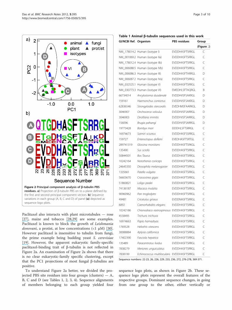
Figure 2 Principal component analysis of β-tubulin PBSresidues. a) Projection of β-tubulin PBS on to a plane defined bythe first and second principal component vectors. b) Sequencevariations in each group (A, B, C and D) of panel (a) depicted assequence logo plots.
Table 1 Animal β-tubulin sequences used in this work
GI/NCBI Ref. Organism PBS residues Group
(Figure 2)
NM_178014.2 Human (isotype I) EVDDHASFTSRRGL C
NM_001069.2 Human (isotype IIa) EVDDHASFTSRRGL C
NM_178012.4 Human (isotype IIb) EVDDHASFTSRRGL C
NM_006088.5 Human (isotype IVb) EVDDHASFTSRRGL C
NM_006086.3 Human (isotype III) EVDDHASFTARRGL D
NM_006087.2 Human (isotype IVa) EVDDHASFTSRRGL C
NM_032525.1 Human (isotype V) EVDDHASFTSRRGL C
NM_030773.3 Human (isotype VI) EMEDHLSFTAQRGL B
66734014 Ancylostoma duodenale EVDDHVSFSAKRGL D
159161 Haemonchus contortus EVDDHVSFSAKRGL D
62836546 Strongyloides stercoralis EVEDHMSFAARKGL D
3046907 Onchocerca volvulus EVDDHVSFSARRGL D
3046903 Dirofilaria immitis EVDDHVSFSARRGL D
156096 Brugia pahangi EVDDHVSFSARRGL D
19773428 Bombyx mori EIDDHLSFTSRRGL C
16974673 Saimiri sciureus EVDDHASFSSRRGL C
159727 Enteroctopus dofleini EVEDHASFTSRTGL C
289741319 Glossina morsitans EVDDHASFTSRKGL C
135490 Sus scrofa EVDDHASFTSRRGL C
50844501 Bos Taurus EVDDHASFTSRRGL C
10242164 Notothenia coriiceps EVDDHASFTSRRGL C
24645350 Drosophila melanogaster EVDDHASFTSRRGL C
1335661 Patella vulgata EVDDHASFTSRRGL C
56603670 Crassostrea gigas EVDDHASFTSRRGL C
51860821 Loligo pealei EVDDHASFTSRRGL C
74136187 Macaca mulatta EVDDHASFTSRRGL C
90960962 Pan troglodytes EVDDHASFTSRRGL C
49481 Cricetulus griseus EVDDNASFTSRRGL C
6892 Caenorhabditis elegans EVDDHASFTSRRGL C
10242186 Chionodraco rastrospinosus EVDDHASFTSRRGL C
4558495 Trichuris trichiura EVDDHASFTSRRGL C
16974663 Papio hamadryas EVDDHASFTSRRGL C
1769528 Heliothis virescens EVDDHASFTSRRGL C
30088884 Aplysia californica EVDDHASFTSRRGL C
17402390 Fasciola hepatica EVDDHASFTSRRGL C
135489 Paracentrotus lividus EVDDHASFTSRRGL C
7838279 Meriones unguiculatus EVDDHASFTSRRGL C
7838199 Echinococcus multilocularis EVDDHASFTSRRGL C
Sequence numbers: 22-23, 26, 226, 229, 233, 236, 272, 276-278, 369-371.
Das et al. BMC Research Notes 2012, 5:395 Page 3 of 10http://www.biomedcentral.com/1756-0500/5/395
Paclitaxel also interacts with plant microtubules — rose[27], maize and tobacco [28,29] are some examples.Paclitaxel is known to block the growth of Leishmaniadonovani, a protist, at low concentrations (<1 μM) [30].However paclitaxel is insensitive to tubulin from fungi,the prime example being budding yeast S. cerevisiae[19]. However, the apparent eukaryotic family-specificpaclitaxel-binding trait of β-tubulin is not reflected inFigure 2a. An examination of Figure 2a shows that thereis no clear eukaryotic-family specific clustering, exceptthat the PC1 projections of most fungal β-tubulins arepositive.To understand Figure 2a better, we divided the pro-
jected PBS site residues into four groups (clusters) — A,B, C and D (see Tables 1, 2, 3, 4). Sequence alignmentsof members belonging to each group yielded four
sequence logo plots, as shown in Figure 2b. These se-quence logo plots represent the overall features of therespective groups. Dominant sequence changes, in goingfrom one group to the other, either vertically or

Table 2 Fungi β-tubulin sequences used in this work
GI/NCBI Ref. Organism PBS residues Group
(Figure 2)
4455142 Lentinus sajor-caju EVDDHISFTARRGC D
173523 Schizophyllum commune EVDDHFSFTARRGL D
74699315 Ustilago maydis EVDDHLSFTARRGL D
11229034 Melampsora lini EVDDHISFTARRGL D
55982602 Cochliobolus heterostrophus EVDDHSSFTSRRGL C
173523 Schizosaccharomyces pombe STDDHAAFAAIKDL B
3435 Saccharomyces cerevisiae ETGDNSSYTAIQGL B
170938 Candida albicans ETGENSSYTSMKDL B
77023508 Verticillium tricorpus QNGDHASFTSRRGL A
299296 Phaeosphaeria nodorum QTGDHASFTSRRGL A
58119500 Phaeosphaeria avenaria QTGDHASFTSRRGL A
170600 Venturia inaequalis QTGDHASFTSRRGL A
61678005 Cercospora beticola QTGDHASFTSRRGL A
30961893 Monilinia fructicola QTGDHASFTSRRGL A
166496 Aspergillus flavus QTGDHASFTSRRGL A
168105 Emericella nidulans QTGDHASFTSRRGL A
639766 Ajellomyces capsulatus QTGDHASFTSRRGL A
2852439 Mycosphaerella pini QTGDHASFTSRRGL A
1002511 Botryotinia fuckeliana QTGDHASFTSRRGL A
602578 Erysiphe pisi QTGDHASFTSRRGL A
1060942 Penicillium digitatum QTGDHASFTSRRGL A
6652864 Pestalotiopsis microspora QTGDHASFTSRRGL A
1263904 Rhynchosporium secalis QTGDHASFTSRRGL A
2718 Epichloe typhina QTGDYASFTSRRGL A
849160 Gibberella fujikuroi QTGDYASFTSRRGL A
2293 Neotyphodium coenophialum QTGDYASFTSRRDL A
32130590 Gibberella zeae QTGDYASFTSRRGL A
167300 Glomerella graminicola QTGHHRSFTS-KGL A
169400 Pneumocystis carinii STGDHASFTSKRGL A
Sequence numbers: 22-23, 26, 226, 229, 233, 236, 272, 276-278, 369-371.
Das et al. BMC Research Notes 2012, 5:395 Page 4 of 10http://www.biomedcentral.com/1756-0500/5/395
horizontally, are: T23V and G26D (horizontal move) andA277S (vertical move). Groups A and B exclusively con-tain members of the fungi family. Group C contains allmembers of the plant family and majority of the animaland the protist family. However, it also contains a fungalpathogen of maize, Cochliobolus heterostrophus. GroupD contains members from all families except plants. Pro-jection of human β-tubulin isotypes on the PC1-PC2plane showed that all isotypes appear in group C exceptβ-III (group D) and β-VI (group B).
Paclitaxel indifference of yeast and βIII-tubulin isotypereflected on the first two principal componentsThe identities of PBS residues were explored along PC1or PC2 axes. Amino acid variations along PC1 and PC2
axes are shown in Figure 3. Among the five residuemutations that altered yeast tubulin affinity towardspaclitaxel (A19K, T23V, G26D, N229H, and Y272F), two(T23V and G26D) are captured by the PC1 axis. Twoother residue variations (N229H, and Y272F) getreflected along the PC2 axis (the fifth residue, A19K, isnot part of the PBS). The only residue that makes theβIII-isotype unique is Ala277 (in place of Ser). This vari-ation is reflected along the PC2 axis. It is noteworthythat the PCA was performed without the inclusion ofthe β-tubulin isotypes. Yet, sequence variations at thePBS in the isotype sequences (position 277) are reflectedin one of the two PC axes. This shows that the two PCaxes are quite robust. The Ala/Ser variation at position277 is naturally present, not only in isotypes βIII and

Table 3 Plant β-tubulin sequences used in this work
GI/NCBI Ref. Organism PBS residues Group
(Figure 2)
4455142 Lentinus sajor-caju EVDDHISFTARRGC C
1488052 Daucus carota EVDDHGSFTSRRGL C
460991 Oryza sativa EVDDHASFTSRRGL C
4415996 Eleusine indica EVDDHASFTSRRGL C
224106013 Populus trichocarpa EVDDHASFTSRRGL C
1403143 Cicer arietinum EVDDHASFTSRKNL C
244539475 Lotus japonicus EVDDHASFTSRKGL C
153799899 Eucalyptus grandis EVDDHASFTSRKGL C
255564502 Ricinus communis EVDDHASFTSRKGL C
312989 Glycine max EVDDHASFTSRKGL C
223018283 Citrus maxima EVDDHASFTSRKGL C
295851 Zea mays EVADHASFTSRHGL C
609270 Solanu tuberosum EVDDHASFTSRTGL C
77963735 Solanum lycopersicum EVDDHASFTSRTGL C
40036995 Nicotiana attenuata EVDDHASFTSRTGL C
1743277 Hordeum vulgare EVDDHASFTSRTGL C
14331109 Medicago sativa EVDDHTSFTSRTGL C
145388977 Capsicum annuum EVDDHASFTSRTGL C
37038246 Physcomitrella patens EVEDHASFTSRTGL C
402636 Lupinus albus EVADHASFTSRTGL C
20758 Pisum sativum EVADHASFTSRTGL C
20148289 Arabidopsis thaliana EVADHASFTSRTGL C
51988178 Setaria viridis EVADHASFTSRTGL C
205326619 Prunus salicina EVADHASFTSRTGL C
5668669 Zinnia elegans EVTDHASFTSRTGL C
296498 Anemia phyllitidis EVTDHASFTSRVGL C
4098333 Triticum aestivum EVGDHASFTSRVGL C
19569609 Gossypium hirsutum EVADHASFTSRIGL C
Sequence numbers: 22-23, 26, 226, 229, 233, 236, 272, 276-278, 369-371.
Table 4 Protist β-tubulin sequences used in this work
GI/NCBI Ref.Organism PBS residues Group
(Figure 2)
155874 Babesia bovis EVDDHASFTSRKGL C
29420520 Babesia microti EVDDHASFTSRKGL C
295762 Plasmodium falciparum EVDDHASFTSRKGL C
4079637 Tetrahymena pyriformis EVDDHASFTSRKGL C
161737 Tetrahymena thermophila EVDDHASFTSRKGL C
6007456 Stylonychia mytilus EVDDHASFTSRKGL C
9309 Euplotes octocarinatus EVDDHASFTSRKGL C
2155306 Chlamydomonas incerta EVDDHASFTSRKGL C
167456 Chlamydomonas reinhardtii EVDDHASFTSRKGL C
166302 Achlya klebsiana EVDDHASFTSRKGL C
68128910 Leishmania major EVDDHASFTSRKGL C
6652866 Pythium ultimum EVDDHASFTSRKGL C
639490 Eimeria tenella EVDDHASFTSRKGL C
161939 Toxoplasma gondii EVDDHASFTSRKGL C
23481527 Plasmodium yoelii yoelii EVDDHASFTSRKGL C
290685 Moneuplotes crassus EVDDHASFTSRKGL C
38520885 Paramecium tetraurelia EVDDHASFTSRKGL C
295443942 Palpitomonas bilix EVDDHASFTSRKGL C
238617571 Leucocryptos marina EVDDHASFTSRKGL C
302849658 Volvox carteri EVDDHASFTSRKGL C
135500 Trypanosoma brucei rhodesiensEVDDHASFTSRKGL C
206598211 Bodo saltans EVDDHASFTSRKGL C
159416 Leishmania mexicana EVDDHASFTSRKGL C
135494 Polytomella agilis EVDDHASFTSRKGL C
829213 Naegleria gruberi EVDDHISFTSRRGL C
8926601 Thalassiosira weissflogii EVDDHACYTSRKGL C
2951981 Phytophthora cinnamomi EVDDHASFTSRQGL C
29150706 Porphyra yezoensis EVEDKGSYSADTGE D
29539330 Cyanidioschyzon merolae EVEDKGSLTATKGL D
1067176 Porphyra purpurea EVEDKGSYSADTGE D
Sequence numbers: 22-23, 26, 226, 229, 233, 236, 272, 276-278, 369-371.
Das et al. BMC Research Notes 2012, 5:395 Page 5 of 10http://www.biomedcentral.com/1756-0500/5/395
βVI, but also in β-tubulin across eukaryotes. In the nextsections we provide structural arguments as to whymembers of groups A, B and D might be paclitaxel-insensitive.
Structural consequences of Val23Thr and Asp26GlymutationCompared to group C that contains paclitaxel-sensitiveβ-tubulin sequences, Val23Thr and Asp26Gly mutationscharacterize groups B and A (positive PC1 values). Ifthese mutations are indeed responsible for paclitaxel-insensitivity, what is the structural mechanism behindpaclitaxel-resistance? In Figure 4a paclitaxel is shownwith Asp26 and Val23 as found in the structure 1JFF.The structural consequences of Asp26Gly and Val23Thrmutations are shown in Figure 4b. The large side chain
of Asp molecule strongly interacts with 3′-phenyl group(Figure 4a) of paclitaxel. Asp26Gly mutation will clearlyhave a major effect since the Asp side-chain makes sev-eral contacts with paclitaxel that will be lost. However,the effect of Val23Thr mutation on paclitaxel-sensitivityof β-tubulin is not apparent from Figure 4a. To fullyunderstand the effect of Val23Thr mutation, one needsto examine alternate rotameric states of Thr23. In thenew rotameric state (Figure 4c) the side-chain of Thr23can form a H-bond with the backbone carbonyl atomsof Lys19 and Phe20. Although this new interaction is alittle distant from paclitaxel, it can have subtle but im-portant effect on paclitaxel-binding. As shown inFigure 4d, Lys19, Phe20, Val23, and Asp26 β-tubulin is

Figure 3 Variations of amino acid composition (from theaverage composition of the entire data set) at the PBS, asprincipal component 1 (PC1) and principal component 2 (PC2)vectors deviate from their respective mean (zero) values.
Das et al. BMC Research Notes 2012, 5:395 Page 6 of 10http://www.biomedcentral.com/1756-0500/5/395
part of a helix in paclitaxel-bound (pdb code: 1JFF) aswell as in paclitaxel-free (pdb code: 1SA0) structure.However, paclitaxel-binding distorts the helix. UponVal23Thr mutation, the side-chain hydroxy group of Thr
Figure 4 Paclitaxel-bound (pdb ID:1JFF) and paclitaxel-free (pdb ID: 1paclitaxel-bound β-tubulin (pdb ID:1JFF). b) Same as panel (a) with two inthe original pdb file (1JFF) was maintained during the mutation. c) Identicato a new rotameric state. In the new rotameric state, the sidechain of Thr2Phe20 and Lys19. d) An α-helix in β-tubulin (containing residues 19, 20 and(paclitaxel-bound).
can potentially form a H-bond with the backbone oxy-gen atom of Phe 20 and Lys 19 (Figure 4c). Therefore,the observed helix distortion is harder to achieve whenVal gets mutated to Thr at position 23. This extra H-bond formed by Thr 23 will be detrimental to paclitaxel-binding. In other words, Val23Thr mutation can affectpaclitaxel sensitivity in an indirect manner.
Structural consequences of Ser277Ala mutationSerine is present at position 277 in βIII and βVI isotypes(the βVI isotype also has amino acid changes at otherpositions) while all other isotypes contain Ala at thisposition (Figure 1). Here we explore mechanisms bywhich Ser277Ala mutation can give rise to paclitaxel re-sistance. This amino acid change was reflected in thePC2 axis. As shown in Figure 5, Ser277 is present in aflexible loop around the bound paclitaxel in 1JFF and isproximal to another loop containing Lys218. That theloops are flexible and become more ordered upon bind-ing paclitaxel is clear from the fact that in the paclitaxel-
SA0) β-tubulin. a) Disposition of Val23, Asp26 and paclitaxel insilico mutations: Val23Thr and Asp26Gly. The rotameric state of Val23 inl to panel (b) except that the side-chain of Thr23 has been changed3 can form a H-bond with the backbone carbonyl oxygen atoms of23) as found in two crystal structures: 1SA0 (paclitaxel-free) and 1JFF

Figure 5 Loop-loop interactions in paclitaxel-bound animal β-tubulin (pdb ID: 1JFF). a) The side-chain of Ser277 is shown in tworotameric states — the first rotamer (as found in 1JFF) lacks Ser277-Lys218 interaction while the second rotamer (generated in silico)shows Ser277-Lys218 H-bond (blue line) interaction. The interactionbetween Ser280 and Lys218 is present in the crystal structure. b)Disposition of loops of panel (a) with two in silico mutations (S277Aand K278I; 277A and 278I are present in yeast β-tubulin) exhibiting athree-center hydrophobic interaction (L219-L217-I278).
Das et al. BMC Research Notes 2012, 5:395 Page 7 of 10http://www.biomedcentral.com/1756-0500/5/395
free state (pdb ID:1SA0), residues 278 to 285 in one ofthe loops is disordered in β-tubulin. Upon paclitaxelbinding (pdb ID: 1JFF) this loop becomes ordered. Inter-actions between the two loops present in 1JFF are shownin Figure 5a. In the paclitaxel-bound state, the side chainhydroxyl group of Ser280 forms a H-bond with the sidechain nitrogen of Lys218 (note that residue 280 is disor-dered in 1SA0 and so no such interaction is present).This indicates that interaction of the two loops is im-portant for paclitaxel binding. Ser277 does not partici-pate in loop-loop interaction in the paclitaxel-boundstructure. Side-chain rotamers are often missed in pro-tein structures, especially in structures like 1JFF, whichwas determined by electron diffraction and has a ratherlow resolution (3.5 Å). So we altered (in silico) the rota-meric state of Ser277 to examine if that would introduceany loop-loop interaction. Indeed, as shown inFigure 5a, one rotameric state of Ser277 showed side-chain H-bonding to the backbone amide of Lys 218. This
indicates that upon Ser277Ala mutation, the possibilityof formation of an additional H-bond formation wouldbe lost. This loss of additional H-bond energy may be in-directly responsible for resistance of isotype βIII by dir-ectly affecting loop-loop interaction.
Why mutated yeast β-tubulin is paclitaxel-sensitive evenwith Ala at position 277S. cerevisiae β-tubulin, a member of group B inFigure 2a, is paclitaxel-insensitive. According to themajor amino acid changes among the quadrants, this isdue to the presence of: i) Ala instead of Ser at position277, and, ii) Thr and Gly (instead of Val and Asp) atpositions 23 and 26 respectively. Paclitaxel-sensitivity inS. cerevisiae was achieved with three single mutations:Thr23Val, Gly26Asp and Tyr272Phe. However, Ala277was unaltered in that experiment. According to thearguments presented above, β-tubulin of the triple mu-tant would still be paclitaxel insensitive since Ala 277(instead of Ser 277) will destabilize the loop-loop inter-action. The reason why this is not the case becomesclear if one looks at the sequence of yeast β-tubulin(Table 2). The presence of Ile in place of Arg/Lys (con-served in all other members except the three quadrant Bsequences) is conspicuous at position 278, a positionthat follows Ala277. If one examines the tubulin struc-ture, Ile278 is in proximity to Leu217 and Leu 219(Figure 5b) allowing the formation of a three-centerhydrophobic staple, stabilizing the loop-loop interaction.In other words, although yeast β-tubulin lacks the po-tential loop-loop interaction mediated by Ser277-Lys218H-bond, its absence is compensated by an alternate po-tential loop-loop interaction mediated by a three-centered hydrophobic staple Ile-Leu-Leu (278-217- 219).
Consequences of mutations in yeast β-tubulin atpositions 19, 229 and 272Of the five mutations that transformed yeast β-tubulinfrom paclitaxel non-binder to paclitaxel-binder, we havediscussed structural consequences of only two: T23Vand G26D. Here we look at the other three mutations.Interactions of residues His229 and Phe272 with pacli-taxel in 1JFF are shown in Figure 6a. While His partici-pates in stacking interactions, Phe participates in anedge-edge interaction with paclitaxel. The His229Asnmutation will clearly disrupt the stacking interaction andaffect paclitaxel-binding affinity. However, it seems thatPhe272Tyr mutation may not play an important role indisrupting paclitaxel-β-tubulin interaction. However,only experiments with single mutants can clearly addressthe importance of the F272Y mutation. We did not con-sider residue 19 in our work since it was not part of thePBS. In fact, as shown in Figure 6b, residue 19 is quitedistant from paclitaxol in 1JFF. Nonetheless, its
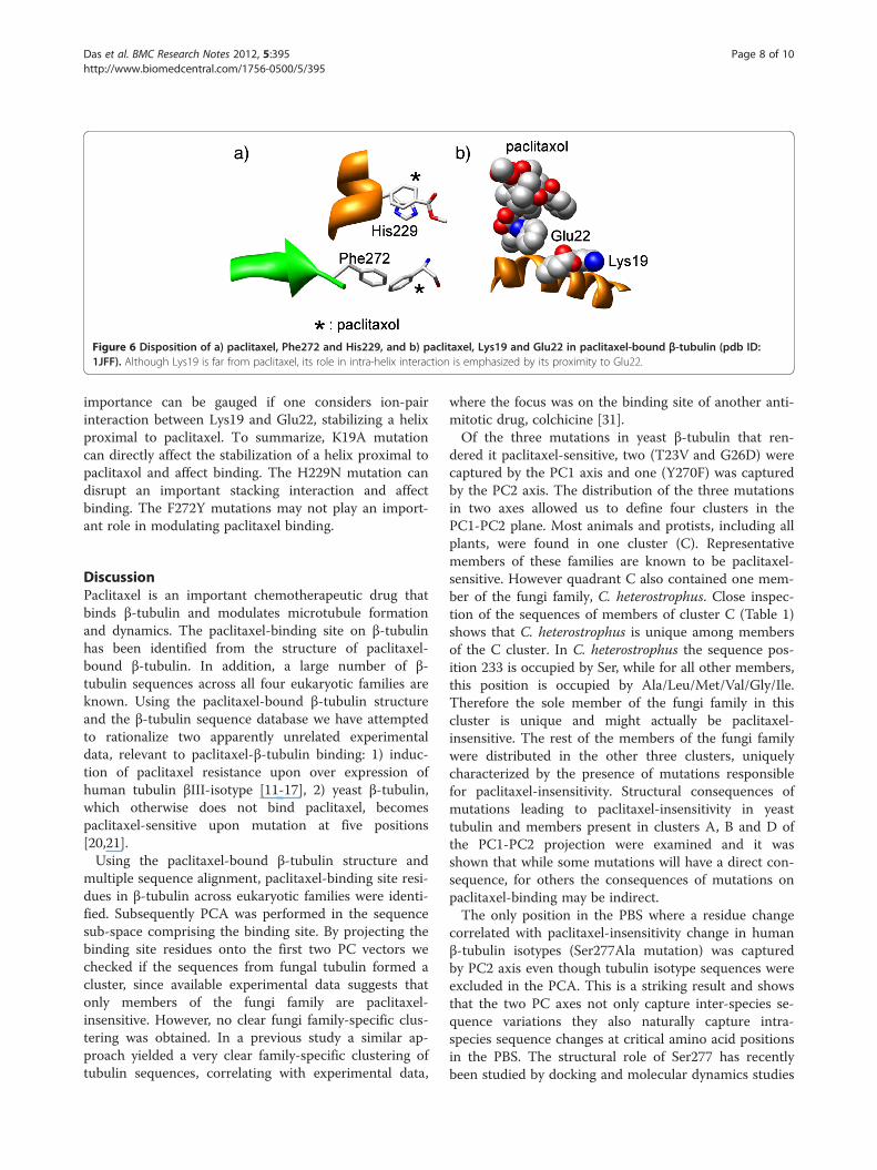
Figure 6 Disposition of a) paclitaxel, Phe272 and His229, and b) paclitaxel, Lys19 and Glu22 in paclitaxel-bound β-tubulin (pdb ID:1JFF). Although Lys19 is far from paclitaxel, its role in intra-helix interaction is emphasized by its proximity to Glu22.
Das et al. BMC Research Notes 2012, 5:395 Page 8 of 10http://www.biomedcentral.com/1756-0500/5/395
importance can be gauged if one considers ion-pairinteraction between Lys19 and Glu22, stabilizing a helixproximal to paclitaxel. To summarize, K19A mutationcan directly affect the stabilization of a helix proximal topaclitaxol and affect binding. The H229N mutation candisrupt an important stacking interaction and affectbinding. The F272Y mutations may not play an import-ant role in modulating paclitaxel binding.
DiscussionPaclitaxel is an important chemotherapeutic drug thatbinds β-tubulin and modulates microtubule formationand dynamics. The paclitaxel-binding site on β-tubulinhas been identified from the structure of paclitaxel-bound β-tubulin. In addition, a large number of β-tubulin sequences across all four eukaryotic families areknown. Using the paclitaxel-bound β-tubulin structureand the β-tubulin sequence database we have attemptedto rationalize two apparently unrelated experimentaldata, relevant to paclitaxel-β-tubulin binding: 1) induc-tion of paclitaxel resistance upon over expression ofhuman tubulin βIII-isotype [11-17], 2) yeast β-tubulin,which otherwise does not bind paclitaxel, becomespaclitaxel-sensitive upon mutation at five positions[20,21].Using the paclitaxel-bound β-tubulin structure and
multiple sequence alignment, paclitaxel-binding site resi-dues in β-tubulin across eukaryotic families were identi-fied. Subsequently PCA was performed in the sequencesub-space comprising the binding site. By projecting thebinding site residues onto the first two PC vectors wechecked if the sequences from fungal tubulin formed acluster, since available experimental data suggests thatonly members of the fungi family are paclitaxel-insensitive. However, no clear fungi family-specific clus-tering was obtained. In a previous study a similar ap-proach yielded a very clear family-specific clustering oftubulin sequences, correlating with experimental data,
where the focus was on the binding site of another anti-mitotic drug, colchicine [31].Of the three mutations in yeast β-tubulin that ren-
dered it paclitaxel-sensitive, two (T23V and G26D) werecaptured by the PC1 axis and one (Y270F) was capturedby the PC2 axis. The distribution of the three mutationsin two axes allowed us to define four clusters in thePC1-PC2 plane. Most animals and protists, including allplants, were found in one cluster (C). Representativemembers of these families are known to be paclitaxel-sensitive. However quadrant C also contained one mem-ber of the fungi family, C. heterostrophus. Close inspec-tion of the sequences of members of cluster C (Table 1)shows that C. heterostrophus is unique among membersof the C cluster. In C. heterostrophus the sequence pos-ition 233 is occupied by Ser, while for all other members,this position is occupied by Ala/Leu/Met/Val/Gly/Ile.Therefore the sole member of the fungi family in thiscluster is unique and might actually be paclitaxel-insensitive. The rest of the members of the fungi familywere distributed in the other three clusters, uniquelycharacterized by the presence of mutations responsiblefor paclitaxel-insensitivity. Structural consequences ofmutations leading to paclitaxel-insensitivity in yeasttubulin and members present in clusters A, B and D ofthe PC1-PC2 projection were examined and it wasshown that while some mutations will have a direct con-sequence, for others the consequences of mutations onpaclitaxel-binding may be indirect.The only position in the PBS where a residue change
correlated with paclitaxel-insensitivity change in humanβ-tubulin isotypes (Ser277Ala mutation) was capturedby PC2 axis even though tubulin isotype sequences wereexcluded in the PCA. This is a striking result and showsthat the two PC axes not only capture inter-species se-quence variations they also naturally capture intra-species sequence changes at critical amino acid positionsin the PBS. The structural role of Ser277 has recentlybeen studied by docking and molecular dynamics studies

Das et al. BMC Research Notes 2012, 5:395 Page 9 of 10http://www.biomedcentral.com/1756-0500/5/395
of βI and βIII tubulin isotypes and their complexes withantimitotic agents, where it was shown that Ser277 playsa crucial role in intra-loop interaction critical for pacli-taxel binding [32]. We showed that Ser277 also plays animportant role in inter-loop interaction that can affectpaclitaxel binding. Despite being a critical residue,Ser277Ala mutation can still allow paclitaxel binding,provided there are alternate mechanisms of loop-loopinteraction, as evident from mutated yeast tubulin wherea Ser277Ala mutation is compensated by Arg278Ile mu-tation, giving rise to a three-center hydrophobic staple.In addition, human βVI tubulin isotype, which also con-tains Ala277, may actually be paclitaxel sensitive due toother compensatory mutations (Arg278Gln, Ala233Leu).
ConclusionsIn summary, we have rationalized two apparently unre-lated experimental data — differential paclitaxel bindingamong mammalian β-tubulin isotypes and that betweenwild type and yeast β-tubulin mutated at six positions —in terms of two collective sequence vectors, PC1 andPC2, spanning the PBS. PCA has previously been suc-cessful in predicting protein functional residues [33] andrationalizing the differential colchicine-binding ofanimal-tubulins [31]. The present analysis extends theapplicability of PCA to paclitaxel-tubulin interactionwith valuable insights and specific predictions.
MethodsA total of 125 β-tubulin (animal: 38, fungi: 29; protists:30; plant: 28) sequences were used in this work. The se-quence accession numbers and the names of the corre-sponding organisms are given in Tables 1, 2, 3, 4. Thestructures for paclitaxel-bound (PDB ID: 1JFF) [23] andpaclitaxel-free (PDB ID: 1SA0) [34] tubulin were down-loaded from the protein data bank [35]. Structural ana-lysis was performed using the program Chimera [36].The primary paclitaxel binding site (PBS) was defined byresidues in 1JFF that were within 5 Å of paclitaxel (dis-tance between atoms of paclitaxel and any heavy atomof tubulin). Amino acid residues within 5 Å from thebound paclitaxel molecule are given in Figure 1. Mul-tiple sequence alignments were performed using theprogram ClustalW [24] to identify PBS residues in all β-tubulin sequences corresponding to that defined in 1JFF.Subsequently, Principal Component Analysis (PCA)
was performed in the multi-dimensional sequence-spaceof PBS, as previously described [31]. Each sequence pos-ition was represented by a binary-vector (of length 21),where the first twenty elements represent the occurrenceof a particular amino acid (1 for presence and 0 for ab-sence), and the last element represents the presence of agap. For example, Ala is represented by {1, 0, 0, 0, 0, 0,0, 0, 0, 0, 0, 0, 0, 0, 0, 0, 0, 0, 0, 0, 0} and a gap is
represented by {0, 0, 0, 0, 0, 0, 0, 0, 0, 0, 0, 0, 0, 0, 0, 0, 0,0, 0, 0, 1}. The PBS residues (22 residue positions, ofwhich 14 positions showed amino acid variations) fromeach β-tubulin sequence (a total of M sequences) wasrepresented by a binary-vector {aik} where the i-th indexrepresents a particular β-tubulin sequence (i= 1, M) andthe k-th index runs from 1 to N (N= 14 × 21). The N ×N variance-covariance matrix was prepared as:
Ckl ¼XM
i¼1
aik � �akð Þ ail � �alð Þ= M � 1ð Þ ð1Þ
where āk and āl are given by:
�ak ¼XM
i¼1
aik=M
�al ¼XM
i¼1
ail=M
ð2Þ
In Eq. 1, the denominator (M - 1), instead of M, isused to yield an unbiased estimator of the (co)variance.The variance-covariance matrix was diagonalized yield-ing N principal component vectors (eigen vectors ! V k ;k= 1, N) and N eigen values (mean square variationassociated with each eigen vector) λk (k= 1, N). The pro-jection of the i-th β-tubulin PBS sequence on the k-thPC vector ! V k was calculated from the dot product ofi-th sequence vector { ai1; ai2 . . . aiN } and the k-th PCvector {vk1; vk2 . . . vkN }.
Competing interestsThe authors declare no competing interests.
Authors’ contributionsGB and BB conceived of the study, GB and LD participated in its design andperformed the sequence analysis, and all authors analyzed the data and GBdrafted the manuscript. All authors read and approved the final manuscript.
AcknowledgementsFinancial assistance for this work came from Department of Science andTechnology, India.
Author details1Department of Biochemistry, Bose Institute, P-1/12 CIT Scheme VIIM, Kolkata70054, India. 2Department of Biophysics, Bose Institute, P-1/12 CIT SchemeVIIM, Kolkata 70054, India.
Received: 10 April 2012 Accepted: 19 July 2012Published: 1 August 2012
References1. Avila J: Microtubule dynamics. FASEB J 1990, 4:3284–3290.2. Havercroft JC, Cleveland DW: Programmed expression of beta-tubulin
genes during development and differentiation of the chicken. J Cell Biol1984, 99:1927–1935.
3. Fulton C, Simpson PA: Selective synthesis and utilization of flagellartubulin. The multi-tubulin hypothesis. In Cell Motility. Edited by GoldmanR, Pollard T, Rosenbaum J. Cold Spring Harbor, NY: Cold Spring HarborLaboratory Press; 1976:987–1006.
4. Banerjee A, Luduena RF: Distinct colchicine binding kinetics of bovinebrain tubulin lacking the type III isotype of beta-tubulin. J Biol Chem1991, 266:1689–1691.

Das et al. BMC Research Notes 2012, 5:395 Page 10 of 10http://www.biomedcentral.com/1756-0500/5/395
5. Banerjee A, Luduena RF: Kinetics of colchicine binding to purified beta-tubulin isotypes from bovine brain. J Biol Chem 1992, 267:13335–13339.
6. Schiff PB, Horwitz SB: Taxol stabilizes microtubules in mouse fibroblastcells. Proc Natl Acad Sci USA 1980, 77:1561–1565.
7. Horwitz SB: Mechanism of action of taxol. Trends Pharmacol Sci 1992,13:134–136.
8. Jordan M, Toso RJ, Thrower D, Wilson L: Mechanism of mitotic block andinhibition of cell proliferation by taxol at low concentrations. Proc NatlAcad Sci USA 1993, 90:9552–9556.
9. McGuire WP, Rowinsky EK, Rosenshein NB, Grumbine FC, Ettinger DS,Armstrong DK, Donehower RC: Taxol: a unique antineoplastic agent withsignificant activity in advanced ovarian epithelial neoplasms. Ann InternMed 1989, 111:273–279.
10. Holmes FA, Walters RS, Theriault RL, Forman AD, Newton LK, Raber MN,Buzdar AU, Frye DK, Hortobagyi GN: Phase II trial of taxol, an active drugin the treatment of metastatic breast cancer. J Natl Cancer Inst 1991,83:1797–1805.
11. Kavallaris M, Burkhart CA, Horwitz SB: Antisense oligonucleotides to classIII beta-tubulin sensitize drug-resistant cells to Taxol. Br J Cancer 1999,80:1020–1025.
12. Hari M, Yang H, Zeng C, Canizales M, Cabral F: Expression of class III beta-tubulin reduces microtubule assembly and confers resistance topaclitaxel. Cell Motil Cytoskeleton 2003, 56:45–56.
13. Kamath K, Wilson L, Cabral F, Jordan MA: BetaIII-tubulin induces paclitaxelresistance in association with reduced effects on microtubule dynamicinstability. J Biol Chem 2005, 280:12902–12907.
14. Panda D, Miller HP, Banerjee A, Ludueña RF, Wilson L: Microtubuledynamics in vitro are regulated by the tubulin isotype composition. ProcNatl Acad Sci USA 1994, 91:11358–11362.
15. Verdier-Pinard P, Wang F, Martello L, Burd B, Orr GA, Horwitz SB: Analysis oftubulin isotypes and mutations from taxol-resistant cells by combinedisoelectrofocusing and mass spectrometry. Biochemistry 2003, 42:5349–5357.
16. Dumontet C, Isaac S, Souquet PJ, Bejui-Thivolet F, Pacheco Y, Peloux N,Frankfurter A, Luduena R, Perol M: Expression of class III beta tubulin innon-small cell lung cancer is correlated with resistance to taxanechemotherapy. Bull Cancer 2005, 92:E25–E30.
17. Mozzetti S, Ferlini C, Concolino P, Filippetti F, Raspaglio G, Prislei S, Gallo D,Martinelli E, Ranelletti FO, Ferrandina G, Scambia G: Class III beta-tubulinoverexpression is a prominent mechanism of paclitaxel resistance inovarian cancer patients. Clin Cancer Res 2005, 11:298–305.
18. Barnes G, Louie KA, Botstein D: Yeast proteins associated withmicrotubules in vitro and in vivo. Mol Biol Cell 1992, 3:29–47.
19. Bode CJ, Gupta ML Jr, Reiff EA, Suprenant KA, Georg GI, Himes RH:Epothilone and paclitaxel: unexpected differences in promoting theassembly and stabilization of yeast microtubules. Biochemistry 2002,41:3870–3874.
20. Gupta ML Jr, Bode CJ, Georg GI, Himes RH: Understanding tubulin-Taxolinteractions: mutations that impart Taxol binding to yeast tubulin. ProcNatl Acad Sci USA 2003, 100:6394–6397.
21. Entwistle RA, Winefield RD, Foland TB, Lushington GH, Himes RH: Thepaclitaxel site in tubulin probed by site-directed mutagenesis ofSaccharomyces cerevisiae beta-tubulin. FEBS Lett 2008, 582:2467–2470.
22. Nogales E, Wolf SG, Downing KH: Structure of the alpha beta tubulindimer by electron crystallography. Nature 1998, 391:199–203.
23. Lowe J, Li H, Downing KH, Nogales E: Refined structure of alpha beta-tubulin at 3.5 Å resolution. J Mol Biol 2001, 313:1045–1057.
24. Thompson JD, Higgins DG, Gibson TJ: CLUSTAL W: improving thesensitivity of progressive multiple sequence alignment throughsequence weighting, position-specific gap penalties and weight matrixchoice. Nucl Acids Res 1994, 22:4673–4680.
25. Crooks GE, Hon G, Chandonia JM, Brenner SE: WebLogo: a sequence logogenerator. Gen Res 2004, 14:1188–1190.
26. Parness J, Horwitz SB: Taxol stabilizes microtubules in mouse fibroblastcells. J Cell Biol 1981, 91:479–487.
27. Morejohn LC, Fosket DE: Taxol-induced rose microtubule polymerizationin vitro and its inhibition by colchicine. J Cell Biol 1984, 99:141–147.
28. Hugdahl JD, Bokros CL, Hanesworth VR, Aalund GR, Morejohn LC: Uniquefunctional characteristics of the polymerization and MAP bindingregulatory domains of plant tubulin. Plant Cell 1993, 5:1063–1080.
29. Gardiner JC, Harper JD, Weerakoon ND, Collings DA, Ritchie S, Gilroy S, CyrRJ, Marc J: A 90-kD phospholipase D from tobacco binds to microtubulesand the plasma membrane. Plant Cell 2001, 13:2143–2158.
30. Werbovetz KA, Brendle JJ, Sackett DL: Purification, characterization, anddrug susceptibility of tubulin from Leishmania. Mol Biochem Parasitol1999, 98:53–65.
31. Banerjee M, Roy D, Bhattacharyya B, Basu G: Differential colchicine-bindingacross eukaryotic families: the role of highly conserved Pro268beta andAla248beta residues in animal tubulin. FEBS Lett 2007, 581:5019–5023.
32. Magnani M, Ortuso F, Soro S, Alcaro S, Tramontano A, Botta M: The betaI/betaIII-tubulin isoforms and their complexes with antimitotic agents.Docking and molecular dynamics studies. FEBS J 2006, 273:3301–3310.
33. Casari G, Sander C, Valencia A: A method to predict functional residues inproteins. Nat Struct Biol 1995, 2:171–178.
34. Ravelli RB, Gigant B, Curmi PA, Jourdain I, Lachkar S, Sobel A, Knossow M:Insight into tubulin regulation from a complex with colchicine and astathmin-like domain. Nature 2004, 428:198–202.
35. Berman HM, Westbrook J, Feng Z, Gilliland G, Bhat TN, Weissig H,Shindyalov IN, Bourne PE: The protein data bank. Nucleic Acids Res 2000,28:235–241.
36. Pettersen EF, Goddard TD, Huang CC, Couch GS, Greenblatt DM, Meng EC,Ferrin TE: UCSF Chimera - a visualization system for exploratory researchand analysis. J Comput Chem 2004, 25:1605–1612.
doi:10.1186/1756-0500-5-395Cite this article as: Das et al.: Rationalization of paclitaxel insensitivity ofyeast β-tubulin and human βIII-tubulin isotype using principalcomponent analysis. BMC Research Notes 2012 5:395.
Submit your next manuscript to BioMed Centraland take full advantage of:
• Convenient online submission
• Thorough peer review
• No space constraints or color figure charges
• Immediate publication on acceptance
• Inclusion in PubMed, CAS, Scopus and Google Scholar
• Research which is freely available for redistribution
Submit your manuscript at www.biomedcentral.com/submit