Lower birth weight as a critical effect of chlorpyrifos: A comparison of human and animal data

TOXICOLOGICAL SCIENCES 115(1), 183–193 (2010)
doi:10.1093/toxsci/kfq032
Advance Access publication February 8, 2010
Mice Treated with Chlorpyrifos or Chlorpyrifos Oxon HaveOrganophosphorylated Tubulin in the Brain and Disrupted MicrotubuleStructures, Suggesting a Role for Tubulin in Neurotoxicity Associated
with Exposure to Organophosphorus Agents
Wei Jiang,* Ellen G. Duysen,* Heidi Hansen,* Luda Shlyakhtenko,† Lawrence M. Schopfer,* and Oksana Lockridge*,1
*Eppley Institute, University of Nebraska Medical Center, Omaha, Nebraska 68198-5950; and †Department of Pharmaceutical Sciences, University of NebraskaMedical Center, Omaha, Nebraska 68198-6025
1 To whom correspondence should be addressed. Fax: (402) 559-4651. E-mail: [email protected].
Received November 30, 2009; accepted January 22, 2010
Exposure to organophosphorus (OP) agents can lead to learning
and memory deficits. Disruption of axonal transport has been
proposed as a possible explanation. Microtubules are an essential
component of axonal transport. In vitro studies have demonstrated
that OP agents react with tubulin and disrupt the structure of
microtubules. Our goal was to determine whether in vivo exposure
affects microtubule structure. One group of mice was treated daily
for 14 days with a dose of chlorpyrifos that did not significantly
inhibit acetylcholinesterase. Beta-tubulin from the brains of these
mice was diethoxyphosphorylated on tyrosine 281 in peptide
GSQQY281RALTVPELTQQMFDSK. A second group of mice was
treated with a single sublethal dose of chlorpyrifos oxon (CPO).
Microtubules and cosedimenting proteins from the brains of these
mice were visualized by atomic force microscopy nanoimaging and
by Coomassie blue staining of polyacrylamide gel electrophoresis
bands. Proteins in gel slices were identified by mass spectrometry.
Nanoimaging showed that microtubules from control mice were
decorated with many proteins, whereas microtubules from CPO-
treated mice had fewer associated proteins, a result confirmed
by mass spectrometry of proteins extracted from gel slices. The
dimensions ofmicrotubules fromCPO-treatedmice (height 8.7± 3.1nm and width 36.5 ± 15.5 nm) were about 60% of those from control
mice (height 13.6 ± 3.6 nm and width 64.8 ± 15.9 nm). A third group
of mice was treated with six sublethal doses of CPO over 50.15 h.
Mass spectrometry identified diethoxyphosphorylated serine 338 in
peptide NS338NFVEWIPNNVK of beta-tubulin. In conclusion,
microtubules from mice exposed to chlorpyrifos or to CPO have
covalently modified amino acids and abnormal structure, suggest-
ing disruption of microtubule function. Covalent binding of CPO to
tubulin and to tubulin-associated proteins is a potential mechanism
of neurotoxicity.
Key Words: chlorpyrifos; tubulin; mass spectrometry;
nanoimaging; neurotoxicity.
Organophosphorus (OP) agents are used as pesticides, in jet
engine oil as a flame retardant, as chemical warfare agents, and
for treatment of Schistosomiasis. The acute toxicity that occurs
with high doses of OP is due to inhibition of acetylcholines-
terase (AChE). Some people who survive acute exposure
describe neurological symptoms that last long after the acute
symptoms have abated. The sarin attack in the Tokyo subway
occurred in 1995, but 5 years later, some victims of this attack
still suffered from blurred vision, easy fatigability, difficulty in
concentration, and insomnia (Kawada et al., 2005; Yanagisawa
et al., 2006). Magnetic resonance imaging revealed structural
changes in the brains of exposed subjects (Yamasue et al.,2007). These long-lasting symptoms could be the consequence
of seizure activity and anoxia initiated by acute inhibition of
AChE (McDonough and Shih, 1997).
Low doses of OP that do not inhibit AChE have also been
associated with neurological dysfunction, including clinically
significant extrapyramidal symptoms, anxiety, depression
(Salvi et al., 2003), memory loss, and learning disability
(Srivastava et al., 2000). The mechanism of low-dose toxicity
is not understood. One hypothesis to explain long-lasting
neurotoxicity from low doses of OP involves OP modification
of proteins in the axonal transport system (Gearhart et al.,2007; Gupta et al., 1997; Prendergast et al., 2007; Terry et al.,2007). The transport system moves organelles from the cell
nucleus to the axon termini and back to the nucleus. Hundreds
of proteins are present in the axoplasm of neurons (Rishal
et al., 2010), suggesting the involvement of a great many
proteins in this transport process.
Indirect evidence supports the hypothesis that organophos-
phorylation of key proteins in the axonal transport system
disrupts the transport mechanism in neurons (Gearhart et al.,2007; Terry et al., 2007). Such a disruption could result in loss of
synaptic contacts, slow dying back of axon structures, and finally
in neuron cell death as has been described by Morfini et al. (2009)
to explain neurodegenerative diseases, including Parkinson’s,
Alzheimer’s disease, and amyotrophic lateral sclerosis.
� The Author 2010. Published by Oxford University Press on behalf of the Society of Toxicology. All rights reserved.For permissions, please email: [email protected]
at University of N
ebraska Medical C
enter on Novem
ber 30, 2010toxsci.oxfordjournals.org
Dow
nloaded from

Rats treated chronically with low doses of chlorpyrifos have
decreased rates of axonal transport measured in sciatic nerves
ex vivo, implicating OP modification of tubulin, kinesin, and
microtubule-associated proteins in this dysfunction (Gearhart
et al., 2007; Terry et al., 2007). The present report focuses on
tubulin in brain because in vitro experiments have shown that
OP modification of tubulin disrupts polymerization of tubulin
into microtubules (Grigoryan and Lockridge, 2009; Prendergast
et al., 2007). Disruption of tubulin polymerization has been
shown to result in neuron dysfunction, cellular apoptosis, and
tissue damage (Conde and Caceres, 2009; Tierno et al., 2009).
Mass spectrometry analysis of a variety of proteins treated
with OP in vitro identified covalent binding of OP to tyrosine
and lysine residues (Grigoryan et al., 2008, 2009a,b,c). The
results suggest that almost any protein can be modified by
OP. Previously, OP adducts were thought to form only with
the active site serine of enzymes in the serine hydrolase
superfamily. In vivo studies have identified OP-tyrosine
adducts in the blood of guinea pigs treated with nerve agents
and have shown that the adducts are detectable 24 days after
exposure at a time when OP-serine adducts of butyrylcholi-
nesterase (BChE) are no longer detectable (Read et al., 2010).
In the present report, we treated mice with the pesticide
chlorpyrifos as well as with its active metabolite, chlorpyrifos
oxon (CPO). The pesticide chlorpyrifos is relatively harmless
to man and rodents because it is rapidly detoxified and
excreted. However, a portion of the chlorpyrifos is bioactivated
through oxidative desulfuration catalyzed by cytochrome P450
enzymes to form CPO, the toxic agent that inhibits AChE
(Sams et al., 2004; Tang et al., 2001) (see Fig. 1).
Our goal was to identify alterations in microtubule structure
following in vivo treatment of mice with chronic low doses of
chlorpyrifos and with sublethal doses of CPO. This is the first
report to identify structural deficits in microtubules of animals
treated in vivo with OP and furthermore to provide mass
spectrometry evidence for OP labeling of tubulin in mice
treated in vivo with chlorpyrifos or CPO.
MATERIALS AND METHODS
Materials. Chlorpyrifos and CPO (PS-674 and MET-674B; ChemService
Inc., West Chester, PA) were stored at �80�C. Guanosine 5#-triphosphate
(GTP) sodium salt hydrate (G8877) � 95% HPLC pure powder and 1,4-
piperazinediethanesulfonic acid (PIPES, P6757) > 99% pure powder were from
Sigma (St Louis, MO). Sequencing grade–modified trypsin (V5113) was
from Promega (Madison, WI). Alpha-cyano-4-hydroxycinnamic acid was from
Applied Biosystems (MDS Sciex; Foster City, CA). All other chemicals were
of analytical grade.
Animal. All animal work was conducted in accordance with the Guide for
the Care and Use of Laboratory Animals as adopted by the National Institutes
of Health. Formal approval to conduct the experiments was obtained from the
Institutional Animal Care and Use Committee of the University of Nebraska
Medical Center. Adult wild-type mice of strain 129Sv were used in all trials.
Mice were bred at the University of Nebraska Medical Center.
Adult female mice treated for 14 days with a low dose of chlorpyrifos
(n ¼ 4) (85.6 ± 11.2 days of age) weighed an average of 19.8 ± 3.2 g, while
female control mice (n ¼ 4) (90.1 ± 5.8 days of age) weighed an average of
18.2 ± 1.4 g. Adult male mice treated with a single sublethal dose of CPO
(n ¼ 3) (72 days of age) weighed an average of 24.6 ± 1.4 g, and control male
mice (n ¼ 3) (72 days of age) weighed an average of 23.8 ± 1.9 g. Two female
mice were treated with six sublethal doses of CPO over a period of 50.15 h
(127 days of age, 23.0 g), and two control female mice (127 days of age, 22.6 g)
were treated at the same time points with an equivalent volume of ethanol.
Chronic low-dose treatment of mice (n ¼ 4) with chlorpyrifos. Female
mice (n ¼ 4) were injected sc with 3 mg/kg chlorpyrifos dissolved in 3%
dimethyl sulfoxide/97% corn oil. Each mouse was treated for 14 consecutive
days between 10 and 11 A.M. Surface body temperature, body weight, and
observations were made every 5 min through 15 min and at 30-min postdosing.
The axial body temperature was measured with a digital thermometer,
Thermalert model TH-5, and a surface Microprobe MT-D, Type T
thermocouple (Physitemp Instruments Inc., Clifton, NJ). Blood was collected
(50 ll) via the saphenous vein prior to dosing each day. On day 15, 24 h after
the final dose, the mice were decapitated, plasma was collected, and the brains
placed on ice. Brains were weighed and processed according to the tubulin
purification protocol. The tubulin was examined for OP modifications.
Single-dose treatment of mice (n ¼ 3) with chlorpyrifos. In three separate
experiments, a mouse was injected ip with CPO dissolved in ethanol at a dose
of 3.0 mg/kg delivered in 50 ll. A control mouse was injected with ethanol
only. Animals were observed for signs of toxicity following injection. About
30-min postinjection, the mice were euthanized and perfused with 50 ml of
0.1M PBS to remove blood from tissues. Tubulin from the brains of these
animals was purified and used for nanoimaging and SDS gel electrophoresis.
Proteins in the SDS bands were analyzed mass spectrally.
Treatment of mice (n ¼ 2) with six doses of CPO. To derive samples for
mass spectral analysis of CPO-labeled tubulin, mice (n ¼ 2) were injected ip
with CPO dissolved in ethanol at a dose of 2.5 mg/kg delivered in 50 ll.
Control mice (n ¼ 2) were injected with ethanol only. Each mouse received six
injections at 0, 1, 22, 48, 50, and 50.15 h. Animals were observed for signs of
toxicity after each injection. Mice were euthanized 5 min after the last dose by
inhalation of carbon dioxide and then perfused via intracardial injection with
50 ml 0.1M PBS solution to remove blood from the tissues. Tubulin from the
brains of these animals was examined for OP modifications.
Enzyme activity assays. AChE and BChE activities in mouse plasma and
brain were measured with 1mM acetylthiocholine and 1mM butyrylthiocholine,
respectively, as described (Peeples et al., 2005). Carboxylesterase activity in
mouse plasma was assayed with 2.5mM p-nitrophenyl acetate after incubating
3 ll plasma for 5 min in 0.1M potassium phosphate (pH 7.0) containing
12.5mM EDTA, to inhibit paraoxonase, and 0.01mM eserine, to inhibit AChE
and BChE. The reaction was started by adding 0.05 ml of 0.1M p-nitrophenyl
acetate dissolved in methanol. The total reaction volume was 2.0 ml, and the
temperature was 25�C. Absorbance increase at 400 nm was measured on
a Gilford spectrophotometer and recorded on a computer interfaced to MacLab/
200 (AD Instruments, Sydney, Australia). Activity in micromoles per minute
was calculated from the extinction coefficient for p-nitrophenol of 9000M-1cm-1.
FIG. 1. Chlorpyrifos is bioactivated to CPO by cytochrome P450. Results
presented below show that the oxon reacts with tubulin by donating its
diethoxyphosphate group to tyrosine 281 and to serine 338 of beta-tubulin in
mouse brain. Each diethoxyphosphate group adds a mass of 136 amu to
tubulin. The 3,5,6-trichloro-2-(O)-pyridine group is released from CPO and
excreted.
184 JIANG ET AL.
at University of N
ebraska Medical C
enter on Novem
ber 30, 2010toxsci.oxfordjournals.org
Dow
nloaded from

Tubulin purification and polymerization. Tubulin from mouse brains was
purified by two cycles of polymerization using the protocol of Shelanski et al.
(1973). Brains from mice were collected and placed on ice. The brains were
homogenized in four volumes of ice-cold PM buffer (80mM PIPES, 2mM
magnesium chloride, and 0.5mM ethylene glycol tetraacetic acid (EGTA), pH
7.0) and centrifuged at 17,000 3 g for 60 min at 4�C. The pellet was discarded,
and the clear supernatant was transferred to a clean ultracentrifuge tube. The
supernatant, in which no microtubules were present, was mixed with an equal
volume of 8M glycerol containing 1.0mM GTP. The samples were incubated for
2 h at 37�C to assemble the microtubules. The solution was centrifuged for 60 min
at 100,0003 g at 29�C to pellet the microtubules and remove the polymerization
inducers glycerol and GTP. The pellet was suspended in 500 ll PM buffer,
depolymerized on ice for 30 min, and centrifuged for 1 h at 4�C to remove
particulates. Tubulin in the supernatant was mixed with an equal volume of 8M
glycerol containing 1.0mM GTP and incubated for 45 min at 37�C to assemble the
microtubules for a second time. The sample was centrifuged for 60 min at 29�Cto pellet the microtubules. The pellet was resuspended in 1 ml of PM buffer
and maintained at room temperature to preserve the microtubules. The
protein concentration of the final microtubule pellet ranged from 3 to 7 lg/ll
(depending on the preparation) as measured by absorbance at 280 nm using the
NanoDrop spectrophotometer ND-1000 (NanoDrop Technologies Inc.; Thermo
Scientific, Waltham, MA). Absorbance at 280 nm was compared to protein
concentration estimated from the Pierce bicinchoninic acid protein assay. Both
methods gave similar results. Bovine albumin was used as the standard for
both assays.
Atomic force microscopy of microtubules. Atomic force microscopy
nanoimages were acquired in the Nanoimaging Core Facility at the University
of Nebraska Medical Center, codirected by L.S. A portion of the microtubule
pellet, prepared by two cycles of assembly, was fixed immediately by addition
of glutaraldehyde to 0.25%. The fixed microtubules were stored at 4�C for 12 h
before an aliquot was diluted 10-fold with double-distilled water to a protein
concentration of 0.5 lg/ll. Ten microliters of the suspension was drawn from
the bottom of the tube and placed in the center of a mica chip that had been
treated with 1-(3-aminopropyl) silatrane. The detailed procedure for mica
surface modification is given in Shlyakhtenko et al. (2003) and Lyubchenko
and Shlyakhtenko (2009). The silatrane modification allows the sample to be
deposited in a wide range of ionic strengths and pH. After 2-min incubation in
a humid chamber, the samples were rinsed with deionized water and dried with
argon gas flow. Images were acquired using an MFP-3DAtomic Force
Microscope (Asylum Research, Santa Barbara, CA) operated in tapping mode.
Pictures were taken from 15 different 5 3 5 lm areas on average. Image
processing and measurement of microtubule height and width were performed
using Femtoscan software (Advanced Technology Center, Moscow, Russia).
Off-line HPLC purification of peptides for determination of labeled
proteins by mass spectrometry. A portion of the microtubule pellet was
denatured in 8M urea, reduced with 10mM dithiothreitol, and carbamidome-
thylated with 50mM iodoacetamide. The sample was diluted to 2M urea and
then digested with trypsin at a ratio of 50:1 (wt/wt) at 37�C for 16 h. The salt
concentration was reduced by dilution rather than by dialysis because dialysis
resulted in loss of up to 95% of the tubulin protein due to sticking to the dialysis
membrane. Peptides were desalted and separated by reverse phase HPLC on
a 100 3 4.60 mm Phenomenex C18 column eluted with a gradient from 0 to
60% (vol/vol) acetonitrile versus 0.1% (vol/vol) trifluoroacetic acid at a flow
rate of 1 ml/min for 60 min on a Waters 625 LC system. Fractions of 1 ml were
collected, dried by vacuum centrifugation, and redissolved in 50 ll of 5%
acetonitrile and 0.1% formic acid. Fractions that eluted between 5 and 40 min
were analyzed by liquid chromatography-tandem mass spectrometry (LC-
MSMS) electrospray ionization. The CPO-labeled peptide in Figure 4 was in
fraction 38, and the CPO-labeled peptide in Figure 7 was in fraction 30. Details
on the method for searching mass spectrometry data to identify labeled peptides
are in the ‘‘Materials and Methods’’ section entitled Q-Trap 4000 and LTQ-
Orbitrap mass spectrometry. In brief, the mass spectrometry data were
compared to the National Center for Biotechnology Information (NCBI)
nonredundant database using Mascot software. Diethoxyphosphate was used as
a variable modification for Ser, Thr, Tyr, and Lys. Each identified peptide was
manually examined to verify that the tandem mass spectral fragmentation
(MSMS) spectrum supported the Mascot assignment.
Polyacrylamide gel electrophoresis of proteins in the microtubule pellet,
followed by in-gel digestion. Two gradient polyacrylamide gels (4–30%)
were cast in a Hoefer gel apparatus to make 15 3 11–cm gels, 0.75-mm thick,
with a single well that spanned the entire gel. Microtubule pellets from control
and CPO-treated mouse brains in 400 ll PM buffer were denatured by addition
of 80 ll of 6 3 SDS gel loading buffer containing 0.2M Tris/Cl (pH 6.8), 10%
SDS, 30% glycerol, 0.6M dithiothreitol, and 0.012% bromophenol blue. The
samples were boiled for 3 min, and each was loaded onto a gel. About 2 mg of
protein were loaded onto each gel. Electrophoresis was for 3000 V/h at 4�C.
Gels were stained with Coomassie blue. Protein bands from the upper 75% of
each gel were cut into slices. Gel slices were destained, dried, treated with
trypsin, and the peptides extracted as previously described (Peeples et al.,
2005). The dry samples were dissolved in 80 ll of 5% acetonitrile and 0.1%
formic acid and placed in autosampler vials for analysis in the Q-Trap 4000
(MDS Sciex; Applied Biosystems) and the LTQ-Orbitrap (Thermo Scientific)
mass spectrometers.
Q-Trap 4000 and LTQ-Orbitrap mass spectrometry. Peptides purified by
off-line HPLC or extracted from gel slices were analyzed by LC-MSMS in the
Q-Trap 4000 linear ion trap mass spectrometer using a nanospray source with
a continuous flow head, at a flow rate of 0.3 ll/min (Li et al., 2009). The four
most intense peptides in each cycle of analysis were fragmented by collision-
induced dissociation using pure nitrogen at 40 l Torr and a collision energy of
20–50 V. Collision voltage was determined by the Analyst software based on
the size and charge state of the peptide. The data were searched against the
NCBI nonredundant database using Mascot v 1.9 (Matrix Science Ltd, London,
UK) for tryptic peptides with an added mass of 136 Da from CPO (Perkins
et al., 1999). The CPO added mass was put into the UNIMOD public database
of protein modifications (http://www.unimod.org) for use with Mascot. Mascot
search parameters included peptides with one missed tryptic cleavage,
carbamidomethylated cysteine as a fixed modification; diethoxyphosphorylated
serine, threonine, tyrosine, and lysine; and oxidized methionine as variable
modifications, a peptide mass tolerance of 1.2 Da and a fragment mass
tolerance of 0.6 Da. The MSMS fragmentation spectra of peptides identified by
Mascot were manually confirmed with the aid of the MS-Product algorithm
from Protein Prospector (v 5.3.2 from the University of California Mass
Spectrometry Facility). Peptides extracted from gel slices were also analyzed in
the LTQ-Orbitrap mass spectrometer as described (Wiederin et al., 2009).
Statistical analysis of microtubule nanoimaging parameters. A two-
independent sample t-test was performed to determine the statistical
significance of differences in the dimensions of the microtubules between the
treated and control groups. The data were normally distributed. SPSS software
16.0 (Microsoft Corp., Redmond, WA) was used for statistical analysis.
RESULTS
Mice Treated Chronically with Low Doses of Chlorpyrifos
Nontoxic dose of chlorpyrifos. Mice injected daily for 14
consecutive days with a nontoxic dose of chlorpyrifos (not the
oxon), 3 mg/kg sc, showed no signs of toxicity with the
exception of hunched posture, which persisted for less than
15 min after treatment. Their body temperature and body
weight remained normal (see Fig. 2).
Plasma AChE activity dropped from 0.3 to 0.2 U/ml after the
first two doses of chlorpyrifos but rebounded to 0.45 U/ml after
9–14 days of treatment (see Fig. 3A). In contrast, plasma BChE
MICROTUBULE STRUCTURE DISRUPTED IN MICE 185
at University of N
ebraska Medical C
enter on Novem
ber 30, 2010toxsci.oxfordjournals.org
Dow
nloaded from

activity steadily declined following each injection of chlorpyr-
ifos (Fig. 3B). This result is consistent with the greater
sensitivity of BChE to inhibition by CPO compared to that of
AChE (Amitai et al., 1998). Plasma carboxylesterase activity
was unaffected (Fig. 3C). Since inhibition of BChE has no
health consequences and since AChE activity was not inhibited
by the 14th day of treatment, it is concluded that the dosing
regimen was nontoxic.
Mass spectrometry identifies CPO-modified tubulin. Tubu-
lin purified from the brains of mice treated with a daily low
dose of chlorpyrifos was digested with trypsin and analyzed by
LC-MSMS. The mass spectrometry data were searched for
diethoxyphosphorylated peptides with an added mass of 136
amu on tyrosine, serine, threonine, or lysine. One modified
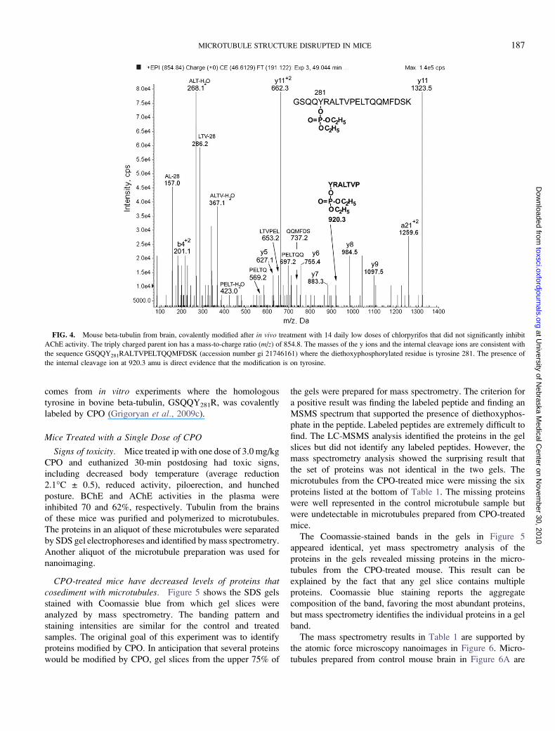
peptide was found. Figure 4 shows the MSMS spectrum for
peptide GSQQY281RALTVPELTQQMFDSK from mouse
beta-tubulin, covalently modified on tyrosine 281 by diethox-
yphosphate. The triply charged parent ion has a mass-to-charge
ratio of 854.8, which is consistent with the peptide sequence
plus 136 amu from the diethoxyphosphate. The y5 to y11 ions
and the internal fragments support the sequence. Internal
fragments are at 157.0 (AL minus 28), 268.1 (ALT minus
water), 286.2 (LTV minus 28), 367.1 (ALTV minus water),
423.0 (PELT minus water), 569.2 (PELTQ), 653.2 (LTVPEL),
697.2 (PELTQQ), 737.2 (QQMFDS), and 920.3 amu
(YRALTVP diethoxyphosphorylated on Y).
Support for modification of tyrosine comes from the internal
fragment at 920.3 amu, which corresponds to the sequence
Y*RALTVP where Y has an added mass of 136 amu. The
threonine in peptide YRALTVP can be ruled out as the site of
diethoxyphosphorylation because internal fragments that in-
clude this threonine (ALT minus water, LTV minus 28, ALTV
minus water, and LTVPEL) do not have an added mass of 136
amu. The second threonine in peptide GSQQY281RALTV-
PELTQQMFDSK can be ruled out because internal fragments
that include this threonine (PELT minus water, PELTQ, and
PELTQQ) do not have an added mass of 136 amu. The serine
near the C-terminus can be ruled out based on the mass of the
internal fragment QQMFDS. The serine near the N-terminal
can be ruled out based on the mass of the doubly charged b4
ion. Precedent for the reactivity of this tyrosine in tubulin
FIG. 3. Plasma esterase activity in mice (n ¼ 4) treated daily for 14 days
with chlorpyrifos, 3 mg/kg sc. (A) Plasma AChE activity. (B) Plasma BChE
activity. (C) Plasma carboxylesterase (CES) activity. Day 0 values are the
activities before treatment was begun. Units of activity are micromoles
substrate hydrolyzed per minute per microliter plasma. Error bars are SD for
four mice.
FIG. 2. Surface body temperature and body weight of mice treated daily
for 14 days sc with 3 mg/kg chlorpyrifos (not the oxon). Values for control
mice (n ¼ 4) treated with vehicle were indistinguishable from values for treated
mice (n ¼ 4). Day 0 values are the temperature and body weight before
treatment. Errors bars are SD.
186 JIANG ET AL.
at University of N
ebraska Medical C
enter on Novem
ber 30, 2010toxsci.oxfordjournals.org
Dow
nloaded from
comes from in vitro experiments where the homologous
tyrosine in bovine beta-tubulin, GSQQY281R, was covalently
labeled by CPO (Grigoryan et al., 2009c).
Mice Treated with a Single Dose of CPO
Signs of toxicity. Mice treated ip with one dose of 3.0 mg/kg
CPO and euthanized 30-min postdosing had toxic signs,
including decreased body temperature (average reduction
2.1�C ± 0.5), reduced activity, piloerection, and hunched
posture. BChE and AChE activities in the plasma were
inhibited 70 and 62%, respectively. Tubulin from the brains
of these mice was purified and polymerized to microtubules.
The proteins in an aliquot of these microtubules were separated
by SDS gel electrophoreses and identified by mass spectrometry.
Another aliquot of the microtubule preparation was used for
nanoimaging.
CPO-treated mice have decreased levels of proteins thatcosediment with microtubules. Figure 5 shows the SDS gels
stained with Coomassie blue from which gel slices were
analyzed by mass spectrometry. The banding pattern and
staining intensities are similar for the control and treated
samples. The original goal of this experiment was to identify
proteins modified by CPO. In anticipation that several proteins
would be modified by CPO, gel slices from the upper 75% of
the gels were prepared for mass spectrometry. The criterion for
a positive result was finding the labeled peptide and finding an
MSMS spectrum that supported the presence of diethoxyphos-
phate in the peptide. Labeled peptides are extremely difficult to
find. The LC-MSMS analysis identified the proteins in the gel
slices but did not identify any labeled peptides. However, the
mass spectrometry analysis showed the surprising result that
the set of proteins was not identical in the two gels. The
microtubules from the CPO-treated mice were missing the six
proteins listed at the bottom of Table 1. The missing proteins
were well represented in the control microtubule sample but
were undetectable in microtubules prepared from CPO-treated
mice.
The Coomassie-stained bands in the gels in Figure 5
appeared identical, yet mass spectrometry analysis of the
proteins in the gels revealed missing proteins in the micro-
tubules from the CPO-treated mouse. This result can be
explained by the fact that any gel slice contains multiple
proteins. Coomassie blue staining reports the aggregate
composition of the band, favoring the most abundant proteins,
but mass spectrometry identifies the individual proteins in a gel
band.
The mass spectrometry results in Table 1 are supported by
the atomic force microscopy nanoimages in Figure 6. Micro-
tubules prepared from control mouse brain in Figure 6A are
FIG. 4. Mouse beta-tubulin from brain, covalently modified after in vivo treatment with 14 daily low doses of chlorpyrifos that did not significantly inhibit
AChE activity. The triply charged parent ion has a mass-to-charge ratio (m/z) of 854.8. The masses of the y ions and the internal cleavage ions are consistent with
the sequence GSQQY281RALTVPELTQQMFDSK (accession number gi 21746161) where the diethoxyphosphorylated residue is tyrosine 281. The presence of
the internal cleavage ion at 920.3 amu is direct evidence that the modification is on tyrosine.
MICROTUBULE STRUCTURE DISRUPTED IN MICE 187
at University of N
ebraska Medical C
enter on Novem
ber 30, 2010toxsci.oxfordjournals.org
Dow
nloaded from

decorated with many proteins. In contrast, microtubules
prepared from the CPO-treated mouse brain in Figure 6B have
few attached proteins.
The microtubules from CPO-treated animals (in Fig. 6B)
appeared to be thinner than those from untreated animals (in
Fig. 6A). The difference was quantified by measuring the width
and height of 100 microtubules at three to four positions along
each tubule for a total of 700 measurements. Figure 6C shows
that the average width was 64.8 ± 15.9 nm for the control and
36.5 ± 15.5 nm for the CPO-treated microtubules. The average
height was 13.6 ± 3.6 nm for the control and 8.7 ± 3.1 nm for
the CPO-treated microtubules.
The atomic force microscopy nanoimaging and the mass
spectrometry experiments to identify proteins that cosediment
with microtubules were conducted by two researchers, each
finding similar results. It is unlikely that the missing proteins
were removed during purification as both control and CPO-
treated samples were handled identically, and the tubulin was
not exposed during the purification process to the high-salt
conditions, which remove microtubule-associated proteins
(Friden and Wallin, 1991).
Mice Treated with Six Sublethal Doses of CPO
Signs of toxicity. Mice treated ip with six doses of 2.5 mg/kg
CPO showed no toxic signs after injections at 0, 1, 22, 48, and
50 h. Following the injection at 50.15 h, CPO-treated mice had
hunched posture, piloerection, decreased activity, ataxic gait,
decrease in body temperature (average reduction 4.2�C ± 1.8),
and lacrimation. Mice were euthanized 5 min after the 50.15-h
FIG. 5. Coomassie blue–stained SDS gels. Tubulin isolated from the
brains of control and CPO-treated mice was polymerized to microtubules. The
microtubule preparations were denatured and loaded on the gels. Each 15-cm
wide gel was loaded with 2 mg of protein. A representative 1.5-cm portion of
each gel is shown. The upper 75% of each gel was sliced horizontally. Proteins
were extracted from the gel slices, digested with trypsin, and analyzed by mass
spectrometry.
TABLE 1
Proteins Identified in Control and CPO-Treated Mouse Brain
Microtubules
Protein
Molecular
weight
(kDa)
Accession
number
Control
Mowse
score
CPO
Mowse
score
Plectin 1 isoform 6 534 gi 41322931 78 110
Dynein, cytoplasmic,
heavy chain 1
527 gi 148686723 1254 589
Titin 300 gi 123232572 112 68
Microtubule-associated
protein 1A
300 gi 122065442 913 841
Microtubule-associated
protein 1B
271 gi 6678946 1066 1573
Microtubule-associated
protein 2
199 gi 126741 4982 4769
Microtubule-associated
protein 4
121 gi 148677083 423 271
mKIAA0325 protein 230 gi 28972155 1424 790
Neurofilament protein 115 gi 200022 964 295
Mtap4 protein 114 gi 29747932 432 308
Stop protein 96 gi 2769587 324 383
Ulip2 protein 62 gi 1915913 362 148
Neurofilament L 61 gi 387492 504 499
Tubulin beta 3 50 gi 12963615 600 672
Tubulin beta 1 50 gi 124430500 89 160
Tubulin beta 5 50 gi 7106439 826 803
Tubulin beta 6 50 gi 27754056 686 467
Tubulin alpha isotype
m-alpha 2
50 gi 202210 923 522
Tubulin alpha 1C 50 gi 6678469 920 672
Tubulin alpha 1B 50 gi 34740335 571 550
Tubulin alpha 1a 50 gi 6678465 909 403
Alpha-tubulin 50 gi 74182829 256 330
Tubulin alpha 8 50 gi 8394493 502 125
Microtubule-associated
protein tau
76 gi 13432200 739 374
Beta-tubulin 42 gi 202229 412 440
Tubulin polymerization–
promoting protein
24 gi 33469051 401 417
Cytoskeleton-associated
protein 5
218 gi 123227410 915 0
Myosin Va 215 gi 148694358 2097 0
Heat-shock protein 84 kDa 84 gi 309317 123 0
Dynein, cytoplasmic, 1
light intermediate chain
57 gi 22122795 422 0
Alpha-internexin 56 gi 94730353 543 0
Microtubule-associated
protein 2 isoform 1
53 gi 90186270 396 0
Note. Accession number is the identifying number in the NCBI
nonredundant protein database. Mowse scores are assigned by Mascot
software to indicate the probability that the assignment is correct. They are
the sum of the scores for all of the peptides associated with a particular protein.
A score of 69 or higher is considered to be a definitive assignment (p < 0.05).
188 JIANG ET AL.
at University of N
ebraska Medical C
enter on Novem
ber 30, 2010toxsci.oxfordjournals.org
Dow
nloaded from
dosing. BChE and AChE plasma activities were completely
inhibited 5 min after the 50.15-h injection. At this same time
point, BChE and AChE activities in the brain were inhibited
50 and 46%, respectively. Brains from these mice were the
source of the tubulin that yielded the CPO-labeled peptide in
Figure 7.
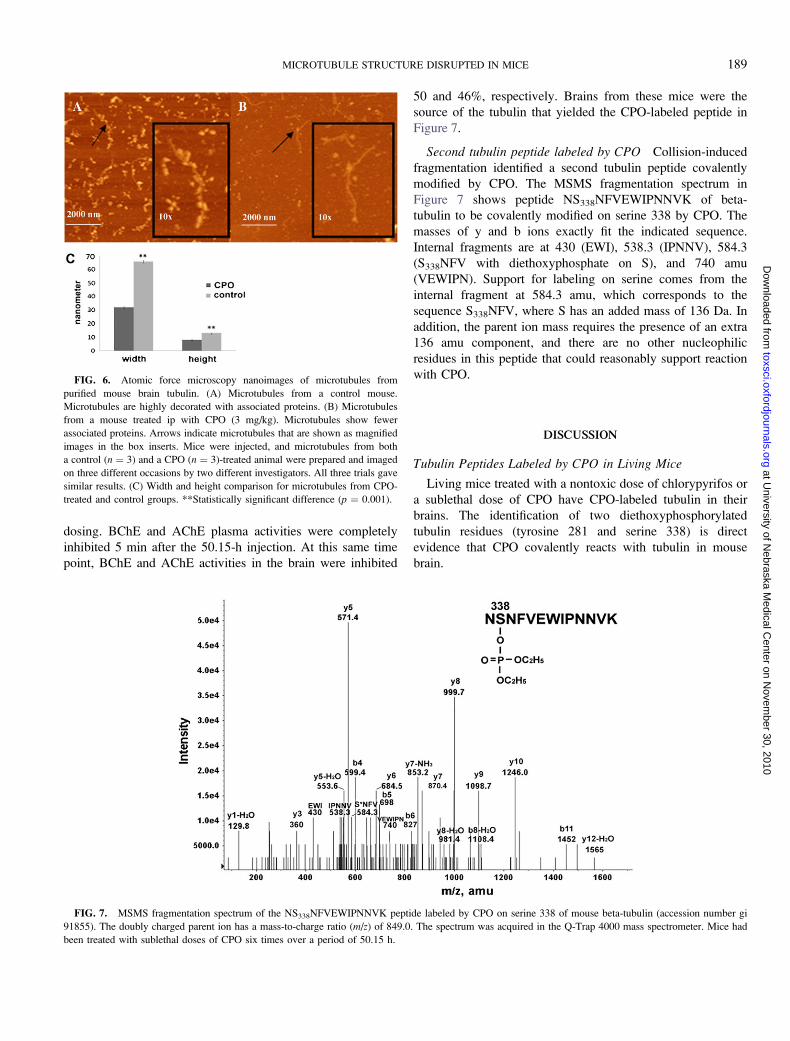
Second tubulin peptide labeled by CPO Collision-induced
fragmentation identified a second tubulin peptide covalently
modified by CPO. The MSMS fragmentation spectrum in
Figure 7 shows peptide NS338NFVEWIPNNVK of beta-
tubulin to be covalently modified on serine 338 by CPO. The
masses of y and b ions exactly fit the indicated sequence.
Internal fragments are at 430 (EWI), 538.3 (IPNNV), 584.3
(S338NFV with diethoxyphosphate on S), and 740 amu
(VEWIPN). Support for labeling on serine comes from the
internal fragment at 584.3 amu, which corresponds to the
sequence S338NFV, where S has an added mass of 136 Da. In
addition, the parent ion mass requires the presence of an extra
136 amu component, and there are no other nucleophilic
residues in this peptide that could reasonably support reaction
with CPO.
DISCUSSION
Tubulin Peptides Labeled by CPO in Living Mice
Living mice treated with a nontoxic dose of chlorypyrifos or
a sublethal dose of CPO have CPO-labeled tubulin in their
brains. The identification of two diethoxyphosphorylated
tubulin residues (tyrosine 281 and serine 338) is direct
evidence that CPO covalently reacts with tubulin in mouse
brain.
FIG. 6. Atomic force microscopy nanoimages of microtubules from
purified mouse brain tubulin. (A) Microtubules from a control mouse.
Microtubules are highly decorated with associated proteins. (B) Microtubules
from a mouse treated ip with CPO (3 mg/kg). Microtubules show fewer
associated proteins. Arrows indicate microtubules that are shown as magnified
images in the box inserts. Mice were injected, and microtubules from both
a control (n ¼ 3) and a CPO (n ¼ 3)-treated animal were prepared and imaged
on three different occasions by two different investigators. All three trials gave
similar results. (C) Width and height comparison for microtubules from CPO-
treated and control groups. **Statistically significant difference (p ¼ 0.001).
FIG. 7. MSMS fragmentation spectrum of the NS338NFVEWIPNNVK peptide labeled by CPO on serine 338 of mouse beta-tubulin (accession number gi
91855). The doubly charged parent ion has a mass-to-charge ratio (m/z) of 849.0. The spectrum was acquired in the Q-Trap 4000 mass spectrometer. Mice had
been treated with sublethal doses of CPO six times over a period of 50.15 h.
MICROTUBULE STRUCTURE DISRUPTED IN MICE 189
at University of N
ebraska Medical C
enter on Novem
ber 30, 2010toxsci.oxfordjournals.org
Dow
nloaded from

In previous studies with purified bovine tubulin, 17 peptides
from bovine alpha- and beta-tubulin were found to have been
labeled with CPO on tyrosine and lysine. Tyrosine 281 was the
most reactive residue in beta-tubulin (Grigoryan et al., 2009c).
However, no serine was found to be organophosphorylated by
CPO in bovine tubulin (Grigoryan et al., 2009c). When live
mice were treated with chlorpyrifos or CPO, the labeled tubulin
residues were tyrosine 281 and serine 338. No other residues
were found to be organophosphorylated in mouse tubulin. It is
possible that more sensitive methods will detect additional
labeled residues in the future.
Table 2 compares the amino acid sequences of bovine
and mouse tubulin peptides that include tyrosine 281 and
serine 338. The sequence of peptide GSQQY281RALTV-
PELTQQMFDSK is conserved in both species, bovine
differing from mouse by virtue of a single conservative change
(alanine to serine) at the second position from the C-terminus.
Tyrosine 281 is present in both bovine and mouse tubulin. Both
bovine beta-tubulin peptide NSSY440FVEWIPNNVK and
mouse beta-tubulin peptide NS338NFVEWIPNNVK have
serine at position 338, but only bovine tubulin has tyrosine at
position 340. Both peptides are labeled by CPO. The label in
bovine tubulin is on tyrosine 440. The difference in target at
this locus could reflect the greater nucleophilicity of tyrosine
compared to serine.
Seventeen sites on bovine tubulin were labeled with CPO
in vitro, while only two sites were labeled on mouse tubulin
in vivo. The larger overall number of labeling sites for bovine
tubulin in vitro could be partly explained by the fact that the
pure bovine tubulin purchased from Cytoskeleton Inc. had
been depleted of microtubule-associated proteins before it was
treated with CPO. Tyrosine sites available for reaction in vitromay be unavailable in vivo. The tyrosines might be protected
by interaction with other proteins or might already be
phosphorylated by kinases (Westermann and Weber, 2003).
Prior phosphorylation could mask other potential serine residue
targets in vivo.
The homologous sequences in human beta-tubulin are nearly
identical to those in bovine and mouse beta-tubulin. Sites of
labeling on human tubulin are unknown, but they would be
expected to be similar to those identified here.
Thin Microtubules and Decreased Proteins on TubulinIsolated from CPO-treated mice
Thirty-two proteins are listed in Table 1. Eleven of these are
forms of tubulin. Two are large proteins (plectin 1 isoform 6 and
Titin) with Mowse scores of about 100. These proteins are
frequently identified in database searches and may be considered
as nonspecific identifications. The remaining 19 proteins appear
to copurify with the microtubules. These proteins are recognized
as being involved in axonal transport (Maccioni and Cambiazo,
1995; Park et al., 2008; Sakamoto et al., 2008).
Six proteins in Table 1 were undetectable in microtubules
prepared from CPO-treated mice. These were heat-shock
protein 84 kDa, alpha-internexin, Myosin Va, dynein cyto-
plasmic 1 light intermediate chain, cytoskeleton-associated
protein 5, and microtubule-associated protein 2 isoform 1.
Heat-shock proteins function as molecular chaperones, able to
direct folding of cytoskeletal proteins, such as alpha/beta/
gamma-tubulin, actin, and centractin (Liang and MacRae,
1997). Internexin is among the five major types of intermediate
filament proteins expressed in mature neurons. Alpha-
internexin, which is highly expressed during mammalian
nervous system development, can coassemble with neurofila-
ment proteins and plays a key role in axonal outgrowth
(Lariviere and Julien, 2004). Myosin is involved in fast axonal/
dendritic transport and can form a complex with kinesin,
a microtubule-based motor. This complex allows long-range
movement of vesicles within axons and dendrites on micro-
tubules (Langford, 2002). The formation and maintenance of
neuronal synapses are dependent on the active transport of
material between the cell body and the axon terminal.
Cytoplasmic dynein is one of the motors for microtubule-
based axonal transport, and it takes part in regulating the
dynamic behavior of microtubules (Hestermann et al., 2002;
Susalka and Pfister, 2000). Cytoskeleton-associated protein
5 plays a major role in organizing spindle poles. Microtubule-
associated protein 2 isoform 1 is a low-molecular weight
alternatively spliced variant that is more abundant in embryonic
brain than in adult brain (Tucker et al., 1988). Thus, the proteins
that are missing from microtubules in CPO-treated mouse brain
are related to microtubule assembly, structure, stability, and
TABLE 2
CPO-Labeled Beta-Tubulin Peptides
Species Sequence
Accession
number
Labeled
amino
acid Reference
Mus musculus GSQQY281RALTVPELTQQMFDSK gi 21746161 Tyr 281 Present work
Bovine GSQQY281RALTVPELTQQMFDAK gi 75773583 Tyr 281 Grigoryan et al. (2009c)
Homo sapiens GSQQY281RALTVPELTQQMFDSK gi 4507729 Unknown
Mus musculus NS338NFVEWIPNNVK gi 91855 Ser 338 Present work
Bovine NSSY340FVEWIPNNVK gi 75773583 Tyr 340 Grigoryan et al. (2009c)
Homo sapiens NSSYFVEWIPNNVK gi 4507729 Unknown
190 JIANG ET AL.
at University of N
ebraska Medical C
enter on Novem
ber 30, 2010toxsci.oxfordjournals.org
Dow
nloaded from

function. Dissociation of these proteins from the microtubules
could account for the abnormally thin microtubules in CPO-
treated mice.
Our observation of a decrease in microtubule cosedimenting
proteins in response to exposure to CPO is supported by other
reports. Prendergast et al. (2007) found that CPO treatment of
organotypic hippocampal slice cultures from rat brain results in
dramatically reduced levels of microtubule-associated protein
2a and 2b. The fluorescence intensity from bound antibody was
reduced up to 85% following 3 days of treatment with 1–10lM
CPO. However, levels of alpha-tubulin were not altered by
CPO exposure, suggesting that the general structure of the
microtubules was not destroyed. A proteomic analysis of
cultured N2a neuroblastoma cells found that treatment with
10lM diazinon (an OP agent) increased the levels of some
proteins, decreased the levels of others, but left tubulin levels
unchanged (Harris et al., 2009). It was suggested that diazinon
exposure interferes with cytoskeletal networks, disrupting the
structure and function of microtubules, microfilaments, and
neurofilaments. Reduced levels of the cytoskeletal protein
microtubule-associated protein 1B were found in differentiat-
ing rat C6 glioma cells following treatment with CPO, thus
demonstrating a potent perturbation effect of CPO on the
microtubule network (Sachana et al., 2008).
Mechanisms of OP Toxicity Independent of AChE Inhibition
Microtubules are polymers of tubulin dimers that serve in
a structural capacity in neurons and as tracks for transporting cell
components from the nucleus to the axons and from the axons
back to the nucleus. Microtubule-associated proteins bind to
tubulin and serve a wide range of functions, including
stabilizing, destabilizing, cross-linking, and facilitating interac-
tions between microtubules and other proteins in the cell. Protein
binding to microtubules is regulated through phosphorylation.
For example, tubulin phosphorylated by calmodulin-dependent
protein kinase is not capable of binding to microtubule-
associated protein 2 or of polymerizing to microtubules
(Wandosell et al., 1986). Detachment of microtubule-associated
proteins from the microtubule is regulated by microtubule
affinity–regulating kinase. Phosphorylation of the associated
proteins and subsequent detachment causes destabilization and
destruction of the microtubule. Hyperphosphorylation of the tau
protein causes disassembly of microtubules, a phenomenon seen
in Alzheimer’s disease (Alonso et al., 1994).
Phosphorylation of tubulin and tubulin-associated proteins
following challenge with OP agents has been studied in vitro,
in brain extracts, and in vivo, in rats and hens. Choudhary et al.(2001) found in rats that dichlorvos induced hyperphosphor-
ylation of tubulin and microtubule-associated protein 2,
resulting in destabilization of the microtubule assembly. An
increase in phosphorylation of microtubule-associated protein 2
was measured in the brain of hens following a single in vivochallenge with diisopropylfluorophosphate (DFP) (Abou-
Donia et al., 1993). An increase in the phosphorylation of
tubulin, myelin basic protein, and tau was found following
in vitroDFP treatment of brain extracts (Gupta and Abou-Donia,
1999). The DFP-treated hen had decreased concentrations of
tau protein as well as decreased tubulin polymerization (Gupta
and Abou-Donia, 1994).
We propose that the reduction in microtubule cosedimenting
proteins for microtubules purified from CPO-treated mice
resulted from direct organophosphorylation of tubulin by CPO
and perhaps from organophosphorylation of the associated
proteins as well. It is also possible that enhanced phosphor-
ylation of tubulin and microtubule-associated proteins by
kinases such as CaM kinase II was triggered by CPO exposure
(Gupta and Abou-Donia, 1999) and that this contributed to the
dissociation of some of the microtubule-associated proteins that
we observed. We further propose that the dissociation of these
vital microtubule-associated proteins resulted in impaired
microtubule structure as demonstrated by the significant
reduction in their width and height. Such structural alterations
imply disruption of microtubule function. These results support
a noncholinergic mechanism for OP neurotoxicity in which
long-lasting neurotoxicity is due to OP modification of proteins
involved in axonal transport.
The question was raised how disruption of axonal transport
provides a better explanation of long-lasting neurotoxicity after
OP exposure when it is likely that both the persistence of
microtubule disruption and of AChE inhibition are short lived?
To address this question, we offer the hypothesis developed by
Scott Brady and his associates to explain neurodegenerative
diseases (Morfini et al., 2009). Their hypothesis is that
alterations in phosphorylation-dependent intracellular signaling
mechanisms result in alterations of axonal transport. Dysregu-
lation of axonal transport causes axons to lose connectivity to
synapses and to slowly die back. The consequence is a slow
neurodegeneration. The abnormality that initiates these events
in diseases such as Alzheimer’s and Huntington’s is a mutation.
We propose that phosphorylation abnormalities can also be
initiated by covalent binding of OP. The present work
demonstrates covalent binding by OP. Other workers have
shown that treatment with OP leads to hyperphosphorylation
of neurofilaments, tau, calcium/cyclic AMP response element
binding protein, tubulin, and microtubule-associated protein 2
(Choudhary et al., 2001; Gupta and Abou-Donia, 1999; Gupta
et al., 1997; Schuh et al., 2002) and to disruption of adenylyl
cyclase signaling and serotonin-mediated signal transduction
(Aldridge et al., 2003; Song et al., 1997). Thus, we propose
a mechanism in which low-dose exposure to OP disrupts
axonal transport for a sufficient length of time to initiate an
irreversible degradation of the neuron, which in turn leads to
permanent loss of neuronal function.
FUNDING
U.S. Army Medical Research and Materiel Command
(W81XWH-07-2-0034); the National Institutes of Health
MICROTUBULE STRUCTURE DISRUPTED IN MICE 191
at University of N
ebraska Medical C
enter on Novem
ber 30, 2010toxsci.oxfordjournals.org
Dow
nloaded from

(NIH) (U01 NS058056 and P30CA36727); NIH (SIG pro-
gram), the UNMC Program of Excellence, the Nebraska
Research Initiative; Mass spectra were obtained with the
support of the Mass Spectrometry; Proteomics Core Facility at
the University of Nebraska Medical Center.
ACKNOWLEDGMENTS
Nanoimages were obtained with the support of L.S.,
codirector, and Dr Yuri L. Lyubchenko, director of the
Nanoimaging Core Facility at the University of
Nebraska Medical Centerin the Department of Pharmaceutical
Sciences. The Nanoimaging Core.
REFERENCES
Abou-Donia, M. B., Viana, M. E., Gupta, R. P., and Anderson, J. K. (1993).
Enhanced calmodulin binding concurrent with increased kinase-dependent
phosphorylation of cytoskeletal proteins following a single subcutaneous
injection of diisopropyl phosphorofluoridate in hens. Neurochem. Int. 22,
165–173.
Aldridge, J. E., Seidler, F. J., Meyer, A., Thillai, I., and Slotkin, T. A. (2003).
Serotonergic systems targeted by developmental exposure to chlorpyrifos:
effects during different critical periods. Environ. Health Perspect. 111,
1736–1743.
Alonso, A. C., Zaidi, T., Grundke-Iqbal, I., and Iqbal, K. (1994). Role of
abnormally phosphorylated tau in the breakdown of microtubules in
Alzheimer disease. Proc. Natl. Acad. Sci. U.S.A. 91, 5562–5566.
Amitai, G., Moorad, D., Adani, R., and Doctor, B. P. (1998). Inhibition of
acetylcholinesterase and butyrylcholinesterase by chlorpyrifos-oxon. Bio-
chem. Pharmacol. 56, 293–299.
Choudhary, S., Joshi, K., and Gill, K. D. (2001). Possible role of enhanced
microtubule phosphorylation in dichlorvos induced delayed neurotoxicity in
rat. Brain Res. 897, 60–70.
Conde, C., and Caceres, A. (2009). Microtubule assembly, organization and
dynamics in axons and dendrites. Nat. Rev. Neurosci. 10, 319–332.
Friden, B., and Wallin, M. (1991). Dependency of microtubule-associated
proteins (MAPs) for tubulin stability and assembly; use of estramustine
phosphate in the study of microtubules. Mol. Cell. Biochem. 105, 149–158.
Gearhart, D. A., Sickles, D. W., Buccafusco, J. J., Prendergast, M. A., and
Terry, A. V., Jr. (2007). Chlorpyrifos, chlorpyrifos-oxon, and diisopropyl-
fluorophosphate inhibit kinesin-dependent microtubule motility. Toxicol.
Appl. Pharmacol. 218, 20–29.
Grigoryan, H., Li, B., Anderson, E. K., Xue, W., Nachon, F., Lockridge, O.,
and Schopfer, L. M. (2009a). Covalent binding of the organophosphorus
agent FP-biotin to tyrosine in eight proteins that have no active site serine.
Chem. Biol. Interact. 180, 492–498. PMCID:2700782.
Grigoryan, H., Li, B., Xue, W., Grigoryan, M., Schopfer, L. M., and
Lockridge, O. (2009b). Mass spectral characterization of organophosphate-
labeled lysine in peptides. Anal. Biochem. 394, 92–100.
Grigoryan, H., and Lockridge, O. (2009). Nanoimages show disruption of
tubulin polymerization by chlorpyrifos oxon: implications for neurotoxicity.
Toxicol. Appl. Pharmacol. 240, 143–148. NIHMS:134468.
Grigoryan, H., Schopfer, L. M., Peeples, E. S., Duysen, E. G., Grigoryan, M.,
Thompson, C. M., and Lockridge, O. (2009c). Mass spectrometry identifies
multiple organophosphorylated sites on tubulin. Toxicol. Appl. Pharmacol.
240, 149–158.
Grigoryan, H., Schopfer, L. M., Thompson, C. M., Terry, A. V., Masson, P.,
and Lockridge, O. (2008). Mass spectrometry identifies covalent binding of
soman, sarin, chlorpyrifos oxon, diisopropyl fluorophosphate, and FP-biotin
to tyrosines on tubulin: a potential mechanism of long term toxicity by
organophosphorus agents. Chem. Biol. Interact. 175, 180–186.
Gupta, R. P., Abdel-Rahman, A., Wilmarth, K. W., and Abou-Donia, M. B.
(1997). Alteration in neurofilament axonal transport in the sciatic nerve of
the diisopropyl phosphorofluoridate (DFP)-treated hen. Biochem. Pharma-
col. 53, 1799–1806.
Gupta, R. P., and Abou-Donia, M. B. (1994). In vivo and in vitro effects of
diisopropyl phosphorofluoridate (DFP) on the rate of hen brain tubulin
polymerization. Neurochem. Res. 19, 435–444.
Gupta, R. P., and Abou-Donia, M. B. (1999). Tau phosphorylation by
diisopropyl phosphorofluoridate (DFP)-treated hen brain supernatant inhibits
its binding with microtubules: role of Ca2þ/Calmodulin-dependent protein
kinase II in tau phosphorylation. Arch. Biochem. Biophys. 365, 268–278.
Harris, W., Sachana, M., Flaskos, J., and Hargreaves, A. J. (2009). Proteomic
analysis of differentiating neuroblastoma cells treated with sub-lethal neurite
inhibitory concentrations of diazinon: identification of novel biomarkers of
effect. Toxicol. Appl. Pharmacol. 240, 159–165.
Hestermann, A., Rehberg, M., and Graf, R. (2002). Centrosomal microtubule
plus end tracking proteins and their role in Dictyostelium cell dynamics.
J. Muscle Res. Cell. Motil. 23, 621–630.
Kawada, T., Katsumata, M., Suzuki, H., Li, Q., Inagaki, H., Nakadai, A.,
Shimizu, T., Hirata, K., and Hirata, Y. (2005). Insomnia as a sequela of sarin
toxicity several years after exposure in Tokyo subway trains. Percept. Mot.
Skills. 100, 1121–1126.
Langford, G. M. (2002). Myosin-V, a versatile motor for short-range vesicle
transport. Traffic 3, 859–865.
Lariviere, R. C., and Julien, J. P. (2004). Functions of intermediate filaments in
neuronal development and disease. J. Neurobiol. 58, 131–148.
Li, B., Schopfer, L. M., Grigoryan, H., Thompson, C. M., Hinrichs, S. H.,
Masson, P., and Lockridge, O. (2009). Tyrosines of human and mouse
transferrin covalently labeled by organophosphorus agents: a new motif for
binding to proteins that have no active site serine. Toxicol. Sci. 107,
144–155. PMCID:2638647.
Liang, P., and MacRae, T. H. (1997). Molecular chaperones and the
cytoskeleton. J. Cell. Sci. 110(Pt 13), 1431–1440.
Lyubchenko, Y. L., and Shlyakhtenko, L. S. (2009). AFM for analysis of
structure and dynamics of DNA and protein-DNA complexes. Methods 47,
206–213.
Maccioni, R. B., and Cambiazo, V. (1995). Role of microtubule-associated
proteins in the control of microtubule assembly. Physiol. Rev. 75, 835–864.
McDonough, J. H., Jr, and Shih, T. M. (1997). Neuropharmacological
mechanisms of nerve agent-induced seizure and neuropathology. Neurosci.
Biobehav. Rev. 21, 559–579.
Morfini, G. A., Burns, M., Binder, L. I., Kanaan, N. M., LaPointe, N.,
Bosco, D. A., Brown, R. H., Jr, Brown, H., Tiwari, A., Hayward, L., et al.
(2009). Axonal transport defects in neurodegenerative diseases. J. Neurosci.
29, 12776–12786.
Park, H., Kim, M., and Fygenson, D. K. (2008). Tau-isoform dependent
enhancement of taxol mobility through microtubules. Arch. Biochem.
Biophys. 478, 119–126.
Peeples, E. S., Schopfer, L. M., Duysen, E. G., Spaulding, R., Voelker, T.,
Thompson, C. M., and Lockridge, O. (2005). Albumin, a new biomarker of
organophosphorus toxicant exposure, identified by mass spectrometry.
Toxicol. Sci. 83, 303–312.
Perkins, D. N., Pappin, D. J., Creasy, D. M., and Cottrell, J. S. (1999).
Probability-based protein identification by searching sequence databases
using mass spectrometry data. Electrophoresis 20, 3551–3567.
192 JIANG ET AL.
at University of N
ebraska Medical C
enter on Novem
ber 30, 2010toxsci.oxfordjournals.org
Dow
nloaded from

Prendergast, M. A., Self, R. L., Smith, K. J., Ghayoumi, L., Mullins, M. M.,
Butler, T. R., Buccafusco, J. J., Gearhart, D. A., and Terry, A. V., Jr. (2007).
Microtubule-associated targets in chlorpyrifos oxon hippocampal neurotox-
icity. Neuroscience 146, 330–339.
Read, R. W., Riches, J. R., Stevens, J. A., Stubbs, S. J., and Black, R. M.
(2010). Biomarkers of organophosphorus nerve agent exposure: comparison
of phosphylated butyrylcholinesterase and phosphylated albumin after oxime
therapy. Arch. Toxicol. 84, 25–36.
Rishal, I., Michaelevski, I., Rozenbaum, M., Shinder, V.,
Medzihradszky, K. F., Burlingame, A. L., and Fainzilber, M. (2010).
Axoplasm isolation from peripheral nerve. Dev. Neurobiol. 70, 126–133.
Sachana, M., Flaskos, J., Sidiropoulou, E., Yavari, C. A., and Hargreaves, A. J.
(2008). Inhibition of extension outgrowth in differentiating rat C6 glioma
cells by chlorpyrifos and chlorpyrifos oxon: effects on microtubule proteins.
Toxicol. In Vitro 22, 1387–1391.
Sakamoto, T., Uezu, A., Kawauchi, S., Kuramoto, T., Makino, K., Umeda, K.,
Araki, N., Baba, H., and Nakanishi, H. (2008). Mass spectrometric analysis
of microtubule co-sedimented proteins from rat brain. Genes Cells 13,
295–312.
Salvi, R. M., Lara, D. R., Ghisolfi, E. S., Portela, L. V., Dias, R. D., and
Souza, D. O. (2003). Neuropsychiatric evaluation in subjects chronically
exposed to organophosphate pesticides. Toxicol. Sci. 72, 267–271.
Sams, C., Cocker, J., and Lennard, M. S. (2004). Biotransformation of
chlorpyrifos and diazinon by human liver microsomes and recombinant
human cytochrome P450s (CYP). Xenobiotica 34, 861–873.
Schuh, R. A., Lein, P. J., Beckles, R. A., and Jett, D. A. (2002).
Noncholinesterase mechanisms of chlorpyrifos neurotoxicity: altered
phosphorylation of Ca2þ/cAMP response element binding protein in
cultured neurons. Toxicol. Appl. Pharmacol. 182, 176–185.
Shelanski, M. L., Gaskin, F., and Cantor, C. R. (1973). Microtubule assembly in
the absence of added nucleotides. Proc. Natl. Acad. Sci. U.S.A. 70, 765–768.
Shlyakhtenko, L. S., Gall, A. A., Filonov, A., Cerovac, Z., Lushnikov, A., and
Lyubchenko, Y. L. (2003). Silatrane-based surface chemistry for immobi-
lization of DNA, protein-DNA complexes and other biological materials.
Ultramicroscopy 97, 279–287.
Song, X., Seidler, F. J., Saleh, J. L., Zhang, J., Padilla, S., and Slotkin, T. A.
(1997). Cellular mechanisms for developmental toxicity of chlorpyrifos:
targeting the adenylyl cyclase signaling cascade. Toxicol. Appl. Pharmacol.
145, 158–174.
Srivastava, A. K., Gupta, B. N., Bihari, V., Mathur, N., Srivastava, L. P.,
Pangtey, B. S., Bharti, R. S., and Kumar, P. (2000). Clinical, biochemical
and neurobehavioural studies of workers engaged in the manufacture of
quinalphos. Food Chem. Toxicol. 38, 65–69.
Susalka, S. J., and Pfister, K. K. (2000). Cytoplasmic dynein subunit
heterogeneity: implications for axonal transport. J. Neurocytol. 29,
819–829.
Tang, J., Cao, Y., Rose, R. L., Brimfield, A. A., Dai, D., Goldstein, J. A., and
Hodgson, E. (2001). Metabolism of chlorpyrifos by human cytochrome P450
isoforms and human, mouse, and rat liver microsomes. Drug Metab. Dispos.
29, 1201–1204.
Terry, A. V., Jr, Gearhart, D. A., Beck, W. D., Jr, Truan, J. N., Middlemore, M. L.,
Williamson, L. N., Bartlett, M. G., Prendergast, M. A., Sickles, D. W., and
Buccafusco, J. J. (2007). Chronic, intermittent exposure to chlorpyrifos in rats:
protracted effects on axonal transport, neurotrophin receptors, cholinergic
markers, and information processing. J. Pharmacol. Exp. Ther. 322,
1117–1128.
Tierno, M. B., Kitchens, C. A., Petrik, B., Graham, T. H., Wipf, P.,
Xu, F. L., Saunders, W. S., Raccor, B. S., Balachandran, R., Day, B. W.,
et al. (2009). Microtubule binding and disruption and induction of
premature senescence by disorazole C(1). J. Pharmacol. Exp. Ther. 328,
715–722.
Tucker, R. P., Binder, L. I., Viereck, C., Hemmings, B. A., and Matus, A. I.
(1988). The sequential appearance of low- and high-molecular-weight forms
of MAP2 in the developing cerebellum. J. Neurosci. 8, 4503–4512.
Wandosell, F., Serrano, L., Hernandez, M. A., and Avila, J. (1986).
Phosphorylation of tubulin by a calmodulin-dependent protein kinase.
J. Biol. Chem. 261, 10332–10339.
Westermann, S., and Weber, K. (2003). Post-translational modifications
regulate microtubule function. Nat. Rev. Mol. Cell Biol. 4, 938–947.
Wiederin, J., Rozek, W., Duan, F., and Ciborowski, P. (2009). Biomarkers
of HIV-1 associated dementia: proteomic investigation of sera. Proteome Sci.
7, 8.
Yamasue, H., Abe, O., Kasai, K., Suga, M., Iwanami, A., Yamada, H.,
Tochigi, M., Ohtani, T., Rogers, M. A., Sasaki, T., et al. (2007). Human
brain structural change related to acute single exposure to sarin. Ann. Neurol.
61, 37–46.
Yanagisawa, N., Morita, H., and Nakajima, T. (2006). Sarin experiences in
Japan: acute toxicity and long-term effects. J. Neurol. Sci. 249, 76–85.
MICROTUBULE STRUCTURE DISRUPTED IN MICE 193
at University of N
ebraska Medical C
enter on Novem
ber 30, 2010toxsci.oxfordjournals.org
Dow
nloaded from
Copyright © 2022 FDOKUMEN