Comparison of Two Surgical Treatments of Gastric Dilatation ...
12
Acta vet. scand. 1996, 37, 415-426. Comparison of Two Surgical Treatments of Gastric Dilatation-Volvulus in Dogs By A. V. Eggertsd6ttir1, 0. Stigen 1 , L. Lenaas 1 , 0. Kolbjernsen 2 and L. Moe 1 1 Department of Small Animal Clinical Sciences, Norwegian College of Veterinary Medicme, and 2 Department of Pathology, Central Veterinary Laboratory, Norway. Introduction Eggertsdottir A. V., 0. Stigen, L. Lenaas, 0. Kolbjernsen and L. Moe: Comparison of two surgical treatments of gastric dilatation-volvulus in dogs. Acta vet. scand. 1996, 37, 415-426. -The purpose of this randomized clmical study was to compare the effect of 2 surgical methods m the treatment of gastric dilatation-volvulus (GDV) in dogs. One group of dogs (group A) was treated with and one group (group B) without fixation of the stomach. Group A cons1stedof21 cases (including2 dropouts) and group B of 10 cases. The dogs in group A received decompression, anatomical repositionmg of the stomach and a cir- cumcostal gastropexy and the dogs m group B (the control group) received the same treatment without gastropexy. Supportive treatment was the same for both groups. The randomization of the dogs in groups A and B was successful with only small differences between the 2 groups m the breed, age, sex and mitial decompression methods. At the end of the study ( censonng time), the median survival times were s1gmficantly different between group A and group B, respectively 549 and 107 days. There were no recurrences in group A while in group B 3 dogs (50%) expenenced a recurrence within 6 months. The overall death rates w1thm the first year were 32% in group A and 80% in group B. The death rates caused by GDV and GDV related causes only, after one year of follow-up, were 19% and 71 % for groups A and B, respectively. This study shows that treatment that included circumcostal gastropexy sigmficantly re- duced the recurrence of GDV and prolonged the postoperative survival time compared with treatment that did not mclude fixation of the stomach. torsion; syndrome; circumcostal gastropexy. Gastric dilatation-volvulus syndrome (GDV) is a medical and surgical emergency which pre- dominantly occurs among large, deep-chested dogs. This syndrome represents a great chal- lenge to the veterinary practitioner in both its acute life-threatening stage and in the preven- tion of its recurrence. 42-71% in dogs that have survived an acute attack of the disease (Funkquist 1969, Wing- field et al. 1975, Dann 1976, Fjeld & Eggertsdottir 1988, Eggertsdottir & Moe 1995). Various surgical techniques to reduce the likelihood of recurrence have therefore been advocated. Most of these techniques in- volve fixation of the stomach. These techniques include gastrocolopexy (Christie & Smith 1976), "permanent" gastropexy (Betts et al. Retrospective studies of conservative and surgi- cal treatment of GDV, without fixation of the stomach, have shown that the recurrence rate is Acta vet scand. vol. 37 no 4, 1996
-
Upload
khangminh22 -
Category
Documents
-
view
0 -
download
0
Transcript of Comparison of Two Surgical Treatments of Gastric Dilatation ...
Acta vet. scand. 1996, 37, 415-426.
Comparison of Two Surgical Treatments of Gastric Dilatation-Volvulus in Dogs By A. V. Eggertsd6ttir1, 0. Stigen1, L. Lenaas1, 0. Kolbjernsen2 and L. Moe1
1 Department of Small Animal Clinical Sciences, Norwegian College of Veterinary Medicme, and 2Department of Pathology, Central Veterinary Laboratory, Norway.
Introduction
Eggertsdottir A. V., 0. Stigen, L. Lenaas, 0. Kolbjernsen and L. Moe: Comparison of two surgical treatments of gastric dilatation-volvulus in dogs. Acta vet. scand. 1996, 37, 415-426. -The purpose of this randomized clmical study was to compare the effect of 2 surgical methods m the treatment of gastric dilatation-volvulus (GDV) in dogs. One group of dogs (group A) was treated with and one group (group B) without fixation of the stomach. Group A cons1stedof21 cases (including2 dropouts) and group B of 10 cases. The dogs in group A received decompression, anatomical repositionmg of the stomach and a circumcostal gastropexy and the dogs m group B (the control group) received the same treatment without gastropexy. Supportive treatment was the same for both groups. The randomization of the dogs in groups A and B was successful with only small differences between the 2 groups m the breed, age, sex and mitial decompression methods. At the end of the study ( censonng time), the median survival times were s1gmficantly different between group A and group B, respectively 549 and 107 days. There were no recurrences in group A while in group B 3 dogs (50%) expenenced a recurrence within 6 months. The overall death rates w1thm the first year were 32% in group A and 80% in group B. The death rates caused by GDV and GDV related causes only, after one year of follow-up, were 19% and 71 % for groups A and B, respectively. This study shows that treatment that included circumcostal gastropexy sigmficantly reduced the recurrence of GDV and prolonged the postoperative survival time compared with treatment that did not mclude fixation of the stomach.
torsion; syndrome; circumcostal gastropexy.
Gastric dilatation-volvulus syndrome (GDV) is a medical and surgical emergency which predominantly occurs among large, deep-chested dogs. This syndrome represents a great challenge to the veterinary practitioner in both its acute life-threatening stage and in the prevention of its recurrence.
42-71% in dogs that have survived an acute attack of the disease (Funkquist 1969, Wingfield et al. 1975, Dann 1976, Fjeld & Eggertsdottir 1988, Eggertsdottir & Moe 1995). Various surgical techniques to reduce the likelihood of recurrence have therefore been advocated. Most of these techniques involve fixation of the stomach. These techniques include gastrocolopexy (Christie & Smith 197 6), "permanent" gastropexy (Betts et al.
Retrospective studies of conservative and surgical treatment of GDV, without fixation of the stomach, have shown that the recurrence rate is
Acta vet scand. vol. 37 no 4, 1996

416 A. V Eggertsdottir et al.
1976), tube gastrostomy (Parks & Greene 1976, Johnson et al. 1984, Flanders & Harvey 1984, Fox 1985, Mm Sluijs 1987), incisional gastropexy (MacCoy et al. 1982), muscular flap gastropexy (Schulman et al. 1986), belt-loop gastropexy (Whitney et al. 1989) and fundic gastropexy (Frendin & Funkquist 1990). Fallah et al. (1982) described an experimental method, later called circumcostal gastropexy, in which a flap from the serosa-muscularis layer of the gastric antrum is created by stump dissection and sutured around a rib. Since this initial description several reports using this technique have been published (Fox 1985, Leib et al. 1985, Woolf,son & Kostolich 1986) These studies concluded that circumcostal gastropexy has several advantages because it is relatively simple and rapid to perform, the stomach is not opened and its physiologic position is only slightly altered. However, the most important advantage of performing a circumcostal gastropexy seems to be that it is effective in preventing or reducing the recurrence of disease. In Norway, most dogs with GDV are treated conservatively, by emptying the stomach with an orogastric tube, or surgically, with gastrostomy in the acute stage of the disease (Fjeld & Eggertsdottir 1988, Eggertsdottir & Moe 1995). Ifa tube can be passed into the stomach surgery is most often avoided, but if gastrostomy is performed and the patient is stabilized, the stomach is repositioned without fixation. Fixation of the stomach has not yet been included in the routine treatment of GDV. To the knowledge of the authors there have not been studies on the treatment of GDV that have systematically compared fixation of the stomach with treatments that do not involve fixation. The purpose of the present study was to compare the results of 2 different surgical methods, one with and one without fixation of the stomach, in the treatment of GDV in dogs.
Acta vet scand vol 37 no 4, 1996
Materials and methods Study design The study was designed as an open, randomized clinical trial with 2 groups. Group A received decompression and anatomical repositioning of the stomach and a circumcostal gastropexy (treatment A) and the control group, group B, received the same treatment without gastropexy (treatment B). The size of the treatment groups was decided on the basis of pre-study calculations that estimated the lowest acceptable difference in recurrence rate between the 2 treatments that the study should be able to determine, and took into account the time available for the study and economic limitations. It was calculated that at least 30 cases should be included and the maximum duration for the collection of cases should be 2 years. All the dogs in the 2 groups received essentially the same physical examination, the same supportive pre- and postoperative treatment and care, and were fed the same food during the entire study period. The study was designed so that the only difference between the 2 groups should be the surgical procedure (see below). The treatment received by a referred case, was determined by a block randomization procedure using a computerized random generator. The results of the randomization procedure were sealed in numbered envelopes. The sequence of treatments A and B was kept blind for the participating veterinarians in inclusion of cases. The owners were informed about the study and if the owner's consent was obtained, the dog was included in the study. After the patient had been prepared for surgery and anaesthetized, the envelope containing the predetermined treatment, was opened. Otherwise the study was conducted in an open manner. Both the veterinarian performing the follow-up examinations of the dogs and usually the owner knew which treatment had been given.

Surgery of gastric di/atation-volvulus in dogs 417
Selection criteria Only dogs that had not experienced an acute attack of GDV in the last 5 months were included in the study. All breeds, ages and both sexes were allowed. The time between the acute stage of the disease and arrival at our clinic had to be less than 10 days. All kinds of preoperative treatments were accepted before inclusion in the study. The material consisted of 31 dogs, from 16 breeds, that were referred to the Department of Small Animal Climcal Sciences, Norwegian College of Veterinary Medicine between March 1991 and November 1992. The diagnosis of GDV was made by the referring veterinarian. Of all the dogs included, 2 cases, numbers 7 and 12 (Table 1), had previously suffered from GDV. For both dogs this was their second occurrence of GDV. The first occurrence was 7 and 5 months earlier, respectively. The initial gastric decompression treatment that the dogs received from the referring veterinarian is listed in Table 1. For 6 of the dogs, the initial decompression of the stomach using an orogastric tube had not been successful. In these dogs the stomach was distended on arrival and the dogs were operated upon immediately. Treatment for shock had been initiated in all the dogs by the referring veterinarians. There were 2 dropouts in the study, cases numbers 17 and 26. These 2 dogs were euthanized during surgery due to severe gastric necrosis (Table 1 ). During the study period, 5 additional dogs with GDV were treated at the clinic. These dogs were not included because they did not fulfil the selection criteria.
Statistics Mean values were compared using a t-test, and recurrence and death rates were compared using a 2-tailed Fisher's exact test (JMP 1989; JMP, SAS Institute Inc., and Epi Info 1990 version 3). A probability was considered
to be statistically significant. Kaplan-Meyer curves and Cox proportional hazard ratio were calculated using Egret version 0.03, Sere & Cytel 1991, (Altman 1991).
Clinical examination and laboratory analyses On arrival, all the dogs received a general physical examination and blood samples for haematology and clinical chemistry were collected. The parameters measured included total number of red blood cells, white blood cells, platelets, neutrophils, lymphocytes, monocytes, eosinophils, basophils and measurement of haematocrit and haemoglobin" and the determination of serum concentrations of alanine aminotransferase, aspartate aminotransferase, alkaline phosphatase, creatine kinase, total bilirubin, cholesterol, plasma proteins, urea, creatinine, calcium, inorganic phosphate, sodium, chloride, potassium, glucose, carbon dioxideb amylase, lipasec and bile acidsd. Blood samples for arterial acid-base values were collected from 26 dogs with a heparinized arterial blood sampler" and analyzedf. The haematology sample for one dog (case number 7) was not analyzed. The blood samples for 2 dogs that were euthanized during the operation (cases numbers 1 7 and 26), were not analyzed. There were no significant differences between the mean values of the blood-parameters in the 2 groups except for globulin and calcium. The mean values in group A were 32.7 g/l for globulin and 2.5 mmol/l for calcium while the re-
• Techmcon H* 1 haematology analyzer. b Techmcon Axon analyzing system, Techmcon re
agents. c Technicon RA 1000 analyzmg system, Boehnnger
reagents. d Techn1con Axon analyzing system, Nycomed re
agents. e Gaslyte, Marquest. r Radiometer ABL 4.
Acta vet scand vol 37 no 4, 1996
418 A. V. Eggertsdottir et al.
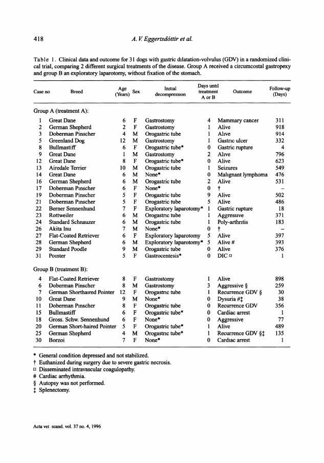
Table 1. Clinical data and outcome for 31 dogs with gastric dilatation-volvulus (GOV) in a randomized clini-cal trial, comparing 2 different surgical treatments of the disease. Group A received a circumcostal gastropexy and group Ban exploratory laparotomy, without fixation of the stomach.
Age Imtial Days until Follow-up Case no Breed treatment Outcome (Years) Sex decompress10n AorB (Days)
Group A (treatment A):
1 Great Dane 6 F Gastrostomy 4 Mammary cancer 311 2 German Shepherd 2 F Gastrostomy l Alive 918 3 Doberman Pinscher 4 M Orogastric tube l Ahve 914 5 Greenland Dog 12 M Gastrostomy l Gastnc ulcer 332 8 Bullmastiff 6 F Orogastric tube* 0 Gastric rupture 4 9 Great Dane l M Gastrostomy 2 Ahve 796
12 Great Dane 8 F Orogastric tube* 0 Alive 623 13 Airedale Terrier 10 M Orogastric tube l Seizures 549 14 Great Dane 6 M None* 0 Malignant lymphoma 476 16 German Shepherd 6 M Orogastric tube 2 Alive 531 17 Doberman Pmscher 6 F None* 0 t 19 Doberman Pinscher 5 F Orogastric tube 9 Alive 502 21 Doberman Pmscher 5 F Orogastric tube 5 Alive 486 22 Berner Sennenhund 7 F Exploratory laparotomy* l Gastric rupture 18 23 Rottweiler 6 M Orogastnc tube l Aggressive 371 24 Standard Schnauzer 6 M Orogastric tube l Poly-arthntis 183 26 Akita Inu 7 M None* 0 t 27 Flat-Coated Retriever 6 F Exploratory laparotomy 5 Alive 397 28 German Shepherd 6 M Exploratory laparotomy* 5 Alive# 393 29 Standard Poodle 9 M Orogastric tube 0 Alive 376 31 Pomter 5 F Gastrocentesis * 0 DICc l
Group B (treatment B):
4 Flat-Coated Retriever 8 F Gastrostomy 1 Alive 898 6 Doberman Pinscher 8 M Gastrostomy 3 Aggressive § 259 7 German Shortha1red Pointer 12 F Orogastnc tube l Recurrence GOV § 30
10 Great Dane 9 M None* 0 Dysuria#t 38 11 Doberman Pmscher 8 F Orogastric tube 0 Recurrence GOV 356 15 Bullmastiff 6 F Orogastric tube* 0 Cardiac arrest l 18 Gross. Schw. Sennenhund 6 F None* 0 Aggressive 77 20 German Short-haired Pointer 5 F Orogastric tube* l Alive 489 25 German Shepherd 4 M Orogastnc tube* l Recurrence GOV §t 135 30 Borzoi 7 F None* 0 Cardiac arrest l
* General condition depressed and not stabilized. t Euthanized during surgery due to severe gastric necrosis. c Disseminated intravascular coagulopathy. # Cardiac arrhythmia. § Autopsy was not performed. t Splenectomy.
Acta vet scand. vol. 37 no. 4, 1996

Surgery of gastric dilatation-volvulus in dogs 419
spective values in group B were 25 .1 g/l and 2.3 mmol/l. A lxl cm biopsy of all the ventricular layers was collected from the corpus of the stomach and fixed with 4% formaldehyde. The biopsy site was closed with interrupted sutures ofpolyglactin 9108. The biopsies were stained with haematoxylin and eosin, Van Gieson, periodic acid Schiff and Martius scarlet blue reagents for histological examination. The results of the histological examination of the biopsies taken from the stomach walls showed no differences between the 2 groups. Electrocardiography was performed on 18 dogs before the operation and on 5 dogs postoperatively. The electrocardiograms were normal for all dogs except 2 (Table 1 ).
Preoperative treatment All dogs received an intravenous (i.v.) infusion of lactated Ringer's solutionh 15-30 ml/kg/h, depending on the status of the patient and ampicillin' 50 mg/kg i.v. The dogs that were operated on immediately also received flumethasoneJ 0.5 mg/kg i.v. The dogs were anaesthetized with propofol k 4 mg/kg i. v. Further anaesthesia was maintained with a mixture of isoflurane1
and oxygen. Nitrous oxide gas was only used after drainage from the stomach had been achieved during surgery. Case number 28 received antiarrhythmic therapy before surgery with propranolol tabletsm 0.5 mg/kg orally. The general condition prior to surgery was not stabilized in 6 dogs (32%) (excluding 2 dropouts) that received treatment A and in 6 dogs ( 60%) that received treatment B (Table 1 ).
Surgical treatment In both groups a standard midline abdominal incision was performed. If the dogs had received a gastrostomy for gastric decompression in the acute stage of the disease, the gastrostomy operation site was first closed routinely.
As for the dogs that had received an exploratory laparotomy in the acute stage, the same midline incision was used to enter the abdominal cavity. The abdominal cavity and organs were examined for abnormalities and the stomach was repositioned. If needed, the stomach was emptied with an orogastric tube or by gastrostomy ifthe food remains did not pass through the tube. Splenectomy was performed in cases numbers 10 and 25 because of severe congestion and traumatic haemorrhage, respectively. The dogs included in treatment B received no further operative treatment, while in the dogs receiving treatment A, a slight modification of the method for circumcostal gastropexy as described by Fallah et al. (1982) was performed (Fig. 1). Traction sutures were placed approximately 10 cm apart on the ventral pyloric antrum. The sutures were held by an assistant. Two 2-3 cm long incisions were made with scissors, 2 cm apart, penetrating the serosal and muscular layers only. Both incisions were parallel with the long axis of the antrum. Cutting blood vessels was avoided as much as possible. The muscle layers between the incisions were undermined with scissors and cut with a scalpel to create 2 flaps, that were large enough to be joined around a rib without excessive tension. The 11th or 12th rib on the right side was used for costal fixation. One of these 2 costal arches was exposed by incising and reflecting the musculature for approximately 5 cm at the costo-
g 00 Vicryl, Etlncon, Inc. Sommerville, New Jersey. h Ringerlactat®, Pharmacia AB, Stockholm, Swe
den.V 1 Doctacillin®, AB Astra, Sodertiilje, Sweden. J Fluvet vet.®, Leo, Levens kemiske Fabnk,
K0benhavn, Ballerup, Danmark. k Rapinovet vet.®, Pitman-Moore, Harefield, Ux
bndge, Middlesex, England. 1 Forene®, Abbott Laboratories, USA. m Inderal®, Zeneca Ltd., Macclesfield, Cheshire,
England.
Acta vet scand vol 37 no 4, 1996

420 A. V. Eggertsdottir et al.
Figure l. Illustration of the crrcumcostal gastropexy flap techmque used m the treatment of dogs with GDV in group A (see Table I)
chondral junction. Care was taken to remain caudal to diaphragmatic attachments. Stay sutures were placed through all stomach layers at each corner where the cranial flap attached to the stomach. These stay sutures were to assist in bringing the flap around the exposed rib. The suture ends were passed around the exposed rib and the stomach was pulled against the right abdominal wall. The cranial flap was carefully passed around the rib using Allis tissue forceps. The 2 flaps were then sutured to each other with simple interrupted sutures (polypropylenen) and the stay sutures were removed. Twenty-three dogs were operated upon within
n 00 Prolene, Ethicon, Inc., Sommervdle, New Jersey.
0 Hill's w/d diet, Hill's pet products Inc., Topeka, Kansas, USA.
Acta vet scand. vol 37 no 4, 1996
one day and the remaining dogs between 2 and 9 days after the acute appearance of the disease. The median time between the initial gastric decompression and the final operation was 1.0 days (mean 1.9 days) in group A and 0.5 days (mean 0.7 days) in group B (Table l).
Postoperative treatment After the operation the dogs were monitored closely in the intensive care unit. An intravenous infusion of lactated Ringer's solution ( 60 ml/kg/day) was maintained for 2 days and on the third day the dogs were allowed for the first time to drink water (50 ml/kg/day) and eat small amounts of a high fibre diet0 • The amount of food was initially limited to 10% of their estimated daily food intake and then to 30% on the fourth day and to 60% on the fifth day. Ampicillin was given intravenously for 6 days (25

Surgery of gastric dilatation-volvulus in dogs 421
mg/kg, twice daily). Blood samples for haematology, clinical chemistry and arterial acid-base values were collected for haematology and clinical chemistry one or 2 days after the operation. Complications were treated immediately, thus case number 10 received antiarrhythmic therapy with lidocaineP 4 mg/kg i.v. as a slow bolus injection. This case and case number 28, received further treatment with propranolol tablets, 0.5 mg/kg, 3 times daily for 10 days postoperatively. If there were no signs of complications, the patients were released after a week. All the dogs were fed a high fibre diet for a minimum of 6 months and the food was supphed by the clime, to encourage the owners' compliance. For the first 4 weeks after the operation, the dogs were fed 3 times a day and after that twice a day. Written instructions were given to the owners with calculations of the daily food and water intake for their dog. The owners were advised to contact the clinic 1mmed1ately if their dogs' condition deteriorated. A routine clinical follow-up examination was performed 3 times during the first year, with the first examination occurring one to 3 months after the operation. In addition to breed, age, sex distribution and follow-up time, the number and the date of recurrence of GDV were recorded. The number and date of death caused by GDV, GDV related causes or other causes were also recorded in both groups. In addition, any gastrointestinal problems following the surgery were recorded.
Results There were 2 dropouts in the study and 5 dogs died 1-18 days postoperatively due to GDV-related complications (Table 1 ). Thus 24 dogs were available for follow-up evaluation. The follow-up time together with the breed, age
P Xylocard®, AB Hassle, Goteborg, Sweden.
and sex distribution are listed in Table 1. The study ended on November 1st 1993 (censoring time) and the median follow-up time for group A was 397 days (mean 430 days) and 106 days (mean 228 days) for group B. The outcome for the dogs in the study is listed in Table 1. At the end of the study 10 dogs were alive in group A and 2 dogs in group B. In group A, 3 dogs died due to GDV-related complications. Two of these had ruptures in the fundus part of the stomach whereas one dog died as a result of a chronic gastric ulcer that perforated 11 months after surgery. The latter dog had been treated with corticosteroids because of coxarthritis and the ulcer was discovered at necropsy. Five dogs were euthanized in this group because of allergy and polyarthritis, mammary cancer, aggressiveness, malignant lymphoma and seizures. In group B, 2 dogs died from GDV-related complications. One dog was euthanized because of dysuria and 2 dogs were euthanized because of aggressive behaviour. The non-recurrence probability within 6 months is shown in Fig. 2. It was unchanged after one year of follow-up. The recurrence rates within 6 months were significantly different between the 2 groups with 0 (0/16) recurrences in group A and 50% (3/6) in group B (Fisher exact test p = 0.01). Three dogs in group A and 4 dogs in group B died within 6 months (withdrawals) and were not available for calculation of the recurrence rate (Table 1). The overall death rates within the first year were 32% ( 6/19) in group A and 80% (8/10) in group B (Fisher exact test p = 0.02). The death rates caused by GDV and GDV-related causes only, after one year of follow-up, were 19% (3/16) and 71 % ( 5/7) for groups A and B respectively (Fisher exact test p = 0.02). Three dogs in both groups died within one year (withdrawals) from causes not related to GDV and were not available for calculation of the death rate due to GDV-related causes (Table 1 ).
Acta vet scand vol 37 no 4, 1996

422 A. V. Eggertsd6ttir et al.
1. 00 -- ···-···-···- ··-···-···-···-···-···-·· -···-···-···-···-···-···-···-···-···-···
>. !: 0.75-.-t ....
M llo
0.50 -:ii M M g I
0.25 -
0.00 20 40 60 80 100 120 140 160 180
TIME (days)
Figure 2. Kaplan-Meyer non-recurrence curves for dogs with GDV (see Table 1) in group A (n = 16) and in group B (n = 6). Withdrawals are not indicated on the figure.
The survival probability including all causes of death is shown in Fig. 3. The median survival times were 549 days in group A and l 07 days in group B. The estimated hazard ratio calculation (R = 0.36 (95% confidence interval 0.14-0.95); p = 0.04) showed that the relative survival for group A was significantly longer than for groupB. Since 5 dogs died postoperatively from GDVrelated complications, the median follow-up time from treatment until death for the remaining 12 dogs was 351 days (mean 370 days) for the dogs that received treatment A and l 06 days (mean 149 days) for the dogs that received treatment B. Three owners complained that their dogs had gastrointestinal problems after treatment A (cases numbers 5,9,19). The dogs were placed
Acta vet scand vol 37 no 4, 1996
on different diets. Two of these dogs regained normal gastrointestinal function, but case number 5 continued to have occasional vomiting problems.
Discussion There is much historical information about the results of treatment ofGDV without fixation of the stomach (treatment B), and the aim of the present study was to investigate the effect of treatment that included fixation (treatment A). From the literature and preliminary data we expected treatment that included fixation to be better in preventing recurrence of disease, than the treatment that did not include fixation. Under these circumstances it seemed unethical to have too large a control group. It was there-

Surgery of gastric dilatation-volvulus in dogs 423
1.00
-··-···-···1
0.75 l.. I
1U __ _
0.50
0.25
0 200 400
1.1.L.l.l
I ··-· .i . _ _ ___ L . -· _ _J
600 800 1000 TIME (days)
Figure 3. Kaplan-Meyer survival curves for dogs with GDV (see Table 1) m group A (n = 19) and in group B (n = 10). All causes of death are included. A vertical bar indicates dogs that were ahve at the end of the study (censonng time).
fore decided to allow twice as many cases to receive treatment A as received the control treatment B. Therefore the groups are unbalanced. In the present study a comparable distribution of the dogs in group A and B was achieved by the randomization procedure. There were only small differences in the breed, age, sex and initial decompression methods between the 2 groups. Thus, the randomization reduced selection biases and improved the reliability of comparisons between the 2 groups. The difference between the 2 groups was the surgical method and there were no recurrences in group A whereas the recurrence rate in group B was 50%. The values in both groups are similar to or better than other recurrence rates that have been reported following surgical repositioning without fixation of the stomach (Wing-
field et al. 1975, Dann 1976, Fjeld & Eggertsdottir 1988) However, it is well recognised that the results of a clinical trial are often better than would be expected from historical data. This is partly explained by the controlled conditions in clinical trials that are not achieved in the normal clinical situation. Nevertheless the significant difference identified between the 2 groups in the present study indicates that for the prevention of recurrence of GDV, a circumcostal gastropexy (treatment A) is better than no fixation of the stomach (treatment B). Other studies have reported low recurrence rates (0%-6.9%) following identical or comparable gastropexy methods (Leib et al. 1985, Schulman et al. 1986, Woolfeon & Kostolich 1986, Whitney et al. 1989, Frendin & Funkquist 1990). These studies, however, were not ran-
Acta vet. scand vol. 37 no. 4, 1996

424 A. V. Eggertsd6ttir et al.
Figure 4. Illustrat10n of the modified c!fcumcostal gastropexy techmque that was developed mstead of the trad1t1onal c!fcumcostal gastropexy with 2 flaps.
domized clinical trials, and so may have been subject to biased selection. A problem in this study, as in many clinical studies, was the early withdrawals that reduced the median follow-up time considerably. Nevertheless the dogs in group A did not experience any recurrences during the median follow-up time, which was about 3 times longer than the median follow-up time for the dogs in group B. The results would have been more reliable if more cases had been included in the study. However, the presence of recurrences only in the group that received treatment B and the demonstration of statistically significant differences between the 2 groups, justified the decision to limit the size of the study. Despite the successful result of the randomization, there were differences in the presurgical condition between the 2 groups. In group B,
Acta vet scand vol 37 no 4, 1996
60% of the dogs had a depressed general condition that was not stabilized before surgery. Among the dogs in group A only 32% belonged to this category. This difference may partly explain why the death rate from all causes and also the death rate from GDV-related causes only, were much higher in group B than in group A. However there were no differences between the 2 groups in the number of dogs that died from causes not related to GDV Thus the main difference in the survival time between the 2 groups ( 546 days in group A versus 104 days in group B) (Fig. 3) can probably be attributed to the difference between the 2 treatments. Since the median survival time was about 5 times longer in group A than in group B, a circumcostal gastropexy (treatment A) is found to be better than no fixation (treatment B) in prolonging postoperative survival time.
Surgery of gastric dilatation-volvulus in dogs 425
The dogs in this study were all fed the same high fibre diet for 6 months postoperatively. All the dogs did well on the diet, except for 2 dogs that had pre-existing gastrointestinal problems and one male Great Dane, that was still growing. A diet high in fibre was used instead of more easily digestible food, because the high fibre content reduces the gastrointestinal transit time and probably increases gastric emptying and motility. These effects of a high fibre diet may partly explain the low recurrence rate in the present study. Still, there are wide individual differences among dogs and some investigators have questioned whether diet influences the outcome of GDV (Burrows et al. 1982, Van Kruiningen et al. 1987, Burrows & Ignaszewski 1990).
Addendum After the clinical trial was ended, an additional modification of the circumcostal procedure was developed. q Instead of creating 2 flaps, a tunnel was made at the same operation site on the ventral pyloric antrum. Then the ventral half of the I Ith or 12th rib on the right side was exposed and cut with a scalpel at the costochondral junction and pulled through the tunnel (Fig. 4). The rib, together with the stomach, is placed back to its natural position without any additional fixation at the costochondraljunction. The exposed rib is covered by suturing the incised parietal peritoneum and muscle with interrupted sutures. In our experience this procedure is less timeconsuming and easier to perform than the traditional circumcostal gastropexy with 2 flaps.
q Fjeld, T.O., Department of Small Ammal Clinical Sciences, Norwegian College ofVetennary Medicine, P.0.Box 8146 Dep., N-0033 Oslo, Norway: Personal communication 1994.
Acknowledgements The authors thank the Central Laboratory, Department of B1ochem1stry, Physiology and Nutrition, Norwegian College of Vetermary Med1cme for analyzmg the blood samples and the Department of Morphology, Genetics and Aquatic Biology for performmg the autopsies and our colleagues W Breda!, S.I. Thoresen, E Skjerve and I Skaar for thelf assistance, constructive cntlc1sm and support.
References Altman DG Analysis of survival times. Ill" Altman
DG ( ed): Practical Stat1st1cs for Medical Research, Chapman & Hall, London, 1991, pp 365-395.
Betts CW, Wingfield WE, Rosin E Permanent gastropexy as a prophylactic measure agamst gastnc volvulus. J Amer. an1m. hosp Ass. 1976, 12, 177-181.
Burrows CF, Ignaszewski LA. Camne gastric d1latat1on-volvulus. J. small an1m. Pract. 1990, 31, 495-501.
Burrows CF, Kronfeld DS, Banta CA, Memtt AM Effects of fiber on d1gestlb1hty and transit time m dogs. J. Nutr. 1982, 112, 1726-1732.
Christie TR, Smith CW Gastrocolopexy for prevent10n of recurrent gastric volvulus. J. Amer. amm hosp. Ass. 1976, 12, 173-176.
Dann JR Medical and surgical treatment of canme acute gastric dilatation. J. Amer. amm. hosp. Ass. 1976, 12, 17-22.
Eggertsd6ttir AV, Moe L: A retrospective study of conservative treatment of gastric dllatat1on-volvulus m the dog Acta vet. scand. 1995, 36, 175-184.
Fallah AM, Lumb WV, Nelson AW, Frandsan RD, Withrow SJ C!fcumcostal gastropexy in the dog - a prehmmary study. Vet. Surg. 1982, 11, 9-12.
Fjeld TO, Eggertsd6tt1r AV VentrikkeldilatasJons/torsJonssyndromet hos hund: en retrospektlv stud1e av operativt behandlede kasus (Gastric d1latat1on-volvulus m the dog: a retrospective study of surgically treated cases). Norsk Vet.-T. 1988, 100, 281-287
Flanders JA, Harvey HJ. Results of tube gastrostomy as treatment for gastric volvulus in the dog. J. Amer. vet. med. Ass. 1984, 185, 74-77.
Fox SM· Gastric dilatat1on-volvulus: results from 31 cases circumcostal gastropexy vs. tube gastrostomy. Cahf Vet. 1985, 39, 8-11.
Acta vet scand vol 37 no 4, 1996

426 A. V. Eggertsdottir et al.
Frendin J, Funl«juist B: Fundic gastropexy for prevention of recurrence of gastric volvulus. J. small anim. Pract. 1990, 31, 78-82.
Funl«juist B: Gastric torsion in the dog. Nonsurgical reposition. J. small anim. Pract. 1969, JO, 507-511.
Johnson RG, Barrus J, Greene RW: Gastric dilatation-volvulus: recurrence rate following tube gastrostomy. J. Amer. anim. hosp. Ass. 1984, 20, 33-37.
Leib MS, Konde LJ, Wingfield WE, Twedt DC: Circumcostal gastropexy for preventing recurrence of gastric dilation-volvulus in the dog: an evaluation of 30 cases. J. Amer. vet. med. Ass. 1985, 187, 245-248.
MacCoy DM, Sykes GP, Hoffer RE, Harvey HJ: A gastropexy technique for permanent fixation of the pyloric antrum. J. Amer. anim. hosp. Ass. 1982, 18, 763-768.
Parks JL, Greene RW: Tube gastrostomy for the treatment of gastric volvulus. J. Amer. anim. hosp. Ass. 1976, 12, 168-172.
Schulman AJ, Lusk R, Lippencott CL, Ettinger SJ: Muscular flap gastropexy: a new surgical technique to prevent recurrences of gastric dilatationvolvulus syndrome. J. Amer. anim. hosp. Ass. 1986, 22, 339-346.
Van Kruzningen HJ, Wojan LD, Stake PE, Lord PF: The influence of diet and feeding frequency on gastric function in the dog. J. Amer. anim. hosp. Ass. 1987, 23, 145-153.
Van Sluijs FJ: Gastric dilatation-volvulus in the dog. Dissertation Utrecht 1987.
Whitney WO, Scavelli TD, Matthiesen DT, Burk RL: Belt-loop gastropexy: technique and surgical results in 20 dogs. J. Amer. anim. hosp. Ass. 1989, 25, 75-83.
Wingfield WE, Betts CW, Greene RW: Operative techniques and recurrence rates associated with gastric volvulus in the dog. J. small anim. Pract. 1975, 16, 427-452.
Wooifson J, Kostolich M: Circumcostal gastropexy: Clinical use of the technique in 34 dogs with gastric dilation-volvulus. J. Amer. anim. hosp. Ass. 1986, 22, 825-830.
Sammendrag Sammenligning av to kirurgiske behandlingsmetoder av ventrikkeldilatasjons/torsjons-syndromet hos hund.
Hensikten med denne prospektive randomiserte kliniske undersekelsen var a sammenligne to kirurgiske metoder i behandlingen av ventrikkeldilatasjon/ torsjon (VDT) hos hund. En gruppe hunder (gruppe A) ble behandlet med fiksering av ventrikkelen (gastropeksi) og en gruppe (gruppe B) uten fiksering. I gruppe A var det 21 hunder (Hvorav to "dropouts") og 1 gruppe B var det 10 hunder. Behandlingen av hundene i gruppe A bestod av dekomprimering av ventrikkelen, korrigering av ventrikkelens leie og gastropeksi. Hundene i gruppe B fikk samme behandling, uten gastropeksi. St0tteterapien var den samme for begge gruppene. Randomiseringen av hundene i gruppe A og B var vellykket idet det var Iiten forskjell mellom de to gruppene med henblikk pa rase, alder, kjenn og dekomprimeringsmetode i akuttfasen. Da undersekelsen ble avsluttet (censoring time) var det statistisk signifikant forskjell pa gjennomsnittlig overlevelsestid (median) i gruppe A og B, henholdsvis 549 og 107 dager. Det var ingen hunder som fikk res1div i gruppe A, men i gruppe B var det tre hunder (50%) som fikk residiv innen seks maneder. Mortaliteten i lepet av ett ar var 32% i gruppe A og 80% i gruppe B. Etter ett ars oppfolgingstid var 19% av hundene i gruppe A og 71% av hundene i gruppe B dede som felge av VDT eller VDT-relaterte 8rsaker. Undersekelsen viser en signifikant forskjell pa behandling som inkluderer "circumcostal" gastropeksi og at den forer til frerre residiv og forlenger den postoperative overlevelsestiden sammenlignet med behandling uten gastropeksi.
(Received January 21, 1996; accepted June 20, 1996).
Reprint may be obtained from: A. V. Eggertsd6ttir, Department of Small Animal Clinical Sciences, Norwegian College of Veterinary Medicine, P.O. Box 8146 Dep., N-0033 Oslo, Norway. Tel: +46 22 96 48 98, e-mail: [email protected].
Acta vet. scand. vol. 37 no. 4, 1996