Induction of high-affinity IgE receptor on lung dendritic cells during viral infection leads to...
11
The Journal of Experimental Medicine ARTICLE JEM © The Rockefeller University Press $30.00 Vol. 204, No. 11, October 29, 2007 2759-2769 www.jem.org/cgi/doi/ 2759 10.1084/jem.20070360 The risk of asthma from severe paramyxoviral infection in both human and experimental models is well documented (1–3). This virally imparted risk presents an interesting paradox; although the primary antiviral response is dom- inated by production of IFN / and IL-12, which are hallmarks of a Th1 response, rhinor- rhea and mucous cell metaplasia also develop. These conditions are driven by IL-13, which is a hallmark Th2 cytokine (4, 5). The produc- tion of antiviral IgE, along with neutralizing IgG antibodies, provides a further link between these disparate responses (6–10). In fact, IgE se- rum concentrations have been correlated with subsequent wheezing in infants with respiratory viral infection and with the risk of otitis media with effusion in children (10, 11). How a Th1- biased response generates a Th2 phenotype is not known, although we now show that the high-affinity receptor for IgE on DCs bridges the antiviral Th1 response to the atopic/pro- asthmatic Th2 response. The role of the high-affinity receptor for IgE (Fc RI) on human conventional DCs (cDCs) has been assumed to be antigen focusing, with expression being tightly regulated by serum IgE levels, much like it is on basophils (12, 13). Fc RI has not been reported on mouse DCs, and little is known of what role it might play during an antiviral response. Indeed, the role of the cDCs in an antiviral immune response is not fully understood. Initial lung cDC migration to draining lymph nodes, and subsequent anti- gen presentation, has been examined (14–16). However, the role of those cDCs that remain in or are attracted to the lung parenchyma dur- ing a primary response has not been evaluated. Although, in the case of secondary viral infec- tions or challenge responses to OVA, the evi- dence suggests that these cells are involved in recruitment of memory effector T cells (17, 18). We have developed a mouse model of viral CORRESPONDENCE Mitchell H. Grayson: [email protected] Abbreviations used: ATCC, American Type Culture Collec- tion; IFNAR, type I IFN recep- tor; PI, postinnoculation; SeV, Sendai virus. The online version of this article contains supplemental material. Induction of high-affinity IgE receptor on lung dendritic cells during viral infection leads to mucous cell metaplasia Mitchell H. Grayson, 1 Dorothy Cheung, 1 Michelle M. Rohlfing, 1 Robert Kitchens, 1 Daniel E. Spiegel, 1 Jennifer Tucker, 2 John T. Battaile, 2 Yael Alevy, 2 Le Yan, 2 Eugene Agapov, 2 Edy Y. Kim, 2 and Michael J. Holtzman 2,3 1 Division of Allergy and Immunology, 2 Division of Pulmonary and Critical Care Medicine, Department of Internal Medicine, and 3 Department of Cell Biology, Washington University School of Medicine, Saint Louis, MO 63110 Respiratory viral infections are associated with an increased risk of asthma, but how acute Th1 antiviral immune responses lead to chronic inflammatory Th2 disease remains unde- fined. We define a novel pathway that links transient viral infection to chronic lung disease with dendritic cell (DC) expression of the high-affinity IgE receptor (Fc RI ). In a mouse model of virus-induced chronic lung disease, in which Sendai virus triggered a switch to persistent mucous cell metaplasia and airway hyperreactivity after clearance of replicating virus, we found that FceRIa / mice no longer developed mucous cell metaplasia. Viral infection induced IgE-independent, type I IFN receptor–dependent expression of Fc RI on mouse lung DCs. Cross-linking DC Fc RI resulted in the production of the T cell chemoat- tractant CCL28. FceRIa / mice had decreased CCL28 and recruitment of IL-13–producing CD4 T cells to the lung after viral infection. Transfer of wild-type DCs to FceRIa / mice restored these events, whereas blockade of CCL28 inhibited mucous cell metaplasia. There- fore, lung DC expression of Fc RI is part of the antiviral response that recruits CD4 T cells and drives mucous cell metaplasia, thus linking antiviral responses to allergic/asth- matic Th2 responses.
-
Upload
independent -
Category
Documents
-
view
3 -
download
0
Transcript of Induction of high-affinity IgE receptor on lung dendritic cells during viral infection leads to...

The
Journ
al o
f Exp
erim
enta
l M
edic
ine
ARTICLE
JEM © The Rockefeller University Press $30.00
Vol. 204, No. 11, October 29, 2007 2759-2769 www.jem.org/cgi/doi/
2759
10.1084/jem.20070360
The risk of asthma from severe paramyxoviral infection in both human and experimental models is well documented ( 1 – 3 ). This virally imparted risk presents an interesting paradox; although the primary antiviral response is dom-inated by production of IFN � / � and IL-12, which are hallmarks of a Th1 response, rhinor-rhea and mucous cell metaplasia also develop. These conditions are driven by IL-13, which is a hallmark Th2 cytokine ( 4, 5 ). The produc-tion of antiviral IgE, along with neutralizing IgG antibodies, provides a further link between these disparate responses ( 6 – 10 ). In fact, IgE se-rum concentrations have been correlated with subsequent wheezing in infants with respiratory viral infection and with the risk of otitis media with eff usion in children ( 10, 11 ). How a Th1-biased response generates a Th2 phenotype is not known, although we now show that the high-affi nity receptor for IgE on DCs bridges
the antiviral Th1 response to the atopic/pro-asthmatic Th2 response.
The role of the high-affi nity receptor for IgE (Fc � RI) on human conventional DCs (cDCs) has been assumed to be antigen focusing, with expression being tightly regulated by serum IgE levels, much like it is on basophils ( 12, 13 ). Fc � RI has not been reported on mouse DCs, and little is known of what role it might play during an antiviral response. Indeed, the role of the cDCs in an antiviral immune response is not fully understood. Initial lung cDC migration to draining lymph nodes, and subsequent anti-gen presentation, has been examined ( 14 – 16 ). However, the role of those cDCs that remain in or are attracted to the lung parenchyma dur-ing a primary response has not been evaluated. Although, in the case of secondary viral infec-tions or challenge responses to OVA, the evi-dence suggests that these cells are involved in recruitment of memory eff ector T cells ( 17, 18 ). We have developed a mouse model of viral
CORRESPONDENCE
Mitchell H. Grayson:
Abbreviations used: ATCC,
American Type Culture Collec-
tion; IFNAR, type I IFN recep-
tor; PI, postinnoculation; SeV,
Sendai virus.
The online version of this article contains supplemental material.
Induction of high-affi nity IgE receptor on lung dendritic cells during viral infection leads to mucous cell metaplasia
Mitchell H. Grayson, 1 Dorothy Cheung, 1 Michelle M. Rohlfi ng, 1 Robert Kitchens, 1 Daniel E. Spiegel, 1 Jennifer Tucker, 2 John T. Battaile, 2 Yael Alevy, 2 Le Yan, 2 Eugene Agapov, 2 Edy Y. Kim, 2 and Michael J. Holtzman 2,3
1 Division of Allergy and Immunology, 2 Division of Pulmonary and Critical Care Medicine, Department of Internal Medicine,
and 3 Department of Cell Biology, Washington University School of Medicine, Saint Louis, MO 63110
Respiratory viral infections are associated with an increased risk of asthma, but how acute
Th1 antiviral immune responses lead to chronic infl ammatory Th2 disease remains unde-
fi ned. We defi ne a novel pathway that links transient viral infection to chronic lung disease
with dendritic cell (DC) expression of the high-affi nity IgE receptor (Fc � RI � ). In a mouse
model of virus-induced chronic lung disease, in which Sendai virus triggered a switch to
persistent mucous cell metaplasia and airway hyperreactivity after clearance of replicating
virus, we found that FceRIa � / � mice no longer developed mucous cell metaplasia. Viral
infection induced IgE-independent, type I IFN receptor – dependent expression of Fc � RI � on
mouse lung DCs. Cross-linking DC Fc � RI � resulted in the production of the T cell chemoat-
tractant CCL28. FceRIa � / � mice had decreased CCL28 and recruitment of IL-13 – producing
CD4 � T cells to the lung after viral infection. Transfer of wild-type DCs to FceRIa � / � mice
restored these events, whereas blockade of CCL28 inhibited mucous cell metaplasia. There-
fore, lung DC expression of Fc � RI � is part of the antiviral response that recruits CD4 � T
cells and drives mucous cell metaplasia, thus linking antiviral responses to allergic/asth-
matic Th2 responses.
2760 Fc � RI � AND VIRUS-INDUCED MUCOUS CELL METAPLASIA | Grayson et al.
of the high-affi nity receptor for IgE (Fc � RI � ), and this ex-pression remained detectable for 2 – 3 wk after SeV inoculation ( Fig. 2, A and B ). This response was specifi c to lung cDCs be-cause there was no detectable expression of Fc � RI � on cDCs isolated from draining lymph nodes or spleen during SeV in-fection (unpublished data). CD23, the low-affi nity receptor for IgE, was not induced during the infection ( Fig. 2 B ). Us-ing intranasal administration of the intravital dye CFSE, which labeled lung cells present at the time of its administration but not cells that subsequently migrated into the lung, we were able to show that Fc � RI � -expressing cDCs were present in the lung at the time of the initial viral inoculation and had not migrated into the lung after the onset of infection ( Fig. 2, C and D ). The level of expression of Fc � RI � on lung cDCs at PI day 7 was similar to levels seen on c-kit � lung mast cells ( Fig. 2, A and E ). Increased expression of Fc � RI after viral in-fection was relatively selective for cDCs and mast cells because we did not detect expression of Fc � RI � on lung CD4 � or CD8 � T cells, B220 � B cells, Mac-3 � macrophages, or Gr-1 � neutrophils (unpublished data). Expression of Fc � RI � was de-tected on 20 – 40% of lung pDCs at PI day 3 (unpublished data),
bronchiolitis that reproduces disease traits associated with asthma ( 2 ). In this model, Fc � RI was expressed on lung cDCs only during the antiviral response, and these cells were criti-cal for the development of postviral mucous cell metaplasia. Indeed, mice defi cient in Fc � RI ( FceRIa – / – ) failed to recruit IL-13 – producing CD4 � T cells to their lungs after viral infec-tion. Cross-linking the � -chain of Fc � RI (Fc � RI � ) on lung cDCs produced a CD4 � T cell chemoattractant, CCL28, and blockade of this chemokine prevented development of post-viral mucous cell metaplasia. Thus, these studies provide a novel insight into how the antiviral response leads to a potent Th2 response through Fc � RI � on lung cDCs.
RESULTS
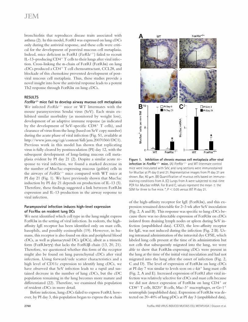
FceRIa – / – mice fail to develop airway mucous cell metaplasia
We infected FceRIa – / – mice or WT littermates with the mouse paramyxovirus Sendai virus (SeV). Each strain ex-hibited similar morbidity (as monitored by weight loss), development of an adaptive immune response (as indicated by the development of SeV-specific CD8 � T cells), and clearance of virus from the lung (based on SeV copy number) during the acute phase of viral infection (Fig. S1, available at http://www.jem.org/cgi/content/full/jem.20070360/DC1). Previous work in this model has shown that replicating virus is fully cleared by postinoculation (PI) day 12, with the subsequent development of long-lasting mucous cell meta-plasia evident by PI day 21 ( 2 ). Despite a similar acute re-sponse to viral infection, we found a marked decrease in the number of Muc5ac-expressing mucous (goblet) cells in the airways of FceRIa – / – mice compared with WT mice at PI day 21 ( Fig. 1 ). We have previously shown that Muc5ac induction by PI day 21 depends on production of IL-13 ( 5 ). Therefore, these fi ndings suggested a link between Fc � RI � expression and IL-13 production in the airway response to viral infection.
Paramyxoviral infection induces high-level expression
of Fc � RI � on resident lung DCs
We next identifi ed which cell type in the lung might express Fc � RI � in the setting of viral infection. In rodents, the high-affi nity IgE receptor has been identifi ed only on mast cells, basophils, and possibly eosinophils ( 19 ). However, in hu-mans, this receptor is also found on skin and peripheral blood cDCs, as well as plasmacytoid DCs (pDCs), albeit as a trimeric form (Fc � RI � � � ) that lacks the Fc � RI � chain ( 13, 20, 21 ). Therefore, we questioned whether this form of the receptor might also be found on lung parenchymal cDCs after viral infection. Using forward/side scatter characteristics and a high level of CD11c expression to identify lung cDCs, we have observed that SeV infection leads to a rapid and sus-tained decrease in the number of lung cDCs, but the cDC population remaining in the lung becomes more mature and diff erentiated ( 22 ). Therefore, we examined this population of resident cDCs in more detail.
Before infection, lung cDCs failed to express Fc � RI; how-ever, by PI day 3, this population began to express the � chain
Figure 1. Inhibition of chronic mucous cell metaplasia after viral
infection in FceRIa � /� mice. (A) FceRIa � / � and WT littermate control
mice were inoculated with SeV, and lung sections were immunostained
for Muc5ac at PI day 0 and 21. Representative images from PI day 21 are
shown. Bar, 40 � m. (B) Quantifi cation of mucous cells based on immuno-
staining conditions from A. (C) Lungs from A were subjected to real-time
PCR for Muc5ac mRNA. For B and C, values represent the mean the
SEM for three to fi ve mice. *, P 0.05 versus WT PI day 21.

JEM VOL. 204, October 29, 2007
ARTICLE
2761
Fc � RI proteins, real-time PCR assays showed that lung cDCs expressed Fc � RI � , but not Fc � RI � mRNA after viral infec-tion, whereas MC9 mast cells contained high levels of both Fc � RI � and Fc � RI � mRNA (Fig. S2 C). Thus, mouse lung cDCs express the trimeric (Fc � RI � � � ) form of the Fc � RI receptor. This is the same form of Fc � RI that is expressed by human DCs, but expression on mouse cDCs requires induc-tion by a productive viral infection. This requirement may explain the failure to detect Fc � RI expression on mouse DCs in previous works.
Type I IFN receptor, but not IgE, regulates DC expression
of Fc � RI �
Next, we explored what component of the antiviral response is responsible for induction of Fc � RI � expression on lung cDCs during a paramyxoviral infection. Serum IgE level tightly regulates expression of the high-affi nity IgE receptor in humans, so we reasoned that total IgE and, more likely, SeV-specifi c IgE might drive Fc � RI receptor expression after viral infection in mice. However, we found that serum total IgE and SeV-specifi c IgE do not increase until PI day 7 ( Fig. 3 A ). This time course lags behind the onset of expression of Fc � RI �
which is consistent with reports of Fc � RI � � � on human pDCs ( 21 ). This pDC population was distinct from the lung cDC population because Fc � RI � -expressing lung cDCs did not express the pDC/B cell marker B220 (Fig. S2 A, available at http://www.jem.org/cgi/content/full/jem.20070360/DC1). Expression of Fc � RI did not infl uence cDC maturation or diff erentiation during viral infection. We found no diff erence in the levels of expression of MHC class II, CD11b, CD80, or CD86 between lung cDCs from FceRIa – / – versus WT mice (Fig. S2, A and B). Similarly, we found no infl uence of Fc � RI on the expression of CD23.
Based on work with isolated cells, mice have been reported to be obligate expressers of the tetrameric form (Fc � RI � � � � ) of Fc � RI ( 23 ). To determine if mouse lung cDCs were, indeed, expressing the tetrameric and not trimeric form of the receptor, we analyzed mouse lung cDCs for expression of each Fc � RI component chain. Immunoprecipitation of Fc � RI with an anti-Fc � RI � antibody, and subsequent Western blot with anti-Fc � RI � or -Fc � RI � antibodies, showed that lung cDCs isolated after SeV infection expressed Fc � RI � , but not Fc � RI � ( Fig. 2 F ). In contrast, MC9 mast cells expressed both Fc � RI � and Fc � RI � . In concert with these fi ndings for
Figure 2. Up-regulation of Fc � RI � expression on lung DCs after viral infection. (A) WT mice were inoculated with SeV, and CD11c � lung cDCs or
c-kit � mast cells (MC) were analyzed by fl ow cytometry using anti-Fc � RI � mAb and isotype control mAb at PI day 0 and 7. (B) Conditions in A were used
for fl ow cytometry for Fc � RI � and CD23 on the indicated PI day. Values represent the mean the SEM for n � 6 mice. *, P 0.05 versus PI day 0. (C) WT
mice were given PBS ([ – ] CFSE) or CFSE ([+] CFSE) intranasally and inoculated with SeV. Lung cDCs were isolated at PI day 7, and expression of CFSE
and Fc � RI � was determined by fl ow cytometry. (D) Quantifi cation of data from C. Values represent the mean the SEM for n � 3 mice. (E) Lung cDCs
(CD11c � ) and mast cells (c-kit � ) were analyzed by fl ow cytometry for Fc � R1 � , as in A. Values represent the mean the SEM for fold mean fl uorescence
intensity Fc � RI � signal versus isotype control mAb for three mice per time point. (F) Cell lysates from lung cDCs (PI day 5) or MC/9 mast cells were sub-
jected to immunoprecipitation with anti-Fc � RI � mAb. Immunoprecipitated complexes (IP) were then Western blotted with anti-Fc � RI � or -Fc � RI � mAb.
Representative blots are shown for two separate experiments.

2762 Fc � RI � AND VIRUS-INDUCED MUCOUS CELL METAPLASIA | Grayson et al.
Therefore, IgE levels do not appear to regulate the appearance of Fc � RI � on lung cDCs after viral infection in mice.
Given that initial expression of Fc � RI � developed at the same time as IFN-dependent responses in this viral model, we examined whether IFN signaling was necessary for Fc � RI ex-pression. Indeed, we found that type I IFN receptor (IFNAR) – defi cient ( IFNAR � / � ) mice no longer exhibited an increase in Fc � RI � expression on lung cDCs during viral infection ( Fig. 3, C – E ). In contrast, mice that were defi cient for CD1d , CD4 , CD8 , perforin , or MyD88 gene expression showed no defect in Fc � RI � expression after viral infection (unpub-lished data). Thus, although IFNAR is critical for expression of Fc � RI � on lung cDCs after viral infection, there is no substantial role for NKT cells, CD4 � T cells, or CD8 � T cells, or for the innate antiviral perforin- or MyD88-dependent Toll-like receptor pathways in this process.
We next examined the mechanism for IFN-dependent expression of Fc � RI on DCs. We recognized that direct IFN treatment of human lung pDCs failed to increase Fc � RI � expression ( 21 ). This fi nding suggested that IFN-dependent increases in Fc � RI expression on lung DCs may involve other cell types besides DCs. Accordingly, we next determined whether IFNAR expression on cDCs was necessary for Fc � RI expression after viral infection. For these experiments, we purifi ed lung cDCs from IFNAR � / � and WT control mice. We verifi ed that the cells we purifi ed exhibited typical DC morphology ( Fig. 4 ). Purifi ed lung cDCs were loaded with CFSE (to discriminate from endogenous cDCs) and transferred into WT or IFNAR � / � recipients. We found that either IFNAR � / � or WT cDC transfer into WT mice allowed for expression of Fc � RI � on cDCs (Fig. S3, available at http://www.jem.org/cgi/content/full/jem.20070360/DC1). In contrast, transfer of either genotype of cDCs into IFNAR � / � mice did not permit expression of Fc � RI � on cDCs after viral infection. These fi ndings imply that IFN acts through another intermediate cell to activate Fc � RI expression on lung cDCs.
Lung cDCs from virus-infected mice drive IL-13 production
from CD4 � T cells
Because IL-13 is thought to drive postviral mucous cell meta-plasia, and Th2 cells are thought to be a likely source of IL-13, we reasoned that generation of Th2 cells might be altered as a result of Fc � RI � engagement on cDCs ( 5 ). CD8 � cDCs, like those found in the lung, have been shown to drive T cell development toward a Th2 phenotype in vitro ( 24 ). There-fore, to determine if induction of IL-13 – producing CD4 � T cells depended on engagement of Fc � RI � , we examined IL-13 production in an in vitro cell culture system that contains CD4 � T cells and cDCs. OVA-specifi c CD4 � T cells (OT-II cells) were cultured with lung cDCs isolated from WT or FceRIa � / � mice with or without SeV infection. We isolated cDCs at PI day 7 and verifi ed that these cDCs had not yet bound IgE at this time point (unpublished data). We found that OT-II CD4 � T cells produced IL-13 when cultured with antigen and cDCs, regardless of whether cDCs were
on lung cDCs, suggesting that the level of IgE is not driving the expression of Fc � RI receptor under these conditions. In fact, we found that IgE-defi cient ( IgE � / � ) mice continued to develop the same increase in Fc � RI � expression on lung cDCs as their WT littermates after viral inoculation ( Fig. 3 B ).
Figure 3. Effect of IgE levels and IFN signaling on up-regulation
of Fc � RI � on lung cDCs after viral infection. (A) WT mice were
inoculated with SeV, and serum was analyzed for total and SeV-specifi c
IgE levels. Values represent the mean the SEM for three to six mice.
(B) IgE � / � and WT control mice were inoculated with SeV, and lung
cDCs were analyzed by fl ow cytometry using anti-Fc � RI � and isotype
control mAbs. (C) IFNAR � / � and WT mice were inoculated with SeV, and
lung cDCs were analyzed at PI day 5, as in B. For conditions in C, quan-
tifi cation of fold increase in mean fl uorescence intensity for Fc � RI �
over isotype control (D) and percentage of lung cDCs expressing Fc � RI �
(E). Values represent the mean the SEM for four to eight mice
per group.

JEM VOL. 204, October 29, 2007
ARTICLE
2763
To establish that CCL28 expression was inducible in cDCs with a more physiological stimulus of Fc � RI activa-tion, we loaded lung cDCs from PI day 7 with IgE against OVA. In this setting, we found that addition of OVA (by cross-linking anti-OVA IgE bound to Fc � RI � on the cDC) caused a marked increase in CCL28 mRNA ( Fig. 5 C ). Sim-ilar results were obtained when using the anti-Fc � RI � anti-body to directly cross-link the receptor (unpublished data). To determine whether Fc � RI activation also caused CCL28 expression in vivo, we returned to experiments with the mouse model of virus-induced lung disease. Again, we found that lung levels of CCL28 mRNA were increased after SeV infec-tion in WT mice, and that this eff ect was lost in FceRIa � / � mice ( Fig. 5 D ). If CCL28 was being released with engagement of Fc � RI, then we would expect diff erences in the frequency of CD4 � T cells in the lungs of FceRIa � / � and WT mice after viral infection. Indeed, we found that CD4 � T cells were signifi cantly decreased in the lungs of FceRIa � / � mice compared with WT mice at PI day 21 ( Fig. 5 E ).
Because we have previously shown that mucous cell metaplasia after viral infection is dependent on IL-13 produc-tion, we next examined the amount of IL-13 and GATA-3 (a Th2-specifi c transcription factor) message in lung CD4 � T cells of WT and FceRIa � / � mice after SeV infection ( 5 ). We found a signifi cant decrease in the levels of IL-13 and GATA-3 mRNA in CD4 � T cells from the lungs of FceRIa � / � com-pared with WT mice ( Fig. 5, F and G ). These fi ndings further supported the proposal that inhibition of mucous cell meta-plasia in FceRIa � / � mice was based on decreased accumula-tion of IL-13 – producing CD4 � Th2 cells caused by a lack of production of CCL28 by the resident Fc � RI � � lung cDCs. We also examined IL-13 and GATA-3 mRNA levels in the CD4 � T cells that had or had not migrated in response to CCL28 in vitro. We found that there was much greater ex-pression of both IL-13 and GATA-3 mRNA in T cells that
isolated from WT or FceRIa � / � mice or from infected or un-infected mice ( Fig. 5 A ). Furthermore, cross-linking Fc � RI � had no eff ect on T cell production of IL-13 or proliferation ( Fig. 5 A and not depicted). Thus, we conclude that lung cDCs are capable of driving the development of IL-13 – pro-ducing CD4 + Th2 cells, but this process does not require cDC expression or activation of Fc � RI � .
Cross-linking Fc � RI � on lung DCs produces the T cell
chemoattractant CCL28
Because we did not detect a requirement for cross-linking Fc � RI � in the development of the T cell cytokine response, we next assessed whether Fc � RI might be involved in the in-duction of T cell chemoattractants. Lung cDCs were purifi ed from WT mice by immunomagnetic selection at PI day 5. The purifi ed cells were then cultured for 18 h with a cross-linking antibody against Fc � RI � or a control hamster IgG. Superna-tants from these cultures were then used in a modifi ed Boyden chamber assay to assess whether a functional T cell chemoat-tractant had been produced. Supernatants from Fc � RI � cross-linked cDCs induced signifi cantly more CD4 � T cell migration than did IgG control supernatants, indicating that engagement of the receptor led to production of a CD4 � T cell chemoat-tractant ( Fig. 5 B ). To identify the chemokine receptor for this T cell chemoattractant, we added various chemokines (CCL5, CCL22, and CCL27) to the upper chamber and evaluated the eff ect on chemotaxis (Fig. S4 A, available at http://www.jem.org/cgi/content/full/jem.20070360/DC1). Only CCL27 (C-TACK) inhibited T cell migration, indicating that the CD4 � T cells were moving in response to a CCR10 agonist. Only two known CCR10 agonists, CCL27 and CCL28 (MEC), have been identifi ed. Using blocking mAbs, we found that the chemotactic activity was entirely caused by CCL28 ( Fig. 5 B ). Of relevance to this work, CCL28 has been associated with both human asthma and mouse models of asthma ( 25 – 27 ).
Figure 4. Morphology of purifi ed lung cDCs versus macrophages. Lung cDCs were isolated by positive immunomagnetic selection with macro-
phages obtained by BAL. Both cell preparations were cytocentrifuged onto slides (left two columns) and cultured in chamber slides with or without
10 � g/ml LPS for 24 h at 37 ° C (right two columns), and then imaged with modifi ed Wright ’ s stain and photomicrography. For each condition, representative
photomicrographs from three separate experiments are shown. Bars, 10 � m.

2764 Fc � RI � AND VIRUS-INDUCED MUCOUS CELL METAPLASIA | Grayson et al.
Figure 5. CCL28 production after activation of Fc � RI � on lung cDCs. (A) Lung cDCs from WT or FceRIa � / � mice that were uninfected (PI day 0) or
SeV-infected (PI day 7) were treated with or without anti-Fc � RI � mAb or control IgG. Cells were loaded with OVA peptide and cocultured with OVA-spe-
cifi c CD4 � T cells (OT-II) for 4 d at 37 ° C, followed by ELISA of cell supernatants for IL-13. Values represent the mean the SEM for four to seven experi-
ments done in triplicate. (B) Lung cDCs were purified from PI day 5 and treated with an anti-Fc � RI � mAb or control IgG for 18 h at 37 ° C. Cell
supernatants were assayed for chemotactic activity for naive CD4 � T cells with and without treatment with anti-CCL28 or -CCL27 mAb. Values represent
the mean the SEM for three to six experiments done in triplicate. *, P 0.05 versus IgG supernatant; **, P 0.05 versus anti-CCL27 and no antibody
treatment. (C) Lung cDCs purifi ed at PI day 7 were loaded with anti-OVA IgE and cultured with (+) or without ( – ) 3 � g/ml OVA protein for 18 h at 37 ° C.
Cellular RNA was subjected to real-time PCR for Ccl28 mRNA. Values are corrected for Gapdh mRNA level and represent the mean the SEM for three
experiments. *, P 0.05 versus no OVA protein. (D) FceRIa � / � and WT mice were inoculated with SeV, and lung levels of Ccl28 mRNA were determined by
real-time PCR at the indicated times. Values represent the mean the SEM for three to fi ve mice. (E) Lung frequencies of CD4 � T cells were quantifi ed by
fl ow cytometry in WT and FceRIa � / � mice at the indicated times. Values represent the mean the SEM for three to nine mice. (F) Lung CD4 � T cells were
purifi ed by positive immunomagnetic selection at PI day 21 and analyzed for Il-13 by real-time PCR. (G) Using conditions in F, expression of GATA-3
mRNA was analyzed by real-time PCR. (H) Percentage of total lung cells in the lymphocyte gate expressing CCR10 and CD4 by fl ow cytometry at PI day 0
or 10 in WT and FceRIa � / � mice. For F – H, values represent the mean the SEM for three mice. *, P 0.05 versus WT mice for D – H. (I) CCR10 � CD4 � T
cells and CCR10 � CD4 � T cells were purifi ed by fl ow cytometry at PI day 10 and subjected to real-time PCR for GATA-3 and T-bet . Values represent the
mean the SEM for the ratio of GATA-3 to T-bet from three experiments with three mice per experiment. *, P 0.05 versus CCR10 � CD4 � T cells.

JEM VOL. 204, October 29, 2007
ARTICLE
2765
DISCUSSION
In this study, we show that respiratory viral infection can lead to expression and activation of Fc � RI on lung cDCs and thereby drive the development of mucous cell metaplasia even after the inciting infection is cleared. We examined lung cDCs because we previously found that the migration and maturation of this cell population was tightly regulated dur-ing respiratory viral infection ( 22 ). Indeed, the potent eff ects of viral infection on DCs might explain why we have detected Fc � RI on DCs in the mouse, whereas others have not previ-ously found this receptor on DCs ( 28 ).
We have also shown that expression of Fc � RI in the set-ting of a productive viral infection requires an active type I IFN response. In fact, expression of Fc � RI on cDCs quickly returns to low levels with resolution of the antiviral response,
migrated compared with cells that did not migrate in response to CCL28 (Fig. S4 B).
Because CCL28 binds to CCR10 to recruit immune cells, we determined the level of CCR10 � CD4 � T cells in the lungs of WT and FceRIa � / � mice before and after viral infection. We found an increase in CCR10 � CD4 � T cells in WT mice after viral infection ( Fig. 5 H ). Furthermore, this increase in CCR10 � CD4 � T cells did not develop in FceRIa � / � mice. We also found that CCR10 � CD4 � lung T cells from WT mice contained increased levels of GATA-3 mRNA (typically found in Th2 cells) compared with T-bet mRNA (typically found in Th1 cells; Fig. 5 I ). Together, these fi nd-ings provide further support for the proposal that activation of Fc � RI � on cDCs leads to preferential accumulation of Th2 cells in the lung after SeV infection.
cDC reconstitution restores CD4 � T cell accumulation
and mucous cell metaplasia after viral infection
To prove that Fc � RI � expression on cDCs was necessary for recruitment of CD4 � Th2 cells to the lung and mucous cell metaplasia after viral infection, we performed adoptive cell transfer experiments with cDCs from WT or FceRIa � / � mice transferred into FceRIa � / � recipients, followed by SeV inocu-lation. We found that reconstitution with WT cDCs restored postviral mucous cell metaplasia in FceRIa � / � recipient mice ( Fig. 6, A and B , and Fig. S5 A, available at http://www.jem.org/cgi/content/full/jem.20070360/DC1). In contrast, trans-fer of FceRIa � / � cDCs were unable to restore the development of mucous cell metaplasia after viral infection. Furthermore, we found that CD4 � T cells isolated at PI day 21 from lungs of FceRIa � / � mice that had received WT cDCs expressed IL-13 and GATA-3 at signifi cantly higher levels than mice that re-ceived cDCs from FceRIa � / � mice ( Fig. 6 C ). We found no diff erence in the engraftment of WT versus FceRIa � / � cDCs, with both genotypes of cDCs persisting for at least 6 d after transfer to the recipient mice (Fig. S5 B). Together, these re-sults indicate that Fc � RI � on lung cDCs is critical for Th2 cell recruitment and mucous cell metaplasia after viral infection.
Blockade of CCL28 inhibits postviral mucous cell metaplasia
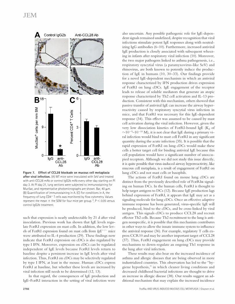
We next determined the role of CCL28 in the cascade lead-ing to mucous cell metaplasia after viral infection. For these experiments, we treated WT mice with an anti-CCL28 – blocking mAb or control IgG2b and monitored the develop-ment of mucous cell metaplasia at PI day 21. We found a signifi cant decrease in mucous cell metaplasia after treatment with anti-CCL28 mAb compared with control IgG ( Fig. 7, A and B , and Fig. S6, available at http://www.jem.org/cgi/content/full/jem.20070360/DC1). We observed no signifi -cant diff erence in the frequency of CD4 � T cells in the lungs of mice treated with either anti-CCL28 mAb or control IgG2b ( Fig. 7 C ). This fi nding suggests that CCL28 is a che-motactic factor for immune cells producing IL-13 and driv-ing mucous cell metaplasia after viral infection, but additional chemoattractants also contribute to CD4 � T cell accumula-tion in the lung in this setting.
Figure 6. Restoration of postviral mucous cell metaplasia in FceRIa � /�
mice after lung cDC transfer. (A) Lung cDCs were isolated from WT or
FceRIa � / � mice and transferred into FceRIa � / � recipients 1 d before
inoculation with SeV. At PI day 21, lung sections were obtained for immuno-
staining for Muc5ac. Representative photomicrographs are shown. Bar,
40 � m. (B) Quantifi cation of immunostaining in A. Values represent the
mean the SEM for two to four recipient mice per group from two sepa-
rate experiments; *, P 0.05 versus WT DCs transferred into FceRIa � / �
recipients. (C) For conditions in A, lung CD4 � T cells were purifi ed at PI
day 21 and analyzed for Il-13, GATA-3, and Gapdh mRNA by real-time
Q-PCR. Values represent the mean the SEM from two separate experi-
ments with two to four recipient mice per experiment. *, P 0.05 versus
FceRI a � / � cDCs transferred into FceRIa � / � recipients.
2766 Fc � RI � AND VIRUS-INDUCED MUCOUS CELL METAPLASIA | Grayson et al.
also uncertain. Any possible pathogenic role for IgE-depen-dent signals remained undefi ned, despite recognition that viral infections stimulate potent IgE responses along with neutral-izing IgG antibodies ( 6 – 10 ). Furthermore, increased antiviral IgE production is closely associated with subsequent wheez-ing in infants after respiratory viral infection ( 10 ). Moreover, the two major pathogens linked to asthma pathogenesis, i.e., respiratory syncytial virus (a paramyxovirus-like SeV) and rhinovirus, are both known to potently induce the produc-tion of IgE in humans ( 10, 30 – 33 ). Our fi ndings provide for a novel IgE-dependent mechanism in which an antiviral response characterized by IFN production drives expression of Fc � RI on lung cDCs. IgE engagement of the receptor leads to release of soluble mediators that generate an atopic response characterized by Th2 cell activation and IL-13 pro-duction. Consistent with this mechanism, others showed that passive transfer of antiviral IgE can increase the airway hyper-reactivity caused by respiratory syncytial virus infection in mice, and that Fc � RI was necessary for this IgE-dependent response ( 34 ). This eff ect was assumed to be caused by mast cell activation during the viral infection. However, given the very low dissociation kinetics of Fc � RI-bound IgE (K d of � 10 � 9 – 10 � 10 M), it is not clear that IgE during a primary vi-ral infection would bind to mast cell Fc � RI in any signifi cant quantity during the acute infection ( 35 ). It is possible that the rapid expression of Fc � RI on lung cDCs would make these cells a better target cell for binding antiviral IgE because this cell population would have a signifi cant number of unoccu-pied receptors. Although we did not study this issue directly, it is quite possible that virus-induced airway hyperreactivity, like mucous cell metaplasia, is a result of engagement of Fc � RI on lung cDCs and not mast cells or basophils.
The actions of Fc � RI found on mouse lung cDCs are distinct from the previously described role of Fc � RI � signal-ing on human DCs. In the human cells, Fc � RI is thought to help target antigens to DCs ( 12 ). Because IgE production lags behind expression of Fc � RI, it appears that IgE may act as a signaling molecule for lung cDCs. Once an eff ective adaptive immune response has been generated, virus-specifi c IgE will be produced, bind to the cDCs, and be cross-linked by viral antigen. This signals cDCs to produce CCL28 and recruit eff ector Th2 cells. Because Th2 recruitment to the lung is anti-gen nonspecifi c, it is possible that this mechanism contributes in other ways to allow the innate immune system to infl uence the antiviral response ( 36 ). For example, regulatory T cells ex-press CCR10 and may be another important target of CCL28 ( 37 ). Thus, Fc � RI engagement on lung cDCs may provide mechanisms to down-regulate an ongoing Th1 response in the lung after viral infection.
These results may also bear on the increased incidence of asthma and allergic diseases that are being observed in more industrialized countries. This observation has led to the “ hy-giene hypothesis, ” in which cleaner living conditions and decreased childhood bacterial infections are thought to drive an increase in allergic disease ( 38 ). Our results suggest an ad-ditional mechanism that may explain the increased incidence
such that expression is nearly undetectable by 21 d after viral inoculation. Previous work has shown that IgE levels regu-late Fc � RI expression on mast cells. In addition, the low lev-els of Fc � RI expression found on mast cells from IgE � / � mice were attributed to IL-4 production ( 29 ). These fi ndings now indicate that Fc � RI expression on cDCs is also regulated by type I IFN. Moreover, expression on cDCs can be regulated independent of IgE levels because Fc � RI levels return to baseline despite a persistent increase in IgE levels after viral infection. Thus, Fc � RI on cDCs may be selectively regulated by type I IFN, at least in the mouse. Human cDCs express Fc � RI at baseline, but whether these levels are increased by viral infection still needs to be determined ( 12, 13 ).
In that regard, the consequences of IgE production and IgE – Fc � RI interaction in the setting of viral infection were
Figure 7. Effect of CCL28 blockade on mucous cell metaplasia
after viral infection. (A) WT mice were inoculated with SeV and treated
with anti-CCL28 mAb or control IgG2b mAb every other day starting on PI
day 3. At PI day 21, lung sections were subjected to immunostaining for
Muc5ac, and representative photomicrographs are shown. Bar, 40 � m.
(B) Quantifi cation of immunostaining in A. (C) For conditions in A, the
frequency of lung CD4 � T cells was monitored by fl ow cytometry. Values
represent the mean the SEM for four mice per group. *, P 0.05 versus
control IgG2b treatment.

JEM VOL. 204, October 29, 2007
ARTICLE
2767
Mouse generation and handling. C57BL/6; BALB/c; B6.129S2-
CD4 +tm1Mak /J (CD4-null); B6.129S2-Cd8a tm1Mak /J (CD8-null); and C57BL/6-
Prf1 tm1Sdz /J (perforin-null) mice were obtained from The Jackson Laboratory.
TgN(OT-II.2a)Rag1 tm1 (OT-II) mice on a C57BL6 background were obtained
from the Taconic NIAID Emerging Models Program (Germantown, NY).
C57BL6 mice defi cient in CD1d were obtained from A. Bendelac (Univer-
sity of Chicago, Chicago, IL); IFNAR-null mice on a C57BL6 background
were obtained from J. Sprent (The Scripps Research Institute, La Jolla, CA);
MyD88-defi cient mice and WT littermates on a mixed B6/129 back-
ground were a gift from M. Colonna (Washington University, St. Louis,
MO); Fc � RI � -defi cient mice on a C57BL6 background were obtained
from J.-P. Kinet (Harvard Medical School, Boston, MA); and IgE-defi cient
mice on a BALB/c background were obtained from H. Oettgen (Harvard
Medical School, Boston, MA). Mice 6 – 20 wk of age were used for all
experiments under protocols approved by the University Animal Stud-
ies Committee.
Mice were inoculated with SeV (Fushimi strain; American Type Cul-
ture Collection [ATCC]) or UV-inactivated SeV, as previously described,
using an inoculum of 2 � 10 5 pfu, and they were monitored daily for weight
and activity. To track lung cDCs, mice were anesthetized and given 30 � l of
5 mM CFSE intranasally, as previously described ( 2, 3, 5, 14 ). For blockade
of CCL28, mice were given 100 � g of anti-CCL28 mAb or control IgG2b
subcutaneously every other day from PI day 3 to 19. Mucous cell metaplasia
and Muc5ac mRNA in lungs were assessed at PI day 21 as previously de-
scribed ( 2, 3, 5 ).
Lung cDC isolation. For cDC isolation, mice were killed, and intracardiac
injection of PBS was used to fl ush out the pulmonary and systemic circula-
tions. BAL was performed to remove any cells in the bronchiolar or alveolar
space. Next, 1 ml of digest media was injected intratracheally. Digest me-
dium consisted of DME supplemented with 5% fetal calf serum, penicillin/
streptokinase, 10 mM Hepes, 250 U/ml collagenase I (Worthington Bio-
chemical), 50 U/ml DNase I (Worthington Biochemical), and 0.01% hyal-
uronidase (Sigma-Aldrich). The lungs were carefully dissected from the
trachea, main-stem bronchi, draining lymph nodes, and surrounding tissue.
The lungs were minced and incubated in digest media for 1 h at 37 ° C. Dur-
ing the fi nal 15 min of incubation time, EDTA was added to a fi nal concen-
tration of 2 mM. After digestion, the cell mixture was passed through a
40- � m pore cell strainer to generate single-cell suspensions, and erythrocytes
were removed by hypotonic lysis. Cell viability was assessed by trypan blue
exclusion. An analogous procedure was performed for removal of DCs from
spleen or draining lymph nodes, as well as for isolation of T cells from
C57BL6 or OT-II splenic tissue. Lung cDCs were identifi ed (and other cell
populations, such as macrophages, were excluded) by fl ow cytometry, as
previously described ( 22 ).
Cell purifi cation and culture. DCs were purifi ed from lung cell suspen-
sions using the MACS system (Miltenyi Biotec) for positive immunomag-
netic selection with anti-CD11c microbeads. Using two serial purifi cations,
cDC purity was � 95%. Cells obtained in this manner had typical cDC mor-
phology ( Fig. 4 ). Purifi ed cDCs were cultured with or without cross-linking
antibodies in complete RPMI (Sigma-Aldrich) supplemented with 10% fetal
calf serum and penicillin/streptokinase (Invitrogen) for 18 h at 37 ° C. For T
cell proliferation experiments, cDCs were subjected to 200 cGyn before be-
ing loaded with 0.3 � M of OVA peptide (OVA 323-339 ). CD4 � T cells were
purifi ed from OT-II spleens using the Miltenyi-Biotec MACS system for
positive immunomagnetic selection. T cell purities were � 95%. Irradiated
cDCs (10 4 per condition) were cultured for 4 d with 10 5 OVA-specifi c
CD4 � T cells with or without cross-linking antibodies. For T cell prolifera-
tion, CD4 � T cells were labeled with 5 � M CFSE (Invitrogen) before cul-
ture, and cell proliferation was monitored by CFSE dilution, as previously
described ( 42 ). For some experiments, lung cDCs were loaded with 10 � g/ml
mouse anti-OVA IgE (Serotec) for 48 h at 37 ° C, washed, and incubated
with or without 3 � g/ml OVA protein (Sigma-Aldrich) for 18 h at 37 ° C,
followed by mRNA isolation from cell pellets. Mouse MC/9 mast cells were
of asthma in the urban environment. In this setting, the in-creased population density, as well as other factors, may lead to increased transmission of respiratory viral infections. If vi-ral respiratory illnesses drive Fc � RI expression on lung cDCs in humans, this process could lead to an increase in lung Th2 responses in individuals with genetic susceptibility to this type of response. This mechanism is consistent with the in-creased incidence of asthma now being found for children living in the inner city ( 39 ).
In summary, we have defi ned a new scheme for mucous cell metaplasia after viral infection that depends on an IF-NAR – Fc � RI – CCL28 – IL-13 immune axis (Fig. S7, available at http://www.jem.org/cgi/content/full/jem.20070360/DC1). This mechanism involves cDC expression of Fc � RI and pro-duction of CCL28 in concert with T cell production of IL-13 and consequent mucous cell metaplasia. This immune axis may be more active after severe infection to help explain why more severe respiratory viral infections are associated with an in-creased risk of asthma. For example, the level of IgE produc-tion after SeV infection in mice appears to depend on the severity of the infection (unpublished data). The scheme also implies that exposure to even nonviral IgE and antigen during the resolution of the viral response might also lead to increased Th2 recruitment to the lung driven by additional Fc � RI cross-linking and CCL28 production. This mechanism would thereby provide for an allergic-type response to nonviral antigens be-cause Th2 cells that had migrated to the lung could be directed against nonviral antigens. In the atopic individual, these Th2 cells could lead to the development of asthma, or an exacerba-tion of the disease in patients with established asthma. Each of these mechanisms is distinct from previous proposals for IgE – Fc � RI function in the traditional response to parasitic in-fection or allergen exposure. Thus, viral antigens and the con-sequent IFN-dependent antiviral response may also trigger an “ allergic ” cascade, leading to disease traits that are characteristic of asthma and other chronic airway diseases.
MATERIALS AND METHODS Reagents and antibodies. Anti-CD16/-CD32 antibodies (clone 2.4G2;
obtained from C. Pham, Washington University, St. Louis, MO) were used
for fl ow cytometry, as previously described ( 40 ). Anti – mouse CCL28 mAb
or control rat IgG2b was obtained from R & D Systems. PE- or allophycocy-
anin-labeled antibodies against mouse CD4, CD8, CD11c, CD23, c-Kit,
Fc � RI � (clone MAR-1), and IgE, as well as isotype control IgGs (rat and
Armenian hamster), were obtained from eBioscience or BD PharMingen.
SeV-specifi c CD8 � T cells were monitored using tetrameric MHC/peptide
reagents for the immunodominant epitope of SeV nucleoprotein (NP 324-332 )
or control OVA peptide (SIINFEKL) complexed with K(b) provided by the
National Institute of Allergy and Infectious Diseases (NIAID) Tetramer
Core Facility ( 41 ). Purifi ed anti-Fc � RI � mAb (clone MAR-1; eBioscience)
was used for immunoprecipitation, and anti-Fc � RI � mAb (clone JRK) ob-
tained from J. Rivera (National Institutes of Health, Bethesda, MD) or rabbit
anti-Fc � RI � Ab (Millipore) was used for detection of the immune complex
in Western blotting. For cross-linking experiments, functional grade Arme-
nian hamster anti – murine Fc � RI � or functional grade Armenian hamster
IgG isotype control antibodies were obtained from eBioscience. A secondary
goat anti – Armenian hamster IgG antibody purchased from Jackson Immuno-
Research Laboratories was also used.

2768 Fc � RI � AND VIRUS-INDUCED MUCOUS CELL METAPLASIA | Grayson et al.
Statistical analyses. Unless otherwise stated, all data are presented as the
mean the SEM. Student ’ s t test was used to assess statistical signifi cance
between means. For nonnormally distributed data, comparison of medians
was made using a Mann-Whitney U test. For comparison of ratios, Wilcoxin
Signed Rank was used. In all cases, signifi cance was set at P 0.05.
Online supplemental material. FceRIa � / � and WT mice respond simi-
larly to SeV infection, as shown in Fig. S1. Surface marker expression on
lung cDCs from FceRIa � / � and WT mice are similar, as shown in Fig. S2.
Fig. S3 shows that IFNAR expression on lung cDCs is not necessary for
Fc � RI � expression on lung cDCs. Fig. S4 shows the eff ect of cDC Fc � RI
activation on chemotaxis of CD4 � T cells. The ability of WT, but not
FceRIa � / � , cDC to restore postviral-induced mucous cell metaplasia in
FceRIa � / � mice is demonstrated in Fig. S5. Fig. S6 shows that postviral
Muc5ac mRNA induction is inhibited with CCL28 blockade. As shown
in Fig. S7, our data support a scheme whereby an IFN – Fc � RI – CCL28 –
IL-13 signaling pathway regulates mucous cell metaplasia after viral in-
fection. The online version of this article is available at http://www.jem
.org/cgi/content/full/jem.20070360/DC1.
The authors wish to thank Drs. Charles Parker and Richard Hotchkiss for helpful
discussions, and Drs. Albert Bendelac, Marco Colonna, Jean-Pierre Kinet, Hans
Oettgen, Christine Pham, Juan Rivera, and Jonathan Sprent for generous gifts of
mice and/or reagents.
This work was supported by grants from the National Institutes of Health
(NIAID and National Heart, Lung, and Blood Institute) and Genentech/Novartis, as
well as the Martin Schaeffer Fund and Alan A. and Edith L. Wolff Charitable Trust.
The authors declare that they have no competing fi nancial interests.
Submitted: 20 February 2007
Accepted: 1 October 2007
REFERENCES 1 . Sigurs , N. 2002 . A cohort of children hospitalised with acute RSV
bronchiolitis: impact on later respiratory disease. Paediatr. Respir. Rev. 3 : 177 – 183 .
2 . Walter , M.J. , J.D. Morton , N. Kajiwara , E. Agapov , and M.J. Holtzman . 2002 . Viral induction of a chronic asthma phenotype and genetic segre-gation from the acute response. J. Clin. Invest. 110 : 165 – 175 .
3 . Patel , A.C. , J.D. Morton , E.Y. Kim , Y. Alevy , S. Swanson , J. Tucker , G. Huang , E. Agapov , T.E. Phillips , M.E. Fuentes , et al . 2006 . Genetic segregation of airway disease traits despite redundancy of calcium-acti-vated chloride channel family members. Physiol. Genomics . 25 : 502 – 513 .
4 . Chen , Y. , P. Thai , Y.H. Zhao , Y.S. Ho , M.M. DeSouza , and R. Wu . 2003 . Stimulation of airway mucin gene expression by inter-leukin (IL)-17 through IL-6 paracrine/autocrine loop. J. Biol. Chem. 278 : 17036 – 17043 .
5 . Tyner , J.W. , E.Y. Kim , K. Ide , M.R. Pelletier , W.T. Roswit , J.D. Morton , J.T. Battaile , A.C. Patel , G.A. Patterson , M. Castro , et al . 2006 . Blocking airway mucous cell metaplasia by inhibiting EGFR antiapop-tosis and IL-13 transdiff erentiation signals. J. Clin. Invest. 116 : 309 – 321 .
6 . Alexeyev , O.A. , C. Ahlm , J. Billheden , B. Settergren , G. Wadell , and P. Juto . 1994 . Elevated levels of total and Puumala virus-specifi c immuno-globulin E in the Scandinavian type of hemorrhagic fever with renal syndrome. Clin. Diagn. Lab. Immunol. 1 : 269 – 272 .
7 . Calenoff , E. , J.C. Zhao , E.L. Derlacki , W.H. Harrison , K. Selmeczi , J.C. Dutra , I.R. Olson , and D.G. Hanson . 1995 . Patients with Meniere ’ s disease possess IgE reacting with herpes family viruses. Arch. Otolaryngol. Head Neck Surg. 121 : 861 – 864 .
8 . Koraka , P. , B. Murgue , X. Deparis , T.E. Setiati , C. Suharti , E.C. van Gorp , C.E. Hack , A.D. Osterhaus , and J. Groen . 2003 . Elevated levels of total and dengue virus-specifi c immunoglobulin E in patients with varying disease severity. J. Med. Virol. 70 : 91 – 98 .
9 . Votava , M. , D. Bartosova , A. Krchnakova , K. Crhova , and L. Kubinova . 1996 . Diagnostic importance of heterophile antibodies and immuno-globulins IgA, IgE, IgM and low-avidity IgG against Epstein-Barr virus capsid antigen in children. Acta Virol. 40 : 99 – 101 .
obtained from the ATCC and were maintained in ATCC complete growth
medium (DME with 2 mM l- glutamine, 0.05 mM 2-ME, 4.5 g/liter glu-
cose, and 1.5 g/liter sodium bicarbonate) supplemented with 10% fetal calf
serum and 10% Rat T-Stim (BD Biosciences) ( 43 ).
Immunoprecipitation and Western blot analysis. Immunoprecipita-
tion of Fc � RI � from lung cDCs and MC/9 mast cells was performed as pre-
viously described ( 44, 45 ). In brief, 3 � 10 6 cells were solubilized in 100 mM
Tris containing 0.5% Nonidet P-40, 10% glycerol, 5 M NaCl, 100 mM
MgCl 2 , and enhanced inhibitor supplement consisting of a 20 � concentrate
of complete edium (80 mM benzamidine HCL, 50 mM � -caproic acid, 16 mM
iodoacetamide, 10 � g/ml leupeptin, 10 � g/ml pepstatin, and 100 � g/ml
soybean trypsin inhibitor). Aliquots of samples containing 500 � g protein
were immunoprecipitated using 20 � l of protein A – Sepharose beads conju-
gated to 25 � g of rabbit secondary antibody. Immune complexes were sepa-
rated by nonreducing PAGE, and then detected by Western blotting onto an
Immobilon-P PVDF membrane (Millipore), probed with anti-Fc � RI � or
-Fc � RI � antibodies, and imaged with chemiluminescence (SuperSignal West
Pico kit; Pierce Chemical Co.).
Adoptive transfer protocol. Lung cDCs were purifi ed from C57BL6,
IFNAR � / � , or FceRIa � / � mice and delivered intranasally to anesthetized
recipient C57BL6 or FceRIa � / � mice (2.5 � 10 5 cells in 30 � l of PBS given
1 d before SeV inoculation). In some experiments, cDCs were loaded with
either CFSE or TAMRA-SE (Invitrogen), as previously described ( 40 ).
No diff erence in transfer effi ciency (engraftment) was seen with either dye
(unpublished data).
Chemotaxis assay. Chemotaxis was monitored with a modifi ed Boyden
chamber, as previously described ( 46 ). For these experiments, CD4 � T cells
were purifi ed from naive C57BL6 spleen, and 2.5 � 10 4 cells in 50 � l were
loaded in the upper chamber, which was separated from the lower chamber
by a 5- � m membrane (Neuro Probe, Inc.). A 25- � l aliquot of a 1:3 dilution
of supernatant from lung cDC cultures was placed in the lower chamber. In
some experiments, control rat IgG2b, rat anti – mouse CCL27 antibody, or rat
anti – mouse CCL28 antibody (100 � g/ml) was added to the lower chamber.
After a 3-h incubation at 37 ° C, the number of cells that had transmigrated
into the lower chamber was determined by fl ow cytometry, as previously
described ( 46 ). Results are calculated as a migration index that represents the
ratio of cells migrating in each condition compared with the number migrat-
ing to CCL21 control (1 � g/ml; R & D Systems).
Real-time PCR assay. mRNA was isolated with the RNeasy kit (Qiagen)
or Trizol (Sigma-Aldrich) and used to generate cDNA with Applied Biosys-
tems cDNA archive kit as per the manufacturer ’ s instructions. Quantitative
real-time PCR assays were performed using an Applied Biosystems 7500
Fast real-time PCR system and TaqMan fast master mix. In addition to
the TaqMan rodent GAPDH control, the following TaqMan gene ex-
pression arrays (mouse primer and probe sets) for PCR were obtained from
Applied Biosystems: IL-13 (assay ID Mm00434204_m1), Muc5ac (assay ID
Mm01276725_g1), CCL28 (assay ID Mm00445039_m1), GATA-3 (assay
ID Mm00484683_m1), Fc � RI � (assay ID Mm00438867_m1), and Fc � RI�
(assay ID Mm00442780_m1). For Fc � RI � and Fc � RI � mRNA levels, results
represent arbitrary units (calculated as 2 [C T
Maximum − C T
gene ]) per ng of total
RNA. For IL-13 , Muc5ac , GATA-3 , and CCL28 mRNA levels, results rep-
resent copy number per Gapdh copy number based on a standard curve with
the known copy number of the target and Gapdh mRNA.
Measurement of IgE and IL-13. Total IgE from mouse serum was deter-
mined by ELISA using capture and detection anti-IgE antibody pairs (BD
PharMingen) in 96-well MaxiSorp plates (Nalge Nunc). Purifi ed mouse IgE
(BD PharMingen) was used as a control. For detection of SeV-specifi c IgE,
SeV-coated plates from Charles River Laboratories were substituted for cap-
ture IgE antibody-coated plates. Because no control anti-SeV IgE antibodies
are available, values represent absolute absorbance units. IL-13 levels were
determined using a commercially available ELISA kit (Invitrogen).

JEM VOL. 204, October 29, 2007
ARTICLE
2769
10 . Welliver , R.C. , M. Sun , D. Rinaldo , and P.L. Ogra . 1986 . Predictive value of respiratory syncytial virus-specifi c IgE responses for recurrent wheezing following bronchiolitis. J. Pediatr. 109 : 776 – 780 .
11 . Chantzi , F.M. , D.A. Kafetzis , T. Bairamis , C. Avramidou , N. Paleologou , I. Grimani , N. Apostolopoulos , and N.G. Papadopoulos . 2006 . IgE sensitization, respiratory allergy symptoms, and heritability inde-pendently increase the risk of otitis media with eff usion. Allergy . 61 : 332 – 336 .
12 . Maurer , D. , C. Ebner , B. Reininger , E. Fiebiger , D. Kraft , J.P. Kinet , and G. Stingl . 1995 . The high affi nity IgE receptor (Fc epsilon RI) medi-ates IgE-dependent allergen presentation. J. Immunol. 154 : 6285 – 6290 .
13 . Foster , B. , D.D. Metcalfe , and C. Prussin . 2003 . Human dendritic cell 1 and dendritic cell 2 subsets express FcepsilonRI: correlation with serum IgE and allergic asthma. J. Allergy Clin. Immunol. 112 : 1132 – 1138 .
14 . Legge , K.L. , and T.J. Braciale . 2003 . Accelerated migration of respira-tory dendritic cells to the regional lymph nodes is limited to the early phase of pulmonary infection. Immunity . 18 : 265 – 277 .
15 . Usherwood , E.J. , T.L. Hogg , and D.L. Woodland . 1999 . Enumeration of antigen-presenting cells in mice infected with Sendai virus. J. Immunol. 162 : 3350 – 3355 .
16 . Crowe , S.R. , S.J. Turner , S.C. Miller , A.D. Roberts , R.A. Rappolo , P.C. Doherty , K.H. Ely , and D.L. Woodland . 2003 . Diff erential antigen presentation regulates the changing patterns of CD8 + T cell immunodominance in primary and secondary infl uenza virus infections. J. Exp. Med. 198 : 399 – 410 .
17 . Zammit , D.J. , L.S. Cauley , Q.M. Pham , and L. Lefrancois . 2005 . Dendritic cells maximize the memory CD8 T cell response to infection. Immunity . 22 : 561 – 570 .
18 . van Rijt , L.S. , S. Jung , A. Kleinjan , N. Vos , M. Willart , C. Duez , H.C. Hoogsteden , and B.N. Lambrecht . 2005 . In vivo depletion of lung CD11c + dendritic cells during allergen challenge abrogates the charac-teristic features of asthma. J. Exp. Med. 201 : 981 – 991 .
19 . Dombrowicz , D. , B. Quatannens , J.-P. Papin , A. Capron , and M. Capron . 2000 . Expression of a functional Fc { epsilon } RI on rat eosino-phils and macrophages. J. Immunol. 165 : 1266 – 1271 .
20 . Bieber , T. , H. de la Salle , A. Wollenberg , J. Hakimi , R. Chizzonite , J. Ring , D. Hanau , and C. de la Salle . 1992 . Human epidermal langerhans cells express the high affi nity receptor for immunoglobulin E (Fc � RI). J. Exp. Med. 175 : 1285 – 1290 .
21 . Schroeder , J.T. , A.P. Bieneman , H. Xiao , K.L. Chichester , K. Vasagar , S. Saini , and M.C. Liu . 2005 . TLR9- and FcepsilonRI-mediated re-sponses oppose one another in plasmacytoid dendritic cells by down-regulating receptor expression. J. Immunol. 175 : 5724 – 5731 .
22 . Grayson , M.H. , M.S. Ramos , M.M. Rohlfi ng , R. Kitchens , H.D. Wang , A. Gould , E. Agapov , and M.J. Holtzman . 2007 . Controls for lung dendritic cell maturation and migration during respiratory viral infection. J. Immunol. 179 : 1438 – 1448 .
23 . Blank , U. , C. Ra , L. Miller , K. White , H. Metzger , and J.P. Kinet . 1989 . Complete structure and expression in transfected cells of high af-fi nity IgE receptor. Nature . 337 : 187 – 189 .
24 . Maldonado-Lopez , R. , T. De Smedt , P. Michel , J. Godfroid , B. Pajak , C. Heirman , K. Thielemans , O. Leo , J. Urbain , and M. Moser . 1999 . CD8 � + and CD8 � � subclasses of dendritic cells direct the development of distinct T helper cells in vivo. J. Exp. Med. 189 : 587 – 592 .
25 . English , K. , C. Brady , P. Corcoran , J.P. Cassidy , and B.P. Mahon . 2006 . Infl ammation of the respiratory tract is associated with CCL28 and CCR10 expression in a murine model of allergic asthma. Immunol. Lett. 103 : 92 – 100 .
26 . Wang , W. , H. Soto , E.R. Oldham , M.E. Buchanan , B. Homey , D. Catron , N. Jenkins , N.G. Copeland , D.J. Gilbert , N. Nguyen , et al . 2000 . Identifi cation of a novel chemokine (CCL28), which binds CCR10 (GPR2). J. Biol. Chem. 275 : 22313 – 22323 .
27 . John , A.E. , M.S. Thomas , A.A. Berlin , and N.W. Lukacs . 2005 . Temporal production of CCL28 corresponds to eosinophil accumula-tion and airway hyperreactivity in allergic airway infl ammation. Am. J. Pathol. 166 : 345 – 353 .
28 . Finkelman , F.D. , M.E. Rothenberg , E.B. Brandt , S.C. Morris , and R.T. Strait . 2005 . Molecular mechanisms of anaphylaxis: lessons from studies with murine models. J. Allergy Clin. Immunol. 115 : 449 – 457 .
29 . Yamaguchi , M. , C.S. Lantz , H.C. Oettgen , I.M. Katona , T. Fleming , I. Miyajima , J.-P. Kinet , and S.J. Galli . 1997 . IgE enhances mouse mast cell Fc � RI expression in vitro and in vivo: evidence for a novel amplifi cation mechanism in IgE-dependent reactions. J. Exp. Med. 185 : 663 – 672 .
30 . Skoner , D.P. , W.J. Doyle , E.P. Tanner , J. Kiss , and P. Fireman . 1995 . Eff ect of rhinovirus 39 (RV-39) infection on immune and infl amma-tory parameters in allergic and non-allergic subjects. Clin. Exp. Allergy . 25 : 561 – 567 .
31 . Las Heras , J. , and V.L. Swanson . 1983 . Sudden death of an infant with rhinovirus infection complicating bronchial asthma: case report. Pediatr. Pathol. 1 : 319 – 323 .
32 . Rager , K.J. , J.O. Langland , B.L. Jacobs , D. Proud , D.G. Marsh , and F. Imani . 1998 . Activation of antiviral protein kinase leads to immuno-globulin E class switching in human B cells. J. Virol. 72 : 1171 – 1176 .
33 . Kusel , M.M. , N.H. de Klerk , T. Kebadze , V. Vohma , P.G. Holt , S.L. Johnston , and P.D. Sly . 2007 . Early-life respiratory viral infections, atopic sensitization, and risk of subsequent development of persistent asthma. J. Allergy Clin. Immunol. 119 : 1105 – 1110 .
34 . Dakhama , A. , J.-W. Park , C. Taube , K. Chayama , A. Balhorn , A. Joetham , X.-D. Wei , R.-H. Fan , C. Swasey , N. Miyahara , et al . 2004 . The role of virus-specifi c immunoglobulin E in airway hyperrespon-siveness. Am. J. Respir. Crit. Care Med. 170 : 952 – 959 .
35 . Garman , S.C. , B.A. Wurzburg , S.S. Tarchevskaya , J.P. Kinet , and T.S. Jardetzky . 2000 . Structure of the Fc fragment of human IgE bound to its high-affi nity receptor Fc epsilonRI alpha. Nature . 406 : 259 – 266 .
36 . Stephens , R. , D.A. Randolph , G. Huang , M.J. Holtzman , and D.D. Chaplin . 2002 . Antigen-nonspecifi c recruitment of Th2 cells to the lung as a mechanism for viral infection-induced allergic asthma. J. Immunol. 169 : 5458 – 5467 .
37 . Eksteen , B. , A. Miles , S.M. Curbishley , C. Tselepis , A.J. Grant , L.S. Walker , and D.H. Adams . 2006 . Epithelial infl ammation is associated with CCL28 production and the recruitment of regulatory T cells ex-pressing CCR10. J. Immunol. 177 : 593 – 603 .
38 . Buff ord , J.D. , and J.E. Gern . 2005 . The hygiene hypothesis revisited. Immunol. Allergy Clin. North Am. 25 : 247 – 262 .
39 . Sly , R.M. 1999 . Changing prevalence of allergic rhinitis and asthma. Ann. Allergy Asthma Immunol. 82 : 233 – 248 .
40 . Grayson , M.H. , R.S. Hotchkiss , I.E. Karl , M.J. Holtzman , and D.D. Chaplin . 2003 . Intravital microscopy comparing T lymphocyte traffi ck-ing to the spleen and the mesenteric lymph node. Am. J. Physiol. Heart Circ. Physiol. 284 : H2213 – H2226 .
41 . Flynn , K.J. , G.T. Belz , J.D. Altman , R. Ahmed , D.L. Woodland , and P.C. Doherty . 1998 . Virus-specifi c CD8+ T cells in primary and sec-ondary infl uenza pneumonia. Immunity . 8 : 683 – 691 .
42 . Lyons , A.B. 2000 . Analysing cell division in vivo and in vitro using fl ow cytometric measurement of CFSE dye dilution. J. Immunol. Methods . 243 : 147 – 154 .
43 . Galli , S.J. , A.M. Dvorak , J.A. Marcum , T. Ishizaka , G. Nabel , H. Der Simonian , K. Pyne , J.M. Goldin , R.D. Rosenberg , H. Cantor , and H.F. Dvorak . 1982 . Mast cell clones: a model for the analysis of cellular maturation. J. Cell Biol. 95 : 435 – 444 .
44 . Gillespie , S.R. , R.R. DeMartino , J. Zhu , H.J. Chong , C. Ramirez , C.P. Shelburne , L.A. Bouton , D.P. Bailey , A. Gharse , P. Mirmonsef , et al . 2004 . IL-10 inhibits Fc epsilon RI expression in mouse mast cells. J. Immunol. 172 : 3181 – 3188 .
45 . Gomez , G. , C.D. Ramirez , J. Rivera , M. Patel , F. Norozian , H.V. Wright , M.V. Kashyap , B.O. Barnstein , K. Fischer-Stenger , L.B. Schwartz , et al . 2005 . TGF-beta 1 inhibits mast cell Fc epsilon RI expression. J. Immunol. 174 : 5987 – 5993 .
46 . Bleul , C.C. , R.C. Fuhlbrigge , J.M. Casasnovas , A. Aiuti , and T.A. Springer . 1996 . A highly effi cacious lymphocyte chemoattractant, stro-mal cell-derived factor 1 (SDF-1). J. Exp. Med. 184 : 1101 – 1109 .