Combined Sellar Fibrosarcoma and Prolactinoma with Neuronal Metaplasia: Report of a Case...
10
Pituitary Fibrosarcoma and Prolactinoma with Neuronal Metaplasia 149 Case Report 149 Department of Pathology (MM, CG, BWS, RVL), Mayo Clinic, Rochester, MN, Departments of Pathology (PR), Endocrinology (CE), and Neurosurgery (AJL), Auckland Hospital, Auckland, New Zealand and St. Michael’s Hospital (KK), University of Toronto, Toronto, ON, Canada Address correspondence to Dr. Bernd W. Scheithauer, Department of Pathology and Laboratory Medicine, 200 First Street, SW, Rochester, MN 55905, USA. E-mail: scheithauer.bernd@ mayo.edu Endocrine Pathology, vol. 15, no. 2, 149–158, Summer 2004 © Copyright 2004 by Humana Press Inc. All rights of any nature whatsoever reserved. 1046–3976/04/15:149–158/ $25.00 Combined Sellar Fibrosarcoma and Prolactinoma with Neuronal Metaplasia: Report of a Case Unassociated with Radiotherapy Mario Moro, Caterina Giannini, Bernd W. Scheithauer, Ricardo V. Lloyd, Paul Restall, Carl Eagleton, Andrew J. Law, and Kalman Kovacs Abstract We report the occurrence of a primary pituitary fibrosarcoma causally unrelated to radiotherapy, admixed in association with a prolactin cell pituitary adenoma showing neuronal metaplasia. These unique findings were associated with multiple endocrine neoplasia type 1 (MEN 1). Primary fibrosarcoma involving the sella is a very rare tumor. The majority of cases have been associated with prior irradiation of either a pituitary adenoma or a craniopharyn- gioma. Pituitary adenoma with neuronal metaplasia is also rare and usually occurs in the setting of acromegaly. Despite the intimate association of both elements in our lesion, no transition of adenoma to sarcoma was demonstrable by immunohistochemistry or in situ hybridization studies. Key Words: Pituitary fibrosarcoma; prolactin cell adenoma with neuronal metaplasia; pituitary adenoma/neuronal choristoma (PANCH). these lesions has been variably referred to as intrasellar gangliocytoma, ganglioglioma, neuronal hamartoma, and choristoma. Recent morphologic evidence suggests that the neuronal component accompanying pituitary adenomas has its origin in neu- ronal metaplasia of the neoplastic adenoma cells [15,17,50]. Such tumors are very rare: fewer than 60 adenomas and only one pituitary carcinoma [52] with neuronal metaplasia have been reported. The most commonly associated endocrine disorder is acromegaly, but examples with Cushing’s disease or hyperprolactinemia have also been described. Two patients having a pituitary adenoma with neuronal metapla- sia were affected by multiple endocrine neo- plasia type 1 (MEN 1) [30,50]. Pituitary Introduction Primary pituitary sarcomas, mainly fibrosarcoma and osteogenic sarcoma, are very uncommon. The majority are post- irradiation sarcomas that occur after treat- ment of sellar region neoplasms, mainly pituitary adenomas and craniopharyngio- mas [2,40,42,43,45,56,61,66]. Fibrosarco- mas unrelated to previous radiotherapy are rare [31,37,68]. Although with rare exceptions [18], the pituitary gland does not contain neurons. However, pituitary tumors composed partly or entirely of neuronal cells have been reported [1,4–11,14,15,17,19– 22,27,29,30,33–36,38,44,46,48,49,51– 55,58,59,63,65]. The neuronal element of
-
Upload
independent -
Category
Documents
-
view
5 -
download
0
Transcript of Combined Sellar Fibrosarcoma and Prolactinoma with Neuronal Metaplasia: Report of a Case...

Pituitary Fibrosarcoma and Prolactinoma with Neuronal Metaplasia 149Case Report
149
Department of Pathology(MM, CG, BWS, RVL),Mayo Clinic, Rochester, MN,Departments of Pathology (PR),Endocrinology (CE),and Neurosurgery (AJL),Auckland Hospital, Auckland,New Zealand and St. Michael’sHospital (KK), University ofToronto, Toronto, ON, Canada
Address correspondence toDr. Bernd W. Scheithauer,Department of Pathologyand Laboratory Medicine,200 First Street, SW,Rochester, MN 55905, USA.E-mail: [email protected]
Endocrine Pathology, vol. 15,no. 2, 149–158, Summer 2004© Copyright 2004 by HumanaPress Inc. All rights of anynature whatsoever reserved.1046–3976/04/15:149–158/$25.00
Combined Sellar Fibrosarcoma and Prolactinomawith Neuronal Metaplasia:Report of a Case Unassociated with Radiotherapy
Mario Moro, Caterina Giannini, Bernd W. Scheithauer,Ricardo V. Lloyd, Paul Restall, Carl Eagleton, Andrew J. Law,and Kalman Kovacs
AbstractWe report the occurrence of a primary pituitary fibrosarcoma causally unrelated toradiotherapy, admixed in association with a prolactin cell pituitary adenoma showingneuronal metaplasia. These unique findings were associated with multiple endocrineneoplasia type 1 (MEN 1).Primary fibrosarcoma involving the sella is a very rare tumor. The majority of cases havebeen associated with prior irradiation of either a pituitary adenoma or a craniopharyn-gioma. Pituitary adenoma with neuronal metaplasia is also rare and usually occurs in thesetting of acromegaly. Despite the intimate association of both elements in our lesion,no transition of adenoma to sarcoma was demonstrable by immunohistochemistry orin situ hybridization studies.Key Words: Pituitary fibrosarcoma; prolactin cell adenoma with neuronal metaplasia;pituitary adenoma/neuronal choristoma (PANCH).
these lesions has been variably referred toas intrasellar gangliocytoma, ganglioglioma,neuronal hamartoma, and choristoma.Recent morphologic evidence suggests thatthe neuronal component accompanyingpituitary adenomas has its origin in neu-ronal metaplasia of the neoplastic adenomacells [15,17,50]. Such tumors are very rare:fewer than 60 adenomas and only onepituitary carcinoma [52] with neuronalmetaplasia have been reported. The mostcommonly associated endocrine disorderis acromegaly, but examples with Cushing’sdisease or hyperprolactinemia have alsobeen described. Two patients having apituitary adenoma with neuronal metapla-sia were affected by multiple endocrine neo-plasia type 1 (MEN 1) [30,50]. Pituitary
Introduction
Primary pituitary sarcomas, mainlyfibrosarcoma and osteogenic sarcoma, arevery uncommon. The majority are post-irradiation sarcomas that occur after treat-ment of sellar region neoplasms, mainlypituitary adenomas and craniopharyngio-mas [2,40,42,43,45,56,61,66]. Fibrosarco-mas unrelated to previous radiotherapy arerare [31,37,68].
Although with rare exceptions [18], thepituitary gland does not contain neurons.However, pituitary tumors composedpartly or entirely of neuronal cells havebeen reported [1,4–11,14,15,17,19–22,27,29,30,33–36,38,44,46,48,49,51–55,58,59,63,65]. The neuronal element of

150 Endocrine Pathology Volume 15, Number 2 Summer 2004
adenomas are a part of MEN 1, a syndromethat also includes parathyroid and pancre-atic endocrine tumors. Among the pitu-itary adenoma subtypes seen associatedwith MEN 1, prolactinomas are the mostfrequent, followed by GH-secreting,ACTH, producing, and non-functioningtumors [13,30,32,39,50].
Herein, we report a unique example ofan MEN 1-associated pituitary prolactincell adenoma exhibiting neuronal metapla-sia and intimately associated with fibrosa-rcoma to form a “composite tumor.”
Case Report
The patient, a 33-yr-old male, first cameto medical attention in 1991 when he pre-sented with a perforated duodenal ulcer.His serum gastrin level was 5240 pmol/L(N<75 pmol/L). Although a CT scan ofthe abdomen was negative, angiographydisclosed several highly vascular pancreaticlesions, consistent with multiple gastrinomas(Zollinger–Ellison syndrome). Omeprazoletreatment (80 mg/d) alleviated his pepticulcer symptoms and stabilized gastrin lev-els. In 1996, a large (11 cm) anteriormediastinal mass was incidentally found.The lesion, a thymic carcinoid tumor withlymph node metastases, was resected, aswas a single parathyroid adenoma. Thepatient received four cycles of cisplatin andetoposide as well as external beam radio-therapy (45 Gy) to the mediastinum. Thetumor did not recur. Postoperatively,primary hyperparathyroidism persisted(serum calcium level 2.6–2.7 mmol/L) andwas treated conservatively. At that time,mild hyperprolactinemia (165 ng/mL;N<25 mg/mL) prompted a CT scan,which disclosed a 2.3 × 1.7 cm sellar mass.A diagnosis of sporadic MEN 1 seemedlikely although no family history of thedisorder was elicited, nor was there evi-
dence of hypercalcemia in his parents, twosiblings, or other second degree relatives.Bromocriptine therapy (2.5 mg bid) sup-pressed the hyperprolactinemia, but causedno significant tumor shrinkage. At age 41,the patient presented with a short historyof headache and diplopia. Neurologicexamination disclosed a left third nervepalsy. Visual fields were normal. The serumprolactin level on bromocriptine was55 ng/mL. No clinical or biochemical fea-tures of acromegaly or Cushing’s syndromewere noted. A urine 5HIAA level was nor-mal. An MRI scan disclosed a large pitu-itary mass extending into the left cavernoussinus and the suprasellar region (Fig. 1).
At transsphenoidal hypophysectomy, afirm tumor was partially debulked resultingin transient resolution of the left third nervepalsy. Two months later, the third nerve palsyreturned. A repeat MRI scan showed furthercavernous sinus invasion. The patient diedat home in 2001. He experienced recurrenceof the tumor with intracranial extension.The patient was treated with radiotherapybut not chemotherapy.
Pathology Findings
Histologic examination revealed thepresence of two intimately associatedprocesses, a pituitary adenoma and a fib-rosarcoma (Fig. 2). Little normal adeno-hypophyseal tissue was noted. Of the twoprincipal elements, the adenoma was leastabundant. It consisted of uniform, oftenclustered cells showing immunoreactivityfor synaptophysin, chromogranin, and,to a lesser extent, prolactin (Fig. 3). Thelatter showed a Golgi pattern of staining.No other pituitary hormones (GH,ACTH, LH, FSH, TSH, α-subunit) werenoted. Immunoreactivity for keratin, CK 7,and CK 20 was seen in a minority ofadenoma cells. Stains for thyroid transcrip-

Pituitary Fibrosarcoma and Prolactinoma with Neuronal Metaplasia 151
tion factor (TTF), EMA, and S-100 pro-tein were negative. Sources of these anti-bodies, specifics of the staining method,
and control procedures have been pub-lished previously [25,26]. No mitoses werepresent in the adenoma component, and
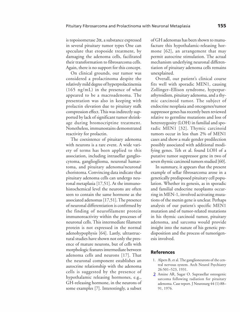
Fig. 1. MRI with contrast showing large tumor extending into the left cavernous sinus and suprasellar region.
Fig. 2. Histologic appearance of the pituitary adenoma on H&E stain (left). Portions of the tumor wereintimately admixed with anaplastic mesenchymal cells (right).

152 Endocrine Pathology Volume 15, Number 2 Summer 2004
Fig. 3. The adenoma cells were strongly immunoreactivity for chromogranin (left) and to a lesser extent forprolactin (right).
Fig. 4. Neurons within the adenoma were large with Nissl-substance-containing amphophilic cyto-plasm and open chromatin with prominent nucleoli (left). Occasional examples were binucleate (center).Neuronal cell processes were strongly immunopositive for neurofilament protein (right).
152

Pituitary Fibrosarcoma and Prolactinoma with Neuronal Metaplasia 153
its MIB1 labeling index was low. Nuclearp53 staining was minimal.
Of particular note was the presence offar larger, full-bodied cells with the featuresof neurons, including ample amphophiliccytoplasm, large open nuclei, and promi-nent nucleoli (Fig. 4). These, too, werereactive for synaptophysin and chromo-granin but not for prolactin. Interveningtissue was seen to contain cell processesstrongly immunoreactive for neurofilamentprotein (Fig. 4). No glial element was seenon glial fibrillary acidic protein (GFAP) stain.
The intimately associated sarcoma com-ponent consisted of nondescript spindlecells with frequent mitotic figures (7/10 HPF)and a high Ki-67 labeling index (60%)(Fig. 5). A reticulin preparation demon-strated the typical pericellular staining pat-tern of sarcoma. On immunohistochemicalstains, these spindle cells were reactive forvimentin but showed no reaction for
smooth muscle actin, keratins, EMA,synaptophysin, chromogranin, GFAP, orpituitary hormones. Lastly, in situ hybrid-ization for chromogranin was positive inthe adenoma cells and neurons, but nega-tive in the sarcoma.
Discussion
Sarcomas of the sellar region are rare.Most occur following irradiation of pitu-itary adenomas or craniopharyngiomas[2,40,42,43,45,56,61,66]. By definition,the tumors occur in the irradiated region,typically after a latency period of 2.5 tomore than 20 yr. Fibrosarcomas predomi-nate over osteosarcomas at a ratio of 4:1[56]. Microscopically, as in the present case,most specimens show an intimate admix-ture of the sarcoma with the adenoma cell.As in our case, no convincing transition of
Fig. 5. The fibrosarcoma element consisted of nondescript spindle cells showing brisk mitotic activity (left)and a high MIB-1 labeling index (right).
154 Endocrine Pathology Volume 15, Number 2 Summer 2004
adenoma to sarcoma has ever beendocumented.
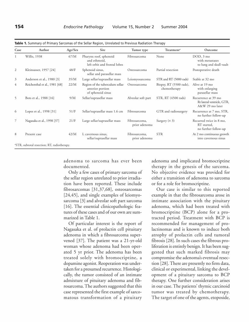
Only a few cases of primary sarcoma ofthe sellar region unrelated to prior irradia-tion have been reported. These includefibrosarcomas [31,37,68], osteosarcomas[24,45], and single examples of leiomyo-sarcoma [3] and alveolar soft part sarcoma[16]. The essential clinicopathologic fea-tures of these cases and of our own are sum-marized in Table 1.
Of particular interest is the report ofNagasaka et al. of prolactin cell pituitaryadenoma in which a fibrosarcoma super-vened [37]. The patient was a 21-yr-oldwoman whose adenoma had been oper-ated 5 yr prior. The adenoma has beentreated solely with bromocriptine, adopamine agonist. Reoperation was under-taken for a presumed recurrence. Histologi-cally, the tumor consisted of an intimateadmixture of pituitary adenoma and fib-rosarcoma. The authors suggested that thiscase represented the first example of sarco-matous transformation of a pituitary
adenoma and implicated bromocriptinetherapy in the genesis of the sarcoma.No objective evidence was provided foreither a transition of adenoma to sarcomaor for a role for bromocriptine.
Our case is similar to this reportedexample in that the fibrosarcoma arose inintimate association with the pituitaryadenoma, which had been treated withbromocriptine (BCP) alone for a pro-tracted period. Treatment with BCP isrecommended for management of pro-lactinomas and is known to induce bothatrophy of prolactin cells and tumoralfibrosis [28]. In such cases the fibrous pro-liferation is entirely benign. It has been sug-gested that such marked fibrosis maycompromise the adenoma’s eventual resec-tion [28]. There are presently no firm data,clinical or experimental, linking the devel-opment of a pituitary sarcoma to BCPtherapy. One further consideration arisesin our case. The patients’ thymic carcinoidtumor was treated by chemotherapy.The target of one of the agents, etoposide,
Table 1. Summary of Primary Sarcomas of the Sellar Region, Unrelated to Previous Radiation Therapy
Case Author Age/Sex Site Tumor type Treatmenta Outcome
1 Willis, 1938 67/M Pharynx roof, sphenoid Fibrosarcoma None DOD, 3 moand ethmoid, with metastasesleft orbit and frontal lobes to lung and skull vault
2 Kleinsasser, 1957 [24] 48/F Sphenoid sinus, Osteosarcoma Partial resection Postoperative deathsellar and parasellar mass
3 Anderson et al., 1980 [3] 35/M Large sellar/suprasellar mass Leiomyosarcoma STR and RT (5000 rads) Stable at 32 mo
4 Reichenthal et al., 1981 [68] 22/M Region of the tuberculum sellae Osteosarcoma Biopsy, RT (5500 rads), Alive at 19 moanterior portion chemotherapy with enlargingof sphenoid sinus parasellar mass
5 Bots et al., 1988 [16] 9/M Sellar/suprasellar mass Alveolar soft part STR, RT (4500 rads) Recurrence at 39 moRt lateral ventricle, GTR,A&W 29 mo later
6 Lopes et al., 1998 [31] 51/F Sellar/suprasellar mass 1.6 cm Fibrosarcoma GTR and radiosurgery Recurrence at 7 mo, STR,no further follow-up
7 Nagasaka et al., 1998 [37] 21/F Large sellar/suprasellar mass Fibrosarcoma, Surgery (× 3) Recurred twice in 8 mo,prior adenoma RT started,
no further follow-up
8 Present case 42/M L cavernous sinus, Fibrosarcoma, STR At 2 mo continuous growthsellar/suprasellar mass prior adenoma into cavernous sinus
aSTR, subtotal resection; RT, radiotherapy.
Pituitary Fibrosarcoma and Prolactinoma with Neuronal Metaplasia 155
is topoisomerase 2α, a substance expressedin several pituitary tumor types One canspeculate that etoposide treatment, bydamaging the adenoma cells, facilitatedtheir transformation to fibrosarcoma cells.Again, there is no support for this concept.
On clinical grounds, our tumor wasconsidered a prolactinoma despite therelatively mild degree of hyperprolactinemia(165 ng/mL) in the presence of whatappeared to be a macroadenoma. Thepresentation was also in keeping withprolactin elevation due to pituitary stalkcompression effect. This was indirectly sup-ported by lack of significant tumor shrink-age during bromocriptine treatment.Nonetheless, immunostains demonstratedreactivity for prolactin.
The coexistence of pituitary adenomawith neurons is a rare event. A wide vari-ety of terms has been applied to thisassociation, including intrasellar ganglio-cytoma, ganglioglioma, neuronal hamar-toma, and pituitary adenoma/neuronalchoristoma. Convincing data indicate thatpituitary adenoma cells can undergo neu-ronal metaplasia [17,51]. At the immuno-histochemical level the neurons are oftenseen to contain the same hormone as theassociated adenomas [17,51]. The presenceof neuronal differentiation is confirmed bythe finding of neurofilament proteinimmunoreactivity within the processes ofneuronal cells. This intermediate filamentprotein is not expressed in the normaladenohypophysis [64]. Lastly, ultrastruc-tural studies have shown not only the pres-ence of mature neurons, but of cells withmorphologic features intermediate betweenadenoma cells and neurons [17]. Thatthe neuronal component establishes anautocrine relationship with the adenomacells is suggested by the presence ofhypothalamic releasing hormones, e.g.,GH-releasing hormone, in the neurons ofsome examples [7]. Interestingly, a subset
of GH adenomas has been shown to manu-facture this hypothalamic-releasing hor-mone [62], an arrangement that maypermit autocrine stimulation. The actualmechanism underlying neuronal differen-tiation of pituitary adenoma cells remainsunexplained.
Overall, our patient’s clinical coursefits well with sporadic MEN1, causingZollinger–Ellison syndrome, hyperpar-athyroidism, pituitary adenoma, and a thy-mic carcinoid tumor. The subject ofendocrine neoplasia and oncogenes/tumorsuppressor genes has recently been reviewedrelative to germline mutations and loss ofheterozygosity (LOH) in familial and spo-radic MEN1 [32]. Thymic carcinoidtumors occur in less than 2% of MEN1cases and show a male gender predilectionpossibly associated with additional modi-fying genes. Teh et al. found LOH of aputative tumor suppressor gene in two ofseven thymic carcinoid tumors studied [60].
In summary, it appears that the presentexample of sellar fibrosarcoma arose in agenetically predisposed pituitary cell popu-lation. Whether its genesis, as in sporadicand familial endocrine neoplasms occur-ring in MEN-1, involved activating muta-tions of the menin gene is unclear. Perhapsanalysis of our patient’s specific MEN1mutation and of tumor-related mutationsin his thymic carcinoid tumor, pituitaryadenoma, and sarcoma would provideinsight into the nature of his genetic pre-disposition and the process of tumorigen-esis involved.
References
1. Alpers B, et al. The ganglioneurons of the cen-tral nervous system. Arch Neurol Psychiatry26:501–523, 1931.
2. Amine AR, Sugar O. Suprasellar osteogenicsarcoma following radiation for pituitaryadenoma. Case report. J Neurosurg 44 (1):88–91, 1976.

156 Endocrine Pathology Volume 15, Number 2 Summer 2004
3. Anderson WR, Cameron JD, Tsai SH. Pri-mary intracranial leiomyosarcoma. Casereport with ultrastructural study. J Neurosurg53(3):401–405, 1980.
4. Angelstein I. [Pathogenesis of acromegaly.].Dtsch Z Nervenheilkd 170(4):337–348, 1953.
5. Arseni C, Horvath L, Carp N, Ciurea V.Intracranial ganglioneuromas in children. ActaNeurochir (Wien) 32(3–4):279–286, 1975.
6. Asa SL, Bilbao JM, Kovacs K, Linfoot JA.Hypothalamic neuronal hamartoma associ-ated with pituitary growth hormone celladenoma and acromegaly. Acta Neuropathol(Berl) 52(3):231–234, 1980.
7. Asa SL, Scheithauer BW, Bilbao JM, et al.A case for hypothalamic acromegaly: a clini-copathological study of six patients withhypothalamic gangliocytomas producinggrowth hormone-releasing factor. J ClinEndocrinol Metab 58(5):796–803, 1984.
8. Asa SL, Kovacs K, Tindall GT, Barrow DL,Horvath E, Vecsei P. Cushing’s disease associ-ated with an intrasellar gangliocytoma produc-ing corticotrophin-releasing factor. Ann InternMed 101(6):789–793, 1984.
9. Baysefer A, Gezen F, Kayali H, Erdogan E,Timurkaynak E, Celasun B. Intrasellargangliocytoma resembling pituitary adenoma.Minim Invasive Neurosurg 40(3):107–109,1997.
10. Bevan JS, Asa SL, Rossi ML, Esiri MM,Adams CB, Burke CW. Intrasellar ganglio-cytoma containing gastrin and growth hor-mone-releasing hormone associated with agrowth hormone-secreting pituitary adenoma.Clin Endocrinol (Oxf) 30(3):213–224, 1989.
11. Burchiel KJ, Shaw CM, Kelly WA. A mixedfunctional microadenoma and ganglion-euroma of the pituitary fossa. Case report.J Neurosurg 58(3):416–420, 1983.
12. Casper J. Ueber neurogene Geschwulste imHinterlappen der Hypopyse. Zentralb AllgPathol 56:404–411, 1933.
13. Corbetta S, Pizzocaro A, Peracchi M, Beck-Peccoz P, Faglia G, Spada A. Multiple endo-crine neoplasia type 1 in patients withrecognized pituitary tumours of differenttypes. Clin Endocrinol (Oxf ) 47(5):507–512,1997.
14. Fischer EG, Morris JH, Kettyle WM. Intra-sellar gangliocytoma and syndromes of pitu-itary hypersecretion. Case report. J Neurosurg59(6):1071–1075, 1983.
15. Geddes JF, Jansen GH, Robinson SF, et al.“Gangliocytomas” of the pituitary: a hetero-geneous group of lesions with differing histo-genesis. Am J Surg Pathol 24(4):607–613,2000.
16. Bots GT, Tijssen CC, Wijnalda D, Teepen JL.Alveolar soft part sarcoma of the pituitarygland with secondary involvement of the rightcerebral ventricle. Br J Neurosurg 2(1):101–107, 1988.
17. Horvath E, Kovacs K, Scheithauer BW, LloydRV, Smyth HS. Pituitary adenoma with neu-ronal choristoma (PANCH): composite lesionor lineage infidelity? Ultrastruct Pathol18(6):565–574, 1994.
18. Horvath E, Kovacs K, Tran A, ScheithauerBW. Ganglion cells in the posterior pituitary:result of ectopia or transdifferentiation? ActaNeuropathol (Berl) 100(1):106–110, 2000.
19. Hourihane M, Moran M, Burke M. Intrasellargangliocytoma associated with functionalpituitary gland microadenoma. J NeuropatholExp Neurol 51:364 (Abstract), 1992.
20. Iwase T, Nishizawa S, Baba S, et al. Intrasellarneuronal choristoma associated with growthhormone-producing pituitary adenoma con-taining amyloid deposits. Hum Pathol26(8):925–928, 1995.
21. Jakumeit HD, Zimmermann V, Guiot G.Intrasellar gangliocytomas. Report of fourcases. J Neurosurg 40(5):626–630, 1974.
22. Kamel OW, Horoupian DS, Silverberg GD.Mixed gangliocytoma-adenoma: a distinctneuroendocrine tumor of the pituitary fossa.Hum Pathol 20(12):1198–1203, 1989.
23. Kiyono H. Die Histopathologie der hypo-physe. Virchows Arch A Pathol Anat Histo-pathol 259:388–465, 1926.
24. Kleinsasser O, Albrecht H. Zur Kenntnis derOsteosarkome des Stirn- und Keilbeines.Arch Ohren Heilk 170:595–603, 1957.
25. Kovacs K, Lloyd R, Horvath E, et al. Silentsomatotroph adenomas of the human pitu-itary. A morphologic study of three casesincluding immunocytochemistry, electronmicroscopy, in vitro examination, and in situhybridization. Am J Pathol 134(2):345–353,1989.
26. Kovacs K, Stefaneanu L, Horvath E, et al.Effect of dopamine agonist medication onprolactin producing pituitary adenomas.A morphological study including immuno-cytochemistry, electron microscopy and in situ

Pituitary Fibrosarcoma and Prolactinoma with Neuronal Metaplasia 157
hybridization. Virchows Arch A Pathol AnatHistopathol 418(5):439–446, 1991.
27. Kroh H, Marchel A. Intrasellar neuronalhamartoma associated with pituitary adenoma.Case report. Folia Neuropathol 38(3):119–124, 2000.
28. Landolt AM, Froesch ER. [Prolactin produc-ing hypophyseal adenoma: diagnosis andtherapeutic possibilities]. Schweiz MedWochenschr 115(23):803–809, 1985.
29. Lapresle J, Racadot J, Said G. [Myasthenia,thymoma and hypophyseal tumor associatedwith heterogenous adenomatous proliferationand ganglioneuroma of the sella turcica]. J NeurolSci 28(2):249–254, 1976.
30. Li JY, Racadot O, Kujas M, Kouadri M,Peillon F, Racadot J. Immunocytochemistryof four mixed pituitary adenomas andintrasellar gangliocytomas associated with dif-ferent clinical syndromes: acromegaly, amen-orrhea-galactorrhea, Cushing’s disease andisolated tumoral syndrome. Acta Neuropathol(Berl) 77(3):320–328, 1989.
31. Lopes MB, Lanzino G, Cloft HJ, WinstonDC, Vance ML, Laws ER, Jr. Primary fibro-sarcoma of the sella unrelated to previousradiation therapy. Mod Pathol 11(6):579–584, 1998.
32. Marx S, Spiegel AM, Skarulis MC, DoppmanJL, Collins FS, Liotta LA. Multiple endocrineneoplasia type 1: clinical and genetic topics.Ann Intern Med 129(6):484–494, 1998.
33. McCowen KC, Glickman JN, Black PM,Zervas NT, Lidov HG, Garber JR. Ganglio-cytoma masquerading as a prolactinoma. Casereport. J Neurosurg 91(3):490–495, 1999.
34. Morikawa M, Tamaki N, Kokunai T, Imai Y.Intrasellar pituitary gangliocyto-adenoma pre-senting with acromegaly: case report. Neuro-surgery 40(3):611–614; discussion 614–615,1997.
35. Muller W, Marcos F. [The occurrence of gan-glion cells in a pituitary tumor.]. VirchowsArch 325(6):733–736, 1954.
36. Muller W. Uber das gemeinsame Vorkommeneines Hypophysenadenomas mit einemGangliocytom in Zwei Fallen: ein Beitrag zurFrage der Neurosekretion. Acta Neurochir(Wien) 7:13–29, 1959.
37. Nagasaka T, Nakashima N, Furui A, Waka-bayashi T, Yoshida J. Sarcomatous transforma-tion of pituitary adenoma after bromocriptinetherapy. Hum Pathol 29(2):190–193, 1998.
38. Nikonov AA. [Hormonally active (somatotro-pin) hypophyseal ganglioneuroma.]. ArkhPatol 43(12):48–52, 1981.
39. Oberg K, Skogseid B, Eriksson B. Multipleendocrine neoplasia type 1 (MEN-1). Clini-cal, biochemical and genetical investigations.Acta Oncol 28(3):383–387, 1989.
40. Paulus W, Slowik F, Jellinger K. Primaryintracranial sarcomas: histopathological fea-tures of 19 cases. Histopathology 18(5):395–402, 1991.
41. Pearce SH. Multiple endocrine neoplasia type1 (MEN1): recent advances. Clin Endocrinol(Oxf ) 47(5):513–514, 1997.
42. Pieterse S, Dinning TA, Blumbergs PC. Postir-radiation sarcomatous transformation of apituitary adenoma: a combined pituitarytumor. Case report. J Neurosurg 56(2):283–286, 1982.
43. Powell HC, Marshall LF, Ignelzi RJ. Post-irradiation pituitary sarcoma. Acta Neuro-pathol (Berl) 39(2):165–167, 1977.
44. Puchner MJ, Herrmann HD. Intrasellarpituitary gangliocyto-adenoma presentingwith acromegaly: case report. Neurosurgery42(5):1197–1199, 1998.
45. Reichenthal E, Cohen ML, Manor CR,Marshak G, Matz S, Shalit MN. Primaryosteogenic sarcoma of the sellar region. Casereport. J Neurosurg 55(2):299–302, 1981.
46. Rhodes RH, Dusseau JJ, Boyd AS, Jr., KniggeKM. Intrasellar neural-adenohypophysealchoristoma. A morphological and immuno-cytochemical study. J Neuropathol ExpNeurol 41(3):267–280, 1982.
47. Russell D, Rubistein L. Pathology of tumoursof the nervous system. In: D Russell, L Rubistein,eds. Pathology of tumours in the nervous sys-tem. 5th ed. London: Edward Arnold; 1989;507–517.
48. Sabel MC, Hans VH, Reifenberger G. Mixedgangliocytoma/pituitary adenoma. ArchNeurol 57(4):587–588, 2000.
49. Saeger W, Puchner MJ, Ludecke DK. Com-bined sellar gangliocytoma and pituitaryadenoma in acromegaly or Cushing’s disease.A report of 3 cases. Virchows Arch 425(1):93–99, 1994.
50. Samaan NA, Ouais S, Ordonez NG, ChoksiUA, Sellin RV, Hickey RC. Multiple endo-crine syndrome type I. Clinical, laboratoryfindings, and management in five families.Cancer 64(3):741–752, 1989.

158 Endocrine Pathology Volume 15, Number 2 Summer 2004
51. Scheithauer BW, Kovacs K, Randall RV,Horvath E, Okazaki H, Laws ER, Jr. Hypo-thalamic neuronal hamartoma and adeno-hypophyseal neuronal choristoma: theirassociation with growth hormone adenoma ofthe pituitary gland. J Neuropathol Exp Neurol42(6):648–663, 1983.
52. Scheithauer BW, Horvath E, Kovacs K, et al.Prolactin-producing pituitary adenoma andcarcinoma with neuronal components—a metaplastic lesion. Pituitary 1(3–4):197–205, 1999.
53. Scindler C, Neurnberger F, Schachenmayer W.Invasive pituitary adenoma in a patient withhypothalamic neuronal hamartoma. ClinNeuropathol 12:264 (Abstract), 1993.
54. Serebrin R, Robertson DM. Ganglioneuromaarising in the pituitary fossa: a twenty yearfollow-up. J Neurol Neurosurg Psychiatry47(1):97–98, 1984.
55. Sharma MC, Karak AK, Mahapatra AK,Sarkar C. Pituitary adenoma with neuronalchoristoma: a report of two rare cases. ClinNeurol Neurosurg 101(2):128–132, 1999.
56. Shi T, Farrell MA, Kaufmann JC. Fibrosar-coma complicating irradiated pituitaryadenoma. Surg Neurol 22(3):277–284, 1984.
57. Skogseid B, Rastad J, Oberg K. Multipleendocrine neoplasia type 1. Clinical featuresand screening. Endocrinol Metab Clin NorthAm 23(1):1–18, 1994.
58. Slowik F, Fazekas I, Balint K, et al. Intrasellarhamartoma associated with pituitary adenoma.Acta Neuropathol (Berl) 80(3):328–333, 1990.
59. Tajika Y, Kubo O, Takeshita M, Tajika T,Shimizu T, Kitamura K. [An intracranialcollision tumor composed of intrasellargangliocytoma and pituitary adenoma.].No Shinkei Geka 17(12):1181–1186, 1989.
60. Teh BT, Zedenius J, Kytola S, et al. Thymiccarcinoids in multiple endocrine neoplasiatype 1. Ann Surg 228(1):99–105, 1998.
61. Terry R, Hyams V, Davidoff L. Combinednon-metastasizing fibrosarcoma and chro-mophobe tumor of the pituitary. Cancer12:791–798, 1958.
62. Thapar K, Kovacs K, Stefaneanu L, et al.Overexpression of the growth-hormone-releasing hormone gene in acromegaly asso-ciated pituitary tumors. An event associatedwith neoplastic progression and aggressivebehavior. Am J Pathol 151(3):769–784,1997.
63. Towfighi J, Salam MM, McLendon RE, Pow-ers S, Page RB. Ganglion cell-containingtumors of the pituitary gland. Arch Pathol LabMed 120(4):369–377, 1996.
64. Trojanowski JQ, Gordon D, Obrocka M, LeeVM. The developmental expression ofneurofilament and glial filament proteins inthe human pituitary gland: an immunohis-tochemical study with monoclonal antibod-ies. Brain Res 315(2):229–239, 1984.
65. Ule G, Waidelich FW. Neurosecretoric nerve-cell choristoma in the anterior pituitary(author’s transl)]. Acta Neuropathol (Berl)36(1):81–84, 1976.
66. Waltz TA, Brownell B. Sarcoma: a possible lateresult of effective radiation therapy forpituitary adenoma. Report of two cases. J Neuro-surg 24(5):901–907, 1966.
67. Weil RJ, Vortmeyer AO, Huang S, et al. 11q13allelic loss in pituitary tumors in patients withmultiple endocrine neoplasia syndrome type1. Clin Cancer Res 4(7):1673–1678, 1998.
68. Willis R. Pathological study of tumors of thepituitary region. Med J Aust 1:281–291,1938.