Role of Phosphoinositide 3Kinase in the Aggressive Tumor Growth of HT1080 Human Fibrosarcoma Cells
12
10.1128/MCB.21.17.5846-5856.2001. 2001, 21(17):5846. DOI: Mol. Cell. Biol. Stanbridge Tencati, Christa Gray, Young E. Whang and Eric J. Swati Gupta, Selma Stuffrein, Rina Plattner, Michael Human Fibrosarcoma Cells Aggressive Tumor Growth of HT1080 Role of Phosphoinositide 3-Kinase in the http://mcb.asm.org/content/21/17/5846 Updated information and services can be found at: These include: REFERENCES http://mcb.asm.org/content/21/17/5846#ref-list-1 at: This article cites 54 articles, 30 of which can be accessed free CONTENT ALERTS more» articles cite this article), Receive: RSS Feeds, eTOCs, free email alerts (when new http://journals.asm.org/site/misc/reprints.xhtml Information about commercial reprint orders: http://journals.asm.org/site/subscriptions/ To subscribe to to another ASM Journal go to: on January 6, 2014 by guest http://mcb.asm.org/ Downloaded from on January 6, 2014 by guest http://mcb.asm.org/ Downloaded from
-
Upload
independent -
Category
Documents
-
view
4 -
download
0
Transcript of Role of Phosphoinositide 3Kinase in the Aggressive Tumor Growth of HT1080 Human Fibrosarcoma Cells

10.1128/MCB.21.17.5846-5856.2001.
2001, 21(17):5846. DOI:Mol. Cell. Biol. StanbridgeTencati, Christa Gray, Young E. Whang and Eric J. Swati Gupta, Selma Stuffrein, Rina Plattner, Michael Human Fibrosarcoma CellsAggressive Tumor Growth of HT1080 Role of Phosphoinositide 3-Kinase in the
http://mcb.asm.org/content/21/17/5846Updated information and services can be found at:
These include:
REFERENCEShttp://mcb.asm.org/content/21/17/5846#ref-list-1at:
This article cites 54 articles, 30 of which can be accessed free
CONTENT ALERTS more»articles cite this article),
Receive: RSS Feeds, eTOCs, free email alerts (when new
http://journals.asm.org/site/misc/reprints.xhtmlInformation about commercial reprint orders: http://journals.asm.org/site/subscriptions/To subscribe to to another ASM Journal go to:
on January 6, 2014 by guesthttp://m
cb.asm.org/
Dow
nloaded from
on January 6, 2014 by guesthttp://m
cb.asm.org/
Dow
nloaded from
MOLECULAR AND CELLULAR BIOLOGY,0270-7306/01/$04.0010 DOI: 10.1128/MCB.21.17.5846–5856.2001
Sept. 2001, p. 5846–5856 Vol. 21, No. 17
Copyright © 2001, American Society for Microbiology. All Rights Reserved.
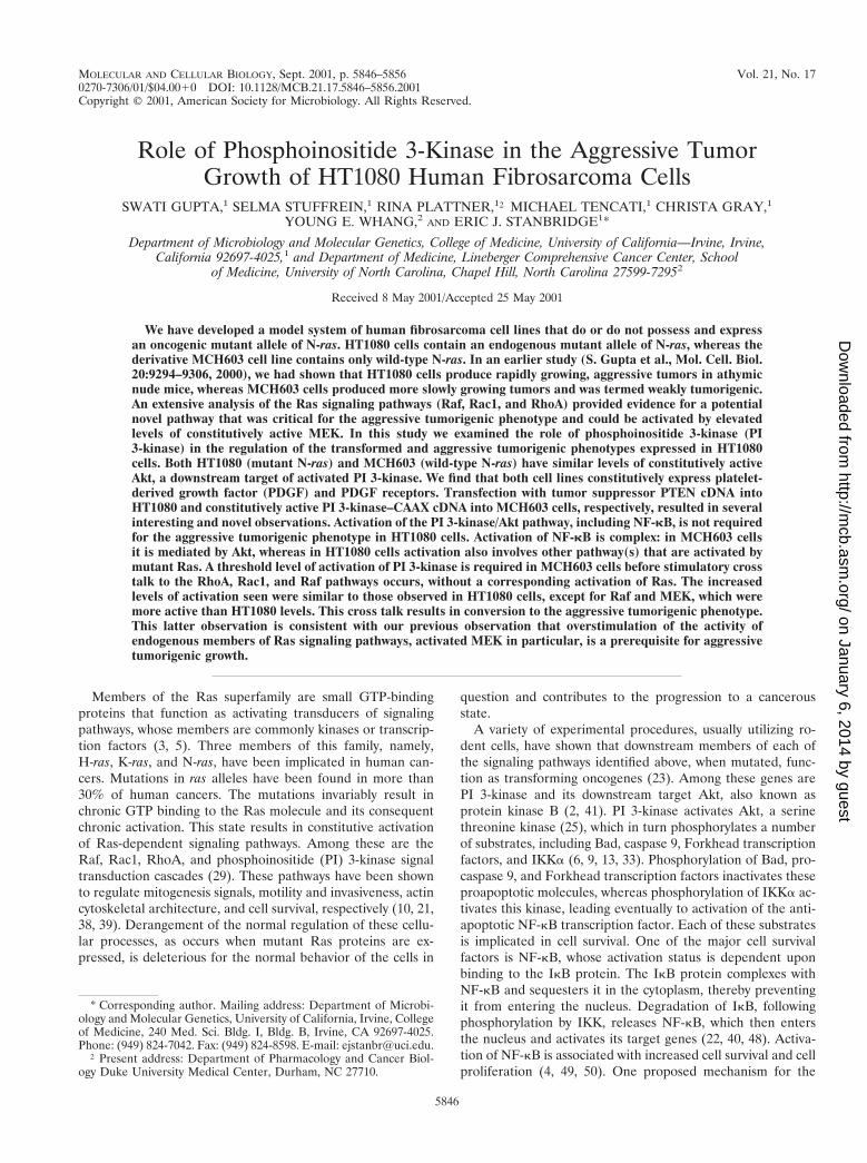
Role of Phosphoinositide 3-Kinase in the Aggressive TumorGrowth of HT1080 Human Fibrosarcoma Cells
SWATI GUPTA,1 SELMA STUFFREIN,1 RINA PLATTNER,1† MICHAEL TENCATI,1 CHRISTA GRAY,1
YOUNG E. WHANG,2 AND ERIC J. STANBRIDGE1*
Department of Microbiology and Molecular Genetics, College of Medicine, University of California—Irvine, Irvine,California 92697-4025,1 and Department of Medicine, Lineberger Comprehensive Cancer Center, School
of Medicine, University of North Carolina, Chapel Hill, North Carolina 27599-72952
Received 8 May 2001/Accepted 25 May 2001
We have developed a model system of human fibrosarcoma cell lines that do or do not possess and expressan oncogenic mutant allele of N-ras. HT1080 cells contain an endogenous mutant allele of N-ras, whereas thederivative MCH603 cell line contains only wild-type N-ras. In an earlier study (S. Gupta et al., Mol. Cell. Biol.20:9294–9306, 2000), we had shown that HT1080 cells produce rapidly growing, aggressive tumors in athymicnude mice, whereas MCH603 cells produced more slowly growing tumors and was termed weakly tumorigenic.An extensive analysis of the Ras signaling pathways (Raf, Rac1, and RhoA) provided evidence for a potentialnovel pathway that was critical for the aggressive tumorigenic phenotype and could be activated by elevatedlevels of constitutively active MEK. In this study we examined the role of phosphoinositide 3-kinase (PI3-kinase) in the regulation of the transformed and aggressive tumorigenic phenotypes expressed in HT1080cells. Both HT1080 (mutant N-ras) and MCH603 (wild-type N-ras) have similar levels of constitutively activeAkt, a downstream target of activated PI 3-kinase. We find that both cell lines constitutively express platelet-derived growth factor (PDGF) and PDGF receptors. Transfection with tumor suppressor PTEN cDNA intoHT1080 and constitutively active PI 3-kinase–CAAX cDNA into MCH603 cells, respectively, resulted in severalinteresting and novel observations. Activation of the PI 3-kinase/Akt pathway, including NF-kB, is not requiredfor the aggressive tumorigenic phenotype in HT1080 cells. Activation of NF-kB is complex: in MCH603 cellsit is mediated by Akt, whereas in HT1080 cells activation also involves other pathway(s) that are activated bymutant Ras. A threshold level of activation of PI 3-kinase is required in MCH603 cells before stimulatory crosstalk to the RhoA, Rac1, and Raf pathways occurs, without a corresponding activation of Ras. The increasedlevels of activation seen were similar to those observed in HT1080 cells, except for Raf and MEK, which weremore active than HT1080 levels. This cross talk results in conversion to the aggressive tumorigenic phenotype.This latter observation is consistent with our previous observation that overstimulation of the activity ofendogenous members of Ras signaling pathways, activated MEK in particular, is a prerequisite for aggressivetumorigenic growth.
Members of the Ras superfamily are small GTP-bindingproteins that function as activating transducers of signalingpathways, whose members are commonly kinases or transcrip-tion factors (3, 5). Three members of this family, namely,H-ras, K-ras, and N-ras, have been implicated in human can-cers. Mutations in ras alleles have been found in more than30% of human cancers. The mutations invariably result inchronic GTP binding to the Ras molecule and its consequentchronic activation. This state results in constitutive activationof Ras-dependent signaling pathways. Among these are theRaf, Rac1, RhoA, and phosphoinositide (PI) 3-kinase signaltransduction cascades (29). These pathways have been shownto regulate mitogenesis signals, motility and invasiveness, actincytoskeletal architecture, and cell survival, respectively (10, 21,38, 39). Derangement of the normal regulation of these cellu-lar processes, as occurs when mutant Ras proteins are ex-pressed, is deleterious for the normal behavior of the cells in
question and contributes to the progression to a cancerousstate.
A variety of experimental procedures, usually utilizing ro-dent cells, have shown that downstream members of each ofthe signaling pathways identified above, when mutated, func-tion as transforming oncogenes (23). Among these genes arePI 3-kinase and its downstream target Akt, also known asprotein kinase B (2, 41). PI 3-kinase activates Akt, a serinethreonine kinase (25), which in turn phosphorylates a numberof substrates, including Bad, caspase 9, Forkhead transcriptionfactors, and IKKa (6, 9, 13, 33). Phosphorylation of Bad, pro-caspase 9, and Forkhead transcription factors inactivates theseproapoptotic molecules, whereas phosphorylation of IKKa ac-tivates this kinase, leading eventually to activation of the anti-apoptotic NF-kB transcription factor. Each of these substratesis implicated in cell survival. One of the major cell survivalfactors is NF-kB, whose activation status is dependent uponbinding to the IkB protein. The IkB protein complexes withNF-kB and sequesters it in the cytoplasm, thereby preventingit from entering the nucleus. Degradation of IkB, followingphosphorylation by IKK, releases NF-kB, which then entersthe nucleus and activates its target genes (22, 40, 48). Activa-tion of NF-kB is associated with increased cell survival and cellproliferation (4, 49, 50). One proposed mechanism for the
* Corresponding author. Mailing address: Department of Microbi-ology and Molecular Genetics, University of California, Irvine, Collegeof Medicine, 240 Med. Sci. Bldg. I, Bldg. B, Irvine, CA 92697-4025.Phone: (949) 824-7042. Fax: (949) 824-8598. E-mail: [email protected].
† Present address: Department of Pharmacology and Cancer Biol-ogy Duke University Medical Center, Durham, NC 27710.
5846
on January 6, 2014 by guesthttp://m
cb.asm.org/
Dow
nloaded from

activation of IKK is phosphorylation mediated by Akt (33, 42).However, other mechanisms also exist that do not involve thedegradation of IkB (27, 44).
In addition to being activated by Ras-GTP, PI 3-kinase mayalso be activated directly by contact with activated growthfactor receptors, including platelet-derived growth factor(PDGF) (20, 46). Dysregulated PI 3-kinase activity is likely toplay an important role in cancer progression. One indication ofthis has been the identification of the PTEN tumor suppressorgene (26, 45). PTEN is a common target of inactivating muta-tions in a variety of sporadic human cancers. In addition, germline mutations in the PTEN gene are associated with Cowden’sdisease, an inherited hamartoma syndrome that includes anelevated risk of breast and thyroid cancers (31). The PTENprotein functions as both a protein and a lipid phosphatase. Itis the lipid phosphatase activity that is critical for its tumor-suppressing function (30). PTEN lipid phosphatase catalyzesthe dephosphorylation of the 3 position of PI 3,4,5-triphos-phate (PIP3) and PI 3,4,-biphosphate (PIP2), both of which arethe lipid byproducts of the lipid kinase activity of PI 3-kinase.The Akt molecule binds to PIP3 via its pleckstrin homology(PH) domain. In this complex with PIP3, Akt is then phos-phorylated and activated by the PI-dependent kinase, PDK-1(1, 8). Thus, normal cells integrate the activities of PI 3-kinaseand PTEN to facilitate homeostasis with respect to PI 3-ki-nase-mediated signal transduction and cell cycle control. Over-activation of PI 3-kinase or loss of PTEN function is likely tocause dysregulation of this finely balanced control. An illustra-tion of this is that expression of wild-type PTEN transfectedinto PTEN-null cancer cells results in induction of G1 arrestand/or apoptosis (12, 16). Conversely, this arrest can be over-ridden by a constitutively active form of Akt (52, 55).
We have developed an experimental model system compris-ing the human fibrosarcoma cell line HT1080, which possessesone mutant N-ras allele, and its derivative, MCH603, which hasdeleted the mutant allele and possesses only wild-type N-ras(35). Examination of these cells has shown that HT1080 has atypical transformed phenotype in culture, including disorga-nized actin stress fibers and the ability to grow in soft agar, plusan aggressive tumorigenic phenotype in vivo in immunodefi-cient mice. By contrast, MCH603 cells have “reversed” theirtransformed phenotype; they have restored a well-organizedactin stress fiber distribution in the cytoplasm and are nolonger able to grow in soft agar. When implanted into immu-nodeficient mice they continue to form tumors but with muchslower kinetics. We have described these cells as having a weaktumorigenic phenotype (35).
When we examined the activation of a number of Ras sig-naling pathways, namely, the Raf, Rac1, and RhoA pathways,we found that all members were constitutively active inHT1080 but had basal activity in MCH603 cells (36). However,we noted that Akt was constitutively active in both cell lines.Since this was not due to oncogenic Ras expression inMCH603 cells, we looked for another explanation. In thisstudy we found that both cell lines constitutively synthesize andsecrete PDGF and contain cell surface PDGF receptor(PDGFR). Thus, this provides a mechanism for constitutiveactivation of PI 3-kinase, resulting in the activation of Akt.
Although HT1080 and MCH603 cells have different trans-formed and tumorigenic phenotypes and yet both have consti-
tutively active Akt, it is formally possible that there may bequantitative and qualitative differences in the activation of PI3-kinase and/or Akt and their downstream substrates in thetwo cell lines that play a role in the expression of these phe-notypes. In order to determine this, we have modulated theactivation of PI 3-kinase and Akt by stable transfection ofHT1080 and MCH603 cells with PTEN and an activated mu-tant of PI 3-kinase (hereafter termed PI3Kact), respectively.Examination of the biochemical and biological properties ofthe parental and transfectant cells has revealed several unex-pected and novel findings with respect to both signal transduc-tion pathways and biological behavior.
MATERIALS AND METHODS
Molecular constructs. The expression plasmids used in this study wereas follows: PI3Kact-pCMV(hyg)P110CAAX59myc is derived from pSG5P110CAAX59myc (51) and encodes the catalytic domain of PI 3-kinase. The consti-tutively active protein product, PI3Kact, is permanently plasma membrane asso-ciated. The construct pCDNA3PTEN(wt) (Neo) encodes a full-length wild-typePTEN cDNA (52), whose expression is driven from a heterologous cytomegalo-virus promoter.
Cell culture and stable transfection. The HT1080 cell line has one mutant andone wild-type N-ras allele (28, 35). MCH603 is a variant of HT1080 and containsonly wild-type N-ras (35). The cell lines were maintained in Dulbecco minimalessential medium (DMEM) supplemented with 10% fetal calf serum (FCS; LifeTechnologies). The HT1080 and MCH603 cell lines were transfected with thePTEN(wt) and PI3Kact plasmids, respectively. Clones from each transfectionwere selected and maintained in medium containing the relevant selective anti-biotic (either 800 mg of Geneticin [Gibco-BRL] or 36 U of hygromycin B[Calbiochem] per ml for the HT1080 and MCH603 transfectants, respectively).Subconfluent (70%) 100-mm dishes of MCH603 cells or HT1080 cells weretransfected with 5 mg of linearized DNA or vector control DNA, using 30 ml ofLipofectin (Gibco-BRL) in Optimem medium (Gibco-BRL).
Growth in soft agar. Logarithmically growing cells (104 or 106) were plated insingle-cell suspension in a 0.3% top agar overlay in DMEM supplemented with10% FCS, above a 0.5% bottom agar layer (in DMEM–10% FCS) in 60-mmdishes as previously described (35). Plates were fed periodically with 1 ml ofDMEM–10% FCS. Colonies (.0.1 mm) were inspected under the microscopeand counted after 3 weeks.
Actin cytoskeleton staining and morphology. Cells grown on glass slides(Nunc) were washed with phosphate-buffered saline (PBS) and fixed with 3.7%paraformaldehyde in PBS for 10 min. After a wash with PBS, cells were perme-abilized with PBS containing 0.1% Triton X-100 for 5 min. The slides were thenwashed, and the actin stress fibers were visualized by staining the cells withfluorescein-conjugated phalloidin (0.005 U/ml; Molecular Probes) for 20 min atroom temperature and mounted in ProLong Fade antifade (Molecular Probes).
Immunoblot analyses. Subconfluent cells were serum starved for 18 h, and thecells were then lysed in lysis buffer comprised of 1% sodium dodecyl sulfate(SDS) in 20 mM Tris (pH 7.4), 1 mM CaCl2, 1 mM phenylmethylsulfonylfluoride, and 0.2 mM sodium orthovanadate. Total cell lysates, each containing60 mg of protein, were electrophoresed by SDS–7.5% polyacrylamide gel elec-trophoresis (PAGE) and transferred to Immobilon-P membranes (Millipore).The membranes were then probed with the relevant antibodies. These includedPDGFR-a and PDGFR-b (Santa Cruz Biotechnology), Akt/PKB, Phospho-Akt/PKB (Ser473), total Bad, Phospho-Bad, total IkBa, and Phospho-IkBa (NewEngland Biolabs). Following incubation with horseradish peroxidase-conjugatedsecondary antibody, bound proteins were detected by incubation with a chemi-luminescent detection system (Pierce) as previously described (7). In order totest for secreted PDGF in the conditioned medium, subconfluent HT1080 andMCH603 cells were exposed to serum-free medium for 18 h. The conditionedmedium was then concentrated in the Centricon (Millipore) apparatus, followedby PAGE under reducing or nonreducing conditions and immunoblotting, usingPDGF-A (E-10) and PDGF-B (P-20) antibodies (Santa Cruz Biotechnology).
Activated Ras, Rac1, and RhoA assays. Subconfluent cells were serum starvedfor 18 h and then lysed with 1 3 Mg21 lysis buffer (Ras and Rac Activation AssayKits; Upstate Biotechnology). Each cell lysate (500 mg) was affinity precipitatedwith 10 ml of Raf-1 RBD, PAK-1 PBD agarose, or glutathione S-transferase(GST)-C21–Sepharose conjugate (43) at 4°C overnight for the Ras, Rac-Cdc42,or RhoA activation assays, respectively. The beads were collected, washed, andresuspended in 63 Laemmli sample buffer. Western blot analysis was performed
VOL. 21, 2001 ROLE OF PI 3-KINASE IN AGGRESSIVE TUMOR GROWTH 5847
on January 6, 2014 by guesthttp://m
cb.asm.org/
Dow
nloaded from

as described elsewhere (7), using 1 mg of mouse monoclonal anti-Ras, anti-Rac1(Upstate Biotechnology), and anti-RhoA (Santa Cruz Biotechnology) antibodiesper ml. Horseradish peroxidase-conjugated anti-mouse immunoglobulin G (San-ta Cruz Biotechnology) was used as the secondary antibody. A chemilumines-cence detection system (Pierce) was used for detection of the relevant proteins.To determine the total Ras, Rac1, or RhoA levels, immunoblots were performedusing N-Ras(F155), Rac1(C-14), or RhoA(26C4) antibodies (Santa Cruz Bio-technology) that recognize total protein.
Kinase assays. MEK, ERK, JNK, and Akt kinase assays were performedaccording to the manufacturer’s protocols (New England Biolabs), using sub-confluent cultures that had been serum starved (0.25% FCS) for 18 h, and havebeen described elsewhere (18). Briefly, cells were washed twice with PBS,scraped into 500 ml of lysis buffer, and incubated on ice for 20 min. Aftercentrifugation at 14,000 3 g for 20 min, the supernatants were incubated with therelevant antibodies. The resulting immunoprecipitates were employed in kinaseassays. The activated MEK assay was carried out by incubating immunoprecipi-tated phospho-MEK with ERK protein and cold ATP (New England BiolabsMEK1/2Kinase Assay Kit). The activated ERK assay was carried out by incu-bating immunoprecipitated phospho-ERK with Elk-1 fusion protein and coldATP (New England Biolabs p44/p42 ERK Assay Kit). The JNK assays werecarried out by incubating the JNK–c-Jun fusion protein complex with cold ATP(New England Biolabs JNK/SAPK Assay Kit). The Akt-P assay was carried outby incubating the immunoprecipitated total Akt with GSK3 protein and coldATP (New England Biolabs Akt Assay Kit). For MEK, ERK, and JNK assays,the relevant gel was transferred onto an Immobilon membrane, and Western blotanalysis was performed. The blots were performed using phospho-ERK (Thr202/Tyr204) monoclonal antibody for the MEK assay, phospho-Elk-1 (Ser383) poly-clonal antibody for the ERK assay, phospho–c-Jun (Ser63) polyclonal antibodyfor the JNK assay, and phospho-GSK3 a/b (Ser219) rabbit polyclonal antibodyfor the Akt assay. The Raf-1 assay was performed as described by Graham et al.(17). For the Raf-1 assay, the g-32P-labeled mitogen-activated protein (MAP)Kinase (ERK) proteins in the gel were visualized by autoradiography. To deter-mine the total Raf, MEK, ERK, JNK, and Akt levels, immunoblots were per-formed using the respective antibodies that recognize total protein.
Elk-1 and NF-kB luciferase reporter assays. To measure Elk-1 activation, adual luciferase reporter assay kit (Promega) was used as previously described(18). For NF-kB assays, approximately 2 3 105 parental HT1080 and MCH603cells and the MCH603/PI3Kact or HT1080/PTEN stable transfectant cells werecotransfected in six-well plates with the pUC13-based D56FosdE-luc plasmid(measures basal expression but is not Ras responsive) and the NF-kB reporter,(HIV-kB)3-luc (54). The latter plasmid has three tandem copies of the twoadjacent NF-kB sites from the human immunodeficiency virus enhancer (six totaltandem NF-kB sites) inserted just upstream of the minimal Fos promoterpresent in D56FosdE-luc. The Effectene kit (Qiagen) was used for these transienttransfections. Following transfection, the cells were kept in serum-starved me-dium for 24 h. Tumor necrosis factor alpha (TNF-a; 10 ng/ml) was then addedto the culture medium, and both treated and untreated control cultures wereincubated for a further 4-h period. The luciferase activity of each sample wasmeasured with the dual luciferase assay kit (Promega) and normalized with aninternal control Renilla luciferase.
Tumorigenicity assays. Cells were trypsinized and resuspended in 0.2 ml ofDMEM, and then 107 cells were injected subcutaneously into the flanks of 4- to6-week-old nude athymic mice. Tumors were measured in three dimensions withlinear calipers at weekly intervals.
RESULTS
We have shown previously that HT1080 (mutant N-ras) cellshave constitutively active Raf-dependent (Raf/MEK/ERK/Elk-1), Rac1 (Rac1/Cdc42/JNK), and RhoA signaling pathways(18). Conversely, MCH603 (wild-type N-ras) cells have basallevels of activity of these signal transduction proteins (18).Interestingly, both HT1080 and MCH603 cells have significantlevels of constitutively active Akt. The fact that MCH603 doesnot possess a mutant ras allele and yet has constitutively activelevels of Akt, approximating those found in HT1080 cells,infers an alternative mechanism of chronic activation.
HT1080 and MCH603 constitutively secrete PDGF. Concen-trated conditioned media and cell lysates from both HT1080and MCH603 serum-starved cell cultures were electropho-
resed and immunoblotted with antibodies to PDGF-A andPDGF-B. Both forms of PDGF were expressed at similar levelsby both cell types. However, whereas PDGF-A is secreted intothe medium, PDGF-B remains associated with the cells (Fig. 1A).Analyses of PDGF dimers under nonreducing conditions indi-cated that the predominant secreted form is PDGF-AA (data notshown). Immunoblotting of cell lysates showed that both the aand b forms of the PDGFR are expressed (Fig. 1B). Constitu-tive secretion of PDGF-A and subsequent binding to and ac-tivation of its cognate receptor is, therefore, the probable mech-anism for downstream activation of PI 3-kinase and Akt. It isknown that the catalytic subunit of PI 3-kinase associates with,and is activated by, the autophosphorylated PDGFR (20, 34, 46).
Modulation of PI 3-kinase and Akt/PKB activity. (i) HT1080cells. We wished to downregulate constitutive activity of PI3-kinase and/or Akt in HT1080 cells. Initially, we attempteddownregulation of PI 3-kinase activity via stable transfectionwith PI 3-kinase dominant-negative cDNAs. Unfortunately,none of the constructs tested (24) had the desired effect (datanot shown). Thus, we resorted to expressing the tumor sup-pressor protein PTEN in these cells. PTEN is a dual-specificityphosphatase that catalyzes the dephosphorylation of PIP3,thereby inhibiting the activation of Akt (30). As shown in Fig.2A, severalfold-higher levels of expression of PTEN were ob-served in the HT1080/PTEN stable transfectants, compared toparental HT1080 cells. Correspondingly, there was a decline inthe level of expression of activated phospho-Akt. This declinein activity was confirmed in Akt assays (Fig. 3A).
(ii) MCH603 cells. Although these cells already express sig-nificant levels of constitutively active Akt, and presumably PI3-kinase, we wanted to elevate the activity levels even furtherin order to determine if this may have an effect on in vitrotransformed phenotypic traits and in vivo tumorigenicity. Toaccomplish this, MCH603 cells were stably transfected with aconstitutively activated PI 3-kinase–CAAX expression vector(PI3Kact) that contains a myc epitope tag. As shown in Fig. 2B,
FIG. 1. Western blot analysis performed on HT1080 and MCH603cell lysates or their respective conditioned media to determine thelevels of secreted PDGF-A and PDGF-B (A) or surface membrane-bound PDGFR-a and PDGFR-b (B). HT, HT1080; 603, MCH603;CM, conditioned medium.
5848 GUPTA ET AL. MOL. CELL. BIOL.
on January 6, 2014 by guesthttp://m
cb.asm.org/
Dow
nloaded from

the transfectants express high levels of the myc epitope tag and,correspondingly, higher levels of activated Akt.
Effects on other Ras-dependent signaling pathways. (i)HT1080/PTEN cells. The lipid phosphatase activity of PTEN
dephosphorylates phosphoinositides and would be expected tohave inhibitory effects on PI 3-kinase-mediated activation ofRhoA-, Rac1-, and Raf-dependent signaling pathways. Theprotein phosphatase acivity of this dual-specificity phosphatase
FIG. 2. Western blot analysis of the cell lysates from HT1080/PTEN transfectants (A) and MCH603/PI3Kact transfectants (B) to determine thelevels of PTEN (A), myc-tagged PI 3-kinase (B), phospho-Akt, and total Akt. Three independent HT1080/PTEN and MCH603/PI3Kact clones wereanalyzed. The fold level of the individual proteins (PTEN and phospho-Akt) is relative to 1.0 for HT1080 control cells. HT, HT1080; 603, MCH603.
FIG. 3. In vitro Raf, MEK, ERK, and JNK kinase assays and Elk-1 activation assays performed on HT1080/PTEN (A) and MCH603/PI3Kact
(B) transfectants. For the kinase assays the fold level is relative to 1.0 for HT1080 control cells, and for the Elk-1 luciferase reporter the activitiesare expressed as the percent relative to 100% for HT1080. Three independent HT1080/PTEN clones and MCH603/PI3Kact clones were analyzed.HT, HT1080; 603, MCH603; V, vector only (control). The error bars indicate the standard deviations.
VOL. 21, 2001 ROLE OF PI 3-KINASE IN AGGRESSIVE TUMOR GROWTH 5849
on January 6, 2014 by guesthttp://m
cb.asm.org/
Dow
nloaded from

may also have PIP3-independent effects on signal transduction.In the case of the HT1080/PTEN transfectants, however, levelsof constitutively active RhoA, Rac1, and JNK and members ofthe Raf-dependent pathway (Raf/MEK/ERK/Elk-1) remainedhigh, approximating the levels found in parental HT1080 cells(Fig. 3A and 4). These levels of constitutive activity are pre-sumably mediated by the mutant N-Ras protein (see Fig. 3A)in a PI 3-kinase-independent manner.
(ii) MCH603/PI3Kact cells. Clear evidence of activation ofmultiple signaling pathways was found in these cells (Fig. 3Band 4). Persistent activation of RhoA, Rac1, and JNK andmembers of the Raf-dependent pathway (Raf/MEK/ERK/Elk-1) were observed. However, no activation of Ras was seen(Fig. 4A). Thus, the activation of these pathways was indepen-dent of Ras activation and was due either to direct signalingfrom activated PI 3-kinase or via cross talk between membersof the distinct pathways. Quantitation of the levels of activity ofthe various members of the signaling pathways examined re-vealed approximately twofold-higher levels of Akt activity inthe transfectants, as expected. Levels of activated RhoA andRac1 approximated that seen in HT1080 cells. A modest butreproducible increase in levels of activated Raf-1 (approxi-mately 1.5-fold) and MEK (1.5- to 1.8-fold) over that seen inHT1080 was observed. The levels of activated ERK and Elk-1were approximately the same as seen in the HT1080 cells. Alllevels of constitutive activity were markedly higher than thosefound in the parental MCH603 cells.
Effects on NF-kB activation. Akt activation, either mediatedby PI 3-kinase or other signal transduction pathways, has beenshown to be an antiapoptotic survival factor via activation ofNF-kB and/or Bad (2, 27, 42). This property may well contrib-ute to the tumor-forming properties of cancer cells. Thus, wewished to determine if activation or downregulation of theactivities of these factors affected the tumorigenic phenotypesof HT1080 and MCH603 cells. Akt activation has been re-ported to activate NF-kB via IkB degradation (33, 42), al-though other mechanisms of activation have been reported(27, 44). In our studies we examined the status of IkB-a andNF-kB in the parental and transfectant cells.
(i) IkB-a phosphorylation. Degradation of IkB subunits isfacilitated by their phosphorylation by IKK (22, 40, 48). Thus,the level of phosphorylated IkB-a, relative to the levels of totalIkB-a protein, is indicative of the degradative process. In Fig.5A we see that HT1080 and MCH603 have comparable levelsof phospho-IkB-a relative to the total IkB protein levels. Incontrast, the HT1080/PTEN transfectants clones have reducedlevels of phospho-IkB-a. Interestingly, the levels of total IkB-aprotein increased in these transfectants. Presumably, this isdue to the increased stability of the unphosphorylated IkB-a.Thus, lowered Akt activity, mediated by the PTEN lipid phos-phatase, results in decreased degradation of IkB-a.
The MCH603/PI3Kact transfectants exhibit the oppositecharacteristics. Increased levels of phospho-IkB-a were seen(Fig. 5B) with correspondingly greatly reduced levels of total
FIG. 4. Pull-down assays of activated Ras, Rac, and Rho. The GTP-bound forms of Ras, Rac, and Rho were pulled down with GST fusionproteins, corresponding to the Ras-binding domain of Raf-1 (Raf-1 RBD), the p21-binding domain (PBD) of human PAK-1, and the C21 bindingdomain of Rho, respectively, conjugated to agarose beads. The Ras-GTP, Rac-GTP, and Rho-GTP proteins bound to the beads were identifiedusing anti-Ras (A), anti-Rac (B), and anti-Rho (C) antibodies, respectively, in a Western immunoblot. Immunoblot analysis of total cell lysatesidentified the levels of total protein.
FIG. 5. Western blot analysis performed on HT1080/PTEN transfectants (A) and MCH603/PI3Kact (B) transfectants to determine the levelsof phospho-IkB and total IkB in these cells.
5850 GUPTA ET AL. MOL. CELL. BIOL.
on January 6, 2014 by guesthttp://m
cb.asm.org/
Dow
nloaded from

IkB-a protein. This is consistent with the increased levels ofconstitutive Akt activity in these transfectants (Fig. 1B) and isindicative of an increased rate of degradation of IkB-a protein,presumably resulting in a release of IkB-bound NF-kB.
(ii) NF-kB activation. The levels of NF-kB activity in theparental and transfectant cells were assayed, using an NF-kBreporter assay (54). The relative fold activity was determinedusing an internal control, the D56FosdE-luc expression vector.
Consistent with their essentially equal levels of constitutiveAkt activity, HT1080 and MCH603 cells had approximately thesame fold NF-kB activities. In the HT1080/PTEN transfectantsthe level of activated NF-kB decreased but did not decline tothe level seen in normal human diploid fibroblast (HDF) cells(Fig. 6A). Since the RhoA, Rac1, and Raf signaling pathwaysremain constitutively active in HT1080 cells, we interpret thisto indicate that activation of NF-kB occurs via Akt-dependentand -independent pathways in these cells. In an attempt toclarify this further, we treated the various cell lines with TNF-a, a cytokine that stimulates NF-kB activation via multiplepathways (15, 22). Both HT1080 and HT1080/PTEN NF-kBactivity levels were elevated by TNF-a, whereas the level ofNF-kB activity in MCH603 cells was unaffected by TNF-a (Fig.6B). However, the level of NF-kB activity in the MCH603/PI3Kact transfectants was substantially increased in the pres-ence of TNF-a (Fig. 6B). It should be noted here that theMCH603/PI3Kact cells possess constitutively active RhoA,Rac1, and Raf pathways but not constitutively active Ras (Fig.3 and 4). Taken together, these data suggest that NF-kB acti-vation in MCH603 cells is Akt dependent, whereas in HT1080and MCH603/PI3Kact cells activation is mediated by both Akt-dependent and independent pathways.
Effects on Bad. Another mechanism whereby activated Aktmay function as a survival factor is by phosphorylating theproapoptotic protein Bad, thereby inactivating it and inhibit-ing the Bad-mediated apoptotic pathway (13). This is, indeed,what was observed: the levels of phosphorylated Bad de-creased in the HT1080/PTEN transfectants and increased inthe MCH603/PI3Kact transfectants, relative to their respectiveparental cells (Fig. 7).
Biological effects of modulating PI 3-kinase and Akt activity.Activation of PI 3-kinase has been shown to have dramaticeffects on the biological behavior of cells, including the trans-formation of rodent cells (24). We therefore examined a num-ber of phenotypic traits expressed in culture that are associatedwith neoplastic transformation, plus tumorigenic growth in vivo.
(i) Actin stress fibers. We had earlier shown (18), as is il-lustrated in Fig. 8A and B, that HT1080 cells have disorganizedactin, whereas MCH603 cells have restored an extensive cy-toskeleton of actin stress fibers. There was no restoration ofactin stress fibers in the HT1080/PTEN transfectants (Fig. 8C).Thus, it appears that phosphoinositide-mediated activation ofthe Akt pathway is not the determining factor with respect to
FIG. 6. In vitro luciferase reporter assays were performed to de-termine NF-kB activities in HT1080, HT1080/PTEN, MCH603, andMCH603/PI3Kact cells with (B) or without (A) TNF-a (TNF) treat-ment. Normal HDFs were used as a control for basal level activity. TheNF-kB luciferase reporter activities are presented as the average foldactivation. HT, HT1080; 603, MCH603.
FIG. 7. Western blot analysis performed on HT1080/PTEN transfectants (A) and 603/PI3Kact transfectants (B) to determine the level ofPhospho-Bad and total Bad in these cells relative to the levels in the parental HT1080 and MCH603 cells.
VOL. 21, 2001 ROLE OF PI 3-KINASE IN AGGRESSIVE TUMOR GROWTH 5851
on January 6, 2014 by guesthttp://m
cb.asm.org/
Dow
nloaded from

regulation of actin stress fiber formation. However, as seen inFig. 8D, increased activation of Akt in the MCH603/PI3Kact
transfectants is associated with a dramatic loss of actin stressfibers. It should be noted that these cells have also constitu-tively activated RhoA, Rac1, and Raf/MEK/ERK/Elk-1 signal-ing pathways (Fig. 3 and 4) and, therefore, more closely re-semble HT1080 cells in this regard.
(ii) Anchorage-independent growth. Our earlier studies hadshown that HT1080 cells grow well in soft agar, whereasMCH603 cells are incapable of forming colonies in this me-dium (18). Downregulation of constitutive Akt activity in theHT1080/PTEN transfectants had no effect on this ability toform colonies in soft agar (Fig. 9), whereas MCH603/PI3Kact
transfectants had a partially restored ability to grow. Colonieswere able to form when cells were plated at high density (106
cells per dish) but not when plated at low density (104 cells perdish). The HT1080 cells form colonies at both plating densities.It should again be noted that the expression of PI3Kact in the
transfectants activates the RhoA, Rac1, and Raf/MEK/ERK/Elk-1 signaling pathways (Fig. 3 and 4). These same pathwaysremain constitutively active in the HT1080/PTEN transfec-tants. Thus, the partial restoration of anchorage-independentgrowth is not dependent on the constitutive activity of Akt perse but is associated with activation of other Ras-associatedsignaling pathways.
(iii) Tumor formation. HT1080 and MCH603 cells bothform tumors in immune-deficient mice. However, the kineticsof tumor formation differ dramatically. HT1080 cells formaggressively growing tumors that reach a large size within 3weeks, whereas MCH603 cells form tumors much more slowly.We have termed these phenotypes as aggressive and weaktumorigenic phenotypes, respectively (18, 35). Stable elevatedlevels of expression of the tumor suppressor protein PTEN inHT1080/PTEN transfectants had no effect on the aggressivetumorigenic phenotype (Fig. 10A). Conversely, elevated levelsof activated PI 3-kinase protein in the MCH603/PI3Kact trans-fectants resulted in a conversion from a weak to an aggressivetumorigenic phenotype, albeit not one as aggressive as thatseen with HT1080 and HT1080/PTEN cells (Fig. 10B). As withthe other biological phenotypes examined, the aggressive andweak tumorigenic phenotypes cannot be a direct consequenceof PI 3-kinase or Akt activity. Thus, the antiapoptotic functionof NF-kB and inactivation of the proapoptotic factor, Bad, donot seem to influence the aggressive and weak tumorigenicphenotypes of HT1080 and MCH603, respectively. As dis-cussed in more detail below, the activation of MEK in theMCH603/PI3Kact transfectants is a likely candidate for orches-trating the conversion from weak to aggressive tumor-formingability.
DISCUSSION
We have developed an experimental model system that uti-lizes the HT1080 human fibrosarcoma cell line, possessing amutant N-ras allele, and its derivative, MCH603, in which themutant N-ras allele has been deleted (35). In the HT1080 cellsall Ras-dependent pathways examined, namely, the Raf, Rac1,RhoA, and PI 3-kinase/Akt pathways, were constitutively ac-tive, presumably as a consequence of the permanent activated
FIG. 8. Actin stress fiber organization in HT1080, MCH603,HT1080/PTEN, and MCH603/PI3Kact cells. The cells were stainedwith fluorescein-conjugated phalloidin. Magnification, 3160.
FIG. 9. Anchorage-independent assays. Totals of 104 cells (A) or 106 cells (B) were plated per 60-mm petri dish in soft agar. Colonies (.0.1mm) were counted after incubation for 3 weeks at 37°C, with periodic refeeding with fresh growth medium. The error bars indicate standarddeviations.
5852 GUPTA ET AL. MOL. CELL. BIOL.
on January 6, 2014 by guesthttp://m
cb.asm.org/
Dow
nloaded from

status of the mutant N-Ras protein (18). In contrast, the de-rivative MCH603 cells exhibit only basal levels of activity ofthese pathways, with the singular exception of Akt and p38MAP kinase. We show here that this is probably due to con-stitutive expression of PDGF and the activation of its cognatePDGFR.
Elevated levels of expression of the lipid phosphatase PTENprotein in HT1080 cells resulted in a significant decrease inactivity of Akt. This indicates that the constitutive activation ofAkt is mediated via PI 3-kinase generated PIP3, rather thansome other pathway, for example Ca21-calmodulin-dependentprotein kinase II (53). Although Akt activity was significantlydecreased, the levels of constitutive activity of the RhoA-,Rac1-, and Raf-dependent pathways remained high. Presum-ably, this is due to the continued stimulation by the endoge-nous mutant N-Ras protein, whose constitutive activity wasunaffected by PTEN.
It is interesting that elevated levels of expression of thePTEN protein did not affect the proliferation of the HT1080/PTEN transfectants, since others have reported that overex-pression of PTEN induces G1 arrest and/or apoptosis (12, 16).However, most of these studies employed transient-transfec-tion methodologies. Also, the cell lines examined were null forPTEN activity (37). Stable transfections of endogenous wild-type PTEN glioma cells with wild-type PTEN cDNA and itssubsequent overexpression did not noticeably affect the prolif-
eration of the cells in culture (16). We experienced a similarlack of effect on the growth of HT1080 cells, which are PTENwild type (data not shown), even though the HT1080/PTENtransfectants express severalfold-higher levels of PTEN pro-tein than the endogenous levels of wild-type PTEN expressedin HT1080. However, the increased levels of PTEN protein didcorrespond with a decrease in Akt activity. This suggests thatthe physiological level of endogenous wild-type PTEN in bothHT1080 and MCH603 cells did not influence the PIP3-medi-ated constitutive activation of Akt and further suggests that athreshold level of PTEN protein is required for its inhibitoryeffect.
Elevating the level of activity of PI 3-kinase in the MCH603/PI3Kact transfectants had dramatic effects on the constitutiveactivities of other putative Ras-dependent pathways examined.The RhoA-, Rac1-, and Raf-dependent pathways were all ac-tivated, presumably in an activated PI 3-kinase-dependentfashion involving positive cross talk (47, 51). Interestingly, en-dogenous Ras was not activated. There has been some debateas to whether low or high levels of activated PI 3-kinase stim-ulate the activation of Ras (51). In these cells there is clearly noactivation of endogenous Ras: thus, PI 3-kinase-mediated ac-tivation of these “Ras-dependent” pathways occurs down-stream of Ras. It is noteworthy that activation of members ofthe Raf pathway, in particular MEK, exceeded the levels seenin HT1080 cells even though Ras itself was not activated.
The fact that MCH603 cells have significant levels of Aktactivity, which is PI 3-kinase mediated, and yet do not exhibitactivation of the RhoA-, Rac1-, and Raf-dependent pathways,suggests that a threshold level of activation is required toinitiate the cross talk activation of multiple pathways. Whetherthis reflects an on/off switch to the activated state, as posited byFerrell (14), will require further experimentation to determine.
PI 3-kinase-mediated activation of Akt and its subsequentupregulation of the activity of the transcription factor, NF-kB,have been shown to be important modulators of antiapoptoticcell survival (33, 42). Additionally, both Akt and PI 3-kinase, intheir activated form, have been shown to have transformingactivity in experimental rodent and avian cell systems (2, 11,24).
Examination of NF-kB activity in the HT1080 and MCH603parental and HT1080/PTEN and MCH603/PI3Kact transfec-tant cells revealed evidence of complex, multiple pathways ofregulation. The complexity of NF-kB activation has been ad-dressed by many investigators. Activation may be effected byoncogenic Ras through Raf-dependent and Raf-independentMAP kinase signaling pathways (15, 19, 32). The Raf-indepen-dent pathway appears to signal via Rac and p38 or a closelyrelated kinase. Raf-dependent activation also converges withRaf-independent activation at the level of p38 activation. Fur-thermore, activation may be effected by PI 3-kinase, either asa consequence of activation of PI 3-kinase by oncogenic Ras orindependently of Ras (27, 44).
In the case of the parental HT1080 and MCH603 cells, thebasal levels of NF-kB activity were similar and significantlyhigher than those of normal HDFs. The fact that HT1080 andMCH603 cells have similar levels of constitutive activity ofNF-kB under conditions of serum starvation is interesting,given that HT1080 has the capacity to stimulate activity viaoncogenic Ras-dependent signaling, as well as PDGF-medi-
FIG. 10. Tumorigenicity assays of HT1080, MCH603, HT1080/PTEN, and 603/PI3Kact cells. Each point is the average size of thetumor sizes of all sites inoculated (total of 6 for the parental cells and18 for the transfectants, combining three independent clones).
VOL. 21, 2001 ROLE OF PI 3-KINASE IN AGGRESSIVE TUMOR GROWTH 5853
on January 6, 2014 by guesthttp://m
cb.asm.org/
Dow
nloaded from

ated PI 3-kinase and Akt signaling, whereas MCH603 cellspossess only the latter mechanism. This presumably reflects anupper threshold level of activity under this physiological con-dition. In both HT1080 and MCH603 cells, activation ofNF-kB appears to be associated with, and presumably depen-dent upon, IkB degradation and the release of NF-kB seques-tered in the cytosol. The parental HT1080 and MCH603 cells,however, differ dramatically in their responsiveness to TNF-astimulation of NF-kB activity. Whereas the activity in HT1080cells is amplified manyfold, the activity in MCH603 cells isunaltered, as is the case with HDF cells. Thus, it would seemthat TNF-a stimulatory effects are mediated only through on-cogenic Ras or one or more downstream signaling partners,independently of PI 3-kinase-mediated Akt activation.
Support for this notion is given by the fact that elevatedPTEN expression in HT1080/PTEN transfectants reduces thelevel of constitutive NF-kB activity below that seen inMCH603 cells but not to the level seen in HDFs. This indicatesthat the constitutive activation of NF-kB in HT1080 is depen-dent on both PI 3-kinase/Akt and oncogenic Ras signaling.Also, overexpression of activated PI 3-kinase in MCH603/PI3Kact transfectants results in levels of constitutive NF-kBactivity that are somewhat higher than those seen in MCH603or HT1080 cells. In these cells exposure to TNF-a does result
in an amplification of NF-kB activity to levels approximatingthose seen in TNF-a-stimulated HT1080 cells. This result isconsistent with the observation that the RhoA, Rac1 and Rafsignaling pathways all become constitutively activated in thesetransfectants. It also indicates that Ras-GTP per se is notdirectly required for TNF-a-mediated stimulation.
A major goal of this study was to determine whether con-stitutive activation of PI 3-kinase and Akt contributed to theaggressive tumorigenic phenotype of HT1080 fibrosarcomacells. Our data, which are summarized in Fig. 11, clearly dem-onstrate that downregulation of this antiapoptotic survivalpathway does not demonstrably affect the aggressive tumori-genic phenotype in HT1080/PTEN transfectants. The fact thatthe HT1080/PTEN transfectants retain the oncogenic Ras-dependent constitutive activation of the RhoA, Rac1, and Rafsignaling pathways seems the most likely mechanism for re-taining the aggressive tumorigenic phenotype. Consistent withthis notion is the observation that overexpression of activatedPI 3-kinase in the MCH603/PI3Kact transfectants results inconstitutive activation of the RhoA-, Rac1-, and Raf-depen-dent signaling pathways, accompanied by a conversion fromthe weak to the aggressive tumorigenic phenotype (Fig. 11).
In earlier studies we have shown that, in the absence ofmutant N-Ras in the MCH603 cells, overexpression of acti-
FIG. 11. Summary of levels of constitutive activity of members of the PI 3-kinase, RhoA, Rac1, and Raf signaling pathways and of thetumorigenic phenotypes in parental HT1080 and MCH603 cells and their respective transfectants, HT1080/PTEN and MCH603/PI3Kact. p, Pro-teins constitutively active in HT1080 or MCH603;2 or1, decrease or increase, respectively, in the constitutive activity of individual factors testedin HT1080/PTEN and MCH603/PI3Kact relative to their respective parental cells;11, activity higher than that seen in HT1080 cells; ‡, not tested.
5854 GUPTA ET AL. MOL. CELL. BIOL.
on January 6, 2014 by guesthttp://m
cb.asm.org/
Dow
nloaded from
vated MEK results in the conversion to an aggressive tumori-genic phenotype (18). This overexpression, coupled with a lackof effect when activated Raf or Rac1 were expressed, led us tospeculate that the overexpression of activated MEK in thesecells stimulated the activation of a possibly novel pathway thatis critical for the conversion to an aggressive tumorigenicphenotype. Consistent with this notion is the observation inthis study that the levels of endogenous activated MEK inMCH603/PI3Kact transfectants are higher than that seen inHT1080 cells. Thus, the same putative novel pathway may beactivated in these cells. Further experimentation is required totest this hypothesis.
The generality of the phenomena described here with re-spect to other human cancers and cell lines must await furtherexamination. If a novel pathway is confirmed and found to begeneral for human cancers that express mutant Ras proteins,this may provide an important target for cancer therapy.
ACKNOWLEDGMENTS
We thank Julian Downward and Craig Hauser for the gifts of plas-mids and Albert Baldwin, Jr., for critical reading of the manuscript.
These studies were supported by NCI grants CA69515 (E.J.S.) andCA85772 (Y.E.W.) and DOD grant DAMD17-00-1-0037.
REFERENCES
1. Alessi, D., M. Deak, A. Casamayor, F. Caudwell, N. Morrice, D. Norman,and P. Gaffney. 1997. 3-Phosphoinositide-dependent protein kinase-1(PDK1): structural and functional homology with the Drosophila DSTPK61kinase. Curr. Biol. 7:776–789.
2. Aoki, M., B. Osvaldo, A. Bellacosa, P. Tsichlis, and P. K. Vogt. 1998. The Aktkinase: molecular determinants of oncogenicity. Proc. Natl. Acad. Sci. USA95:14950–14955.
3. Bar-Sagi, D., and A. Hall. 2000. Ras and Rho GTPases: a family reunion.Cell 103:227–238.
4. Beg, A. A,, and D. Baltimore. 1996. An essential role of NF-kappaB inpreventing TNF-alpha induced cell death. Science 274:782–784.
5. Boettner, B., and L. Van Aelst. 1999. Rac and Cdc42 effectors. Prog. Mol.Subcell. Biol. 22:135–158.
6. Brunet, A., A. Bonni, M. J. Zigmond, M. Z. Lin, P. Juo, L. S. Hu, M. J.Anderson, K. C. Arden, J. Blenis, and M. E. Greenberg. 1999. Akt promotescell survival by phosphorylating and inhibiting a Forkhead transcriptionfactor. Cell 96:857–868.
7. Burnett, W. N. 1981. “Western blotting”: electrophoretic transfer of proteinsfrom sodium dodecyl sulfate-polyacrylamide gels to unmodified nitrocellu-lose and radiographic detection with antibody and radioiodinated protein A.Anal. Biochem. 112:195–203.
8. Cantley, L. C., and B. G. Neel. 1999. New insights into tumor suppression:PTEN suppresses tumor formation by restraining the phosphoinositide 3-ki-nase/Akt pathway. Proc. Natl. Acad. Sci. USA 96:4240–4245.
9. Cardone, M. H., N. Roy, H. R. Stennicke, G. S. Salvesen, T. F. Franke, E. J.Stanbridge, S. Frisch, and J. C. Reed. 1998. Regulation of cell death pro-tease caspase-9 by phosphorylation. Science 282:1318–1321.
10. Carpenter, C. L., and L. C. Cantley. 1996. Phosphoinositide kinases. Curr.Opin. Cell Biol. 8:153–158.
11. Chang, H. W., M. Aoki, D. Fruman, K. R. Auger, A. Bellacosa, P. N. Tsichlis,L. C. Cantley, T. M. Roberts, and P. K. Vogt. 1997. Transformation ofchicken cells by the gene encoding the catalytic subunit of PI 3-kinase.Science 276:1848–1850.
12. Davies, M. A., D. Koul, H. Dhesi, R. Berman, T. J. McDonnell, D. McCon-key, W. K. Yung, and P. A. Steck. 1999. Regulation of Akt/PKB activity,cellular growth, and apoptosis in prostate carcinoma cells by MMAC/PTEN.Cancer Res. 59:2551–2556.
13. del Peso, L., M. Gonzalez-Garcia, C. Page, R. Herrera, and G. Nunez. 1997.Interleukin-3-induced phosphorylation of BAD through the protein kinaseAkt. Science 278:687–689.
14. Ferrell, J. E., Jr. 1997. How responses get more switch-like as you movedown a protein kinase cascade. Trends Biochem. Sci. 22:288–289.
15. Frost, J. A, J. L. Swantek, S. Stippec, M. J. Yin, R. Gaynor, and M. H. Cobb.2000. Stimulation of NFkappa B activity by multiple signaling pathwaysrequires PAK1. J. Biol. Chem. 275:19693–19699.
16. Furnari, F. B., H. Lin, H. J. S. Huang, and W. K. Cavenee. 1997. Growthsuppression of glioma cells by PTEN requires a functional phosphatasecatalytic domain. Proc. Natl. Acad. Sci. USA 94:12479–12484.
17. Graham, A., A. McCleese, K. Malarkey, G. W. Gould, and R. Plevin. 1996.Role of receptor desensitization, phosphatase induction and intracellularcyclic AMP in the termination of mitogen-activated protein kinase activity inUTP-stimulated Eahy 9266 endothelial cells. Biochem. J. 315:563–569.
18. Gupta, S., R. Plattner, C. J. Der, and E. J. Stanbridge. 2000. Dissection ofras-dependent signaling pathways controlling aggressive tumor growth ofhuman fibrosarcoma cells: evidence for a potential novel pathway. Mol. Cell.Biol. 20:9294–9306.
19. Jefferies, C. A., and L. A. O’Neill. 2000. Rac1 regulates interleukin 1-inducednuclear factor kappaB activation in an inhibitory protein kappaBalpha-in-dependent manner by enhancing the ability of the p65 subunit to transacti-vate gene expression. J. Biol. Chem. 275:3114–3120.
20. Jones, S. M., R. Klinghoffer, G. D. Prestwich, A. Toker, and A. Kazlauskas.1999. PDGF induces an early and a late wave of PI 3-kinase activity, and onlythe late wave is required for progression through G1. Curr. Biol. 9:512–521.
21. Joneson, T., and D. Bar-Sagi. 1997. Ras effectors and their role in mitogen-esis and oncogenesis. J. Mol. Med. 75:587–593.
22. Karin, M., and Y. Ben-Neriah. 2000. Phosphorylation meets ubiquitination:the control of NF-kB activity. Annu. Rev. Immunol. 18:621–663.
23. Khosravi-Far, R., P. A. Solski, G. J. Clark, M. S. Kinch, and C. J. Der. 1995.Activation of Rac1, RhoA, and mitogen-activated protein kinases is requiredfor Ras transformation. Mol. Cell. Biol. 15:6443–6453.
24. Klippel, A., M. A. Escobedo, M. S. Wachowicz, G. Apell, T. W. Brown, M. A.Giedlin, W. M. Kavanaugh, and L. T. Williams. 1998. Activation of phos-phatidylinositol 3-kinase is sufficient for cell cycle entry and promotes cel-lular changes characteristic of oncogenic transformation. Mol. Cell. Biol.18:5699–5711.
25. Klippel, A., W. Kavanaugh, D. Pot, and L. T. Williams. 1997. A specificproduct of phosphatidylinositol 3-kinase directly activates the protein kinaseAkt through its pleckstrin homology domain. Mol. Cell. Biol. 17:338–345.
26. Li, J., C. Yen, D. Liaw, K. Podsypanina, S. Bose, S. I. Wang, J. Puc, C.Miliaresis, L. Rodgers, R. McCombie, S. H. Bigner, B. C. Giovanella, M.Ittmann, B. Tycko, H. Hibshoosh, M. H. Wigler, and R. Parsons. 1997.PTEN, a putative protein tyrosine phosphatase gene mutated in humanbrain, breast, and prostate cancer. Science 275:1943–1947
27. Madrid, L. V., C. Y. Wang, D. C. Guttridge, A. J. G. Schottelius, A. S.Baldwin, Jr., and M. W. Mayo. 2000. Akt suppresses apoptosis by stimulatingthe transactivation potential of the RelA/p65 subunit of NF-kB. Mol. Cell.Biol. 20:1626–1638.
28. Marshall, C. J., A. Hall, and R. A. Weiss. 1982. A transforming gene presentin human sarcoma cell lines. Nature 299:171–173.
29. McCormick, F. 1994. Activators and effectors of ras p21 proteins. Curr.Opin. Genet. Dev. 4:71–76.
30. Myers, M. P., I. Pass, I. H. Batty, J. Van der Kaay, J. P. Stolarov, B. A.Hemmings, M. H. Wigler, C. P. Downes, and N. K. Tonks. 1998. The lipidphosphatase activity of PTEN is critical for its tumor suppressor function.Proc. Natl. Acad. Sci. USA 95:13513–13518.
31. Nelen, M. R., W. C. van Staveren, E. A. Peeters, M. B. Hassel, R. J. Gorlin,H. Hamm, C. F. Lindboe, J. P. Fryns, R. H. Sijmons, D. G. Woods, E. C.Mariman, G. W. Padberg, and H. Kremer. 1997. Germline mutations in thePTEN/MMAC1 gene in patients with Cowden disease. Hum. Mol. Genet.6:1383–1387.
32. Norris, J. L., and A. S. Baldwin, Jr. 1999. Oncogenic Ras enhances NF-kBtranscriptional activity through Raf-dependent and Raf-independent mito-gen-activated protein kinase signaling pathways. J. Biol. Chem. 274:13841–13846.
33. Ozes, O. N., L. D. Mayo, J. A. Gustin, S. R. Pfeffer, L. M. Pfeffer, and D. B.Donner. 1999. NF-kappaB activation by tumour necrosis factor requires theAkt serine-threonine kinase. Nature 401:82–85.
34. Palmer, H. J., V. M. Maher, and J. McCormick. 1989. Effect of retinoids ongrowth factor-induced anchorage independent growth of human fibroblasts.J. In Vitro Cell. Dev. Biol. 25:1009–1015.
35. Plattner, R., M. J. Anderson, K. Y. Sato, C. L. Fasching, C. J. Der, and E. J.Stanbridge. 1996. Loss of oncogenic ras expression does not correlate withloss of tumorigenicity in human cells. Proc. Natl. Acad. Sci. USA 93:6665–6670.
36. Plattner, R., S. Gupta, R. Khosravi-Far, K. Y. Sato, M. Perucho, C. J. Der,and E. J. Stanbridge. 1999. Contribution of the ERK and JNK mitogen-activated protein kinase cascades to Ras transformation of HT1080 fibro-sarcoma and DLD-1 colon carcinoma cells. Oncogene 18:1807–1817.
37. Ramaswamy, S., N. Nakamura, F. Vazquez, D. B. Batt, S. Perera, T. M.Roberts, and W. R. Sellers. 1999. Regulation of G1 progression by the PTENtumor suppressor protein is linked to inhibition of the phosphatidylinositol3-kinase/Akt pathway. Proc. Natl. Acad. Sci. USA 96:2110–2115.
38. Ridley, A. J., and A. Hall. 1992. The small GTP-binding protein rho regulatesthe assembly of focal adhesions and actin stress fibers in response to growthfactors. Cell 70:389–99.
39. Ridley, A. J., H. F. Paterson, C. L. Johnston, D. Diekmann, and A. Hall.1992. The small GTP-binding protein rac regulates growth factor-inducedmembrane ruffling. Cell 70:401–410.
40. Rodriguez, M. S., J. Wright, J. Thompson, D. Thomas, F. Baleux, J. L.Virelizier, R. T. Hay, and F. Arenzana-Seisdedos. 1996. Identification of
VOL. 21, 2001 ROLE OF PI 3-KINASE IN AGGRESSIVE TUMOR GROWTH 5855
on January 6, 2014 by guesthttp://m
cb.asm.org/
Dow
nloaded from

lysine residues required for signal-induced ubiquitination and degradation ofI kappa B-alpha in vivo. Oncogene 12:2425–2435.
41. Rodriguez-Viciana, P., P. H. Warne, A. Khwaja, B. M. Marte, D. Pappin, P.Das, M. D. Waterfield, A. Ridley, and J. Downward. 1997. Role of phospho-inositide 3-OH kinase in cell transformation and control of the actin cy-toskeleton by Ras. Cell 89:457–467.
42. Romashkova, J. A., and S. S. Makarov. 1999. NF-kappaB is a target of AKTin anti-apoptotic PDGF signalling. Nature 401:86–90.
43. Sander, E. E., S. Van Delft, J. P. Ten Klooster, T. Reid, R. A. Van derKammen, F. Michiels, and J. G. Collard. 1998. Matrix-dependent Tiam1/Rac signaling in epithelial cells promotes either cell-cell adhesion or cellmigration and is regulated by phosphatidylinositol 3-kinase. J. Cell Biol.143:1385–1398.
44. Sizemore, N., S. Leung, and G. R. Stark. 2000. Activationof phosphatidyl-inositol 3-kinase in response to interleukin-1 leads to phosphorylation andactivation of the NF-kB p65/RelA subunit. Mol. Cell. Biol. 19:4798–4805.
45. Steck, P. A., M. A. Pershouse, S. A. Jasser, W. K. Yung, H. Lin, A. H. Ligon,L. A. Langford, M. L. Baumgard, T. Hattier, T. Davis, C. Frye, R. Hu, B.Swedlund, D. H. Teng, and S. V. Tavtigian. 1997. Identification of a candi-date tumour suppressor gene, MMAC1, at chromosome 10q23.3 that ismutated in multiple advanced cancers. Nat. Genet. 15:356–362.
46. Susa, M., M. Keeler, and L. Varticovski. 1992. Platelet-derived growth factoractivates membrane-associated phosphatidylinositol 3-kinase and mediatesits translocation from the cytosol. Detection of enzyme activity in detergent-solubilized cell extracts. J. Biol. Chem. 267:22951–22956.
47. Tang, Y., J. Yu, and J. Field. 1999. Signals from the Ras, Rac, and RhoGTPases converge on the Pak protein kinase in Rat-1 fibroblasts. Mol. Cell.Biol. 19:1881–1891.
48. Traenckner, E. B., H. L. Pahl, T. Henkel, K. N. Schmidt, S. Wilk, and P. A.Baeuerle. 1995. Phosphorylation of human I kappa B-alpha on serines 32 and36 controls I kappa B-alpha proteolysis and NF-kappa B activation in re-sponse to diverse stimuli. EMBO. J. 14:2876–2883.
49. Van Antwerp, D. J., S. J. Martin, T. Kafri, D. R. Green, and I. M. Verma.1996. Suppression of TNF-alpha-induced apoptosis by NF-kappa-B. Science274:787–789.
50. Wang, C. Y., M. W. Mayo, and A. S. Baldwin, Jr. 1996. TNF-alpha andcancer therapy-induced apoptosis: potentiation by inhibition of NF-kappaB.Science 274:784–787.
51. Wennstrom, S., and J. Downward. 1999. Role of phosphoinositide 3-kinasein activation of ras and mitogen-activated protein kinase by epidermalgrowth factor. Mol. Cell. Biol. 6:4279–4288.
52. Wu, X., K. Senechal, M. S. Neshat, Y. E. Whang, and C. L. Sawyers. 1998.The PTEN/MMAC1 tumor suppressor phosphatase functions as a negativeregulator of the phosphoinositide 3-kinase/Akt pathway. Proc. Natl. Acad.Sci. USA 95:15587–15591.
53. Yano, S., H. Tokumitsu, and T. R. Soderling. 1998. Calcium promotes cellsurvival through CaM-K kinase activation of the protein-kinase-B pathway.Nature 39:584–587.
54. Ye, X., P. Mehlen, S. Rabizadeh, T. VanArsdale, H. Zhang, H. Shin, J. J.Wang, E. Leo, J. Zapata, C. A. Hauser, J. C. Reed, and D. E. Bredesen. 1999.TRAF family proteins interact with the common neurotrophin receptor andmodulate apoptosis induction. J. Biol. Chem. 274:30202–30208.
55. Zhang, P., and B. M. Steinberg. 2000. Overexpression of PTEN/MMAC1and decreased activation of Akt in human papillomavirus-infected laryngealpapillomas. Cancer Res. 60:1457–1462.
5856 GUPTA ET AL. MOL. CELL. BIOL.
on January 6, 2014 by guesthttp://m
cb.asm.org/
Dow
nloaded from