Phosphoinositide binding and phosphorylation act sequentially in the activation mechanism of ezrin
Upload
independentCategory
view
2download
0
The Role of Phosphoinositide 3-Kinase C2� inInsulin Signaling*□S
Received for publication, May 29, 2007, and in revised form, July 13, 2007 Published, JBC Papers in Press, July 20, 2007, DOI 10.1074/jbc.M704357200
Marco Falasca‡1,2, William E. Hughes§3, Veronica Dominguez‡4, Gianluca Sala‡2, Florentia Fostira‡4,Michelle Q. Fang§3, Rosanna Cazzolli§3, Peter R. Shepherd¶, David E. James�3, and Tania Maffucci‡4
From the ‡Inositide Signalling Group, Centre for Diabetes and Metabolic Medicine, Institute of Cell and Molecular Science, Barts andThe London, Queen Mary’s School of Medicine and Dentistry, University of London, 4 Newark Street, London E1 2AT, United Kingdom,the §Phospholipid Biology Group and the �Diabetes and Obesity Research Program, Garvan Institute of Medical Research,384 Victoria Street, Sydney, New South Wales 2010, Australia, and the ¶Department of Molecular Medicine and Pathology,Faculty of Medical and Health Sciences, University of Auckland, Auckland 1142, New Zealand
Themembers of the class II phosphoinositide 3-kinase (PI3K)family can be activated by several stimuli, indicating that theseenzymes can regulate many intracellular processes. Neverthe-less, to date, there has been no definitive identification of theirin vivo product, their mechanism(s) of activation, or their pre-cise intracellular roles. By metabolic labeling, we here identifyphosphatidylinositol 3-phosphate as the sole in vivo product ofthe insulin-dependent activation of PI3K-C2�, confirming theemerging role of such a phosphoinositide in signaling.We dem-onstrate that activation of PI3K-C2� involves its recruitment tothe plasma membrane and that activation is mediated by theGTPase TC10. This is the first report showing a membrane tar-geting-mediated mechanism of activation for PI3K-C2� andthat a small GTP-binding protein can activate a class II PI3Kisoform. We also demonstrate that PI3K-C2� contributes tomaximal insulin-induced translocation of the glucose trans-porterGLUT4 to the plasmamembrane and subsequent glucoseuptake, definitely assessing the role of this enzyme in insulinsignaling.
Phosphoinositide 3-kinases (PI3Ks)5 are a conserved familyof lipid kinases that catalyze the phosphorylation of the 3�-po-sition of the inositol ring of phosphoinositides. Their actionleads to the generation of 3-phosphorylated phosphoinositides
at the plasmamembrane or in specific cellularmembrane com-partments. Interaction between these lipids and distinct struc-tural motifs such as pleckstrin homology and FYVE domainscan regulate the activity of different proteins through theirmembrane targeting or direct modulation of their enzymaticactivity. Not surprisingly then, PI3Ks play key roles in manyphysiological events, including cell proliferation and differenti-ation, apoptosis, cytoskeletal organization, andmembrane traf-ficking (1, 2). Moreover, alterations in PI3K-dependent path-ways are implicated in different diseases, including cancer anddiabetes (3).Eight mammalian PI3Ks have been identified, and they are
now grouped into three classes according to their sequencehomology and in vitro substrate specificity (4, 5). Class I PI3Ksare activated downstreamof tyrosine kinases orG-protein-cou-pled receptors. Although in vitro they can phosphorylate phos-phatidylinositol (PtdIns), PtdIns-4-P, and PtdIns-4,5-P2, it isgenerally accepted that PtdIns-4,5-P2 is their main substrate invivo. The onlymember of the class III PI3K family is the humanhomolog of the yeast vesicular protein-sorting protein Vps34, amonomer that is generally considered to be responsible for gen-eration of the constitutive pool of PtdIns-3-P in endosomes andto be involved in intracellular trafficking.Class II PI3Ks were first identified by sequence homology to
other PI3Ks (6–8). The three members of class II (PI3K-C2�,PI3K-C2�, and PI3K-C2�) are monomers of high molecularweight due to extensions at both the N and C termini. Onefeature of these enzymes is their insensitivity to PI3K inhibitors,with the�-isoform exhibiting high resistance to treatment withboth wortmannin and LY294002 (9–11) and the �-isoformexhibiting resistance to LY294002 (12, 13). In vitro class IIPI3Ks are able to catalyze the phosphorylation of both PtdInsand PtdIns-4-P, whereas they do not appear to phosphorylatePtdIns-4,5-P2 (6–8). The main in vivo substrate of class IIPI3Ks has remained elusive for a long time, and we proposedonly recently that PtdIns-3-Pmight be the in vivoproduct of theclass II enzyme PI3K-C2� at least in the lysophosphatidic acidsignaling cascade (13). A definitive identification of the in vivoproducts of class II PI3Ks is still missing.Similarly, although several stimuli have been identified as
being able to activate class II PI3Ks (8, 10, 14–18), only a fewreports have actually revealed the physiological consequencesof such activation and defined the precise intracellular role of
* This work was supported in part by Diabetes UK (RD Lawrence FellowshipBDA:RD04/0002884), British Heart Foundation Grant PG/06/022/20348 (toM. F.) and the Fondazione Carichieti. The costs of publication of this articlewere defrayed in part by the payment of page charges. This article musttherefore be hereby marked “advertisement” in accordance with 18 U.S.C.Section 1734 solely to indicate this fact.
□S The on-line version of this article (available at http://www.jbc.org) containssupplemental Figs. S1–S3.
1 To whom correspondence should be addressed. Tel.: 44-20-7882-8243; Fax:44-20-7882-2186; E-mail: [email protected].
2 Supported by Association for International Cancer Research Grant 05-127.3 Supported by the National Health and Medical Research Council, Australia.4 Supported by RD Lawrence Fellowship Grant BDA:RD04/0002884 from Dia-
betes UK.5 The abbreviations used are: PI3Ks, phosphoinositide 3-kinases; PtdIns, phos-
phatidylinositol; GSVs, GLUT4 storage vesicles; HPLC, high pressure liquidchromatography; GSK-3�, glycogen synthase kinase-3�; HA, hemaggluti-nin; shRNA, short hairpin RNA; EGFP, enhanced green fluorescent protein;GFP-2�FYVEHrs, green fluorescent protein-fused double FYVE domainfrom the hepatocyte growth factor-regulated tyrosine kinase substrate;TIRFM, total internal reflection fluorescence microscopy; PDGF, platelet-derived growth factor.
THE JOURNAL OF BIOLOGICAL CHEMISTRY VOL. 282, NO. 38, pp. 28226 –28236, September 21, 2007© 2007 by The American Society for Biochemistry and Molecular Biology, Inc. Printed in the U.S.A.
28226 JOURNAL OF BIOLOGICAL CHEMISTRY VOLUME 282 • NUMBER 38 • SEPTEMBER 21, 2007
at UC
L Library Services on January 16, 2008
ww
w.jbc.org
Dow
nloaded from
http://www.jbc.org/cgi/content/full/M704357200/DC1Supplemental Material can be found at:
these enzymes (8). Examples include demonstration that PI3K-C2� is required for migration of cancer cells (13) and cytoskel-etal organization (19) and evidence that PI3K-C2� is requiredfor clathrin assembly (20), clathrin-mediated trafficking (16),ATP-dependent priming of neurosecretory granule exocytosis(21), and contraction of vascular smooth muscle cells (22).Much remains to be understood regarding the activation of theclass II PI3Ks in other signaling pathways and their mechanismof activation. For example, it is known that insulin can activatePI3K-C2� in adipocytes andmuscle cells (10, 11), but the phys-iological consequences of such activation are still not defined.Similarly, the in vivo product of PI3K-C2� in insulin signalingand its mechanism of regulation remain to be clarified.Glucose disposal in skeletal muscle and adipose tissues, a
critical event for the maintenance of glucose homeostasiswithin the body, requires the insulin-mediated translocation ofthe glucose transporter GLUT4 from intracellular storage sitesto the plasma membrane (23, 24). GLUT4 translocationinvolves several steps, including reorganization of actin, forma-tion of GLUT4 storage vesicles (GSVs), andmovement of GSVsclose to the plasma membrane. Once at the cell periphery, aregulated process of tethering, docking, and fusion of the vesi-cles allows the insertion of GLUT4 into the membrane andexternalization, finally leading to glucose transport. Withnumerous molecules reported to be involved in this process,GLUT4 translocation/insertion/externalization is one of themost complicated and tightly regulated intracellular events,requiring a fine cross-talk between signaling and traffickingprocesses through mechanisms that are still poorly defined.It is well established that GLUT4 translocation/externaliza-
tion requires the activation of both a class I PI3K (25) and thesmall GTPase TC10 (26). The involvement of the class I PI3Ktarget protein kinase B/Akt was proposed/demonstratedalmost 10 years ago (27–30), but some Akt downstream effec-tors able to regulate GLUT4 translocation/externalization havebeen identified only recently (31, 32), and the potential Akt-de-pendent step in this process has been described (33). Similarly,there are still few examples of downstream effectors of TC10activation (34–37). In this latter respect, we have demonstratedpreviously that insulin-mediated TC10 activation leads to gen-eration of a pool of PtdIns-3-P at the plasmamembrane of insu-lin-responsive cells (38), identifying this lipid as a downstreammediator of TC10 signals. We also reported that exogenousPtdIns-3-P is able to induceGLUT4 translocation (38), suggest-ing that the insulin/TC10-dependent pool of PtdIns-3-P couldplay a role in this process. The importance of PtdIns-3-P inglucose transport was subsequently confirmed by the observa-tion that overexpression of myotubularin, a specific PtdIns-3-Pphosphatase, impairs insulin-induced GLUT4 translocation(39). More recent data further suggested a critical role forPtdIns-3-P in GLUT4 translocation (40–42), although the roleof the endogenously generated, insulin-dependent pool ofPtdIns-3-P in GLUT4 translocation remains unclear.By directly analyzing the phosphoinositides, we here identify
PtdIns-3-P as the sole lipid product generated through PI3K-C2� activation in the insulin signaling pathway. This is the firsttime that such analysis has been performed upon activation of aclass II PI3K by in vivo metabolic labeling of cells and high
pressure liquid chromatography (HPLC). Our data also indi-cate that activation of PI3K-C2� involves its insulin-dependenttargeting to the lipid raft subdomains of the plasma membraneand is mediated by the small GTPase TC10. Finally, we demon-strate that PI3K-C2� plays an important role in regulatingGLUT4 translocation and glucose uptake, identifying, for thefirst time, the role of both this isoform and the endogenouslygenerated, insulin-dependent pool of PtdIns-3-P in insulinsignaling.
EXPERIMENTAL PROCEDURES
Materials—Anti-PI3K-C2� antibody for confocal micros-copy was a kind gift of Dr. Marcus Thelen (Institute forResearch in Biomedicine, Bellinzona, Switzerland), and anti-PI3K-C2� antibody for immunoprecipitation has beendescribed (10). Anti-PI3K-C2� antibody for Western blottingand anti-PI3K-C2� and anti-flotillin antibodies were from BDTransduction Laboratories. Anti-p110� (sc-7174), anti-p110�((sc-7177), anti-actin (sc-8432), and anti-Akt (sc-8312) anti-bodies were from Santa Cruz Biotechnology, Inc. Anti-phospho-Ser473 Akt, anti-phospho-Ser9 glycogen synthasekinase-3� (GSK-3�), and anti-GSK-3� antibodies were fromCell Signaling Technology. Anti-hemagglutinin (HA) peptideantibody (16B12) was from Babco. Alexa 488-conjugated goatanti-mouse antibody and Alexa 594-conjugated wheat germagglutinin were from Molecular Probes. Insulin was fromSigma, or alternatively, the human insulin analog ActrapidTMfrom Novo Nordisk was used.Plasmids—Two distinct constructs were generated to specif-
ically knock down PI3K-C2�. First, the following oligomers,designed based on the sequence of rat PI3K-C2�, were sub-cloned into a pSuper vector: PI3K-C2� short hairpin RNA(shRNA), 5�-GATCCCCGTCCAGTCACAGTGCAAAG-TTCAAGAGACTTTGCACTGTGACTGGACTTTTTGG-AAA-3� (forward) and 5�-AGCTTTTCCAAAAAGTCCAGT-CACAGTGCAAAGTCTCTTGAACTTTGCACTGTGACT-GGACGGG-3� (reverse). The corresponding scrambledoligomers were 5�-GATCCCCGCCTTGAACACTGGACGA-ATTCAAGAGATTCGTCCAGTGTTCAAGGCTTTTTGG-AAA-3� (forward) and 5�-AGCTTTTCCAAAAAGCCTTGA-ACACTGGACGATCTCTTGAATTCGTCCAGTGTTCAA-GGCGGG-3� (reverse). Second, the following oligomers,designed based on the sequence of rat PI3K-C2�, were sub-cloned into a pSuperior vector: 5�-GATCCCCGTACAGAAT-GAGGAGGTGGTTCAAGAGACCACCTCCTCATTCTGT-ACTTTTTGGAAA-3� (forward) and 5�-AGTCTTTCCAAA-AAGTACAGAATGAGGAGGTGGTCTCTTGAACCACCT-CCTCATTCTGTACGGG-3� (reverse). Scrambled sequencesin the pSuperior vector were also generated. TC10mutant con-structs were a kind gift of Dr. Jeffrey Pessin (Stony Brook Uni-versity, Stony Brook, NY). HA-GLUT4 from pBABE-puro-HA-GLUT4 (43) was cloned into pBABE-hygro via restrictionenzyme digestion with BamHI and SalI. Enhanced green fluo-rescent protein (EGFP)-GLUT4 has been described (44).Cell Cultures and Transfection—L6 cells were maintained in
Dulbecco’s modified Eagle’s medium containing 10% fetalbovine serum plus penicillin/streptomycin and glutamine.Transfection of the pSuper-based recombinant vectors was
PI3K-C2�/PtdIns-3-P Pathway in Insulin Signaling
SEPTEMBER 21, 2007 • VOLUME 282 • NUMBER 38 JOURNAL OF BIOLOGICAL CHEMISTRY 28227
at UC
L Library Services on January 16, 2008
ww
w.jbc.org
Dow
nloaded from
performed using Lipofectamine (Invitrogen) according to themanufacturer. Single clones were selected in medium supple-mented with 1.5 �g/ml puromycin. To generate pSuperior-scrambled shRNA and pSuperior-shRNA PI3K-C2�, L6 cellswere infected with retroviral stocks generated by transfec-tion of pVSVG (Clontech), pGag-pol (Clontech), and pSupe-rior-based vectors into human embryonic kidney 293 cells.Mixed cell populations were selected in medium supple-mented with 1.5 �g/ml puromycin. Electroporation ofEGFP-GLUT4 DNA was performed as described (45).Briefly, cells from a confluent 10-cm dish were washed twicewith �-minimal essential medium containing 10% fetal calfserum and then resuspended in 600 �l of cytomix (120 mMKCl, 0.15 mM CaCl2, 10 mM K2HPO4/KH2PO4 (pH 7.6), 25mM HEPES (pH 7.6), 2 mM EGTA (pH 7.6), 5 mM MgCl2, 2mMATP, and 5mM glutathione) before use. Cells were addedto a 0.4-cm electroporation cuvette (Bio-Rad) containing100 �g of DNA, and electroporation was carried out at roomtemperature at 200 V, one pulse, and 10 ms with a BTX ECM830 pulse generator. Cells were finally resuspended in�-minimal essential medium containing 10% fetal calf serumand plated in 2 � 6-cm dishes.HPLC Analysis—Cells plated in a 12-well plate were labeled
with 15 �Ci/wellmyo-[3H]inositol (PerkinElmer Life Sciences)in inositol-free medium M199 containing antibiotics and glu-tamine. After 24 h, cells were stimulated with 100 nM insulin,and phospholipids were extracted at different times of stimula-tion. After deacylation, samples were separated by HPLC on aPartiSphere 5-�m strong anion exchange column (Whatman)using a nonlinear gradient of 1 mM EDTA (buffer A) and 1.3 M(NH4)2HPO4 � 1 mM EDTA (pH 3.8) (buffer B) (0–1 min, 0%buffer B; 1–40min, 0–5%buffer B; 40–41min, 5–15%buffer B;41–75 min, 15–24% buffer B; 75–76 min, 24–33% buffer B;76–95 min, 33–60% buffer B; 95–96 min, 60–100% bufferB; 96–100 min, 100% buffer B; 100–101 min, 100 to 0% bufferB; and 101–121 min, 0% buffer B wash). The levels of glycero-PtdIns-3-P in each samplewere normalized to the levels of glyc-ero-PtdIns that were unaffected by insulin treatment or PI3K-C2� knockdown. For the experiment shown in supplementalFig. S2, parental L6 cells were plated in a 6-well plate andlabeled with 30 �Ci/wellmyo-[3H]inositol.Confocal Microscopy Analysis—Microscopy was performed
as described (38) using a Zeiss LSM 510 laser confocal micro-scope system connected to an Zeiss Axiovert 100Mmicroscopeand a Zeiss 63� objective. Images were acquired and loadedusing the TCSNT program (Version 1.6.587). No further proc-essing of the images was done except for changes in brightness/contrast to better visualize the data. Images were collected at3.5 �m from the bottom, and all conditions were imaged at thesame distance from the glass. Quantitative analysis of cells dis-playing localization of the green fluorescent protein-fused dou-ble FYVE domain from the hepatocyte growth factor-regulatedtyrosine kinase substrate (GFP-2�FYVEHrs) to the plasmamembrane was performed as described (38).Subcellular Fractionation and Detergent-free Cell Fractiona-
tion—For subcellular fractionation, cells were scraped in 255mM sucrose, 20 mM HEPES, and 1 mM EDTA containing pro-tease inhibitors and lysed by 12 passages with a 23-gauge nee-
dle. Crude plasma membranes were collected as a pellet aftercentrifugation at 20,000 � g for 20 min at �4 °C. Supernatantswere centrifuged at 40,000 � g for 20 min at �4 °C to pellet thehigh density membranes. An additional centrifugation step ofthe corresponding supernatants (50,000 � g, 75 min, �4 °C)separated the low density membranes from the cytosol. Sepa-ration of lipid raft fractions by detergent-free cell fractionationwas performed as described (38).PI3K-C2� in Vitro Assay—PI3K-C2� was immunoprecipi-
tated fromMyc- orMyc-TC10mutant-transfected cells using aspecific antibody, and in vitro assay was performed as described(10).EGFP-GLUT4 Total Internal Reflection FluorescenceMicros-
copy (TIRFM) Analysis—EGFP-GLUT4-expressing cells weregrown on 42-mm No. 1 glass coverslips and starved for 2 h inserum-free Dulbecco’s modified Eagle’s medium. Coverslipswere gently washed with warm 0.5% bovine serum albumin inKrebs-Ringer/HEPES buffer (from a 10�Krebs-Ringer/HEPESstock of 1.36MNaCl, 47mMKCl, 12.5mMMgSO4�7H2O, 12mMCaCl2�2H2O, and 200 mM HEPES) and mounted in a pre-warmed ROC chamber (PeCon, Erbach, Germany) with 1ml ofKrebs-Ringer/HEPES buffer in an insert P heated stage(PeCon) on a Axiovert 200M microscope fitted with an XLincubator (PeCon). Single isolated cells were identified andchosen with differential interference contrast and epifluo-rescence (excitation, 488/10 nm; and emission, 525/25 nm).For TIRFM imaging, cells were excited with the 488-nm lineof a 100-milliwatt argon ion laser introduced at the appro-priate incident angle through the TIRF slider and an alphaPlan-FLUAR 100�/1.45 oil objective and imaged (emission,525/25 nm) using a Zeiss AxioCam MRm digital camera.Single or multiple (up to 20) cells were imaged every 0.5 minfor 15 min; insulin (100 nM) was introduced (100 �l of a 10�stock); and cells were imaged for an additional 30 min.Images were taken with an exposure time of 40 ms using�12.5% maximal laser output. Under such conditions, noreduction in detected fluorescence through “bleaching” wasobserved (data not shown). Image stacks were background-subtracted using Image J software (rsb.info.nih.gov/ij/), andblinded, cells and regions of interest were chosen to excludeany excessive movement and increases in cell area seen afterinsulin stimulus. Approximately one-third of either scram-bled or PI3K-C2� knockdown cells from a single experimentwere not further processed. Total fluorescence within thearea of interest at each time point was normalized to theaverage total fluorescence of pre-stimulated cells (�5 to 0min, f0). Representative images were processed using AdobePhotoshop.HA-GLUT4Translocation andGlucoseUptakeAssays—HA-
GLUT4 translocation assay was performed as described (43).Glucose transport was determined by a method adapted fromRef. 46. Myoblasts in 6-well plates were serum-starved for 2 hand then incubated for 30 min in Krebs-Ringer/HEPES buffercontaining 2% bovine serum albumin with or without 100 nMinsulin. Glucose uptake was measured over 10 min in the pres-ence of 40 �M 2-deoxy[3H]glucose (0.5 �Ci/well). Cells werewashed with ice-cold phosphate-buffered saline and extractedwith 1 ml of phosphate-buffered saline containing 0.05% SDS.
PI3K-C2�/PtdIns-3-P Pathway in Insulin Signaling
28228 JOURNAL OF BIOLOGICAL CHEMISTRY VOLUME 282 • NUMBER 38 • SEPTEMBER 21, 2007
at UC
L Library Services on January 16, 2008
ww
w.jbc.org
Dow
nloaded from
After incubation at 37 °C for 30 min, extracts were subjected toliquid scintillation counting. Protein determination was bybicinchoninic acid assay (Pierce). Nonspecific glucose uptake
was determined using 10 �Mcytochalasin B and subtracted fromthe total rates observed.
RESULTS
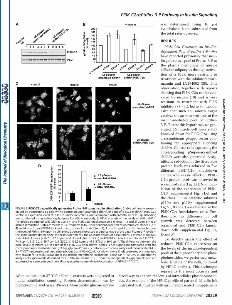
PI3K-C2� Generates an Insulin-dependent Pool of PtdIns-3-P—Wehave reported previously that insu-lin generates a pool of PtdIns-3-P atthe plasma membrane of musclecells and adipocytes through activa-tion of a PI3K more resistant totreatment with the inhibitors wort-mannin and LY294002 (38). Thisobservation, together with reportsshowing that PI3K-C2� can be acti-vated by insulin (10) and is veryresistant to treatment with PI3Kinhibitors (9–11), led us to hypoth-esize that such an isoform mightcatalyze the de novo synthesis of theinsulin-mediated pool of PtdIns-3-P. To test this hypothesis, we gen-erated L6 muscle cell lines stablyknocked down for PI3K-C2� usinga recombinant pSuper vector con-taining the appropriate silencingshRNA.Control cells expressing thecorresponding pSuper-scrambledshRNA were also generated. A sig-nificant reduction in the detectableprotein levels was achieved in fivedifferent PI3K-C2� knockdownclones, whereas no effect on PI3K-C2� protein levels was observed inscrambled cells (Fig. 1A). Nomodu-lation of the expression of PI3K-C2� (supplemental Fig. S1A) andthe class I PI3K catalytic subunitsp110� and p110� (supplementalFig. S1,B andC) was observed in thePI3K-C2� knockdown cells. Fur-thermore, no difference in cellgrowth was detected betweenscrambled and PI3K-C2� knock-down cells (supplemental Fig. S1,D and E).
To directly assess the effect ofreduced PI3K-C2� expression onthe levels of the insulin-dependentpools of the 3-phosphorylated phos-phoinositides, we performed meta-bolic labeling of the cells, followedby HPLC analysis. This techniquerepresents the most accurate and
direct way to analyze the levels of intracellular phosphoinositi-des. An example of the HPLC profile of parental L6 cells leftuntreated or stimulatedwith insulin is presented in supplemen-
FIGURE 1. PI3K-C2� specifically generates PtdIns-3-P upon insulin stimulation. Stable cell lines were gen-erated by transfecting L6 cells with a control pSuper-scrambled shRNA or a specific pSuper-shRNA PI3K-C2�vector. A, expression levels of PI3K-C2� in the indicated clones compared with parental L6 cells. Equal loadingwas confirmed using anti-phospholipase C� (PLC�) antibody. B, HPLC analysis of the levels of PtdIns-3-P in3H-labeled scrambled cells (clones 2 and 4) and PI3K-C2� knockdown cells (clones 1–3 and 5) upon 3 min ofinsulin stimulation. Data are means � S.E. from two to four independent experiments (scrambled, clones 2 (n �4) and 4 (n � 3); and PI3K-C2� knockdown, clones 1 (n � 4), 2 (n � 2), 3 (n � 3), and 5 (n � 3)). For each clone,the levels of PtdIns-3-P upon insulin stimulation are expressed as a percentage of the basal PtdIns-3-P levels inthe same unstimulated clone. In these experiments, the absolute values of basal PtdIns-3-P were as follows:scrambled clones 2 (256.7 � 47.5 cpm) and clone 4 (269 � 175.5) and PI3K-C2� knockdown clones 1 (367.2 �74.8 cpm), 2 (212.2 � 49.7 cpm), 3 (252.2 � 133.5 cpm), and 5 (176.2 � 86.0 cpm). The difference between thebasal levels of PtdIns-3-P in each of the PI3K-C2� knockdown clones is not significant compared with thecorresponding scrambled clone. gPtdIns, glycero-PtdIns. C, confocal microscopy analysis of the indicated GFP-2�FYVEHrs-expressing cells (scrambled clone 2 and PI3K-C2� knockdown clone 1) left untreated or stimulatedwith insulin for 3 min. Arrows mark the plasma membrane localization. Scale bar � 10 �m. D, quantitativeanalysis of experiments described for C. Data are means � S.E. from five independent experiments and areexpressed as a percentage of cells displaying plasma membrane localization of GFP-2�FYVEHrs.
PI3K-C2�/PtdIns-3-P Pathway in Insulin Signaling
SEPTEMBER 21, 2007 • VOLUME 282 • NUMBER 38 JOURNAL OF BIOLOGICAL CHEMISTRY 28229
at UC
L Library Services on January 16, 2008
ww
w.jbc.org
Dow
nloaded from
tal Fig. S2, specifically showing the insulin-induced increase inPtdIns-3-P levels, as reported previously (38). Scrambled andPI3K-C2� knockdown cloneswere labeledwithmyo-[3H]inosi-tol, and phosphoinositides from untreated and insulin-stimu-lated cells were extracted. HPLC analysis revealed that insulinincreased the levels of PtdIns-3-P in two distinct scrambledclones (Fig. 1B). In contrast, knockdown of PI3K-C2� com-pletely inhibited the insulin-dependent generation of PtdIns-3-P (Fig. 1B) in three clones (clones 1, 3, and 5) expressing verylow levels of PI3K-C2� (Fig. 1A). Strikingly, insulin-mediatedPtdIns-3-P synthesis was only slightly affected in one PI3K-C2�knockdown cell line (clone 2) (Fig. 1B), consistent with the factthat this clone still retained appreciable levels of PI3K-C2�expression (Fig. 1A). No difference in the basal levels of PtdIns-3-P was observed between scrambled and PI3K-C2� knock-down cells (see the figure legend). Taken together, these datareveal that the levels of the insulin-dependent pool of PtdIns-3-P correlate with the levels of PI3K-C2� expression and there-fore demonstrate that PI3K-C2� mediates the insulin-inducedgeneration of PtdIns-3-P. To further assess the effect of PI3K-C2� down-regulation on PtdIns-3-P generation, silenced andscrambled cells were transfected with the PtdIns-3-P-bindingprotein GFP-2�FYVEHrs (13, 38, 47). Confocal microscopyanalysis revealed that GFP-2�FYVEHrs was localized exclu-sively in endosomal structures in resting cells, as described pre-viously (13, 38, 47). A clear translocation of GFP-2�FYVEHrs tothe plasma membrane was observed in scrambled cells uponinsulin stimulation (Fig. 1C). These data indicated that, in thesecells, the probewas able to detect the insulin-dependent pool ofPtdIns-3-P generated at the cell surface, as already reported(38). No GFP-2�FYVEHrs translocation was observed in insu-lin-stimulated PI3K-C2� knockdown cells (Fig. 1C). Quantita-tive analysis of these experiments revealed that there was nodifference in the number of knockdown cells showing basalGFP-2�FYVEHrs staining at the plasma membrane comparedwith the scrambled cells (Fig. 1D). Upon insulin stimulation, aclear increase in the number of cells displaying plasma mem-brane localization of GFP-2�FYVEHrs was detected for scram-bled but not knockdown cells (Fig. 1D), indicating that noPtdIns-3-P was generated at the plasma membrane of theknockdown cells upon insulin stimulation. Down-regulation ofPI3K-C2� did not appear to affect the endosomal localizationof GFP-2�FYVEHrs, suggesting that PI3K-C2� does not regu-late the corresponding pool of PtdIns-3-P.HPLC analysis also revealed that silencing PI3K-C2� did not
block the insulin-induced generation of PtdIns-3,4-P2 andPtdIns-3,4,5-P3 (data not shown), indicating that PI3K-C2� isnot required for the insulin-mediated synthesis of these twophosphoinositides. In addition, no difference in the platelet-derived growth factor (PDGF)-dependent generation ofPtdIns-3,4-P2 and PtdIns-3,4,5-P3 was observed (data notshown), indicating that down-regulation of PI3K-C2� does notaffect other class I PI3K-dependent pathways. The levels ofPtdIns-3-P did not change in PDGF-stimulated scrambled orPI3K-C2� knockdown cells (data not shown), in agreementwith our observation that PDGF does not generate PtdIns-3-Pin L6 cells (38).
To further assess the specificity of the effect, we generated asecond recombinant vector targeting a distinct sequence in PI3K-C2� mRNA. L6 cells were retrovirally infected with such a vector(pSuperior-shRNA PI3K-C2�) and a control pSuperior-scram-bled shRNA, and the corresponding mixed populations wereselected. No difference in cell growth was observed between thepSuperior-scrambled shRNA and pSuperior-shRNA PI3K-C2�cells (data not shown). HPLC analysis revealed that the insulin-dependent generation of PtdIns-3-P was completely inhibitedin pSuperior-shRNA PI3K-C2� cells (supplemental Fig. S3A).The lack of PtdIns-3-P generation in these cells was not due toa reduced responsiveness to insulin because we detected a clearAkt phosphorylation as well as phosphorylation of its down-stream target GSK-3� in pSuperior-shRNA PI3K-C2� cells atdifferent times of insulin stimulation (supplemental Fig. S3B).Indeed, no effect on the insulin-induced synthesis of PtdIns-3,4,5-P3 and PtdIns-3,4-P2 was detected in pSuperior-shRNAPI3K-C2� cells (supplemental Fig. S3, C–E), further indicatingthat PI3K-C2� is not involved in the insulin-mediated synthesisof these phosphoinositides. These data indicate that the block-ade of PtdIns-3-P production upon insulin stimulation is spe-cifically associated with down-regulation of PI3K-C2�.Taken together, these data demonstrate that PI3K-C2� cat-alyzes specifically the de novo synthesis of an insulin-de-pendent pool of PtdIns-3-P at the plasma membrane.Because the insulin-dependent generation of PtdIns-3,4-P2or PtdIns-3,4,5-P3 does not appear to be affected in PI3K-C2� knockdown cells, these data identify, for the first time,PtdIns-3-P as the sole in vivo product of PI3K-C2�, at least inthe insulin signaling cascade.PI3K-C2� Translocates to the PlasmaMembrane upon Insu-
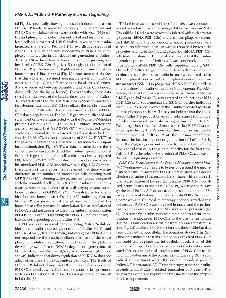
lin Stimulation—In an effort to better understand the mecha-nism of the insulin-mediated PI3K-C2� regulation, we assessedwhether activation of the enzyme is associated with an intracel-lular redistribution of the protein. Because PI3K-C2� is local-ized intracellularly in resting cells (48, 49), whereas the de novosynthesis of PtdIns-3-P occurs at the plasma membrane (38),we hypothesized that insulin might relocate PI3K-C2� to sucha compartment. Confocal microscopy analyses revealed thatendogenous PI3K-C2� was localized in nuclei and the perinu-clear region in resting cells (Fig. 2A), as reported previously (48,49). Interestingly, insulin induced a rapid and transient trans-location of endogenous PI3K-C2� to the plasma membrane(Fig. 2A). Translocation was visible from 1.5 min after stimula-tion (Fig. 2A) and lasted�10min (data not shown). Similar datawere obtained in subcellular fractionation studies (Fig. 2B).These data indicated that insulin not only activated PI3K-C2�,but could also regulate the intracellular localization of thisenzyme. More specifically, sucrose gradient fractionation indi-cated that insulin induced translocation of PI3K-C2� to thelipid raft subdomain of the plasma membrane (Fig. 2C), a spe-cialized compartment where the insulin-dependent pool ofPtdIns-3-P isgenerated (38).Thesedatasuggest that the insulin-dependent, PI3K-C2�-mediated generation of PtdIns-3-P atthe plasmamembrane requires the translocation of the enzymein this compartment.
PI3K-C2�/PtdIns-3-P Pathway in Insulin Signaling
28230 JOURNAL OF BIOLOGICAL CHEMISTRY VOLUME 282 • NUMBER 38 • SEPTEMBER 21, 2007
at UC
L Library Services on January 16, 2008
ww
w.jbc.org
Dow
nloaded from
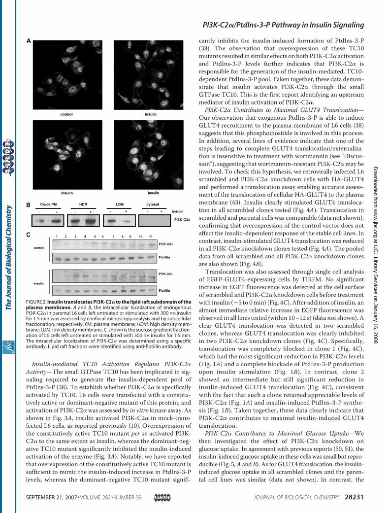
Insulin-mediated TC10 Activation Regulates PI3K-C2�Activity—The small GTPase TC10 has been implicated in sig-naling required to generate the insulin-dependent pool ofPtdIns-3-P (38). To establish whether PI3K-C2� is specificallyactivated by TC10, L6 cells were transfected with a constitu-tively active or dominant-negative mutant of this protein, andactivation of PI3K-C2�was assessed by in vitro kinase assay. Asshown in Fig. 3A, insulin activated PI3K-C2� in mock-trans-fected L6 cells, as reported previously (10). Overexpression ofthe constitutively active TC10 mutant per se activated PI3K-C2� to the same extent as insulin, whereas the dominant-neg-ative TC10 mutant significantly inhibited the insulin-inducedactivation of the enzyme (Fig. 3A). Notably, we have reportedthat overexpression of the constitutively active TC10mutant issufficient to mimic the insulin-induced increase in PtdIns-3-Plevels, whereas the dominant-negative TC10 mutant signifi-
cantly inhibits the insulin-induced formation of PtdIns-3-P(38). The observation that overexpression of these TC10mutants resulted in similar effects on both PI3K-C2� activationand PtdIns-3-P levels further indicates that PI3K-C2� isresponsible for the generation of the insulin-mediated, TC10-dependent PtdIns-3-P pool. Taken together, these data demon-strate that insulin activates PI3K-C2� through the smallGTPase TC10. This is the first report identifying an upstreammediator of insulin activation of PI3K-C2�.PI3K-C2� Contributes to Maximal GLUT4 Translocation—
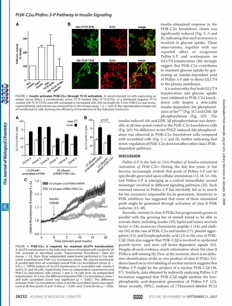
Our observation that exogenous PtdIns-3-P is able to induceGLUT4 recruitment to the plasma membrane of L6 cells (38)suggests that this phosphoinositide is involved in this process.In addition, several lines of evidence indicate that one of thesteps leading to complete GLUT4 translocation/externaliza-tion is insensitive to treatment with wortmannin (see “Discus-sion”), suggesting that wortmannin-resistant PI3K-C2�may beinvolved. To check this hypothesis, we retrovirally infected L6scrambled and PI3K-C2� knockdown cells with HA-GLUT4and performed a translocation assay enabling accurate assess-ment of the translocation of cellular HA-GLUT4 to the plasmamembrane (43). Insulin clearly stimulated GLUT4 transloca-tion in all scrambled clones tested (Fig. 4A). Translocation inscrambled and parental cells was comparable (data not shown),confirming that overexpression of the control vector does notaffect the insulin-dependent response of the stable cell lines. Incontrast, insulin-stimulated GLUT4 translocation was reducedin all PI3K-C2� knockdown clones tested (Fig. 4A). The pooleddata from all scrambled and all PI3K-C2� knockdown clonesare also shown (Fig. 4B).Translocation was also assessed through single cell analysis
of EGFP-GLUT4-expressing cells by TIRFM. No significantincrease in EGFP fluorescence was detected at the cell surfaceof scrambled and PI3K-C2� knockdown cells before treatmentwith insulin (�5 to 0min) (Fig. 4C). After addition of insulin, analmost immediate relative increase in EGFP fluorescence wasobserved in all lines tested (within 10–12 s) (data not shown). Aclear GLUT4 translocation was detected in two scrambledclones, whereas GLUT4 translocation was clearly inhibitedin two PI3K-C2� knockdown clones (Fig. 4C). Specifically,translocation was completely blocked in clone 1 (Fig. 4C),which had the most significant reduction in PI3K-C2� levels(Fig. 1A) and a complete blockade of PtdIns-3-P productionupon insulin stimulation (Fig. 1B). In contrast, clone 2showed an intermediate but still significant reduction ininsulin-induced GLUT4 translocation (Fig. 4C), consistentwith the fact that such a clone retained appreciable levels ofPI3K-C2� (Fig. 1A) and insulin-induced PtdIns-3-P synthe-sis (Fig. 1B). Taken together, these data clearly indicate thatPI3K-C2� contributes to maximal insulin-induced GLUT4translocation.PI3K-C2� Contributes to Maximal Glucose Uptake—We
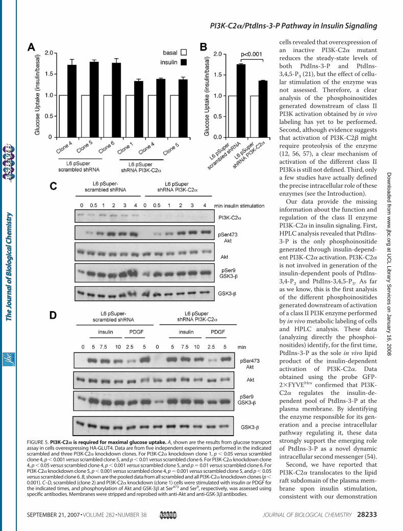
then investigated the effect of PI3K-C2� knockdown onglucose uptake. In agreement with previous reports (50, 51), theinsulin-induced glucose uptake in these cells was small but repro-ducible (Fig. 5,A andB). As for GLUT4 translocation, the insulin-induced glucose uptake in all scrambled clones and the paren-tal cell lines was similar (data not shown). In contrast, the
FIGURE 2. Insulin translocates PI3K-C2� to the lipid raft subdomain of theplasma membrane. A and B, the intracellular localization of endogenousPI3K-C2� in parental L6 cells left untreated or stimulated with 300 nM insulinfor 1.5 min was assessed by confocal microscopy analysis and by subcellularfractionation, respectively. PM, plasma membrane; HDM, high density mem-brane; LDM, low density membrane. C, shown is the sucrose gradient fraction-ation of L6 cells left untreated or stimulated with 300 nM insulin for 1.5 min.The intracellular localization of PI3K-C2� was determined using a specificantibody. Lipid raft fractions were identified using anti-flotillin antibody.
PI3K-C2�/PtdIns-3-P Pathway in Insulin Signaling
SEPTEMBER 21, 2007 • VOLUME 282 • NUMBER 38 JOURNAL OF BIOLOGICAL CHEMISTRY 28231
at UC
L Library Services on January 16, 2008
ww
w.jbc.org
Dow
nloaded from
insulin-stimulated response in thePI3K-C2� knockdown clones wassignificantly reduced (Fig. 5, A andB), indicating that such an enzyme isinvolved in glucose uptake. Theseobservations, together with ourreported effect of exogenousPtdIns-3-P and wortmannin onGLUT4 translocation (38), stronglysuggest that PI3K-C2� contributesto maximal glucose uptake by gen-erating an insulin-dependent poolof PtdIns-3-P able to direct GLUT4to the plasma membrane.It is noteworthy that bothGLUT4
translocation and glucose uptakewere inhibited in PI3K-C2� knock-down cells despite a detectableinsulin-dependent Akt phosphoryl-ation at Ser473 (Fig. 5C) andGSK-3�phosphorylation (Fig. 5D). The
insulin-induced Akt and GSK-3� phosphorylation was detect-able at all time points tested in the PI3K-C2� knockdown cells(Fig. 5D). No difference in the PDGF-induced Akt phosphoryl-ation was observed in PI3K-C2� knockdown cells comparedwith scrambled cells (Fig. 5, C and D), further indicating thatdown-regulationofPI3K-C2�doesnot affect other class IPI3K-dependent pathways.
DISCUSSION
PtdIns-3-P Is the Sole in Vivo Product of Insulin-stimulatedActivation of PI3K-C2�—During the last few years, it hasbecome increasingly evident that pools of PtdIns-3-P can bespecifically generated upon cellular stimulation (13, 38, 52–54),and PtdIns-3-P is emerging as a critical intracellular secondmessenger involved in different signaling pathways (54). Suchrenewed interest in PtdIns-3-P has inevitably led us to searchfor the enzyme(s) responsible for its generation. Sensitivity toPI3K inhibitors has suggested that some of these stimulatedpools might be generated through activation of class II PI3Kisoforms (13, 38).Recently, interest in class II PI3Ks has progressively grown in
parallel with the growing list of stimuli found to be able toactivate them, including insulin (10), leptin and tumor necrosisfactor-� (18), monocyte chemotactic peptide-1 (14), and clath-rin (16) in the case of PI3K-C2� and insulin (17), platelet aggre-gation (15), and lysophosphatidic acid (13) in the case of PI3K-C2�. Data also suggest that PI3K-C2� is involved in epidermalgrowth factor- and stem cell factor-dependent signals (55).Despite all such evidence, much information about the class IIPI3Ks is still missing (8). First, at themoment, there is no defin-itive identification of the in vivo product of class II PI3Ks. Evi-dence based on in vitro labeling of nuclear extracts suggests thatPtdIns-3-P might be the product of a nuclear PI3K-C2� (56,57). Similarly, data obtained by indirectly analyzing PtdIns-3-Pformation suggested that PI3K-C2� might mediate the lyso-phosphatidic acid-dependent generation of PtdIns-3-P (13).More recently, HPLC analyses of [3H]inositol-labeled PC12
FIGURE 3. Insulin activates PI3K-C2� through TC10 activation. A, serum-starved L6 cells expressing anempty vector (Myc), a constitutively active TC10 mutant (Myc-TC10 Q75L), or a dominant-negative TC10mutant (HA-TC10 T31N) were left untreated or stimulated with 300 nM insulin for 2 min. PI3K-C2� was immu-noprecipitated, and activity was assessed by in vitro kinase assay. *, p � 0.05. B, the representative images areof transfected L6 cells showing the efficiency of transfection of the indicated constructs.
FIGURE 4. PI3K-C2� is required for maximal GLUT4 translocation.A, GLUT4 translocation in the indicated clones stimulated with insulin for 20min assessed as described under “Experimental Procedures.” Data aremeans � S.E. from three independent experiments performed in the indi-cated scrambled and PI3K-C2� knockdown clones. PM, plasma membrane.B, pooled data from all scrambled and all PI3K-C2� knockdown clones (p �0.01). C, TIRFM analysis of GLUT4 translocation in scrambled cells (clones 4and 6; 37 and 38 cells, respectively, from six independent experiments) andPI3K-C2� knockdown cells (clones 1 and 2; 18 cells from six independentexperiments). At 5 min, the difference between PI3K-C2� knockdown clone 1and both scrambled clones was significant (p � 0.001). The differencebetween PI3K-C2� knockdown clone 2 and the scrambled clones was signif-icant at all time points (5 and 10 min, p � 0.001; and 15 and 20 min, p � 0.05).
PI3K-C2�/PtdIns-3-P Pathway in Insulin Signaling
28232 JOURNAL OF BIOLOGICAL CHEMISTRY VOLUME 282 • NUMBER 38 • SEPTEMBER 21, 2007
at UC
L Library Services on January 16, 2008
ww
w.jbc.org
Dow
nloaded from
cells revealed that overexpression ofan inactive PI3K-C2� mutantreduces the steady-state levels ofboth PtdIns-3-P and PtdIns-3,4,5-P3 (21), but the effect of cellu-lar stimulation of the enzyme wasnot assessed. Therefore, a clearanalysis of the phosphoinositidesgenerated downstream of class IIPI3K activation obtained by in vivolabeling has yet to be performed.Second, although evidence suggeststhat activation of PI3K-C2� mightrequire proteolysis of the enzyme(12, 56, 57), a clear mechanism ofactivation of the different class IIPI3Ks is still not defined. Third, onlya few studies have actually definedthe precise intracellular role of theseenzymes (see the Introduction).Our data provide the missing
information about the function andregulation of the class II enzymePI3K-C2� in insulin signaling. First,HPLC analysis revealed that PtdIns-3-P is the only phosphoinositidegenerated through insulin-depend-ent PI3K-C2� activation. PI3K-C2�is not involved in generation of theinsulin-dependent pools of PtdIns-3,4-P2 and PtdIns-3,4,5-P3. As faras we know, this is the first analysisof the different phosphoinositidesgenerated downstream of activationof a class II PI3K enzyme performedby in vivometabolic labeling of cellsand HPLC analysis. These data(analyzing directly the phosphoi-nositides) identify, for the first time,PtdIns-3-P as the sole in vivo lipidproduct of the insulin-dependentactivation of PI3K-C2�. Dataobtained using the probe GFP-2�FYVEHrs confirmed that PI3K-C2� regulates the insulin-de-pendent pool of PtdIns-3-P at theplasma membrane. By identifyingthe enzyme responsible for its gen-eration and a precise intracellularpathway regulating it, these datastrongly support the emerging roleof PtdIns-3-P as a novel dynamicintracellular secondmessenger (54).Second, we have reported that
PI3K-C2� translocates to the lipidraft subdomain of the plasma mem-brane upon insulin stimulation,consistent with our demonstration
FIGURE 5. PI3K-C2� is required for maximal glucose uptake. A, shown are the results from glucose transportassay in cells overexpressing HA-GLUT4. Data are from five independent experiments performed in the indicatedscrambled and three PI3K-C2� knockdown clones. For PI3K-C2� knockdown clone 1, p � 0.05 versus scrambledclone 4, p � 0.001 versus scrambled clone 5, and p � 0.01 versus scrambled clone 6. For PI3K-C2� knockdown clone4, p � 0.05 versus scrambled clone 4, p � 0.001 versus scrambled clone 5, and p � 0.01 versus scrambled clone 6. ForPI3K-C2� knockdown clone 5, p � 0.001 versus scrambled clone 4, p � 0.001 versus scrambled clone 5, and p � 0.05versus scrambled clone 6. B, shown are the pooled data from all scrambled and all PI3K-C2� knockdown clones (p �0.001). C–D, scrambled (clone 2) and PI3K-C2� knockdown (clone 1) cells were stimulated with insulin or PDGF forthe indicated times, and phosphorylation of Akt and GSK-3� at Ser473 and Ser9, respectively, was assessed usingspecific antibodies. Membranes were stripped and reprobed with anti-Akt and anti-GSK-3� antibodies.
PI3K-C2�/PtdIns-3-P Pathway in Insulin Signaling
SEPTEMBER 21, 2007 • VOLUME 282 • NUMBER 38 JOURNAL OF BIOLOGICAL CHEMISTRY 28233
at UC
L Library Services on January 16, 2008
ww
w.jbc.org
Dow
nloaded from

that the de novo synthesis of PtdIns-3-P occurs in such a com-partment (38). Relocation of PI3K-C2� in such a specializedcompartment is critical for activation of the enzyme. This is thefirst time that a membrane targeting-mediated mechanism ofactivation has been reported for PI3K-C2�. These data,together with our previous observation that lysophosphatidicacid induces translocation of overexpressed PI3K-C2� to thecell surface (13), suggest that recruitment to the membranemight represent a general mechanism of activation for class IIPI3Ks.Moreover, our data clearly demonstrate that the insulin-dependent activation of PI3K-C2� is mediated by the smallGTPase TC10, in agreement with the role of this protein ininsulin-induced PtdIns-3-P generation (38). Although there aredata questioning the role of TC10 in insulin signaling (58, 59),more recent evidence shows that knockdownof TC10� inhibitsbothGLUT4 translocation and glucose uptake (60), confirmingthe role of such a GTPase in insulin signaling. In this respect, itis important to emphasize that most of the previous reportsactually suggested that some of the proteins proposed to benecessary for TC10 activation are not required for insulin sig-nals (59), but they did not investigate the role of TC10 itself.Therefore, even taking into account such evidence, there is stillthe possibility that TC10 can be activated through a differentmechanism. This is supported by the observation that overex-pressed TC10 is activated by insulin in L6 myoblasts (38, 58)even in the absence of Cbl-associated protein expression andCbl phosphorylation (58). Alternatively, we cannot rule out thepossibility that the overexpressed TC10 mutant proteins used
in this study are mimicking theaction of a different endogenoussmall GTPase.Nevertheless, our data suggest
thatTC10 is required for the insulin-dependent activation of PI3K-C2�,and they represent the first identifi-cation of an upstream mediator ofthe insulin-dependent activation ofPI3K-C2�. Furthermore, to ourknowledge, this is the first reportshowing that a small GTPase canactivate a class II PI3K isoform. Inthis respect, this work opens up newfields of investigation about theintriguing possibility that GTP-binding proteins can regulate classII PI3K activation.The PI3K-C2�-dependent Pool of
PtdIns-3-P Is Necessary for Maxi-malGLUT4Translocation andGlu-cose Uptake—The last unansweredquestion about PI3K-C2� and insu-lin signaling concerned its role andfunction in such a pathway. Accu-mulating evidence suggests thatPtdIns-3-P plays an important rolein GLUT4 translocation (38–42).GLUT4 translocation/external-
ization is a complex process, likelyrequiring tight cooperation between different pathways.Indeed, it has been shown that activation of a class I PI3K,which is necessary for the whole process to occur (25), is notsufficient to fully activate it (61–63). Similarly, although Akt isa key player in GLUT4 translocation (27–32), inhibition of itsactivity either by a specific chemical compound (33) or by genesilencing (33, 64) does not completely inhibit GLUT4 translo-cation, indicating that other, Akt-independent signals are alsorequired. On the other hand, although data have revealed animportant role for the GTPase TC10 in insulin signaling (26),knockdown of TC10� only partially inhibits GLUT4 transloca-tion and uptake (60), further supporting the hypothesis of aconcerted activation of different pathways needed to properlyactivate glucose transport.Although many intermediate steps have been described,
recent data suggest that the whole process known as “GLUT4translocation” might be broken down into two different anddiscrete steps: movement of GSVs to the cell periphery, i.e.close to the plasmamembrane (step 1), and then their tethering,docking, and fusion with such a compartment (togetherreferred here as step 2) (24, 41, 65, 66). Once correctly insertedinto the plasmamembrane and partly externalized, GLUT4 canmediate glucose transport. A simplifiedmodel ofGLUT4 trans-location is depicted in Fig. 6.Notably, movement of the GSVs to the membrane is not
inhibited by the PI3K inhibitors wortmannin (38, 41) andLY294002 (67) and does not appear to involve Akt activation(65), indicating that relocation of GSVs to the plasma mem-
FIGURE 6. Schematic representation of GLUT4 translocation. GLUT4 translocation involves formation ofGSVs and their movement to the cell periphery (step 1). Once close to the plasma membrane, GSVs need to betethered and docked (primarily through an interaction between VAMP2 on GSVs and syntaxin-4/SNAP23 at theplasma membrane) to fuse with such a compartment (step 2). When GLUT4 is correctly inserted into the plasmamembrane and partly externalized, it can mediate glucose transport. Evidence suggests that PI3K-C2� andPtdIns-3-P might be involved in step 1 (see “Discussion”). On the other hand, data indicate that class I PI3K is notinvolved in step 1, whereas it is necessary for step 2. This representation is a simplified model of the process thatactually involves several different proteins and different intermediate steps, including actin polymerization;GSV formation; and trafficking between the trans-Golgi network, endosomes, and GSVs.
PI3K-C2�/PtdIns-3-P Pathway in Insulin Signaling
28234 JOURNAL OF BIOLOGICAL CHEMISTRY VOLUME 282 • NUMBER 38 • SEPTEMBER 21, 2007
at UC
L Library Services on January 16, 2008
ww
w.jbc.org
Dow
nloaded from
brane (step 1) does not require activation of the class I PI3Kpathway but rather involves motor proteins and microtubules(68, 69) and cytoskeletal rearrangement, possibly throughTC10 activation (26). On the contrary, PI3K inhibitors blockGLUT4 externalization (41, 67), indicating that class I PI3Kactivation is strictly necessary in the second step (Fig. 6). In thisrespect, recent evidence suggests that Akt might be the PI3Ktarget necessary for tethering and docking of GSVs (33),whereas protein kinase C� and Munc18c (41, 70) might be thePI3K targets involved in fusion. A critical role for Akt in fusionhas also been proposed (66), indicating that work still needs tobe done to precisely define the PI3K-dependent players in thedifferent steps. Furthermore, data have also suggested a role forTC10-dependent effectors in someof the step 2-associated pro-cesses (37), further supporting the hypothesis of a concertedaction of the two pathways or even a cross-talk between them.Here, we have shown that down-regulation of PI3K-C2�
inhibits GLUT4 translocation and glucose uptake even in thepresence of a detectable insulin-dependent activation of Aktand GSK-3�. It should be noted that inhibition of GLUT4translocation in PI3K-C2� knockdown cells was total or partialdepending on the assays performed, and this is likely due to thedifferent experimental conditions of the assays performed.Nevertheless, the key point is that all our data consistently dem-onstrate that knockdown of PI3K-C2� impairs insulin-inducedGLUT4 translocation and glucose uptake. The observation thatknockdown of PI3K-C2� resulted in only partial inhibition ofglucose uptake but complete inhibition of insulin-inducedPtdIns-3-P synthesis is consistent with the reported partialeffect of TC10� knockdown on glucose uptake (60) and withthe hypothesis of different pathways required to fully activateglucose uptake.These data, together with our previous observations (38),
indicate that activation of a PI3K-C2�/PtdIns-3-P pathway isrequired in some step(s) leading to GLUT4 translocation andglucose uptake. The precise mechanism of action of the PI3K-C2�/PtdIns-3-P pathway, the intracellular effectors of PtdIns-3-P, and the precise step(s) of GLUT4 translocation involvingPtdIns-3-P remain to be addressed. On the basis of the wort-mannin insensitivity and our observation that exogenousPtdIns-3-P is able to move GLUT4 to the cell periphery but isnot sufficient to induce glucose uptake (38), it is tempting tospeculate that PI3K-C2�/PtdIns-3-P might be involved in step1 in the GLUT4 route to the plasma membrane (Fig. 6). Thiswould be consistent with data suggesting a role for the TC10-dependent pathway in this step (26, 34) andwith the hypothesisthat PtdIns-3-P is important for translocation and possiblyunmasking of the C-terminal domain of GLUT4 but not inser-tion into the plasma membrane (40). Other evidence suggeststhat PtdIns-3-Pmight be sufficient to induce both translocationand insertion into the membrane in the absence of Munc18c(41) or in 3T3-L1 adipocytes overexpressing 72-kDa inositol-polyphosphate 5-phosphatase (42).We cannot rule out the pos-sibility that PI3K-C2�/PtdIns-3-P might also play a role insome processes of step 2, acting in parallel with class I PI3K. Inthis respect, it is noteworthy that PI3K-C2� is necessary forpriming neurosecretory granule exocytosis (21) and that a role
for TC10 in exocytic vesicle fusion has been proposed recently(71).In summary, this work defines, for the first time, the role of
both PI3K-C2� and the endogenously generated, insulin-de-pendent PtdIns-3-P pool in insulin signaling. Our data,together with all previous evidence, suggest that a parallel acti-vation of a class I PI3K and PI3K-C2� might be required forproper GLUT4 translocation and glucose uptake. Glucosetransport appears to fail in insulin resistance accompanyingseveral forms of diabetes. There is therefore a huge interest inunderstanding the alterations in signaling ultimately leading tosuch impairment. In this respect, the identification of PI3K-C2� as a novel crucial player in insulin actionwould definitivelyprove useful in understanding the molecular mechanismsunderlying insulin resistance to develop approaches to preventtype 2 diabetes.
Acknowledgments—We thank Dr. M. Thelen for the anti-PI3K-C2�antibody; Dr. J. Pessin for the TC10 constructs; Dr. F. Cooke for helpwith HPLC experiments; C. Hohnen-Behrens, J. Davey, and D. Blairfor help implementing the GLUT4 translocation assays; and L. Mon-tuno (Carl Zeiss Australasia) for assistance with TIRFM.
REFERENCES1. Rameh, L. E., and Cantley, L. C. (1999) J. Biol. Chem. 274, 8347–83502. Cantley, L. C. (2002) Science 296, 1655–16573. Katso, R., Okkenhaug, K., Ahmadi, K., White, S., Timms, J., and Water-
field, M. D. (2001) Annu. Rev. Cell Dev. Biol. 17, 615–6754. Vanhaesebroeck, B., Leevers, S. J., Ahmadi, K., Timms, J., Katso, R.,
Driscoll, P. C., Woscholski, R., Parker, P. J., and Waterfield, M. D. (2001)Annu. Rev. Biochem. 70, 535–602
5. Foster, F. M., Traer, C. J., Abraham, S. M., and Fry, M. J. (2003) J. Cell Sci.116, 3037–3040
6. MacDougall, L. K., Domin, J., and Waterfield, M. D. (1995) Curr. Biol. 5,1404–1415
7. Virbasius, J. V., Guilherme, A., and Czech, M. P. (1996) J. Biol. Chem. 271,13304–13307
8. Falasca, M., and Maffucci, T. (2007) Biochem. Soc. Trans. 35, 211–2149. Domin, J., Pages, F., Volinia, S., Rittenhouse, S. E., Zvelebil, M. J., Stein,
R. C., and Waterfield, M. D. (1997) Biochem. J. 326, 139–14710. Brown, R. A., Domin, J., Arcaro, A.,Waterfield, M. D., and Shepherd, P. R.
(1999) J. Biol. Chem. 274, 14529–1453211. Soos, M. A., Jensen, J., Brown, R. A., O’Rahilly, S., Shepherd, P. R., and
Whitehead, J. P. (2001) Arch. Biochem. Biophys. 396, 244–24812. Arcaro, A., Volinia, S., Zvelebil, M. J., Stein, R., Watton, S. J., Layton, M. J.,
Gout, I., Ahmadi, K., Downward, J., and Waterfield, M. D. (1998) J. Biol.Chem. 273, 33082–33090
13. Maffucci, T., Cooke, F. T., Foster, F.M., Traer, C. J., Fry,M. J., and Falasca,M. (2005) J. Cell Biol. 169, 789–799
14. Turner, S. J., Domin, J., Waterfield, M. D., Ward, S. G., and Westwick, J.(1998) J. Biol. Chem. 273, 25987–25995
15. Zhang, J., Banfic, H., Straforini, F., Tosi, L., Volinia, S., and Rittenhouse,S. E. (1998) J. Biol. Chem. 273, 14081–14084
16. Gaidarov, I., Smith, M. E., Domin, J., and Keen, J. H. (2001) Mol. Cell 7,443–449
17. Brown, R. A., and Shepherd, P. R. (2001)Biochem. Soc. Trans. 29, 535–53718. Ktori, C., Shepherd, P. R., and O’Rourke, L. (2003) Biochem. Biophys. Res.
Commun. 306, 139–14319. Katso, R. M., Pardo, O. E., Palamidessi, A., Franz, C., Marinov, M., De
Laurentiis, A., Downward, J., Scita, G., Ridley, A. J., Waterfield, M. D., andArcaro, A. (2006)Mol. Biol. Cell 17, 3729–3744
20. Gaidarov, I., Zhao, Y., and Keen, J. H. (2005) J. Biol. Chem. 280,40766–40772
PI3K-C2�/PtdIns-3-P Pathway in Insulin Signaling
SEPTEMBER 21, 2007 • VOLUME 282 • NUMBER 38 JOURNAL OF BIOLOGICAL CHEMISTRY 28235
at UC
L Library Services on January 16, 2008
ww
w.jbc.org
Dow
nloaded from
21. Meunier, F. A., Osborne, S. L., Hammond, G. R., Cooke, F. T., Parker, P. J.,Domin, J., and Schiavo, G. (2005)Mol. Biol. Cell 16, 4841–4851
22. Wang, Y., Yoshioka, K., Azam,M.A., Takuwa,N., Sakurada, S., Kayaba, Y.,Sugimoto, N., Inoki, I., Kimura, T., Kuwaki, T., and Takuwa, Y. (2006)Biochem. J. 394, 581–592
23. Bryant, N. J., Govers, R., and James, D. E. (2002)Nat. Rev.Mol. Cell Biol. 3,267–277
24. James, D. E. (2005) J. Clin. Investig. 115, 219–22125. Shepherd, P. R., Withers, D. J., and Siddle, K. (1998) Biochem. J. 333,
471–49026. Saltiel, A. R., and Pessin, J. E. (2003) Traffic 4, 711–71627. Kohn, A.D., Summers, S. A., Birnbaum,M. J., andRoth, R. A. (1996) J. Biol.
Chem. 271, 31372–3137828. Cong, L. N., Chen, H., Li, Y., Zhou, L., McGibbon, M. A., Taylor, S. I., and
Quon, M. J. (1997)Mol. Endocrinol. 11, 1881–189029. Wang, Q., Somwar, R., Bilan, P. J., Liu, Z., Jin, J., Woodgett, J. R., and Klip,
A. (1999)Mol. Cell. Biol. 19, 4008–401830. Foran, P. G., Fletcher, L. M., Oatey, P. B., Mohammed, N., Dolly, J. O., and
Tavare, J. M. (1999) J. Biol. Chem. 274, 28087–2809531. Kane, S., Sano, H., Liu, S. C., Asara, J. M., Lane, W. S., Garner, C. C., and
Lienhard, G. E. (2002) J. Biol. Chem. 277, 22115–2211832. Berwick, D. C., Dell, G. C., Welsh, G. I., Heesom, K. J., Hers, I., Fletcher,
L. M., Cooke, F. T., and Tavare, J. M. (2004) J. Cell Sci. 117, 5985–599333. Gonzalez, E., and McGraw, T. E. (2006)Mol. Biol. Cell 17, 4484–449334. Chang, L., Adams, R. D., and Saltiel, A. R. (2002) Proc. Natl. Acad. Sci.
U. S. A. 99, 12835–1284035. Chiang, S. H., Hwang, J., Legendre, M., Zhang, M., Kimura, A., and Saltiel,
A. R. (2003) EMBO J. 22, 2679–269136. Kanzaki, M., Mora, S., Hwang, J. B., Saltiel, A. R., and Pessin, J. E. (2004)
J. Cell Biol. 164, 279–29037. Inoue, M., Chiang, S. H., Chang, L., Chen, X. W., and Saltiel, A. R. (2006)
Mol. Biol. Cell 17, 2303–231138. Maffucci, T., Brancaccio, A., Piccolo, E., Stein, R. C., and Falasca,M. (2003)
EMBO J. 22, 4178–418939. Chaussade, C., Pirola, L., Bonnafous, S., Blondeau, F., Brenz-Verca, S.,
Tronchere, H., Portis, F., Rusconi, S., Payrastre, B., Laporte, J., and VanObberghen, E. (2003)Mol. Endocrinol. 17, 2448–2460
40. Ishiki,M., Randhawa,V. K., Poon,V., Jebailey, L., andKlip, A. (2005) J. Biol.Chem. 280, 28792–28802
41. Kanda, H., Tamori, Y., Shinoda, H., Yoshikawa, M., Sakaue, M., Udagawa,J., Otani, H., Tashiro, F., Miyazaki, J., and Kasuga, M. (2005) J. Clin. Inves-tig. 115, 291–301
42. Kong, A. M., Horan, K. A., Sriratana, A., Bailey, C. G., Collyer, L. J., Nan-durkar, H. H., Shisheva, A., Layton,M. J., Rasko, J. E., Rowe, T., andMitch-ell, C. A. (2006)Mol. Cell. Biol. 26, 6065–6081
43. Govers, R., Coster, A. C., and James, D. E. (2004) Mol. Cell. Biol. 24,6456–6466
44. Li, C. H., Bai, L., Li, D. D., Xia, S., and Xu, T. (2004) Cell Res. 14, 480–48645. van den Hoff, M. J., Moorman, A. F., and Lamers, W. H. (1992) Nucleic
Acids Res. 20, 290246. Moyers, J. S., Bilan, P. J., Reynet, C., and Kahn, C. R. (1996) J. Biol. Chem.
271, 23111–2311647. Gillooly, D. J., Morrow, I. C., Lindsay,M., Gould, R., Bryant, N. J., Gaullier,
J. M., Parton, R. G., and Stenmark, H. (2000) EMBO J. 19, 4577–458848. Domin, J., Gaidarov, I., Smith, M. E., Keen, J. H., and Waterfield, M. D.
(2000) J. Biol. Chem. 275, 11943–1195049. Didichenko, S. A., and Thelen,M. (2001) J. Biol. Chem. 276, 48135–4814250. Maffucci, T., Razzini, G., Ingrosso, A., Chen, H., Iacobelli, S., Sciacchitano,
S., Quon, M. J., and Falasca, M. (2003)Mol. Endocrinol. 17, 1568–157951. Niu,W., Huang, C., Nawaz, Z., Levy,M., Somwar, R., Li, D., Bilan, P. J., and
Klip, A. (2003) J. Biol. Chem. 278, 17953–1796252. Ellson, C. D., Anderson, K. E., Morgan, G., Chilvers, E. R., Lipp, P., Ste-
phens, L. R., and Hawkins, P. T. (2001) Curr. Biol. 11, 1631–163553. Vieira, O. V., Botelho, R. J., Rameh, L., Brachmann, S. M., Matsuo, T.,
Davidson, H.W., Schreiber, A., Backer, J.M., Cantley, L. C., andGrinstein,S. (2001) J. Cell Biol. 55, 19–25
54. Falasca,M., andMaffucci, T. (2006)Arch. Physiol. Biochem. 112, 274–28455. Arcaro, A., Khanzada, U. K., Vanhaesebroeck, B., Tetley, T. D.,Waterfield,
M. D., and Seckl, M. J. (2002) EMBO J. 21, 5097–510856. Sindic, A., Aleksandrova, A., Fields, A. P., Volinia, S., and Banfic, H. (2001)
J. Biol. Chem. 276, 17754–1776157. Visnjic, D., Crljen, V., Curic, J., Batinic, D., Volinia, S., and Banfic, H.
(2002) FEBS Lett. 529, 268–27458. Jebailey, L., Rudich, A., Huang, X., Di Ciano-Oliveira, C., Kapus, A., and
Klip, A. (2004)Mol. Endocrinol. 18, 359–37259. Mitra, P., Zheng, X., and Czech, M. P. (2004) J. Biol. Chem. 279,
37431–3743560. Chang, L., Chiang, S. H., and. Saltiel, A. R. (2007) Endocrinology 148,
27–3361. Isakoff, S. J., Taha, C., Rose, E., Marcusohn, J., Klip, A., and Skolnik, E. Y.
(1995) Proc. Natl. Acad. Sci. U. S. A. 92, 10247–1025162. Jiang, T., Sweeney, G., Rudolf, M. T., Klip, A., Traynor-Kaplan, A., and
Tsien, R. Y. (1998) J. Biol. Chem. 273, 11017–1102463. Czech, M. P., and Corvera, S. (1999) J. Biol. Chem. 274, 1865–186864. Jiang, Z. Y., Zhou, Q. L., Coleman, K. A., Chouinard, M., Boese, Q., and
Czech, M. P. (2003) Proc. Natl. Acad. Sci. U. S. A. 100, 7569–757465. van Dam, E. M., Gover, R., and James, D. E. (2005) Mol. Endocrinol. 19,
1067–107766. Koumanov, F., Jin, B., Yang, J., and Holman, G. D. (2005) Cell Metab. 2,
179–18967. Bose, A., Robida, S., Furcinitti, P. S., Chawla, A., Fogarty, K., Corvera, S.,
and Czech, M. P. (2004)Mol. Cell. Biol. 24, 5447–545868. Semiz, S., Park, J. G., Nicoloro, S. M., Furcinitti, P., Zhang, C., Chawla, A.,
Leszyk, J., and Czech, M. P. (2003) EMBO J. 22, 2387–239969. Bose, A., Guilherme, A., Robida, S. I., Nicoloro, S. M., Zhou, Q. L., Jiang,
Z. Y., Pomerleau, D. P., and Czech, M. P. (2002) Nature 420, 821–82470. Hodgkinson, C. P., Mander, A., and Sale, G. J. (2005) Diabetologia 48,
1627–163671. Kawase, K., Nakamura, T., Takaya, A., Aoki, K., Namikawa, K., Kiyama,H.,
Inagaki, S., Takemoto, H., Saltiel, A. R., and Matsuda, M. (2006) Dev. Cell11, 411–421
PI3K-C2�/PtdIns-3-P Pathway in Insulin Signaling
28236 JOURNAL OF BIOLOGICAL CHEMISTRY VOLUME 282 • NUMBER 38 • SEPTEMBER 21, 2007
at UC
L Library Services on January 16, 2008
ww
w.jbc.org
Dow
nloaded from
Copyright © 2022 FDOKUMEN