Phosphoinositide Metabolism Links cGMP-Dependent Protein Kinase G to Essential Ca 2+ Signals at Key...
15
Phosphoinositide Metabolism Links cGMP-Dependent Protein Kinase G to Essential Ca 2+ Signals at Key Decision Points in the Life Cycle of Malaria Parasites Mathieu Brochet 1 *, Mark O. Collins 1¤ , Terry K. Smith 2 , Eloise Thompson 3 , Sarah Sebastian 1 , Katrin Volkmann 1 , Frank Schwach 1 , Lia Chappell 1 , Ana Rita Gomes 1 , Matthew Berriman 1 , Julian C. Rayner 1 , David A. Baker 3 , Jyoti Choudhary 1 , Oliver Billker 1 * 1 Wellcome Trust Sanger Institute, Hinxton, Cambridge, United Kingdom, 2 Schools of Biology and Chemistry, Biomedical Sciences Research Complex, The North Haugh, The University of Saint Andrews, St. Andrews, Fife United Kingdom, 3 Faculty of Infectious and Tropical Diseases, London School of Hygiene & Tropical Medicine, London, United Kingdom Abstract Many critical events in the Plasmodium life cycle rely on the controlled release of Ca 2+ from intracellular stores to activate stage-specific Ca 2+ -dependent protein kinases. Using the motility of Plasmodium berghei ookinetes as a signalling paradigm, we show that the cyclic guanosine monophosphate (cGMP)-dependent protein kinase, PKG, maintains the elevated level of cytosolic Ca 2+ required for gliding motility. We find that the same PKG-dependent pathway operates upstream of the Ca 2+ signals that mediate activation of P. berghei gametocytes in the mosquito and egress of Plasmodium falciparum merozoites from infected human erythrocytes. Perturbations of PKG signalling in gliding ookinetes have a marked impact on the phosphoproteome, with a significant enrichment of in vivo regulated sites in multiple pathways including vesicular trafficking and phosphoinositide metabolism. A global analysis of cellular phospholipids demonstrates that in gliding ookinetes PKG controls phosphoinositide biosynthesis, possibly through the subcellular localisation or activity of lipid kinases. Similarly, phosphoinositide metabolism links PKG to egress of P. falciparum merozoites, where inhibition of PKG blocks hydrolysis of phosphatidylinostitol (4,5)-bisphosphate. In the face of an increasing complexity of signalling through multiple Ca 2+ effectors, PKG emerges as a unifying factor to control multiple cellular Ca 2+ signals essential for malaria parasite development and transmission. Citation: Brochet M, Collins MO, Smith TK, Thompson E, Sebastian S, et al. (2014) Phosphoinositide Metabolism Links cGMP-Dependent Protein Kinase G to Essential Ca 2+ Signals at Key Decision Points in the Life Cycle of Malaria Parasites. PLoS Biol 12(3): e1001806. doi:10.1371/journal.pbio.1001806 Academic Editor: David S. Schneider, Stanford University, United States of America Received September 18, 2013; Accepted January 23, 2014; Published March 4, 2014 Copyright: ß 2014 Brochet et al. This is an open-access article distributed under the terms of the Creative Commons Attribution License, which permits unrestricted use, distribution, and reproduction in any medium, provided the original author and source are credited. Funding: This work was funded by grants from the Wellcome Trust (WT098051 and 079643/Z/06/Z) and the Medical Research Council (G0501670) to OB, a Wellcome Trust project grant to DB (WT094752), a Wellcome Trust Grant (WT093228) to TKS, a Marie Curie Fellowship (PIEF-GA-2008-220180) to SS, and a Marie Curie Fellowship (PIEF-GA-2009-253899) and an EMBO Long Term Fellowship (ALTF 45-2009) to MBr. C2 was synthesised and kindly provided by Katy Kettleborough and colleagues at MRC Technology through an MRC grant to DB (G10000779). The funders had no role in study design, data collection and analysis, decision to publish, or preparation of the manuscript. Competing Interests: The authors have declared that no competing interests exist. Abbreviations: ARF, ADP-ribosylation factor; ARF-GAP, ADP-ribosylation factor GTPase activating protein; ARF-GEF, ADP-ribosylation factor guanine exchange factor; CDPK, Ca 2+ -dependent protein kinase; cGMP, 39-59-cyclic guanosine monophosphate; GC, guanylyl cyclase; IMAC, immobilised metal ion chromatography; IP 3 , inositol (1,4,5)-trisphosphate; PDE, phosphodiesterase; PI, phosphatidyl-1D-myo-inositol; PI(4,5)P 2 , phosphatidylinositol (4,5)-bisphosphate; PI4K, phosphatidylinositol 4-kinase; PI4P, phosphatidylinositol 4-phosphate; PI-PLC, PI-specific phospholipase C; PIP5K, phosphatidylinositol 5-kinase; PKG, cGMP- dependent protein kinase G; SILAC, stable isotope labelling in culture. * E-mail: [email protected] (M.Br.); [email protected] (O.B.) ¤ Current address: Department of Biomedical Science, The University of Sheffield, Sheffield, United Kingdom Introduction Malaria is caused by vector-born protozoan parasites of the genus Plasmodium, which cycle between mosquitoes and humans. Waves of fever arise from the synchronised egress of merozoites from erythrocytes, an event that must be followed by the invasion of fresh red blood cells (RBCs) for the asexual replicative cycle to continue. Precise timing of egress is crucial for parasite survival as premature or late egress leads to noninvasive merozoites [1]. Parasite transmission to mosquitoes relies on gametocytes, sexual precursor stages that are developmentally arrested in the blood but that resume their development within seconds of being taken up into a mosquito blood meal. Gametocytes respond rapidly to environmental signals including a small mosquito molecule, xanthurenic acid (XA), and a concomitant drop in temperature [2]. Egress of gametes from the host erythrocyte occurs within 10 min of gametocyte ingestion by the mosquito and is followed by fertilisation. Within 24 h zygotes transform into ookinetes, which move actively through the blood meal to colonise the epithelial monolayer of the mosquito midgut. Each successful ookinete transforms into an extracellular cyst that undergoes sporogony. Eventually thousands of sporozoites are released from each cyst and invade the salivary glands of the mosquito. Once transmitted back into another human, they first replicate in the liver before invading the blood stream. This complex life cycle requires a high degree of coordination to allow the parasites to recognise and PLOS Biology | www.plosbiology.org 1 March 2014 | Volume 12 | Issue 3 | e1001806
-
Upload
washington -
Category
Documents
-
view
3 -
download
0
Transcript of Phosphoinositide Metabolism Links cGMP-Dependent Protein Kinase G to Essential Ca 2+ Signals at Key...

Phosphoinositide Metabolism Links cGMP-DependentProtein Kinase G to Essential Ca2+ Signals at Key DecisionPoints in the Life Cycle of Malaria ParasitesMathieu Brochet1*, Mark O. Collins1¤, Terry K. Smith2, Eloise Thompson3, Sarah Sebastian1,
Katrin Volkmann1, Frank Schwach1, Lia Chappell1, Ana Rita Gomes1, Matthew Berriman1,
Julian C. Rayner1, David A. Baker3, Jyoti Choudhary1, Oliver Billker1*
1 Wellcome Trust Sanger Institute, Hinxton, Cambridge, United Kingdom, 2 Schools of Biology and Chemistry, Biomedical Sciences Research Complex, The North Haugh,
The University of Saint Andrews, St. Andrews, Fife United Kingdom, 3 Faculty of Infectious and Tropical Diseases, London School of Hygiene & Tropical Medicine, London,
United Kingdom
Abstract
Many critical events in the Plasmodium life cycle rely on the controlled release of Ca2+ from intracellular stores to activatestage-specific Ca2+-dependent protein kinases. Using the motility of Plasmodium berghei ookinetes as a signalling paradigm,we show that the cyclic guanosine monophosphate (cGMP)-dependent protein kinase, PKG, maintains the elevated level ofcytosolic Ca2+ required for gliding motility. We find that the same PKG-dependent pathway operates upstream of the Ca2+
signals that mediate activation of P. berghei gametocytes in the mosquito and egress of Plasmodium falciparum merozoitesfrom infected human erythrocytes. Perturbations of PKG signalling in gliding ookinetes have a marked impact on thephosphoproteome, with a significant enrichment of in vivo regulated sites in multiple pathways including vesiculartrafficking and phosphoinositide metabolism. A global analysis of cellular phospholipids demonstrates that in glidingookinetes PKG controls phosphoinositide biosynthesis, possibly through the subcellular localisation or activity of lipidkinases. Similarly, phosphoinositide metabolism links PKG to egress of P. falciparum merozoites, where inhibition of PKGblocks hydrolysis of phosphatidylinostitol (4,5)-bisphosphate. In the face of an increasing complexity of signalling throughmultiple Ca2+ effectors, PKG emerges as a unifying factor to control multiple cellular Ca2+ signals essential for malariaparasite development and transmission.
Citation: Brochet M, Collins MO, Smith TK, Thompson E, Sebastian S, et al. (2014) Phosphoinositide Metabolism Links cGMP-Dependent Protein Kinase G toEssential Ca2+ Signals at Key Decision Points in the Life Cycle of Malaria Parasites. PLoS Biol 12(3): e1001806. doi:10.1371/journal.pbio.1001806
Academic Editor: David S. Schneider, Stanford University, United States of America
Received September 18, 2013; Accepted January 23, 2014; Published March 4, 2014
Copyright: � 2014 Brochet et al. This is an open-access article distributed under the terms of the Creative Commons Attribution License, which permitsunrestricted use, distribution, and reproduction in any medium, provided the original author and source are credited.
Funding: This work was funded by grants from the Wellcome Trust (WT098051 and 079643/Z/06/Z) and the Medical Research Council (G0501670) to OB, aWellcome Trust project grant to DB (WT094752), a Wellcome Trust Grant (WT093228) to TKS, a Marie Curie Fellowship (PIEF-GA-2008-220180) to SS, and a MarieCurie Fellowship (PIEF-GA-2009-253899) and an EMBO Long Term Fellowship (ALTF 45-2009) to MBr. C2 was synthesised and kindly provided by KatyKettleborough and colleagues at MRC Technology through an MRC grant to DB (G10000779). The funders had no role in study design, data collection andanalysis, decision to publish, or preparation of the manuscript.
Competing Interests: The authors have declared that no competing interests exist.
Abbreviations: ARF, ADP-ribosylation factor; ARF-GAP, ADP-ribosylation factor GTPase activating protein; ARF-GEF, ADP-ribosylation factor guanine exchangefactor; CDPK, Ca2+-dependent protein kinase; cGMP, 39-59-cyclic guanosine monophosphate; GC, guanylyl cyclase; IMAC, immobilised metal ion chromatography;IP3, inositol (1,4,5)-trisphosphate; PDE, phosphodiesterase; PI, phosphatidyl-1D-myo-inositol; PI(4,5)P2, phosphatidylinositol (4,5)-bisphosphate; PI4K,phosphatidylinositol 4-kinase; PI4P, phosphatidylinositol 4-phosphate; PI-PLC, PI-specific phospholipase C; PIP5K, phosphatidylinositol 5-kinase; PKG, cGMP-dependent protein kinase G; SILAC, stable isotope labelling in culture.
* E-mail: [email protected] (M.Br.); [email protected] (O.B.)
¤ Current address: Department of Biomedical Science, The University of Sheffield, Sheffield, United Kingdom
Introduction
Malaria is caused by vector-born protozoan parasites of the
genus Plasmodium, which cycle between mosquitoes and humans.
Waves of fever arise from the synchronised egress of merozoites
from erythrocytes, an event that must be followed by the invasion
of fresh red blood cells (RBCs) for the asexual replicative cycle to
continue. Precise timing of egress is crucial for parasite survival as
premature or late egress leads to noninvasive merozoites [1].
Parasite transmission to mosquitoes relies on gametocytes, sexual
precursor stages that are developmentally arrested in the blood but
that resume their development within seconds of being taken up
into a mosquito blood meal. Gametocytes respond rapidly to
environmental signals including a small mosquito molecule,
xanthurenic acid (XA), and a concomitant drop in temperature
[2]. Egress of gametes from the host erythrocyte occurs within
10 min of gametocyte ingestion by the mosquito and is followed by
fertilisation. Within 24 h zygotes transform into ookinetes, which
move actively through the blood meal to colonise the epithelial
monolayer of the mosquito midgut. Each successful ookinete
transforms into an extracellular cyst that undergoes sporogony.
Eventually thousands of sporozoites are released from each cyst
and invade the salivary glands of the mosquito. Once transmitted
back into another human, they first replicate in the liver before
invading the blood stream. This complex life cycle requires a high
degree of coordination to allow the parasites to recognise and
PLOS Biology | www.plosbiology.org 1 March 2014 | Volume 12 | Issue 3 | e1001806

respond appropriately to stimuli from their environment. Howev-
er, the underlying signal transduction pathways remain poorly
understood.
Reverse genetics and pharmacological studies have identified
39-59-cyclic guanosine monophosphate (cGMP) as an important
second messenger for regulating the development of malaria
parasites. In Plasmodium, cGMP levels are tightly controlled at the
level of synthesis by two membrane-associated guanylyl cyclases
(GCs) and degradation by four cyclic nucleotide phosphodiester-
ases (PDEs), all of which show stage specificity in their expression
[3]. GCa has resisted knockout attempts in the human malaria
parasite Plasmodium falciparum [4] and in Plasmodium berghei [5], a
parasite infecting rodents, suggesting GCa is essential in asexual
blood stages. In contrast gcb and pded could be deleted in asexual
blood stages and the mutants revealed critical functions for both
enzymes in gametocytes of P. falciparum [6] and in ookinetes of P.
berghei, where the deletion of gcb results in a marked reduction of
gliding motility that could be reversed by the additional deletion of
pded [5], demonstrating a key role for cGMP in regulating ookinete
gliding.
The only known downstream target of cGMP in malaria
parasites is a cGMP-dependent protein kinase, PKG [7], which
according to current evidence is essential in asexual blood stages of
P. falciparum [6] and P. berghei [5]. Work in Toxoplasma gondii and
Eimeria tenella, coccidian parasites that are related to Plasmodium,
identified PKG as the primary target for two structurally distinct
anticoccidal compounds, the trisubstituted pyrrole compound 1
(C1) and the imidazopyridine-based inhibitor compound 2 (C2).
Both compounds achieve high selectivity over PKG of humans by
exploiting an unusually small gatekeeper residue within the active
site of all apicomplexan PKG enzymes [8]. Mutating the threonine
gatekeeper residue of apicomplexan PKG to a larger residue
renders parasites resistant to both inhibitors. This provided a
powerful genetic tool to study PKG function, first in tachyzoites of
Toxoplasma gondii, where PKG was found to be important for egress
from the host cell, secretion of micronemes, and gliding motility
[9], and later in P. falciparum, where PKG was shown to be
important for the initial activation of gametocytes in response to
environmental triggers and for replication of asexual blood stages
[4,10]. Inhibition of PKG in P. falciparum resulted in the
accumulation of mature segmented schizonts, which did not
rupture and failed to release merozoites. A C1-insensitive PKG
allele also reversed inhibition of schizont rupture, ruling out off-
target effects of C1 as responsible. Recently PKG was shown to
operate upstream of a Ca2+-dependent protein kinase, CDPK5
[11], and to control exocytosis of two secretory organelles, called
exonemes and micronemes, which contain proteins essential for
merozoite egress [1].
In mammals cGMP regulates diverse and important cellular
functions, ranging from smooth muscle contractility [12] to retinal
phototransduction [13]. It mediates cellular response to a range of
agonists including peptide hormones and nitric oxide [14]. In
Plasmodium neither the upstream regulators of cGMP signalling have
been identified, nor the cellular targets and downstream effector
pathways through which PKG regulates the two distinct biological
processes that are schizont egress and gametocyte activation. In the
present study, we generate P. berghei transgenic lines that express a
resistant PKG allele. Using a chemical genetic approach we first
show that PKG controls the gliding motility that ookinetes rely on to
reach and penetrate the midgut epithelium of the mosquito during
transmission. We then use a global analysis of protein phosphory-
lation by quantitative mass spectrometry to identify pathways that
operate downstream of PKG in gliding ookinetes. We chose
phosphoinositide metabolism as a putative effector pathway for
further validation and demonstrate that PKG controls phosphoino-
sitide synthesis including the production of phosphatidylinositol (4,5)-
biphosphate (PI(4,5)P2), the precursor of inositol (1,4,5)-trispho-
sphate (IP3), whose synthesis triggers mobilisation of intracellular
Ca2+ [15]. This leads us to hypothesise that a major function for
PKG is to control intracellular Ca2+ levels in malaria parasites,
through the regulation of phosphoinositide metabolism by lipid
kinases. This study presents strong evidence in support of this idea by
showing in three life cycle stages and two Plasmodium species that
activation of PKG is critically required to regulate cytosolic Ca2+
levels. PKG emerges as a universal regulator that controls ookinete
gliding, gametocyte activation, and schizont rupture.
Results
PKG Regulates Gliding Motility of OokinetesThe pkg gene appears to be essential in blood stages of P. berghei
since it could not be disrupted. So far only pharmacological
evidence implicates PKG as the effector kinase of cGMP in gliding
ookinetes [5]. To facilitate genetic studies in P. berghei we replaced
pkg with a modified allele, pkgT619Q-HA, in which the threonine
gatekeeper residue was mutated to a larger glutamine residue
together with a C-terminal triple HA epitope tag (Figure S1 and
Figure S2A). The equivalent gatekeeper mutation in P. falciparum
PKG confers resistance to the selective inhibitors C1 and C2
[4,10]. A transgenic control line without the T619Q mutation, pkg-
HA, was also generated and the resistance marker was removed
from both cloned lines by negative selection to enable subsequent
genetic modifications (Figure S2A). We observed no effect of the
T619Q mutation on asexual growth rate, gametocyte and
ookinete formation, midgut oocyst numbers, salivary gland
sporozoite numbers, and sporozoite infectivity to mice (Figure S3).
To assess the role of PKG in gliding we recorded time-lapse
movies of in vitro cultured ookinetes in thin layers of matrigel.
Ookinetes expressing PKG-HA were strongly inhibited by C2
(Figure 1A and Figure 1B) with a half-maximal effect of ,100 nM
(Figure 1C). Expression of PKGT619Q-HA, in contrast, conferred
complete resistance to C2 up to at least 5 mM, demonstrating that
Author Summary
Malaria, caused by Plasmodium spp. parasites, is aprofound human health problem. Plasmodium parasitesprogress through a complex life cycle as they movebetween infected humans and blood-feeding mosquitoes.We know that tight regulation of calcium ion levels withinthe cytosol of the parasite is critical to control multiplesignalling events in their life cycle. However, how thesecalcium levels are controlled remains a mystery. Here, weshow that a single protein kinase, the cGMP-dependentprotein kinase G (PKG), controls the calcium signals thatare critical at three different points of the life cycle: (1) forthe exit of the merozoite form of the parasite from humanerythrocytes (red blood cells), (2) for the cellular activationthat happens when Plasmodium sexual transmission stagesare ingested by a blood-feeding mosquito, and (3) for theproductive gliding of the ookinete, which is the parasitestage that invades the mosquito midgut. We provide initialevidence that the universal role of PKG relies on theproduction of lipid precursors which then give rise toinositol (1,4,5)-trisphosphate (IP3), a messenger moleculethat serves as a signal for the release of calcium fromstores within the parasite. This signalling pathway providesa potential target to block both malaria development inthe human host and transmission to the mosquito vector.
Plasmodium PKG Controls Essential Calcium Signals
PLOS Biology | www.plosbiology.org 2 March 2014 | Volume 12 | Issue 3 | e1001806
PKG is the critical target for C2 and essential for ookinete gliding.
Inhibition of PKG by C2 was as potent as disrupting cGMP
production genetically by deleting gcb (Figure 1D). In contrast,
interfering with degradation of cyclic nucleotides through com-
plete deletion of pded (Figure S2B) had the opposite effect, resulting
in a marked increase in average gliding speed (Figure 1D). These
results show that PKG is a key effector kinase for cGMP in
regulating ookinete gliding.
Identification of Putative PKG-Regulated Pathways byQuantitative Mass Spectrometry
In some animal cells, activated PKG can translocate to the
nucleus and control transcription [16,17]. We therefore sequenced
mRNA from wild-type and gcb mutant ookinetes but failed to
reveal a notable pattern of differential expression (Figure S4A and
Figure S4B), suggesting PKG does not regulate gene expression in
ookinetes. We next designed two experiments to measure the effect
of altered cGMP signalling on the global phosphorylation state of
ookinete proteins using mass spectrometry. In the first experiment,
we looked for long-term molecular changes in nonmotile gcbmutant parasites as compared to gliding wild-type ookinetes
(Figure 2A). We used triplex stable isotope labelling in culture
(SILAC) to measure differences between wild-type (medium label)
and mutant parasites (heavy label) from five biological replicates.
By performing SILAC-based quantitative proteome profiling on
1% of the material, we first identified labelled tryptic peptides from
1,312 proteins. Of these proteins, 763 could be quantified with
high stringency, which revealed no notable differences between the
mutant and the wild-type proteomes (Figure S4C and Table S1),
suggesting the overall protein composition of the gcb mutant was
normal. To compare phosphorylation patterns we next performed
SILAC-based quantitative phosphoproteomics using the remain-
ing material from each replicate (Figure 2A). Analysing phospho-
peptides enriched by immobilised metal ion chromatography
(IMAC), we identified 6,375 phosphorylation sites, 5,002 of which
were detected with high confidence (class I sites according to [18]).
Only 96 class I sites exhibited significantly altered phosphorylation
in the gcb mutant as compared with wild-type ookinetes (Figure 2B).
Table S1 lists ookinete proteins and their phosphorylation sites, and
Table S2 shows the significantly regulated sites in the gcb mutant.
In a second experiment, we asked which ookinete proteins show
rapid changes in phosphorylation when PKG is inhibited by C2
Figure 1. Role of PKG in regulating ookinete gliding. (A) Gliding traces of ookinetes in matrigel recorded for 20 min from a representative fieldof view. Scale bar, 50 mm. The coloured tracks were created by superimposing individual images from a time series, each marking the tip of eachookinete. (B) Effect of C2 on the gliding speed of ookinetes. (C) Average gliding speed of ookinetes at increasing concentrations of C2. Error bars showstandard deviations of 20 ookinetes from each of two independent biological replicates. (D) Gliding speeds of mutant ookinetes. The range of whiskerplots in (B) and (D) indicates the 2.5 and 97.5 percentiles, the box includes 50% of all values, and the horizontal line shows median values obtained for 20ookinetes from each of two independent biological replicates. Statistical analyses in (B) and (D) were carried out using a two-tailed t test.doi:10.1371/journal.pbio.1001806.g001
Plasmodium PKG Controls Essential Calcium Signals
PLOS Biology | www.plosbiology.org 3 March 2014 | Volume 12 | Issue 3 | e1001806
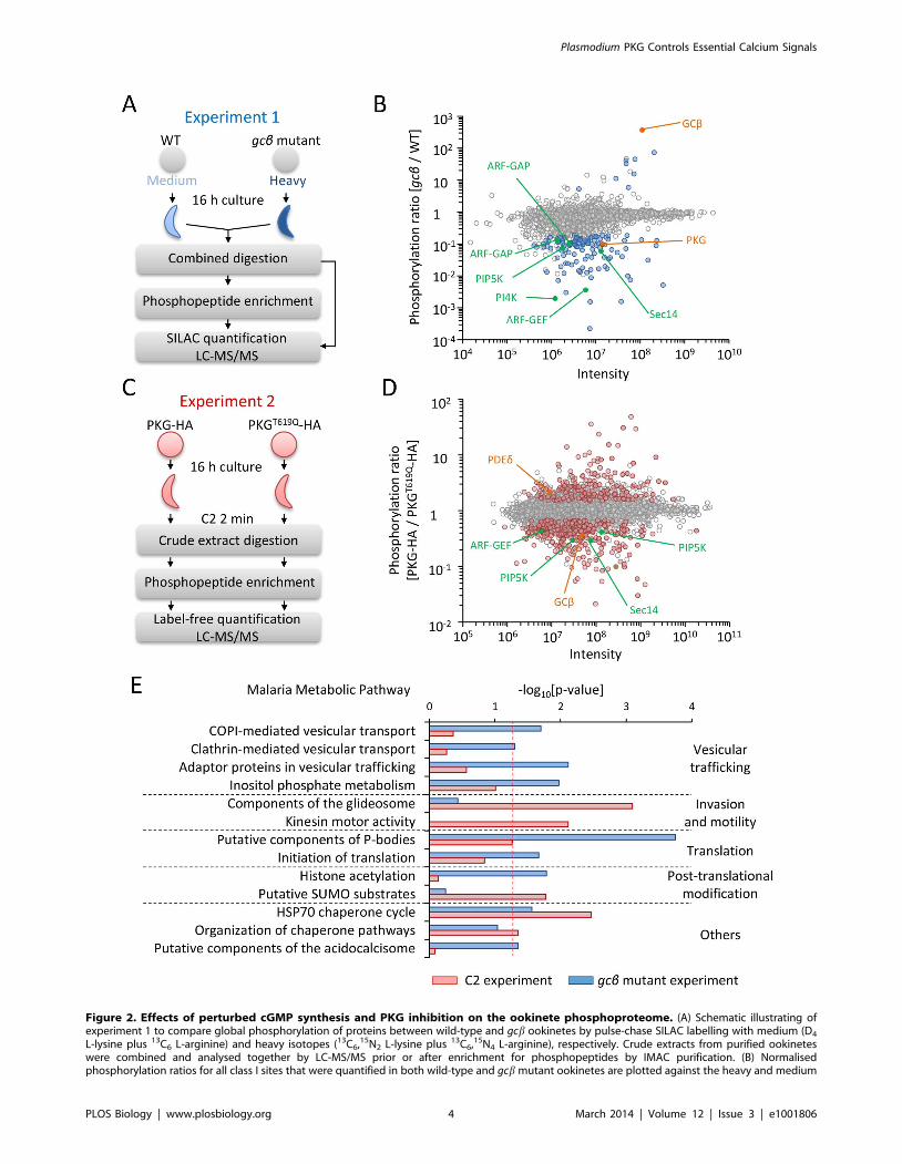
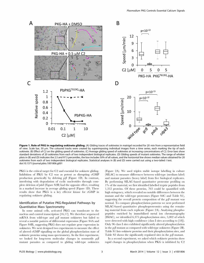
Figure 2. Effects of perturbed cGMP synthesis and PKG inhibition on the ookinete phosphoproteome. (A) Schematic illustrating ofexperiment 1 to compare global phosphorylation of proteins between wild-type and gcb ookinetes by pulse-chase SILAC labelling with medium (D4
L-lysine plus 13C6 L-arginine) and heavy isotopes (13C6,15N2 L-lysine plus 13C6,15N4 L-arginine), respectively. Crude extracts from purified ookineteswere combined and analysed together by LC-MS/MS prior or after enrichment for phosphopeptides by IMAC purification. (B) Normalisedphosphorylation ratios for all class I sites that were quantified in both wild-type and gcb mutant ookinetes are plotted against the heavy and medium
Plasmodium PKG Controls Essential Calcium Signals
PLOS Biology | www.plosbiology.org 4 March 2014 | Volume 12 | Issue 3 | e1001806

for 2 min (Figure 2C). For this experiment, we exposed ookinetes
expressing either PKG-HA or PKGT619Q-HA to 0.5 mM C2,
which blocks gliding only in PKG-HA parasites. Recognising that
in the first experiment SILAC had potentially favoured the
identification of regulated sites in the part of the proteome that
turns over most rapidly and thus incorporates more isotope label
[19], we now opted for a label-free strategy. We detected an even
larger number of 1,634 phosphorylated proteins, on which we
mapped 7,277 unique phosphorylation sites with high confidence
(Table S1). Data from six biological replicates lead us to conclude
that 266 sites belonging to 193 different proteins were reproduc-
ibly regulated in response to inhibition of PKG (Figure 2D and
Table S2).
A significantly less phosphorylated site (6-fold, p = 0.03) in gcbookinetes was serine S694 in the activation loop of the kinase
catalytic domain of PKG itself. Activation loop phosphorylation is
a common mechanism for regulating protein kinase activity,
including in mammalian PKG [20]. Down-regulation of S694
probably reflects a state of reduced PKG activity, as was expected
in the gcb mutant. In contrast, phosphorylation of PKG S694 was
not affected within 2 min of adding C2, suggesting the kinetics of
PKG dephosphorylation is slow. However, C2 reduced phosphor-
ylation of S2072 in GCb and increased phosphorylation of S310 in
PDEd, suggesting possible mechanisms for rapid feedback
regulation of cGMP levels, as happens in mammalian cells
[21,22], reinforcing the notion that these enzymes act in the same
pathway as PKG to regulate ookinete gliding. Inhibition of PKG
also resulted in a rapid 6-fold reduction in the phosphorylation of
S11 in the N-terminal leader peptide upstream of the kinase
domain of CDPK3, a Ca2+-dependent protein kinase important
for ookinete gliding [23], indicating its function is linked closely
with PKG.
To identify mechanisms of regulation by PKG we asked which
cellular pathways were enriched among the proteins with
regulated phosphorylation sites (Figure 2E, see Table S2 for gene
IDs and site information). Treatment with C2 had the greatest
impact on phosphorylation sites of inner membrane complex
(IMC) proteins and components of the gliding motor, such as the
two glideosome-associated proteins GAP45 and GAPM2, and
IMC1b. Microtubule-associated proteins were also enriched,
including several dynein and kinesin-related putative motor
proteins of unknown function. In marked contrast, regulated
phosphosites in the gcb mutant were most abundant in mRNA-
interacting proteins involved in splicing and 39 polyadenylation,
and in components of Plasmodium P-bodies, such as the RNA
helicase, DOZI (development of zygote inhibited), and a putative
trailer hitch homolog, CITH, which are both essential for ookinete
formation by stabilising translationally repressed mRNAs in the
female gametocyte [24]. Also deregulated were multiple sites in a
family of Alba domain-containing proteins that form part of the
DOZI snRNP complex [24]. Furthermore, regulated phospho-
proteins in the gcb mutant were enriched for components of
clathrin and COPI-coated vesicles, including a putative clathrin
coat assembly protein, AP180, the beta subunit of the coatomer
complex, as well as a number of putative regulators of vesicular
trafficking, which together point to an important role for protein
trafficking in ookinete gliding. Finally, the gcb mutant had a
notable abundance of regulated phosphorylation sites in enzymes
involved in the metabolism of inositol phospholipids and their
regulators. Importantly, many representatives from this group of
proteins were also regulated in response to C2 (highlighted in
Figure 2B and Figure 2D), suggesting they may be more direct
targets of PKG than some of the other regulated phosphoproteins.
PKG Controls PI4P and PI(4,5)P2 Levels in MotileP. berghei Ookinetes
Given that enzymes in the inositol phospholipid biosynthetic
pathway were identified by both phosphoproteomic approaches,
we chose to investigate this pathway in more detail. Phosphory-
lated phosphatidylinositol lipids have important roles in vesicle
trafficking and as a source of secondary messengers in signal
transduction. Their biosynthesis from phosphatidyl-1D-myo-inosi-
tol (PI) is mediated by lipid kinases. The P. berghei genome encodes
four putative lipid kinases to convert PI first to phosphatidylino-
sitol 4-phosphate (PI4P) and then to phosphatidylinositol (4,5)-
bisphosphate (PI(4,5)P2) (Figure 3). Hydrolysis of the latter by a PI-
specific phospholipase C (PI-PLC) gives rise to the secondary
messenger inositol (1,4,5)-trisphosphate (IP3), which plays an
important role in P. berghei gametocytes, where it is responsible
for the mobilisation of Ca2+ from internal stores, leading to
activation and gametogenesis [25]. All four PI kinases were
detected in the ookinete phosphoproteome and three contained
sites that were less phosphorylated upon inhibition of PKG or
disruption of gcb (Figure 3). An important regulator of phospho-
inositide metabolism and membrane trafficking is the phosphati-
dylinositol transfer protein Sec14 [26], the phosphorylation of
which was reduced in closely adjacent sites in both experiments.
PIP5K activity of the P. falciparum orthologue of PBANKA_
020310 is controlled by a small G protein of the ADP-ribosylation
factor (ARF) family [27], which cycles between an inactive GDP-
bound and an active GTP-bound form. In other eukaryotes, the
active state of ARF results from its interaction with a guanine
nucleotide exchange factor (ARF-GEF) that forces ARF to adopt a
new GTP molecule in place of a bound GDP, whereas the inactive
state results from hydrolysis of GTP facilitated by a GTPase
activating protein, ARF-GAP. Putative ARF-GEF and ARF-GAP
proteins are encoded in the P. berghei genome, and these also have
GCb/PKG-dependent phosphosites (Figure 3). Taken together
these data led us to hypothesise that phosphoinositide metabolism
is important for ookinete gliding and regulated by PKG.
To test whether the PKG-dependent phosphorylation of
enzymes associated with phosphoinositide metabolism has a direct
role in ookinete motility, we used experimental genetics to infer the
role of putative PI kinases. Both the putative PI4K (PBANKA_
110940) and the putative PIP5K (PBANKA_020310) were unable
to be genetically disrupted (unpublished data), suggesting these
intensities for each site. Data points are coloured to indicate significance of regulation as determined from five biological replicates: blue circles showsignificantly regulated sites (p,0.01, ratio count $6, and fold change .3). Labelled sites are in enzymes linked to cGMP signalling (orange) orphosphoinositide metabolism (green). (C) Schematic illustrating experiment 2 to measure the effect of C2 on global protein phosphorylation usinglabel-free quantification. Purified ookinetes expressing PKG-HA or PKGT619Q-HA were snap-frozen after a 2 min exposure to C2. (D) Normalisedphosphorylation ratios for all class I phosphorylation sites that were quantified in both lines in experiment 2 are plotted against the intensity for eachsite. Data points are coloured to indicate significance of regulation as determined across six biological replicates: red circles show significantlyregulated sites (false discovery rate #0.05 and fold change $1.5). Proteins with likely roles in cGMP signalling and phosphoinositide metabolism arecoloured as in (B). (E) Functional categories from the Malaria Parasite Metabolic Pathway database that were enriched among proteins with regulatedphosphorylation sites in experiments 1 (blue bars) or 2 (red bars). The dashed line shows the chosen significance cutoff of p,0.05.doi:10.1371/journal.pbio.1001806.g002
Plasmodium PKG Controls Essential Calcium Signals
PLOS Biology | www.plosbiology.org 5 March 2014 | Volume 12 | Issue 3 | e1001806

genes may be essential for asexual growth, although both loci
could be modified (Figure S2C and Figure S2D). One of the most
strongly down-regulated phosphorylation sites in the gcb mutant
was S534 of PI4K. To assess the importance of this residue, we
generated allelic replacement constructs to mutate S534 to
alanine, either on its own or in combination with a nearby
phosphorylation site, S538 (Figure S2D). At the ookinete stage,
pi4kS534A and pi4kS534A/S538A clonal mutants showed a significant
decrease in gliding speed compared with a control line, pi4kS534
(Figure 4A and Figure S2D), which would be consistent with
phosphorylation of S534 in PI4K contributing to the regulation of
phosphoinositide metabolism in vivo. A direct link between PKG
and phosphoinositide metabolism was also supported by the
location of PIP5K-HA, which rapidly redistributed from the cell
periphery to the ookinete cytosol in ookinetes treated with 0.5 mM
C2 (Figure 4B and Figure 4C).
To define more precisely the role of PKG in regulating
phosphoinositide metabolism, we examined the effect of C2 on the
phospholipid composition of gliding ookinetes. Analysing total
lipid extracts from purified ookinetes by mass spectrometry, we
detected PI, phosphatidylcholine (PC), phosphatidylethanolamine
(PE), phosphatidylglycerol (PG), cardiolipin (CL), and several
other minor phospholipids. For all of these we then determined
changes in their relative abundance upon inhibition of PKG
(Figure S5A and Figure S5B). Exposing gliding ookinetes to
0.5 mM C2 for 10 min resulted in a marked relative increase in PI,
while peaks corresponding to PIP and PIP2 molecular species were
reduced (Figure 4D). No significant differences in PC, PE, CL, or
PG were detected (unpublished data). In control experiments, C2
had no effect on phospholipid composition of ookinetes expressing
PKGT619Q-HA (Figure 4E). These data suggest a link between
PKG and PI4P synthesis and therefore probably with Ca2+
signalling in ookinetes. We therefore examined next whether PKG
is a positive regulator of Ca2+ release.
PKG Activity Maintains High Cytosolic Ca2+ Levels in P.berghei Ookinetes
To measure Ca2+ levels in life ookinetes, we inserted an
expression cassette for the free Ca2+ reporter pericam into the
redundant p230p locus of the PKG-HA and PKGT619Q-HA lines
(Figure S2E). Pericam is a fusion protein comprising calmodulin,
GFP, and the M13 peptide corresponding to the calmodulin-
binding domain of skeletal muscle myosin light chain kinase [28].
Binding of Ca2+ to the EF hands of calmodulin causes the latter to
interact with the M13 peptide, which in turn modulates the
fluorescence properties of GFP. We expressed a ratiometric form
of pericam, in which Ca2+ shifts the excitation peak from 415 to
494 nm. Dual excitation imaging detects changes in Ca2+ levels
through shifts in the ratio of the Ca2+ bound to the unbound form
of pericam [28]. Ca2+-bound and -unbound pericam were
uniformly distributed throughout the ookinete cytosol (Figure
S6A) and were sensitive to changes in intracellular Ca2+ induced
by the Ca2+ ionophore ionomycin (Figure S6B). Addition of
0.5 mM C2 to PKG-HA-pericam ookinetes resulted in a rapid shift
within 15 s from Ca2+-bound to -unbound reporter if compared to
C2-resistant PKGT619Q-HA-pericam parasites (Figure 5A). There
Figure 3. Phosphorylation sites in proteins with likely roles in phosphoinositide metabolism. All class I phosphorylation sites are shownas squares next to the schematic illustrations of the relevant proteins and their annotated functional domains. PI-PLC was not detected and is shownin light green. Each phosphorylation site is represented by a divided square, the colour of which shows the degree of regulation upon inhibition ofPKG by C2 or in the gcb mutant. Failure to quantify a phosphorylation site with one of the two experimental designs is shown in white.doi:10.1371/journal.pbio.1001806.g003
Plasmodium PKG Controls Essential Calcium Signals
PLOS Biology | www.plosbiology.org 6 March 2014 | Volume 12 | Issue 3 | e1001806

was no such response in the solvent control (Figure 5B). These
results demonstrate that PKG activity is critical for high cytosolic
Ca2+ levels to be maintained in gliding ookinetes.
PKG Controls Agonist-Induced Ca2+ Mobilisation UponGametocyte Activation
It remains unknown whether ookinetes glide constitutively in vivo
or whether their behaviour responds to internal or external stimuli.
In contrast, gametocytes, the developmentally arrested sexual
precursor stages that circulate in the blood stream in a
developmentally arrested form, become activated by well-defined
environmental triggers within seconds of being taken up by a
feeding mosquito. Activation is mediated by a drop in temperature
and the concomitant exposure to a chemical stimulus from the
mosquito, XA [2]. These well-defined triggers offer an opportunity
to ask how PKG and Ca2+ interact in response to physiological
agonists. At a permissive temperature XA triggers PI(4,5)P2
hydrolysis in P. berghei gametocytes, presumably by activation of
PI-PLC [25], which results in the rapid mobilisation of Ca2+ from
internal stores. In P. falciparum, on the other hand, XA enhances
GC activity in membrane preparations [29], and PKG regulates
gametocyte activation [10]. If and how signalling through cGMP
and Ca2+ are linked during gametocyte activation has not been
addressed.
To ask if in P. berghei gametocytes PKG regulates Ca2+ release in
response to XA, we introduced into the dssu or cssu locus of the
marker-free PKG-HA and PKGT619Q-HA lines an expression
cassette for a reporter protein that is based on the Ca2+-dependent
photoprotein, aequorin (Figure S2F) [30]. XA triggered a transient
luminescence response that peaked rapidly after a characteristic
lag phase 10 s after stimulation (Figure 5C), as described
previously [31]. In parasites expressing PKG-HA this response
was dose-dependently blocked by C2 with an IC50 of around
3 mM (Figure 5C). C2 acted through inhibition of PKG, because
the PKGT619Q-HA-GFPaeq line was completely resistant to even
higher concentrations of the inhibitor. We conclude that the rapid
activation of PKG within seconds of exposing gametocytes to their
natural agonist mediates gametocyte activation through the
mobilisation of Ca2+ and is therefore most likely a key event for
the transmission of Plasmodium to the mosquito.
PKG Controls Phosphoinositide and Ca2+ Levels in P.falciparum Schizonts
To explore whether Ca2+ release via PKG signalling regulates
blood stages in Plasmodium parasites, we turned to the major
human pathogen P. falciparum. In asexual blood stages of P.
falciparum PKG is critically required for schizonts to rupture and
for merozoites to egress [4]. C1 and C2 were shown to block
schizont rupture by inhibiting PKG, while conversely, an inhibitor
of cGMP-PDE, zaprinast, raises cellular cGMP levels and triggers
premature egress through the rapid discharge of micronemes and
exonemes from the intracellular parasite that is strictly dependent
on parasite PKG [1]. Since discharge of secretory organelles by
Plasmodium schizonts also depends on Ca2+ we asked whether
activation of PKG by zaprinast triggers egress by controlling
intracellular Ca2+ levels. In synchronised P. falciparum schizonts
loaded with the fluorescent Ca2+ sensor Fluo-4, addition of
100 mM zaprinast led to a marked increase in free cytosolic Ca2+
within 20 s (Figure S6C). This rapid Ca2+ response was mediated
by PKG, because it could be inhibited by the simultaneous
administration of C2 with a half-maximal effect of ,2 mM
(Figure 6A). Importantly, C2 was completely ineffective in
blocking zaprinast-induced Ca2+ release in parasites expressing a
resistant T618Q allele of PfPKG.
To ask if PKG controlled schizont Ca2+ mobilisation by
regulating phosphoinositide metabolism, we studied the schizont
lipidome (Figure S5C). Zaprinast triggered a rapid depletion of
Figure 4. Phosphoinositide phosphorylation links PKG to glidingin P. berghei ookinetes. (A) Ookinete gliding speed of PI4KS534A andPI4KS534A/S538A ookinete mutants. Values are representative of 20individual ookinetes from two independent biological replicates. (B) Ratioof peripheral to cytosolic fluorescence intensity from optical sectionstaken of different ookinetes from the experiment shown in (C); n = 10sections from different ookinetes. Statistical analysis was carried out usinga two-tailed t test. (C) Confocal immunofluorescence images of fixedookinetes showing the effect of 0.5 mM C2 on the cellular distribution of aC-terminally HA tagged PIP5K (PBANKA_020310) expressed from itsendogenous promoter. Scale bar, 5 mm. (D) Relative quantification of PI,PIP, PIP2, and PIP3 levels after 10 min treatment with 0.5 mM C2 or DMSOin PKG-HA ookinetes. (E) As in (D) but for PKGT619Q-HA ookinetes. Errorbars in (D) and (E) show standard deviations of two biological replicates.The p values are from two-tailed t test. See also Figure S5.doi:10.1371/journal.pbio.1001806.g004
Plasmodium PKG Controls Essential Calcium Signals
PLOS Biology | www.plosbiology.org 7 March 2014 | Volume 12 | Issue 3 | e1001806

PIP, PIP2, and PIP3, and a concomitant rise in PI from total lipid
extracts within 10 s, consistent with the kinetics of Ca2+
mobilisation by the same treatment. Pre-exposing infected
erythrocytes to 1 mM C2 blocked the zaprinast-induced depletion
of phosphorylated PIs, but only in parasites expressing wild-type
PKG and not in parasites expressing the C2-resistant T618Q allele
of PKG (Figure 6B). Uninfected erythrocytes, in contrast,
contained very little phosphorylated PIs (Figure S5D). Taken
together, these data show that activation of PKG by the PDE
inhibitor zaprinast triggers a cellular Ca2+ response in P. falciparum
schizonts that is accompanied by the rapid initiation of PIP2
hydrolysis.
Discussion
PKG was discovered in Eimeria and Toxoplasma as the target for
potent anticoccidial inhibitors that achieve selectivity over
vertebrate PKG by exploiting the small gatekeeper residue
typically found in PKG of apicomplexa, including in malaria
parasites [8]. PKG has since been shown to have essential
functions in egress, microneme secretion, and gliding of T. gondii
tachyzoites [9], as well as in biological processes as diverse as
merozoite egress and gametocyte activation in Plasmodium [1,10].
As a result PKG is considered as a promising drug target also in
malaria parasites [4,33], yet how PKG performs its wide range of
cellular functions has remained elusive.
In this study we demonstrate that PKG is the cGMP effector
kinase that regulates gliding of P. berghei ookinetes downstream of
GCb and PDEd. Ookinetes are relatively tractable cells by both
biochemical and genetic methods. We therefore used the
comparative analysis of total ookinete phosphoproteomes to
identify proteins whose phosphorylation is directly or indirectly
dependent on PKG. In one type of experiment we compared the
abundance of individual phosphopeptides between wild-type and
gcb mutant ookinetes. With a second experimental design we
assessed the sensitivity of global phosphorylation events in gliding
ookinetes to chemical inhibition of PKG by C2. Importantly, by
comparing two parasite lines in the presence of C2 that differed in
only a single amino acid, the gatekeeper residue of PKG, we could
focus our analysis on on-target effects of the inhibitor. Our
combined experiments identified .9,000 different phosphoryla-
tion sites on nearly 2,000 ookinete proteins. In view of this large
number of phosphorylated proteins and the many cellular
pathways potentially involved in gliding, it was not surprising to
find 250 phosphoproteins reproducibly regulated under either one
or both experimental conditions. Statistical pathway enrichment
analysis, followed by experimental validation, proved crucial for
extracting biological meaning from these complex global data sets.
Figure 5. PKG controls cytosolic Ca2+ levels in ookinete and upon gametocyte activation in P. berghei. (A and B) Determination ofrelative cytosolic Ca2+ levels in purified ookinetes expressing PKG-HA or PKGT619Q-HA using pericam, a dual excitation Ca2+ reporter. We added 0.5 mMC2 treatment (A) or DMSO treatment (B) at t = 20 s. Fluorescence was normalised as follows: DF = (Fn2F20)/F20, in which Fn is the fluorescence at t = ns; F20 is the reference time before addition of C2 or DMSO, and was normalised to the baseline provided by the C2-resistant parasites expressingPKGT619Q-HA. Error bars show standard errors from three independent replicates each using 10 ookinetes per condition. See Figure S6A and FigureS6B for a validation of the pericam reporter in P. berghei. (C) Luminescence responses of gametocytes expressing GFP-aequorin to differentconcentrations of C2. Gametocytes were stimulated with 50 mM XA at t = 0 s. Data are representative of at least four independent experiments.doi:10.1371/journal.pbio.1001806.g005
Plasmodium PKG Controls Essential Calcium Signals
PLOS Biology | www.plosbiology.org 8 March 2014 | Volume 12 | Issue 3 | e1001806
From the significantly enriched cellular pathways downstream of
PKG, we selected phosphoinositide metabolism for experimental
validation because enzymes in this pathway showed robust signs of
differential phosphorylation under both experimental conditions.
Importantly, PI(4,5)P2 hydrolysis by PI-PLC generates IP3, a
second messenger that is important for Ca2+-dependent gameto-
cyte activation of P. berghei [25] and that also elicits Ca2+ responses
in intraerythrocytic asexual stages of P. falciparum [34]. A role for
PKG in phosphoinositide metabolism upstream of Ca2+ signalling
could therefore provide a unifying explanation for the seemingly
disparate roles of PKG in different life cycle stages.
A Universal Role for PKG in Ca2+ Mobilisation FromInternal Stores Reconciles Its Diverse Functions AcrossDifferent Plasmodium Species and Stages
All biological processes in apicomplexan parasites known to
require PKG are also thought to rely on the release of Ca2+ from
intracellular stores [11,31,32,35,36], which in turn activates
distinct stage-specific effector pathways including members of a
family of plant-like Ca2+-dependent protein kinases (CDPKs) [37].
Merozoite egress requires CDPK5 [11], cell cycle progression to
S-phase in activated male gametocytes is mediated by CDPK4
[31], and ookinete gliding relies on CDPK3 [23,35]. By combining
a range of genetically encoded and chemical Ca2+ reporter systems
with PKG gatekeeper mutants in both P. berghei and P. falciparum,
this study demonstrates clearly that PKG controls cytosolic Ca2+
levels in all these life cycle stages. The critical importance of PKG
upstream of parasite cytosolic Ca2+ is thus of universal relevance to
parasite development in blood and transmission stages, as it holds
true for agonist-induced signalling in gametocytes, in constitutively
gliding ookinetes, and after artificial PKG activation triggered by a
PDE inhibitor in erythrocytic schizonts.
Cross-talk between second messengers is common in eukaryotic
cells. In mammalian vascular smooth muscle cells, for instance,
cGMP regulates cytosolic Ca2+ negatively, chiefly through
PKG1b, which reduces Ca2+ release from internal stores by
phosphorylating the IP3 receptor in the ER membrane [38].
Phototransduction, in contrast, relies on a direct inhibitory
interaction of cGMP with a cyclic nucleotide-gated cation channel
in the plasma membrane, which appears to have been lost from
the apicomplexan genomes during evolution [39]. We here
present evidence for a positive interaction between cGMP and
Ca2+ signalling in malaria parasites that invokes a different
mechanism by involving regulation of phosphoinositide metabo-
lism. Studying gametocyte activation in P. berghei, we previously
identified hydrolysis of PIP2 and generation of IP3 by PI-PLC as a
critical event upstream of Ca2+ mobilisation by XA [25]. IP3-
dependent Ca2+ release also operates in P. falciparum blood stages
[34], although genetic evidence for an IP3 receptor in Plasmodium is
still missing. Because canonical G-protein coupled receptors and
heterotrimeric G-proteins, which typically regulate PI-PLC, are
absent from the genomes of Plasmodium species, it has remained
Figure 6. PKG controls phosphoinositide metabolism and Ca2+ mobilisation in P. falciparum schizonts prior to merozoite egress. (A)Relative fluorescence intensity of ,108 Fluo-4–loaded synchronised P. falciparum schizonts in response to simultaneous exposure to 100 mMzaprinast and increasing concentrations of C2. Data are representative of two independent experiments. (B) Relative abundance over time of PI, PIP(left panel), PIP2, and PIP3 (right panel, note different scale) after simultaneous inhibition of PDEs by zaprinast and PKG by C2. The response of P.falciparum 3D7 (dashed lines) is compared to a transgenic clone expressing the C2-resistant PKGT618Q allele (solid lines).doi:10.1371/journal.pbio.1001806.g006
Plasmodium PKG Controls Essential Calcium Signals
PLOS Biology | www.plosbiology.org 9 March 2014 | Volume 12 | Issue 3 | e1001806

unclear how IP3 production in apicomplexan parasites is
regulated. Our lipidome analysis of P. falciparum schizonts provides
strong biochemical evidence that activating PKG with the help of
a PDE inhibitor that raises cellular levels of cGMP [1] leads to the
rapid and PKG-dependent hydrolysis of PIP2 at the same time as
cellular Ca2+ increases sharply. This would be consistent with a
role for PKG for the activation of PI-PLC. Whether this involves
phosphorylation remains unknown, as our proteometric studies
failed to detect PI-PLC with confidence.
Intriguingly, inhibiting PKG in gliding ookinetes, where Ca2+ is
already elevated, revealed a different link between PKG and
phosphoinositide metabolism. In this situation, inhibiting PKG did
not cause PIP2 to accumulate, as would be expected if its primary
role was to promote PI-PLC activity. Instead, inhibition of PKG
revealed phosphorylation of PI as a rate-limiting step. More
tentative evidence that in ookinetes PKG regulates signalling at the
point of PI phosphorylation comes from two additional observa-
tions. First, mutations in phosphorylated serine residues in PI4K
reduce ookinete gliding speed. Second, a type I PIP5K of
ookinetes localises to the cell periphery in a PKG-dependent
manner. Dissociation of PIP5K from the plasma membrane or
possibly the IMC of the ookinete could merely be a consequence of
substrate depletion [40]. On the other hand, PIP5K, the P.
falciparum ortholog of which encodes a functional type I PIP5K that
is activated by ARF [27], may also provide a more active link to
cGMP signalling, as it contains EF-hand-like motifs of a kind
typically found in the neuronal Ca2+ sensor family of proteins,
which intriguingly can function as activators of membrane GCs in
other eukaryotes [41].
More work is clearly required to evaluate lipid kinases and PI-
PLC as direct substrates of PKG; to establish their precise roles,
and those of their products, in linking PKG to Ca2+ in Plasmodium;
and to determine their relative importance in schizonts, ookinetes,
and gametocytes. A recent study has proposed that in the
mammalian central nervous system PKG regulates presynaptic
vesicle endocytosis by controlling PI(4,5)P2 synthesis indirectly via
the small GTPase RhoA and Rho kinase [42]. However, a
different mechanism must operate in apicomplexan parasites,
which appear to lack a Rho signalling pathway.
Our identification of PKG as a positive upstream regulator of
cytosolic Ca2+ levels extends current models of how schizonts
rupture in the bloodstream and how XA activates gametocytes in
the mosquito. Importantly, it puts a spotlight on the GCs and
PDEs that control cGMP levels in the parasite and which must
provide the next layer of regulation for some of the key events in
the life cycle of malaria parasites.
Vesicular Trafficking and Microneme BiogenesisApicomplexan zoites glide with the help of transmembrane
adhesins, which are secreted apically from micronemes, and then
translocate posteriorly, where they are shed and left behind in a
trail of vesicles that also contain other membrane proteins [43].
Sustained gliding must be fuelled by the continuous synthesis of
substantial quantities of proteins and lipids that need to be
trafficked to new micronemes, which are then transported to the
apical pole of the ookinete for regulated secretion. In the gcbmutant the striking abundance of regulated phosphoproteins with
likely functions in vesicular transport (Figure 7A), but also of
chaperones (Figure 2E), reflects a profoundly dysregulated
secretory system (Figure 7B), which may result from the role of
PKG as a regulator of phosphoinositide metabolism in at least two
ways. First, the changed phosphorylation pattern in vesicular
transport proteins may result from a ‘‘traffic jam’’ following a
block in IP3/Ca2+-dependent regulated secretion of micronemes at
the apical end of the ookinete. Second, because in other
eukaryotes PIP and PIP2 control many essential cellular processes
in regulating membrane dynamic and vesicular trafficking [44],
PKG-mediated changes in PI phosphorylation could alter
vesicular transport more directly and at different stages in the
secretory pathway.
Microneme biogenesis is poorly understood in Plasmodium, but
work in T. gondii tachyzoites has identified endosomal sorting
signals that traffic micronemal proteins from the Golgi to an
endosomal-like compartment before they are packaged into
micronemes. In the absence of either dynamin-related protein B
(DrpB) or the VPS10/sortilin homolog TgSORTLR, proteins
destined for micronemes fail to be targeted from the Golgi to
secretory organelles and instead enter the constitutive secretion
pathway. TgSORTLR is thought to be an essential cargo receptor
to transport microneme and rhoptry proteins to endosomal-like
compartments of the T. gondii tachyzoite. In other eukaryotes
retrograde transport of sortilin to the Golgi relies on its interaction
with the conserved retromer complex. We here report regulated
phosphorylation for P. berghei homologues for another retromer
component, VPS35, as well as for DrpB (T690 and S736,
respectively; Figure 7A). Similarly, a VPS9 homolog we found
regulated in S767 is a likely conserved activator of Rab5 GTPases,
which is relevant because in T. gondii Rab5A and Rab5C are
essential for targeting proteins to unique subsets of micronemes
[45]. Although many of the regulated phosphosites we report here
are probably not phosphorylated directly by PKG, these examples
illustrate that our study provides a rich source of leads for future
research into gliding motility and microneme biogenesis in
Plasmodium.
Our data demonstrate that by regulating phosphoinositide
metabolism PKG controls multiple essential cellular processes
including critical Ca2+ signals and probably vesicular trafficking.
Members of a promising class of new antimalarials, the
imidazopyrazines, target PI4K [46]. Our analysis provides a
rationale for the activity of imidazopyrazines against multiple life
cycle stages of Plasmodium and highlights how a stage transcending
signalling pathway can regulate different critical steps during
parasite development. Future work will have to refine the
knowledge about the nature of PKG-mediated signalling by
identifying direct PKG substrates and the genetic or environmen-
tal factors controlling PKG activity.
Materials and Methods
Ethics StatementAll animal experiments were conducted under a license from
the UK Home Office in accordance with national and European
animal welfare guidelines.
ParasitesP. berghei ANKA wild-type strain 2.34 and transgenic lines made
in the same background were maintained in female Theiler’s
Original outbred mice and infections monitored on Giemsa-
stained blood films. Exflagellation was quantified 3 d postinfection
by adding 4 ml of blood from a superficial tail vein to 150 ml
exflagellation medium (RPMI 1640 containing 25 mM HEPES,
4 mM sodium bicarbonate, 5% FCS, 100 mM XA, pH 7.4).
Between 13 and 16 min after activation, the number of
exflagellating microgametocytes was counted in a haemocytometer
and the RBC count determined. The percentage of RBCs
containing microgametocytes was assessed on Giemsa-stained
smears, and the number of exflagellations per 100 microgameto-
cytes was then calculated. For Ca2+ assays, gametocytes were
Plasmodium PKG Controls Essential Calcium Signals
PLOS Biology | www.plosbiology.org 10 March 2014 | Volume 12 | Issue 3 | e1001806

separated from uninfected erythrocytes on a nycodenz cushion
made up from 48% of a nycodenz stock (27.6% w/v Nycodenz in
5.0 mM Tris-HCl [pH 7.20], 3.0 mM KCl, 0.3 mM EDTA) and
RPMI1640 medium containing 25 mM HEPES, 5% FCS, 4 mM
sodium bicarbonate, pH 7.30. Gametocytes were harvested from
the interphase.
For ookinete cultures, parasites were maintained in phenyl
hydrazine-treated mice. Ookinetes were produced in vitro by
adding one volume of high gametocyteamia blood in 20 volumes
of ookinete medium (RPMI1640 containing 25 mM HEPES, 10%
FCS, 100 mM XA, pH 7.5) and incubated at 19uC for 16–18 h.
For the gcb mutant experiment, customized RPMI1640 medium
(Invitrogen) containing 13C6,15N2 L-lysine and 13C6,15N4 L-
arginine (gcb mutant) or D4 L-lysine and 13C6 L-arginine (wild-
type) was used. Conversion efficiency was determined by live
staining of ookinetes and activated macrogametes with Cy3-
conjugated 13.1 monoclonal antibody against p28. The conversion
rate was determined as the number of banana-shaped ookinetes as
a percentage of the total number of Cy3-fluorescent cells. For
biochemical analysis and Ca2+ assays, ookinetes were purified
using paramagnetic anti-mouse IgG beads (Life Technologies)
coated with anti-p28 mouse monoclonal antibody (13.1). For
motility assays, ookinete cultures were added to an equal volume
of Matrigel (BDbioscience) containing DMSO or C2 on ice, mixed
thoroughly, dropped onto a slide, covered with a cover slip, and
sealed with nail polish. After identifying a field containing
ookinetes, time-lapse videos were taken (1206; 1 frame every
20 s, for 20 min) on a Leica M205A at 19uC. Movies were
analysed with Fiji and the Manual Tracking plugin (http://pacific.
mpi-cbg.de/wiki/index.php/Manual_Tracking).
For transmission experiments batches of ,50 female Anopheles
stephensi, strain SD500, mosquitoes were allowed to feed on infected
mice 3 d after intraperitoneal injection of infected blood. Unfed
mosquitoes were removed the day after. Infected mosquitoes were
maintained on fructose at 19uC and oocyst numbers were counted
on dissected midguts 7 d after feeding. Sporozoite numbers were
determined on day 21 by homogenising dissected salivary glands
and counting the released sporozoites. To determine sporozoite
infectivity to mice 21 d after infection, infected mosquitoes were
allowed to feed on naıve mice or 20,000 freshly isolated sporozoites
in RPMI 1640 containing 1% penicillin/streptomycin were injected
in a volume of 100 ml into the tail vein. Mice were then monitored
daily for blood stage parasites.
Targeting Vector Construction and TransgenicGeneration
Tagging, knockout, and allelic replacement constructs were
generated using phage recombinase mediated engineering in Esche-
richia coli TSA (Figure S1); PlasmoGEM vectors PbG01-2397b11,
Figure 7. Proteins with PKG-dependent phosphorylation sites and their hypothetical functions in gliding ookinetes. (A) Proteins withphosphorylation sites that are significantly regulated in response to C2 (red dots) or deletion of gcb (blue dots) and that belong either to knownsignalling pathways are linked to the glideosome, or which belong to the enriched functional groups of proteins with likely roles in vesiculartrafficking. The numeric part of the PBANKA gene ID is shown in grey. The amino acid numbers for the regulated sites in each protein are stated nextto a letter indicating if the phosphorylated residue is a serine (S), threonine (T), or tyrosine (Y). (B) Model illustrating hypothetical functions for theproteins in (A) in the molecular motor or during microneme biogenesis in a gliding ookinete.doi:10.1371/journal.pbio.1001806.g007
Plasmodium PKG Controls Essential Calcium Signals
PLOS Biology | www.plosbiology.org 11 March 2014 | Volume 12 | Issue 3 | e1001806
PbG02_C-11d08, PbG02_C-25e06, and PbG01-2399h12 con-
taining PBANKA_100820 (PKG), PBANKA_110940 (PI4K),
PBANKA_020310 (PIP5K), and PBANKA_100820 (PDEa)
encoding genes, respectively, (Figure S2) were generated from
genomic DNA library clones in linear pJAZZ-OK vectors
(Lucigen) as described (Ref. 47 and http://plasmogem.sanger.ac.
uk/). Oligonucleotides used are shown in Table S3.
Point mutations were introduced with a two-step strategy using
l Red-ET recombineering in E. coli. The first step involved the
insertion by homologous recombination of a Zeocin-resistance/
Phe-sensitivity cassette surrounded by sequences 59 and 39 of the
codon of interest amplified using primer pairs specific for the gene
of interest (GOI): goi-delF/goi-delR. Recombinant bacteria were
then selected on Zeocin. After verification of the recombination
event by PCR, a second round of recombination exchanged the
Zeocin-resistance/Phe-sensitivity cassette with a PCR product
containing the desired mutation surrounding the codon of interest
amplified using goi-mutF/goi-mutR primer pairs. Bacteria were
selected on YEG-Cl kanamycin plates. Mutations were confirmed
by sequencing vectors isolated from colonies sensitive to Zeocin.
Generation of knockout and tagging constructs was performed
using sequential recombineering and gateway steps as previously
described [47]. Lambda red-ET recombineering was first used to
introduce a bacterial selection marker amplified into the gDNA
insert, such that the target gene is either deleted or prepared for
39-end tagging. The bacterial marker was then replaced with a
selection cassette for P. berghei in a Gateway LR Clonase reaction in
vitro. The modified library inserts were then released from the
plasmid backbone using NotI and used to transfect P. berghei.
For the pericam construct, the P. berghei hsp70 promoter
(PBANKA_071190) was first cloned using SacII/XhoI and hsp70-
F/hsp70-R primers into a p230p targeting vector, which also
contains a resistance cassette encoding human DHFR [48]. This
was followed by the pericam coding sequence [28] downstream of
the promoter, using XhoI only and primers pc-F and pc-R. The
vector was linearised using HindIII/EcoRI. The expression vector
for the GFP-aequorin chimeric gene was transfected in PKG-HA
and PKGT619Q-HA parasites as previously described [31].
Schizonts for transfection were purified from overnight cultures
and transfected with 1–5 mg of linearised DNA as previously
described [49]. Electroporated merozoites were injected intravei-
nously into a naıve mouse. Resistant parasites were selected by
pyremithamine supplied in the drinking water. All transgenic
parasites were cloned by limiting dilution cloning, and correct
integrations of the targeting constructs were verified by diagnostic
PCRs and sequencing. Negative selection of PKG-HA and
PKGT619Q-HA parasites expressing yFCU was performed through
the administration of 5 fluorocytosine via the drinking water [50],
and dilution cloning was subsequently carried out allowing for
further genetic modifications.
Ca2+ AssaysFluorescence measurements of purified ookinetes expressing
Pericam were performed on single cells. Purified ookinetes were
immobilised on Lab-Tek II chambered coverglass slides using
Cell-Tak as described previously [51]. Immobilised ookinetes were
imaged in PBS (Ca2+ and Mg2+ free) supplemented with 10 mM
glucose. Confocal images were acquired with a LSM510 laser
scanning confocal microscope (Zeiss), a Plan-Apochromat 636/
1.4 oil Ph3 objective and a LP505 filter using 405 nm or 488 nm
excitation. For time lapse image acquisition, cells were imaged
with a Fluar 406/1.30 oil objective on a Zeiss Axiovert 200 M
inverted microscope equipped with an AxioCam MRm camera
using the multidimensional acquisition mode of the Axiovision
software. Reporter fluorescence was monitored using a 21HE
FURA filter set (exposure time, 50 ms) and a 38HE filter set
(exposure time, 300 ms) at a rate of 1 frame per 5 s for 35 cycles.
The fluorescence of individual ookinetes was subsequently
analysed using the Axiovision Physiology Module.
Aequorin reconstitution and luminometric Ca2+ detection from
purified P. berghei gametocytes were performed as previously
described [31]. First, purified gametocytes were washed 3 times in
coelenterazine loading buffer (CLB - PBS, 20 mM HEPES,
20 mM Glucose, 4 mM sodium bicarbonate, 1 mM EGTA, 0.1%
w/v bovine serum albumin, pH 7.2). Reconstitution was then
achieved by shaking ,108 gametocytes, in 0.5 ml CLB, supple-
mented with 5 mM coelenterazine for 30 min at 19uC. Loaded
gametocytes were washed twice in CLB and were then suspended
in 10 ml RPMI 1640, 5% FBS, 4 mM sodium bicarbonate,
pH 7.2. For luminescence measurements, 150 ml of the gameto-
cyte suspension were injected into the same volume of ookinete
medium containing DMSO or C2 in a 96-well assay plate of an
Orion II microplate system luminometer. For each sample 50
luminescence readings were acquired over 35 s.
Changes in the levels of intracellular free Ca2+ were measured
using Fluo-4 (Sigma) loaded P. falciparum late stage schizonts that
had been cultured in albumax II (Fisher Scientific) and human A+erythrocytes (washed whole blood from the National Blood service)
as previously described [4]. Excitation was measured using a
SPECTRAmax microplate fluorometer. Levels of free Ca2+ were
compared to a baseline read prior to the addition of a test reagent.
Schizonts were purified magnetically (Macs; Milteny Biotec) and
pelleted by centrifugation for 2 min at 500 g. Parasites were
resuspended in 106 warm Ringer Buffer (122.5 mM NaCl,
5.4 mM KCl, 0.8 mM MgCl2, 11 mM HEPES, 10 mM D-
Glucose, 1 mM NaH2PO4) to 1–26108 parasites/ml, and 2 ml of
5 mM Fluo-4 was added to 1 ml of parasite preparation. Cells
were incubated with Fluo-4 at 37uC for 45 min and washed twice
in warm Ringer buffer and incubated for 20 min for de-
esterification followed by a further two washes. The pellet was
resuspended in Ringer buffer and plated out on the bottom half of
a 96-well plate. Compound dilutions were plated out on the top of
the plate and the excitation of the cells measured at 20 s intervals
for a period of 3 min to achieve a baseline read. The cells were
then transferred onto the test compound and read for a further
5 min.
Immunofluorescence Staining and Confocal MicroscopyOokinete immunofluorescence assays were performed as
previously described [52]. For HA staining after fixation with
3% paraformaldehyde in PBS, ookinetes were permeabilised with
0.1% Triton X-100/PBS and blocked with 2% BSA/PBS.
Primary antibodies were diluted in blocking solution (rat anti-
HA, 1:200). Anti-rat Alexa488 was used as a secondary antibody
together with DAPI (all from Life Technologies), all diluted 1:200
in blocking solution. Confocal images of ookinetes were acquired
with a LSM510 laser scanning confocal microscope (Zeiss).
RNA SequencingApproximately 10 mg of wild-type and gcb mutant RNA were
extracted in duplicate from purified ookinetes using the RNeasy kit
(Qiagen). Depletion of ribosomal RNA and highly abundant
transcripts and sequencing library construction were performed as
previously described [53]. The library was end-sequenced on an
Illumina GAII instrument. TopHat [54] was used to map the
Illumina reads against the P. berghei ANKA reference genome.
Read counts and reads per kilo base per million mapped reads
(RPKM) values were calculated for each gene. Differential
Plasmodium PKG Controls Essential Calcium Signals
PLOS Biology | www.plosbiology.org 12 March 2014 | Volume 12 | Issue 3 | e1001806
expression was analysed using DESeq [55]. Data plots were
created using R, and Artemis [56] was used to visualise
transcriptome data.
Quantitative Protein Mass SpectrometryFor the gcb mutant proteome profiling, SILAC-based quanti-
tative proteome profiling was performed essentially as previously
described [19]. Briefly, 40 mg of total protein was used for each of
the five replicates (20 mg from wild-type (K4/R6) and 20 mg gcbmutant (K8/R10)) pooled. Protein gels were stained with colloidal
Coomassie blue, and each lane was excised and cut into 12 bands
that were destained and in-gel digested overnight using trypsin.
Extracted peptides were suspended using 0.5% formic acid were
analysed online using an Ultimate 3000 Nano/Capillary LC
System (Dionex) coupled to an LTQ Orbitrap Velos hybrid mass
spectrometer (Thermo Electron) equipped with a nanospray ion
source. Peptides were desalted online using a micro-Precolumn
cartridge (C18 Pepmap 100, LC Packings) and then separated
using a 70 min RP gradient (4%–32% acetonitrile/0.1% formic
acid) on a BEH C18 analytical column (1.7 mm, 75,235 mm
id610 cm, Waters) and analysed using a Top10 CID method.
For the phosphoproteomic analyses, ,5 mg of total proteins
were extracted as previously described [19] from ,5.108 purified
ookinetes for each of the five and six biological replicates of the gcbmutant and C2 experiments, respectively. Solubilised proteins
were processed according to the FASP procedure [57,58] and
digested with Trypsin Gold (Promega). Peptides were collected by
centrifugation and addition of ammonium bicarbonate and further
desalted using Sep-Pak Light C18 cartridge (Waters).
IMAC purifications were performed as described previously
[59], with the following modifications: Peptides were resuspended
in IMAC loading buffer (50% acetonitrile, 0.1% TFA) and
incubated with pre-equilibrated Phos-Select beads (Sigma) for 1 h
at room temperature. The beads were then transferred to a
TopTip (Glygen) and washed once with IMAC loading buffer, 1%
acetic acid, and then water. Phosphopeptides were eluted with
100 ml ammonia water pH 11 and acidified using formic acid.
Phosphopeptide samples were analysed online using an
Ultimate 3000 Nano/Capillary LC System (Dionex) coupled to
an LTQ Orbitrap Velos hybrid mass spectrometer (Thermo
Scientific) equipped with a nanospray ion source. Data were
analysed using MaxQuant version 1.0.13.13 and Mascot server
2.2 (Matrix Science) and MaxQuant version 1.3.0.5 for the gcbmutant experiment and C2 experiment, respectively [60].
MaxQuant processed data were searched against a combined
mouse and P. berghei protein identified by the GeneDB database. A
protein false discovery rate (FDR) of 0.01 and a peptide FDR of
0.01 were used for identification level cutoffs. Class I phosphor-
ylation site was defined with a localisation probability of .0.75
and a score difference of .5 [18]. For the gcb mutant experiment,
phosphorylation sites with a p value for detection of significant
outlier ratio $0.01, a ratio count $6, and at least a 3-fold change
were defined as being regulated in the mutant. For the C2
experiment, data were filtered, and two-sample t testing was
performed with a permutation-based FDR calculation in Perseus
(1.3.0.4) as described previously [61]. Phosphorylation sites within
an FDR of 0.05 with at least a 1.5-fold change were defined as
being regulated by C2 treatment. See Text S1 for details of protein
extraction, data acquisition, and analysis.
Lipid Extraction and Electrospray-Mass SpectrometryAnalysis
Total lipids were extracted using a modified Bligh and Dyer
method from ,5.108 purified P. berghei ookinetes or 50 ml of
packed cell volume of purified P. falciparum schizont per replicate.
Ookinetes were first washed with PBS, suspended in 100 ml PBS,
and transferred to a glass tube. Cells were lysed with 375 ml of 1:2
(v/v) chloroform:methanol and vortexed for 15 min. Samples
were made biphasic by the addition of 125 ml of CHCl3 and
125 ml of H2O. After centrifugation at 1,000 g at room
temperature for 5 min, the lower phase was transferred to a new
glass vial and dried under nitrogen.
Lipids were subsequently dissolved in a mixture of chloroform:-
methanol (1:2) and acetonitrile:iso-propanol:water (6:7:2) and
analysed with a Absceix 4000 QTrap, a triple quadrupole mass
spectrometer equipped with a nanoelectrospray source. Samples
were delivered using either thin-wall nanoflow capillary tips or a
Nanomate interface in direct infusion mode (125 nl/min). The
lipid extracts were analysed in both positive and negative ion
modes using a capillary voltage of 1.25 kV. Tandem mass spectra
(MS/MS) scanning (daughter, precursor, and neutral loss scans)
was performed using nitrogen as the collision gas with collision
energies between 35 and 90 V. Each spectrum encompasses at
least 50 repetitive scans.
Tandem mass spectra (MS/MS) were obtained with collision
energies as follows: 35–45 V, PC/SM in positive ion mode,
parent-ion scanning of m/z 184; 35–55 V, PI/IPC in negative ion
mode, parent-ion scanning of m/z 241; 35–65 V, PE in negative
ion mode, parent-ion scanning of m/z 196; 20–35 V, PS in
negative ion mode, neutral loss scanning of m/z 87; and 40–90 V,
all glycerophospholipids (including PA, PG, and CL) were
detected by precursor scanning for m/z 153 in negative ion
mode. MS/MS daughter ion scanning was performed with
collision energies between 35 and 90 V. Assignment of phospho-
lipid species is based upon a combination of survey, daughter,
precursor, and neutral loss scans, as well as previous assignments
[62]. The identity of phospholipid peaks was verified using the
LIPID MAPS: Nature Lipidomics Gateway (www.lipidmaps.org).
Pathway Enrichment AnalysisThe enrichment analysis was based on a list of 3,167 P.
falciparum genes assigned to 212 metabolic pathways and functional
groups present in Malaria Parasite Metabolic Pathways database
(http://priweb.cc.huji.ac.il/malaria, June 2013). Annotations were
transferred to P. berghei orthologs identified by the GeneDB
database (http://www.genedb.org). Enrichment for pathways in
sets of genes was calculated using a one-tailed Fisher’s exact test.
Supporting Information
Figure S1 Workflow for genetically modifying largeinserts from a P. berghei genomic DNA library by lRed-ET recombinase-mediated engineering. This protocol
was developed to generate allelic exchange vectors for PKG and
PI4K that mutate the gatekeeper residue or selected phosphory-
lation sites. Steps 1–7 can be carried out as shown to first mutate a
site and then turn the modified alleles into a complementation
vector. Alternatively, steps 1–4 can be used to modify an existing
PlasmoGEM tagging vector.
(TIF)
Figure S2 Production and genotyping of transgenic P.berghei lines. (A–F) Genetic modification vectors and strategies
used in this study and genotyping data for each transgenic
parasite. Oligonucleotides used for PCR genotyping are indicated
and agarose gels of corresponding PCR products from genotyping
reactions are shown. For pkg and pi4k point mutations, sequence
chromatograms of the modified sites are also shown.
(TIF)
Plasmodium PKG Controls Essential Calcium Signals
PLOS Biology | www.plosbiology.org 13 March 2014 | Volume 12 | Issue 3 | e1001806

Figure S3 Fitness of PKG-HA and PKGT619Q-HA lines atdifferent life cycle stages. (A) Blood parasitaemia after
intraperitoneal injection of 107 parasites. Error bars show standard
deviations from three infections. (B) Zygote-to-ookinete conversion
as assessed 18 h after inducing gametogenesis in vitro by scoring the
developmental states of 100 parasites labelled with anti–p28-Cy3
monoclonal antibody by fluorescence microscopy. Error bars show
standard deviations from six cultures. (C) Number of oocysts
formed per mosquito midgut. Whiskers show 2.5 and 97.5
percentiles, the box includes 50% of all values, and the horizontal
line shows median values; n = 22 mosquitoes from two separate
infection replicates. (D) Number of sporozoites per infected
salivary gland. Errors show standard deviations from 44 dissected
glands from two biological replicates. (E) Blood parasitaemia after
mosquito bite. Error bars show standard deviations from three
infections. (F) Blood stages parasitaemia after intravenous injection
of 20,000 salivary gland sporozoites. Error bars as in (E).
(TIF)
Figure S4 Molecular phenotyping of the gcb mutant. (A)
Normalised transcript abundance expressed as reads per kilo base
per million (RPKM) for wild-type and gcb mutant ookinetes,
showing no gross differences in transcription levels. A represen-
tative experiment of two replicates is shown. (B) Coverage plot
showing mRNA sequencing reads mapping to a 39 fragment of the
disrupted gcb gene of the gcb mutant. Translation of partial
mRNAs from the disrupted gene would be predicted to result in a
nonfunctional protein lacking a complete cyclase domain, which
explains the detection of peptides from GCb in the proteome of
the mutant (see, e.g., Figure 2B). (C) Normalised protein ratios
deduced from five biological replicates are plotted against the
heavy/medium ratio counts for each protein.
(TIF)
Figure S5 Effect of zaprinast and C2 on phospholipidsof P. berghei ookinetes, P. falciparum schizont–infectederythrocytes, and uninfected erythrocytes. (A) Total
phospholipid analysis of P. berghei ookinetes. (B) Phosphorylated
PI analysis in the presence of C2 of P. berghei ookinetes expressing
PKG-HA or PKGT619Q-HA over a m/z range of 1,000–1,300. (C)
Phospholipid analysis of P. falciparum schizont–infected erythro-
cytes over an m/z range of 600–1,000. (D) Phospholipid analysis
of human uninfected RBCs in negative ion mode (600–1,000 m/z,
left panel; 1,000–1,300 m/z, right panel), showing low or
undetectable levels of PIPs.
(TIF)
Figure S6 Characterisation of Ca2+ reporters in P.berghei ookinetes and P. falciparum schizonts. (A)
Confocal immunofluorescence images of a live ookinete expressing
the dual excitation ratiometric calcium reporter pericam showing
unbound reporter from excitation at 405 nm and bound reporter
from excitation at 488 nm. Scale bar, 5 mm. (B) Fluorescence
response of wild-type purified ookinetes expressing pericam after
the addition of 0.5 mM ionomycin (t = 20 s), followed by 10 mM
calcium (t = 60 s). Fluorescence was normalised as follow:
DF = (Fn2F20)/F20, in which Fn is the fluorescence at t = n s and
F20 is the reference time t = 20 s before addition of C2. Error bars
indicate the standard error of the mean from three independent
replicates each representative of 10 ookinetes. (C) Fluorescence
response of synchronised P. falciparum 3D7 schizonts loaded with
Fluo-4 to varying concentrations of zaprinast and to the A23187
ionophore.
(TIF)
Table S1 Transcriptome, proteome, and phosphopro-teome analyses of altered cGMP signalling in ookinetes.The table displays the full list of transcripts, proteins, and
phosphorylation sites that were detected in the gcb or C2
experiments. Fold changes are indicated as FC, and S(A) is the p
value for detection of significant outlier ratio.
(XLS)
Table S2 Phosphorylation sites significantly regulatedin the gcb mutant and C2 experiments. The table displays
the list of phosphorylation ratios for significantly regulated
phosphorylated sites in the gcb and the C2 experiments,
respectively. Annotation from the Malaria Parasite Metabolic
Pathway database is indicated.
(XLSX)
Table S3 Oligonucleotides used in this study. The table
displays the list of oligonucleotides used in this study.
(XLS)
Text S1 Supplementary methods.
(DOCX)
Acknowledgments
We would like to thank Matthew Jones for many helpful discussions and
Hagai Ginsburg for providing data from the Malaria Parasite Metabolic
Pathways database. We would also like to thank members of the Rayner,
Choudhary, Berriman, and Billker groups for their support and Christine
Hopp for initiating the development of Fluo-4 fluorescence monitoring in
P. falciparum.
Author Contributions
The author(s) have made the following declarations about their
contributions: Conceived and designed the experiments: MBr MOC
TKS DAB MBe JCR JC OB. Performed the experiments: MBr MOC
TKS ET SS KV LC ARG. Analyzed the data: MBr MOC TKS ET FS
OB. Contributed reagents/materials/analysis tools: DAB. Wrote the
paper: MBr OB.
References
1. Collins CR, Hackett F, Strath M, Penzo M, Withers-Martinez C, et al. (2013)
Malaria parasite cGMP-dependent protein kinase regulates blood stagemerozoite secretory organelle discharge and egress. PLoS Pathog 9:
e1003344.
2. Billker O, Lindo V, Panico M, Etienne AE, Paxton T, et al. (1998) Identification
of xanthurenic acid as the putative inducer of malaria development in the
mosquito. Nature 392: 289–292.
3. Baker DA (2011) Cyclic nucleotide signalling in malaria parasites. Cell Microbiol13: 331–339.
4. Taylor HM, McRobert L, Grainger M, Sicard A, Dluzewski AR, et al. (2010)The malaria parasite cyclic GMP-dependent protein kinase plays a central role
in blood-stage schizogony. Eukaryot Cell 9: 37–45.
5. Moon RW, Taylor CJ, Bex C, Schepers R, Goulding D, et al. (2009) A cyclic
GMP signalling module that regulates gliding motility in a malaria parasite.
PLoS Pathog 5: e1000599.
6. Taylor CJ, McRobert L, Baker DA (2008) Disruption of a Plasmodium falciparum
cyclic nucleotide phosphodiesterase gene causes aberrant gametogenesis. MolMicrobiol 69: 110–118
7. Deng W, Baker DA (2002) A novel cyclic GMP-dependent protein kinase isexpressed in the ring stage of the Plasmodium falciparum life cycle. Mol Microbiol
44: 1141–1151.
8. Donald RG, Allocco J, Singh SB, Nare B, Salowe SP, et al. (2002) Toxoplasma
gondii cyclic GMP-dependent kinase: chemotherapeutic targeting of an essentialparasite protein kinase. Eukaryot Cell 1: 317–328.
9. Wiersma HI, Galuska SE, Tomley FM, Sibley LD, Liberator PA, et al. (2004) Arole for coccidian cGMP-dependent protein kinase in motility and invasion.
Int J Parasitol 34: 369–380.
10. McRobert L, Taylor CJ, Deng W, Fivelman QL, Cummings RM, et al. (2008)
Gametogenesis in malaria parasites is mediated by the cGMP-dependent protein
kinase. PLoS Biol 6: e139.
Plasmodium PKG Controls Essential Calcium Signals
PLOS Biology | www.plosbiology.org 14 March 2014 | Volume 12 | Issue 3 | e1001806
11. Dvorin JD, Martyn DC, Patel SD, Grimley JS, Collins CR, et al. (2010) A plant-
like kinase in Plasmodium falciparum regulates parasite egress from erythrocytes.Science 328: 910–912.
12. Carvajal JA, Germain AM, Huidobro-Toro JP, Weiner CP (2000) Molecular
mechanism of cGMP-mediated smooth muscle relaxation. J Cell Physiol 184:409–420.
13. Zhang X, Cote RH (2005) cGMP signaling in vertebrate retinal photoreceptorcells. Front Biosci 10: 1191–1204.
14. Potter LR (2011) Guanylyl cyclase structure, function and regulation. Cell Signal
23: 1921–1926.15. Martin SK, Jett M, Schneider I (1994) Correlation of phosphoinositide
hydrolysis with exflagellation in the malaria microgametocyte. J Parasitol 80:371–378.
16. O’Halloran DM, Altshuler-Keylin S, Lee JI, L’Etoile ND (2009) Regulators ofAWC-mediated olfactory plasticity in Caenorhabditis elegans. PLoS Genet 5:
e1000761.
17. Schwappacher R, Weiske J, Heining E, Ezerski V, Marom B, et al. (2009) Novelcrosstalk to BMP signalling: cGMP-dependent kinase I modulates BMP receptor
and Smad activity. EMBO J 28: 1537–1550.18. Olsen JV, Blagoev B, Gnad F, Macek B, Kumar C, et al. (2006) Global, in vivo,
and site-specific phosphorylation dynamics in signaling networks. Cell 127: 635–
648.19. Sebastian S, Brochet M, Collins MO, Schwach F, Jones ML, et al. (2012) A
Plasmodium calcium-dependent protein kinase controls zygote development andtransmission by translationally activating repressed mRNAs. Cell Host Microbe
12: 9–19.20. Nolen B, Taylor S, Ghosh G (2004) Regulation of protein kinases; controlling
activity through activation segment conformation. Mol Cell 15: 661–675.
21. Kotera J, Grimes KA, Corbin JD, Francis SH (2003) cGMP-dependent proteinkinase protects cGMP from hydrolysis by phosphodiesterase-5. Biochem J 372:
419–426.22. Zhou Z, Sayed N, Pyriochou A, Roussos C, Fulton D, et al. (2008) Protein kinase
G phosphorylates soluble guanylyl cyclase on serine 64 and inhibits its activity.
Arterioscler Thromb Vasc Biol 28: 1803–1810.23. Siden-Kiamos I, Ecker A, Nyback S, Louis C, Sinden RE, et al. (2006)
Plasmodium berghei calcium-dependent protein kinase 3 is required for ookinetegliding motility and mosquito midgut invasion. Mol Microbiol 60: 1355–1363.
24. Mair GR, Lasonder E, Garver LS, Franke-Fayard BM, Carret CK, et al. (2010)Universal features of post-transcriptional gene regulation are critical for
Plasmodium zygote development. PLoS Pathog 6: e1000767.
25. Raabe AC, Wengelnik K, Billker O, Vial HJ (2011) Multiple roles for Plasmodium
berghei phosphoinositide-specific phospholipase C in regulating gametocyte
activation and differentiation. Cell Microbiol 13: 955–966.26. Bankaitis VA, Mousley CJ, Schaaf G (2010) The Sec14 superfamily and
mechanisms for crosstalk between lipid metabolism and lipid signaling. Trends
Biochem Sci 35: 150–160.27. Leber W, Skippen A, Fivelman QL, Bowyer PW, Cockcroft S, et al. (2009) A
unique phosphatidylinositol 4-phosphate 5-kinase is activated by ADP-ribosyla-tion factor in Plasmodium falciparum. Int J Parasitol 39: 645–653.
28. Nagai T, Sawano A, Park ES, Miyawaki A (2001) Circularly permuted greenfluorescent proteins engineered to sense Ca2+. Proc Natl Acad Sci U S A 98:
3197–3202.
29. Muhia DK, Swales CA, Deng W, Kelly JM, Baker DA (2001) The gametocyte-activating factor xanthurenic acid stimulates an increase in membrane-
associated guanylyl cyclase activity in the human malaria parasite Plasmodium
falciparum. Mol Microbiol 42: 553–560.
30. Baubet V, Le Mouellic H, Campbell AK, Lucas-Meunier E, Fossier P, et al.
(2000) Chimeric green fluorescent protein-aequorin as bioluminescent Ca2+reporters at the single-cell level. Proc Natl Acad Sci U S A 97: 7260–7265.
31. Billker O, Dechamps S, Tewari R, Wenig G, Franke-Fayard B, et al. (2004)Calcium and a calcium-dependent protein kinase regulate gamete formation and
mosquito transmission in a malaria parasite. Cell 117: 503–514.
32. Glushakova S, Lizunov V, Blank PS, Melikov K, Humphrey G, et al. (2013)Cytoplasmic free Ca2+ is essential for multiple steps in malaria parasite egress
from infected erythrocytes. Malar J 12: 41.33. Diaz CA, Allocco J, Powles MA, Yeung L, Donald RG, et al. (2006)
Characterization of Plasmodium falciparum cGMP-dependent protein kinase(PfPKG): antiparasitic activity of a PKG inhibitor. Mol Biochem Parasitol
146: 78–88.
34. Alves E, Bartlett PJ, Garcia CR, Thomas AP (2011) Melatonin and IP3-inducedCa2+ release from intracellular stores in the malaria parasite Plasmodium
falciparum within infected red blood cells. J Biol Chem 286: 5905–5912.35. Ishino T, Orito Y, Chinzei Y, Yuda M (2006) A calcium-dependent protein
kinase regulates Plasmodium ookinete access to the midgut epithelial cell. Mol
Microbiol 59: 1175–1184.
36. Lourido S, Tang K, Sibley LD (2012) Distinct signalling pathways control
Toxoplasma egress and host-cell invasion. Embo J 31: 4524–4534.
37. Billker O, Lourido S, Sibley LD (2009) Calcium-dependent signaling and kinases
in apicomplexan parasites. Cell Host Microbe 5: 612–622.
38. Schlossmann J, Ammendola A, Ashman K, Zong X, Huber A, et al. (2000)
Regulation of intracellular calcium by a signalling complex of IRAG, IP3
receptor and cGMP kinase Ibeta. Nature 404: 197–201.
39. Johnson JL, Leroux MR (2010) cAMP and cGMP signaling: sensory systems
with prokaryotic roots adopted by eukaryotic cilia. Trends Cell Biol 20: 435–
444.
40. Kunz J, Fuelling A, Kolbe L, Anderson RA (2002) Stereo-specific substrate
recognition by phosphatidylinositol phosphate kinases is swapped by changing a
single amino acid residue. J Biol Chem 277: 5611–5619.
41. Fik-Rymarkiewicz E, Duda T, Sharma RK (2006) Novel frequenin-modulated
Ca2+-signaling membrane guanylate cyclase (ROS-GC) transduction pathway
in bovine hippocampus. Mol Cell Biochem 291: 187–204.
42. Taoufiq Z, Eguchi K, Takahashi T (2013) Rho-kinase accelerates synaptic
vesicle endocytosis by linking cyclic GMP-dependent protein kinase activity to
phosphatidylinositol-4,5-bisphosphate synthesis. J Neurosci 33: 12099–12104.
43. Stewart MJ, Vanderberg JP (1992) Electron microscopic analysis of circumspor-
ozoite protein trail formation by gliding malaria sporozoites. J Protozool 39:
663–671.
44. Di Paolo G, De Camilli P (2006) Phosphoinositides in cell regulation and
membrane dynamics. Nature 443: 651–657.
45. Kremer K, Kamin D, Rittweger E, Wilkes J, Flammer H, et al. (2013) An
overexpression screen of Toxoplasma gondii Rab-GTPases reveals distinct
transport routes to the micronemes. PLoS Pathog 9: e1003213.
46. McNamara CW, Lee MCS, Lim CS, Lim SH, Roland J, et al. (2013) Targeting
Plasmodium PI(4)K to eliminate malaria. Nature, Advance online publication.
47. Pfander C, Anar B, Schwach F, Otto TD, Brochet M, et al. (2011) A scalable
pipeline for highly effective genetic modification of a malaria parasite. Nat
Methods 8: 1078–1082.
48. Pino P, Sebastian S, Kim EunBin A, Bush E, Brochet M, et al. (2012) A
tetracycline-repressible transactivator system to study essential genes in malaria
parasites. Cell Host Microbe 12: 824–834.
49. Janse CJ, Ramesar J, Waters AP (2006) High-efficiency transfection and drug
selection of genetically transformed blood stages of the rodent malaria parasite
Plasmodium berghei. Nat Protoc 1: 346–356.
50. Orr RY, Philip N, Waters AP (2012) Improved negative selection protocol for
Plasmodium berghei in the rodent malarial model. Malar J 11: 103.
51. Louise Meyer R, Zhou X, Tang L, Arpanaei A, Kingshott P, et al. (2010)
Immobilisation of living bacteria for AFM imaging under physiological
conditions. Ultramicroscopy 110: 1349–1357.
52. Volkmann K, Pfander C, Burstroem C, Ahras M, Goulding D, et al. (2012) The
alveolin IMC1h is required for normal ookinete and sporozoite motility
behaviour and host colonisation in Plasmodium berghei. PLoS One 7: e41409.
53. Otto TD, Wilinski D, Assefa S, Keane TM, Sarry LR, et al. (2010) New insights
into the blood-stage transcriptome of Plasmodium falciparum using RNA-Seq. Mol
Microbiol 76: 12–24.
54. Trapnell C, Pachter L, Salzberg SL (2009) TopHat: discovering splice junctions
with RNA-Seq. Bioinformatics 25: 1105–1111.
55. Anders S, Huber W (2010) Differential expression analysis for sequence count
data. Genome Biol 11: R106.
56. Rutherford K, Parkhill J, Crook J, Horsnell T, Rice P, et al. (2000) Artemis:
sequence visualization and annotation. Bioinformatics 16: 944–945.
57. Manza LL, Stamer SL, Ham AJ, Codreanu SG, Liebler DC (2005) Sample
preparation and digestion for proteomic analyses using spin filters. Proteomics 5:
1742–1745.
58. Wisniewski JR, Zougman A, Nagaraj N, Mann M (2009) Universal sample
preparation method for proteome analysis. Nat Methods 6: 359–362.
59. Kokubu M, Ishihama Y, Sato T, Nagasu T, Oda Y (2005) Specificity of
immobilized metal affinity-based IMAC/C18 tip enrichment of phosphopep-
tides for protein phosphorylation analysis. Anal Chem 77: 5144–5154.
60. Cox J, Mann M (2008) MaxQuant enables high peptide identification rates,
individualized p.p.b.-range mass accuracies and proteome-wide protein
quantification. Nat Biotechnol 26: 1367–1372.
61. Lundby A, Andersen MN, Steffensen AB, Horn H, Kelstrup CD, et al. (2013) In
vivo phosphoproteomics analysis reveals the cardiac targets of beta-adrenergic
receptor signaling. Sci Signal 6: rs11.
62. Richmond GS, Gibellini F, Young SA, Major L, Denton H, et al. (2010)
Lipidomic analysis of bloodstream and procyclic form Trypanosoma brucei.
Parasitology 137: 1357–1392.
Plasmodium PKG Controls Essential Calcium Signals
PLOS Biology | www.plosbiology.org 15 March 2014 | Volume 12 | Issue 3 | e1001806