Common blood and tissue parasites of man - CDC stacks
40
-
Upload
khangminh22 -
Category
Documents
-
view
1 -
download
0
Transcript of Common blood and tissue parasites of man - CDC stacks


Common
BLOOD AND TISSUE PARASITES
of Man
Life Cycle Charts
Prepared by
Dorothy M. Melvin, M. M. Brooke, and G. R. Healy
Laboratory Consultation and Development Section
Laboratory Branch
U. S. DEPARTMENT OF HEALTH, EDUCATION, AND WELFAREPUBLIC HEALTH SERVICECommunicable Disease Center
Atlanta, Georgia 30333

These charts were originally issued in 1961 as an unnumbered publi-
cation by the Laboratory Branch of the Communicable Disease Center
for use in training courses.
Public Health Service Publication No. 1234
April 1965
U.S. Government Printing Office, Washington: 1965
For sale by the Superintendent of Documents, U.S. Government Printing OfficeWashington, D.C., 20402 - Price 20 cents

Contents
I. Introduction 1
II. Blood Parasites 3
Malaria 3
Hemoflagellates 8
Leishmania 8
Trypanosomes 9
Filaria 14
Dracunculus 22
III. Tissue Parasites 24
Trichinella spiralis 24
Toxocara canis 24
Echinococcus granulosus 25
Life Cycle Charts
Malaria 7
Leishmania 11
Trypanosoma gambiense and T. rhodesiense 12
Trypanosoma cruzi 13
Wuchereria bancrofti 16
Brugia malayi 17
Loa loa 18
Acanthocheilonema perstans 19
Mansonella ozzardi 20
Onchocerca volvulus 21
Dracunculus medinensis 23
Trichinella spiralis 27
Toxocara canis 28
Echinococcus granulosus 29


Life Cycle Charts
COMMON BLOOD AND TISSUE PARASITES OF MAN
I. Introduction
The blood and tissue parasites of man include representatives of
two groups of helminths, nematodes and cestodes, and of two groups
of protozoa, hemoflagellates and sporozoa. In general, the life cycles
tend to be more complex than those of the intestinal parasites and,
in the case of the blood parasites, involve an arthropod vector as
well as a human host. Intermediate hosts are necessary for the trans-
mission of most of the blood and tissue organisms and in several
cases, reservoir hosts are important. With the exception of Toxocara
canis , no external development takes place.
Like those of the intestinal parasites,* the life cycle charts of
the blood and tissue parasites are intended for use by students of
parasitology, laboratory technicians, public health workers, and
physicians. They are designed as simple, basic patterns that pur-
posely omit details of epidemiology, incubation periods, patent
periods, and exceptions to the usual pattern. The individual user can
add any needed or desired details, obtaining this information from
lectures or from the literature. Toxoplasma gondii has been excluded
from this presentation since neither the correct classification nor
the complete life history has been determined.
The design of the charts conforms to the following general rules,
insofar as possible:
1. The diagnostic and infective stages are indicated and empha-
sized. These stages are in proportion with regard to species
within a given group but, because of size variations, the scale
is not uniform between groups. The sizes of the nematode male
and female adults are relative to each other and, within the
filariae, are drawn to a single scale.
Melvin, D. M., Brooke, M. M. and Sadun, E. H. 1965. Common Intestinal Helminthsof Man - Life Cycle Charts. PHS Publication No. 1235.
Brooke, M. M. and Melvin, D. M. 1964. Common Intestinal Protozoa of Man - Life
Cycle Charts. PHS Publication No. 1140.
1

2. Morphological details are included in a diagrammatic fashion
in most of the stages of the protozoa, but only in the diagnos-
tic and infective stages of the helminths.
3. Survival times, pre-patent and patent periods, and developmen-
tal times are omitted.
4. Not all of the embryonic and larval stages of the helminths are
indicated. For example, the number of nematode larval stages
are not recorded.
5. Only broad groups of organisms are indicated as invertebrate
intermediate hosts. More specific names are applied to mamma-lian hosts.
6. Reservoir hosts are not listed on the charts, but in cases
where man is an accidental or abnormal host, the common
hosts are indicated.
7. No general references are listed since the material incorpo-
rated into the charts is commonly found in most parasitology
textbooks. However, where necessary, specific references
have been included.

II. Blood Parasites
The blood parasites presented here include malaria, hemoflagel-
lates, and filaria. Not all of the species of each group actually
inhabit the blood stream (for example, adult filaria, Leishmania spp.)
but most are associated with the circulatory system at some stage
of their development. Possibly the leishmania might be considered
as tissue rather than blood parasites, but since they are classified
as hemoflagellates they are presented here with the blood inhabiting
forms. Another tissue parasite is Dracunculus medinensis which is
usually put in the same general category as the filariae, and is also
included with the blood parasites in these charts.
Malaria
The life history of malaria is similar to that of the intestinal
sporozoa, the coccidia, and involves an asexual cycle, schizogony,
in the human host and a sexual cycle, sporogony, in the vector, a
species of Anopheles mosquito. The pattern presented here is that
of Plasmodium vivax but, in general, it is the same for all four
species.
Immediately after the introduction of the infective sporozoites
through the bite of the mosquito, a pre-erythrocytic (exo-erythrocytic)
development occurs. In man, these exo-erythrocytic stages have been
demonstrated in the parenchymal cells of the liver. On the basis of
the knowledge of the prepatent period, this pre-erythrocytic phase
probably requires a week or more depending on the species involved.
In P . vivax,Plasmodium ovale , and Plasmodium malariae residual
exo-erythrocytic stages probably continue in fixed cells during the
erythrocytic phases, while in Plasmodium falciparum , on the other
hand, residual exo-erythrocytic stages probably do not occur. The
presence or absence of residual exo-erythrocytic stages affects both
the clinical course and the therapy of the disease.
The asexual stages of malaria include trophozoites and schizonts
and, except in P. falciparum, all stages of growth may be found in
peripheral blood. P. falciparum organisms complete their schizogony
in the capillaries of the internal organs so that usually only the
rings (young trophozoites) of the asexual forms are seen in circu-
lating blood.
In addition to the asexual stages, gametocytes (sexual forms)
develop in man and in all four species, may be found in peripheral
blood. Their exact origin is unknown. Some workers believe that
they develop from certain of the merozoites produced in the erythro-
cytic schizonts; others, that they come from merozoites formed in
3

exo-erythrocytic schizogony. Gametocytes are the infective stages
for the mosquito and, in the arthropod stomach, develop into gametes
which initiate the sexual cycle. They do not multiply in the humanhost, and unless ingested by the appropriate mosquito, will degener-
ate and die within a few days. The sexual development, ending in
production of sporozoites, the infective stage for man, is influenced
by such extrinsic factors as temperature and humidity. The average
length of both sexual and asexual cycles for each species is in-
cluded in the table below. These are based on reports in the liter-
ature (by various investigators) and represent the more commonaverage development periods as recorded.
The Anopheles species involved as hosts for human malaria vary
with the geographical area. There are over sixty species which are
considered vectors of malaria in different areas of the world. In the
United States, there are only two that are considered to be important
vectors: A . quadrimaculatus in the east and A . freebomi in the west.
Not all species of Anopheles serve as malaria vectors, either be-
cause they are not good biological hosts, or because they normally
do not feed on human blood.
Malaria infections can be spread from man to man via blood
inoculations, for example, transfusions or common hypodermic needles
used by drug addicts, but an exo-erythrocytic phase does not occur.
Exo-erythrocytic development takes place only after sporozoite
inoculation.
Man is considered to be the only natural vertebrate host for the
four species of human malaria. However, recently it has been found
that P. cynomolgi,a parasite of monkeys resembling P. vivax, can
be transmitted by mosquitoes to humans (Eyles, et al., 1960). While
there is at present no clear-cut evidence that transmission occurs
naturally, the possibility of monkey to man infection exists. Since
this early report of Eyles, man to man transmission of P. cynomolgi
by mosquitoes has been demonstrated by Contacos et al. (1962).
The susceptibility of humans to simian malaria species may influence
control and eradication programs in areas in which they are endemic.
References
Eyles, D. E., Coatney, G. R., and Getz, M. 1960. Vivax-type malariaparasites of Macaques transmissible to man. Science 131 : 18 1 2-1 813.
Contacos, P. G., Elder, H. A., Coatney, G. R., and Genther, C.
1962. Man to man transfer of P lasmodium cynomolgi by mosquitobite. Am. J. Trop. Med. Hyg. 11 ( 2) : 1 86 -1 93.
4

AVERAGE
DEVELOPMENT
TIMES*
- 771-344 0 - 65-2 5
Boyd,
M.
F.
1949.
Malariology.
W.
B.
Saunders
Co.,
Philadelphia.
Wilcox,
A.
1960.
Manual
for
the
Microscopical
Diagnosis
of
Malaria
in
Man.
PHS
Publication
No.
796.
Government
Printing
Office,
Washington.


LIFE CYCLE of—
Malaria
Microgamete
Based on life cycle of Plasmodium vivax
1

Hemoflage Mates
The life history patterns of the hemoflagellates, like that of
malaria, involve an arthropod vector which is chiefly responsible for
the transmission and spread of the infection. The vectors for the
hemoflagellates are various species of flies, and the specificity of
the vector usually limits the geographical distribution of the parasite.
Unlike most of the other blood parasites, the hemoflagellates do
not possess sexual forms and multiplication occurs entirely through
binary fision in one or more of the morphological stages. Species of
two genera, Trypanosoma and Leishmania parasitize man. Four mor-
phological stages are described: leishmania, leptomonas, crithidia,
and trypanosome. Of the four, the leishmania are the only non-
flagellated forms and both crithidial and trypanosomal stages pos-
sess an undulating membrane in addition to the flagellum. Except
for Trypanosoma cruzi
,
no more than two of the four stages are
associated with each genus of organisms.
Leishmania
In the human host, Leishmania spp. exist only in the leishmania
form and are intracellular parasites of reticulo-endothelial cells:
L. donovani predominantly in bone marrow and internal organs such
as spleen and liver, L. tropica in skin and subcutaneous tissue, and
L. hrasiliensis in cutaneous and mucocutaneous tissues. Three
other species of Leishmania,
L. peruana, L. guyanensis, and L.
mexicana, have been involved in cutaneous leishmaniasis in Peru,
in Panama, and in Mexico, Guatemala, and British Honduras, respec-
tively. The cycles of all species are similar.
In the arthropod vectors, which are certain species of Phlebotomus
(sand-flies), the leishmania stages taken up during the insect bite
transform into the leptomonas forms. These leptomonads divide in the
midgut and in 3 to 5 days move to the proboscis of the sand-fly. When
the insect feeds again, the leptomonads (infective forms) are injected
into the vertebrate host where they again become leishmania within
reticulo-endothelial cells.
Several species of Phlebotomus have been incriminated as vectors
of leishmania, among them P . argentipes and P . chinensis for L.
donovani, P. intermedins for L. hrasiliensis
,and P. papatasii and
P. sergenti for L. tropica.
Dogs, rodents, and possibly other mammals may serve as reser-
voir hosts.
8

Trypanosomes
Three species of trypanosomes are known to infect man: Trypano-
soma gambiense and Trypanosoma rhodesiense
,
etiologic agents of
African sleeping sickness (African trypanosomiasis) and Trypano-
soma cruzi, ethologic agent of Chagas’ Disease (American trypanoso-
miasis). In addition to T. cruzi, a second species, Trypanosoma
rangeli, has been reported from man and other animals in the western
hemisphere but has not been included in the charts presented here.
The life cycles of the two African trypanosomes are similar
except in the specific fly vector and are shown in a single chart.
These forms occur in man only in the trypanosomal form and are
ordinarily located in the blood stream and lymph nodes in the early
phases of infection and in the central nervous system (primarily
T. gambiense ) in the chronic phases. They multiply in man by longi-
tudinal binary fission of the trypanosomes.
In the arthropod vector, species of tsetse flies, the trypanosomes,
taken up during the bite, multiply by binary fission in this stage in
the midgut. They then migrate to the salivary glands where they be-
come crithidial forms and undergo a second multiplication. In about 2
to 3 weeks they become metacyclic trypanosomes. These infective
stage trypanosomes are introduced into the vertebrate host when the
fly bites again.
The principal species of tsetse flies which serve as vectors for
the two trypanosome species are Glossina palpalis and its sub-
species, G. palpalis fuscipes and G. tachinoides for T. gambiense
and G. morsitans, G. pallidipes and G. swynnertoni for T, rho-
desiense, The choice of specific vectors determines to a degree the
geographical distribution of the trypanosome infections: T, gambiense
is found chiefly in tropical West and Central Africa and T, rho-
desiense in northeastern and southern Rhodesia, Nyasaland, Portu-
guese East Africa, Tanganyika, and Eastern Uganda.
Wild game mammals such as antelope, probably are reservoir
hosts for T, rhodesiense, and cattle, hogs, and goats may possibly
serve as reservoir hosts for T, gambiense, although this has not
been definitely established.
The life cycle of T. cruzi is markedly different from that of the
African trypanosomes. In the mammalian host, two forms, the trypano-
some and leishmania stages, may be found. The trypanosome form
usually occurs in the blood stream during the early acute phase and
during febrile periods. The leishmania stage is found in the tissue,
usually either reticulo-endothelial cells or heart muscle cells. Occa-
sionally, they are found within macrophages in the blood. In the
vertebrate host, the parasite divides only in the leishmania stage.
771-344 0 - 65-3 9

The vector, a species of triatomid bug, may ingest the typanosome
stage in the blood or the leishmania stage within a macrophage
during feeding. In the bug midgut the parasite becomes the flagellated
crithidial form and multiplies. The organisms then migrate to the
hindgut where they become metacyclic or infective trypanosomes.
These forms passed in the feces discharged as the bug feeds,
ordinarily enter the mammalian host by being rubbed into the bite
wound.
The triatomid species incriminated as vectors are Panstrongylus
megistus,Triatoma infestans (southern South America), T. dimidiata
(Central America) and Rhodnius prolixus (northern South America
and Central America).
Both domestic and wild mammals serve as reservoir hosts
including dogs, cats, pigs, armadillos, and rodents among others.
The second American species, T. rangeli, morphologically is
more nearly like the African trypanosomes than like T. cruzi . How-
ever, in its choice of a vector, a genus of triatomid bugs, it resembles
the latter. The life cycle differs from that of T. cruzi in the following
aspects: the metacyclic trypanosomes in the vector gain access to
the vertebrate host through the bite of the bug as well as through
fecal contamination of the bite wound as occurs in T. cruzi; no
leishmania stages have been found in the vertebrate host. T. rangeli
has been reported from northern South America and from Central
America.
10

LIFE CYCLE of
Leishmania
Reproduction and

LIFE CYCLE of-
Trypanosoma gambiense and T. rhodesiense
Trypanosome
in blood, lymph
(eventually invade
central nervous system)
Metacyclic trypanosome
in salivary gland
(infective stage)
Crithidial stage in salivary gland
12

LIFE CYCLE of-
Tr^yganosoma cruzi
13

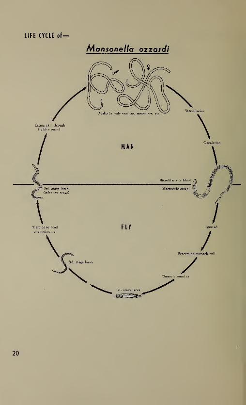
Filaria
The life histories of the various species of human filaria are
similar except for the specific arthropod host and the location of the
adults within the human body. These blood nematodes differ from the
intestinal species in that the diagnostic stage is a prelarval form
called a microfilaria and there is no external environment period.
Most of the structural details have been included in the drawings of
the microfilariae but other stages are more diagrammatic. Within the
group, the diagnostic and infective stages have been drawn to scale,
and to a lesser degree, the adults.
Like other helminths, filariae do not multiply in man. Furthermore,
passage through the arthropod host is necessary for transmission of
the infection. For example, microfilariae in transfused blood will
circulate in the peripheral blood of the recipient but are unable to
cause infection and will die within a relatively short time.
Within the human host, the worms mature slowly requiring several
months to a year before diagnostic stages (microfilariae) can be
demonstrated. The location of adults varies with the species (see
table below) but microfilariae are found in the peripheral blood in
all species except Onchocerca volvulus where they are present in the
cutaneous tissues. The appearance of microfilariae in the blood is
periodic in certain species (Wuchereria bancrofti, Brugia malayi, and
Loa loa) and non-periodic in others (Acanthocheilonema perstans
,
Mansonella ozzardi,and the South Pacific strain of W. bancrofti ).
The reasons for this periodicity are not clearly understood.
The arthropod host may be a species of mosquito (IT. bancrofti
and B . malayi), of flies (0. volvulus and L. loa), or biting midges
C4. perstans and M . ozzardi ). There is no multiplication of the
filariae within the vector as occurs with the blood protozoa. The in-
gested microfilariae penetrate the stomach wall of the insect after
losing the sheath if one is present and develop to the infective third
stage larvae in the thoracic muscles. The infective larvae then mi-
grate to the proboscis, and when the insect bites again, they actively
move down the proboscis to the skin surface and probably enter the
human host through the bite wound. The development within the insect
is influenced by such extrinsic factors as temperature and humidity.
The average developmental time and some of the chief vectors for
each species are included in the table below.
While several species of arthropods may be involved as interme-
diate hosts for the filariae, man is the usual definitive host in most
instances. A. perstans has been reported in the gorilla, L. loa in the
baboon (although their role as reservoir host is yet to be proved)
and, recently, B. malayi has been recovered from cats and monkeys.
Of the six species, A. perstans and M. ozzardi are the only ones
usually considered non-pathogenic, although the possible pathoge-
nicity of A. perstans has recently been suggested.
14

CHARACTERISTICS
OF
FILARIAE
° ? o
8 J? Z•C-O S
< §<
M43 fc
~- E
.2
J
i 1
C3 3O
- - c «-2 6 § -c xTT •— D *>» CLCQ CO ^3 O <A
Oa>
<A tA
>s J W-g |-2o- I 500 o
<A
3o0)cO W3 *>
U 3-3 «3 .2CO
IA
3Od)
c53U_Q
« OC w~v IA
CO
&
eZ
34/5 o
S':^ E< <
S E•• °2 *5 ‘S "§
U 43 u-g
1
^ t;®
s o § U
O0)>S
</)
>S •
o X2
* &h*. o
1
1
2?° I4) OC0)uj •-
« 5>S CL
5 8. •3
Z V)
4) .2co
S-= Oo .y
3 C ®O © Eco U <
J 8 gS'a S^ u v2
>* jo X-O O
* ao
(A
2
l!i
°
s-fr
£.8 CQ
"O0
1I
coZ
— ^ o^ v> O 43
“o a t; u- o .2 oO 5.-0 --2 b ’p *»
o -c .2 .
I— U ti U
o4)
W >s>»D —
Zto &— O
(A
3O4)
!
§
U 3_5 ««
3 .£co 4=
“Oo_o
CQ
• &00
"oS
o(A*
*5
43 <T3 C-E a>
tillS to
.. CLIA IA
8 .2
I §cr uj
w c
S 5
o4)>N
(A'
o X-O O
* &NO D
O-s.
E>s
“Oc —o o
uO *Q_
.2 OQ. -fc
O -Q
h- w
COo00CM
o zo
«A43 <A
3 y 2. 2 w SS’® f .2*5 XO .3 3 ° “o2: U cr <C a
o43
(A X2
mol5 I &•— n
CL
E>N_l
c_ oo ^O 3IE ~2 (A
85
o
(A
01“OoCLo
c
£ CLE o
6
? < s
C 4-O —
-O u
•§s43
Q_
15
*A
second
species
of
Acanthocheilonema,
A.
streptocerca,
whpse
microfilariae
are
found
in
subcutaneous
tissue
rather
than
blood,
has
been
reported
from
man
but
is
not
included
here.

LIFE CYCLE of—
Wuchereria bancrofti
16

LIFE CYCLE of
Brugja malayj
Enters skin through
mosquito bite wound
\Migrates to head
and proboscis
\
Microfilariae
\Circulation
MOSQUITOES
3rd. stage larva
/Sheds sheath;
penetrates stomach wall
Thoracic muscles
1st. stage larva
17

LIFE CYCLE of-
Loa log

LIFE CYCLE of-
Acanthocheilonema perstans
LIFE CYCLE of—
Mansonella ozzardi
20

LIFE CYCLE of—Onchocerca volvulus
1st sta^e larva

Dracunculus
Dracunculus medinensis is often grouped with the filariae, but
since the life cycle differs significantly it is discussed separately.
The diagnostic and infective stages, however, are drawn in proportion
to those of the filariae. The sizes of the adult Dracunculus worms are
relative to each other but not to filariae adults.
D. medinensis, or the guinea worm, is probably the “fiery serpent”
referred to in biblical writings and is a parasite of man in Africa,
southwestern Asia, northeastern South America and West Indies.
Man acquires infections with Dracunculus by ingestion of the
arthropod host containing infective larvae, rather than by bite of the
vector as is true of most of the other blood parasites. After slowly
maturing in the loose connective tissue or serous cavities, the
gravid female migrates to the superficial cutaneous tissue. First
stage larvae are liberated from the female worm directly into the
external environment, in this case into water, through an ulcer or
blister which forms on the skin over the worm’s vaginal opening.
The free-swimming larvae are ingested by a species of Cyclops and
mature in the body cavity in about three weeks.
In many respects, Dracunculus differs from other blood and tissue
parasites. While it requires an intermediate host for completion of its
life cycle like the filariae, its choice of host (a crustacean rather
than a species of Diptera), mode of entry into both arthropod and
human (by ingestion), and the existence of free-swimming larvae are
markedly different from the patterns of other blood and tissue
parasites.
Man is not the only definitive host of Dracunculus . Both domestic
and wild animals — dogs, cats* foxes, mink, even horses and cattle —
have been reported infected.
22

LIFE CYCLE of—
Dracunculus medinensis
23

III. Tissue Parasites
The three species of helminths included here inhabit human body
tissues in the larval form. The adults of all three are normally in-
habitants of the intestine of the definitive host. Man may be an
intermediate host (Echinococcus ) or both intermediate and definitive
hosts (Trichinella ) or simply an accidental host (Toxocara ). In all
three charts, the cycle in man has been left unclosed to denote the
“blind alley” ending of the parasites in humans.
Trichinella spiralis
T. spiralis is a nematode parasite which goes through both adults
and larval stages within a single animal host but two hosts are
necessary to continue the infection. Except in the tropics, it is
world-wide in distribution. The cycle is a relatively simple one
with a short adult life span and a considerable longer larval life.
The host, which is both the definitive and intermediate host, may be
any carnivorous or omnivorous animal, but chiefly, man, hogs, rats,
bears, foxes, dogs, and cats. From the standpoint of man, the hog is
the primary source of infection, and the life cycle chart has been
prepared on this basis, including only the most important and basic
steps.
It is the general consensus, that the infection is maintained in
swine primarily through ingestion of the infective larvae in meat
scraps (usually pork) in uncooked garbage. This swine to swine
transmission is shown in the life cycle chart.
The cycle in man is initiated by ingestion of meat (pork) con-
taining encysted larvae (diagnostic and infective stages). In the
intestine the liberated larvae mature very rapidly and by the fifth
day the females begin to deposit larvae, a process which continues
for about 4 weeks or longer. The males live for a relatively short
time and usually are passed out soon after fertilization. The young
larvae reach the tissues by way of the lymphatics and blood. Although
they are carried to all parts of the body, they ordinarily develop only
in striated muscle. Encystment may begin at about 3 weeks and
calcification of the cyst often starts as early as 6 months and is
usually completed within 18 months. The majority of the encysted
larvae probably die within 1 to 2 years after infection.
Toxocara canis (Larva migrans)
Human infections with larvae of nematode parasites of lower
animals are called larva migrans. Larva migrans may be caused by
many different species of parasites and may be either cutaneous or
24

visceral depending on the body area affected and the parasite spe-cies concerned. Cutaneous larva migrans is commonly due to filari-
form larvae of the dog and cat hookworms. The cycle in the normal
hosts is the same as that for human hookworms (included in the
intestinal helminth series). In man the larvae are unable to proceed
further than the cutaneous layers in the region of penetration. Theprincipal agent of visceral larva migrans appears to be the dog
ascarid, Toxocara canis . The life cycle of T. canis is included here
as representative of the larva migrans group.
T. canis is an intestinal nematode having a life cycle similar to
Ascaris lumbricoides, the human ascarid species. It is cosmopolitan
in distribution. The one-celled egg (diagnostic stage) is passed in
the feces of the dog and undergoes development in the external
environment to the embryonated stage (infective stage). Upon in-
Igestion by the normal host, these embryonated eggs hatch in the
intestine and the liberated larvae undergo a lung migration before
maturing in the lumen of the intestine.
Man becomes an accidental and abnormal host through ingestion
of the embryonated eggs. In the human intestine, the eggs hatch and
the larvae penetrate into the mucosa and the circulation. However,
since they are not in a normal host they do not complete the lung
migration but rather are filtered out in various organs, chiefly the
liver. They remain immature and eventually die in the tissues. The
infection is more common in children than in adults and is character-
ized by a persistent high eosinophilia.
Echinococcus granulosus
Echinococcus spp. (E. granulosus and E. multilocularis) are the
etiologic agents of hydatid disease in man. E, granulosus is widely
distributed in temperate and subtropical regions and other areas
where sheep, cattle, and hogs are raised. E , multilocularis is preva-
lent in southern Europe, Russia, Alaska and neighboring territories.
Only the larval stages infect humans and the hydatid cysts may be
found in various tissue, chiefly liver and lungs. Like most cestodes,
the normal life cycle of Echinococcus involves two hosts, definitive
and intermediate. The adults are found in the intestines of various
carnivora, especially dogs and foxes, and larval development occurs
in sheep, cattle or swine (E . granulosus) or in rodents (E . multilocu-
laris), The life histories of the two species are similar (except for
the choice of intermediate host) so only that of E, granulosus is
represented here.
The eggs of the worm (diagnostic stage) are passed in the feces
of the definitive host. These are ingested by the intermediate host
in which the infective larvae (hydatid cysts) develop. The larval
25

growth usually requires about 5 months. These larval forms differ
from those of other human cestode infections in that multiple rather
than single scoleces develop within the cyst. When hydatid cysts
containing the scoleces are ingested by carnivora, adult wormsmature in the small intestine in about 7 weeks.
Man may become an accidental intermediate host by ingestion of
the eggs from contact with contaminated food, drink, or other
materials.
In addition to Echinococcus larvae, man may also become the
intermediate host of the larval stages of Taenia solium which in the
adult stage normally infects the human intestine. The extra-intestinal
phase of the T, solium cycle in man has been presented in the chart
included with the intestinal helminths and is not repeated here.
26

LIFE CYCLE of—
Tnchinella spiralis
27

LIFE CYCLE of—
Toxocara canis

LIFE CYCLE of—
Echinococcus granulosus
U. S. GOVERNMENT PRINTING OFFICE : 1965 O - 771-344


NIH LIBRARY


1 0 Center DriveBethesda, MD 20892-1150
301-496-1080

Public Health Service Publication No. 1234