Multidrug resistance-associated protein 1 expression is under the control of the phosphoinositide 3...
12
ORIGINAL ARTICLE Multidrug resistance-associated protein 1 expression is under the control of the phosphoinositide 3 kinase/Akt signal transduction network in human acute myelogenous leukemia blasts PL Tazzari 1 , A Cappellini 2 , F Ricci 1 , C Evangelisti 3 , V Papa 3 , T Grafone 4 , G Martinelli 4 , R Conte 1 , L Cocco 3 , JA McCubrey 5 and AM Martelli 3,6 1 Servizio di Immunoematologia e Trasfusionale, Policlinico S.Orsola-Malpighi, Bologna, Italy; 2 Dipartimento di Scienze Motorie e della Salute, Sezione di Anatomia, Universita ` di Cassino, Cassino, Italy; 3 Dipartimento di Scienze Anatomiche Umane e Fisiopatologia dell’Apparato Locomotore, Universita ` di Bologna, Bologna, Italy; 4 Istituto di Ematologia ed Oncologia Medica Sera `gnoli, Universita ` di Bologna, Bologna, Italy; 5 Department of Microbiology and Immunology, Brody School of Medicine at East Carolina University, Greenville, USA and 6 ITOI-C.N.R., c/o I.O.R., Bologna, Italy A high incidence of relapses following induction chemotherapy is a major hindrance to patient survival in acute myelogenous leukemia (AML). There is strong evidence that activation of the phosphoinositide 3 kinase (PI3K)/Akt signaling network plays a significant role in rendering AML blasts drug resistant. An important mechanism underlying drug resistance is repre- sented by overexpression of membrane drug transporters such as multidrug resistance-associated protein 1 (MRP1) or 170-kDa P-glycoprotein (P-gp). Here, we present evidence that MRP1, but not P-gp, expression is under the control of the PI3K/Akt axis in AML blasts. We observed a highly significant correlation between levels of phosphorylated Akt and MRP1 expression in AML cells. Furthermore, incubation of AML blasts with wortmannin, a PI3K pharmacological inhibitor, resulted in lower levels of phosphorylated Akt, downregulated MRP1 expression, and decreased Rhodamine 123 extrusion in an in vitro functional dye efflux assay. We also demonstrate that wort- mannin-dependent PI3K/Akt inhibition upregulated p53 protein levels in most AML cases, and this correlated with diminished MRP1 expression and enhanced phosphorylation of murine double minute 2 (MDM2). Taken together, these data suggest that PI3K/Akt activation may lead to the development of chemoresistance in AML blasts through a mechanism involving a p53-dependent suppression of MRP1 expression. Leukemia advance online publication, 11 January 2007; doi:10.1038/sj.leu.2404523 Keywords: PI3K/Akt; AML; drug resistance; p53; MDM2 Introduction The phosphoinositide 3 kinase (PI3K)/Akt signaling pathway is a key mediator of cell growth, survival, and apoptosis, and its constitutive activation has been implicated in both the pathogenesis and the progression of a wide variety of neoplasias. 1 Upon activation by different growth factors and cytokines, PI3K generates phosphatidylinositol 3–5 trisphosphate (PtdIns(3,4,5) P 3 ), which in turn activates a number of important downstream proteins. 2 The serine (Ser)/threonine (Thr) kinase Akt (also known as protein kinase B or PKB) is a well- characterized downstream target of PI3K, and binding of PtdIns(3,4,5) P 3 to the pleckstrin homology domain of Akt results in its translocation to the proximity of the plasma membrane, where it subsequently undergoes phosphorylation within its catalytic loop at Thr 308, and C-terminally at Ser 473. 2 The former phosphorylation step is effected by phosphoinosi- tide-dependent kinase 1 which is recruited to the vicinity of the plasma membrane by PtdIns 3,4,5 P 3 , whereas the latter is effected by a kinase not yet conclusively identified. Activated Akt targets many proteins that are deeply involved in cell growth, survival and apoptosis, including Bad, caspase-9, FoxO transcription factors, glycogen synthase kinase 3b, and Inhibitor kinase B (IkB) kinase (IKK), which then regulates nuclear factor kappa B (NF-kB) function. 3–5 Acute myelogenous leukemia (AML) is a clonal hematological disease characterized by multiple genetic anomalies resulting in alterations in transcription factor regulation and in signal transduction pathways that lead to impaired cell differentiation, excessive proliferation, and inadequate apoptosis. 6,7 Recent studies showed that PI3K/Akt signaling is frequently activated in AML cell lines and patient blasts and strongly contributes to proliferation, survival, and drug resistance of these cells 8–12 (see Martelli et al. 13 for an updated review on this issue). Moreover, constitutive activation of PI3K/Akt is associated with a poorer prognosis of AML. 14 Drug resistance owing to PI3K/Akt upregulation has been generally interpreted as being the consequence of Akt-depen- dent blockage of proapoptotic signaling cascades, 10 given that most chemotherapeutic drugs kill cancer cells by inducing apoptosis. 15 However, recent evidence has highlighted that in prostate cancer cell lines, PI3K/Akt activation leads to multidrug resistance-associated protein 1 (MRP1) expression and chemo- resistance. 16 MRP1 is a well-characterized member of ATP- binding cassette (ABC) membrane transporters that function as drug efflux pumps. These membrane proteins are responsible for the transport of hundreds of substrates, including hormones, lipids, drugs, and other toxins, across both extracellular and intracellular membranes. 17,18 Several studies have highlighted that overexpression of ABC transporters correlates with in vitro and in vivo drug resistance of AML cells as well as with poorer prognosis. 19–22 With the above in mind, we decided to investigate whether or not there was a relationship between PI3K/Akt activation and MRP1 expression in AML blasts. Here, we show that in these cells a strong correlation exists between upregulation of the PI3K/Akt survival pathway and MRP1 expression, whereas such a relationship was not found for the Received 28 September 2006; revised 3 November 2006; accepted 8 November 2006 Correspondence: Professor AM Martelli, Dipartimento di Scienze Anatomiche Umane e Fisiopatologia dell’Apparato Locomotore, Sezione di Anatomia, Cell Signalling Laboratory, Universita ` di Bologna, via Irnerio 48, 40126 Bologna, Italy. E-mail: [email protected] Leukemia (2007), 1–12 & 2007 Nature Publishing Group All rights reserved 0887-6924/07 $30.00 www.nature.com/leu
-
Upload
independent -
Category
Documents
-
view
3 -
download
0
Transcript of Multidrug resistance-associated protein 1 expression is under the control of the phosphoinositide 3...
ORIGINAL ARTICLE
Multidrug resistance-associated protein 1 expression is under the control of thephosphoinositide 3 kinase/Akt signal transduction network in human acute myelogenousleukemia blasts
PL Tazzari1, A Cappellini2, F Ricci1, C Evangelisti3, V Papa3, T Grafone4, G Martinelli4, R Conte1, L Cocco3, JA McCubrey5
and AM Martelli3,6
1Servizio di Immunoematologia e Trasfusionale, Policlinico S.Orsola-Malpighi, Bologna, Italy; 2Dipartimento di Scienze Motoriee della Salute, Sezione di Anatomia, Universita di Cassino, Cassino, Italy; 3Dipartimento di Scienze Anatomiche Umane eFisiopatologia dell’Apparato Locomotore, Universita di Bologna, Bologna, Italy; 4Istituto di Ematologia ed Oncologia MedicaSeragnoli, Universita di Bologna, Bologna, Italy; 5Department of Microbiology and Immunology, Brody School of Medicine at EastCarolina University, Greenville, USA and 6ITOI-C.N.R., c/o I.O.R., Bologna, Italy
A high incidence of relapses following induction chemotherapyis a major hindrance to patient survival in acute myelogenousleukemia (AML). There is strong evidence that activation of thephosphoinositide 3 kinase (PI3K)/Akt signaling network plays asignificant role in rendering AML blasts drug resistant. Animportant mechanism underlying drug resistance is repre-sented by overexpression of membrane drug transporters suchas multidrug resistance-associated protein 1 (MRP1) or 170-kDaP-glycoprotein (P-gp). Here, we present evidence that MRP1,but not P-gp, expression is under the control of the PI3K/Aktaxis in AML blasts. We observed a highly significant correlationbetween levels of phosphorylated Akt and MRP1 expression inAML cells. Furthermore, incubation of AML blasts withwortmannin, a PI3K pharmacological inhibitor, resulted in lowerlevels of phosphorylated Akt, downregulated MRP1 expression,and decreased Rhodamine 123 extrusion in an in vitrofunctional dye efflux assay. We also demonstrate that wort-mannin-dependent PI3K/Akt inhibition upregulated p53 proteinlevels in most AML cases, and this correlated with diminishedMRP1 expression and enhanced phosphorylation of murinedouble minute 2 (MDM2). Taken together, these data suggestthat PI3K/Akt activation may lead to the development ofchemoresistance in AML blasts through a mechanism involvinga p53-dependent suppression of MRP1 expression.Leukemia advance online publication, 11 January 2007;doi:10.1038/sj.leu.2404523Keywords: PI3K/Akt; AML; drug resistance; p53; MDM2
Introduction
The phosphoinositide 3 kinase (PI3K)/Akt signaling pathway is akey mediator of cell growth, survival, and apoptosis, and itsconstitutive activation has been implicated in both thepathogenesis and the progression of a wide variety ofneoplasias.1 Upon activation by different growth factors andcytokines, PI3K generates phosphatidylinositol3–5 trisphosphate(PtdIns(3,4,5) P3), which in turn activates a number of importantdownstream proteins.2 The serine (Ser)/threonine (Thr) kinaseAkt (also known as protein kinase B or PKB) is a well-
characterized downstream target of PI3K, and binding ofPtdIns(3,4,5) P3 to the pleckstrin homology domain of Aktresults in its translocation to the proximity of the plasmamembrane, where it subsequently undergoes phosphorylationwithin its catalytic loop at Thr 308, and C-terminally at Ser 473.2
The former phosphorylation step is effected by phosphoinosi-tide-dependent kinase 1 which is recruited to the vicinity of theplasma membrane by PtdIns 3,4,5 P3, whereas the latter iseffected by a kinase not yet conclusively identified. ActivatedAkt targets many proteins that are deeply involved in cellgrowth, survival and apoptosis, including Bad, caspase-9, FoxOtranscription factors, glycogen synthase kinase 3b, and Inhibitorkinase B (IkB) kinase (IKK), which then regulates nuclear factorkappa B (NF-kB) function.3–5
Acute myelogenous leukemia (AML) is a clonal hematologicaldisease characterized by multiple genetic anomalies resulting inalterations in transcription factor regulation and in signaltransduction pathways that lead to impaired cell differentiation,excessive proliferation, and inadequate apoptosis.6,7 Recentstudies showed that PI3K/Akt signaling is frequently activated inAML cell lines and patient blasts and strongly contributes toproliferation, survival, and drug resistance of these cells8–12 (seeMartelli et al.13 for an updated review on this issue). Moreover,constitutive activation of PI3K/Akt is associated with a poorerprognosis of AML.14
Drug resistance owing to PI3K/Akt upregulation has beengenerally interpreted as being the consequence of Akt-depen-dent blockage of proapoptotic signaling cascades,10 given thatmost chemotherapeutic drugs kill cancer cells by inducingapoptosis.15 However, recent evidence has highlighted that inprostate cancer cell lines, PI3K/Akt activation leads to multidrugresistance-associated protein 1 (MRP1) expression and chemo-resistance.16 MRP1 is a well-characterized member of ATP-binding cassette (ABC) membrane transporters that function asdrug efflux pumps. These membrane proteins are responsible forthe transport of hundreds of substrates, including hormones,lipids, drugs, and other toxins, across both extracellular andintracellular membranes.17,18 Several studies have highlightedthat overexpression of ABC transporters correlates with in vitroand in vivo drug resistance of AML cells as well as with poorerprognosis.19–22 With the above in mind, we decided toinvestigate whether or not there was a relationship betweenPI3K/Akt activation and MRP1 expression in AML blasts. Here,we show that in these cells a strong correlation exists betweenupregulation of the PI3K/Akt survival pathway and MRP1expression, whereas such a relationship was not found for the
Received 28 September 2006; revised 3 November 2006; accepted 8November 2006
Correspondence: Professor AM Martelli, Dipartimento di ScienzeAnatomiche Umane e Fisiopatologia dell’Apparato Locomotore,Sezione di Anatomia, Cell Signalling Laboratory, Universita diBologna, via Irnerio 48, 40126 Bologna, Italy.E-mail: [email protected]
Leukemia (2007), 1–12& 2007 Nature Publishing Group All rights reserved 0887-6924/07 $30.00
www.nature.com/leu
170-kDa P-glycoprotein (P-gp), the product of the MDR1 gene,which is another well-characterized ABC transporter. We alsodemonstrate that PI3K/Akt downregulated by means of wort-mannin, upregulated p53 in most AML cases, and this correlatedwith diminished MRP1 levels and enhanced phosphorylation ofMDM2. Our results highlight a possible mechanism by whichupregulation of the PI3K/Akt pathway leads to drug resistance ofAML blasts.
Materials and methods
Antibodies and chemicalsThe following antibodies were employed in this study. ForWestern blot analysis with MRP1, a mouse monoclonal (cloneMRPm5) from Chemicon International, Temecula, CA, USA; forflow cytometric analysis with MRP1, a fluorescein isothiocya-nate (FITC)-conjugated mouse monoclonal (clone QCRL-3) fromBD Biosciences Pharmingen, Milan, Italy. For Western blotanalysis with P-gp, a mouse monoclonal (clone F4) from Sigma-Aldrich, St Louis, MO, USA; for flow cytometric analysis with P-gp, a phycoerythrin (PE)-conjugated mouse monoclonal (clone17F9) from BD Biosciences Pharmingen. For Western blotanalysis with Akt, a rabbit polyclonal (catalog number 9272)from Cell Signaling Technology, Beverly, MA, USA. For Westernblot analysis with Ser 473 p-Akt, a rabbit polyclonal (catalog92719) from Cell Signaling Technology). For flow cytometricanalysis with Ser 473 p-Akt, either an Alexa Fluor 488-conjugated rabbit polyclonal (catalog 2336) or an Alexa Fluor647-conjugated rabbit polyclonal (catalog 2337) from CellSignaling Technology. For Western blot and flow cytometricanalysis with p53, a rabbit polyclonal from Cell SignalingTechnology (catalog 9282). For Western blot with MDM2, amouse monoclonal (clone 2A10) from Calbiochem, La Jolla, CA,USA. For Western blot and flow cytometric analysis with Ser166 p-MDM2, a rabbit polyclonal from Cell Signaling Technol-ogy (catalog 3521). PE-conjugated anti-human CD33 (cloneP67.6, mouse monoclonal) was from BD Biosciences. Normalrabbit IgG, Alexa Fluor 488-conjugated (catalog number 16–237),was from Upstate, Lake Placid, NY, USA; normal mouse IgG,either FITC- or PE-conjugated and FITC-conjugated anti-rabbitor anti-mouse IgG were from BD Biosciences. For Western blotanalysis to b-tubulin, a mouse monoclonal (clone 2-28-33) fromSigma-Aldrich. The MRP1 inhibitor MK571 was from Calbio-chem. The Multidrug Resistance Direct Dye Efflux Assay wasfrom Chemicon International, Temecula, CA, USA. Wortmanninwas from Sigma.
Cell culture and MRP1 cDNA transfectionNB4 acute human promyelocytic leukemia cells, wild-type(WT) CEM acute human T-lymphoblastic leukemia cells andtheir multidrug resistant subclone (VBL100),23 and Jurkat cellswere routinely maintained in (RPMI) 1640 supplemented with10% fetal calf serum (FCS) at an optimal cell density of 3–8� 105 cells/ml. NB4 cells were transiently transfected withhuman MRP1 cDNA cloned in pcDNA3.1 (a kind gift form DrSusan PC Cole, Queen’s University, Kingston, Ontario, Canada,USA see ref.24). Briefly, cells were transfected with NucleofactorR using an Amaxa electroporator device set at program T20(Amaxa, Koln, Germany). Two microgram of cDNA were usedfor 7� 106 cells. Saos2 human osteosarcoma cells were culturedin Dulbecco’s modified Eagle’s medium (DMEM) supplementedwith 10% FCS.
Isolation of mononuclear cells from bone marrow andperipheral bloodAll samples were obtained at diagnosis before chemotherapyinduction, after informed consent of the patients in accordancewith institutional guidelines. Peripheral blood mononuclearcells (PBMC) or bone marrow mononuclear cells (BMMC) wereisolated by density gradient centrifugation (Ficoll-Paque Plus,1.077 g/ml, from Amersham Biosciences, Milan, Italy) and werefrozen at a concentration of 30–60� 106cells/ml in 10%dimethylsulfoxide (DMSO), 45% RPMI Rosewell Park MemorialInstitute medium 1640, and 45% FCS. The AML cases weredefined according to the classification of the French–American–British (FAB) committee. Percentage of blasts in the samplesranged between 75 and 90% and was checked by flowcytometry surface immunostaining, depending on the pheno-type of the leukemia (usually CD13, CD33, CD34, alone or incombination). AML blasts (1� 106/ml) were cultured in methyl-cellulose medium (Methocult, Stem Cell Technologies, Van-couver, Canada, USA) supplemented with human recombinantgrowth factors: interleukin (IL)-3 (20 ng/ml), IL-6 (20 ng/ml), andstem cell factor (50 ng/ml) (complete medium). CD34þ cellsfrom normal bone marrow and peripheral blood leukocytes(PBL) were isolated from healthy donors after informed consentusing magnetic cell separation (Miltenyii Biotec, BergischGladbach, Germany) or by density gradient centrifugation,respectively.
Flow cytometric analysis of MRP1 and P-gp expressionFor detection of MRP1 intracellular epitope, cells were washedwith phosphate-buffered saline (PBS, pH 7.4), fixed withReagent 1 of the Intraprep kit, according to the manufacturer’sinstructions (Beckman Coulter, Miami, FL, USA), permeabilizedwith saponin-based Reagent 2, and incubated at 41C for 12 hwith FITC-conjugated anti-MRP1 monoclonal antibody (2 ml/105 cells). A double immunostaining procedure combiningsurface staining with PE-conjugated anti-CD33 (BeckmanCoulter) and cytoplasmic staining for MRP1 was also performed.In brief, 5� 105 AML blast cells were incubated with 10 ml of PE-conjugated anti-CD33 for 20 min at room temperature. Cellswere washed with PBS and then fixed and stained for MRP1 asdescribed above. For detection of P-gp cell surface expression,cells were washed with PBS and incubated with 4 ml/106cells ofPE-conjugated anti-P-gp monoclonal antibody for 1 h at 41C.After incubation with antibodies, cells were washed with PBSand analyzed on an Epics XL flow cytometer (Beckman Coulter).Appropriately isotype-matched controls (same IgG subclass atthe same concentration as the antibody tested) were used ascontrol in all assays. At least 5000 events were analyzed foreach sample.
Flow cytometric detection of intracellular Ser 473 p-Akt,p53, and Ser 166 p-MDM2 levelsThis was performed essentially as reported previously25 How-ever, an Alexa Fluor 488-conjugated rabbit polyclonal antibodyto Ser 473 p-Akt was employed (5 ml/105cells). In some cases,samples were double stained with FITC-conjugated anti-MRP1and Alexa Fluor 647-conjugated anti-Ser473 p-Akt. They werethen washed with PBS and analyzed by flow cytometry, using adual-laser FC500 flow cytometer (Beckman Coulter), equippedwith CXP software. For detection of p53 or Ser 166–MDM2, theprimary antibody (3 ml/105cells) was revealed by means of anFITC-conjugated anti-rabbit IgG. At least 5000 events wereanalyzed for each sample.
PI3K/Akt activation and MRP1 expression in AMLPL Tazzari et al
2
Leukemia
Multidrug resistance direct dye efflux assayTo assess functional drug efflux, the ability of leukemic blasts toefflux Rhodamine 123 was measured in single-color flowcytometric assay. Rhodamine 123 is a substrate for both P-gpand MRP1.20 The assay was performed essentially as describedin the manufacturer’s instructions, with some modifications.Briefly, approximately 5� 105 AML blasts were centrifuged, andthe pellet was resuspended in cold Loading Buffer containingRhodamine 123 and incubated at 41C for 45 min for measure-ment of basal dye uptake. Subsequently, cells were washedtwice with PBS and incubated for 3 h at 371C, as it has beenreported that MRP1-mediated functional efflux is slower than P-gp-dependent efflux.20 Then, test tubes were put on ice to stopthe reaction, samples were washed twice with ice-cold PBS, andcells resuspended in cold PBS. Cellular fluorescence wasanalyzed on gated leukemic blasts, selected by forward andside-scatter characteristics, after baseline dye uptake (41C) andafter efflux (371C), using an Epics XL flow cytometer. The MRP1inhibitor MK571 was used at 20mM.26 At least 5000 events wereanalyzed for each sample.
Preparations of cell homogenates for Western blotanalysisCells (at least 2� 106/sample) were resuspended at B107/ml in10 mM Tris–HCl, pH 7.4, 1 mM MgCl2, 1 mM ethylene glycolbis(2-aminoethylether)-N,N,N’,N’,-tetraacetic acid (EGTA), 1%Triton X-100, 250 mM sucrose, 25 mM Na pyrophosphate,supplemented with the COMPLETE Protease Inhibitor Cocktail(Roche Applied Science, Milan, Italy). Samples were incubatedat 41C for 15 min, then centrifuged at 10 000 g for 10 min at 41C.Protein concentration was assayed using the Protein Assay kit(detergent compatible, from Bio-Rad, Hercules, CA, USA).Lysates were stored at �801C until Western blot analysis.
Western blot analysisProtein (40 mg/sample), separated on sodium dodecyl sulfate-polyacrylamide gel electrophoresis (SDS-PAGE), was transferredto nitrocellulose membranes. The membranes were saturated inPBS containing 0.1% Tween-20 and 5% w/v nonfat dry milk(blocking buffer) for 60 min at 371C, then incubated overnight at41C with the primary antibodies diluted in PBS containing 0.1%Tween-20 and 5% w/v nonfat dry milk for the monoclonals, orin PBS containing 0.1% Tween-20 and 5% bovine serumalbumin for the polyclonals. Dilutions of the antibodies used forWestern blotting were as follows: Anti-MRP1: 1:250; anti-P-gp:1:5000; anti-Akt and anti-Ser 473 p-Akt: 1:1000; anti-p53:1:500; anti-MDM2: 1:100; anti Ser-166 p-MDM2: 1:500; anti-b-tubulin: 1:10 000. After four washes in PBS containing 0.1%Tween-20, membranes were incubated for 30 min at roomtemperature with the appropriate peroxidase-conjugated sec-ondary antibodies (Cell Signaling Technology), diluted 1:2000in PBS, 0.1% Tween-20, and washed as described above.Protein bands were visualized by the enhanced chemilumines-cence (ECL) method (Phototope-HRP Western blot detection kit,from Cell Signaling Technology). Blots shown are representativeof three different experiments.
Statistical analysisThe Pearson rank correlation and the two-tailed Students t-testwere used to describe correlations between variables andevaluate their significance. Analyses were performed using the
Statistical Package for the Social Sciences (SPSS) software,version 11 � 0.1. A Po0.05 was considered to be significant.
Results
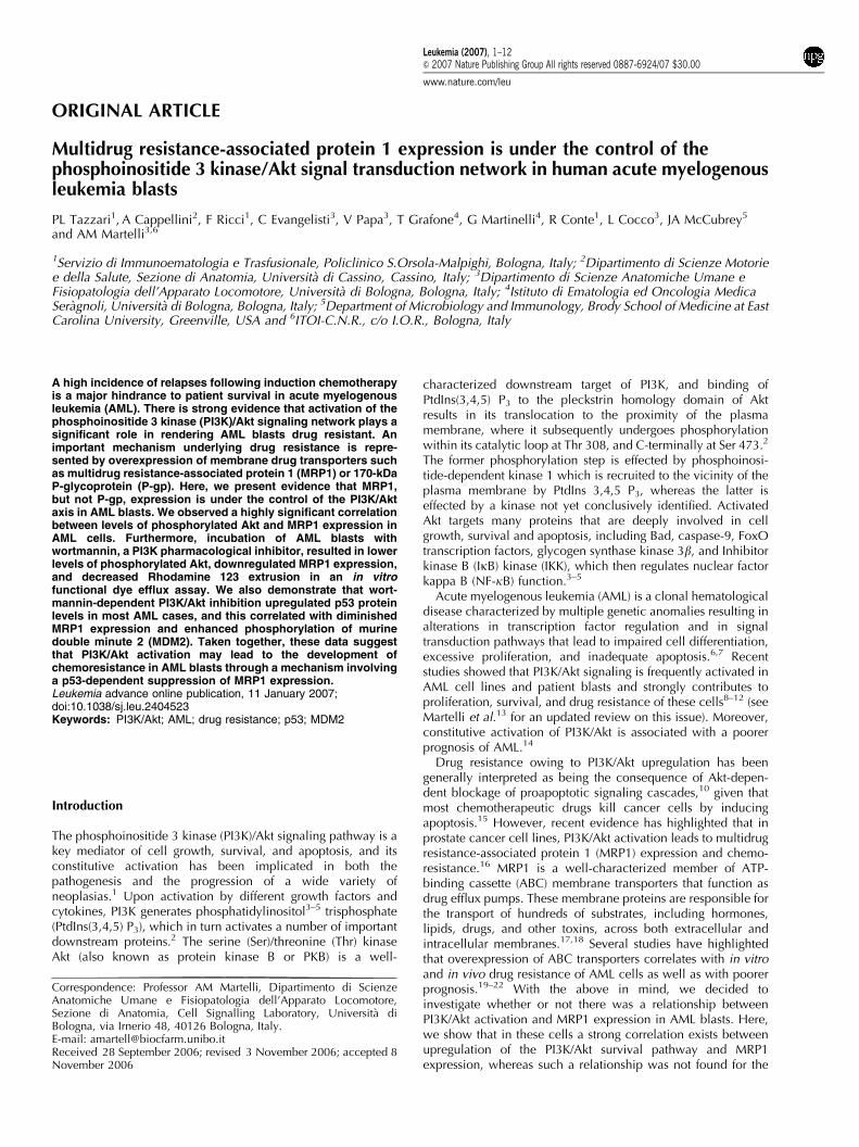
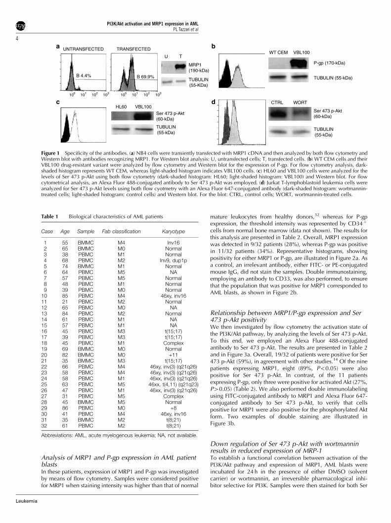
Specificity of the antibodiesAs in some cases of AML, the number of blasts available foranalytical purposes represents a limiting factor, we decided toevaluate MRP1, P-gp, and Ser 473 p-Akt levels in AML patientprimary cells by flow cytometry. Indeed, this approach allowsthe use of less cells when compared with Western blot and israpidly becoming the preferred technique for the analysis ofdrug resistance-linked proteins27 and phosphorylated pro-teins28–30 in AML. Therefore, a very critical issue concernedthe specificity of the antibodies used in our study. For thisreason, a series of control experiments were first performed tovalidate the antibodies employed in this investigation. NB4human leukemia cells were transiently transfected with a humanMRP1 cDNA and the MRP1 protein expression was analyzed24 h after transfection by both flow cytometry and Western blot(Figure 1a). Although control (untransfected) cells were practi-cally negative by both techniques, flow cytometric analysis withan FITC-conjugated antibody revealed that approximately 70%of the cells expressed MRP1. Western blot with a monoclonalantibody to human MRP1 supported this conclusion, byshowing that transfected cells expressed a predominant bandmigrating at approximately 190-kDa.
To assess specificity of the antibody employed to detect P-gp,we took advantage of CEM cells that are P-gp negative, and oftheir drug-resistant variant, VBL100 cells, which overexpressP-gp.23 As shown in Figure 1b, flow cytometric analysisdemonstrated that WT CEM cells stained negatively, whereasVBL100 stained positively for P-gp. Once again, Western blotconfirmed these findings.
To control for Ser 473 p-Akt, we took advantage of HL60cells, which have low or undetectable levels of this phosphory-lated Akt form,25 and of VBL100 cells, which, on the contrary,have elevated levels, because they lack PTEN lipid phospha-tase.23,31 As illustrated in Figure 1c, flow cytometric analysis ofsamples stained with an Alexa Fluor 488-conjugated antibody toSer 473 p-Akt showed HL60 cells to be negative, whereasVBL100 cells were positive. Western blot analysis corroboratedthese findings. The specificity of the Alexa Fluor 647-conjugatedantibody to Ser 473 p-Akt was also tested, employing Jurkat cellswhich, like CEM and VBL-100 cells, lack PTEN and conse-quently have elevated levels of Ser 473 p-Akt.31 When treatedwith wortmannin (300 nM for 24 h), a selective PI3K pharmaco-logical inhibitor, Jurkat cells displayed a much lower level of Ser473 p-Akt, as revealed by flow cytometric analysis of samplesstained with the Alexa Fluor 647-conjugated antibody to thephosphorylated Akt form (Figure 1d). Once again, Western blotanalysis confirmed these results. Overall, these experimentsvalidated the antibodies we used for cytofluorimetric investiga-tions aimed at evaluating the levels of MRP1, P-gp, and Ser 473p-Akt.
PatientsBMMC and PBMC fractions from 32 patients diagnosed withAML were examined in this study. Median age was 55.3 (range:21–85 years). For patient demographics and disease character-istics, refer to Table 1.
PI3K/Akt activation and MRP1 expression in AMLPL Tazzari et al
3
Leukemia
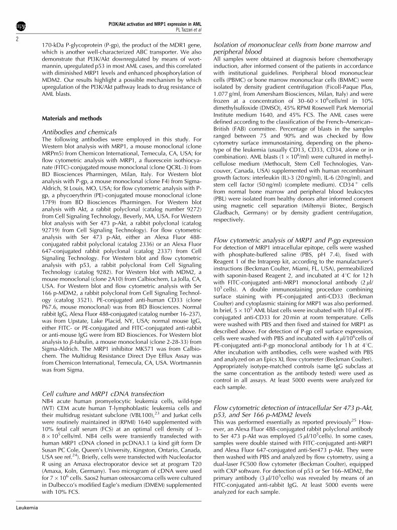
Analysis of MRP1 and P-gp expression in AML patientblastsIn these patients, expression of MRP1 and P-gp was investigatedby means of flow cytometry. Samples were considered positivefor MRP1 when staining intensity was higher than that of normal
mature leukocytes from healthy donors,32 whereas for P-gpexpression, the threshold intensity was represented by CD34þ
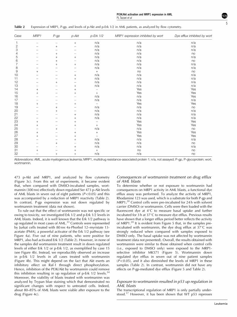
cells from normal bone marrow (data not shown). The results forthis analysis are presented in Table 2. Overall, MRP1 expressionwas detected in 9/32 patients (28%), whereas P-gp was positivein 11/32 patients (34%). Representative histograms, showingpositivity for either MRP1 or P-gp, are illustrated in Figure 2a. Asa control, an irrelevant antibody, either FITC- or PE-conjugatedmouse IgG, did not stain the samples. Double immunostaining,employing an antibody to CD33, was also performed, to ensurethat the population that was positive for MRP1 corresponded toAML blasts, as shown in Figure 2b.
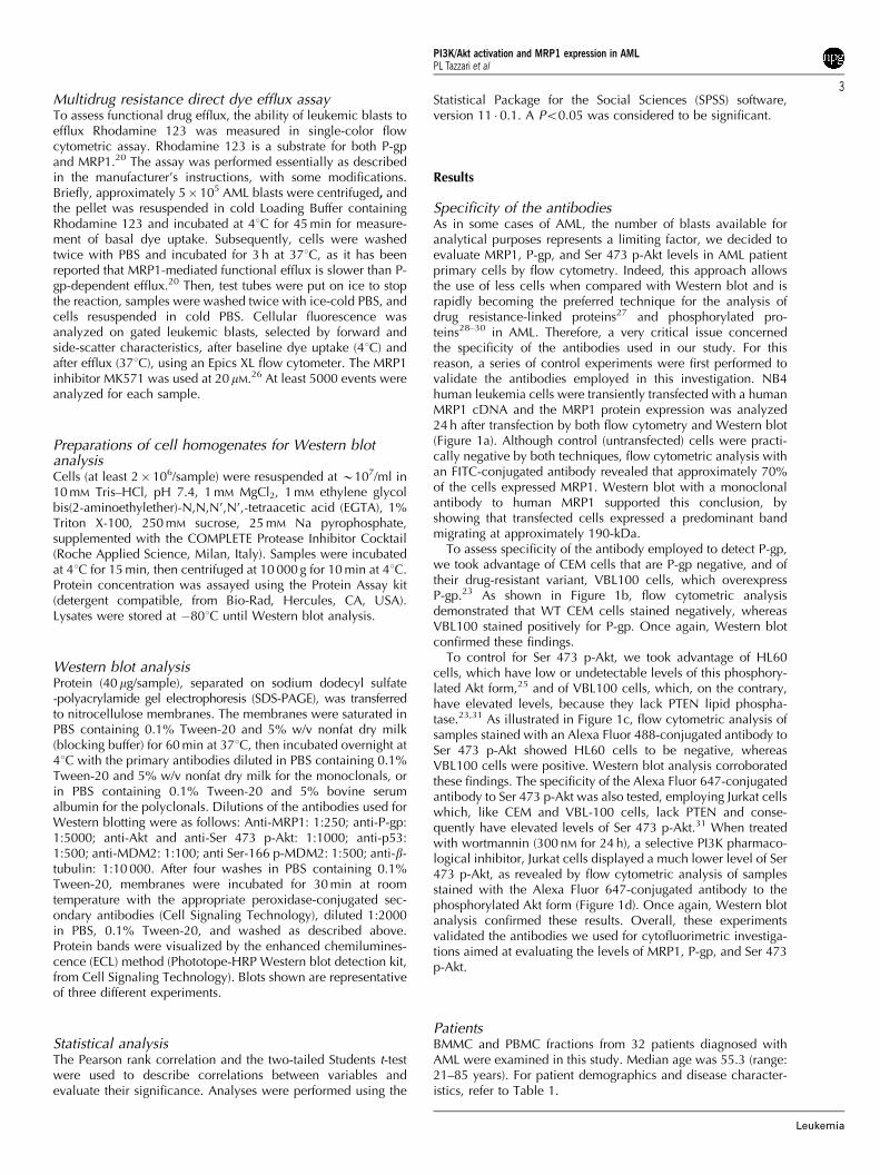
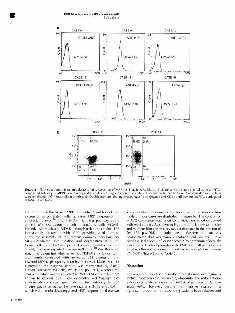
Relationship between MRP1/P-gp expression and Ser473 p-Akt positivityWe then investigated by flow cytometry the activation state ofthe PI3K/Akt pathway, by analyzing the levels of Ser 473 p-Akt.To this end, we employed an Alexa Fluor 488-conjugatedantibody to Ser 473 p-Akt. The results are presented in Table 2and in Figure 3a. Overall, 19/32 of patients were positive for Ser473 p-Akt (59%), in agreement with other studies.14 Of the ninepatients expressing MRP1, eight (89%, Po0.05) were alsopositive for Ser 473 p-Akt. In contrast, of the 11 patientsexpressing P-gp, only three were positive for activated Akt (27%,P40.05) (Table 2). We also performed double immunolabelingusing FITC-conjugated antibody to MRP1 and Alexa Fluor 647-conjugated antibody to Ser 473 p-Akt, to verify that cellspositive for MRP1 were also positive for the phosphorylated Aktform. Two examples of double staining are illustrated inFigure 3b.
Down regulation of Ser 473 p-Akt with wortmanninresults in reduced expression of MRP-1To establish a functional correlation between activation of thePI3K/Akt pathway and expression of MRP1, AML blasts wereincubated for 24 h in the presence of either DMSO (solventcarrier) or wortmannin, an irreversible pharmacological inhi-bitor selective for PI3K. Samples were then stained for both Ser
UNTRANSFECTED TRANSFECTED
B 4.4% B 69.9%
U T
MRP1(190-kDa)
TUBULIN(55-KDa)
100 101 102 103
HL60 VBL100Ser 473 p-Akt(60-kDa)
TUBULIN(55-kDa)
WT CEM VBL100
P-gp (170-kDa)
TUBULIN (55-kDa)
CTRL WORT
Ser 473 p-Akt(60-kDa)
TUBULIN(55-kDa)
100 101 102 103
a
c
b
d
Figure 1 Specificity of the antibodies. (a) NB4 cells were transiently transfected with MRP1 cDNA and then analyzed by both flow cytometry andWestern blot with antibodies recognizing MRP1. For Western blot analysis: U, untransfected cells; T, transfected cells. (b) WT CEM cells and theirVBL100 drug-resistant variant were analyzed by flow cytometry and Western blot for the expression of P-gp. For flow cytometry analysis, dark-shaded histogram represents WT CEM, whereas light-shaded histogram indicates VBL100 cells. (c) HL60 and VBL100 cells were analyzed for thelevels of Ser 473 p-Akt using both flow cytometry (dark-shaded histogram: HL60; light-shaded histogram: VBL100) and Western blot. For flowcytometrical analysis, an Alexa Fluor 488-conjugated antibody to Ser 473 p-Akt was employed. (d) Jurkat T-lymphoblastoid leukemia cells wereanalyzed for Ser 473 p-Akt levels using both flow cyotmetry with an Alexa Fluor 647-conjugated antibody (dark-shaded histogram: wortmannin-treated cells; light-shaded histogram: control cells) and Western blot. For the blot: CTRL, control cells; WORT, wortmannin-treated cells.
Table 1 Biological characteristics of AML patients
Case Age Sample Fab classification Karyotype
1 55 BMMC M4 Inv162 65 BMMC M0 Normal3 38 PBMC M1 Normal4 68 PBMC M2 Inv9, dup1p5 74 BMMC M1 Normal6 64 PBMC M5 NA7 57 PBMC M5 Normal8 48 PBMC M1 Normal9 39 PBMC M0 Normal
10 85 PBMC M4 46xy, inv1611 21 PBMC M2 Normal12 65 PBMC M0 NA13 84 PBMC M2 Normal14 61 PBMC M1 NA15 57 PBMC M1 NA16 45 PBMC M3 t(15;17)17 39 PBMC M3 t(15;17)18 45 PBMC M1 Complex19 69 BMMC M0 Normal20 82 BMMC M0 +1121 35 BMMC M3 t(15;17)22 66 PBMC M4 46xy, inv(3) (q21q26)23 58 PBMC M4 46xy, inv(3) (q21q26)24 58 PBMC M1 46xx, inv(3) (q21q26)25 63 PBMC M5 46xx, t(4,11) (q21q23)26 47 PBMC M1 46xx, inv(3) (q21q26)27 31 PBMC M5 Complex28 45 BMMC M5 Normal29 86 PBMC M0 +830 41 PBMC M4 46xy, inv1631 35 BMMC M2 t(8;21)32 61 PBMC M2 t(8;21)
Abbreviations: AML, acute myelogenous leukemia; NA, not available.
PI3K/Akt activation and MRP1 expression in AMLPL Tazzari et al
4
Leukemia
473 p-Akt and MRP1, and analyzed by flow cytometry(Figure 3c). From this set of experiments, it became evidentthat, when compared with DMSO-incubated samples, wort-mannin (300 nM) effectively down regulated Ser 473 p-Akt levelsof AML blasts in seven out of eight patients (Po0.05) and thiswas accompanied by a reduction of MRP1 reactivity (Table 2).In contrast, P-gp expression was not down regulated bywortmannin treatment (data not shown).
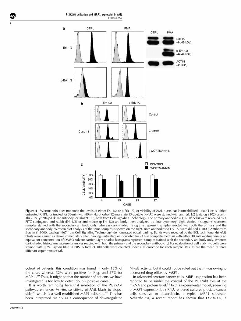
To rule out that the effect of wortmannin was not specific orowing to toxicity, we investigated Erk 1/2 and p-Erk 1/2 levels inAML blasts. Indeed, it is well known that the Erk 1/2 pathway isup regulated in most cases of AML.33 Controls were representedby Jurkat cells treated with 80 nM 4a-Phorbol 12–myristate 13-acetate (PMA), a powerful activator of the Erk 1/2 pathway (seeFigure 4a). Five out of nine patients, who were positive forMRP1, also had activated Erk 1/2 (Table 2). However, in none ofthe samples did wortmannin treatment result in down regulatedlevels of either Erk 1/2 or p-Erk 1/2, as exemplified by case 15(see Figure 4b). Instead, we reproducibly observed an increasein p-Erk 1/2 levels in all cases treated with wortmannin(Figure 4b). This might depend on the fact that Akt exerts aninhibitory effect on Raf-1 through direct phosphorylation.Hence, inhibition of the PI3K/Akt by wortmannin could removethis inhibition resulting in up regulation of p-Erk 1/2 levels.34
Moreover, the viability of blasts treated with wortmaninn wasanalyzed by Trypan blue staining which that demonstrated nosignificant changes with respect to untreated cells. Indeed,about 80–85% of AML blasts were viable after exposure to thedrug (Figure 4c).
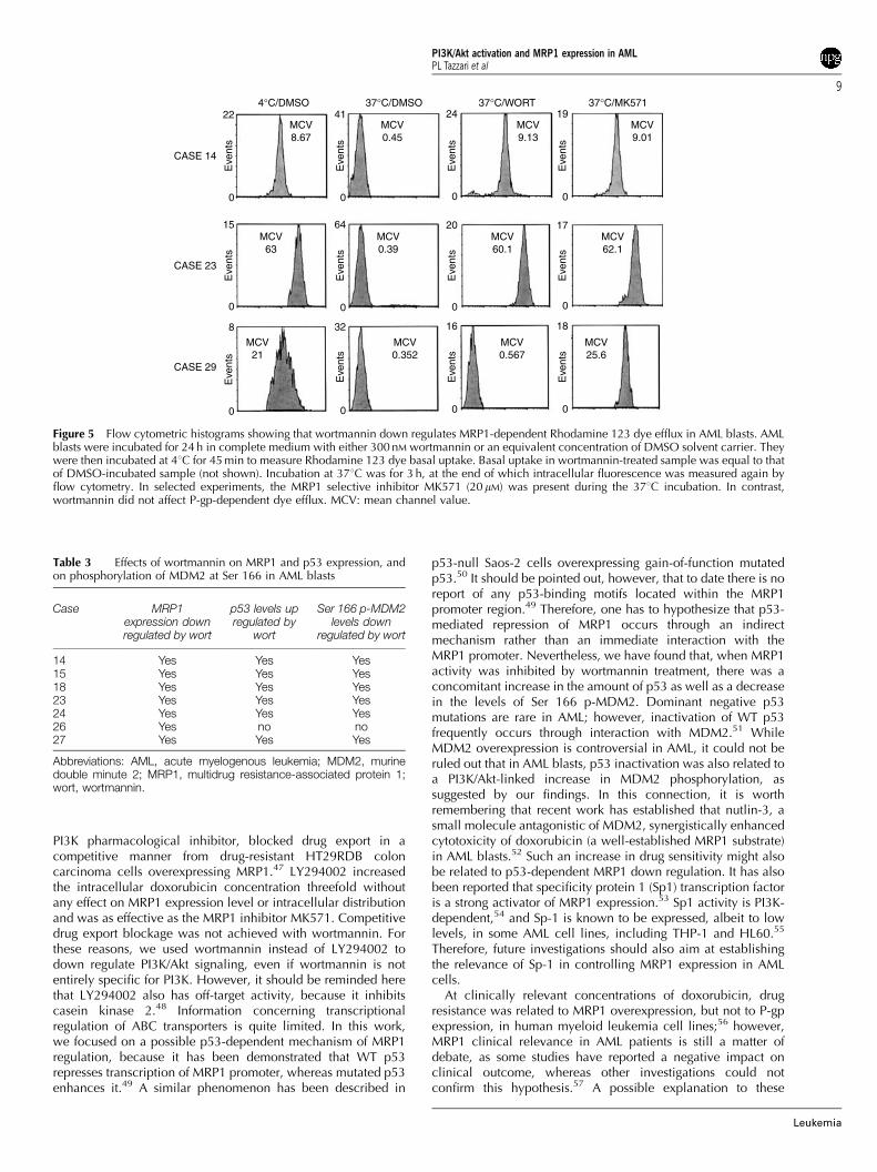
Consequences of wortmannin treatment on drug effluxof AML blastsTo determine whether or not exposure to wortmannin hadconsequences on MRP1 activity in AML blasts, a functional dyeefflux assay was performed. To analyze the activity of MRP1,Rhodamine 123 was used, which is a substrate for both P-gp andMRP1.20 Control cells were pre-incubated for 24 h with solventcarrier (DMSO) or wortmannin. Cells were then loaded with thefluorescent dye at 41C to measure basal uptake and furtherincubated for 3 h at 371C to measure dye efflux. Previous resultshave shown that a longer efflux period better reflects the activityof MRP1.20 It is evident from Figure 5 that, in the samples pre-incubated with wortmannin, the dye drug efflux at 371C wasstrongly reduced when compared with samples exposed toDMSO only. The basal uptake was not affected by wortmannintreatment (data not presented). Overall, the results obtained withwortmannin were similar to those obtained when control cells(i.e., exposed to DMSO only) were exposed to the MRP1-selective inhibitor MK571 (Figure 5). Wortmannin downregulated dye efflux in seven out of nine patient samples(Po0.05), and it also diminished the levels of MRP1 in thesesamples (Table 2). In contrast, wortmannin did not have anyeffects on P-gp-mediated dye efflux (Figure 5 and Table 2).
Exposure to wortmannin resulted in p53 up regulation inAML blastsThe transcriptional regulation of MRP1 is only partially under-stood.17 However, it has been shown that WT p53 represses
Table 2 Expression of MRP1, P-gp, and levels of p-Akt and p-Erk 1/2 in AML patients, as analyzed by flow cytometry.
Case MRP1 P-gp p-Akt p-Erk 1/2 MRP1 expression inhibited by wort Dye efflux inhibited by wort
1 � � + n/a n/a n/a2 � + � n/a n/a n/a3 � � � n/a n/a n/a4 � + � n/a n/a no5 � + + n/a n/a n/a6 � + � n/a n/a no7 � + + n/a n/a n/a8 � � � n/a n/a n/a9 + � � + no no
10 � � + n/a n/a n/a11 � � + n/a n/a n/a12 � + � n/a n/a n/a13 � � + n/a n/a n/a14 + � + � Yes Yes15 + � + + Yes Yes16 � � � n/a n/a Yes17 � � � n/a n/a n/a18 + � + � Yes Yes19 � + � n/a n/a no20 � + � n/a n/a n/a21 � � + n/a n/a n/a22 � � + n/a n/a n/a23 + � + � Yes Yes24 + � + + Yes Yes25 � + - n/a n/a no26 + � + + Yes Yes27 + � + � Yes Yes28 � � + n/a n/a n/a29 � + � n/a n/a no30 � - + n/a n/a n/a31 + � + + no no32 � + + n/a n/a no
Abbreviations: AML, acute myelogenous leukemia; MRP1, multidrug resistance-associated protein 1; n/a, not assayed; P-gp, P-glycoprotein; wort,wortmannin.
PI3K/Akt activation and MRP1 expression in AMLPL Tazzari et al
5
Leukemia
transcription of the human MRP1 promoter35 and loss of p53expression is correlated with increased MRP1 expression incolorectal cancer.36 The PI3K/Akt signaling pathway couldcontrol p53 expression through interactions with MDM2.Indeed, Akt-mediated MDM2 phosphorylation at Ser 166increases its interaction with p300, providing a platform toallow the assembly of the protein complex necessary forMDM2-mediated ubiquitination and degradation of p53.37
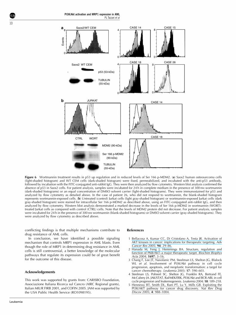
Consistently, a PI3K/Akt-dependent down regulation of p53activity has been reported in some AML cases10 We, therefore,sought to determine whether or not PI3K/Akt inhibition withwortmannin correlated with increased p53 expression andlowered MDM2 phosphorylation levels in AML blasts. For p53expression, the negative control was represented by Saos2human osteosarcoma cells, which are p53 null, whereas thepositive control was represented by WT CEM cells, which areknown to express p53. Flow cytometry and Western blotanalysis demonstrated specificity of the antibody to p53(Figure 6a). In six out of the seven patients (85%, Po0.05) inwhich wortmannin down regulated MRP1 expression, there was
a concomitant increase in the levels of 53 expression (seeTable 3). Four cases are illustrated in Figure 6a. The control forMDM2 expression was Jurkat cells, either untreated or treatedwith wortmannin. As shown in Figure 6b, both flow cytometryand Western blot analysis revealed a decrease in the amount ofSer 166 p-MDM2 in Jurkat cells. Western blot analysisdemonstrated that wortmannin treatment did not result in adecrease in the levels of MDM2 protein. Wortmannin effectivelyreduced the levels of phosphorylated MDM2 in all patient casesin which there was a concomitant increase in p53 expression(Po0.05) (Figure 6b and Table 3).
Discussion
Conventional induction chemotherapy with intensive regimensincluding doxorubicin, idarubicin, etoposide, and mitoxantroneinduces complete remission in 65–75% of adults with de novoacute AML. However, despite the intensive treatments, asignificant proportion of responding patients have relapses and
CASE 9 CASE 9 CASE 10
IRRELEVANT
MCV=0.35
ANTI-MRP1
MCV=2.53 MCV=0.39
ANTI-MRP1
MCV=0.36
IRRELEVANT ANTI-P-gp ANTI-P-gp
MCV=2.68 MCV=0.41
CASE 12CASE 12 CASE 16
CASE 9 CASE 10
CD
33
100
Cou
nt
0
68
0
Cou
nt
–1 10000–1 1000
Cou
nt
104
0
Cou
nt
160
0
Cou
nt
108
–1 1000 –1 1000 –1 1000
–1 1000
103
102
101
100
l12.1% 96.3%
l2
l3 l40.1% 1.5%
100 101 102 103
103
102
101
100
100 101 102 103
MRP1
B1 B299.6% 0.0%
B3 B40.4% 0.0%
0
Cou
nt
108
a
b
Figure 2 Flow cytometric histograms demonstrating detection of MRP1 or P-gp in AML blasts. (a) Samples were single stained using an FITC-conjugated antibody to MRP1 or a PE-conjugated antibody to P-gp. As controls, irrelevant antibodies (either FITC- or PE-conjugated mouse IgG)were employed. MCV: mean channel value. (b) Double immunolabeling employing a PE-conjugated anti-CD33 antibody and an FITC-conjugatedanti-MRP1 antibody.
PI3K/Akt activation and MRP1 expression in AMLPL Tazzari et al
6
Leukemia
ultimately die of therapy-refractory disease, so that disease-freesurvival at 5 years ranges from 25 to 35%.38 Therefore, AMLremains a clinical challenge with poor long-term survival, andthe identification of aberrantly activated signaling pathways,which contribute to drug resistance, could provide an opportu-nity to improve the standard therapeutic treatments. The ABCfamily of drug transporters confer resistance to multiplechemotherapeutic agents when overexpressed in human tumors,including AML. The best-characterized ABC family member is170-kDa P-gp, the product of the MDR1 gene. However, it hasbecome clear that additional ABC transporters could beinvolved in drug resistance of AML blasts. MRP1 is encodedby the MRP1 gene located on chromosome 16p13.39
More recently, additional members, MRP2–MRP9, have beenidentified; however,their role in multidrug resistance awaitselucidation.40
The PI3K/Akt signal transduction pathway plays a key role incontrolling neoplastic cell growth, survival, motility andinvasion.41 There is now increasing evidence for a PI3K/Akt-mediated role in the regulation of AML cell cycle, survival, anddrug resistance.13,42 In this study, we showed that, in AMLblasts, the expression of MRP1, but not of P-gp, is under thecontrol of the PI3K/Akt axis. As far as we know, this is the firstreport showing a relationship between MRP1 expression andactivation of the PI3K/Akt axis in primary tumor cells. Severalobservations support this conclusion. There was a statistically
significant correlation (Po0.05) between the levels of Ser 473 p-Akt and the expression of MRP1 in AML primary cells, whereasthis was not true for P-gp. Moreover, treatment of AML blastswith wortmannin, a selective PI3K pharmacological inhibitor,resulted in lower levels of MRP1 expression and in a decreasedcapability of these cells to extrude Rhodamine 123 in an in vitrofunctional dye efflux assay. This reduced dye extrusion wassimilar to that obtained with MK571, a chemical that selectivelyinhibits MRP1 function. In our study, we have analyzed 32patients for MRP1 and P-gp expression. We found MRP1expression in 9/32 patients (28%), whereas P-gp was positivein 11/32 patients (34%). These values are consistent with muchlarger studies demonstrating that MRP1 is overexpressed(defined as a level surpassing that of healthy blood leukocytes)in 7–30% of patients with de novo AML,43 whereas P-gppositivity, exceeding the positivity of normal bone marrowCD34þ cells, is found in 25–40% of patients.19 The expressionof P-gp has been reported to be linked with increasing age of thepatients44 reaching 71% in a group with median age 68 years.Interestingly, although we have investigated a much smallercohort of patients, the median age of patients with P-gpexpression was 68.5 years, whereas the median age of patientswith MRP1 expression was 48. Indeed, MRP1 expression isknown to be more frequent in patients who are younger.44 Wedid not find patients positive for both P-gp and MRP-1.However, according to a study performed on a much larger
MRP1 Ser 473 p-Akt
CASE 15
CASE 18
CASE 14 CASE 15103
102
101
100
MR
P1
l17.0%
l247.8%
l33.1%
l442.1%
100 101 102 103
103
102
101
100
100 101 102 103
l1
l3
16.1%
0.3% 0.3%
83.3%l2
l4
Ser 473 p-Akt
MRP1 Ser 473 p-Akt
CASE 23 + DMSO
CASE 23 + WORT
a b
c
Figure 3 Flow cytometric histograms showing the relationship between MRP1 expression and activation of the PI3K/Akt pathway in AML blasts.(a) Samples were either stained with FITC-conjugated anti-MRP1 antibody or Alexa Fluor 488-conjugated anti-Ser 473 antibody (dark-shadedhistograms). Irrelevant antibodies (FITC-conjugated mouse IgG or Alexa Fluor 488-conjugated rabbit IgG) were employed as control (light-shadedhistograms). (b) Double labeling analysis for FITC-conjugated anti-MRP1 antibody and Alexa Fluor 647-conjugated anti-Ser 473 p-Akt antibody. (c)Samples were incubated for 24 h in complete medium in the presence of 300 nM wortmannin or an equivalent concentration of DMSO solventcarrier. They were then stained for either MRP1 or Ser 473 p-Akt (dark-shaded histograms). Appropriate irrelevant antibodies, as described above,were employed (light-shaded histograms).
PI3K/Akt activation and MRP1 expression in AMLPL Tazzari et al
7
Leukemia
cohort of patients, this condition was found in only 15% ofthe cases whereas 32% were positive for P-gp and 27% forMRP-1.19 Thus, it might be that the number of patients we haveinvestigated is too low to detect doubly positive cases.
It is worth reminding here that inhibition of the PI3K/Aktpathway enhances in vitro sensitivity of AML blasts to etopo-side,45 which is a well-established MRP1 substrate.46 This hasbeen interpreted mainly as a consequence of downregulated
NF-kB activity, but it could not be ruled out that it was owing todecreased drug efflux by MRP1.
In advanced prostate cancer cells, MRP1 expression has beenreported to be under the control of the PI3K/Akt axis at themRNA and protein level.16 In this experimental model, silencingof MRP1 expression by siRNA rendered cultured prostate cancercells sensitive to doxorubicin, a typical MRP1 substrate.Nevertheless, a recent report has shown that LY294002, a
Erk 1/2
CTRL PMA
CTRL PMA
Erk 1/2(44/42-kDa)
p-Erk 1/2(44/42-kDa)
ACTIN(45-kDa)
p-Erk 1/2
Erk 1/2 p-Erk 1/2
Control
+DMSO
+WORTMANNIN
CONTROLWORTMANNIN
Case 15
100%
80%
60%
40%
20%
0%
CE
LL V
IAB
ILIT
Y
14 15 23 27
CASE
a
b
c
Figure 4 Wortmannin does not affect the levels of either Erk 1/2 or p-Erk 1/2, or viability of AML blasts. (a) Permeabilized Jurkat T cells (eitheruntreated, CTRL, or treated for 30 min with 80 nM 4a-phorbol 12-myristate 13-acetate (PMA)) were stained with anti-Erk 1/2 (catalog 9102) or anti-Thr 202/Tyr 204 p-Erk 1/2 antibody (catalog 9106), both from Cell Signaling Technology. The primary antibodies (3ml/105 cells) were revealed by aFITC-conjugated anti-rabbit (Erk 1/2) or anti-mouse (p-Erk 1/2) antibody, then analyzed by flow cytometry. Light-shaded histograms representsamples stained with the secondary antibody only, whereas dark-shaded histograms represent samples reacted with both the primary and thesecondary antibody. Western blot analysis of the same samples is shown on the right. Both antibodies to Erk 1/2 were diluted 1:1000. Antibody tob-actin (1:1000, catalog 4967 from Cell Signaling Technology) demonstrated equal loading. Bands were revealed by the ECL technique. (b) AMLblasts were stained as above immediately after thawing (untreated) or incubated for 24 h in complete medium with either 300 nM wortmannin or anequivalent concentration of DMSO solvent carrier. Light-shaded histograms represent samples stained with the secondary antibody only, whereasdark-shaded histograms represent samples reacted with both the primary and the secondary antibody. (c) For evaluation of cell viability, cells werestained with 0.2% Trypan blue in PBS. A total of 300 cells were counted under a microscope for each sample. Results are the mean of threedifferent experiments7s.d.
PI3K/Akt activation and MRP1 expression in AMLPL Tazzari et al
8
Leukemia
PI3K pharmacological inhibitor, blocked drug export in acompetitive manner from drug-resistant HT29RDB coloncarcinoma cells overexpressing MRP1.47 LY294002 increasedthe intracellular doxorubicin concentration threefold withoutany effect on MRP1 expression level or intracellular distributionand was as effective as the MRP1 inhibitor MK571. Competitivedrug export blockage was not achieved with wortmannin. Forthese reasons, we used wortmannin instead of LY294002 todown regulate PI3K/Akt signaling, even if wortmannin is notentirely specific for PI3K. However, it should be reminded herethat LY294002 also has off-target activity, because it inhibitscasein kinase 2.48 Information concerning transcriptionalregulation of ABC transporters is quite limited. In this work,we focused on a possible p53-dependent mechanism of MRP1regulation, because it has been demonstrated that WT p53represses transcription of MRP1 promoter, whereas mutated p53enhances it.49 A similar phenomenon has been described in
p53-null Saos-2 cells overexpressing gain-of-function mutatedp53.50 It should be pointed out, however, that to date there is noreport of any p53-binding motifs located within the MRP1promoter region.49 Therefore, one has to hypothesize that p53-mediated repression of MRP1 occurs through an indirectmechanism rather than an immediate interaction with theMRP1 promoter. Nevertheless, we have found that, when MRP1activity was inhibited by wortmannin treatment, there was aconcomitant increase in the amount of p53 as well as a decreasein the levels of Ser 166 p-MDM2. Dominant negative p53mutations are rare in AML; however, inactivation of WT p53frequently occurs through interaction with MDM2.51 WhileMDM2 overexpression is controversial in AML, it could not beruled out that in AML blasts, p53 inactivation was also related toa PI3K/Akt-linked increase in MDM2 phosphorylation, assuggested by our findings. In this connection, it is worthremembering that recent work has established that nutlin-3, asmall molecule antagonistic of MDM2, synergistically enhancedcytotoxicity of doxorubicin (a well-established MRP1 substrate)in AML blasts.52 Such an increase in drug sensitivity might alsobe related to p53-dependent MRP1 down regulation. It has alsobeen reported that specificity protein 1 (Sp1) transcription factoris a strong activator of MRP1 expression.53 Sp1 activity is PI3K-dependent,54 and Sp-1 is known to be expressed, albeit to lowlevels, in some AML cell lines, including THP-1 and HL60.55
Therefore, future investigations should also aim at establishingthe relevance of Sp-1 in controlling MRP1 expression in AMLcells.
At clinically relevant concentrations of doxorubicin, drugresistance was related to MRP1 overexpression, but not to P-gpexpression, in human myeloid leukemia cell lines;56 however,MRP1 clinical relevance in AML patients is still a matter ofdebate, as some studies have reported a negative impact onclinical outcome, whereas other investigations could notconfirm this hypothesis.57 A possible explanation to these
4°C/DMSO 37°C/DMSO 37°C/WORT 37°C/MK571
CASE 14
CASE 23
MCV63
MCV0.39
MCV60.1
MCV62.1
MCV9.01
MCV9.13
MCV0.45
MCV8.67
MCV21
MCV0.352
MCV0.567
MCV25.6
22
0
Eve
nts
15
0
Eve
nts
8
0
Eve
nts
32
0
Eve
nts
64
0
Eve
nts
41
0
Eve
nts
24
0
Eve
nts
20
0
Eve
nts
16
0
Eve
nts
CASE 29
19
0
Eve
nts
17
0
Eve
nts
18
0
Eve
nts
Figure 5 Flow cytometric histograms showing that wortmannin down regulates MRP1-dependent Rhodamine 123 dye efflux in AML blasts. AMLblasts were incubated for 24 h in complete medium with either 300 nM wortmannin or an equivalent concentration of DMSO solvent carrier. Theywere then incubated at 41C for 45 min to measure Rhodamine 123 dye basal uptake. Basal uptake in wortmannin-treated sample was equal to thatof DMSO-incubated sample (not shown). Incubation at 371C was for 3 h, at the end of which intracellular fluorescence was measured again byflow cytometry. In selected experiments, the MRP1 selective inhibitor MK571 (20 mM) was present during the 371C incubation. In contrast,wortmannin did not affect P-gp-dependent dye efflux. MCV: mean channel value.
Table 3 Effects of wortmannin on MRP1 and p53 expression, andon phosphorylation of MDM2 at Ser 166 in AML blasts
Case MRP1expression downregulated by wort
p53 levels upregulated by
wort
Ser 166 p-MDM2levels down
regulated by wort
14 Yes Yes Yes15 Yes Yes Yes18 Yes Yes Yes23 Yes Yes Yes24 Yes Yes Yes26 Yes no no27 Yes Yes Yes
Abbreviations: AML, acute myelogenous leukemia; MDM2, murinedouble minute 2; MRP1, multidrug resistance-associated protein 1;wort, wortmannin.
PI3K/Akt activation and MRP1 expression in AMLPL Tazzari et al
9
Leukemia
conflicting findings is that multiple mechanisms contribute todrug resistance of AML cells.
In conclusion, we have identified a possible signalingmechanism that controls MRP1 expression in AML blasts. Eventhough the role of MRP1 in determining drug resistance in AMLcells is still controversial, a better knowledge of the molecularpathways that regulate its expression could be of great benefitfor the outcome of this disease.
Acknowledgements
This work was supported by grants from: CARISBO Foundation,Associazione Italiana Ricerca sul Cancro (AIRC Regional grants),Italian MIUR FIRB 2001, and COFIN 2005. JAM was supported bythe USA Public Health Service (RO1098195).
References
1 Bellacosa A, Kumar CC, Di Cristofano A, Testa JR. Activation ofAKT kinases in cancer: implications for therapeutic targeting. AdvCancer Res 2005; 94: 29–86.
2 Hanada M, Feng J, Hemmings BA. Structure, regulation andfunction of PKB/AKT–a major therapeutic target. Biochim BiophysActa 2004; 1697: 3–16.
3 Chang F, Lee JT, Navolanic PM, Steelman LS, Shelton JG, BlalockWL et al. Involvement of PI3K/Akt pathway in cell cycleprogression, apoptosis, and neoplastic transformation: a target forcancer chemotherapy. Leukemia 2003; 17: 590–603.
4 Steelman LS, Pohnert SC, Shelton JG, Franklin RA, Bertrand FE,McCubrey JA. JAK/STAT, Raf/MEK/ERK, PI3K/Akt and BCR-ABL in cellcycle progression and leukemogenesis. Leukemia 2004; 18: 189–218.
5 Hennessy BT, Smith DL, Ram PT, Lu Y, Mills GB. Exploiting thePI3K/AKT pathway for cancer drug discovery. Nat Rev DrugDiscov 2005; 4: 988–1004.
8
0
Eve
nts
Saos2/WT CEM
Saos2 WT CEM
CASE 14 CASE 15
CASE 18
CASE 18
CASE 14
CASE 26
4
0
8
0
9
0
4
0
p53 (53-kDa)
TUBULIN(55-kDa)
CTRL WORT
MDM2 (90-kDa)
Ser 166 p-MDM2(90-kDa)
(55-kDa)TUBULIN
JURKAT
a
b
Figure 6 Wortmannin treatment results in p53 up regulation and in reduced levels of Ser 166 p-MDM2. (a) Saos2 human osteosarcoma cells(light-shaded histogram) and WT CEM cells (dark-shaded histogram) were fixed, permeabilized, and incubated with the anti-p53 antibody,followed by incubation with the FITC-conjugated anti-rabbit IgG. They were then analyzed by flow cytometry. Western blot analysis confirmed theabsence of p53 in Saos2 cells. For patient analysis, samples were incubated for 24 h in complete medium in the presence of 300 nM wortmannin(dark-shaded histograms) or an equal concentration of DMSO solvent carrier (light-shaded histograms). They were immunostained for p53 andanalyzed by flow cytometry as detailed above. In the case of patient 26, who did not respond to wortmannin, the blank-shaded histogramrepresents wortmannin-exposed cells. (b) Untreated (control) Jurkat cells (light gray-shaded histogram) or wortmannin-exposed Jurkat cells (darkgray-shaded histogram) were stained for intracellular Ser 166 p-MDM2 as described above, using an FITC-conjugated anti-rabbit IgG, and thenanalyzed by flow cytometry. Western blot analysis demonstrated a marked decrease in the levels of Ser 166 p-MDM2 in wortmannin (WORT)-treated Jurkat cells as compared with control (CTRL) cells. Note that the levels of MDM2 protein did not decrease. For patient analysis, sampleswere incubated for 24 h in the presence of 300 nM wortmannin (blank-shaded histograms) or DMSO solvent carrier (gray-shaded histograms). Theywere analyzed by flow cytometry as described above.
PI3K/Akt activation and MRP1 expression in AMLPL Tazzari et al
10
Leukemia
6 Steffen B, Muller-Tidow C, Schwable J, Berdel WE, Serve H. Themolecular pathogenesis of acute myeloid leukemia. Crit RevOncol Hematol 2005; 56: 195–221.
7 Lelievre H, Ferrand A, Mozziconacci MJ, Birnbaum D, Delaval B.Myeloproliferative disorders: premalignant, stem cell, G1 diseases?Leukemia 2006; 20: 1475–1480.
8 Xu Q, Simpson SE, Scialla TJ, Bagg A, Carroll M. Survival of acutemyeloid leukemia cells requires PI3 kinase activation. Blood 2003;102: 972–980.
9 Zhao S, Konopleva M, Cabreira-Hansen M, Xie Z, Hu W, MilellaM et al. Inhibition of phosphatidylinositol 3-kinase dephosphor-ylates BAD and promotes apoptosis in myeloid leukemias.Leukemia 2004; 18: 267–275.
10 Grandage VL, Gale RE, Linch DC, Khwaja A. PI3-kinase/Akt isconstitutively active in primary acute myeloid leukaemia cells andregulates survival and chemoresistance via NF-kB, Mapkinase andp53 pathways. Leukemia 2005; 19: 586–594.
11 Karajannis MA, Vincent L, Direnzo R, Shmelkov SV, Zhang F,Feldman EJ et al. Activation of FGFR1b signaling pathwaypromotes survival, migration and resistance to chemotherapy inacute myeloid leukemia cells. Leukemia 2006; 20: 979–986.
12 Matkovic K, Brugnoli F, Bertagnolo V, Banfic H, Visnjic D. Therole of the nuclear Akt activation and Akt inhibitors in all-trans-retinoic acid-differentiated HL-60 cells. Leukemia 2006; 20:941–951.
13 Martelli AM, Nyakern M, Tabellini G, Bortul R, Tazzari PL,Evangelisti C et al. Phosphoinositide 3-kinase/Akt signaling path-way and its therapeutical implications for human acute myeloidleukemia. Leukemia 2006; 20: 911–928.
14 Min YH, Eom JI, Cheong JW, Maeng HO, Kim JY, Jeung HK et al.Constitutive phosphorylation of Akt/PKB protein in acute myeloidleukemia: its significance as a prognostic variable. Leukemia 2003;17: 995–997.
15 Kim R. Recent advances in understanding the cell death pathwaysactivated by anticancer therapy. Cancer 2005; 103: 1551–1560.
16 Lee Jr JT, Steelman LS, McCubrey JA. Phosphatidylinositol3’-kinase activation leads to multidrug resistance protein-1expression and subsequent chemoresistance in advanced prostatecancer cells. Cancer Res 2004; 64: 8397–8404.
17 Scotto KW. Transcriptional regulation of ABC drug transporters.Oncogene 2003; 22: 7496–7511.
18 Bonhoure E, Pchejetski D, Aouali N, Morjani H, Levade T,Kohama T et al. Overcoming MDR-associated chemoresistance inHL-60 acute myeloid leukemia cells by targeting sphingosinekinase-1. Leukemia 2006; 20: 95–102.
19 Legrand O, Simonin G, Beauchamp-Nicoud A, Zittoun R, MarieJP. Simultaneous activity of MRP1 and Pgp is correlated with invitro resistance to daunorubicin and with in vivo resistance inadult acute myeloid leukemia. Blood 1999; 94: 1046–1056.
20 Leith CP, Kopecky KJ, Chen IM, Eijdems L, Slovak ML, McConnellTS et al. Frequency and clinical significance of the expression ofthe multidrug resistance proteins MDR1/P-glycoprotein, MRP1,and LRP in acute myeloid leukemia: a Southwest Oncology GroupStudy. Blood 1999; 94: 1086–1099.
21 van der Kolk DM, de Vries EG, van Putten WJ, Verdonck LF,Ossenkoppele GJ, Verhoef GE et al. P-glycoprotein and multidrugresistance protein activities in relation to treatment outcome inacute myeloid leukemia. Clin Cancer Res 2000; 6: 3205–3214.
22 Mahadevan D, List AF. Targeting the multidrug resistance-1transporter in AML: molecular regulation and therapeutic strate-gies. Blood 2004; 104: 1940–1951.
23 Mantovani I, Cappellini A, Tazzari PL, Papa V, Cocco L, MartelliAM. Caspase-dependent cleavage of 170-kDa P-glycoproteinduring apoptosis of human T-lymphoblastoid CEM cells. J CellPhysiol 2006; 207: 836–844.
24 Ito K, Olsen SL, Qiu W, Deeley RG, Cole SP. Mutation of a singleconserved tryptophan in multidrug resistance protein 1 (MRP1/ABCC1) results in loss of drug resistance and selective loss oforganic anion transport. J Biol Chem 2001; 276: 15616–15624.
25 Nyakern M, Tazzari PL, Finelli C, Bosi C, Follo MY, Grafone Tet al. Frequent elevation of Akt kinase phosphorylation in bloodmarrow and peripheral blood mononuclear cells from high-riskmyelodysplastic syndrome patients. Leukemia 2006; 20: 230–238.
26 van der Kolk DM, de Vries EG, Noordhoek L, van den Berg E, vander Pol MA, Muller M et al. Activity and expression of the
multidrug resistance proteins P-glycoprotein, MRP1, MRP2, MRP3and MRP5 in de novo and relapsed acute myeloid leukemia.Leukemia 2001; 15: 1544–1553.
27 Funato T, Harigae H, Abe S, Sasaki T. Assessment of drugresistance in acute myeloid leukemia. Expert Rev Mol Diagn 2004;4: 705–713.
28 Krutzik PO, Irish JM, Nolan GP, Perez OD. Analysis of proteinphosphorylation and cellular signaling events by flow cytometry:techniques and clinical applications. Clin Immunol 2004; 110:206–221.
29 Ricciardi MR, McQueen T, Chism D, Milella M, Estey E, Kaldjian Eet al. Quantitative single cell determination of ERK phosphoryla-tion and regulation in relapsed and refractory primary acutemyeloid leukemia. Leukemia 2005; 19: 1543–1549.
30 Kornblau SM, Womble M, Cade JS, Lemker E, Qiu YH.Comparative analysis of the effects of sample source and testmethodology on the assessment of protein expression in acutemyelogenous leukemia. Leukemia 2005; 19: 1550–1557.
31 Uddin S, Hussain A, Al-Hussein K, Platanias LC, Bhatia KG.Inhibition of phosphatidylinositol 3’-kinase induces preferentiallykilling of PTEN-null T leukemias through AKT pathway. BiochemBiophys Res Commun 2004; 320: 932–938.
32 Laupeze B, Amiot L, Payen L, Drenou B, Grosset JM, Lehne G et al.Multidrug resistance protein (MRP) activity in normal matureleukocytes and CD34-positive hematopoietic cells from peripheralblood. Life Sci 2001; 68: 1323–1331.
33 Lunghi P, Tabilio A, Dall’Aglio PP, Ridolo E, Carlo-Stella C, PelicciPG et al. Downmodulation of ERK activity inhibits the proliferationand induces the apoptosis of primary acute myelogenous leukemiablasts. Leukemia 2003; 17: 1783–1793.
34 Romano D, Pertuit M, Rasolonjanahary R, Barnier JV, Magalon K,Enjalbert A et al. Regulation of the Rap1/Raf-1/Erk-1/2 cascade andprolactin release by the Pi3k/Akt pathway in pituitary cells.Endocrinology 2006; 147: 6036–6045.
35 Bahr O, Wick W, Weller M. Modulation of MDR/MRP by wild-type and mutant p53. J Clin Invest 2001; 107: 643–646.
36 Fukushima Y, Oshika Y, Tokunaga T, Hatanaka H, Tomisawa M,Kawai K et al. Multidrug resistance-associated protein (MRP)expression is correlated with expression of aberrant p53 protein incolorectal cancer. Eur J Cancer 1999; 35: 935–938.
37 Grossman SR, Perez M, Kung AL, Joseph M, Mansur C, Xiao ZXet al. p300/MDM2 complexes participate in MDM2-mediated p53degradation. Mol Cell 1998; 2: 405–415.
38 Tallman MS, Gilliland DG, Rowe JM. Drug therapy for acutemyeloid leukemia. Blood 2005; 106: 1154–1163.
39 Haimeur A, Conseil G, Deeley RG, Cole SP. The MRP-related andBCRP/ABCG2 multidrug resistance proteins: biology, substratespecificity and regulation. Curr Drug Metab 2004; 5: 21–53.
40 Kruh GD, Belinsky MG. The MRP family of drug efflux pumps.Oncogene 2003; 22: 7537–7552.
41 Nicholson KM, Anderson NG. The protein kinase B/Akt signallingpathway in human malignancy. Cell Signal 2002; 14: 381–395.
42 Bertrand FE, Steelman LS, Chappell WH, Abrams SL, Shelton JG,White ER et al. Synergy between an IGF-1R antibody and Raf/MEK/ERK and PI3K/Akt/mTOR pathway inhibitors in suppressing IGF-1R-mediated growth in hematopoietic cells. Leukemia 2006; 20:1254–1260.
43 Walter RB, Raden BW, Hong TC, Flowers DA, Bernstein ID,Linenberger ML. Multidrug resistance protein attenuates gemtuzu-mab ozogamicin-induced cytotoxicity in acute myeloid leukemiacells. Blood 2003; 102: 1466–1473.
44 Leith CP, Kopecky KJ, Godwin J, McConnell T, Slovak ML, ChenIM et al. Acute myeloid leukemia in the elderly: assessment ofmultidrug resistance (MDR1) and cytogenetics distinguishesbiologic subgroups with remarkably distinct responses to standardchemotherapy. A Southwest Oncology Group study. Blood 1997;89: 3323–3329.
45 Billottet C, Grandage VL, Gale RE, Quattropani A, Rommel C,Vanhaesebroeck B et al. A selective inhibitor of the p110d isoformof PI 3-kinase inhibits AML cell proliferation and survival andincreases the cytotoxic effects of VP16. Oncogene 2006; 25:6648–6659.
46 Szentpetery Z, Kern A, Liliom K, Sarkadi B, Varadi A, Bakos E.The role of the conserved glycines of ATP-binding cassettesignature motifs of MRP1 in the communication between the
PI3K/Akt activation and MRP1 expression in AMLPL Tazzari et al
11
Leukemia

substrate-binding site and the catalytic centers. J Biol Chem 2004;279: 41670–41678.
47 Abdul-Ghani R, Serra V, Gyorffy B, Jurchott K, Solf A, Dietel Met al. The PI3K inhibitor LY294002 blocks drug export fromresistant colon carcinoma cells overexpressing MRP1. Oncogene2006; 25: 1743–1752.
48 Tolloczko B, Turkewitsch P, Al-Chalabi M, Martin JG. LY-294002[2-(4-morpholinyl)-8-phenyl-4H-1-benzopyran-4-one] affects cal-cium signaling in airway smooth muscle cells independently ofphosphoinositide 3-kinase inhibition. J Pharmacol Exp Ther 2004;311: 787–793.
49 Sullivan GF, Yang JM, Vassil A, Yang J, Bash-Babula J, Hait WN.Regulation of expression of the multidrug resistance protein MRP1by p53 in human prostate cancer cells. J Clin Invest 2000; 105:1261–1267.
50 Tsang WP, Chau SP, Fung KP, Kong SK, Kwok TT. Modulation ofmultidrug resistance-associated protein 1 (MRP1) by p53 mutant inSaos-2 cells. Cancer Chemother Pharmacol 2003; 51: 161–166.
51 Faderl S, Kantarjian HM, Estey E, Manshouri T, Chan CY, RahmanElsaied A et al. The prognostic significance of p16(INK4a)/p14(ARF) locus deletion and MDM-2 protein expression in adultacute myelogenous leukemia. Cancer 2000; 89: 1976–1982.
52 Kojima K, Konopleva M, Samudio IJ, Shikami M, Cabreira-HansenM, McQueen T et al. MDM2 antagonists induce p53-dependentapoptosis in AML: implications for leukemia therapy. Blood 2005;106: 3150–3159.
53 Wang Q, Beck WT. Transcriptional suppression of multidrugresistance-associated protein (MRP) gene expression by wild-typep53. Cancer Res 1998; 58: 5762–5769.
54 Tiwari G, Sakaue H, Pollack JR, Roth RA. Gene expressionprofiling in prostate cancer cells with Akt activation revealsFra-1 as an Akt-inducible gene. Mol Cancer Res 2003; 1:475–484.
55 Furukawa Y, Sutheesophon K, Wada T, Nishimura M, Saito Y, IshiiH et al. Methylation silencing of the Apaf-1 gene in acuteleukemia. Mol Cancer Res 2005; 3: 325–334.
56 Zhou DC, Ramond S, Viguie F, Faussat AM, Zittoun R, Marie JP.Sequential emergence of MRP- and MDR1-gene over-expressionas well as MDR1-gene translocation in homoharringtonine-selected K562 human leukemia cell lines. Int J Cancer 1996; 65:365–371.
57 Sonneveld P, List AF. Chemotherapy resistance in acutemyeloid leukaemia. Best Pract Res Clin Haematol 2001; 14:211–233.
PI3K/Akt activation and MRP1 expression in AMLPL Tazzari et al
12
Leukemia