Synergistic Proapoptotic Activity of Recombinant TRAIL Plus the Akt Inhibitor Perifosine in Acute...
11
2008;68:9394-9403. Cancer Res Pier Luigi Tazzari, Giovanna Tabellini, Francesca Ricci, et al. Leukemia Cells Plus the Akt Inhibitor Perifosine in Acute Myelogenous Synergistic Proapoptotic Activity of Recombinant TRAIL Updated version http://cancerres.aacrjournals.org/content/68/22/9394 Access the most recent version of this article at: Material Supplementary http://cancerres.aacrjournals.org/content/suppl/2008/11/14/68.22.9394.DC1.html Access the most recent supplemental material at: Cited Articles http://cancerres.aacrjournals.org/content/68/22/9394.full.html#ref-list-1 This article cites by 47 articles, 18 of which you can access for free at: Citing articles http://cancerres.aacrjournals.org/content/68/22/9394.full.html#related-urls This article has been cited by 9 HighWire-hosted articles. Access the articles at: E-mail alerts related to this article or journal. Sign up to receive free email-alerts Subscriptions Reprints and . [email protected] Department at To order reprints of this article or to subscribe to the journal, contact the AACR Publications Permissions . [email protected] Department at To request permission to re-use all or part of this article, contact the AACR Publications Research. on July 2, 2014. © 2008 American Association for Cancer cancerres.aacrjournals.org Downloaded from Research. on July 2, 2014. © 2008 American Association for Cancer cancerres.aacrjournals.org Downloaded from
-
Upload
independent -
Category
Documents
-
view
3 -
download
0
Transcript of Synergistic Proapoptotic Activity of Recombinant TRAIL Plus the Akt Inhibitor Perifosine in Acute...
2008;68:9394-9403. Cancer Res Pier Luigi Tazzari, Giovanna Tabellini, Francesca Ricci, et al. Leukemia CellsPlus the Akt Inhibitor Perifosine in Acute Myelogenous Synergistic Proapoptotic Activity of Recombinant TRAIL
Updated version
http://cancerres.aacrjournals.org/content/68/22/9394
Access the most recent version of this article at:
Material
Supplementary
http://cancerres.aacrjournals.org/content/suppl/2008/11/14/68.22.9394.DC1.html
Access the most recent supplemental material at:
Cited Articles
http://cancerres.aacrjournals.org/content/68/22/9394.full.html#ref-list-1
This article cites by 47 articles, 18 of which you can access for free at:
Citing articles
http://cancerres.aacrjournals.org/content/68/22/9394.full.html#related-urls
This article has been cited by 9 HighWire-hosted articles. Access the articles at:
E-mail alerts related to this article or journal.Sign up to receive free email-alerts
Subscriptions
Reprints and
To order reprints of this article or to subscribe to the journal, contact the AACR Publications
Permissions
To request permission to re-use all or part of this article, contact the AACR Publications
Research. on July 2, 2014. © 2008 American Association for Cancercancerres.aacrjournals.org Downloaded from Research. on July 2, 2014. © 2008 American Association for Cancercancerres.aacrjournals.org Downloaded from
Synergistic Proapoptotic Activity of Recombinant TRAIL
Plus the Akt Inhibitor Perifosine in Acute Myelogenous
Leukemia Cells
Pier Luigi Tazzari,1Giovanna Tabellini,
5Francesca Ricci,
1Veronica Papa,
2Roberta Bortul,
6
Francesca Chiarini,2Camilla Evangelisti,
2Giovanni Martinelli,
3Andrea Bontadini,
1
Lucio Cocco,2James A. McCubrey,
7and Alberto M. Martelli
2,4
1Servizio di Immunoematologia e Trasfusionale, Policlinico S. Orsola-Malpighi, 2Dipartimento di Scienze Anatomiche Umane eFisiopatologia dell’Apparato Locomotore and 3Dipartimento di Ematologia ed Oncologia Medica ‘‘L. and A. Seragnoli’’, Universita diBologna, 4Istituto di Genetica Molecolare del Consiglio Nazionale delle Ricerche, Sezione di Bologna c/o Istituti OrtopediciRizzoli, Bologna, Italy; 5Dipartimento di Scienze Biomediche e Biotecnologie, Sezione di Istologia, Universita di Brescia,Brescia, Italy; 6Dipartimento Clinico di Biomedicina, Universita di Trieste, Trieste, Italy; and 7Department of Microbiologyand Immunology, Brody School of Medicine at East Carolina University, Greenville, North Carolina
Abstract
To potentiate the response of acute myelogenous leukemia(AML) cells to tumor necrosis factor–related apoptosis-inducing ligand (TRAIL) cytotoxicity, we have examined theefficacy of a combination with perifosine, a novel phosphati-dylinositol-3-kinase (PI3K)/Akt signaling inhibitor. The ratio-nale for using such a combination is that perifosine wasrecently described to increase TRAIL-R2 receptor expressionand decrease the cellular FLICE-inhibitory protein (cFLIP) inhuman lung cancer cell lines. Perifosine and TRAIL bothinduced cell death by apoptosis in the THP-1 AML cell line,which is characterized by constitutive PI3K/Akt activation,but lacks functional p53. Perifosine, at concentrations belowIC50, dephosphorylated Akt and increased TRAIL-R2 levels,as shown by Western blot, reverse transcription-PCR, and flowcytometric analysis. Perifosine also decreased the long iso-form of cFLIP (cFLIP-L) and the X-linked inhibitor ofapoptosis protein (XIAP) expression. Perifosine and TRAILsynergized to activate caspase-8 and induce apoptosis, whichwas blocked by a caspase-8–selective inhibitor. Up-regulationof TRAIL-R2 expression was dependent on a protein kinaseCA/c-Jun-NH2-kinase 2/c-Jun signaling pathway activated byperifosine through reactive oxygen species production. Peri-fosine also synergized with TRAIL in primary AML cellsdisplaying constitutive activation of the Akt pathway byinducing apoptosis, Akt dephosphorylation, TRAIL-R2 up-regulation, cFLIP-L and XIAP down-regulation, and c-Junphosphorylation. The combined treatment negatively affectedthe clonogenic activity of CD34+ cells from patients with AML.In contrast, CD34+ cells from healthy donors were resistant toperifosine and TRAIL treatment. Our findings suggest that thecombination of perifosine and TRAIL might offer a noveltherapeutic strategy for AML. [Cancer Res 2008;68(22):9394–403]
Introduction
The tumor necrosis factor (TNF) family member, TNF-relatedapoptosis-inducing ligand (TRAIL), was originally reported toinduce apoptosis in many tumor cells but not in normal cells bothin vitro and in vivo and thus represents a promising anticancercytokine (1). TRAIL is expressed as a type II TNF trans-membraneprotein, however, its extracellular domain can be proteolyticallycleaved from the cell surface and acts as a soluble cytokine.TRAIL transmits the death signal via TRAIL-R1 and TRAIL-R2 (alsoreferred to as DR4 and DR5, respectively) receptors, which, uponTRAIL binding, are oligomerized at the cell surface, therebyenabling the recruitment of the adaptor molecule Fas-associateddeath domain (FADD) and assembly of the death-inducingsignaling complex (2). Two other TRAIL receptors, TRAIL-R3 andTRAIL-R4 (also referred to as DcR1 and DcR2) contain no or only atruncated death domain and do not induce apoptosis upon TRAILbinding. TRAIL-R3 and TRAIL-R4, therefore, act as decoy receptors(3). It has been suggested that preferential expression of decoyreceptors on normal cells is one of the mechanisms underlying theproapoptotic action of TRAIL in neoplastic cells but not in healthycells (4). Upon binding of TRAIL to R1 and R2 receptors, theextrinsic apoptosis pathway is activated (3). In recent years, TRAILhas stimulated hope for its therapeutic potential as an antineo-plastic agent in different types of tumors, including hematologicmalignancies such as acute myelogenous leukemia (AML; ref. 5).The in vitro cytotoxic response of AML cell lines to recombinantTRAIL varies from good to moderate (6, 7); however, a number ofin vitro studies have convincingly shown that AML primary cellsare resistant to the proapoptotic activity of TRAIL used as a singleagent (e.g., ref. 8). TRAIL sensitivity of AML blasts could beincreased by cotreatment with cytotoxic drugs such as daunoru-bicin (9) or histone deacetylase inhibitors (10). A recent report hashighlighted that TRAIL sensitivity of human lung cancer cell linescould be considerably increased by cotreatment with the novel Aktinhibitor, perifosine (11). The phosphatidylinositol-3-kinase (PI3K)/Akt signaling pathway is activated in many patients with AML(12–14), and markedly influences AML sensitivity to various drugs,including TRAIL (6). Therefore, small molecules which inhibit thispathway are currently being developed for clinical use, one suchinhibitor is perifosine (15). Perifosine is a phospholipid analoguewhich has shown promising preclinical activity and is alsocurrently undergoing phase I/II clinical evaluation for AMLtreatment. Serum concentrations of up to 20 Amol/L of perifosine
Note: Supplementary data for this article are available at Cancer Research Online(http://cancerres.aacrjournals.org/).Requests for reprints: Alberto M. Martelli, Dipartimento di Scienze Anatomiche
Umane e Fisiopatologia dell’Apparato Locomotore, Sezione di Anatomia Umana, CellSignalling Laboratory, Universita di Bologna, 40126 Bologna, Italy. Phone: 39-51209-1580; Fax: 39-51209-1695; E-mail: [email protected].
I2008 American Association for Cancer Research.doi:10.1158/0008-5472.CAN-08-2815
Cancer Res 2008; 68: (22). November 15, 2008 9394 www.aacrjournals.org
Research Article
Research. on July 2, 2014. © 2008 American Association for Cancercancerres.aacrjournals.org Downloaded from
have been reached during clinical evaluation (16, 17). We haverecently shown the cytotoxic activity of perifosine, alone or incombination with chemotherapeutic drugs, in AML cells (18).Therefore, it was investigated whether perifosine could increaseAML cell sensitivity to recombinant TRAIL. Here, we show inTHP-1 AML cells that perifosine increased TRAIL-R2 expressionand decreased levels of the long isoform of the cellular FLICE-inhibitory protein (cFLIP-L) and X-linked inhibitor of apoptosisprotein (XIAP) at concentrations below the IC50. When perifosinewas combined with TRAIL, there was a synergistic induction ofapoptosis and increased levels of caspase-8 activation. Similarresults were obtained using AML blasts with a constitutively activePI3K/Akt pathway. Perifosine and TRAIL combined treatment alsodecreased the clonogenic activity of CD34+ cells from patients withAML with active Akt, whereas it had no effect on CD34+ cells fromhealthy donors. Therefore, our findings suggest that perifosine, incombination with TRAIL, may represent an effective approach forthe treatment of patients with AML.
Materials and Methods
Chemicals and antibodies. Perifosine was provided by AEternaZentaris GmbH. For cell viability determination, Cell Viability kit I
3-(4, 5-dimethylthiazol-2-yl)-2, 5-diphenyltetrazolium bromide (MTT) was
purchased from Roche Applied Science. Propidium iodide (PI; DNA-Prepkit) was from Beckman Coulter Immunology. The Annexin V-FITC staining
kit was from Tau Technologies BV, whereas carboxyfluorescein fluorescent-
labeled inhibitor of caspases (FLICA) apoptosis detection kit for caspase
activity assay was from AbD Serotec. Recombinant human TRAIL, the c-JunNH2-terminal kinase (JNK) inhibitor SP600125, and the p38 mitogen-
activated protein kinase (MAPK) inhibitor SB203580, were from EMD
Biosciences. The protein kinase C (PKC) inhibitor Go6976, phorbol 12-
myristate 13-acetate (PMA), the reactive oxygen species (ROS) scavengerN-acetyl-L-cysteine (NAC), and dichlorodihydrofuorescein diacetate were
from Sigma-Aldrich. Antibodies to the following proteins were used for
Western blot analysis: Akt, Ser473 p-Akt, XIAP, FADD, PKCa, caspase-8,Ser638/641 p-PKCa/h, c-Jun, Ser63 p-c-Jun, Thr183/Tyr185 p-JNK 1/2, and
h-tubulin were from Cell Signaling; PKCh2 was from Santa Cruz
Biotechnology; TRAIL receptors R1, R2, R3, R4, and cFLIP-S/L (which
recognizes both the short and the long isoforms of the protein) were fromProSci, Inc. Horseradish peroxidase–conjugated anti-rabbit and anti-mouse
secondary antibodies were purchased from Cell Signaling Technology. For
flow cytometric analysis, AlexaFluor 488–conjugated anti-Ser473 p-Akt was
from Cell Signaling; mouse anti-human TRAIL receptor antibodies conjugat-ed to phycoerythrin were from R&D Systems; mouse anti-human CD33 FITC-
conjugated antibody was from BD Biosciences PharMingen. Controls were
performed with normal rabbit IgG conjugated to AlexaFluor 488 or normal
mouse IgG conjugated to either phycoerythrin or FITC (Upstate).Cell culture, patients, and clonogenic assays. THP-1 human acute
monocytic leukemia cells were grown as previously reported (18). Samples
were obtained from patients upon presentation of AML, before chemother-apy treatment. Informed consent was obtained from all patients prior to
obtaining the samples, according to institutional guidelines. Bone marrow or
peripheral blood mononuclear cells were isolated by Ficoll-Paque (Amer-
sham Biosciences) density gradient centrifugation. The percentage of blastsin the samples ranged between 75% and 91% and was checked by flow
cytometry staining, depending on the phenotype of the leukemia (usually
CD13, CD33, CD34, CD45, alone or in combination). Blast cells were cultured
in RPMI 1640 supplemented with 20% FCS. CD34+ progenitor cells frompatients with AML or from cord blood were isolated using immunomagnetic
cell separation (Miltenyi Biotec) and cultured as reported previously (14).
Cell viability analysis by MTTassay. An MTTassay was used to analyzecell growth and viability, as reported elsewhere (19).
Flow cytometric detection of apoptosis and ROS generation. Thiswas performed as previously reported (18, 20).
Whole cell lysate preparation, cell fractionation, Western blot, anddensitometric analysis of blots. This was performed as previously
described (14). For analysis of THP-1 cells, 40 Ag of protein per lane wasloaded, whereas for AML blasts, 80 Ag of protein per lane was loaded.
Densitometric analysis was as reported by Nyakern and colleagues (21). Foreach blot, the band with the highest intensity was normalized to 1, whereas
other bands were expressed as a fraction. Values from densitometric
scanning are indicated above each protein band. All the blots shown are
representative of at least three separate experiments.Immunoprecipitation. Cells were lysed in 50 mmol/L of Tris (pH 8.0),
50 mmol/L of KCl, 10 mmol/L of EDTA, 1% Nonidet P-40, protease inhibitor
cocktail, and 2 mmol/L of Na3VO4. Immunoprecipitation was performed
overnight using polyclonal antibodies to either PKCa or h2, according tostandard procedures. Antibodies were captured using protein A/G-agarose
and immunoprecipitates washed with lysis buffer.
Caspase activity assay. Flow cytometric assays were performed todetermine caspase activity, using the FLICA Apoptosis Detection kit
according to the manufacturer’s instructions, as reported elsewhere (18).
Flow cytometric detection of Ser473 p-Akt, TRAIL receptors, andCD33. P-Akt detection was performed essentially as previously reported(13). Anti-TRAIL, TRAIL receptors, and CD33 antibodies ( final concentra-
tion, 10 Ag/mL) were used on fresh, unfixed cells (5 � 105) according to the
manufacturer’s procedure. Then, cells were washed thrice with PBS, fixed
with 0.5% paraformaldehyde in PBS, again washed thrice with PBS, andimmunostained for p-Akt. At least 5,000 events were analyzed for each
sample in all flow cytometric analyses. All the flow cytometric data are
representative of three different experiments.Reverse transcription-PCR analysis for TRAIL-RI and TRAIL-R2
mRNA. This was performed exactly according to Zhang and colleagues (22).PCR-amplified products were electrophoresed on a 1.5% agarose gel
containing 0.5 Ag/mL ethidium bromide and were visualized under UVlight.
Combined drug effects analysis. To characterize the interactions
between TRAIL and perifosine, the combination effect and a potential
synergy was evaluated from quantitative analysis of dose-effect relation-ships described by Chou and Talalay (23). CalcuSyn software (Biosoft) was
used to calculate combination indices (CI).
Transient protein down-regulation by short interfering RNA.Scrambled (sc-44230) and specific short interfering RNAs (siRNA) to either
PKCa (sc-36243) or c-Jun (sc-29223) were from Santa Cruz Biotechnology.
Transfection of THP-1 cells was performed using the Amaxa system
(Amaxa) following their specifications (24). Briefly, 106 cells in 100 AL ofmedium was mixed with 3 Ag of siRNA and transferred to an Amaxa-
certified cuvette. For transfection, we used the program V-01. Transfection
efficiency was between 75% and 85% (data not shown), as checked by
flow cytometry, using a fluorescein-labeled nontargeted siRNA control(Cell Signaling). Cells were examined for gene down-regulation and other
properties 48 h after transfection.
Statistical evaluation. The data are presented as mean values from
three separate experiments F SD. Data were statistically analyzed by aDunnet test after one-way ANOVA at a level of significance of P < 0.05
versus control samples.
Results
Effect of TRAIL and perifosine on THP-1 cell survival.Exposure for 24 hours to increasing concentrations of recombinantTRAIL or perifosine induced a dose-dependent decrease in cellsurvival, as evaluated by MTT assays (Fig. 1A). At the highest testedconcentration of TRAIL (800 ng/mL) or perifosine (16 Amol/L),survival was 60% or 32%, respectively. To establish whether acombined treatment consisting of TRAIL and perifosine wassynergistic, THP-1 cells were cultured with serial concentrations ofTRAIL (range, 12.5–800 ng/mL) and perifosine (range, 0.25–16.0 Amol/L) at a constant ratio for 24 hours and data wereanalyzed by the method of Chou and Talalay (23). The combined
TRAIL/Perifosine Combination Therapy in AML
www.aacrjournals.org 9395 Cancer Res 2008; 68: (22). November 15, 2008
Research. on July 2, 2014. © 2008 American Association for Cancercancerres.aacrjournals.org Downloaded from

treatment was much more cytotoxic than either of the singletreatments. All the combinations gave an effect which ranged fromsynergistic (CI < 0.6) to highly synergistic (CI < 0.3; Fig. 1B). Todetermine whether decreased cell survival was related to apoptosis,an Annexin V-FITC/PI analysis was performed. When samples wereanalyzed by flow cytometry, it became evident that the combinedTRAIL and perifosine treatment induced apoptotic cell death ofTHP-1 cells, whereas when the single drugs were used alone, muchlower effects were observed (Fig. 1C). Western blot analysis showeda marked decrease in Ser473 p-Akt phosphorylation at 0.5 Amol/L ofperifosine. Akt dephosphorylation was complete at 1.0 Amol/L,whereas total Akt levels remained unchanged (Fig. 1D).Perifosine increases TRAIL-R2 expression in THP-1 cells and
down-regulates cFLIP-L and XIAP levels. Given that perifosineup-regulates TRAIL-R2 expression in human lung carcinomacells (11), we investigated if this was also true for THP-1 cells.The expression of TRAIL receptors in untreated THP-1 cellswas examined by flow cytometry. Under basal conditions, it wasobserved that these cells expressed TRAIL-R1, TRAIL-R2, andTRAIL-R4, but no TRAIL-R3 (data not shown), in agreement with
others (25). Western blot analysis showed that perifosine increasedthe levels of TRAIL-R2 in a dose-dependent manner. The amount ofthe other TRAIL receptors expressed by THP-1 cells was almostunchanged (Fig. 2A). A dose-dependent decrease in c-FLIP-L andXIAP expression was also observed in THP-1 cells treated withperifosine, whereas FADD levels were not affected. Increasedexpression of TRAIL-R2 protein by Western blot was corroboratedby reverse transcription-PCR (RT-PCR) analysis, which showed anincrease in TRAIL-R2, but not in TRAIL-R1, mRNA (Fig. 2A). Also,flow cytometric analysis highlighted selective enhanced TRAIL-R2expression in response to perifosine treatment (Fig. 2B). Moreover,this technique showed no changes in surface TRAIL expression byperifosine (data not shown).Perifosine and TRAIL combined treatment results in
enhanced caspase-8 activation. The combined treatment wasassociated with increased activation of caspase-8, as shown byFLICA assay (Fig. 2C). Western blot analysis corroborated flowcytometric findings, demonstrating a dramatic decrease inprocaspase-8 levels and the appearance of the p18 cleavedfragment of caspase-8 in cells treated with perifosine and TRAIL
Figure 1. Perifosine synergistically enhances TRAIL-induced cell death in THP-1 cells. A, cells were treated for 24 h with either single agent alone or in combinationat the indicated concentrations. Cell viability was then analyzed by MTT assays. B, CI as calculated from experiments reported in A. C, Annexin V-FITC/PI staininganalysis of THP-1 cells treated with either perifosine or TRAIL alone and with two drugs together for 24 h. Bottom right quadrants, percentages of cells which areAnnexin V–positive and PI-negative (early apoptotic cells). D, Western blot analysis demonstrating Ser473 p-Akt and total Akt levels in THP-1 cells treated withincreasing concentrations of perifosine for 16 h. h-Tubulin served as loading control.
Cancer Research
Cancer Res 2008; 68: (22). November 15, 2008 9396 www.aacrjournals.org
Research. on July 2, 2014. © 2008 American Association for Cancercancerres.aacrjournals.org Downloaded from
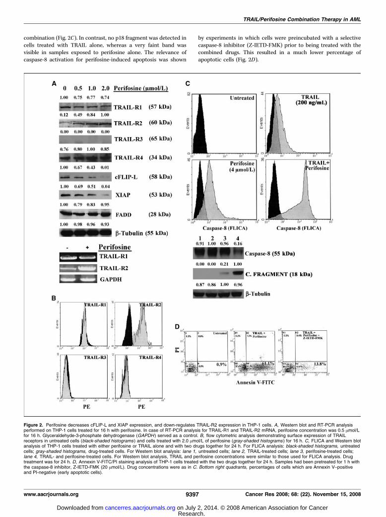
combination (Fig. 2C). In contrast, no p18 fragment was detected incells treated with TRAIL alone, whereas a very faint band wasvisible in samples exposed to perifosine alone. The relevance ofcaspase-8 activation for perifosine-induced apoptosis was shown
by experiments in which cells were preincubated with a selectivecaspase-8 inhibitor (Z-IETD-FMK) prior to being treated with thecombined drugs. This resulted in a much lower percentage ofapoptotic cells (Fig. 2D).
Figure 2. Perifosine decreases cFLIP-L and XIAP expression, and down-regulates TRAIL-R2 expression in THP-1 cells. A, Western blot and RT-PCR analysisperformed on THP-1 cells treated for 16 h with perifosine. In case of RT-PCR analysis for TRAIL-R1 and TRAIL-R2 mRNA, perifosine concentration was 0.5 Amol/Lfor 16 h. Glyceraldehyde-3-phosphate dehydrogenase (GAPDH ) served as a control. B, flow cytometric analysis demonstrating surface expression of TRAILreceptors in untreated cells (black-shaded histograms ) and cells treated with 2.0 Amol/L of perifosine (gray-shaded histograms ) for 16 h. C, FLICA and Western blotanalysis of THP-1 cells treated with either perifosine or TRAIL alone and with two drugs together for 24 h. For FLICA analysis: black-shaded histograms, untreatedcells; gray-shaded histograms, drug-treated cells. For Western blot analysis: lane 1, untreated cells; lane 2, TRAIL-treated cells; lane 3, perifosine-treated cells;lane 4, TRAIL- and perifosine-treated cells. For Western blot analysis, TRAIL and perifosine concentrations were similar to those used for FLICA analysis. Drugtreatment was for 24 h. D, Annexin V-FITC/PI staining analysis of THP-1 cells treated with the two drugs together for 24 h. Samples had been pretreated for 1 h withthe caspase-8 inhibitor, Z-IETD-FMK (20 Amol/L). Drug concentrations were as in C. Bottom right quadrants, percentages of cells which are Annexin V–positiveand PI-negative (early apoptotic cells).
TRAIL/Perifosine Combination Therapy in AML
www.aacrjournals.org 9397 Cancer Res 2008; 68: (22). November 15, 2008
Research. on July 2, 2014. © 2008 American Association for Cancercancerres.aacrjournals.org Downloaded from

Up-regulation of TRAIL-R2 requires PKC activity. A recentreport has highlighted that TRAIL-R2 up-regulation in non–smallcell lung cancer cells required PKC activity (26). Therefore, weinvestigated whether this was true also in THP-1 cells. Preliminaryexperiments indicated that THP-1 cells expressed only two ofthe conventional PKC isoforms, a and h2, whereas h1 and g werenot expressed (data not shown). When cells were treated withperifosine and Go6976, a well-established inhibitor of PKCconventional isoforms (27), the increase in TRAIL-R2 expressionwas lower. No changes in the expression of other TRAIL receptors(R1 and R4) were observed in response to Go6976 and perifosinetreatment, whereas cFLIP-L and XIAP levels did not significantlychange in cells treated with Go6976 and perifosine when comparedwith perifosine alone (Fig. 3A). A time-dependent increase inTRAIL-R2, but not TRAIL-R1, expression levels was detected whenTHP-1 cells were treated with PMA, an activator of PKCconventional isoforms (ref. 28; Fig. 3B). Down-regulation of PKCalevels by specific siRNA, but not treatment with scrambled siRNA,resulted in a much lower induction of TRAIL-R2 by perifosine(Fig. 3C). MTT assays showed a lower cytotoxic effect of the
perifosine and TRAIL combined treatment in cells with down-regulated PKCa expression, but not in those treated withscrambled siRNA (Fig. 3D). The efficacy of PKCa down-regulationby specific siRNA was evaluated by Western blot analysis (Fig. 3D).Overall, these findings indicated that up-regulation of TRAIL-R2 byperifosine was dependent on PKCa and was required to maximallypotentiate the proapoptotic effect of TRAIL.Up-regulation of TRAIL-R2 by perifosine is dependent on a
ROS/PKCA/JNK 2/c-Jun pathway. The mechanism of PKCaactivation by perifosine was next investigated. It has been shownthat PKCa could be activated (phosphorylated) by ROS (29, 30),which also induced its membrane binding. Moreover, perifosinecaused ROS production in U937 AML cells (31). Therefore, it wasinvestigated if perifosine also caused ROS production in THP-1cells. ROS generation was analyzed by flow cytometry after labelingof cells with the ROS-selective probe, DCFH-DA. Perifosine(2 Amol/L) caused an increase in ROS levels, which was blockedby the ROS scavenger, NAC (Supplementary Fig. S1A). Cellfractionation experiments showed that in response to perifosinetreatment, the amount of membrane-bound PKCa increased,
Figure 3. Increased TRAIL-R2 expression in THP-1 cells is dependent on PKCa. A, Western blot analysis of THP-1 cell extracts. Lane 1, untreated cells; lane 2, cellstreated with perifosine (2 Amol/L); lane 3, cells treated with perifosine (2 Amol/L) + Go6976 (0.5 Amol/L). Treatments were for 16 h. B, Western blot analysis forTRAIL-R1 and TRAIL-R2 expression in THP-1 cells treated with PMA (100 ng/mL) for increasing periods of time. C, Western blot analysis for TRAIL-R2 expressionlevels in cells incubated with perifosine (2 Amol/L for 16 h). Cells treated for 48 h with PKCa-specific siRNA or scrambled siRNA. D, results from MTT assays incells treated with TRAIL and perifosine for 24 h at the indicated concentrations. Western blot analyses for PKCa levels in THP-1 cell extracts also shown.Lane 1, untreated cells; lane 2, cells treated with siRNA specific for PKCa; lane 3, cells treated with scrambled siRNA. Cells were analyzed 48 h after transfection.h-Tubulin served as loading control.
Cancer Research
Cancer Res 2008; 68: (22). November 15, 2008 9398 www.aacrjournals.org
Research. on July 2, 2014. © 2008 American Association for Cancercancerres.aacrjournals.org Downloaded from
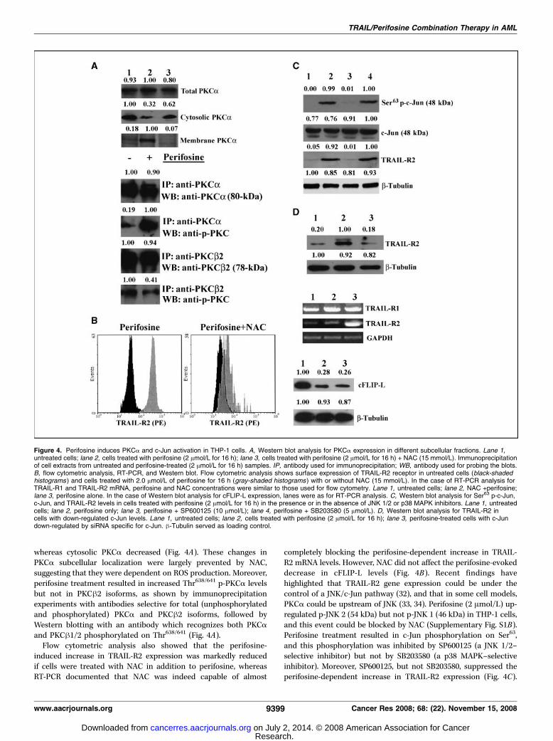
whereas cytosolic PKCa decreased (Fig. 4A). These changes inPKCa subcellular localization were largely prevented by NAC,suggesting that they were dependent on ROS production. Moreover,perifosine treatment resulted in increased Thr638/641 p-PKCa levelsbut not in PKCh2 isoforms, as shown by immunoprecipitationexperiments with antibodies selective for total (unphosphorylatedand phosphorylated) PKCa and PKCh2 isoforms, followed byWestern blotting with an antibody which recognizes both PKCaand PKCh1/2 phosphorylated on Thr638/641 (Fig. 4A).Flow cytometric analysis also showed that the perifosine-
induced increase in TRAIL-R2 expression was markedly reducedif cells were treated with NAC in addition to perifosine, whereasRT-PCR documented that NAC was indeed capable of almost
completely blocking the perifosine-dependent increase in TRAIL-R2 mRNA levels. However, NAC did not affect the perifosine-evokeddecrease in cFLIP-L levels (Fig. 4B). Recent findings havehighlighted that TRAIL-R2 gene expression could be under thecontrol of a JNK/c-Jun pathway (32), and that in some cell models,PKCa could be upstream of JNK (33, 34). Perifosine (2 Amol/L) up-regulated p-JNK 2 (54 kDa) but not p-JNK 1 (46 kDa) in THP-1 cells,and this event could be blocked by NAC (Supplementary Fig. S1B).Perifosine treatment resulted in c-Jun phosphorylation on Ser63,and this phosphorylation was inhibited by SP600125 (a JNK 1/2–selective inhibitor) but not by SB203580 (a p38 MAPK–selectiveinhibitor). Moreover, SP600125, but not SB203580, suppressed theperifosine-dependent increase in TRAIL-R2 expression (Fig. 4C).
Figure 4. Perifosine induces PKCa and c-Jun activation in THP-1 cells. A, Western blot analysis for PKCa expression in different subcellular fractions. Lane 1,untreated cells; lane 2, cells treated with perifosine (2 Amol/L for 16 h); lane 3, cells treated with perifosine (2 Amol/L for 16 h) + NAC (15 mmol/L). Immunoprecipitationof cell extracts from untreated and perifosine-treated (2 Amol/L for 16 h) samples. IP, antibody used for immunoprecipitation; WB, antibody used for probing the blots.B, flow cytometric analysis, RT-PCR, and Western blot. Flow cytometric analysis shows surface expression of TRAIL-R2 receptor in untreated cells (black-shadedhistograms ) and cells treated with 2.0 Amol/L of perifosine for 16 h (gray-shaded histograms ) with or without NAC (15 mmol/L). In the case of RT-PCR analysis forTRAIL-R1 and TRAIL-R2 mRNA, perifosine and NAC concentrations were similar to those used for flow cytometry. Lane 1, untreated cells; lane 2, NAC +perifosine;lane 3, perifosine alone. In the case of Western blot analysis for cFLIP-L expression, lanes were as for RT-PCR analysis. C, Western blot analysis for Ser63 p-c-Jun,c-Jun, and TRAIL-R2 levels in cells treated with perifosine (2 Amol/L for 16 h) in the presence or in the absence of JNK 1/2 or p38 MAPK inhibitors. Lane 1, untreatedcells; lane 2, perifosine only; lane 3, perifosine + SP600125 (10 Amol/L); lane 4, perifosine + SB203580 (5 Amol/L). D, Western blot analysis for TRAIL-R2 incells with down-regulated c-Jun levels. Lane 1, untreated cells; lane 2, cells treated with perifosine (2 Amol/L for 16 h); lane 3, perifosine-treated cells with c-Jundown-regulated by siRNA specific for c-Jun. h-Tubulin served as loading control.
TRAIL/Perifosine Combination Therapy in AML
www.aacrjournals.org 9399 Cancer Res 2008; 68: (22). November 15, 2008
Research. on July 2, 2014. © 2008 American Association for Cancercancerres.aacrjournals.org Downloaded from

Finally, when c-Jun levels were down-regulated by siRNA specificfor c-Jun (Supplementary Fig. S1C), the perifosine-evoked increasein TRAIL-R2 expression was reduced significantly (Fig. 4D). Takentogether, these findings strongly suggested that perifosine couldup-regulate TRAIL-R2 expression through a ROS! PKCa ! JNK 2! c-Jun signaling pathway.Synergistic cytotoxic effects of TRAIL/perifosine combined
treatment on AML blasts with activated Akt. The efficacy ofthe perifosine and TRAIL combined treatment was then analyzedon samples obtained from patients with AML. Samples from 12patients were studied (Table 1). Because levels of caspase-8 couldinfluence the TRAIL sensitivity of AML blasts (35), we analyzedsamples with comparable expression of caspase-8, as evaluated byWestern blot analysis (data not shown). Activation of PI3K/Aktsignaling was studied by Western blot and/or flow cytometricanalysis. Seven patients were positive for Ser473 p-Akt (Table 1).The combination of perifosine and TRAIL was much more effectivethan either drug alone, as shown by MTT assays (Fig. 5A ,patient M4#1). Cytotoxicity was due to apoptotic cell death, asdocumented by Annexin V-FITC/PI staining (Fig. 5B), and wascharacterized by increased TRAIL-R2 expression which wasdetected in CD33+ AML cells by flow cytometric analysis ofsamples double-stained for TRAIL-R2 and CD33 (Fig. 5C ).Combined treatment also resulted in higher levels of caspase-8 activation compared with single treatment, as shown by FLICAanalysis (data not shown). Western blot analysis showed Ser473
p-Akt down-regulation by perifosine, in patient blasts with Aktactivation, as well as a decrease of both cFLIP-L and XIAP, and up-regulation of Ser63 p-c-Jun (Fig. 5D). FADD levels were not affectedby perifosine. In contrast, in patients with no activated Akt, we didnot detect increased apoptosis in response to the combinedtreatment (Supplementary Fig. S2A , patient M4#2). In samplesfrom these patients, there was neither increased TRAIL-R2expression nor decreased cFLIP-L and XIAP expression. Moreover,
c-Jun phosphorylation on Ser63 was not increased after treatmentwith perifosine (Supplementary Fig. S2B and C). FADD levels didnot change in a significant manner. In Table 1, we summarize theresults obtained by treating AML samples, showing the effects ofperifosine on TRAIL-R2 expression and the average CI of thecombined treatment.Perifosine/TRAIL combined treatment negatively affects
clonogenic activity of CD34+ cells from patients with AML.Finally, we investigated the cytotoxic effect of perifosine/TRAILcombined treatment on CD34+ cell clonogenic activity from cordblood and from patients with AML. As expected, neither TRAIL norperifosine alone influenced the clonogenic activity of CD34+ cellsfrom healthy donors, and the same was true of the combinedtreatment (Supplementary Fig. S2D). In contrast, in leukemicCD34+ cells, TRAIL moderately affected clonogenic activity andperifosine alone exhibited a statistically significant inhibitory effectin some samples (see for example, patient M2#7). However, in allpatient samples with activated Akt, we consistently observed astrong inhibitory effect of the perifosine and TRAIL combinedtreatment on the CD34+ cell clonogenic activity.
Discussion
In this study, we showed that perifosine sensitizes AML cellsfrom both the THP-1 cell line and patients, to TRAIL-inducedapoptosis, at least in part via a decrease in cFLIP-L and XIAP levels,and through a PKCa-mediated increase in TRAIL-R2 expression.cFLIP-L, which is structurally similar to caspase-8, can be recruitedto the death-inducing signaling complex to inhibit the binding andactivation of caspase-8 and acts as a powerful repressor of TRAIL-induced death signaling (3). Also XIAP, a potent cellular caspaseinhibitor, is an important factor in TRAIL-induced cell death (36).A recent report from Carter and colleagues (36) has shown that
triptolide, an anticancer agent from a Chinese herb, sensitized AMLcells to TRAIL by decreasing XIAP levels and increasing TRAIL-R2expression through a p53-mediated mechanism. However, peri-fosine was able to up-regulate TRAIL-R2 in THP-1 cells which havea nonfunctional p53 (37). It has been established that TRAIL-R2levels could be regulated through mechanisms which are p53-dependent or -independent (38). Among p53-independent mech-anisms, it has been shown that the JNK/c-Jun axis could positivelyregulate TRAIL-R2 transcription (32). A functional activatorprotein-1 (AP-1) binding site has been shown in the promoterregion of TRAIL-R2. JNK, by increasing c-Jun phosphorylation,leads to an increase in AP-1 activity (39). Interestingly, we haverecently shown JNK-dependent increased AP-1 activity inT-lymphoblastic acute leukemia cells treated with perifosine (19).Our findings point to PKCa as an important mediator of TRAIL-
R2 up-regulation in THP-1 cells, as PKCa down-regulation bysiRNA resulted in a much lower induction of TRAIL-R2 and adecrease in perifosine-dependent sensitization to TRAIL cytotoxiceffect, whereas PMA, a conventional PKC isoform activator,increased TRAIL-R2 expression. Perifosine increased ROS produc-tion in THP-1 cells, and ROS could promote PKCa binding to theplasma membrane. Perifosine also induced increased levels ofphosphorylation of JNK 2 (but not JNK 1) and c-Jun in THP-1 cells.NAC impaired the up-regulation of TRAIL-R2 by perifosine. Theinvolvement of JNK 2/c-Jun signaling in the increased expression ofTRAIL-R2 was shown by the fact that a JNK 1/2–selective inhibitor,but not a p38 MAPK inhibitor, blocked both c-Jun phosphorylationon Ser63 induced by perifosine and TRAIL-R2 up-regulation.
Table 1. Patient classification and response to combinedtreatment
FAB
type
Ser473
p-Akt
TRAIL-R2
up-regulated
P + T synergism,
average CI(ED50 + ED75 + ED90)
M1#1 ++ ++++ 0.23 F 0.03M1#2 + ++ 0.51 F 0.05
M2#1 � � 0.95 F 0.10
M2#2 +++ ++++ 0.26 F 0.04
M2#3 +++ ++++ 0.15 F 0.02M2#4 � � 1.08 F 0.15
M2#5 + +/� 0.84 F 0.09
M2#6 � � 0.99 F 0.12M2#7 ++ +++ 0.39 F 0.05
M4#1 +++ ++++ 0.18 F 0.02
NOTE: Patients were classified according to the French-American-
British (FAB) classification. The levels of Ser473 p-Akt and TRAIL-R2were evaluated by flow cytometry and/or Western blotting. A CI < 0.9
was considered synergistic, whereas a CI between 0.9 and 1.1
was considered additive. Results are from three different experimentsF SD.
Abbreviations: P, perifosine; T, TRAIL; ED, effective dose.
Cancer Research
Cancer Res 2008; 68: (22). November 15, 2008 9400 www.aacrjournals.org
Research. on July 2, 2014. © 2008 American Association for Cancercancerres.aacrjournals.org Downloaded from

Furthermore, down-regulation of c-Jun by siRNA also opposed theincrease in TRAIL-R2. Given that in cells with down-regulatedPKCa, perifosine was unable to increase JNK 2 phosphorylation, wepropose a mechanism whereby perifosine generates ROS, which in
turn, activates a PKCa ! JNK 2 ! c-Jun signaling pathway whichleads to increased expression of TRAIL-R2.XIAP and c-FLIP-L down-regulation caused by perifosine in THP-
1 cells, could be due to an inhibition of the nuclear factor nB activity,
Figure 5. Perifosine and TRAIL combination is synergistic in AMLprimary cells with activated Akt. A, cells from patient M4#1 weretreated for 48 h with either single agent alone or in combination at theindicated concentrations. Cell viability was analyzed by MTT assays.B, Annexin V-FITC/PI staining analysis of cells from patient M4#1treated with either perifosine or TRAIL alone and with two drugstogether for 48 h. Bottom right quadrants, percentages of cells whichare Annexin V–positive and PI-negative (early apoptotic cells).C, flow cytometric analysis showing surface expression of TRAILreceptors and CD33 in cells from patient M4#1, either untreated ortreated with 4.0 Amol/L of perifosine for 24 h. Anti-TRAIL receptorantibodies were conjugated to phycoerythrin, whereas anti-CD33antibody was FITC-conjugated. D, Western blot analysis forSer473 p-Akt, cFLIP-L, XIAP, FADD, and Ser63 p-c-Jun in extractsfrom AML patient primary cells. Perifosine treatment was for 24 hat 4.0 Amol/L concentration. h-Tubulin served as loading control.
TRAIL/Perifosine Combination Therapy in AML
www.aacrjournals.org 9401 Cancer Res 2008; 68: (22). November 15, 2008
Research. on July 2, 2014. © 2008 American Association for Cancercancerres.aacrjournals.org Downloaded from

which is under the control of the PI3K/Akt axis in AML cells (12). Arecent investigation carried out on Waldenstrom macroglobuline-mia cells has indeed shown that perifosine targets nuclear factor nB(40). Future studies should address the mechanism(s) whichunderlie XIAP and cFLIP-L down-regulation by perifosine in AMLcells. Nevertheless, our unpublished findings, obtained by siRNAtechnology, have indicated that down-regulation of these twoproteins was not as critical as that of PKCa for the potentiatingeffect of perifosine on TRAIL cytotoxicity on THP-1 cells.Our results showed that a perifosine and TRAIL combination
was also much more effective than either treatment alone inprimary AML cells. Even though the analysis we performed was notas comprehensive as the one we did in THP-1 cells, due to theinsufficient amount of cells recovered from most of the patients,perifosine dephosphorylated Akt, down-regulated XIAP and cFLIP-L expression, and up-regulated the levels of TRAIL-R2 and Ser63
p-c-Jun in some primary AML cells. AML blasts died by apoptosisand the combined treatment was much more effective in activatingcaspase-8 than either treatment alone. All the patient samplesexpressed TRAIL-R2 to some extent (data not shown); however,perifosine increased TRAIL-R2 expression only in samples withactivated PI3K/Akt signaling. Accordingly, synergism was onlyobserved in those AML samples which displayed activated Akt. Thefact that despite the expression of TRAIL-R2 even under basalconditions, AML blasts were not sensitive to TRAIL alone, could beexplained by the contemporaneous expression of TRAIL decoyreceptors (8). After perifosine treatment, TRAIL-R2 was markedlyup-regulated in AML blasts, and most likely this could overcomethe antiapoptotic effect played by high levels of decoy receptors.However, it should not be ruled out that other proteins, which arecritically important for TRAIL sensitivity (35, 41), are down-regulated by perifosine in AML primary cells.At present, it is unclear why perifosine increased ROS generation
only in primary AML cells with up-regulated PI3K/Akt signaling.Nevertheless, a recent report has highlighted that 7-ketochetos-terol, which is incorporated into the lipid rafts of THP-1 cells (42),was able to increase ROS production by up-regulating the levels ofNAD(P)H oxidase (NOX-4) in THP-1 cells. Interestingly, this wasaccompanied by a down-regulation of Akt (43). It might be thatperifosine, by disrupting PI3K/Akt signaling at the lipid rafts,positively affects NOX-4 gene expression. Therefore, it would beinteresting to investigate if NOX-4 gene expression is under thecontrol of the PI3K/Akt axis in AML cells.
Our results point to the fact that a combination consisting ofTRAIL and perifosine had no effect on the clonogenic activity ofCD34+ cells from healthy donors, whereas it was markedlycytotoxic for CD34+ cells isolated from leukemic patients. Previousresults have shown that TRAIL was not cytotoxic for normal CD34+
cells (44, 45), reflecting the lack of TRAIL receptors expressed inthese cells (46). In contrast, TRAIL displayed proapoptotic activityin CD34+ cells from patients with AML (45), and we have confirmedthese findings. Therefore, CD34+ AML cells express TRAILreceptors. Future investigations should be aimed towards investi-gating whether leukemic stem cells also express TRAIL receptorsand whether they could be targeted by the combination of TRAILand perifosine.In conclusion, we have shown the in vitro efficacy of a TRAIL
and perifosine combination treatment in AML cells. Thiscombination was also synergistic in cells (THP-1) lackingfunctional p53. Even if p53 deletion and/or inactivating mutationsare observed in only f10% of patients with AML, p53 levels arefrequently low in AML blasts due to overexpression of the negativeregulator murine double minute (MDM2; ref. 47). Thus, the use of adrug which could up-regulate TRAIL-R2 levels independently ofp53 could be extremely useful in leukemia therapy. Accordingly, itmust be emphasized that the cytotoxic effect of triptolide andTRAIL combination was enhanced by the addition of the MDM2antagonist, Nutlin-3a (36). In case of triptolide, another drug wasrequired to maximize the effects of TRAIL, which could result inadditional toxic side effects if administered to patients.In summary, the combination of perifosine and TRAIL could
represent a novel strategy for treating patients with AML byovercoming critical mechanisms of apoptosis resistance.
Disclosure of Potential Conflicts of Interest
No potential conflicts of interest were disclosed.
Acknowledgments
Received 7/23/2008; revised 8/27/2008; accepted 9/15/2008.Grant support: Fondazione CARISBO (A.M. Martelli), Progetti Strategici Universita
di Bologna EF2006 (A.M. Martelli), European LeukemiaNet (G. Martinelli), and NIHgrant RO1CA091025 (J.A. McCubrey).
The costs of publication of this article were defrayed in part by the payment of pagecharges. This article must therefore be hereby marked advertisement in accordancewith 18 U.S.C. Section 1734 solely to indicate this fact.
Cancer Research
Cancer Res 2008; 68: (22). November 15, 2008 9402 www.aacrjournals.org
References
1. Kelley SK, Ashkenazi A. Targeting death receptors incancer with Apo2L/TRAIL. Curr Opin Pharmacol 2004;4:333–9.2. Almasan A, Ashkenazi A. Apo2L/TRAIL: apoptosissignaling, biology, and potential for cancer therapy.Cytokine Growth Factor Rev 2003;14:337–48.3. LeBlanc HN, Ashkenazi A. Apo2L/TRAIL and itsdeath and decoy receptors. Cell Death Differ 2003;10:66–75.4. Koschny R, Walczak H, Ganten TM. The promise ofTRAIL-potential and risks of a novel anticancer therapy.J Mol Med 2007;85:923–35.5. Kaufmann SH, Steensma DP. On the TRAIL of a newtherapy for leukemia. Leukemia 2005;19:2195–202.6. Bortul R, Tazzari PL, Cappellini A, et al. Constitutivelyactive Akt1 protects HL60 leukemia cells from TRAIL-induced apoptosis through a mechanism involving NF-
nB activation and cFLIP(L) up-regulation. Leukemia2003;17:379–89.7. Secchiero P, Zerbinati C, di Iasio MG, et al. Synergisticcytotoxic activity of recombinant TRAIL plus the non-genotoxic activator of the p53 pathway nutlin-3 in acutemyeloid leukemia cells. Curr Drug Metab 2007;8:395–403.8. Riccioni R, Pasquini L, Mariani G, et al. TRAIL decoyreceptors mediate resistance of acute myeloid leukemiacells to TRAIL. Haematologica 2005;90:612–24.9. Jones DT, Ganeshaguru K, Mitchell WA, et al.Cytotoxic drugs enhance the ex vivo sensitivity ofmalignant cells from a subset of acute myeloidleukaemia patients to apoptosis induction by tumournecrosis factor receptor-related apoptosis-inducingligand. Br J Haematol 2003;121:713–20.10. Guo F, Sigua C, Tao J, et al. Cotreatment with histonedeacetylase inhibitor LAQ824 enhances Apo-2L/tumornecrosis factor-related apoptosis inducing ligand-induced death inducing signaling complex activity and
apoptosis of human acute leukemia cells. Cancer Res2004;64:2580–9.11. Elrod HA, Lin YD, Yue P, et al. The alkylphospholipidperifosine induces apoptosis of human lung cancer cellsrequiring inhibition of Akt and activation of theextrinsic apoptotic pathway. Mol Cancer Ther 2007;6:2029–38.12. Martelli AM, Nyakern M, Tabellini G, et al. Phos-phoinositide 3-kinase/Akt signaling pathway and itstherapeutical implications for human acute myeloidleukemia. Leukemia 2006;20:911–28.13. Tazzari PL, Cappellini A, Ricci F, et al. Multidrugresistance-associated protein 1 expression is under thecontrol of the phosphoinositide 3 kinase/Akt signaltransduction network in human acute myelogenousleukemia blasts. Leukemia 2007;21:427–38.14. Tazzari PL, Tabellini G, Bortul R, et al. The insulin-like growth factor-I receptor kinase inhibitor NVP-AEW541 induces apoptosis in acute myeloid leukemia
Research. on July 2, 2014. © 2008 American Association for Cancercancerres.aacrjournals.org Downloaded from

TRAIL/Perifosine Combination Therapy in AML
www.aacrjournals.org 9403 Cancer Res 2008; 68: (22). November 15, 2008
cells exhibiting autocrine insulin-like growth factor-Isecretion. Leukemia 2007;21:886–96.15. Sale EM, Sale GJ. Protein kinase B: signallingroles and therapeutic targeting. Cell Mol Life Sci 2008;65:113–27.16. Vink SR, van Blitterswijk WJ, Schellens JH, Verheij M.Rationale and clinical application of alkylphospholipidanalogues in combination with radiotherapy. CancerTreat Rev 2007;33:191–202.17. van der Luit AH, Vink SR, Klarenbeek JB, et al. A newclass of anticancer alkylphospholipids uses lipid rafts asmembrane gateways to induce apoptosis in lymphomacells. Mol Cancer Ther 2007;6:2337–45.18. Papa V, Tazzari PL, Chiarini F, et al. Proapoptoticactivity and chemosensitizing effect of the novel Aktinhibitor perifosine in acute myelogenous leukemiacells. Leukemia 2008;22:147–60.19. Chiarini F, Del Sole M, Mongiorgi S, et al. The novelAkt inhibitor, perifosine, induces caspase-dependentapoptosis and downregulates P-glycoprotein expressionin multidrug-resistant human T-acute leukemia cellsby a JNK-dependent mechanism. Leukemia 2008;22:1106–16.20. Ko CH, Shen SC, Chen YC. Hydroxylation at C4¶ or C6is essential for apoptosis-inducing activity of flavanonethrough activation of the caspase-3 cascade andproduction of reactive oxygen species. Free Radic BiolMed 2004;36:897–910.21. Nyakern M, Cappellini A, Mantovani I, Martelli AM.Synergistic induction of apoptosis in human leukemia Tcells by the Akt inhibitor perifosine and etoposidethrough activation of intrinsic and Fas-mediatedextrinsic cell death pathways. Mol Cancer Ther 2006;5:1559–70.22. Zhang XD, Franco A, Myers K, Gray C, Nguyen T,Hersey P. Relation of TNF-related apoptosis-inducingligand (TRAIL) receptor and FLICE-inhibitory proteinexpression to TRAIL-induced apoptosis of melanoma.Cancer Res 1999;59:2747–53.23. Chou TC, Talalay P. Quantitative analysis of dose-effect relationships: the combined effects of multipledrugs or enzyme inhibitors. Adv Enzyme Regul 1984;22:27–55.24. Theurl I, Theurl M, Seifert M, et al. Autocrineformation of hepcidin induces iron retention in humanmonocytes. Blood 2008;111:2392–9.25. Phillips TA, Ni J, Pan G, et al. TRAIL (Apo-2L) andTRAIL receptors in human placentas: implications forimmune privilege. J Immunol 1999;162:6053–9.26. Chen W, Wang X, Zhuang J, Zhang L, Lin Y. Induction
of death receptor 5 and suppression of survivincontribute to sensitization of TRAIL-induced cytotox-icity by quercetin in non-small cell lung cancer cells.Carcinogenesis 2007;28:2114–21.27. Mahanivong C, Chen HM, Yee SW, Pan ZK, Dong Z,Huang S. Protein kinase Ca-CARMA3 signaling axislinks Ras to NF-nB for lysophosphatidic acid-inducedurokinase plasminogen activator expression in ovariancancer cells. Oncogene 2008;27:1273–80.28. Cuschieri J, Billigren J, Maier RV. Endotoxin toleranceattenuates LPS-induced TLR4 mobilization to lipid rafts:a condition reversed by PKC activation. J Leukoc Biol2006;80:1289–97.29. Kitatani K, Idkowiak-Baldys J, Hannun YA. Mecha-nism of inhibition of sequestration of protein kinase Ca/hII by ceramide. Roles of ceramide-activated proteinphosphatases and phosphorylation/dephosphorylationof protein kinase C a/hII on threonine 638/641. J BiolChem 2007;282:20647–56.30. Johann AM, von Knethen A, Lindemann D, Brune B.Recognition of apoptotic cells by macrophages activatesthe peroxisome proliferator-activated receptor-g andattenuates the oxidative burst. Cell Death Differ 2006;13:1533–40.31. Rahmani M, Reese E, Dai Y, et al. Coadministration ofhistone deacetylase inhibitors and perifosine synergis-tically induces apoptosis in human leukemia cellsthrough Akt and ERK1/2 inactivation and the genera-tion of ceramide and reactive oxygen species. CancerRes 2005;65:2422–32.32. Zou W, Liu X, Yue P, et al. c-Jun NH2-terminal kinase-mediated up-regulation of death receptor 5 contributesto induction of apoptosis by the novel synthetictriterpenoid methyl-2-cyano-3,12-dioxooleana-1,9-dien-28-oate in human lung cancer cells. Cancer Res 2004;64:7570–8.33. Mauro A, Ciccarelli C, De Cesaris P, et al. PKCa-mediated ERK, JNK and p38 activation regulates themyogenic program in human rhabdomyosarcoma cells.J Cell Sci 2002;115:3587–99.34. Chow JM, Shen SC, Wu CY, Chen YC. 12-O -tetradecanoylphorbol 13-acetate prevents baicalein-induced apoptosis via activation of protein kinase Cand JNKs in human leukemia cells. Apoptosis 2006;11:1999–2011.35. Riccioni R, Senese M, Diverio D, et al. Resistance ofacute myeloid leukemic cells to the triterpenoid CDDO-imidazolide is associated with low caspase-8 and FADDlevels. Leuk Res 2008;32:1244–58.36. Carter BZ, Mak DH, Schober WD, et al. Triptolide
sensitizes AML cells to TRAIL-induced apoptosis viadecrease of XIAP and p53-mediated increase of DR5.Blood 2008;111:3742–50.37. Akashi M, Osawa Y, Koeffler HP, Hachiya M.p21WAF1 expression by an activator of protein kinaseC is regulated mainly at the post-transcriptional level incells lacking p53: important role of RNA stabilization.Biochem J 1999;337:607–16.38. Kim YH, Jung EM, Lee TJ, et al. Rosiglitazonepromotes tumor necrosis factor-related apoptosis-in-ducing ligand-induced apoptosis by reactive oxygenspecies-mediated up-regulation of death receptor 5 anddown-regulation of c-FLIP. Free Radic Biol Med 2008;44:1055–68.39. Verde P, Casalino L, Talotta F, Yaniv M, Weitzman JB.Deciphering AP-1 function in tumorigenesis: fra-terniz-ing on target promoters. Cell Cycle 2007;6:2633–9.40. Leleu X, Eeckhoute J, Jia X, et al. Targeting NF-nBin Waldenstrom macroglobulinemia. Blood 2008;111:5068–77.41. Riccioni R, Senese M, Diverio D, et al. M4 and M5acute myeloid leukaemias display a high sensitivity tobortezomib-mediated apoptosis. Br J Haematol 2007;139:194–205.42. Berthier A, Lemaire-Ewing S, Prunet C, et al.Involvement of a calcium-dependent dephosphorylationof BAD associated with the localization of Trpc-1 withinlipid rafts in 7-ketocholesterol-induced THP-1 cellapoptosis. Cell Death Differ 2004;11:897–905.43. Palozza P, Serini S, Verdecchia S, et al. Redoxregulation of 7-ketocholesterol-induced apoptosis byh-carotene in human macrophages. Free Radic Biol Med2007;42:1579–90.44. Zang DY, Goodwin RG, Loken MR, Bryant E, Deeg HJ.Expression of tumor necrosis factor-related apoptosis-inducing ligand, Apo2L, and its receptors in myelodys-plastic syndrome: effects on in vitro hemopoiesis. Blood2001;98:3058–65.45. Plasilova M, Zivny J, Jelinek J, et al. TRAIL (Apo2L)suppresses growth of primary human leukemia andmyelodysplasia progenitors. Leukemia 2002;16:67–73.46. Secchiero P, Vaccarezza M, Gonelli A, Zauli G. TNF-related apoptosis-inducing ligand (TRAIL): a potentialcandidate for combined treatment of hematologicalmalignancies. Curr Pharm Des 2004;10:3673–81.47. Kojima K, Konopleva M, Samudio IJ, Ruvolo V,Andreeff M. Mitogen-activated protein kinase kinaseinhibition enhances nuclear proapoptotic function ofp53 in acute myelogenous leukemia cells. Cancer Res2007;67:3210–9.
Research. on July 2, 2014. © 2008 American Association for Cancercancerres.aacrjournals.org Downloaded from