Ultra-High-Throughput Screening of Natural Product Extracts to Identify Proapoptotic Inhibitors of...
11
Journal of Biomolecular Screening 1–11 © 2014 Society for Laboratory Automation and Screening DOI: 10.1177/1087057114536227 jbx.sagepub.com Original Research Introduction Natural product extracts (NPEs) represent a tremendous resource for novel chemical matter with potential appli- cation toward therapeutic development. In fact, 50% of drugs approved in the United States from 1981 to 2010 were either natural products or their derivatives. 1 Despite this strong track record, most pharmaceutical firms have stopped natural product research in the past decade, which signifies a 30% drop in activity between 2001 and 2008, as a consequence of changes in the drug discovery paradigm and other technical limitations. 2 This decline represents an opportunity for government and noncom- mercial entities to drive this important area of drug dis- covery research. Several academic organizations maintain natural product libraries of considerable scale and diversity, including the Spanish Fundación Medina, the plant extract bank of the Korean Institute of Bioscience and Biotechnology, and the Australian Eskitis Institute. 3 However, industrial-scale high-throughput screening of these collections has been limited. Recently, the National Cancer Institute (NCI) assembled the Chemical Biology Consortium (CBC) to conduct drug discovery within the academic and nonprofit 536227JBX XX X 10.1177/1087057114536227Journal of Biomolecular ScreeningHassig et al. research-article 2014 Corresponding Author: Christian A. Hassig, Sanford Burnham Medical Research Institute, 10901 N. Torrey Pines Road, La Jolla, CA 92037, USA. Email: [email protected] Ultra-High-Throughput Screening of Natural Product Extracts to Identify Proapoptotic Inhibitors of Bcl-2 Family Proteins Christian A. Hassig 1 , Fu-Yue Zeng 1 , Paul Kung 1 , Mehrak Kiankarimi 1 , Sylvia Kim 1 , Paul W. Diaz 1 , Dayong Zhai 1 , Kate Welsh 1 , Shana Morshedian 1 , Ying Su 1 , Barry O’Keefe 2 , David J. Newman 2 , Yudi Rusman 3 , Harneet Kaur 3 , Christine E. Salomon 3 , Susan G. Brown 4 , Beeraiah Baire 4 , Andrew R. Michel 4 , Thomas R. Hoye 4 , Subhashree Francis 5 , Gunda I. Georg 5 , Michael A. Walters 5 , Daniela B. Divlianska 6 , Gregory P. Roth 6 , Amy E. Wright 7 , and John C. Reed 1* Abstract Antiapoptotic Bcl-2 family proteins are validated cancer targets composed of six related proteins. From a drug discovery perspective, these are challenging targets that exert their cellular functions through protein-protein interactions (PPIs). Although several isoform-selective inhibitors have been developed using structure-based design or high-throughput screening (HTS) of synthetic chemical libraries, no large-scale screen of natural product collections has been reported. A competitive displacement fluorescence polarization (FP) screen of nearly 150,000 natural product extracts was conducted against all six antiapoptotic Bcl-2 family proteins using fluorochrome-conjugated peptide ligands that mimic functionally relevant PPIs. The screens were conducted in 1536-well format and displayed satisfactory overall HTS statistics, with Z′- factor values ranging from 0.72 to 0.83 and a hit confirmation rate between 16% and 64%. Confirmed active extracts were orthogonally tested in a luminescent assay for caspase-3/7 activation in tumor cells. Active extracts were resupplied, and effort toward the isolation of pure active components was initiated through iterative bioassay-guided fractionation. Several previously described altertoxins were isolated from a microbial source, and the pure compounds demonstrate activity in both Bcl-2 FP and caspase cellular assays. The studies demonstrate the feasibility of ultra-high-throughput screening using natural product sources and highlight some of the challenges associated with this approach. Keywords ultra-high-throughput screening, fluorescence polarization, natural product extracts, Bcl-2 family, apoptosis, bioassay- guided fractionation at UNIV OF CHICAGO LIBRARY on June 11, 2014 jbx.sagepub.com Downloaded from
-
Upload
independent -
Category
Documents
-
view
2 -
download
0
Transcript of Ultra-High-Throughput Screening of Natural Product Extracts to Identify Proapoptotic Inhibitors of...

Journal of Biomolecular Screening 1 –11© 2014 Society for LaboratoryAutomation and ScreeningDOI: 10.1177/1087057114536227jbx.sagepub.com
Original Research
Introduction
Natural product extracts (NPEs) represent a tremendous resource for novel chemical matter with potential appli-cation toward therapeutic development. In fact, 50% of drugs approved in the United States from 1981 to 2010 were either natural products or their derivatives.1 Despite this strong track record, most pharmaceutical firms have stopped natural product research in the past decade, which signifies a 30% drop in activity between 2001 and 2008, as a consequence of changes in the drug discovery paradigm and other technical limitations.2 This decline represents an opportunity for government and noncom-mercial entities to drive this important area of drug dis-covery research.
Several academic organizations maintain natural product libraries of considerable scale and diversity, including the Spanish Fundación Medina, the plant extract bank of the Korean Institute of Bioscience and Biotechnology, and the Australian Eskitis Institute.3 However, industrial-scale high-throughput screening of these collections has been limited. Recently, the National Cancer Institute (NCI) assembled the Chemical Biology Consortium (CBC) to conduct drug discovery within the academic and nonprofit
536227 JBXXXX10.1177/1087057114536227Journal of Biomolecular ScreeningHassig et al.research-article2014
Corresponding Author:Christian A. Hassig, Sanford Burnham Medical Research Institute, 10901 N. Torrey Pines Road, La Jolla, CA 92037, USA. Email: [email protected]
Ultra-High-Throughput Screening of Natural Product Extracts to Identify Proapoptotic Inhibitors of Bcl-2 Family Proteins
Christian A. Hassig1, Fu-Yue Zeng1, Paul Kung1, Mehrak Kiankarimi1, Sylvia Kim1, Paul W. Diaz1, Dayong Zhai1, Kate Welsh1, Shana Morshedian1, Ying Su1, Barry O’Keefe2, David J. Newman2, Yudi Rusman3, Harneet Kaur3, Christine E. Salomon3, Susan G. Brown4, Beeraiah Baire4, Andrew R. Michel4, Thomas R. Hoye4, Subhashree Francis5, Gunda I. Georg5, Michael A. Walters5, Daniela B. Divlianska6, Gregory P. Roth6, Amy E. Wright7, and John C. Reed1*
AbstractAntiapoptotic Bcl-2 family proteins are validated cancer targets composed of six related proteins. From a drug discovery perspective, these are challenging targets that exert their cellular functions through protein-protein interactions (PPIs). Although several isoform-selective inhibitors have been developed using structure-based design or high-throughput screening (HTS) of synthetic chemical libraries, no large-scale screen of natural product collections has been reported. A competitive displacement fluorescence polarization (FP) screen of nearly 150,000 natural product extracts was conducted against all six antiapoptotic Bcl-2 family proteins using fluorochrome-conjugated peptide ligands that mimic functionally relevant PPIs. The screens were conducted in 1536-well format and displayed satisfactory overall HTS statistics, with Z′-factor values ranging from 0.72 to 0.83 and a hit confirmation rate between 16% and 64%. Confirmed active extracts were orthogonally tested in a luminescent assay for caspase-3/7 activation in tumor cells. Active extracts were resupplied, and effort toward the isolation of pure active components was initiated through iterative bioassay-guided fractionation. Several previously described altertoxins were isolated from a microbial source, and the pure compounds demonstrate activity in both Bcl-2 FP and caspase cellular assays. The studies demonstrate the feasibility of ultra-high-throughput screening using natural product sources and highlight some of the challenges associated with this approach.
Keywordsultra-high-throughput screening, fluorescence polarization, natural product extracts, Bcl-2 family, apoptosis, bioassay-guided fractionation
at UNIV OF CHICAGO LIBRARY on June 11, 2014jbx.sagepub.comDownloaded from
2 Journal of Biomolecular Screening
sectors. The CBC’s directive includes screening to identify novel natural products with potential utility as anticancer agents. A large-scale pilot project was undertaken to screen the NCI NPE collection of approximately 150,000 unique samples against a panel of 10 antiapoptosis cancer targets, including six members of the Bcl-2 family proteins and the BIR2 and BIR3 domains of cIAP1 and cIAP2. This report describes the screening and hit follow-up studies for the Bcl-2 family targets (Bcl-2, Bcl-W, Bcl-XL, Bcl-B, Mcl-1, and Bfl-1).
Overexpression of antiapoptotic proteins is a common feature in the pathogenesis and progression of malignan-cies, making these proteins promising anticancer targets.4,5 BCL-2 was the first antideath gene discovered and consti-tutes a new anticancer target class with far-reaching impli-cations for tumor biology.6 Multiple members of the human Bcl-2 family proteins have since been identified, including six antiapoptotic members. The Bcl-2 proteins bind and sequester the proapoptotic BH3-only proteins such as Bim, thus blocking cell death.7 Bcl-2 family proteins are regu-lated through myriad posttranslational modifications and interactions with other proteins, but most compellingly, Bcl-2 family proteins regulate all major types of cell death, including apoptosis, necrosis, and autophagy. Therefore, these proteins operate as nodal points at the convergence of multiple pathways with a broad and profound relevance to oncology.
Small-molecule BH3 mimetics that antagonize the inter-action between antiapoptotic Bcl-2 proteins and proapop-totic BH3-only proteins represent potential anticancer therapeutics.8 The rationale for screening natural product collections for compounds targeting the Bcl-2 family pro-teins has both a biological and a biophysical foundation. From a biological standpoint, it is noteworthy that Bcl-2 family genes are conserved throughout the animal kingdom and are found in insects, nematodes, and simple marine organisms.9,10 Plants and microbes have evolved chemical biosynthetic pathways that use natural products to defend themselves against predatory or pathological attack by competing animal species, and thus gene products required for cell survival are ideal targets of such agents. Indeed, examples of natural products targeting Bcl-2 have been found, including those with known anticancer activity. The
most advanced of these is gossypol, a Bcl-2 inhibitory natu-ral product from cottonseeds with a history of use in Chinese herbal medicine. Gossypol has advanced into phase III clin-ical trials for cancer; however, three other phase II trials were either suspended or terminated, casting doubt on the future development of this agent.11 From a biophysical standpoint, natural products are attractive as candidate inhibitors of Bcl-2 family proteins because strategies for neutralizing these proteins are predicated on mimicking protein-protein interactions, a task for which more complex chiral molecules found in nature are best suited.
A limited number of synthetic small-molecule inhibitors of Bcl-2 family proteins have been described and are in various stages of preclinical and clinical development, the most advanced of which is ABT-199 (GDC-0199), cur-rently in phase III clinical trials (Abbvie/Roche). ABT-199 is a highly selective and potent inhibitor of Bcl-2, which was generated using nuclear magnetic resonance (NMR)–based chemical fragment screening and structure-based drug design technologies.12–14 There remains a need for potent agents that act on other members of Bcl-2 family proteins—including Bfl-1 and Mcl-1, which are upregu-lated in many cancers—but are not blocked by existing compounds. In addition, a potent broad-spectrum Bcl-2 family inhibitor could be superior to chemical entities that target only one member of the family due to the simultane-ous overexpression of several members in many tumors. To enable the identification of agents functioning as broad-spectrum and isoform-specific inhibitors of these key anti-apoptotic proteins, we took a multitarget parallel HTS approach.
This report describes the large-scale crude extract library reformatting, assay optimization, multitarget parallel ultra-high-throughput screening (uHTS), and bioassay-guided fractionation of active extracts of interest. These efforts have led to the identification of several known, as well as novel, natural products with potential anti–Bcl-2 family activity. Despite efforts to minimize potential interference from nui-sance compounds, it was noted that many of the compounds isolated contain structural features that could cause assay interference. Nonetheless, several of the known compounds that were identified have been described previously to pos-sess anticancer activity via unknown mechanisms. Moreover,
1Sanford Burnham Medical Research Institute, La Jolla, CA, USA2National Cancer Institute, Frederick, MD, USA3Center for Drug Design, University of Minnesota, Minneapolis, MN, USA4Department of Chemistry, University of Minnesota, Minneapolis, MN, USA5Institute for Therapeutics Discovery and Development, Department of Medicinal Chemistry, University of Minnesota, Minneapolis, MN, USA6Sanford-Burnham Medical Research Institute at Lake Nona, Orlando, FL, USA7Harbor Branch Oceanographic Institute at Florida Atlantic University, Fort Pierce, FL, USA*Current address: Roche Pharmaceuticals, Basel, Switzerland
Received Mar 10, 2014, and in revised form Apr 8, 2014. Accepted for publication Apr 26, 2014.
Supplementary material for this article is available on the Journal of Biomolecular Screening Web site at http://jbx.sagepub.com/supplemental.
at UNIV OF CHICAGO LIBRARY on June 11, 2014jbx.sagepub.comDownloaded from
Hassig et al. 3
the purified altertoxins identified from this screen demon-strated broad-spectrum activity against Bcl-2 family proteins and activated caspase-3/7 in cells, providing the intriguing possibility that their apoptosis-inducing properties may in part be due to the direct inhibition of Bcl-2 family proteins.
Materials and Methods
Natural Product Library Reformatting
The collection of NPEs shipped from the NCI Natural Products Branch contained a total of 148,250 extracts in 1685 microplates of 96 wells at 0.5 mg/well. This collection was composed of 825 organic NPE plates and 860 aqueous NPE plates. Reformatting protocols (96 to 384 and 384 to 1536 plate formats) were optimized with a Biomek FX liquid handler (Beckman Coulter, Brea, CA). Additional information on library reformatting is available in the sup-plemental materials (Suppl. Table 1, Suppl. Table 2). NPE transfers from 1536-well source plates into assay plates used an acoustic dispenser Echo 555 (Labcyte, Sunnyvale, CA) or a pintool (High Resolution Biosciences, Woburn, MA).
Protein Purification
GST-tagged proteins containing Bcl-Xl, Bcl-2, Bcl-W, Bcl-B, Bfl-1, and Mcl-1 lacking their C-terminal transmem-brane domains (last 20 amino acids; ΔTM) were expressed from pGEX 4T-1 plasmid in XL-1 Blue cells (Stratagene, La Jolla, CA), as previously reported.15 Additional details of protein expression and purification are available in the supplemental materials.
Peptide Synthesis and Purification
N-terminal rhodamine, fluorescein isothiocyanate (FITC), and Cy5-labeled Bim BH3 peptides were synthesized on Rink Amide-MBHA resin using Fmoc synthesis and DIC/HOBt coupling with an Advanced Chem Tech 396 multiple peptide synthesizer. Mass spectrometry of dye-labeled pep-tides was performed using an AutoFlex II MALDI-TOF (Bruker Daltonics, Billerica, MA), employing α-cyano-4-hydroxycinnamic acid as matrix. High-performance liquid chromatography (HPLC) of the same sample was accom-plished using a C18 column with a gradient of 0.1% trifluo-roacetic acid (TFA) in water to 0.1% TFA in acetonitrile for 20 min at a flow rate of 1.5 mL/min.
Competitive Peptide Displacement Fluorescence Polarization Assays
Methods for competitive peptide displacement using vari-ous Bcl-2 family proteins and FITC assays were similar to
those described in a previous publication.15 The Kd for each assay was estimated by varying the protein concentrations by factors of 2 while maintaining the concentration of the N-terminally labeled FITC-Bim or Cy-5-Bim peptide at 10 nM. Determinations were carried out in phosphate-buffered saline buffer plus 0.005% Tween 20. Fluorescence polariza-tion (FP) was measured after a 20 min incubation. Reactions were run in a total volume of 6 µL per well in 1536-well black solid-bottom microplates (Corning No. 3724, Corning, NY). Additional information is available in the supplementary materials.
High-Throughput Screen
For high-throughput screening (HTS), an acoustic dispenser (Echo 555, Labcyte) was used to transfer organic extracts, and a pintool device (High Resolution Biosciences) was used to transfer aqueous extracts from 1536-well Echo-qualified NPE plates into 1536-well assay plates (Corning No. 3724). The Multidrop Combi bulk dispenser (Thermo Fisher Scientific, Hudson, NH) was then used to dispense buffer, protein, and tracer solutions. A volume of 10 nL of organic NPEs (5 mg/mL stock) or 20 nL of aqueous NPEs (5 mg/mL stock) was dispensed into the assay plates predis-pensed with 2 µL of 0.15% gamma-globulin (Sigma, St. Louis, MO) followed by the addition of 2 µL of GST-Bcl-2 proteins, then followed by the addition of 2 µL of a mixture of FITC or Cy5-Bim BH3 peptide (each at 10 nM in 25 mM Bis-Tris, pH 7.0, containing 0.005% Tween-20) with a final volume of 6 µL. Each well thus contained 8.3 µg/mL of organic NPEs and 16.7 µg/mL of aqueous NPEs and various concentrations of GST-Bcl-2 proteins and 10 nM each of FITC- and Cy5-Bim BH3 peptide. After 20 min of incuba-tion, FP was measured using the Envision Microplate Reader for FITC channel and the PHERAstarPlus Microplate Reader (for Cy5 channel) with an excitation filter (485 nm) and an emission filter (530 nm) for the FITC channel and an excita-tion filter (590 nm) and an emission filter (675 nm) for the Cy5 channel. Data analysis was performed using CBIS soft-ware (ChemInnovations Software, Inc.).
Hit Confirmation
Hits were cherry-picked with a Biomek FX liquid handler (Beckman Coulter) from 384-well source NPE plates into 384-well polypropylene microplates (Greiner No. 781280, Monroe, NC). To use acoustic dispensing (Echo 555) for transfering hits into 1536-well assay plates, one set of Echo-certified hit plates (8 µL/well) was generated. The remain-ing polypropylene hit plates were kept at –80 °C for secondary assays of confirmed hits. For confirmation fluo-rescence polarization assays (FPAs), an acoustic dispenser (Echo 555, Labcyte) was used to transfer both organic NPE hits and aqueous NPE hits from 384-well Echo-certified hit
at UNIV OF CHICAGO LIBRARY on June 11, 2014jbx.sagepub.comDownloaded from
4 Journal of Biomolecular Screening
plates into 1536-well assay plates (Corning No. 3724) in quadruplicate. The FPAs were performed as described above.
Cell Culture and Caspase-3/7 Assay
Reconfirmed NPE hits (30 µL each) were cherry-picked again from the hit plates (384-well polypropylene plates; see the Hit Confirmation section) into 384-well polypropyl-ene plates (Greiner No. 781280) to generate hit dose-response (DR) plates at five concentrations (5, 2.5, 1.25, 0.625, and 0.3125 mg/mL). Cells were seeded into 384-well microplates (Corning No. 3570) at a cell density of 10,000 cells/well for RS11846 or 5000 cells/well for Caco-2 in RPMI or MEM medium (25 µL/well), respectively. Cells were cultured at 37 °C with 5% CO2 overnight prior to addi-tion of NPE hits from DR plates in duplicate using a Biomek FX liquid handler (Beckman Coulter). Final NPE concen-trations tested were 50, 16.7, 5.6, 1.9, and 0.6 µg/mL. After incubation at 37 °C with 5% CO2, 25 µL of ApoLive-Glo reagent (Promega, Madison, WI) was added, and lumines-cent signals were measured with an Envision microplate reader. The cell viability and caspase-3/7 activation were simultaneously determined according to the manufacturer’s instructions.
Solid Phase Extraction Filtration and Bioassay-Guided Fractionation
Approximately 20 mg of the resupplied crude extracts was suspended in an appropriate solvent to a known concentra-tion. An aliquot of the original crude extract was transferred to a preweighed, bar-coded vial for evaporation as the assay validation sample. The remaining solution was filtered using an appropriate solid phase extraction (SPE) cartridge and eluted with additional solvent. All samples were filtered using commercially available SPE cartridges and accesso-ries. The filtrate was then collected and placed into a pre-weighed, bar-coded vial. The solvent from the vials was removed under reduced pressure using a Genevac EZ 2 cen-trifugal evaporator with mild convection heating. All final vial content weights were determined on a FlexiWeigh automated vial-weighing workstation. Vials 1 (crude retest) and 2 (SPE filtered) were then tested in the appropriate FPAs after resuspension in appropriate solvents. FPAs were conducted as described above.
Extracts resupplied at 100 mg were filtered through the appropriate SPE cartridges as established for the corre-sponding 20 mg samples. The post-SPE filter eluent was evaporated to dryness to obtain an accurate sample weight. The residue was resuspended in a solvent compatible with the HPLC mobile phase (DMSO, MeOH, or aqueous mix-tures of these solvents) and at a known concentration for
injection onto a Waters AutoPurification preparative liquid chromatography–mass spectrometry instrument with ultra-violet detection. The final step of the sample preparation for HPLC injection was particulate filtration using a 0.4 µm PTFE cartridge. Fraction collection started at 0 min, stopped at 12 min, and was timed at 0.5 min intervals, generating a total of 24 fractions. Fractions (as well as crude and post-SPE samples) were retested at three concentrations in the appropriate FPAs (4, 12, and 25 µg/mL) in triplicate. The Echo 555 was used to transfer both organic and aqueous samples from source plates to assay plates. Additional information on the processing of 100 mg samples is described in the supplemental materials.
Isolation and Characterization of Altertoxins
A detailed summary of the isolation and purification of altertoxins will be described elsewhere (manuscript in prep-aration). Briefly, the crude extract of the fungus was frac-tionated by C18 vacuum chromatography (Discovery DSC18 SPE) with a step gradient of H2O/MeOH to give fractions 2 (100% H2O), 3 (50 % H2O/MeOH), and 4 (100% MeOH). Fractions 2 and 3 were combined and subjected to Sephadex LH-20 chromatography (MeOH) to give alter-toxin I. Additional purification of adjacent LH-20 fractions with semipreparative HPLC resulted in isolation of alterph-erylenol. Fraction 4 was purified using semipreparative C18 HPLC to give altertoxin II. Compounds were identified by comparison of one- and two-dimensional NMR spectra and mass spectrometric data to published reports.16
Results and Discussion
NPE Library Reformatting
The Natural Products Branch of the NCI has amassed one of the largest and most diverse collections of NPEs in the world, totaling more than 180,000 samples from more than 50,000 source organisms. The library is composed of solvent-extracted samples obtained from terrestrial and marine biomass resources. The library samples were gener-ated by taking a portion of each source material and dissolv-ing it either with aqueous (milliQ water) or organic (typically 50:50 methylene chloride:methanol) extraction solvents to yield two extract solute classes, referred to as aqueous NPE and organic NPE, respectively. The majority of test samples are derived from terrestrial plants (approxi-mately 100,000), with the remainder derived from marine plant, marine invertebrate, and microbial sources, and they are predominantly crude extracts (Suppl. Fig. 1). A total of 148,250 NPEs were transferred as dry films from NCI to our facility in a total of 1685 plates of 96 wells. Given the large number of samples in the NPE collection, the multitar-get nature of the pilot screen, and the limited room
at UNIV OF CHICAGO LIBRARY on June 11, 2014jbx.sagepub.comDownloaded from

Hassig et al. 5
temperature stability of crude extracts, converting the extract library and the assays into ultra-high-throughput 1536-well format was deemed paramount. To our knowl-edge, this high-density format has never been described with an NPE-based screen of this magnitude.
A series of experiments was conducted to determine the feasibility of solubilizing, replating, and transferring NPEs from 1536-well source plates to 1536-well assay plates. Methods were established to accurately and reproducibly transfer the NPEs in this high-density format using both pintool and acoustic transfer technologies (supplemental materials). Once transfer methods were validated, the dry-film NPE 96-well plates received from the NCI repository were solubilized in bulk and stamped into Labcyte Echo-certified 1536-well plates for future screening efforts. When not in use, all crude extract plates were maintained at –80 °C, and samples were thawed and visually assessed for sol-ubility immediately prior to use. Efforts were also taken to limit freeze-thawing to two cycles prior to testing in bioassays.
Assay Optimization and Miniaturization
The antiapoptotic Bcl-2 family proteins function by seques-tering various proapoptotic BH3-only proteins such as Bim and Bid, thereby modulating processes that control perme-ability of mitochondrial membranes and thus determine cell viability.7 FP 384-well assays were developed previously to monitor the protein-protein interaction between antiapop-totic Bcl-2 proteins and the BH3-only proteins by using FITC-labeled Bim-derived peptides.17 Assay windows were based on “no protein” controls, but these matched well with the positive control small molecule, ABT-737, a potent BH3 mimetic and inhibitor of certain Bcl-2 family proteins (Bcl-2, Bcl-XL, and, to a lesser extent, Bcl-W). Assay optimiza-tion was conducted to ensure that the FPAs could be miniaturized to 1536-well format with suitable assay statis-tics to ensure quality data sets from screening.
Despite the advantage of unparalleled chemical diversity offered by natural product mixtures, the presence of poten-tial assay-interfering substances makes HTS of these col-lections challenging. Therefore, we took several steps to mitigate the interference of nuisance compounds during screening and later downstream assays. Two assay buffer components were included to limit the potential for interfer-ence from nonspecific binding and aggregation,18,19 includ-ing a non-ionic detergent (0.005% Tween 20) and an inert protein “scrubber,” gamma globulin (0.05%).
Published reports have suggested NPEs interfere with blue-shifted fluorescence wavelengths.20,21 To minimize interfer-ence by substances causing fluorescence or light scattering, we generated an N-terminal–conjugated Bim peptide cou-pled to the red-shifted Cy5 dye. Protein titration studies indicated similar apparent Kd values for FITC-conjugated
and Cy5-conjugated Bim peptides against the six Bcl-2 family targets. Titration curves were similar for both Bim peptides when tested alone or in combination, demonstrating the feasi-bility of testing samples with both peptides simultaneously (dual-color FPA; Fig. 1A; Suppl. Fig. 2; Suppl. Table 3). We observed that a subset of assays had a high hit rate (>2%), sur-prisingly even using the red-shifted detection wavelengths. This issue was partially resolved through the application of the dual-color FPA. Reading assay wells in both red and green channels, we identified potential hits in either channel. Extracts that hit in both channels were potentially more attractive
Figure 1. (A) Bcl-2 family dual-channel fluorescence polarization assay schematic showing a representative target protein (Bcl-2) expressed as a GST fusion protein. The BH3-only proapoptotic Bim peptide with conjugated fluorescent label (fluorescein isothiocyanate [FITC] or Cy5) is incubated with the Bcl-2 binding partner, resulting in binding and high-milliP (mP) polarization. Incubation with natural product extracts (NPEs; represented by blue geometric shapes) that contain the BH3 mimetic compound results in binding of the mimetic and abrogation of the Bim interaction, leading to low mP and reduction in fluorescence polarization (FP). (B) Two-dimensional scatterplot representation of a dual-color FP screening experiment of an NPE test plate against the Bcl-2 assay. Data are plotted as the percentage inhibition of the Cy5-Bim readout (y axis) versus the percentage inhibition of the FITC readout (x axis). Values scoring >50% inhibition in both channels are boxed and used to calculate the hit rate (1.8%). (C) Calculation of percentage inhibition of all NPEs tested from the same test plate as determined for FITC-Bim (top, green) or Cy5-Bim (bottom, red) channels alone.
at UNIV OF CHICAGO LIBRARY on June 11, 2014jbx.sagepub.comDownloaded from
6 Journal of Biomolecular Screening
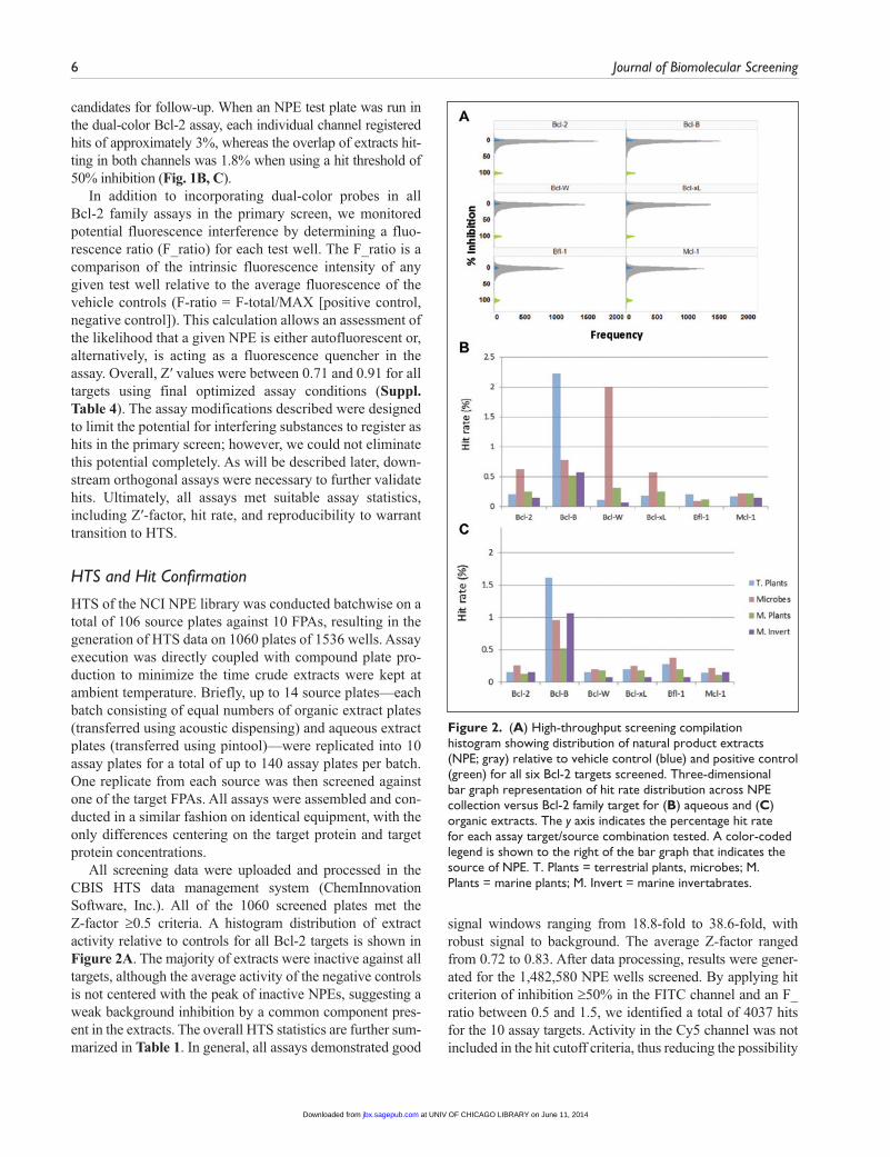
candidates for follow-up. When an NPE test plate was run in the dual-color Bcl-2 assay, each individual channel registered hits of approximately 3%, whereas the overlap of extracts hit-ting in both channels was 1.8% when using a hit threshold of 50% inhibition (Fig. 1B, C).
In addition to incorporating dual-color probes in all Bcl-2 family assays in the primary screen, we monitored potential fluorescence interference by determining a fluo-rescence ratio (F_ratio) for each test well. The F_ratio is a comparison of the intrinsic fluorescence intensity of any given test well relative to the average fluorescence of the vehicle controls (F-ratio = F-total/MAX [positive control, negative control]). This calculation allows an assessment of the likelihood that a given NPE is either autofluorescent or, alternatively, is acting as a fluorescence quencher in the assay. Overall, Z′ values were between 0.71 and 0.91 for all targets using final optimized assay conditions (Suppl. Table 4). The assay modifications described were designed to limit the potential for interfering substances to register as hits in the primary screen; however, we could not eliminate this potential completely. As will be described later, down-stream orthogonal assays were necessary to further validate hits. Ultimately, all assays met suitable assay statistics, including Z′-factor, hit rate, and reproducibility to warrant transition to HTS.
HTS and Hit Confirmation
HTS of the NCI NPE library was conducted batchwise on a total of 106 source plates against 10 FPAs, resulting in the generation of HTS data on 1060 plates of 1536 wells. Assay execution was directly coupled with compound plate pro-duction to minimize the time crude extracts were kept at ambient temperature. Briefly, up to 14 source plates—each batch consisting of equal numbers of organic extract plates (transferred using acoustic dispensing) and aqueous extract plates (transferred using pintool)—were replicated into 10 assay plates for a total of up to 140 assay plates per batch. One replicate from each source was then screened against one of the target FPAs. All assays were assembled and con-ducted in a similar fashion on identical equipment, with the only differences centering on the target protein and target protein concentrations.
All screening data were uploaded and processed in the CBIS HTS data management system (ChemInnovation Software, Inc.). All of the 1060 screened plates met the Z-factor ≥0.5 criteria. A histogram distribution of extract activity relative to controls for all Bcl-2 targets is shown in Figure 2A. The majority of extracts were inactive against all targets, although the average activity of the negative controls is not centered with the peak of inactive NPEs, suggesting a weak background inhibition by a common component pres-ent in the extracts. The overall HTS statistics are further sum-marized in Table 1. In general, all assays demonstrated good
signal windows ranging from 18.8-fold to 38.6-fold, with robust signal to background. The average Z-factor ranged from 0.72 to 0.83. After data processing, results were gener-ated for the 1,482,580 NPE wells screened. By applying hit criterion of inhibition ≥50% in the FITC channel and an F_ratio between 0.5 and 1.5, we identified a total of 4037 hits for the 10 assay targets. Activity in the Cy5 channel was not included in the hit cutoff criteria, thus reducing the possibility
Figure 2. (A) High-throughput screening compilation histogram showing distribution of natural product extracts (NPE; gray) relative to vehicle control (blue) and positive control (green) for all six Bcl-2 targets screened. Three-dimensional bar graph representation of hit rate distribution across NPE collection versus Bcl-2 family target for (B) aqueous and (C) organic extracts. The y axis indicates the percentage hit rate for each assay target/source combination tested. A color-coded legend is shown to the right of the bar graph that indicates the source of NPE. T. Plants = terrestrial plants, microbes; M. Plants = marine plants; M. Invert = marine invertabrates.
at UNIV OF CHICAGO LIBRARY on June 11, 2014jbx.sagepub.comDownloaded from
Hassig et al. 7
of missing false-negatives and allowing retrospective com-parison of dual-color versus single-color hit confirmation rates. By running all targets simultaneously, an initial assess-ment of target selectivity for extracts was possible. Many of the hits register as active on more than one target. As a result, the number of unique Bcl-2 family active hits was 2962. Of these hits, 2312 NPEs were active against only one Bcl-2 family target assay. Interestingly 524 NPEs hit more than one target but were restricted to members of the Bcl-2 family over the cIAP targets (not shown).
To identify trends, screening data were analyzed in sev-eral ways to assess hit distribution across the targets, extract solvents, and extract sources. Among all targets screened, Bcl-B has a significantly higher number of hits, approxi-mately three times higher than the next highest target, Bcl-W. Interestingly, among the antiapoptotic Bcl-2 family proteins, Bcl-B displays differences in its BH3 binding domain and binding partner preference relative to other family members. This may be related to its apparent pro-miscuity.22,23 With respect to extract solvent, the hit rate was higher for aqueous samples versus organic (ranging from 20% to 45% higher) for all targets except Bfl-1. For techni-cal reasons, aqueous samples were tested at twice the con-centration relative to organic samples, which may in part account for the relative hit distribution.
Figures 2B and 2C show the distribution of hit rate per extract source in aqueous and organic solvent, respectively. The majority of Bcl-B hits come from terrestrial plant extracts. As previously alluded, there may be a common plant-derived agent(s) (e.g., tannins) for which this target
(and/or assay) is particularly sensitive. On average, the hit rates of the microbial-derived extracts were higher than the other three types of extracts and appear disproportionally high relative to their representation in the library. Only one hit was found in aqueous extracts derived from marine plant sources for Bcl-XL and Bfl-1 targets. Interestingly, the aver-age hit rates from organic extractions of the microbial sources were higher than the other three sources, with the exception of Bcl-B. This was also generally true in the aqueous extracts and was particularly striking in the case of Bcl-W (nearly 2% hit rate). The direct explanation for these findings is unclear at this time.
Cherry-pick confirmation studies were conducted in qua-druplicate on all hits identified in the primary HTS. Data from confirmation assays on 2962 Bcl-2 family active NPEs were uploaded into the CBIS database to identify confirmed NPEs and determine the confirmation rate and other screen-ing statistics. Data for all test plates were of high quality (Z′ factor ≥0.5) with an average Z′ factor of 0.7. Final confirma-tion criteria were chosen to be more inclusive with respect to confirmed hits, specifically, using the criteria in which two of the four replicates satisfied inhibition of ≥40% and an F_ratio between 0.5 and 1.5 resulted in 994 unique NPE to follow up in orthogonal assays. Surprisingly, the confirmation rate for the dual positive extracts was not consistently higher than that for the FITC channel alone, with only Bcl-2 showing a higher confirmation rate and Bcl-W, Mcl-1, and Bfl-1 show-ing notably lower confirmation rates (Table 2). The confir-mation rate was not uniform across targets, with Bfl-1 and Mcl-1 having a lower hit confirmation rate (16% and 17%,
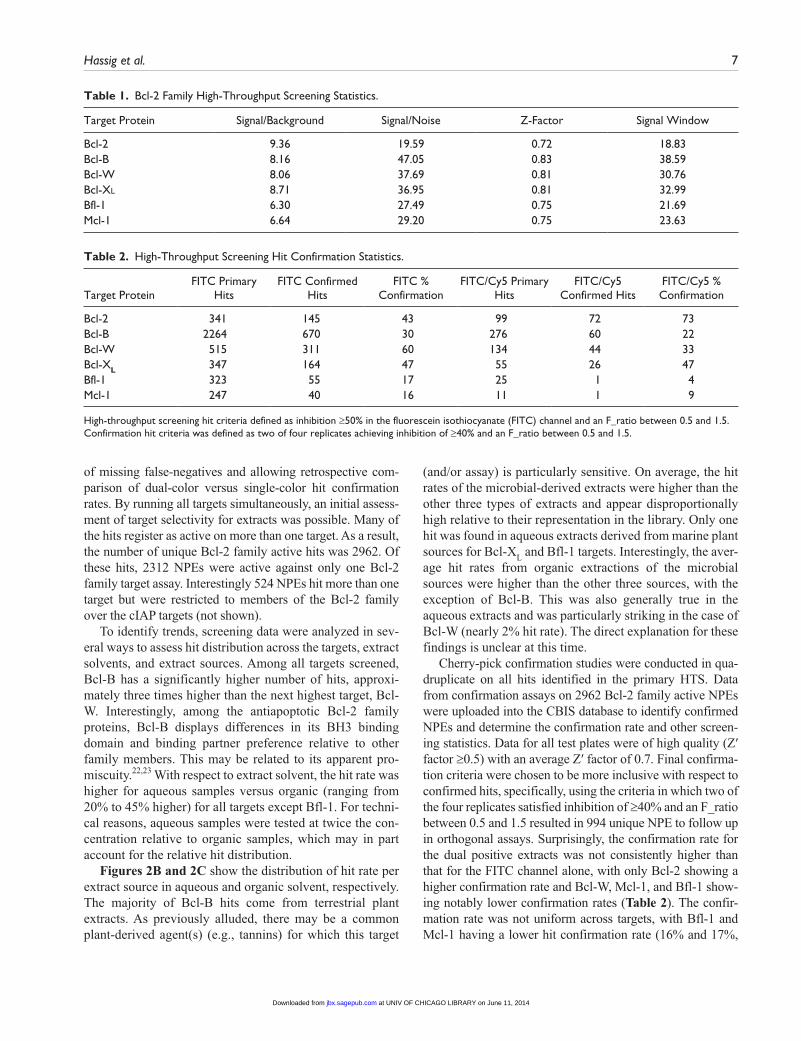
Table 1. Bcl-2 Family High-Throughput Screening Statistics.
Target Protein Signal/Background Signal/Noise Z-Factor Signal Window
Bcl-2 9.36 19.59 0.72 18.83Bcl-B 8.16 47.05 0.83 38.59Bcl-W 8.06 37.69 0.81 30.76Bcl-Xl 8.71 36.95 0.81 32.99Bfl-1 6.30 27.49 0.75 21.69Mcl-1 6.64 29.20 0.75 23.63
Table 2. High-Throughput Screening Hit Confirmation Statistics.
Target ProteinFITC Primary
HitsFITC Confirmed
HitsFITC %
ConfirmationFITC/Cy5 Primary
HitsFITC/Cy5
Confirmed HitsFITC/Cy5 % Confirmation
Bcl-2 341 145 43 99 72 73Bcl-B 2264 670 30 276 60 22Bcl-W 515 311 60 134 44 33Bcl-XL 347 164 47 55 26 47Bfl-1 323 55 17 25 1 4Mcl-1 247 40 16 11 1 9
High-throughput screening hit criteria defined as inhibition ≥50% in the fluorescein isothiocyanate (FITC) channel and an F_ratio between 0.5 and 1.5. Confirmation hit criteria was defined as two of four replicates achieving inhibition of ≥40% and an F_ratio between 0.5 and 1.5.
at UNIV OF CHICAGO LIBRARY on June 11, 2014jbx.sagepub.comDownloaded from

8 Journal of Biomolecular Screening
respectively) relative to other targets. This was also true for these targets when applying the dual positive confirmation criteria. Reasons for the very low confirmation rates of these targets/assays are not clear, although the signal to background for these two assays was somewhat lower than the other assays screened, potentially giving rise to a higher false- positive rate. Overall, the FITC hit confirmation rate ranged from 16% to 60% across the targets.
Cell-Based Orthogonal Assays
After confirming the activity of crude extracts in the FPAs, active samples were re–cherry-picked and tested in a func-tional cellular assay to further validate them as containing proapoptotic Bcl-2 family inhibitory activity. Inhibitors of the Bcl-2 family are reported to induce caspase-dependent cell death by liberating the proapoptotic BH3-only proteins (e.g., Bim, Bid, Bad) from their antiapoptotic partners, leading to mitochondrial outer membrane permeabilization (MOMP) via Bax and Bak. MOMP results in the release of cytochrome-c and other proapoptotic factors in the assem-bly of the apoptosome. This in turn activates caspase-9 and subsequent downstream executioner caspases (caspases-3 and -7).7,24 Therefore, we tested confirmed hits for their ability to activate caspase-3/7 in tumor cells. We simultane-ously monitored cell viability and caspase-3/7 activation using a single multiplex assay (ApoLive-Glo, Promega) to determine the ability of extracts to induce programmed cell death. Importantly, in addition to being an orthogonal cell-based assay, the caspase-3/7 activity was monitored using a luminescent readout, thereby providing a non–fluorescent-based measurement of extract bioactivity.
Previous studies have demonstrated that human leuke-mia and lymphoma-derived cancer cell lines overexpress a diversity of antiapoptotic Bcl-2 family members, which likely contributes to their resistance to apoptosis.25 As such, the lymphoma cell line RS11846 was used to evaluate NPE-induced apoptosis via interference with Bcl-2, Bcl-XL, Bcl-W, Mcl-1, and Bfl-1, because these cells have been shown to express detectable levels of these antiapoptotic genes.25 Caco-2 cells express higher levels of Bcl-B relative to other cell lines and were therefore selected as a second cell line to assess cellular activity against this target.
We confirmed the ability of the potent nanomolar Bcl-2 inhibitors, ABT-737 and ABT-263, to induce caspase-3/7 activity in RS11846 cells within 6 h of treatment (Suppl. Fig. 3A). As expected, ABT-737 and ABT-263 activated caspases much more potently than a less active enantiomer (ABT-ent). A similar level of caspase induction was observed in Caco-2 cells, albeit effects on cell viability remained modest at these time points (Suppl. Fig. 3B). Gossypol, a broad-spectrum micromolar inhibitor of Bcl-2 family proteins, displayed much less potent caspase activa-tion and modest effects on viability.
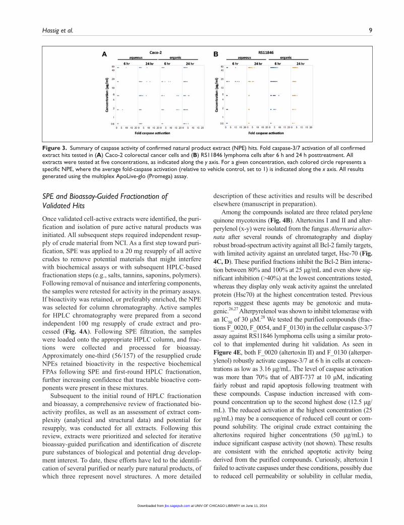
The highest NPE concentration tested was 50 µg/mL, which is approximately fivefold higher than the NPE concen-trations used in primary screening. All cell-based assays were performed in 384-well format, and confirmed hits were directly cherry-picked from intermediate plates in five-point concen-tration response in triplicate. Effects on viability in this assay using potent target selective inhibitors was modest at both the 6 h and 24 h time points. Moreover, relatively few NPEs tested showed significant effects on cell viability under the condi-tions tested. As a consequence, hit validation criteria focused on caspase activation wherein an extract registered as a hit if the average caspase induction (measured as an increase in luminescence) was greater than the average induction of the negative control plus three times the standard deviation of the sample and negative control. The range of caspase induction achieved by NPEs (2- to 10-fold) was generally less than that observed for 10 µM ABT-737 (15-fold induction) but was sim-ilar or superior to effects observed using gossypol and related analogs at ~20 µM final concentration. In fact, some NPEs actually demonstrated greater than 10-fold induction of cas-pase activity, even at concentrations below 10 µg/mL. A total of 167/994 NPEs met validation criteria, representing a hit validation rate of approximately 17%. This hit validation rate demonstrates a considerable level of enrichment given that only 1 of 128 (<1%) randomly selected NPEs was active in the caspase-3/7 assay (data not shown). Figure 3 shows the distri-bution of caspase-3/7 activation across both cell lines for NPEs meeting threshold criteria. Caspase activation was observed to similar extents in both Caco-2 (Fig. 3A) and RS11846 cells (Fig. 3B). Maximal caspase induction was not necessarily observed at the highest concentration tested; in fact, some of the most effective extracts induced maximal activation between 1.9 µg/mL and 5.6 µg/mL. Although the majority of NPEs induced modest caspase activation (2- to 5-fold), some induced caspases up to 15-fold. In general, caspase induction was more dramatic at 6 h versus 24 h, particularly for organic extracts. The decline in caspase activity at 24 h may be a reflec-tion of reduced cell health or extract stability at later time points.
The majority of cell-active NPEs were derived from organic extracts, possibly as a consequence of better cell per-meability properties intrinsic to these agents. Nonetheless, sev-eral potent actives were identified from aqueous sources. Terrestrial plant extracts represented the source with the most abundant cell-active samples, 108 in total. This was followed by microbial samples, with 34 total extracts. Both single-target and broad-spectrum active NPEs were repre-sented in the validated hit set. At this stage, validated hit extracts demonstrated reproducible activity in the biochem-ical assay as well as activity in the cellular caspase assay. The cell-based caspase-3/7 induction data—in conjunction with source extraction information and resupply availability—was used to guide the selection of extracts for bioassay-guided fractionation.
at UNIV OF CHICAGO LIBRARY on June 11, 2014jbx.sagepub.comDownloaded from
Hassig et al. 9
SPE and Bioassay-Guided Fractionation of Validated Hits
Once validated cell-active extracts were identified, the puri-fication and isolation of pure active natural products was initiated. All subsequent steps required independent resup-ply of crude material from NCI. As a first step toward puri-fication, SPE was applied to a 20 mg resupply of all active crudes to remove potential materials that might interfere with biochemical assays or with subsequent HPLC-based fractionation steps (e.g., salts, tannins, saponins, polymers). Following removal of nuisance and interfering components, the samples were retested for activity in the primary assays. If bioactivity was retained, or preferably enriched, the NPE was selected for column chromatography. Active samples for HPLC chromatography were prepared from a second independent 100 mg resupply of crude extract and pro-cessed (Fig. 4A). Following SPE filtration, the samples were loaded onto the appropriate HPLC column, and frac-tions were collected and processed for bioassay. Approximately one-third (56/157) of the resupplied crude NPEs retained bioactivity in the respective biochemical FPAs following SPE and first-round HPLC fractionation, further increasing confidence that tractable bioactive com-ponents were present in these mixtures.
Subsequent to the initial round of HPLC fractionation and bioassay, a comprehensive review of fractionated bio-activity profiles, as well as an assessment of extract com-plexity (analytical and structural data) and potential for resupply, was conducted for all extracts. Following this review, extracts were prioritized and selected for iterative bioassay-guided purification and identification of discrete pure substances of biological and potential drug develop-ment interest. To date, these efforts have led to the identifi-cation of several purified or nearly pure natural products, of which three represent novel structures. A more detailed
description of these activities and results will be described elsewhere (manuscript in preparation).
Among the compounds isolated are three related perylene quinone mycotoxins (Fig. 4B). Altertoxins I and II and alter-perylenol (x-y) were isolated from the fungus Alternaria alter-nata after several rounds of chromatography and display robust broad-spectrum activity against all Bcl-2 family targets, with limited activity against an unrelated target, Hsc-70 (Fig. 4C, D). These purified fractions inhibit the Bcl-2 Bim interac-tion between 80% and 100% at 25 µg/mL and even show sig-nificant inhibition (>40%) at the lowest concentrations tested, whereas they display only weak activity against the unrelated protein (Hsc70) at the highest concentration tested. Previous reports suggest these agents may be genotoxic and muta-genic.26,27 Alterpyrelenol was shown to inhibit telomerase with an IC50 of 30 µM.28 We tested the purified compounds (frac-tions F_0020, F_0054, and F_0130) in the cellular caspase-3/7 assay against RS11846 lymphoma cells using a similar proto-col to that implemented during hit validation. As seen in Figure 4E, both F_0020 (altertoxin II) and F_0130 (alterper-ylenol) robustly activate caspase-3/7 at 6 h in cells at concen-trations as low as 3.16 µg/mL. The level of caspase activation was more than 70% that of ABT-737 at 10 µM, indicating fairly robust and rapid apoptosis following treatment with these compounds. Caspase induction increased with com-pound concentration up to the second highest dose (12.5 µg/mL). The reduced activation at the highest concentration (25 µg/mL) may be a consequence of reduced cell count or com-pound solubility. The original crude extract containing the altertoxins required higher concentrations (50 µg/mL) to induce significant caspase activity (not shown). These results are consistent with the enriched apoptotic activity being derived from the purified compounds. Curiously, altertoxin I failed to activate caspases under these conditions, possibly due to reduced cell permeability or solubility in cellular media,
Figure 3. Summary of caspase activity of confirmed natural product extract (NPE) hits. Fold caspase-3/7 activation of all confirmed extract hits tested in (A) Caco-2 colorectal cancer cells and (B) RS11846 lymphoma cells after 6 h and 24 h posttreatment. All extracts were tested at five concentrations, as indicated along the y axis. For a given concentration, each colored circle represents a specific NPE, where the average fold-caspase activation (relative to vehicle control, set to 1) is indicated along the x axis. All results generated using the multiplex ApoLive-glo (Promega) assay.
at UNIV OF CHICAGO LIBRARY on June 11, 2014jbx.sagepub.comDownloaded from

10 Journal of Biomolecular Screening
although further studies are necessary to confirm these hypoth-eses. Caspase activation has not been reported previously for any of the altertoxin compounds. The altertoxins share some structural similarity to the known Bcl-2 family inhibitor gos-sypol, suggesting a potentially related mechanism of inhibi-tion. Like the altertoxins and gossypol, many of the samples isolated thus far contain polyphenolic substructures, likely reflecting a combination of the inherent sensitivity of the assays and/or targets to this class of compounds and the abun-dance of these compounds within the extract collection.
In summary, we have completed a parallel, multitarget HTS campaign against nearly 150,000 NPEs in high- density 1536-well format. The antiapoptosis Bcl-2 family proteins screened in this project represent oncology targets that garner significant clinical interest. A total of 994 NPEs were confirmed as active against one or more Bcl-2 family targets, with 17% of these also inducing caspase-3/7 activa-tion in tumor cells. Reconfirmation and orthogonal testing in cell-based assays enabled the identification of NPEs for bioassay-guided fractionation and isolation. However, the complex nature of crude extract mixtures and the potential
for isolating nuisance compounds make this screening approach challenging. Further studies are required to eluci-date the mechanisms of action of the pure natural products identified. Notwithstanding these limitations, our results demonstrate the feasibility of uHTS of natural product libraries to discover leads for therapeutically active natural product substances.
Acknowledgments
We thank Antimone Dewing for technical assistance with cellular assays and Drs. Barbara Mroczkowski, Shizuko Sei, Neal Green, and Bill Moore for scientific discussions.
Declaration of Conflicting Interests
The authors declared no potential conflicts of interest with respect to the research, authorship, and/or publication of this article.
Funding
The authors disclosed receipt of the following financial support for the research, authorship, and/or publication of this article: This project has been funded in whole or in part with federal funds from
Figure 4. (A) Standard workflow for first-round natural product extract (NPE) fractionation. A 100 mg resupply of crude NPE was processed using solid phase extraction (SPE), and precleaned material was further filtered using PTFE membrane prior to high-performance liquid chromatography fractionation into an initial 24 fractions. Samples of the crude NPE and all subsequent purification steps were retained and, all samples were tested in the fluorescence polarization (FP) assay. (B) Chemical structures of altertoxins and gossypol. (C) Fractions (F_0020, F_0054, F_0130) containing purified altertoxins were tested at three concentrations in Bcl-2 family FP assays, and percentage inhibition of fluorescein isothiocyanate (FITC)–Bim binding is indicated on the y axis. The F_ratios for all samples were within the acceptable range of 0.5 to 1.5. (D) Fractions (F_0020, F_0054, F_0130) containing purified altertoxins were tested at three concentrations in the FP counterscreen assay (Hsc70), and percentage inhibition of FITC-Bim binding is indicated on the y axis. (E) Activity of isolated altertoxin-containing fractions (F_0020, F_0054, F_0130) in cell-based caspase activation assay at concentrations indicated in RS11846 lymphoma cells. Caspase fold-activation (relative to ABT-737 at 10 µM = 100%) was determined at 6 h posttreatment.
at UNIV OF CHICAGO LIBRARY on June 11, 2014jbx.sagepub.comDownloaded from

Hassig et al. 11
the National Cancer Institute NExT Program, National Institutes of Health, under contract No. HHSN261200800001E. The content of this publication does not necessarily reflect the views or poli-cies of the Department of Health and Human Services, nor does mention of trade names, commercial products, or organizations imply endorsement by the U.S. government.
References
1. Newman, D. J.; Cragg, G. M. Natural Products as Sources of New Drugs over the 30 Years from 1981 to 2010. J. Nat. Prod. 2012, 75, 311–335.
2. Li, J. W.; Vederas, J. C. Drug Discovery and Natural Products: End of an Era or an Endless Frontier? Science 2009, 325, 161–165.
3. Henrich, C. J.; Beutler, J. A. Matching the Power of High Throughput Screening to the Chemical Diversity of Natural Products. Nat. Prod. Rep. 2013, 30, 1284–1298.
4. Leibowitz, B.; Yu, J. Mitochondrial Signaling in Cell Death via the Bcl-2 Family. Cancer Biol. Ther. 2010, 9, 417–422.
5. Adams, J. M.; Cory, S. The Bcl-2 Apoptotic Switch in Cancer Development and Therapy. Oncogene 2007, 26, 1324–1337.
6. Cleary, M. L.; Sklar, J. Nucleotide Sequence of a t(14;18) Chromosomal Breakpoint in Follicular Lymphoma and Demonstration of a Breakpoint-Cluster Region Near a Transcriptionally Active Locus on Chromosome 18. Proc. Natl. Acad. Sci. U. S. A. 1985, 82, 7439–7443.
7. Ni Chonghaile, T.; Letai, A. Mimicking the BH3 Domain to Kill Cancer Cells. Oncogene 2008, 27, Supplement 1, S149–S157.
8. Bodur, C.; Basaga, H. Bcl-2 Inhibitors: Emerging Drugs in Cancer Therapy. Curr. Med. Chem. 2012, 19, 1804–1820.
9. Aouacheria, A.; Brunet, F.; Gouy, M. Phylogenomics of Life-or-Death Switches in Multicellular Animals: Bcl-2, BH3-Only, and BNip Families of Apoptotic Regulators. Mol. Biol. Evol. 2005, 22, 2395–2416.
10. Lanave, C.; Santamaria, M.; Saccone, C. Comparative Genomics: The Evolutionary History of the Bcl-2 Family. Gene 2004, 333, 71–79.
11. Kitada, S.; Leone, M.; Sareth, S.; et al. Discovery, Characterization, and Structure-Activity Relationships Studies of Proapoptotic Polyphenols Targeting B-Cell Lymphocyte/Leukemia-2 Proteins. J. Med. Chem. 2003, 46, 4259–4264.
12. Davids, M. S.; Letai, A. ABT-199: Taking Dead Aim at BCL-2. Cancer Cell 2013, 23, 139–141.
13. Balakrishnan, K.; Gandhi, V. Bcl-2 Antagonists: A Proof of Concept for CLL Therapy. Invest. New Drugs 2013, 31, 1384–1394.
14. Vandenberg, C. J.; Cory, S. ABT-199, a New Bcl-2-Specific BH3 Mimetic, Has In Vivo Efficacy against Aggressive
Myc-Driven Mouse Lymphomas without Provoking Thrombocytopenia. Blood 2013, 121, 2285–2288.
15. Zhai, D.; Jin, C.; Satterthwait, A. C.; et al. Comparison of Chemical Inhibitors of Antiapoptotic Bcl-2-Family Proteins. Cell Death Differ. 2006, 13, 1419–1421.
16. Stack, M. E.; Prival, M. J. Mutagenicity of the Alternaria Metabolites Altertoxins I, II, and III. Appl. Environ. Microbiol. 1986, 52, 718–722.
17. Zhai, D.; Godoi, P.; Sergienko, E.; et al. High-Throughput Fluorescence Polarization Assay for Chemical Library Screening against Anti-Apoptotic Bcl-2 Family Member Bfl-1. J. Biomol. Screen. 2012, 17, 350–360.
18. Balunas, M. J.; Su, B.; Landini, S.; et al. Interference by Naturally Occurring Fatty Acids in a Noncellular Enzyme-Based Aromatase Bioassay. J. Nat. Prod. 2006, 69, 700–703.
19. Coan, K. E.; Ottl, J.; Klumpp, M. Non-stoichiometric inhi-bition in Biochemical High-Throughput Screening. Expert Opin. Drug Discov. 2011, 6, 405–417.
20. Grant, S. K.; Sklar, J. G.; Cummings, R. T. Development of Novel Assays for Proteolytic Enzymes Using Rhodamine-Based Fluorogenic Substrates. J. Biomol. Screen. 2002, 7, 531–540.
21. Turek-Etienne, T. C.; Lei, M.; Terracciano, J. S.; et al. Use of Red-Shifted Dyes in a Fluorescence Polarization AKT Kinase Assay for Detection of Biological Activity in Natural Product Extracts. J. Biomol. Screen. 2004, 9, 52–61.
22. Zhai, D.; Jin, C.; Huang, Z.; et al. Differential Regulation of Bax and Bak by Anti-Apoptotic Bcl-2 Family Proteins Bcl-B and Mcl-1. J. Biol. Chem. 2008, 283, 9580–9586.
23. Zhai, D.; Ke, N.; Zhang, H.; et al. Characterization of the Anti-Apoptotic Mechanism of Bcl-B. Biochem. J. 2003, 376(Pt 1), 229–236.
24. Leber, B.; Lin, J.; Andrews, D. W. Still Embedded Together Binding to Membranes Regulates Bcl-2 Protein Interactions. Oncogene 2010, 29, 5221–5230.
25. Placzek, W. J.; Wei, J.; Kitada, S.; et al. A Survey of the Anti-Apoptotic Bcl-2 Subfamily Expression in Cancer Types Provides a Platform to Predict the Efficacy of Bcl-2 Antagonists in Cancer Therapy. Cell Death Dis. 2010, 1, e40.
26. Fleck, S. C.; Burkhardt, B.; Pfeiffer, E.; et al. Alternaria Toxins: Altertoxin II Is a Much Stronger Mutagen and DNA Strand Breaking Mycotoxin Than Alternariol and Its Methyl Ether in Cultured Mammalian Cells. Toxicol. Lett. 2012, 214, 27–32.
27. Schwarz, C.; Tiessen, C.; Kreutzer, M.; et al. Characterization of a Genotoxic Impact Compound in Alternaria alternata Infested Rice as Altertoxin II. Arch. Toxicol. 2012, 86, 1911–1925.
28. Togashi, K.; Kakeya, H.; Morishita, M.; et al. Inhibition of Human Telomerase Activity by Alterperylenol. Oncol. Res. 1998, 10, 449–453.
at UNIV OF CHICAGO LIBRARY on June 11, 2014jbx.sagepub.comDownloaded from