Improving colour image segmentation on acute myelogenous leukaemia images using contrast enhancement...
6
Improving Colour Image Segmentation on Acute Myelogenous Leukaemia Images Using Contrast Enhancement Techniques Aimi Salihah, A.N. #1 , M.Y.Mashor #2 , Nor Hazlyna Harun #3 , Azian Azamimi Abdullah #4 , H.Rosline *5 # Electronic & Biomedical Intelligent Systems (EBItS) Research Group, School of Mechatronics Engineering, University Malaysia Perlis, 02600 Ulu Pauh, Perlis, Malaysia. 1 [email protected] * Department of Haematology, School of Medical Sciences, University Science Malaysia, Kubang Kerian, Kelantan, Malaysia. 5 [email protected] Abstract—Contrast enhancement and image segmentation play an important process in most medical image analysis tasks. One of the main tasks is the analyzing of white blood cells (WBC) where the WBC composition reveals important diagnostic information of a patient. This paper presents a two phase methodology in order to obtain a fully segmented abnormal white blood cell (blast) and nucleus in acute leukaemia images. In the first phase, the three contrast enhancement techniques which are partial contrast, bright stretching and dark stretching were used to improve the image quality. Contrast enhancement techniques enhanced the area of interest of acute leukaemia for easing the segmentation process. In the second phase, image segmentation based on HSI (Hue, Saturation, Intensity) colour space is proposed. The proposed technique helps to improve the image visibility and has successfully segmented the acute leukaemia images into two main components: blast and nucleus. The combination between contrast enhancements and image segmentation has good effect on improving the accuracy of segmentation. Hence, information gain from the resultant images would become useful for haematologists to further analysis the types of acute leukaemia. Keywords—Acute leukaemia, Image processing, Contrast enhancement, Colour segmentation, HSI colour space I. INTRODUCTION Leukaemia is a cancer of the marrow and blood. It is caused by an excessive production of immature white blood cells (blast) that replace the normal blood cells in the body. The two main types of acute leukaemia are Acute Lymphoblastic Leukaemia (ALL) and Acute Myelogenous Leukaemia (AML) [1]. Leukaemia can be cured if it is detected and treated at the early stage. Generally in leukaemia screening process, haematologists will first perform the complete blood count process [2]. If there are abnormalities in this count, a study of morphological bone marrow smear is done to confirm the present of leukaemia cells. By observing morphological features of acute leukaemia, a haematologist will classify the samples as either ALL or AML [3]. Currently, the microscopic investigation to identify the types and maturity of blood cells is performed manually by haematologists through visual identification under the light microscope. However, the manual recognition method has an error rate between 30% and 40% depending on the haematologists experience and difficulties to distinguish the types of leukaemia [4]. Contrast of the leukaemia image is one of the factors that may influence the accuracy of interpretation by haematologists. The resulting enhanced medical images will provide clearer and cleaner images for better and easier disease screening process by doctor. Thus, contrast enhancement at the pre-processing stage becomes the most important process for a successful feature extraction and diagnosis of leukaemia. Several previous studies had proved that contrast enhancement technique is capable to improve the medical image quality [5][6]. Harun et al. [7] stated that partial contrast technique has been shown to be good for enhancing the contrast for the three different types of acute leukaemia images which are normal, bright and dark images. Segmentation of an image refers to the separation of regions with similar characteristics. There are several segmentation techniques such as thresholding, edge detection, pixel clustering and region growing that have been combined in order to segment region of interest in medical images [8][9]. Nallaperumal and Krishnaveni [10] proposed a novel watershed segmentation algorithm to segment the nucleus from the surrounding cytoplasm of cervical cancer images. The proposed method converts the input RGB image into HSI image which contains the hue, saturation and intensity components. A segmentation technique based on extracting the region of interest from a larger image around thresholded cell nuclei was proposed by Katz [11]. The process of segmentation into cell or non-cell regions was carried out using canny edge detection followed by a circle identification algorithm. Some of the proposed segmentation techniques could be applied if the images showed high contrast background that is suitable to define the region of interest. The assumption of circular shape for cell segmentation is unsuitable in the case of abnormal blood cells [12]. In addition, many algorithms for segmentation have been developed for gray level images to extract the blood cell features. However, the actual screening 2010 IEEE EMBS Conference on Biomedical Engineering & Sciences (IECBES 2010), Kuala Lumpur, Malaysia, 30th November - 2nd December 2010. 978-1-4244-7600-8/10/$26.00 ©2010 IEEE 246
-
Upload
independent -
Category
Documents
-
view
0 -
download
0
Transcript of Improving colour image segmentation on acute myelogenous leukaemia images using contrast enhancement...

Improving Colour Image Segmentation on Acute Myelogenous Leukaemia Images Using Contrast
Enhancement Techniques Aimi Salihah, A.N. #1, M.Y.Mashor#2, Nor Hazlyna Harun#3, Azian Azamimi Abdullah#4, H.Rosline*5
#Electronic & Biomedical Intelligent Systems (EBItS) Research Group, School of Mechatronics Engineering, University Malaysia Perlis, 02600 Ulu Pauh, Perlis, Malaysia.
[email protected] *Department of Haematology, School of Medical Sciences, University Science Malaysia,
Kubang Kerian, Kelantan, Malaysia. [email protected]
Abstract—Contrast enhancement and image segmentation play an important process in most medical image analysis tasks. One of the main tasks is the analyzing of white blood cells (WBC) where the WBC composition reveals important diagnostic information of a patient. This paper presents a two phase methodology in order to obtain a fully segmented abnormal white blood cell (blast) and nucleus in acute leukaemia images. In the first phase, the three contrast enhancement techniques which are partial contrast, bright stretching and dark stretching were used to improve the image quality. Contrast enhancement techniques enhanced the area of interest of acute leukaemia for easing the segmentation process. In the second phase, image segmentation based on HSI (Hue, Saturation, Intensity) colour space is proposed. The proposed technique helps to improve the image visibility and has successfully segmented the acute leukaemia images into two main components: blast and nucleus. The combination between contrast enhancements and image segmentation has good effect on improving the accuracy of segmentation. Hence, information gain from the resultant images would become useful for haematologists to further analysis the types of acute leukaemia. Keywords—Acute leukaemia, Image processing, Contrast enhancement, Colour segmentation, HSI colour space
I. INTRODUCTION Leukaemia is a cancer of the marrow and blood. It is caused by an excessive production of immature white blood cells (blast) that replace the normal blood cells in the body. The two main types of acute leukaemia are Acute Lymphoblastic Leukaemia (ALL) and Acute Myelogenous Leukaemia (AML) [1]. Leukaemia can be cured if it is detected and treated at the early stage. Generally in leukaemia screening process, haematologists will first perform the complete blood count process [2]. If there are abnormalities in this count, a study of morphological bone marrow smear is done to confirm the present of leukaemia cells. By observing morphological features of acute leukaemia, a haematologist will classify the samples as either ALL or AML [3]. Currently, the microscopic investigation to identify the types and maturity of blood cells is performed manually by haematologists through visual identification under the light
microscope. However, the manual recognition method has an error rate between 30% and 40% depending on the haematologists experience and difficulties to distinguish the types of leukaemia [4]. Contrast of the leukaemia image is one of the factors that may influence the accuracy of interpretation by haematologists. The resulting enhanced medical images will provide clearer and cleaner images for better and easier disease screening process by doctor. Thus, contrast enhancement at the pre-processing stage becomes the most important process for a successful feature extraction and diagnosis of leukaemia. Several previous studies had proved that contrast enhancement technique is capable to improve the medical image quality [5][6]. Harun et al. [7] stated that partial contrast technique has been shown to be good for enhancing the contrast for the three different types of acute leukaemia images which are normal, bright and dark images. Segmentation of an image refers to the separation of regions with similar characteristics. There are several segmentation techniques such as thresholding, edge detection, pixel clustering and region growing that have been combined in order to segment region of interest in medical images [8][9]. Nallaperumal and Krishnaveni [10] proposed a novel watershed segmentation algorithm to segment the nucleus from the surrounding cytoplasm of cervical cancer images. The proposed method converts the input RGB image into HSI image which contains the hue, saturation and intensity components. A segmentation technique based on extracting the region of interest from a larger image around thresholded cell nuclei was proposed by Katz [11]. The process of segmentation into cell or non-cell regions was carried out using canny edge detection followed by a circle identification algorithm. Some of the proposed segmentation techniques could be applied if the images showed high contrast background that is suitable to define the region of interest. The assumption of circular shape for cell segmentation is unsuitable in the case of abnormal blood cells [12]. In addition, many algorithms for segmentation have been developed for gray level images to extract the blood cell features. However, the actual screening
2010 IEEE EMBS Conference on Biomedical Engineering & Sciences (IECBES 2010), Kuala Lumpur, Malaysia, 30th November - 2nd December 2010.
978-1-4244-7600-8/10/$26.00 ©2010 IEEE 246

process by haematologists is performed on stained slide where the leukaemia is detected based on colour and size of blast [3]. Thus, the current study proposes and investigates the performance of the combination between contrast enhancement techniques that have been applied to enhance area of interest in blood images and colour image segmentation using HSI colour space in order to obtain the fully segmented blast and nucleus of Acute Myelogenous Leukaemia (AML).
II. METHODOLOGY In this paper, the proposed methodologies are divided into two image processing parts. The first part is to enhance the area of interest by applying contrast enhancement techniques on acute leukaemia images. The second part will focus on colour image segmentation based on HSI colour space. Image segmentation is partitioned into two categories which consist of blast and nucleus segmentation. A typical acute leukaemia sample consists of three main regions: abnormal white blood cells (blast), red blood cell (RBC) and background areas. Nucleus and cytoplasm regions formed a blast and contain important information to be observed by haematologists. However, the RBC and background regions contain no information and can be eliminated from the image. The characteristic of features that can be obtained from blast observation are [3]: a) The size of blast, the shape of nucleus and the ratio of
size between nucleus and cytoplasm. b) The presence of Auer rode and multiple nucleoli inside
the nucleus are prominent in AML.
Fig. 1 The proposed image processing steps for blast and nucleus segmentation
The procedures used to develop the image processing for blast and nucleus segmentation are illustrated by the flow chart in Fig. 1. In order to obtain the segmented blast, the AML image will first be implemented with two contrast enhancement techniques which are partial contrast and bright stretching technique. Then, the area of interest will be segmented by applying image segmentation using HSI colour space. If the result from the segmentation is satisfied, the resultant image will be processed using N x N (N=7) median filter to produce a cleaner and clearer image. The filtering operation using median filter does not only smooth the image but can also preserve the edges. Region growing technique is used in order to obtain the region area in pixels for blast and nucleus. Finally, the colour for the segmented blast is retrieved from the partial contrast image. To obtain the segmented nucleus, the area of interest in AML image will first be processed using partial contrast and dark stretching technique. The subsequent steps for nucleus segmentation are the same as shown in Fig. 1. The details of contrast enhancement techniques and segmentation using HSI colour space are discussed in the following section.
III. CONTRAST ENHANCEMENT AND IDENTIFICATION OF WHITE BLOOD CELLS
The procedures used to develop the contrast enhancement techniques are as follows:
Step 1: Step 2: Step 3: Step 4:
Capture the acute leukaemia slide image at 40X magnification and save as bitmap (*.bmp) format with the 800 x 600 resolution. Apply the partial contrast technique on original AML image. Develop the intensity histogram from the resultant partial contrast image to obtain the threshold value, TH for area of interest. Apply the bright stretching or dark stretching technique by implementing the threshold and stretching value on the resultant partial contrast image.
Partial contrast is a linear mapping function that is used to increase the contrast and brightness level of the image. The technique is based on the original contrast and brightness level of the images to be adjusted [7]. By implementing this technique, the narrow range of data in original image will be stretched linearly to a new wider range of pixels data so that the dynamic range of the histogram (0-255) is fulfilled.
A. Contrast Enhancement for Blast Detection The combination between partial contrast and bright stretching techniques are used to enhance the brighter area (cytoplasm) for further blast segmentation process. Bright stretching technique is based on linear mapping function [7]. The bright stretching technique is applied on resultant partial contrast image in order to enhance cytoplasm area of the images.
Original AML image
Contrast enhancement techniques
Segmentation using HSI colour space
Image filtering
Region growing
Retrieve the colour based from the partial contrast image
Start
End
247
Consider the mapping function as given in Equation 1:
minmin
fk
qminfmaxf
minmaxkP +−−
−=
⎟⎟⎟⎟
⎠
⎞
⎜⎜⎜⎜
⎝
⎛
⎟⎠⎞
⎜⎝⎛
⎟⎠⎞⎜
⎝⎛
(1)
Pk: qk f max f min min max
: Colour level of the output pixel : Colour level of the input pixel : Maximum colour level values in the input image : Minimum colour level values in the input image : Desired minimum colour levels in the output image : Desired maximum colour levels in the output image
For bright stretching technique, Equation 1 can be further interpreted as follows:
( )⎪⎪⎩
⎪⎪⎨
⎧
>+⎥⎦⎤
⎢⎣⎡ −∗
−−
<∗=
THyxinforSFbSFbTH
THyxin
THyxinforSFbTH
yxin
yxout),()255(
255),(
),(),(
),(
(2) in(x,y) out(x,y) TH SFb
: Colour level for the input pixel : Colour level for the output pixel : Threshold value : Bright stretching factor
In leukaemia image, most pixels in cytoplasm region lies on the bright side of the histogram while pixels for the nucleus region lies on the dark side of the histogram. During the bright stretching process, the pixel values which are less than threshold value will be compressed while the pixel values which are greater than the threshold value will be stretched.
B. Contrast Enhancement for Nucleus Detection The combination between partial contrast and dark stretching techniques are used to enhance area of interest (nucleus) for further nucleus segmentation process. Dark stretching is a reverse process of bright stretching technique. Similar to the bright stretching, dark stretching technique is also involves linear mapping function. The dark stretching technique has been applied to the area of interest on the resultant partial contrast image in order to enhance the contrast between the nucleus region and background. Dark stretching is governed by Equation 3. During the dark stretching process, the pixel which is less than threshold value will be stretched while the pixel values which are greater than the threshold value will be compressed.
( )⎪⎪⎩
⎪⎪⎨
⎧
⎥⎦⎤
⎢⎣⎡ >+−∗
−−
<∗−
−
=THyxinforSFdSFd
THTHyxin
THyxinforSFdTH
THyxin
yxout),()255(
255),(
),(255
),(
),(
(3) in(x,y) out(x,y) TH SFd
: Colour level for the input pixel : Colour level for the output pixel : Threshold value : Dark stretching factor
IV. BLAST AND NUCLEUS SEGMENTATION To utilise the colour contents in an image, the colour image segmentation for both blast and nucleus are performed based on the HSI (Hue, Saturation, Intensity) colour space image. Hue is a colour attribute that describes a pure colour (pure yellow, orange or red), whereas saturation gives a measure of degree to which a pure colour is diluted by white colour [13]. According to the fact, the H component in HSI colour space contains most of the white blood cells information while the S component contains the structure information of the white blood cells nucleus [14]. Based on this information, colour segmentation based on S component image for both blast and nucleus is proposed. Thus, out of these three colour spaces, only the S component is used for transforming the RGB image. The procedures used to develop the segmentation techniques using HSI colour space are as follows: Step 1:
Step 2:
Step 3:
Extract the S component information from the enhanced RGB image. Develop the S component histogram (S-plot) from S component image to obtain threshold value. Apply the thresholding technique by implementing the threshold value on S component image.
The conversion from RGB image to S component image can be carried out according to the following equation:
( )BGRBGR
Saturation ,,min3
1++
−= (4)
By using the partition of S component, it can divide the image into high S region and low S region. The enhanced RGB image is converted into S component image so that the blast will appear as the brighter part of the image (high S region). A basic analysis method to analyze the colour distribution in an image can be obtained from the evaluation of the histogram. The histogram of S image (S-plot) will shows the distinct modes corresponding to each of the region in acute leukaemia which consist of abnormal WBC, RBC and background. Thus, S-plot will be utilized in order to select the threshold value for both blast and nucleus segmentation.
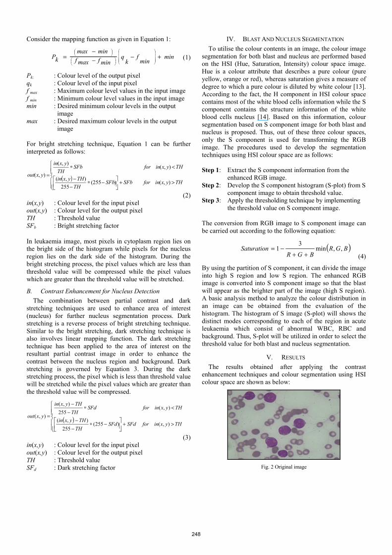
V. RESULTS The results obtained after applying the contrast enhancement techniques and colour segmentation using HSI colour space are shown as below:
Fig. 2 Original image
248
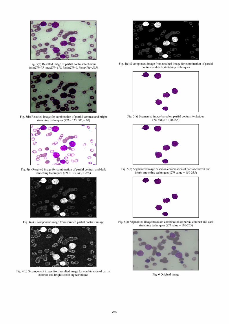
Fig. 3(a) Resulted image of partial contrast technique
(minTH=75, maxTH=175, NminTH=0, NmaxTH=255)
Fig. 3(b) Resulted image for combination of partial contrast and bright stretching techniques (TH = 125, SFb = 10)
Fig. 3(c) Resulted image for combination of partial contrast and dark
stretching techniques (TH = 125, SFd = 255)
Fig. 4(a) S component image from resulted partial contrast image
Fig. 4(b) S component image from resulted image for combination of partial
contrast and bright stretching techniques
Fig. 4(c) S component image from resulted image for combination of partial
contrast and dark stretching techniques
Fig. 5(a) Segmented image based on partial contrast technique
(TH value = 100-255)
Fig. 5(b) Segmented image based on combination of partial contrast and
bright stretching techniques (TH value = 150-255)
Fig. 5(c) Segmented image based on combination of partial contrast and dark
stretching techniques (TH value = 100-255)
Fig. 6 Original image
249

Fig. 7(a) Segmented image based on partial contrast technique
(TH value = 100-255)
Fig. 7(b) Segmented image based on combination of partial contrast and bright stretching techniques (TH value = 150-255)
Fig. 7(c) Segmented image based on combination of partial contrast and dark
stretching techniques (TH value = 100-255)
Fig. 8 Original image
Fig. 9(a) Segmented image based on partial contrast technique
(TH value = 100-255)
Fig. 9(b) Segmented image based on combination of partial contrast and bright stretching techniques (TH value = 150-255)
Fig. 9(c) Segmented image based on combination of partial contrast and dark
stretching techniques (TH value = 100-255)
VI. DISCUSSION In this study, the three image enhancement techniques have been applied on acute leukaemia images. Comparisons between the combinations of two image enhancement techniques have been made in order to recognize the significance of enhancement techniques on image segmentation. The qualities of segmented blast and nucleus were determined based on human visual interpretation by comparing the resultant segmented image with its original image. Fig. 2 shows the original image of Acute Myelogenous Leukaemia. The implementation result of partial contrast technique on AML image is shown in Fig. 3(a). The result shows that the contrast of the overall acute leukaemia images improved significantly. Thus, contrasts of the nucleus and the cytoplasm region have successfully been increased compare to the original image. The implementation result for the combination of partial contrast and bright stretching techniques on AML image is shown in Fig. 3(b). When applying the bright stretching technique, the RGB values of the cytoplasm pixels that lie at the bright side of the histogram will be stretched while the RGB values of nucleus pixels will be compressed. As a result for bright stretching technique, the contrast of cytoplasm area is increased and the shape of blast can be seen easier than before the implementation of the technique. The significant of the bright stretching technique on enhancing the contrast of cytoplasm can be easily seen after transforming the RGB image into S component image as shown in Fig. 4(b). Since the contrast of the blast component had been increased due to the application of contrast enhancement techniques, it will be easy to suppress the background from the image. The result of fully segmented blast obtained in Fig. 5(b) shows that the proposed combination methods between contrast
250

enhancement and segmentation based on HSI colour space produced good segmentation performance. The implementation result for the combination of partial contrast and dark stretching techniques on AML image is shown in Fig. 3(c). When applying the dark stretching technique, the RGB values of the nucleus pixels that lie at the dark side of the histogram will be stretched, for which result in increasing the contrast of nucleus region. The RGB values for cytoplasm and RBC pixels have been compressed and cause the decreased in contrast for both regions. Therefore, after applying the dark stretching technique, the morphological changes between the nucleus and cytoplasm component in the blast can be seen. As a result for dark stretching, shape of nucleus can easily be seen as shown in Fig. 4(c) which represents the S component image. The implementation of dark stretching technique will result in obtaining the segmented nucleus as shown in Fig. 5(c). Based on the resulted image, the segmentation process will result on the elimination of cytoplasm and RBC component in AML images. Fig. 5(a)-(c), show the final results of the segmentation process. Since the cytoplasm tends to have similar saturation value with the RBC, segmentation based on partial contrast image will result on elimination of cytoplasm area in blast as shown in Fig. 5(a). However, this situation does not appear on Fig. 5(b). Thus, segmentation based on partial contrast image is unsuitable to be applied for the purpose of obtaining fully segmented blast. Fig. 7(a)-(c) and Fig. 9(a)-(c) show the resultant segmented image for original image in Fig. 6 and Fig. 8, respectively. The fully segmented blast had been achieved by combining the partial contrast and bright stretching techniques while the fully segmented nucleus can be obtained by combining the partial contrast and dark stretching techniques.
VII. CONCLUSION The results show that the proposed combination methods between contrast enhancements and image segmentation work successfully in the segmentation of Acute Myelogenous Leukaemia images. Partial contrast algorithm can be used to improve the overall contrast of acute leukaemia images. The changes of contrast in cytoplasm and nucleus regions can easily be seen after applying the bright and dark stretching techniques, respectively. The fully segmented blast which consist of cytoplasm and nucleus region can be achieved by using the combination of partial contrast and bright stretching techniques. For nucleus segmentation, these can be achieved by using the combination of partial contrast and dark stretching techniques. Hence, the proposed segmentation technique based on HSI helps to improve the image visibility and has successfully segmented the acute leukaemia images while preserving significant features.
ACKNOWLEDGMENT The authors gratefully acknowledge and thank the team members of acute leukaemia research and University Science of Malaysia (USM). We also would like to acknowledge the Malaysian Government for providing the financial support under Fundamental Research Grant Scheme under the Ministry of Higher Education.
REFERENCES [1] G. C. C. Lim, “Overview of Cancer in Malaysia,” Japanese Journal of
Clinical Oncology, Department of Radiotherapy and Oncology, Hospital Kuala Lumpur, Kuala Lumpur, Malaysia, 2002.
[2] G. P. M. Priyankara, O. W Seneviratne, R. K. O. H Silva, W. V. D Soysa and C. R. D. Silva, “An Extensible Computer Vision Application for Blood Cell Recognition and Analysis”, 2006.
[3] S. Miwa, Atlas of Blood Cells. Tokyo, Japan: Bunkodo Co., Ltd., 1998. [4] C. Reta, L. Altamirano, J. A. Gonzalez, R. Diaz, and J. S. Guichard,
“Segmentation of Bone Marrow Cell Images for Morphological Classification of Acute Leukemia,” in Proceedings of the Twenty-Third International Florida Artificial Intelligence Research Society Conference (FLAIRS 2010), USA, 2010.
[5] N. Mustafa, N. A. Mat Isa, M. Y. Mashor, and N. H. Othman, “Colour Contrast Enhancement on Preselected Cervical Cell for ThinPrep® Images,” in Proceedings of the Third International Conference on International Information Hiding and Multimedia Signal Processing (IIH-MSP 2007), Kaoshing, Taiwan, 2007.
[6] G. D. Joshi and J. Sivaswamy, “Colour Retinal Image Enhancement based on Domain Knowledge,” in Sixth Indian Conference on Computer Vision, Graphics & Image Processing, India, 2008.
[7] N. H. Harun, N. R. Mokhtar, M. Y. Mashor, H. Rosline, H. Adilah, R. Adollah, N. Mustafa, N. F. M. Nasir, “Colour Image Enhancement Techniques Based on Partial Contrast and Contrast Stretching for Acute Leukemia Image,” in International Conference on Postgraduate Education (ICPE 2008), Penang, Malaysia, 2008.
[8] H. T. Madhloom, S. A. Kareem, H. Ariffin, A. A. Zaidan, H. O. Alanazi, and B. B. Zaidan, “An Automated White Blood Cell Nucleus Localization and Segmentation using Image Arithmetic and Automatic Threshold,” Journal of Applied Sciences. vol. 10, pp. 959-966, 2010.
[9] F. Sadeghian, Z. Seman, A. B. Ramli, B. H. A. Kahar, and M-Iqbal Saripan, “A Framework for White Blood Cell Segmentation in Microscopic Blood Images Using Digital Image Processing,” Biological Procedures Online. vol. 11, pp.196-206, 2009.
[10] K. Nallaperumal and K. Krishnaveni, “Watershed Segmentation of Cervical Images Using Multiscale Morphological Gradient and HSI Color Space,” International Journal of Imaging Science and Engineering (IJISE). vol. 2, pp. 212-216, 2008.
[11] A. R. J. Katz, “Image Analysis and Supervised Learning in the Automated Differentiation of White Blood Cells from Microscopic Images” Master’s thesis, Department of Computer Science, RMIT, Feb. 2000.
[12] N. Sinha and A. G. Ramakrishnan, “Blood Cell Segmentation using EM Algorithm,” in Proc. Indian Conf Vision, Graphics and Image Processing (ICVGIP-02), 2002.
[13] R. C. Gonzalez and R. E. Woods, Digital Image Processing, 3rd ed: Prentice Hall, 2007.
[14] J. Wu, P. Zeng, Y. Zhou, and C. Olivier, “A Novel Color Image Segmentation Method and Its Application to White Blood Cell Image Analysis,” in Proceedings of 8th IEEE ICSP, 2006.
251