Polarization of Migrating Monocytic Cells Is Independent of PI 3Kinase Activity
Upload
independentCategory
view
2download
0
Metallothionein Expression Is Suppressed in Primary Human
Hepatocellular Carcinomas and Is Mediated through
Inactivation of CCAAT/Enhancer Binding Protein A by
Phosphatidylinositol 3-Kinase Signaling Cascade
Jharna Datta,1Sarmila Majumder,
1Huban Kutay,
1Tasneem Motiwala,
1Wendy Frankel,
2
Robert Costa,4Hyuk C. Cha,
5Ormond A. MacDougald,
5Samson T. Jacob,
1,3
and Kalpana Ghoshal1
1Department of Molecular and Cellular Biochemistry, 2Department of Pathology, 3Comprehensive Cancer Center, College of Medicine, OhioState University, Columbus, Ohio; 4Department of Biochemistry and Molecular Genetics, University of Illinois at Chicago, Chicago, Illinois;and 5Department of Molecular and Integrative Physiology and Internal Medicine, University of Michigan Medical School,Ann Arbor, Michigan
Abstract
Reactive oxygen species (ROS) resulting from chronic inflam-mation cause liver injury leading to transformation ofregenerating hepatocytes. Metallothioneins (MT), induced athigh levels by oxidative stress, are potent scavengers of ROS.Here, we report that the levels of MT-1 and MT-2A are dras-tically reduced in primary human hepatocellular carcinomas(HCCs) and in diethylnitrosamine-induced liver tumors inmice, which is primarily due to transcriptional repression.Expression of the transcription factor, MTF-1, essential for MTexpression, and its target gene Zn-T1 that encodes the zinctransporter-1 was not significantly altered in HCCs. Inhibitorsof both phosphatidylinositol 3-kinase (PI3K) and its down-stream target AKT increased expression of MT genes in HCCcells but not in liver epithelial cells. Suppression of MT-1 andMT-2A by ectopic expression of the constitutively active PI3Kor AKT and their up-regulation by dominant-negative PI3Kor AKT mutant confirmed negative regulation of MT expres-sion by PI3K/AKT signaling pathway. Further, treatment ofcells with a specific inhibitor of glycogen synthase kinase-3(GSK-3), a downstream effector of PI3K/AKT, inhibited MTexpression specifically in HCC cells. Short interfering RNA–mediated depletion of CCAAT/enhancer binding protein A
(C/EBPA), a target of GSK-3, impeded MT expression, whichcould not be reversed by PI3K inhibitors. DNA binding activityof C/EBPA and its phosphorylation at T222 and T226 byGSK-3 are required for MT expression. MTF-1 and C/EBPA actin concert to increase MT-2A expression, which probablyexplains the high level of MT expression in the liver. This studyshows the role of PI3K/AKT signaling pathway and C/EBPAin regulation of MT expression in hepatocarcinogenesis.[Cancer Res 2007;67(6):2736–46]
Introduction
Hepatocellular carcinoma (HCC) is the fifth most prevalentcancer in the world and is most common form of liver cancer with5-year survival rate of only 7% (1–3). The high mortality is due tolate-stage detection of this cancer when most of the therapiesavailable are not effective (4, 5). Hepatitis B (HBV) or hepatitis C(HCV) viral infection that causes chronic liver disease andinflammation and cirrhosis plays an important role in etiology ofHCC. In United States, alcoholism is the most common cause ofHCC, whereas consumption of food contaminated with aflatoxin isthe major cause of HCC in Africa and Asia. Alcohol or aflatoxinintake further increases the risk for HCC in individuals exposed tochronic HCV (6). Metastasis from other cancerous organs thatinclude colorectal, pancreas, and breast and diabetes (3) alsocontribute to liver cancer. The disease is progressive and deathusually occurs within 10 months of initial diagnosis from cachexia,gastrointestinal bleeding, liver failure, or rupture of the tumor withmassive hemorrhage.Generation of excessive free radicals in tissues resulting from
chronic inflammation damages cellular macromolecules, includingDNA, which lead to mutation, apoptosis, hyperproliferation, andultimately cancer (7). Metallothioneins (MT), a group of stressresponse proteins induced at a high level by oxidative stress, areefficient scavengers of reactive oxygen species (ROS) and reactivenitrogen species (8–10). These are evolutionarily conserved,ubiquitously expressed, and cysteine-rich, heavy metal bindingproteins. Four isoforms of MT are arranged in tandem on mousechromosome 8 and on human chromosome 16 (11). Humangenome contains several MT-1 variants of which some arepseudogenes. MT-2A is the major MT isoform expressed in human.Expression of MT-1 and MT-2 are coordinately regulated in alltissues. An important role of MTs is to preserve homeostasis ofbiologically essential metals, such as zinc and copper, and toscavenge the toxic metals, such as cadmium and mercury (8, 9).The major function of MT is to scavenge free radicals by usingits unique metal-thiolate clusters that act as redox sensor andare rapidly oxidized by ROS releasing apo-MT and the metal ions(12, 13). Overproduction of MT at a high level selectively in theheart can protect mice from the cardiotoxic effects of the potentanticancer drug Adriamycin (13).MTs are expressed at high levels in the liver and are dramatically
induced by a variety of agents (8, 9), whereas these genes are notinduced in rodent hepatomas following exposure to heavy metals
Note: Supplementary data for this article are available at Cancer Research Online(http://cancerres.aacrjournals.org/).
This work is dedicated to the memory of Dr. Robert Costa.Requests for reprints: Samson T. Jacob, Department of Molecular and Cellular
Biochemistry, College of Medicine, The Ohio State University, Columbus, OH 43210.Phone: 614-688-5494; Fax: 614-688-5600; E-mail: [email protected] and KalpanaGhoshal, Department of Molecular and Cellular Biochemistry, College of Medicine,The Ohio State University, Columbus, OH 43210. Phone: 614-292-8865; Fax: 614-292-4118; E-mail: [email protected].
I2007 American Association for Cancer Research.doi:10.1158/0008-5472.CAN-06-4433
Cancer Res 2007; 67: (6). March 15, 2007 2736 www.aacrjournals.org
Research Article
Research. on February 11, 2016. © 2007 American Association for Cancercancerres.aacrjournals.org Downloaded from

(14). We explored the molecular mechanism of MT down-regulation in a transplanted rat hepatoma (15). This study showedthat methylation of CpG islands (CGI) located on MT-1 genepromoter played a causal role in silencing this gene, which wasdemethylated and reactivated by treatment of animals bearing thetumor with DNA hypomethylating agent 5-azacytidine.The primary objective of the present study was to determine
whether down-regulation of MTs is a common event in primaryHCCs and to elucidate the underlying mechanism. This studyshowed that MT is suppressed in primary HCC by transcriptionalrepression rather than by promoter methylation and it is mediatedthrough the activation of phosphatidylinositol 3-kinase (PI3K)/AKTpathway inducing dephosphorylation of the transcription factorCCAAT/enhancer binding protein (C/EBP) a.
Materials and Methods
Inhibitors of PI3K (LY294002 and Wortmannin) and glycogen synthasekinase-3 (GSK-3; LiCl) were from Sigma (St. Louis, MO). AKT inhibitor [1L-6-hydroxymethyl-chiro-inositol 2-(R)-2-O -methyl-3-O -octadecylcarbonate]and extracellular signal-regulated kinase 1 (Erk1) inhibitors (U0126 andPD98059) were from EMD Biosciences (La Jolla, CA). C/EBPahy and h-actinantibodies were obtained from Santa Cruz Biotechnology (Santa Cruz, CA),anti-Myc (9E10) and anti-Flag (M2) antibodies were from Sigma, and anti-HA antibody was from Covance (Philadelphia, PA). Phospho-T222/T226 andphospho–S21-C/EBPa were from Cell Signaling (Danvers, MA).
Plasmid construction.Mouse MTF-1 cDNA and rat C/EBPa cDNA were
PCR amplified with gene-specific primers with Accuprime polymerase
(Invitrogen, Carlsbad, CA) and cloned into pcDNA-Flagx3 (Sigma) andpcDNA3.1-HA, respectively (Invitrogen).
Human HCC samples and immunohistochemical analysis. HCC and
matching liver tissues were obtained from Ohio State University Tissue
Bank (Institutional Review Board protocol no. 2004C0081). Formalin-fixedtissue sections from cancerous liver were stained with MT (E9) antibody
(DAKO, Carpinteria, CA) and developed using avidin-biotin method (16).
Mouse primary tumors. Liver tumor was induced in C57B/6XSLJ miceby diethylnitrosamine following published protocol (17).
Real-time reverse transcription-PCR of MT-2A and MT-1. An aliquot
of cDNA synthesized from total RNA was subjected to real-time SYBR
Green PCR using gene-specific primers following published protocol(18, 19). Reverse transcription-PCR (RT-PCR) primers for human MT-2A,
MT-1, and C/EBPa are provided in the Supplementary Materials and
Methods.
Cell culture, treatment with inhibitors, and transfection. HumanHCC cell line (HepG2 and Hep3B) were cultured according to supplier’s
[American Type Culture Collection (ATCC), Manassas, VA] instruction.
Immortalized human liver derived THLE-2 cells (ATCC) were cultured inthe recommended medium (Cambrex, Walkersville, MD). These cells
maintain properties of hepatocytes and express liver-specific markers
(20). Exponentially growing cells were treated with 50 Amol/L LY290002,
DMSO (vehicle), or 100 nmol/L Wortmannin for 1 h followed by treatmentwith ZnSO4 (50 Amol/L) for 3 h. Similarly, cells were treated with 25 mmol/L
LiCl or NaCl for 1 h before zinc treatment. H293T cells were transfected with
constitutively active or dominant-negative PI3K, AKT expression vector, or
the empty vector using LipofectAMINE 2000 (Invitrogen) followingmanufacturer’s protocol. After 36 h, cells were harvested and the RNA
isolated was subjected to RT-PCR analysis.
Western blot analysis. The whole-cell extracts were immunoblotted with
different antibodies as described earlier (21, 22). The signal was developedwith enhanced chemiluminescence (GE Healthcare, Little Chalfont, Buck-
inghamshire, United Kingdom) after incubation with appropriate secondary
antibodies.Generation of Hep3B cells depleted of C/EBPA by short hairpin
RNA. A short hairpin RNA (shRNA) specific for C/EBPa (23) cloned into
pRetroSuper vector (24) and transfected Phoenix cells were used to
generate infectious viral particles using calcium phosphate precipitation
method. After 24 h, the cells were washed and allowed to grow. Theretroviral particles collected after 24 h from the culture medium were used
to infect Hep3B cells. Cells infected with C/EBPa-specific shRNA or
pRetroSuper were selected with puromycin (2 Ag/mL) and the expression of
C/EBPa and MT-2A was analyzed in the selected cells.Ectopic expression of C/EBPA mutants and their effect on MT
expression. The C/EBPa mutants (S193A and S193A+R290A) were a
generous gift of Dr. Nicholai Timchenko (Baylor College of Medicine,
Houston, TX; ref. 23). The generation of S21A and T222A/T226A mutants ofC/EBPa has been described earlier (21, 22). The mutants along with the
wild-type (WT) C/EBPa or empty vector were transfected to C/EBPa-depleted Hep3B cells using LipofectAMINE 2000. After 24 h, cells were split
into three plates, and 12 h later, they were either treated with ZnSO4
(50 Amol/L) or left untreated for an additional 3 h. Whole-cell extracts were
subjected to Western blot analysis with anti-C/EBPa antibody. Real-time
RT-PCR analyses were for MT-1 and MT-2A done with total RNA.Immunoprecipitation. C/EBPa-depleted Hep3B cells were transfected
with pcMTF-1–Flag and pcC/EBPa-HA separately or in combination. Cells
transfected with the empty vector were used as control. After 48 h, cells
were harvested and washed, and extracts made in TNN buffer weresubjected to immunoprecipitation with anti-Flag or anti-HA antibodies (25).
Immunocomplexes were pulled down with protein G-agarose and washed
with TNN buffer followed by Western blot analysis.
Chromatin immunoprecipitation. Formaldehyde cross-linked chroma-tin was prepared from THLE-2 cells following published protocol (26) and
subjected to immunoprecipitation with anti-C/EBPa, MTF-1 IgG, or normal
rabbit IgG. The immunocomplexes were pulled down by protein A-agaroseand DNA was purified (26). Input and pulled-down DNA were subjected
to PCR with primers specific for MT-2A, albumin, and glyceraldehyde-
3-phosphate dehydrogenase (GAPDH) promoters. The primer sequences are
provided in the Supplementary Data.Methylation analysis. Genomic DNA from liver and HCCs were
subjected to bisulfite conversion as described (26–28) followed by
amplification of the promoter region of each MT isoform using strand-
specific primers (see Supplementary Data). MT-2A PCR product was alsosubjected to sequencing using dideoxy termination kit (26).
Results
Expression of MTs is significantly reduced in primary HCCsof both human and mouse origin. We have shown previouslythat MT genes are suppressed in a transplanted rodent tumor (15).To investigate whether similar down-regulation of MT expressionalso occurs in primary human HCC, immunohistochemical analysisof 25 paraffin-embedded HCC samples was done with anti-MTantibody. A representative immunohistochemical profile is shownin Fig. 1A . Tumor cells were devoid of MT, whereas adjacent livertissues expressed MT abundantly. Quantitative analysis showedcomparable levels of MT in most liver samples (Table 1). MTlevel decreased significantly in all 25 HCC samples compared withthe adjacent normal liver tissues (Table 1). MT expression in 18samples was 0% to 20% of that observed in the matching livertissues, and the expression in the remaining eight tumors was 30%to 50% of that in the matching control livers.To elucidate the underlying mechanism of MT down-regulation
in HCCs, we investigated whether reduced MT protein level inHCCs correlated with the RNA level of MT isoforms. RT-PCRanalysis with isoform-specific primers showed significant decreasein MT-2A and all MT-1 isoforms (A, E, G, H, and X) in HCCs (T)compared with the matching normal livers (N; SupplementaryFig. S1). Real-time RT-PCR analysis with primers specific for MT-2Aor common to all isoforms of MT-1 showed significant decrease inMT-2A (P = 0.0006) as well as MT-1 (P = 0.001) mRNA levels in thetumors compared with the matching livers (Fig. 1B). These data
PI3K/AKT Negatively Regulates MT Expression in HCC
www.aacrjournals.org 2737 Cancer Res 2007; 67: (6). March 15, 2007
Research. on February 11, 2016. © 2007 American Association for Cancercancerres.aacrjournals.org Downloaded from

suggest that down-regulation of MT level in the liver tumors isprobably due to reduced gene expression.To examine whether down-regulation of MT is a common
molecular event in the etiology of HCCs, we used diethylnitros-amine-induced hepatocarcinogenesis in mice as a model system.These mice develop liver tumors after 30 weeks of diethylnitros-amine injection (see Materials and Methods for details). Real-timeRT-PCR analysis showed significant suppression (92%) of MT-1expression in all four tumors compared with the three age-matchedcontrol livers (Fig. 1C), which is consistent with the data obtainedwith primary human HCCs (Fig. 1B).Expression of the key transcription factor, MTF-1, or one of
its target genes, Zn-T1, is not significantly altered in humanprimary HCCs. MTF-1 is the transcription factor required for bothbasal and heavy metal-induced expression of MT genes (8, 29). Thisubiquitously expressed transcription factor binds to the metalresponse elements (MRE) on the MT promoters and activatesexpression of these genes. To examine whether decreased MT levelsin HCCs are due to reduced expression of MTF-1 in primary HCCs,we did real-time RT-PCR analysis. The results showed that MTF-1
level was not significantly altered (P = 0.075) in HCCs relative tothe matching normal tissues (Fig. 1D).To rule out the possibility that the transactivation potential of
MTF-1 is compromised in HCCs, we measured mRNA level of zinctransporter-1 (Zn-T1), another MTF-1 target gene (30). Real-timeRT-PCR analysis showed that the expression of Zn-T1 in tumorswas not significantly different from the liver tissues (Fig. 1D). Thus,it is unlikely that MT down-regulation in HCCs is due toinactivation of MTF-1.Inhibitors of PI3K and AKT up-regulate both basal and zinc-
induced expression of MT-2A and MT-1 in HCC. PI3K/AKTsignaling pathway, which regulates cell growth and cell survival,is activated in different forms of cancer, including HCC (31).We tested the possibility that the activation of PI3K/AKT pathwayin HCCs may play a key role in down-regulation of MT expres-sion. To address this issue, we compared the effect of LY294002,a specific inhibitor of PI3K, on MT expression in human liver-derived epithelial cell line THLE-2 and HCC cell line Hep3B. Real-time RT-PCR analysis showed 12- to 13-fold increase in basalexpression of MT-2A and MT-1 genes in the inhibitor-treated
Figure 1. A, immunohistochemicalanalysis shows dramatic decrease of MTprotein level in HCCs. Formalin-fixedtissue sections were incubated withanti-MT mouse monoclonal antibodyfollowed by horseradish peroxidase–conjugated antimouse IgG. The color wasdeveloped by avidin-biotin method. Thearrow indicates the cancerous region ofthe tissue section and the adjacent areais the normal liver tissue. B, real-timePCR analysis showed significant down-regulation of MT-2A and MT-1 expressionin human HCC compared with matchingnormal liver tissues. cDNA (f100 ng) wassubjected to real-time RT-PCR withMT-2A–specific primers and primerscommon to all isoforms of MT-1. 18S rRNAwas amplified from the same cDNA(f1 ng), and copy numbers of MT-2A andMT-1 were normalized to that of 18SrRNA. C, MT-1 expression is dramaticallydecreased in diethylnitrosamine-inducedliver tumors in mice. cDNAs from tumorsand age-matched control livers weresubjected to real-time RT-PCR withprimers specific for MT-1 and h-actin.Each sample was assayed in triplicate.Columns, mean of three values; bars, SD.D, mRNA levels of MTF-1 and Zn-T1 arenot significantly altered between the tumorand normal tissues. Total RNA isolatedfrom 12 HCCs and matching livers weresubjected to real-time RT-PCR withMTF-1– and Zn-T1–specific primers andnormalized to 18S rRNA. Columns, meanof three values; bars, SD.
Cancer Research
Cancer Res 2007; 67: (6). March 15, 2007 2738 www.aacrjournals.org
Research. on February 11, 2016. © 2007 American Association for Cancercancerres.aacrjournals.org Downloaded from

Hep3B cells (Fig. 2A), suggesting that PI3K negatively regulatestheir expression. As anticipated, zinc induced expression of bothMT-2A (f53-fold) and MT-1 (f50-fold), which was further up-regulated (f6-fold and f4-fold, respectively) in cells pretreatedwith LY294002 (Fig. 2A). In contrast, LY294002 exerted minimaleffect on both basal and zinc-induced MT-2A and MT-1 expressionin a human liver-derived epithelial cell line THLE-2 (Fig. 2A),supporting the notion that higher activity of PI3K indeed down-regulates MT expression in HCC cells. The higher level of MT-2Aand MT-1 mRNA in THLE-2 cells (liver epithelial) comparedwith Hep3B (HCC) cells concurs with the observation in primaryliver tumors (Fig. 1B and C). Treatment of THLE-2 cells withzinc further stimulated expression of MT-2A and MT-1 f2.4- andf3.8-fold, respectively (Fig. 2A). The relatively lower level ofstimulation of MT expression in these cells compared with thatin Hep3B cells is probably due to their higher basal expressionin THLE-2 cells. LY294002 exerted similar effect on the expres-sion of MT genes in other HCC cell lines, such as HepG2 (human),Hepa (mouse), and H4 (rat; data not shown). We also testedanother potent PI3K inhibitor, Wortmannin, on MT expressionin HCC and liver epithelial cells. Like LY294002, Wortmannintreatment increased MT-2A and MT-1 levels 7.8- and 2.6-fold,respectively, in Hep3B cells without any significant effect on THLE-2 cells (Fig. 2B). These results suggest that relatively high PI3Kactivity suppresses MT expression specifically in hepatocarcinomacells.AKT is the immediate downstream effector of PI3K. To address
its role on MT expression, cells were treated with a specificinhibitor of AKT [1L-6-hydroxymethyl-chiro-inositol 2-(R)-2-O-methyl-3-O -octadecylcarbonate]. Real-time RT-PCR analysisshowed that the inhibitor significantly up-regulated both basal(f10-fold) and zinc-induced (f9.6-fold) MT-2A mRNA level inHep3B cells (Fig. 2C). Similarly, AKT inhibitor induced basal andzinc-stimulated expression of MT-1 f5- and f5.7-fold, respec-tively (Fig. 2C). Comparatively lesser stimulation of MT-1 by PI3Kand AKT inhibitors is probably due to their differential effects on
MT-1 variants. AKT inhibitor did not have any significant effect onMT induction in THLE-2 cells (Fig. 2D). Taken together, theseresults indicate that the activation of PI3K/AKT signaling cascadeleads to down-regulation of MT expression in HCC cells.Constitutively active and dominant-negative mutants of
PI3K and AKT reciprocally regulate MT gene expression. Toconfirm the role of PI3K/AKT in MT gene regulation, we expressedconstitutively active (P110* and AKT*) and dominant-negative(kinase dead) mutants (P110*KR and AKTKR; ref. 32) separately inH293T cells. We used H293T cells, as detectable expression of theectopic kinases was achieved in these cells. Western blot analysisshowed that the levels of both PI3K and AKT variants werecomparable (Fig. 3A, top). The size of the constitutively active AKTprotein was higher than the dominant-negative mutant due to itsmyristoylation (32). Because the basal expression of MT is very lowin H293T cells, their zinc-induced expression was analyzed. Theresults showed that both constitutively active PI3K and AKTimpeded MT-2A expression by f54% and f60%, respectively,compared with the vector-transfected cells (Fig. 3A). The inhibitoryeffects of P110* and AKT* on MT-1 mRNA level were comparativelyless prominent (28% and 21%, respectively) than those observed onMT-2A expression. In contrast, MT-2A mRNA level increased 83-and 43-fold in cells expressing dominant-negative mutants of PI3Kand AKT, respectively (Fig. 3A). MT-1 expression also increased4- and 7-fold, respectively, when KR mutants of P13K and AKT wereectopically expressed (Fig. 3A, bottom). Relatively low level of MT-1induction might be due to differential effect of PI3K/AKT ondifferent MT isoforms. These results further confirm that MT-2Aand MT-1 genes are subjected to negative regulation by PI3K/AKTsignaling pathway.Depletion of C/EBPA, a downstream target of PI3K/AKT,
reduces MT expression. We entertained the possibility thatC/EBPa may be one of the transactivators involved in MTexpression in the liver for the following reasons. First, it is a majortranscription factor that negatively regulates hepatocyte prolifer-ation by controlling cell cycle progression and maintains terminal
Table 1. Immunohistochemical analysis of 25 human primary HCC samples shows down-regulation of MT expression in thetumors compared with the adjacent normal liver tissue (control)
Case no. MT expression (%) Case no. MT expression (%)
Tumor Control Tumor Control
1 10 90 14 0 60
2 5 100 15 30 1003 5 100 16 0 100
4 5 90 17 30 90
5 0 100 18 50 100
6 40 100 19 40 1007 15 100 20 20 100
8 0 100 21 15 80
9 0 80 22 0 10010 30 100 23 30 100
11 40 100 24 0 100
12 40 100 25 0 100
13 0 100
NOTE: MT expression was abolished in 10 of 25 cases. Intensity in the surrounding liver tissues was comparable among most of the samples and was
assigned a value of 100%, and intensity of MT staining in HCCs was compared with the controls.
PI3K/AKT Negatively Regulates MT Expression in HCC
www.aacrjournals.org 2739 Cancer Res 2007; 67: (6). March 15, 2007
Research. on February 11, 2016. © 2007 American Association for Cancercancerres.aacrjournals.org Downloaded from

differentiation state ( for review, see ref. 33). Second, its functionis compromised in different rodent and human malignancies dueto down-regulation of its expression (34, 35). Third, its activity isnegatively regulated in HCC cells by PI3K/AKT signaling (23).Fourth, both MT-2A and MT-1 promoters harbor multiple C/EBPcognate sites (analyzed by TRANSFAC database).As a first step to determine whether the PI3K-mediated
inactivation of C/EBPa results in down-regulation of MT genes inHCCs, we depleted C/EBPa from Hep3B cells by RNA interference.Hep3B cells expressing C/EBPa-specific shRNA were generatedusing a retroviral vector (23). C/EBPa RNA was reduced by 88% inpuromycin-resistant pool of cells infected with pRetroSuperharboring C/EBPa-shRNA compared with the cells infected withthe vector alone (Fig. 3B, top). Two isoforms of C/EBPa (42 and30 kDa) that arise due to usage of alternate ATG codon areexpressed in Hep3B cells (Fig. 3B). The level of both polypeptides
was significantly reduced (f70%) in depleted cells (sh) comparedwith the vector-infected cells or parental Hep3B cells. Protein levelsof other C/EBP isoforms (h and y) and MTF-1 were unaltered inC/EBPa-depleted cells (Fig. 3B), showing its specific depletion byshRNA. Real-time RT-PCR analysis showed that the basal MT-2Aand MT-1 RNA levels were reduced by 52% and 55%, respectively,in C/EBPa-depleted cells compared with the control cells (Fig. 3B,bottom). Zinc-induced expression of MT-2A and MT-1 was alsodecreased by 48% and 74%, respectively, in C/EBPa-depleted cells(Fig. 3B, bottom). These results were reproducible in differentbatches of cells. These results indicate that C/EBPa is one of thekey transcription factors involved in regulating MT expression inhuman HCC cells.C/EBPA is the target of PI3K/AKT signaling to the MT
promoter. Next, we investigated whether the observed decrease inMT level in HCCs was due to depletion or inactivation of C/EBPa.
Figure 2. A, specific inhibitor of PI3K(LY294002) activates both basal andzinc-induced expression of MT-2A/MT-1 inHCC (Hep3B) cells but not in normal liverepithelial (THLE-2) cells. Cells weretreated for 4 h with DMSO (C) or 50 Amol/LLY294002 (LY ) for 1 h followed by ZnSO4
treatment (Zn ; 50 Amol/L) for another 3 h.Control cells were treated with the vehicle(DMSO). DNase I–treated RNA wassubjected to real-time RT-PCR. Eachsample was assayed in triplicate.Columns, mean of three independentexperiments; bars, SD. B, Wortmanninincreased expression of MT genes inHep3B cells but not in THLE-2 cells.Cells were treated with 100 nmol/L ofWortmannin (Wort. ) for 3 h followed byreal-time RT-PCR analysis. Each samplewas assayed in triplicate. Columns, meanof three independent experiments; bars,SD. C and D, treatment of cells with AKTinhibitor (AKTI ) increases expression ofMT genes in Hep3B cells but not inTHLE-2 cells. Cells were treated with20 Amol/L of the inhibitor for 1 h followedby ZnSO4 (50 Amol/L) treatment for 3 h.Control cells were treated with the vehicle(DMSO). DNase I–treated RNA wassubjected to real-time RT-PCR. Eachsample was assayed in triplicate.Columns, mean of three independentexperiments; bars, SD.
Cancer Research
Cancer Res 2007; 67: (6). March 15, 2007 2740 www.aacrjournals.org
Research. on February 11, 2016. © 2007 American Association for Cancercancerres.aacrjournals.org Downloaded from
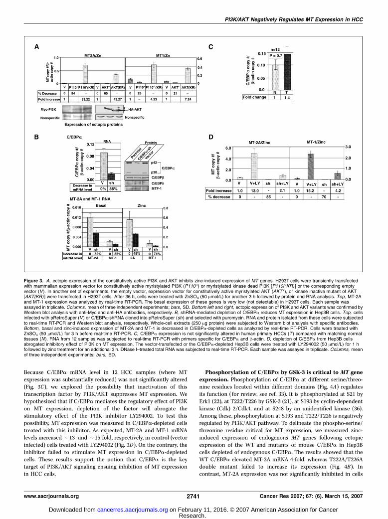
Because C/EBPa mRNA level in 12 HCC samples (where MTexpression was substantially reduced) was not significantly altered(Fig. 3C), we explored the possibility that inactivation of thistranscription factor by PI3K/AKT suppresses MT expression. Wehypothesized that if C/EBPa mediates the regulatory effect of PI3Kon MT expression, depletion of the factor will abrogate thestimulatory effect of the PI3K inhibitor LY294002. To test thispossibility, MT expression was measured in C/EBPa-depleted cellstreated with this inhibitor. As expected, MT-2A and MT-1 mRNAlevels increasedf13- andf15-fold, respectively, in control (vectorinfected) cells treated with LY294002 (Fig. 3D). On the contrary, theinhibitor failed to stimulate MT expression in C/EBPa-depletedcells. These results support the notion that C/EBPa is the keytarget of PI3K/AKT signaling ensuing inhibition of MT expressionin HCC cells.
Phosphorylation of C/EBPA by GSK-3 is critical to MT geneexpression. Phosphorylation of C/EBPa at different serine/threo-nine residues located within different domains (Fig. 4A) regulatesits function ( for review, see ref. 33). It is phosphorylated at S21 byErk1 (22), at T222/T226 by GSK-3 (21), at S193 by cyclin-dependentkinase (Cdk) 2/Cdk4, and at S248 by an unidentified kinase (36).Among these, phosphorylation at S193 and T222/T226 is negativelyregulated by PI3K/AKT pathway. To delineate the phospho-serine/threonine residue critical for MT expression, we measured zinc-induced expression of endogenous MT genes following ectopicexpression of the WT and mutants of mouse C/EBPa in Hep3Bcells depleted of endogenous C/EBPa. The results showed that theWT C/EBPa elevated MT-2A mRNA 4-fold, whereas T222A/T226Adouble mutant failed to increase its expression (Fig. 4B). Incontrast, MT-2A expression was not significantly inhibited in cells
Figure 3. A, ectopic expression of the constitutively active PI3K and AKT inhibits zinc-induced expression of MT genes. H293T cells were transiently transfectedwith mammalian expression vector for constitutively active myristylated PI3K (P110* ) or myristylated kinase dead PI3K [P110(*KR) ] or the corresponding emptyvector (V). In another set of experiments, the empty vector, expression vector for constitutively active myristylated AKT (AKT* ), or kinase inactive mutant of AKT[AKT(KR) ] were transfected in H293T cells. After 36 h, cells were treated with ZnSO4 (50 Amol/L) for another 3 h followed by protein and RNA analysis. Top, MT-2Aand MT-1 expression was analyzed by real-time RT-PCR. The basal expression of these genes is very low (not detectable) in H293T cells. Each sample wasassayed in triplicate. Columns, mean of three independent experiments; bars, SD. Bottom left and right, ectopic expression of PI3K and AKT variants was confirmed byWestern blot analysis with anti-Myc and anti-HA antibodies, respectively. B, shRNA-mediated depletion of C/EBPa reduces MT expression in Hep3B cells. Top, cellsinfected with pRetroSuper (V ) or C/EBPa-shRNA cloned into pRetroSuper (sh ) and selected with puromycin. RNA and protein isolated from these cells were subjectedto real-time RT-PCR and Western blot analysis, respectively. Whole-cell extracts (250 Ag protein) were subjected to Western blot analysis with specific antibodies.Bottom, basal and zinc-induced expression of MT-2A and MT-1 is decreased in C/EBPa-depleted cells as analyzed by real-time RT-PCR. Cells were treated withZnSO4 (50 Amol/L) for 3 h before real-time RT-PCR. C, C/EBPa expression is not significantly altered in human primary HCCs (T ) compared with matching normaltissues (N ). RNA from 12 samples was subjected to real-time RT-PCR with primers specific for C/EBPa and h-actin. D, depletion of C/EBPa from Hep3B cellsabrogated inhibitory effect of PI3K on MT expression. The vector-transfected or the C/EBPa-depleted Hep3B cells were treated with LY294002 (50 Amol/L) for 1 hfollowed by zinc treatment for an additional 3 h. DNase I–treated total RNA was subjected to real-time RT-PCR. Each sample was assayed in triplicate. Columns, meanof three independent experiments; bars, SD.
PI3K/AKT Negatively Regulates MT Expression in HCC
www.aacrjournals.org 2741 Cancer Res 2007; 67: (6). March 15, 2007
Research. on February 11, 2016. © 2007 American Association for Cancercancerres.aacrjournals.org Downloaded from

expressing S21A, which corroborated with the inability of Erk1inhibitors to affect MT expression in Hep3B cells (data not shown).S193A mutant that inhibits its interaction with Cdk2 and Brm(23, 36) did not alter MT-2A expression (Fig. 4C). It is, therefore,unlikely that recruitment of these proteins by CEBPa is involved inMT expression. T222A/T226A mutant also failed to stimulate basalMT-2A expression (data not shown). Western blot analysis showedthat the expression of the WT and mutant proteins wascomparable (Fig. 4D). These results show that phosphorylation atT222/T226 by GSK-3 is required for MT gene activation.To confirm that GSK-3 is indeed involved in MT expression, we
treated Hep3B cells with LiCl, a specific inhibitor of this kinase.Real-time RT-PCR analysis showed that inactivation of GSK-3significantly inhibited both basal and zinc-induced MT-2A and MT-1 levels by 74% and 48%, respectively (Fig. 5A). Western blotanalysis with phosphorylated specific antibodies showed thattreatment of cells with LiCl dramatically reduced (f90%) the levelof phospho-T222/T226 C/EBPa (both p42 and p30) withoutaltering C/EBPa protein level (Fig. 5B). A 2-fold increase inphospho-T222/T226 level in cells treated with PI3K inhibitor(Fig. 5B) substantiates the conclusion that PI3K/AKT negativelyregulates activity of C/EBPa by inhibiting GSK-3 (31). How-ever, zinc treatment did not significantly change C/EBPa proteinlevel or its phosphorylation status at either site (Fig. 5B). Asexpected, phosphorylation of C/EBPa at S21, catalyzed by Erk1(22), was not significantly affected by inhibitors of GSK-3 or PI3K(Fig. 5B).
DNA binding activity of C/EBPA is essential for MT geneactivation. C/EBPa is a unique transcription factor that regulatesgene expression by directly binding to cognate cis elements as wellas indirectly by interacting with other transcription factors orchromatin-modifying factors (different domains of C/EBPa aredepicted in Fig. 4A ; ref. 37). To identify the underlying mechanismof regulation of MT genes by C/EBPa, we ectopically expressed amutant (S193A and R290A) devoid of DNA binding activity due tomutation of R290 in the basic region (23). Real-time RT-PCRanalysis showed that MT-2A mRNA levels increased in cellsexpressing the WT or S193A mutant compared with the controlcells transfected with the vector alone (Fig. 4C). In contrast, MT-2Adid not increase in cells expressing the double mutant (S193A/R290A), indicating that the DNA binding activity of C/EBPa isessential for MT gene transactivation. Western blot analysisshowed that expression of the WT and mutant proteins wascomparable (Fig. 4D).To confirm that C/EBPa indeed binds to MT promoters in vivo ,
we did chromatin immunoprecipitation assay. Formaldehydecross-linked chromatin was immunoprecipitated with specificantibody or normal rabbit IgG (negative control). Both theprecipitated DNA and the input DNA were subjected to PCR withMT-2A promoter-specific primers. The results showed that C/EBPaindeed interacts with MT-2A promoter although at a reduced levelcompared with MTF-1 (Fig. 5C). Inability of rabbit IgG to pull downthe promoter suggests specific interaction of these transcriptionfactors to the promoter. Lack of amplification of GAPDH promoter
Figure 4. Phosphorylation of C/EBPa at T222/T226 is essential for MT gene transactivation. A, schematic representation of serine/threonine phosphorylation sites ofC/EBPa. TE, transregulatory domain; BR, basic region (DNA binding); Zip, leucine zipper. B, T222A and T226A double mutant of C/EBPa cannot induce MT-2Aexpression. C/EBPa-depleted cells were transfected with expression vectors specific for the WT or mutant proteins. Cells transfected with the vector (pcDNA3.1) wereused as control. After 24 h, cells were split into two, and 12 h later, one group was subjected to Western blot analysis and the other part was subjected to real-timeRT-PCR after zinc treatment for 3 h. Each sample was analyzed in triplicate. Columns, mean of three data; bars, SD. C, Western blot analysis with C/EBPa-specificantibody (25 Ag protein). Phosphorylation at S193, another PI3K regulatable site, is not required for MT-2A expression. C/EBPa-depleted cells were transfectedwith the empty vector (pAdTrack), WT, or mutant constructs and subjected to real-time RT-PCR analysis after zinc treatment as described in (B ). Each sample wasassayed in triplicate. Columns, mean of three independent experiments; bars, SD. D, expression of ectopic proteins are comparable. Western blot analysis ofwhole-cell extracts (50 Ag protein) using C/EBPa antibody.
Cancer Research
Cancer Res 2007; 67: (6). March 15, 2007 2742 www.aacrjournals.org
Research. on February 11, 2016. © 2007 American Association for Cancercancerres.aacrjournals.org Downloaded from

from DNA pulled down by C/EBPa or MTF-1 antibody furtherconfirms specific association of these transcription factors withMT-2A promoter. However, MTF-1 was not associated withALBUMIN , another C/EBPa target gene, showing specificity ofthe antibodies. Real-time PCR analysis showed that associations ofC/EBPa and MTF-1 with MT-2A promoter (normalized to input)were f20- and f45-fold, respectively, compared with the negativecontrol (rabbit IgG; Fig. 5D). Altogether, these results show thatC/EBPa activates MT-2A gene by directly binding to its promoter.C/EBPA and MTF-1 cooperatively activate MT genes. C/
EBPa interacts with many transcription factors through its trans-regulatory domains, basic region, and leucine zipper (Fig. 4A) tomodulate gene expression (33, 37). Because its depletion reducedboth basal and heavy metal-induced expression of MT genes, wetested whether it modulates the transactivation potential of MTF-1,a transcription factor essential for the basal and metal-inducedexpression of MT (8). For this purpose, we overexpressed these twofactors alone or in combination in C/EBPa-depleted Hep3B cellsand measured endogenous MT-2A and MT-1 levels by real-timeRT-PCR. As expected, MTF-1 increased endogenous MT-2A level(5.5- and 8-fold by 3 and 6 Ag MTF-1, respectively; Fig. 6A, left).Similarly, C/EBPa also elevated MT-2A mRNA 3.6- and 5.1-fold, at3 and 6 Ag, respectively. Basal MT-2A expression was synergisticallyactivated (f16-fold) when both transcription factors were coex-pressed (Fig. 6A). Zinc-stimulated expression of MT-2A alsoincreased 3.7- and 2.2-fold by 6 Ag MTF-1 and C/EBPa, respectively(Fig. 6A, right). On the other hand, these transcription factorsexerted only additive effect on zinc-stimulated expression of MT-2A(5.7-fold). Western blot analysis showed that the expression ofectopic MTF-1 and C/EBPa was proportional to the amount ofplasmids transfected (Fig. 6B , compare lanes 2 and 3, lanes 4 and 5 ,and lanes 6 and 7 , respectively).
To determine whether these two factors interact in vivo , MTF-1–Flag and C/EBPa-HA were coexpressed in C/EBPa-depleted cellsand the extracts were immunoprecipitated with anti-Flag or anti-HA antibody. Western blot analysis of the precipitated proteinsconfirmed that each antibody specifically pulled down only therespective polypeptide (Fig. 6C , compare lanes 6 and 7 and lanes 10and 11 , respectively). As expected, neither antibody precipitatedany protein from cells transfected with the vectors (Fig. 6C, lanes 1,5 , and 9). Anti-HA antibody precipitated both C/EBPa and MTF-1when these proteins were coexpressed (Fig. 6C, lane 8), implicatingtheir interaction in vivo . Similarly, anti-Flag antibody pulled downboth MTF-1 and C/EBPa from this extract (Fig. 6C, lane 12).Detection of the ectopic proteins (in extracts and in immunopre-cipitates) by anti–MTF-1 and C/EBPa antibodies show authenticityof the recombinant proteins. The endogenous MTF-1 in 50 Agwhole-cell extracts was too little to be detected by MTF-1 antibody(Fig. 6C, lanes 1 and 2), whereas C/EBPa was not detected invector-transfected cells because we used C/EBPa-depleted cells tomaximize the effect of ectopic protein on MT expression.Based on the results generated in the present study, we propose
a model (Fig. 6D) that depicts regulation of MT expression by PI3Kand its downstream effectors. To our knowledge, the data haverevealed for the first time that phosphorylation of C/EBPa atthreonines by GSK-3 is essential for MT expression and PI3K/AKTnegatively regulates MT expression by inactivating GSK-3.
Discussion
The data presented here have clearly shown that, like rodenthepatomas, primary human HCCs also do not express MT orinduce MT in response to heavy metals. Further, this study hasprovided a molecular mechanism for the down-regulation of MT
Figure 5. A, LiCl inhibits MT expression inHep3B cells. Real-time RT-PCR analysisof MT-2A and MT-1 in cells treated withLiCl. Hep3B cells were treated with 25mmol/L LiCl (a specific inhibitor of GSK-3)and NaCl for 1 h followed by zinc-treatmentfor 3 h. B, phosphorylation at T222/T226 isspecifically decreased in cells treated withLiCl but increased in cells treated withLY294002. Whole-cell extracts (250 Ag)were subjected to SDS-PAGE in parallellanes and subjected to immunoblotanalysis with anti–phospho-T222/T226,anti–phospho-S21, and C/EBPaantibodies. C, C/EBPa associates withMT-2A promoter in vivo. Formaldehydecross-linked chromatin was precipitatedwith anti-C/EBPa, MTF-1 IgG, or normalrabbit IgG followed by amplification ofMT-2A, albumin (ALB ), or GAPDHpromoter with specific primers. An aliquotof input chromatin (1:100) was used ascontrol. The experiment was repeatedthrice. Representative data. D, quantitativeanalysis of the association of C/EBPa andMTF-1 with MT-2A promoter. Chromatinimmunoprecipitated DNA and input DNA(100-fold dilution) were subjected toreal-time PCR with MT-2A promoter-specific primers. The results arenormalized to the MT-2A copy number ofinput DNA and fold association wasdetermined compared with the copiespulled down by rabbit IgG.
PI3K/AKT Negatively Regulates MT Expression in HCC
www.aacrjournals.org 2743 Cancer Res 2007; 67: (6). March 15, 2007
Research. on February 11, 2016. © 2007 American Association for Cancercancerres.aacrjournals.org Downloaded from

expression in liver tumors. The relative lack of MT expression inprimary HCC is consistent with similar observations on hepato-cellular, colorectal, and papillary thyroid carcinomas (38–40).Histochemical analysis has shown that MT expression is signifi-cantly reduced even in the serum of patients with HCCs and liveradenocarcinomas and MT level correlates inversely with tumorgrade in HCC (41, 42). Based on these observations, it is temptingto conclude that the reduced level or absence of MTs can bepotential biomarker for HCCs. Further, down-regulation of MTexpression in primary liver tumors developed in mice on treatmentwith diethylnitrosamine suggests a protective role for MTs againstchemical carcinogens.We have shown previously that MT expression is suppressed in
mouse and rat cell lines (27, 43) and in a transplanted rat hepatoma(15) due to promoter methylation. One of the goals of the presentstudy was to determine whether similar mechanism is operative forMT suppression in primary human HCCs. Interestingly, combinedbisulfite restriction analysis and bisulfite genomic sequencing ofMT-2A and MT-1 promoters did not, however, reveal tumor-specificmethylation of CGIs in their promoters (see Supplementary Fig. S2and Supplementary Data). The results presented here show thattranscriptional repression is the primary mechanism of down-regulation of MT expression in HCCs, which may be permanentlysilenced by promoter methylation during subsequent rounds of
replication in the host or in cell culture. This notion is furthersupported by the observation that MT-1 suppression in primaryhepatomas developed in rat livers in response to folate/methyl-deficient diet was also caused by transcriptional mechanism ratherthan methylation of its promoter region (data not shown).An important finding here is the potential role of PI3K/AKT
signaling in the regulation of MT expression. Studies to date haveemphasized the role of the metal transcription factor MTF-1in thebasal and induced expression of MT. The lack of significant changein the level and activity of MTF-1 led us to search for other factor(s)that mediates suppression of MT in the liver tumors. C/EBPa wasconsidered a potential candidate for the following reasons. First,C/EBPa is an abundant transcription factor in the liver whosegrowth-inhibitory property is compromised in primary HCCs byPI3K signaling cascade (23). Second, C/EBPa is essential for themaintenance of liver energy metabolism and regulation ofhepatocyte proliferation (33). Third, a recent report indicatessignificantly diminished C/EBPa level in diethylnitrosamine-induced hepatic tumors (44), which is correlated with drasticreduction of MT level in these tumors (Fig. 1D). Further, ectopicC/EBPa has been shown to inhibit HCC formation in a knockinmouse model where C/EBPa is expressed from a-fetoproteinpromoter, a functional promoter in liver tumors (44). It is,therefore, conceivable that down-regulation of MT, a C/EBPa
Figure 6. A to C, MTF-1 and C/EBPainteract in vivo and synergistically activateMT expression. A, C/EBPa-depletedHep3B cells were transfected with thesetwo transcription factors alone or incombination. After 24 h, cells were splitinto two, and 12 h later, one aliquot wastreated with zinc for 3 h. MT mRNA levelwas measured by real-time RT-PCR.Each sample was assayed in triplicate.Columns, mean of three independentexperiments; bars, SD. B, Western blotanalysis of ectopic MTF-1 and C/EBPain whole-cell extracts (50 Ag protein).C, MTF-1–Flag and C/EBPa-HA weretransfected alone or together in Hep3Bcells. After 48 h, cells were washed withPBS and whole-cell extracts wereprecipitated with anti-Flag or anti-HAantibody followed by Western blot analysiswith anti–MTF-1 and anti-C/EBPaantibodies. An aliquot of the extracts(50 Ag) was used for Western blot analysisto detect expression of ectopic proteins.The experiment was repeated thrice.Representative coimmunoprecipitationdata. D, a model depicting regulation ofMT genes in HCC cells by PI3K and itsdownstream effectors based on the resultsof the present study. GSK-3 activates MTexpression by phosphorylating C/EBPa atT222/T226. PI3K/AKT negatively regulatesMT activation by inactivating GSK-3.Interaction of C/EBPa with MTF-1 is likelyto be responsible for relatively higher MTexpression in the liver. Because T222Aand T226A mutants alter proteinconformation (21), phosphorylation atthese sites probably affects MT expressionby altering protein-protein interactioneither with basal transcription factors orwith MTF-1. The repressed promoterpermanently gets silenced due tomethylation in some cancer cells (15, 43).
Cancer Research
Cancer Res 2007; 67: (6). March 15, 2007 2744 www.aacrjournals.org
Research. on February 11, 2016. © 2007 American Association for Cancercancerres.aacrjournals.org Downloaded from
target and abundantly expressed protein in the liver, may play acausal role in hepatocarcinogenesis. Abundance of C/EBPa in theliver may explain relatively high MT level in this tissue.The present study also established that inhibition of MT expres-
sion in HCC cells is mediated by activation of the PI3K/AKTpathway as a result of inhibition of GSK-3 activity and subsequentphosphorylation of C/EBPa at T222/T226. In contrast, phosphor-ylation at Ser193 by cyclin D3-Cdk4/Cdk6 complex (36) that is alsonegatively regulated by PI3K independent of GSK-3 activity is notessential for MT activation. These findings reveal a specific role ofGSK-3 in suppression of MT in HCC.There is one report that suggests positive regulation of mouse
MT promoter activity by PI3K (45). This conclusion was based ondecreased reporter gene activity driven by several copies of MRE-d(the MTF-1 binding site) in cells treated with PI3K inhibitor. Wealso observed inhibitory effect of PI3K inhibitor, LY294002, onluciferase activity driven by five copies of MRE-d in tandem [(MRE-d)5-Luc] when transfected into HepG2 cells (data not shown). Incontrast, luciferase expression transcribed by the native MT-1promoter (�457 to +67) was stimulated by LY294002 (data notshown), mimicking the endogenous gene expression in thechromatin context. These results show that the artificial promoter(MRE-d–Luc) behaves very differently from the endogenouspromoter at least with respect to PI3K signaling. It is, therefore,
critical to study the effect of any inhibitors in the context of thenative promoter.A common characteristic of the liver cancer–causing agents, such
as HBVand HCV infection and alcohol, is the induction of oxidativestress by inflammatory cells, which results in chronic hepatic injuryand eventually transformation of regenerating hepatocytes to HCC(46, 47). Mice overexpressing MT are relatively resistant to agentsthat cause oxidative stress and hepatic hyperplasia (48), whereasMT-1 and MT-2 knockout mice are markedly more sensitive tochemical carcinogenesis (49, 50). Future studies withMT-1 andMT-2null or MT-1 overexpressor mice in different animal models ofhepatocarcinogenesis will further elucidate the role of C/EBPa andP13K/AKT signalling in the protective function of MTs againstmalignant transformation of hepatocytes.
Acknowledgments
Received 12/1/2006; revised 12/27/2006; accepted 1/10/2007.Grant support: NIH grants CA86978 and CA122523.The costs of publication of this article were defrayed in part by the payment of page
charges. This article must therefore be hereby marked advertisement in accordancewith 18 U.S.C. Section 1734 solely to indicate this fact.
We thank Drs. Nicholai Timchenko, Anke Klippel (Merck Research Laboratories,Boston, MA), Rene Bernards (The Netherlands Cancer Institute, Plesmanlann,Amsterdam, The Netherlands), and Glen Andrews (University of Kansas MedicalCenter, Kansas City, KS) for providing us C/EBPa mutants, PI3K and AKT mutants,pRetroSuper, and anti–MTF-1 antibody, respectively.
References1. Levrero M. Viral hepatitis and liver cancer: the case ofhepatitis C. Oncogene 2006;25:3834–47.2. Kremsdorf D, Soussan P, Paterlini-Brechot P, BrechotC. Hepatitis B virus-related hepatocellular carcinoma:paradigms for viral-related human carcinogenesis.Oncogene 2006;25:3823–33.3. Seeff LB, Hoofnagle JH. Epidemiology of hepatocellularcarcinoma in areas of low hepatitis B and hepatitis Cendemicity. Oncogene 2006;25:3771–7.4. Avila MA, Berasain C, Sangro B, Prieto J. Newtherapies for hepatocellular carcinoma. Oncogene2006;25:3866–84.5. Bruix J, Hessheimer AJ, Forner A, et al. New aspects ofdiagnosis and therapy of hepatocellular carcinoma.Oncogene 2006;25:3848–56.6. Yu MC, Yuan JM. Environmental factors and risk forhepatocellular carcinoma. Gastroenterology 2004;127:S72–8.7. Moss SF, Blaser MJ. Mechanisms of disease: inflam-mation and the origins of cancer. Nat Clin Pract Oncol2005;2:90–7; quiz 1 p following 113.8. Andrews GK. Regulation of metallothionein geneexpression by oxidative stress and metal ions. BiochemPharmacol 2000;59:95–104.9. Ghoshal K, Jacob ST. Regulation of metallothioneingene expression. Prog Nucleic Acid Res Mol Biol 2001;66:357–84.10. Sato M, Kondoh M. Recent studies on metallothio-nein: protection against toxicity of heavy metals andoxygen free radicals. Tohoku J Exp Med 2002;196:9–22.11. Quaife CJ, Findley SD, Erickson JC, et al. Induction ofa new metallothionein isoform (MT-IV) occurs duringdifferentiation of stratified squamous epithelia. Bio-chemistry 1994;33:7250–9.12. Maret W, Vallee BL. Thiolate ligands in metal-lothionein confer redox activity on zinc clusters. ProcNatl Acad Sci U S A 1998;95:3478–82.13. Kang YJ. The antioxidant function of metallothioneinin the heart. Proc Soc Exp Biol Med 1999;222:263–73.14. Waalkes MP, Rehm S, Cherian MG. Repeatedcadmium exposures enhance the malignant progressionof ensuing tumors in rats. Toxicol Sci 2000;54:110–20.15. Ghoshal K, Majumder S, Li Z, Dong X, Jacob ST.
Suppression of metallothionein gene expression in a rathepatoma because of promoter-specific DNA methyla-tion. J Biol Chem 2000;275:539–47.16. Tan G, Yilmaz A, De Young BR, et al. Immunohisto-chemical analysis of biliary tract lesions. Appl Immu-nohistochem Mol Morphol 2004;12:193–7.17. Kalinichenko VV, Major ML, Wang X, et al. Foxm1btranscription factor is essential for development ofhepatocellular carcinomas and is negatively regulatedby the p19ARF tumor suppressor. Genes Dev 2004;18:830–50.18. Bai S, Ghoshal K, Datta J, et al. DNA methyltransfer-ase 3b regulates nerve growth factor-induced differen-tiation of PC12 cells by recruiting histone deacetylase 2.Mol Cell Biol 2005;25:751–66.19. Ghoshal K, Datta J, Majumder S, et al. 5-Aza-deoxycytidine induces selective degradation of DNAmethyltransferase 1 by a proteasomal pathway thatrequires the KEN box, bromo-adjacent homologydomain, and nuclear localization signal. Mol Cell Biol2005;25:4727–41.20. Pfeifer AM, Cole KE, Smoot DT, et al. Simian virus 40large tumor antigen-immortalized normal human liverepithelial cells express hepatocyte characteristics andmetabolize chemical carcinogens. Proc Natl Acad SciU S A 1993;90:5123–7.21. Ross SE, Erickson RL, Hemati N, MacDougald OA.Glycogen synthase kinase 3 is an insulin-regulated C/EBPa kinase. Mol Cell Biol 1999;19:8433–41.22. Ross SE, Radomska HS, Wu B, et al. Phosphorylationof C/EBPa inhibits granulopoiesis. Mol Cell Biol 2004;24:675–86.23. Wang GL, Iakova P, Wilde M, Awad S, Timchenko NA.Liver tumors escape negative control of proliferation viaPI3K/Akt-mediated block of C/EBPa growth inhibitoryactivity. Genes Dev 2004;18:912–25.24. Brummelkamp TR, Bernards R, Agami R. A systemfor stable expression of short interfering RNAs inmammalian cells. Science 2002;296:550–3. Epub 2002Mar 21.25. Datta J, Majumder S, Bai S, et al. Physical andfunctional interaction of DNA methyltransferase 3Awith Mbd3 and Brg1 in mouse lymphosarcoma cells.Cancer Res 2005;65:10891–900.26. Ghoshal K, Datta J, Majumder S, et al. Inhibitors of
histone deacetylase and DNA methyltransferase syner-gistically activate the methylated metallothionein Ipromoter by activating the transcription factor MTF-1and forming an open chromatin structure. Mol Cell Biol2002;22:8302–19.27. Majumder S, Ghoshal K, Li Z, Jacob ST. Hyper-methylation of metallothionein-I promoter and suppres-sion of its induction in cell lines overexpressing the largesubunit of Ku protein. J Biol Chem 1999;274:28584–9.28. Ghoshal K, Majumder S, Jacob ST. Analysis ofpromoter methylation and its role in silencing metal-lothionein I gene expression in tumor cells. MethodsEnzymol 2002;353:476–86.29. Lichtlen P, Schaffner W. The ‘‘metal transcriptionfactor’’ MTF-1: biological facts and medical implica-tions. Swiss Med Wkly 2001;131:647–52.30. Langmade SJ, Ravindra R, Daniels PJ, Andrews GK.The transcription factor MTF-1 mediates metal regula-tion of the mouse ZnT1 gene. J Biol Chem 2000;275:34803–9.31. Vivanco I, Sawyers CL. The phosphatidylinositol 3-kinase AKT pathway in human cancer. Nat Rev Cancer2002;2:489–501.32. Hu Q, Klippel A, Muslin AJ, Fantl WJ, Williams LT.Ras-dependent induction of cellular responses byconstitutively active phosphatidylinositol-3 kinase. Sci-ence 1995;268:100–2.33. Johnson PF. Molecular stop signs: regulation of cell-cycle arrest by C/EBP transcription factors. J Cell Sci2005;118:2545–55.34. Tomizawa M, Watanabe K, Saisho H, Nakagawara A,Tagawa M. Down-regulated expression of the CCAAT/enhancer binding protein a and h genes in humanhepatocellular carcinoma: a possible prognostic marker.Anticancer Res 2003;23:351–4.35. Xu L, Hui L, Wang S, et al. Expression profilingsuggested a regulatory role of liver-enriched transcrip-tion factors in human hepatocellular carcinoma. CancerRes 2001;61:3176–81.36. Wang GL, Shi X, Salisbury E, et al. Cyclin D3maintains growth-inhibitory activity of C/EBPa bystabilizing C/EBPa-cdk2 and C/EBPa-Brm complexes.Mol Cell Biol 2006;26:2570–82.37. McKnight SL. McBindall-a better name for CCAAT/enhancer binding proteins? Cell 2001;107:259–61.
PI3K/AKT Negatively Regulates MT Expression in HCC
www.aacrjournals.org 2745 Cancer Res 2007; 67: (6). March 15, 2007
Research. on February 11, 2016. © 2007 American Association for Cancercancerres.aacrjournals.org Downloaded from

Cancer Research
Cancer Res 2007; 67: (6). March 15, 2007 2746 www.aacrjournals.org
38. Zhang L, Zhou W, Velculescu VE, et al. Geneexpression profiles in normal and cancer cells. Science1997;276:1268–72.39. Cherian MG, Jayasurya A, Bay BH. Metallothioneinsin human tumors and potential roles in carcinogenesis.Mutat Res 2003;533:201–9.40. Huang Y, de la Chapelle A, Pellegata NS. Hyper-methylation, but not LOH, is associated with the lowexpression of MT1G and CRABP1 in papillary thyroidcarcinoma. Int J Cancer 2003;104:735–44.41. Endo T, Yoshikawa M, Ebara M, et al. Immunohis-tochemical metallothionein expression in hepatocellularcarcinoma: relation to tumor progression and chemo-resistance to platinum agents. J Gastroenterol 2004;39:1196–201.42. Nakayama A, Fukuda H, Ebara M, et al. A newdiagnostic method for chronic hepatitis, liver cirrhosis,
and hepatocellular carcinoma based on serum metal-lothionein, copper, and zinc levels. Biol Pharm Bull 2002;25:426–31.43. Majumder S, Ghoshal K, Li Z, Bo Y, Jacob ST. Silencingof metallothionein-I gene in mouse lymphosarcoma cellsby methylation. Oncogene 1999;18:6287–95.44. Tan EH, Hooi SC, Laban M, et al. CCAAT/enhancerbinding protein a knock-in mice exhibit early liverglycogen storage and reduced susceptibility to hepato-cellular carcinoma. Cancer Res 2005;65:10330–7.45. LaRochelle O, Gagne V, Charron J, Soh JW, Seguin C.Phosphorylation is involved in the activation of metal-regulatory transcription factor 1 in response to metalions. J Biol Chem 2001;276:41879–88.46. Block TM, Mehta AS, Fimmel CJ, Jordan R. Molecularviral oncology of hepatocellular carcinoma. Oncogene2003;22:5093–107.
47. Varela M, Sala M, Llovet JM, Bruix J. Treatment ofhepatocellular carcinoma: is there an optimal strategy?Cancer Treat Rev 2003;29:99–104.48. Quaife CJ, Cherne RL, Newcomb TG, Kapur RP,Palmiter RD. Metallothionein overexpression suppresseshepatic hyperplasia induced by hepatitis B surfaceantigen. Toxicol Appl Pharmacol 1999;155:107–16.49. Suzuki JS, Nishimura N, Zhang B, et al. Metallothioneindeficiency enhances skin carcinogenesis induced by7,12-dimethylbenz[a ]anthracene and 12-O -tetradeca-noylphorbol-13-acetate in metallothionein-null mice.Carcinogenesis 2003;24:1123–32. Epub 2003 Mar 28.50. Waalkes MP, Liu J, Kasprzak KS, Diwan BA.Hypersusceptibility to cisplatin carcinogenicity in met-allothionein-I/II double knockout mice: production ofhepatocellular carcinoma at clinically relevant doses. IntJ Cancer 2006;23:23.
Research. on February 11, 2016. © 2007 American Association for Cancercancerres.aacrjournals.org Downloaded from

2007;67:2736-2746. Cancer Res Jharna Datta, Sarmila Majumder, Huban Kutay, et al. Phosphatidylinositol 3-Kinase Signaling Cascade
byαInactivation of CCAAT/Enhancer Binding Protein Hepatocellular Carcinomas and Is Mediated through Metallothionein Expression Is Suppressed in Primary Human
Updated version
http://cancerres.aacrjournals.org/content/67/6/2736
Access the most recent version of this article at:
Material
Supplementary
http://cancerres.aacrjournals.org/content/suppl/2007/03/19/67.6.2736.DC1.html
Access the most recent supplemental material at:
Cited articles
http://cancerres.aacrjournals.org/content/67/6/2736.full.html#ref-list-1
This article cites 49 articles, 24 of which you can access for free at:
Citing articles
http://cancerres.aacrjournals.org/content/67/6/2736.full.html#related-urls
This article has been cited by 13 HighWire-hosted articles. Access the articles at:
E-mail alerts related to this article or journal.Sign up to receive free email-alerts
Subscriptions
Reprints and
To order reprints of this article or to subscribe to the journal, contact the AACR Publications
Permissions
To request permission to re-use all or part of this article, contact the AACR Publications
Research. on February 11, 2016. © 2007 American Association for Cancercancerres.aacrjournals.org Downloaded from
Copyright © 2022 FDOKUMEN