

Analgesic Compound from Sea Anemone Heteractis crispa Is the First Polypeptide Inhibitor of...
16
1 ANALGESIC COMPOUND FROM SEA ANEMONE HETERACTIS CRISPA IS THE FIRST POLYPEPTIDE INHIBITOR OF VANILLOID RECEPTOR 1 (TRPV1) * Yaroslav A. Andreev 1 , Sergey A. Kozlov 1 , Sergey G. Koshelev 1 , Ekaterina A. Ivanova 1 , Margarita M. Monastyrnaya 2 , Emma P. Kozlovskaya 2 , Eugene V. Grishin 1 1 Shemyakin-Ovchinnikov Institute of Bioorganic Chemistry, Russian Academy of Sciences, ul. Miklukho-Maklaya, 16/10, 117997 Moscow, Russia 2 Pacific Institute of Bioorganic Chemistry of the Far Eastern Branch of the Russian Academy of Sciences, 690022, pr. 100 let Vladivostoku, 159, Russia. Running title: Analgesic polypeptide from sea anemone Address correspondence to: Andreev Ya.A, ul. Miklukho-Maklaya, 16/10, 117997 Moscow, Russia, tel: +7495-336-40-22, fax: +7495 330 7301. E-mail: [email protected] Venomous animals from distinct phyla such as spiders, scorpions, snakes, cone snails or sea anemones produce small toxic proteins interacting with a variety of cell targets. Their bites often cause pain. One of the ways of pain generation is the activation of TRPV1 channels. Screening of 30 different venoms from spiders and sea anemones for modulation of TRPV1 activity revealed inhibitors in tropical sea anemone Heteractis crispa venom. Several separation steps resulted in isolation of an inhibiting compound. This is a 56 residues length polypeptide named APHC1 which has BPTI/Kunitz-type fold mostly represented by serine protease inhibitors and ion channels blockers. APHC1 acted as a partial antagonist of capsaicin induced currents (32±9% inhibition) with half-maximal effective concentration (EC 50 ) 54±4 nM. In vivo 0.1 mg/kg dose of APHC1 significantly prolonging tail-flick latency and reducing capsaicin- induced acute pain. Therefore, our results can make an important contribution to the research into molecular mechanisms of TRPV1 modulation and help to solve the problem of overactivity of this receptor during a number of pathological processes in the organism. During the evolutionary process different poisonous animals combined a set of bioactive compounds in their venoms used mainly to paralyze prey and/or as a defense against predators (1,2). Bites of these creatures may induce inflammation, pain, tissue necrosis, allergic reactions and neurotoxic effects such as convulsions, paralysis, respiratory failure, and cardiovascular stroke (3). Numerous toxic peptides are found within these venoms, and some them can discriminate between closely related cellular targets that make them attractive for drug development and scientific use (4). Molecules accounting for lethal and inflammation effects of venoms have been extensively characterized but less is known about the properties of other compounds. We concentrated on searching the compounds able to reduce TRPV1 1 conductivity. These receptors are expressed in mammalians in small and medium size dorsal root ganglion (DRG) neurons and are localized in peripheral and central neuronal system (5,6,7). At present, it is accepted that TRPV1-receptors are molecular integrators of pain stimulus and initiate neuronal response during inflammation. Experiments with knockout mice lacking the gene of vanilloid receptor clearly demonstrate its role in pain perception (8,9). Since vanilloid receptor had been disclosed and cloned in 1997, it became an object of numerous investigations as a potential target for novel drugs against pain of different origin (10). As recently reported, vanillotoxins from a tarantula Psalmopoeus cambridgei directly activate TRPV1 in micromolar concentrations causing pain effect in the same way as capsaicin does (11). Venoms of several jellyfish also seem to interact with TRPV1, knocking down its desensitization (12). A number of small molecules were synthesized that selectively inhibit TRPV1 in nanomolar concentration in vitro and some of them have significant in vivo effects (13). Two acylpolyamine toxins from Agelenopsis aperta spider venom were shown to inhibit TRPV1 channels from extracellular side (14). The search of selective and potent polypeptide antagonists acting extracellularly hasn’t been successful until now. We have found a sea anemone polypeptide representing the first polypeptide inhibitor of TRPV1. This compound, http://www.jbc.org/cgi/doi/10.1074/jbc.M800776200 The latest version is at JBC Papers in Press. Published on June 25, 2008 as Manuscript M800776200 Copyright 2008 by The American Society for Biochemistry and Molecular Biology, Inc. by on September 15, 2008 www.jbc.org Downloaded from
Transcript of Analgesic Compound from Sea Anemone Heteractis crispa Is the First Polypeptide Inhibitor of...
1
ANALGESIC COMPOUND FROM SEA ANEMONE HETERACTIS CRISPA IS THE FIRST POLYPEPTIDE INHIBITOR OF VANILLOID RECEPTOR 1 (TRPV1) *
Yaroslav A. Andreev1, Sergey A. Kozlov1, Sergey G. Koshelev1, Ekaterina A. Ivanova1, Margarita M. Monastyrnaya2, Emma P. Kozlovskaya2, Eugene V. Grishin1 1 Shemyakin-Ovchinnikov Institute of Bioorganic Chemistry, Russian Academy of Sciences, ul. Miklukho-Maklaya, 16/10, 117997 Moscow, Russia 2 Pacific Institute of Bioorganic Chemistry of the Far Eastern Branch of the Russian Academy of Sciences, 690022, pr. 100 let Vladivostoku, 159, Russia. Running title: Analgesic polypeptide from sea anemone Address correspondence to: Andreev Ya.A, ul. Miklukho-Maklaya, 16/10, 117997 Moscow, Russia, tel: +7495-336-40-22, fax: +7495 330 7301. E-mail: [email protected] Venomous animals from distinct phyla such as spiders, scorpions, snakes, cone snails or sea anemones produce small toxic proteins interacting with a variety of cell targets. Their bites often cause pain. One of the ways of pain generation is the activation of TRPV1 channels. Screening of 30 different venoms from spiders and sea anemones for modulation of TRPV1 activity revealed inhibitors in tropical sea anemone Heteractis crispa venom. Several separation steps resulted in isolation of an inhibiting compound. This is a 56 residues length polypeptide named APHC1 which has BPTI/Kunitz-type fold mostly represented by serine protease inhibitors and ion channels blockers. APHC1 acted as a partial antagonist of capsaicin induced currents (32±9% inhibition) with half-maximal effective concentration (EC50) 54±4 nM. In vivo 0.1 mg/kg dose of APHC1 significantly prolonging tail-flick latency and reducing capsaicin-induced acute pain. Therefore, our results can make an important contribution to the research into molecular mechanisms of TRPV1 modulation and help to solve the problem of overactivity of this receptor during a number of pathological processes in the organism. During the evolutionary process different poisonous animals combined a set of bioactive compounds in their venoms used mainly to paralyze prey and/or as a defense against predators (1,2). Bites of these creatures may induce inflammation, pain, tissue necrosis, allergic reactions and neurotoxic effects such as convulsions, paralysis, respiratory failure, and cardiovascular stroke (3). Numerous toxic peptides are found within these venoms, and some them can discriminate between closely related cellular
targets that make them attractive for drug development and scientific use (4). Molecules accounting for lethal and inflammation effects of venoms have been extensively characterized but less is known about the properties of other compounds. We concentrated on searching the compounds able to reduce TRPV11 conductivity. These receptors are expressed in mammalians in small and medium size dorsal root ganglion (DRG) neurons and are localized in peripheral and central neuronal system (5,6,7). At present, it is accepted that TRPV1-receptors are molecular integrators of pain stimulus and initiate neuronal response during inflammation. Experiments with knockout mice lacking the gene of vanilloid receptor clearly demonstrate its role in pain perception (8,9). Since vanilloid receptor had been disclosed and cloned in 1997, it became an object of numerous investigations as a potential target for novel drugs against pain of different origin (10). As recently reported, vanillotoxins from a tarantula Psalmopoeus cambridgei directly activate TRPV1 in micromolar concentrations causing pain effect in the same way as capsaicin does (11). Venoms of several jellyfish also seem to interact with TRPV1, knocking down its desensitization (12). A number of small molecules were synthesized that selectively inhibit TRPV1 in nanomolar concentration in vitro and some of them have significant in vivo effects (13). Two acylpolyamine toxins from Agelenopsis aperta spider venom were shown to inhibit TRPV1 channels from extracellular side (14). The search of selective and potent polypeptide antagonists acting extracellularly hasn’t been successful until now. We have found a sea anemone polypeptide representing the first polypeptide inhibitor of TRPV1. This compound,
http://www.jbc.org/cgi/doi/10.1074/jbc.M800776200The latest version is at JBC Papers in Press. Published on June 25, 2008 as Manuscript M800776200
Copyright 2008 by The American Society for Biochemistry and Molecular Biology, Inc.
by on Septem
ber 15, 2008 w
ww
.jbc.orgD
ownloaded from

2
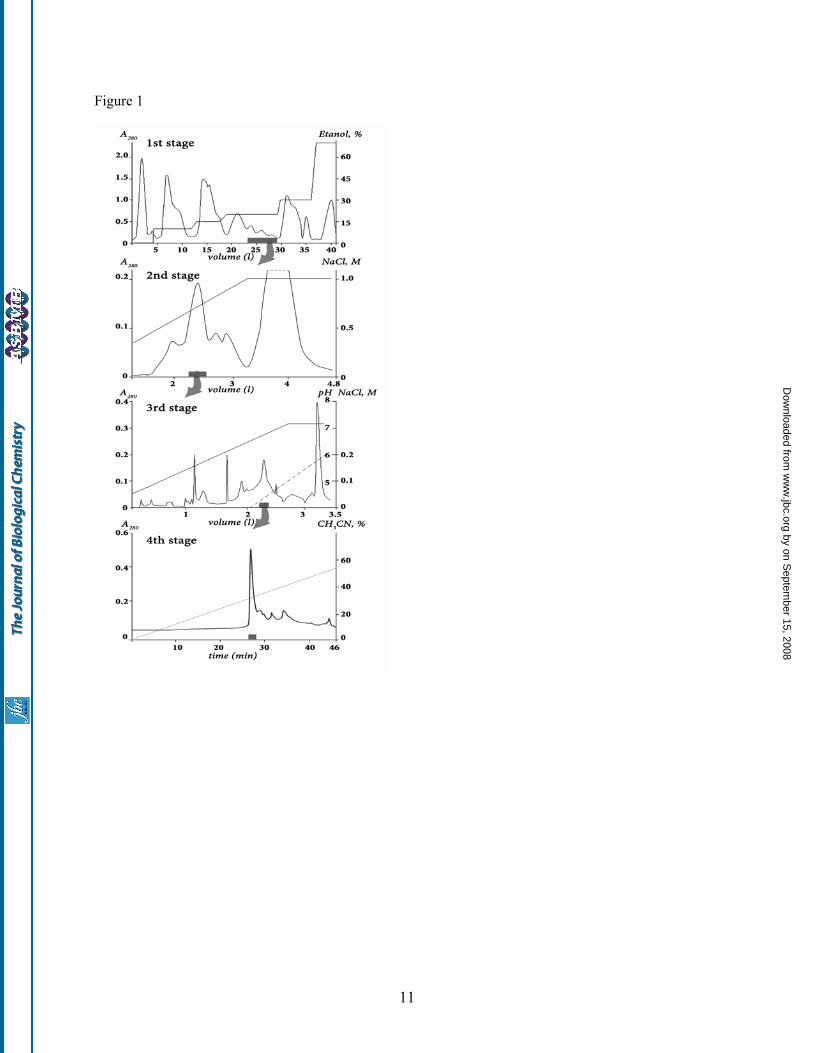
named analgesic polypeptide HC1 (APHC1), had analgesic effect in vivo experiment. Various Conus peptides have reached human clinical trials and one is already approved as a commercial drug for intractable pain. All these peptides act through distinct mechanisms none of which is opioid-based (15). It was also reported that peptide APETx2 from the sea anemone Anthopleura elegantissima inhibits ASIC3 channel that takes part in the transduction of acid-induced pain and hyperalgesia (16). And now novel TRPV1 peptide inhibitor APHC1 from sea anemone Heteractis crispa enriches the toolbox for pain and inflammation study and control. EXPERIMENTAL PROCEDURES Polypeptide purification Polypeptide isolation was performed from lyophilized 70%-ethanol extract of Heteractis crispa nematocysts collected on a littoral zone of Seychelles islands. Crude polypeptide fraction was produced by hydrophobic chromatography on a Polychrom-1 (CromLab, Russia) 7×30 cm column by stepwise gradient of ethanol. Chromatography profile, gradient condition and active fraction elution time are shown on fig. 1 stage 1. Further separation steps included cation exchange chromatography, first on Bio-Rex 70 (Bio-Rad) 2.5×60 cm column equilibrated with 5 mM ammonium acetate buffer pH 4.5 in a linear gradient NaCl (fig.1 stage 2), and then on SP-Sephadex С-25 (Pharmacia) 2.5×40 cm column in the same ammonium acetate buffer (fig.1 stage 3). Elution of compounds on stage 3 was performed in the ascent NaCl concentration accompanied with a linear gradient of pH from 4.5 to 7.3 just after half of separation process. The next stage of active compound isolation was carrying out on reverse phase column Jupiter C5 (Phenomenex) 4.6x150 mm in linear gradient of acetonitrile concentration in presence of 0.1% TFA (fig.1 stage 4). Mass Spectrometry Separated fractions, as well as purified polypeptides, were analyzed by matrix-assisted laser desorption ionization (MALDI) time-of-flight mass spectrometry to determine fractions composition or final product purity. MALDI-LR (Micromass UK Ltd.) and Ultraflex TOF-TOF (Bruker Daltonik GmbH, Bremen, Germany) spectrometers were used. Tandem mass spectrometric (MS/MS) analysis was performed according to the manufacturer’s guidelines.
Amino Acid Sequence Analysis N-terminal sequencing was carried out by automated stepwise Edman degradation using a Procise Model 492 protein sequencer (Applied Biosystems) according to the manufacturer’s protocol. Cysteine residues were reduced and alkylated by 4-vynil pyridine to determine their sequence position and improve a yield of conversion prior to analysis. Determination of APHC1 precursor Total RNA was purified using RNAwiz (Ambion, Canada) as directed by manufacturer protocols. First strand cDNA was synthesized from 5 µg of total RNA by MMLV reverse transcriptase Superscript II (Gibco) following the protocol recommended (17). Rapid amplification of cDNA ends was carried out using the universal primer T7cap (GTA ATA CGA CTC ACT ATA GGG CAA GCA GTG GTA ACA ACG CAG AGT) and a gene-specific degenerated primers Ing1 (ATA TGT CTA GAA CCT AAR GTN GTN GG), Ing2 (CCT AAG GTT GTA GGA CCN TGY CAN GC) for 3`-terminus determination (3`-RACE), and IngR (GGC ATG CAC GCA GGG TCTC) for 5`-terminus determination (5`-RACE). Taq Platinum polymerase (Gibco) was used for chain amplification. DNA sequencing was carried out on ABI PRISM 3100-Avant. APHC1 gene synthesis The DNA encoding APHC1 was constructed from 5 synthetic oligonucleotides using the PCR technique. The target PCR fragment was amplified using forward primer AL (5’-GGAATTCC ATG GGT TCT ATC TGC CTG GAA CCG AAA GTT GTTG) containing EcoRI restriction enzyme site (underlined), Met-codon for BrCN cleavage, and reverse primer AR (5’-CTCTCGAG TCA AGC ACG GCA GAT AGC ACG GCA AGC ACG CAG) containing XhoI restriction enzyme site (underlined) and stop-codon. The PCR fragment encoding mature APHC1 was gel purified, digested by EcoRI/XhoI and cloned into the expression vector pET32b+ (Novagen). The resulted construct was checked by sequencing. Production of recombinant APHC1 Recombinant APHC1 was produced as a fusion with a thioredoxin domain. E.coli BL21(DE3) cells transformed with the expression vector were cultured at 370C in LB medium containing 100 µg/ml ampicillin up to reaching the culture density of OD600 ~0.4–0.8. Expression was induced by adding IPTG up to 0.1 mM. The cells were cultured at 25 0C for 12–14 h, harvested,
by on Septem
ber 15, 2008 w
ww
.jbc.orgD
ownloaded from

3
resuspended in the start buffer for affinity chromatography (300 mM NaCl, 50 mM sodium phosphate buffer, pH 8.0) and ultrasonicated. Then lysed cells were centrifuged for 15 min at 15,000 rpm to remove all insoluble particles. The supernatant was applied to a TALON Superflow Metal Affinity Resin (Clontech), and the fusion protein was purified according to the protocol supplied by the manufacturer. The hybrid protein was quickly desalted on a Jupiter C5 column (Phenomenex) 4.6×150 mm, using a stepwise gradient of acetonitrile in 0.1% TFA. The collected fusion protein was vacuum dried and dissolved in 0.1M HCl solution. Protein cleavage by CNBr was performed over night at room temperature with molar ratio CNBr to protein – 600:1. Recombinant APHC1 was purified from reaction mixture on reverse-phase column Jupiter C4 (Phenomenex) 250×10 mm. The purity of the target polypeptide was checked by MALDI-TOF mass-spectrometry, as well as by N-terminal sequencing. Oocyte electrophysiology Xenopus laevis oocytes were removed surgically, defolliculated and injected with 2.5 – 10 ng of human TRPV1 cRNA (AJ272063). cRNA transcripts were synthesized from NotI-linearized TRPV1 cDNA template (EX-W1312-B02 from RZPD) using RiboMAXTM Large Scale RNA production system T7 (Promega) according to a protocol for capped transcripts supplied by manufacturers. After injection, oocytes were kept for 2 – 7 days at 180C in ND-96 medium containing (in mM) NaCl 96, KCl 2, CaCl2 1.8, MgCl2 1 HEPES 5 titrated to pH 7.4 with NaOH supplemented with gentamycin (50 µg/ml). Two-electrode voltage clamp recordings were performed using GeneClamp 500 amplifier (Axon Instruments, CA), data was filtered at 500 Hz and digitized at 100 Hz by AD Converter L780 (LCard, Moscow) using software created in our laboratory. Microelectrodes were filled with 3 M KCl solution. Ca2+-free ND-96 containing 0.1 mM BaCl2 was used as bath solution. To induce ligand-activated currents, short application (20 – 40 s) of 2 µM capsaicin (Sigma) solution in Ca2+-free ND-96 supplemented with BSA (0.1%) was used. Each oocyte was tested first by applying capsaicin solution 3 – 4 times and only the ones with appropriate current amplitude (200 – 1000 nA) were used in further experiments. Animals Adult male wild-type mice (19-21 g) were used. The animals were housed at room temperature of
23±2°C subjected to a 12 h light–dark cycle with food and water available ad libitum. Each animal was used for experimentation once only. Tail-flick test Lyophilized APHC1 was dissolved in 0.9% sterile NaCl solution and administered intramuscularly into the root of the tail 100 µl/mouse. Control animals received 0.9 % sterile NaCl solution 100 µl/mouse. For the tail-flick test, the mouse was restrained in a soft tissue pocket and the distal half of the tail was immersed into water heated up to 500C. Latency for tail-flick was measured with a 10-s cutoff time to avoid animal injury. Capsaicin-induced acute pain Intraplantar injection of capsaicin (3µg/10µl in 10% ethanol/90% saline) was used to produce a capsaicin-induced acute pain. Immediately after the injection of capsaicin, mice were placed inside glass cylinders. Intraplantar injection of capsaicin evoked a licking and shaking of the injected paw in mice. The number and duration of episodes of licking and shaking the paw in response to the injection were recorded. APHC1 was administered intravenously 15 min before capsaicin. Control mice received an equal volume of 0.9 % sterile NaCl solution. Statistic The significance of the data was determined by ANOVA followed by Tukey’s test. Data are presented as mean ± SEM. Serine protease inhibitory activity The enzymatic activity of trypsin and chymotrypsin as well as their inhibition by APHC1 peptide was measured spectrophotometrically in 96 well plates. Aliquots of trypsin dissolved in 90 µl in 50 mM Tris-HCl, pH 8.0 at the final concentration of about 0.5 µM were incubated for 10 min at 37°C, in the presence of various concentrations of the peptide. After incubation, the remaining tryptic activity was determined by addition of 10 µl of 3 mM BAPNA solution. Kinetics of paranitroaniline release was measured at 405 nm. For chymotrypsin the same incubation buffer was used. After incubation, the remaining proteolytic activity was determined by adding 1.3 mM BTEE solution. Kinetics of tyrosine release was measured at 250 nm. BPTI was used as control in all experiments. Inhibition constants for APHC1 were calculated for trypsin and chymotrypsin by method (18).
by on Septem
ber 15, 2008 w
ww
.jbc.orgD
ownloaded from
4
RESULTS Isolation of APHC1 Thirty different venoms from poisonous animals were tested for TRPV1 inhibition activity. The screening was performed by two-electrode voltage clamp standard technique on Xenopus oocytes expressed vanilloid receptors. The most attractive inhibitory action was noted for nematocysts ethanol extract from tropical sea anemone Heteractis crispa. After the inhibitory action was evaluated, next step of fractionation was performed up to single active component purification. Polypeptide fraction was separated from a lot of protein compounds, lipids, pigments and various organics molecules on Polychrom-1 chromatographic media in accordance by the method earlier developed for purification of sea anemone neurotoxins or protease inhibitors (19,20). Then cation exchange separation technique in median acidic pH was used for gentle purification of active compound. As a result, the TRPV1 inhibitory fraction, which consisted of about 20 related components with measured molecular weight around 6 kDa, was isolated. The next purification step on a reverse phase column was performed in 0.1% TFA solution. Total purification procedure is shown on fig.1. Novel sea anemone polypeptide, able to reduce capsaicin-induced response of Xenopus oocytes expressed TRPV1 channels, was named as APHC1. Average molecular weight estimated by MALDI mass spectrometry was equal to 6187.0 Da. Primary structure determination Since vinylpyridine alkilated APHC1 was produced, the sequence of fist 15 amino acid residues from N-terminus was recognized by automatic method: GSICLEPKVVGPCTA The complete structure definition was performed by 3-RACE with degenerate primers, which were synthesized according to the structural information of N-terminal peptide, and universal primer T7Cap (as described in (17)). PCR fragments (~350bp) were cloned into pBS-SK+. Further clones sequencing revealed a variety of closely relative sequences that had diverse point mutation at 19 positions in ~300 bp length consensus, 11 of them are in coding part. One sequence was chosen as putative APHC1 since it encoded 6 Cys family polypeptide with calculated average molecular mass 6187.08 Da and the same N-terminus. To confirm the sequence, polypeptide chain of natural
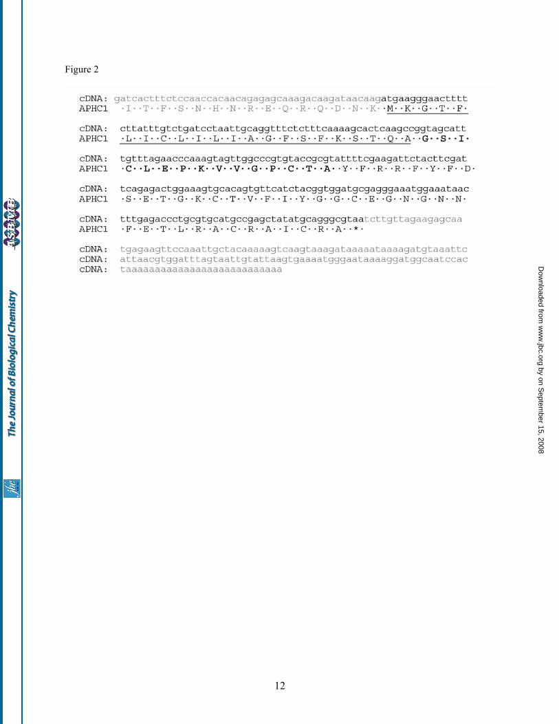
APHC1 was subjected to limited enzymatic digestion and MS-MS fragmentation. Both MS-MS spectrum and molecular weight analysis of peptide map verify the accuracy of APHC1 mature structure determination. 5-RACE performed on IngR and T7cap primers followed by isolation and sequencing of a PCR fragment about 300 bp. This fragment was aligned with sequence of 3-RACE fragment so that complete precursor structure of APHC1 was deduced together with 3’ and 5’ untranslated regions (see Fig. 2). The precursor has a very simple organization consisting of signal peptide of 22 amino acids restudies and a mature chain. No posttranslational modifications were determined. Primary structure homology APHC1 has a primary structure highly homologous to BPTI/Kunitz -type trypsin inhibitors from sea anemones, such as Kunitz-type trypsin inhibitor IV from H. crispa (Uniprot ID P16344) (85% identities) and SHPI-1 from Stichodactyla helianthus (Uniprot ID P31713) (81% identities) (20,21). Homology with sea anemones is evident, but what is more important, is that APHC1 has the features of structure firstly described for protease inhibitor from bovine pancreas. This disulfide rich alpha/beta structure is termed BPTI/Kunitz -type fold. The special features of this fold group are the length of about 60 amino acid residues, the presence of 6 cysteine residues, glycine in -2 position to second cysteine, triplet of aromatic residues close to molecule center and a typical location of several aromatic, glycine and asparagine residues around 4th cysteine (pfam 00014, cd00109). This structural features correlate with spatial polypeptide chain fold in ellipsoid globule but not with its functions. A number of polypeptide molecules of different functions have this fold. Thus the alignment on Fig.3 include not only protease inhibitors’ structures but also K+ channels blockers structure KAL1, DTX-α, DTXK (50-62% homology with APHC1) and Ca2+ channel inhibitor CAC (52% homology with APHC1). Recombinant polypeptide production To provide sufficient material for functional investigations, recombinant APHC1 was produced in the prokaryotic expression system. Thioredoxin (Trx) was chosen as the fusion partner for expression, since it is known to ensure high yields of cysteine-containing polypeptides with native conformation. A synthetic gene coding for APHC1 was constructed and cloned into pET-32b(+)
by on Septem
ber 15, 2008 w
ww
.jbc.orgD
ownloaded from

5
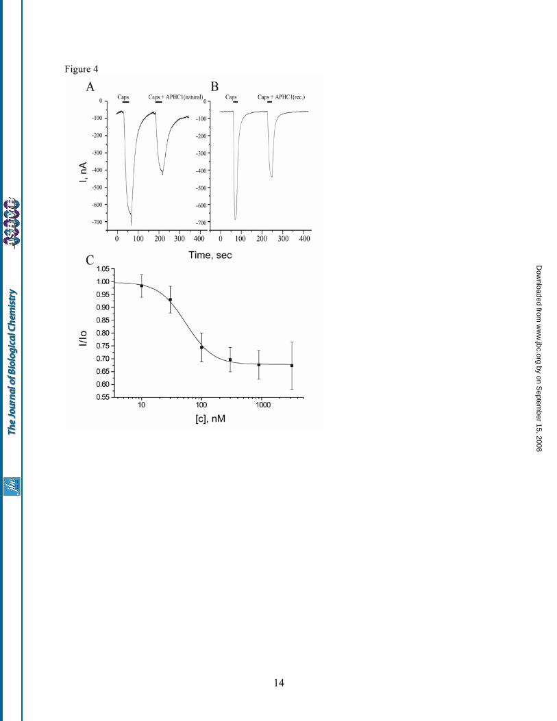
expression vector, and the resulting plasmid (pET- 32b+APHC1) was used to transform E. coli BL21(DE3) cells. Trx-APHC1 fusion protein production and purification was followed by CNBr cleavage. The recombinant APHC1 was purified by reverse-phase HPLC. The final yield of purified recombinant APHC1 was estimated to be ~0.5 mg/l of cell culture. Molecular weight of recombinant product was equal to native molecule, and amino acid sequence of five N-terminal residues was determined as well. The proper peptide folding was checked by experiments on serine protease inhibition and preliminary electrophysiology tests. In both tests the recombinant APHC1 and the natural polypeptide were equally active. Moreover both polypeptides had the same retention time while were co-injected on reverse phase column. Therefore, the obtained recombinant polypeptide was widely utilized in all experiments. Electrophysiological study Capsaicin (2 µM) was used as channel activator for human TRPV1 channels expressed in Xenopus laevis oocytes. The inhibition activity was calculated as I/I0, where I is ionic current evoked by co-application of diluted venom, fractions or APHC1 with agonist, and I0 is ionic current evoked by agonist alone on the same oocyte. Fractions of the first and the second purification steps were able to reduce capsaicin-induced currents up to 25%, while the active fraction of the third stage up to 50%. Purified APHC1 was shown to be a partial inhibitor of TRPV1 capsaicin-induced ionic currents in the most of oocytes (fig. 4). There was no difference in APHC1 action on TRPV1 when it was co-applied with capsaicin or when a 2 minute preincubation with APHC1 was performed before the capsaicin/APHC1 application. APHC1 alone (without capsaicin) did not affect TRPV1. APHC1 binding seems to be completely reversible, since 2-minute wash recovered capsaicin-induced currents to the control level. Usually the difference between control and test applications was observed only in response amplitude. Dose-response analysis of an inhibitory activity of recombinant APHC1 estimates the maximal inhibitory effect 32±9%, half-maximal effective concentration (EC50) 54±4 nM and Hill coefficient 2.12±0.19. Increasing APHC1 concentrations up to 3.2 µM or crude fractions of venom did not provide complete inhibition of capsaicin induced currents. The maximal observed inhibition was about 50% at
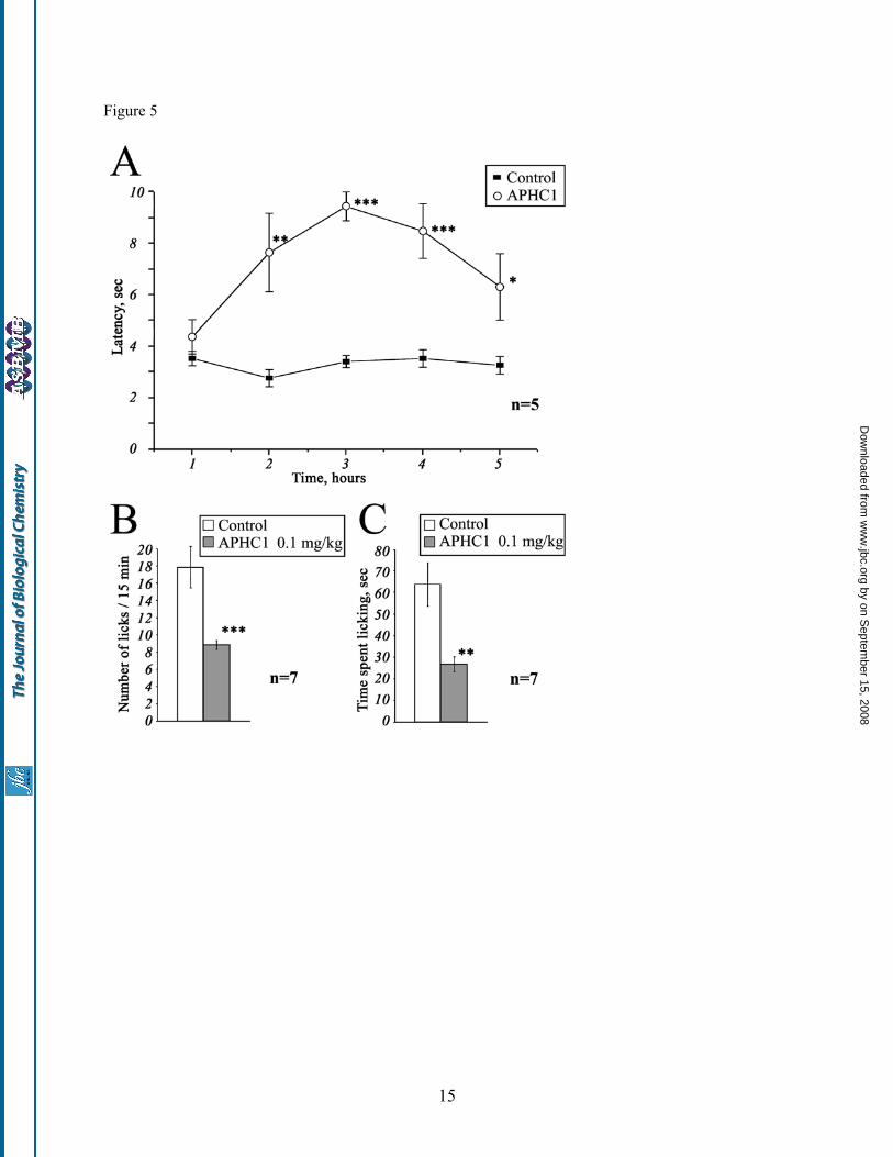
concentrations ≥300nM but did not occur in every oocyte. Thus, APHC1 takes an effect on TRPV1 as a modulator and not as a real blocker. So far as APHC1 has significant homology with serine protease inhibitors and also has a weak protease-inhibiting activity, we carried out additional study of protease inhibitor BPTI influence on capsaicin-induced currents. Since no effect was detected, indirectly action of APHC1 on TRPV1 via intermediate systems was excluded. In vivo studies Lyophilized APHC1 dissolved in NaCl saline didn’t provide any toxic effects on mice up to 1 mg/kg. Tail-flick test. The basal reaction time of tested animals was in 2.5 to 4 sec range. Intramuscular administration of APHC1 (0.1mg/kg) in root of tail considerably increased the tail-flick latency when compared to the saline-treated control (Fig. 5, A). Significant antinociception effect began within 2 hours following the injection, lasted all the remaining observation time (3 hours) and reached maximal effect 3 hours after administration. Effect began to grow weak at the time between 4th and 5th hour of observation and in the 5th hour the experimental data the degree of the difference of the experimental data from the control one was not high enough (p<0.06). Capsaicin-induced pain-related behavior. Intraplantar injection of capsaicin rapidly produced distinct paw-licking behavior, mediated by direct activation of local TRPV1 receptors. Intravenous administration of APHC1 (0.1 mg/kg) 15 min before capsaicin injection significantly reduced capsaicin-induced pain-related behavior (fig.5 B,C). Serine protease inhibition APHC1 shows a very weak inhibition of trypsin and chymotrypsin activity. Only 8-fold molar excess of APHC1 over trypsin provides complete inhibition of paranitroaniline release from BAPNA substrate. The same APHC1 exceeds to chymotrypsin reduce enzyme activity only to 50%. At the same time BPTI completely blocks trypsin/ chymotrypsin in molar ratio 1:1. Constants (Ki) and inhibition types of APHC1 were determined with Dicson method (18): trypsyn inhibition was competitive with Ki = 1x10⁻⁶, but chemotrypsin inhibition was non–competitive with Ki ≈ 5x10⁻⁶.
by on Septem
ber 15, 2008 w
ww
.jbc.orgD
ownloaded from
6
DISCUSSION Stings of poisonous animals usually evoke a sensation of pain. In some cases this sensation is connected with venom peptides. So, peptide vanillotoxins isolated from tarantula venom were shown to activate the TRPV1 channels and produce pain (11). Tentacle extract of jellyfish also provokes TRPV1 activation comparable with action of capsaicin (12). Along with activators two polyamine inhibitors of TRPV1 channels were identified in the spider venom (14) while polyamines as spermine, spermidine and putrescine directly activated TRPV1 in a charge-dependent manner (28). Sea anemones also produce a sensation of pain after contact with a victim. These coelenterates have large tentacles that contain venom-producing nematocysts which are well-known sources for the isolation of toxic proteins including pore-forming cytolysins (29,30), phospholipases (31), Na+-channel toxins (32,33,34), K+-channel inhibitors (23,35,36,37), acid-sensing ion channels (ASICs) inhibitor (16) and proteinase inhibitors (20,21,38). It was suggested that the mixture of sea anemone toxins acting on Na+ and K+ channels has destructive neurotoxic effects due to a massive release of neurotransmitters and a resulting break of the signal transduction process. In addition, sea anemone cytolysins and phospholipases produce massive cytolytic effects on different cells (30,39) that cause tissues damage and inflammation. In view of pain producing effect of sea anemone it was interesting to find in it a molecule that downregulate such an important integrator of various pain and inflammation stimuli as TRPV1. There are several possible explanations for this phenomenon. One of them is that sea anemone nematocysts can provide an array of peptide components with distinguished activity. Another possibility is that a single peptide can activate or inhibit more than one ion channel. APHC1 as analgesic agent TRPV1 antagonists can apparently produce pain relief during a number of pathological states. Extensive research of small-molecule inhibitors of TRPV1 provides intriguing evidence that TRPV1 blockade can be a useful therapeutic approach for inflammatory, cancer and possibly for neuropathic pain (13,40). APHC1 showed significant antinociception to noxious thermal stimuli, since it efficiently prolonged the latencies of tail withdrawal in a 50°C tail-flick tests (see Fig.5A). This anesthetic
effect has been observed in a minimal dose of 0.05 mg/kg (data not shown). At dose of 0.1 mg/kg the difference of tail flick latency for APHC1-treated and control mice was close to reported latency for TRPV1 knockout mice (8). But possible additional influence of APHC1 on other thermal nociception pathways should not be excluded. Only several small molecule TRPV1 antagonists have been reported to provide antinociception in tail-flick test - IBTU (41), and/or in hot plate test - DD161515, DD191515 (42). So, IBTU is essential for prolongation of tail flick latency at about ~30-60 mg/kg, DD161515, DD191515 are also active in high dose (0.2 mmol/kg). Analgesia effect produced by intramuscular administration of APHC1 appears slowly (within 1-2 h after injection) but is extended for up to more then 2 h suggesting that APHC1 may have rather a long pharmacokinetic half-life and its diffusion in organism is quite slow, which is typical for the most part of polypeptide molecules. Capsaicin is a selective agonist of TRPV1 receptor (8). Therefore we tested whether APHC1 was capable of blocking capsaicin-induced behavior. Intravenous administration of APHC1 was used to provide quick distribution of the polypeptide in the organism. Dose of 0.1 mg/kg APHC1 significantly suppressed the capsaicin-induced behavior. Significant antinociception to capsaicin and noxious thermal stimuli could be an evidence of the fact that APHC1 in small doses essentially blocks TRPV1 in vivo. Polypeptide ligands specific to TRPV1 can be of great importance as research tools or pro-drugs. To our knowledge, no selective polypeptides inhibiting TRPV1 have been reported so far. It is known that high positively charged lysine/arginine rich peptides interacting with some Ca2+ channels also provide nonselective binding to TRPV1 (43). Mode of action To date TRPV1 is considered as polymodal detector of noxious physical and chemical stimuli (44). Vanilloid receptor can be directly activated by various ligands: exogenous (capsaicin, EC50 = 0.7 µM, resiniferatoxin, EC50 = 40 nM and others) as well as endogenous (anandamide and others), by heat (>43ºC) and extracellular protons (pHout <6.5) (13). TRPV1 is sensitive to some chemical compounds of varios nature, including inflammatory agents (13), polyamines (28) and venoms of jellyfish (12) and spiders (11). APHC1 interaction with TRPV1 channels results in reduction of capsaicin induced responses. It is
by on Septem
ber 15, 2008 w
ww
.jbc.orgD
ownloaded from

7
most likely that APHC1 action occurs through receptor extracellular domains which form a channel vestibule. It is also highly probable that the pore of the channel remains free, otherwise complete blockade could occur, as reported for channel blocker Ruthenium Red (45). APHC1 was found to be high-affinity (EC50=54±4 нМ) but not very potent (maximum observed inhibition ~50%, maximum mean inhibition 32%±9%). Rabbit anti-rat TRPV1 polyclonal antibodies (Ab-156H) specific to TRPV1 loop (considered as pH sensor) have something similar in inhibition result to APHC1. These antibodies were not able to block completely capsaicin- and anandamide-induced response in saturating concentrations and had maximum inhibition of approximately 55% (46). Probably APHC1 shares the mechanism of action suggested to Ab-156H that probably partially lock the channel conformation in the closed state. Other molecules which are likely to affect TRPV1 through extracellular prepore regions are vanillotoxins (11) DD161515, DD191515,H-Arg-15-15C and arginine/lysine rich peptides (42,43,47). Ability of APHC1 to inhibit activity of serine proteases is apparently not able to influence channel response that was indirectly proved in experiments with BPTI – TRPV1 interaction. Spatial structure and functional important residues The structure of APHC1, like proteinase inhibitors or K+-channel inhibitors, has BPTI/Kunitz–type fold that was extensively studied on BPTI and dendrotoxins models (48,49,50). As mentioned above, various molecules with different functions from diverse species have this fold: BPTI is the most potent trypsin inhibitor; snakes dendrotoxins (DTX I, DTXK, DTXα) are some of the most potent K+-channels blockers (49,50); calcicludine from Dendroaspis angusticeps snake venom that is a potent blocker of high-voltage-activated calcium L-type ion channels (27); protease inhibitors from artropoda, vertebrata, sea anemones; K+-channel blockers from sea anemone (23). The fact that distinct phyla adopt the same fold for the similar functions underlines that this fold gives significant advantages in a compact variable binding site
construction. APHC1, that modulates activity of TRPV1, should be also added to the list of molecules with this fold. Despite high primary structure homology with trypsin inhibitor SHPI-1 the most part of functional important residues of APHC1 are changed. So 4 out of 6 apparently important for protease amino acid residues (fig. 3) differ in APHC1 and SHPI-1. On SHPI-1 NMR determined spatial structure (fig. 6) is shown that these 6 residues form mutual surface that interact with enzyme cleft and its environment. It is likely that two residues of APHC1 which remained unchanged retain, together with other residues, an ability of molecule to interact with proteolytic enzymes. Thus, inhibition constants for trypsin and chymotrypsin are decreased more than 1000 times: APHC1 ~10-6; 5x10⁻⁶, respectively, (measured in this work) in compassion with SHPI-1 ~10-10, 2,3x10⁻⁹ (51). Alignment and spatial structure analysis revealed potential residues necessary for TRPV1 binding. We are of the opinion that these are Arg18, Arg 48, and Val31 (shown in blue on fig 7 and as asterisk mark on fig. 3). Their side chains are located on the same molecule interface and oriented outside. It is known that a combination of positively charged and hydrophobic residues often form active sites of toxin, for example, K+ channel blockers (49,50). Some authors also reported that arginine/lysine rich peptides and peptoid molecule have moderate blockade potency on TRPV1 channel in in vitro and in vivo experiments (42,43,47). In our opinion, one or several positively charged residues may be in an active site of APHC1 too. We want to underscore that active sites of two highly homologue molecules SHPI-1 and APHC1 are probably located on different molecular interfaces. The same fact was reported for active sites of kalicludines that were found to have K+ channels blockers interface together with trypsin inhibitor site. Finally, we would like to highlight that APHC1 is the first polypeptide inhibitor of TRPV1 channels and can play a great role in further studies of TRPV1 properties, as well as a model for designing a new generation of analgesic drugs.
REFERENCES
1. Kawai, N., and Nakajima, T. (1993) In: A.L. Harvey, Editor, Natural and Synthetic Neurotoxins, Acad. Press, London, 319–345 2. Miljanich, G. P. (1997) Sci. Med. 4(5), 6–15
by on Septem
ber 15, 2008 w
ww
.jbc.orgD
ownloaded from

8
3. Mortaria, M. R., Cunhaa, A. O., Ferreiraa, L. B., and Ferreira dos Santos, W. (2007), Pharmacology & Therapeutics 114(2), 171-183 4. Watters, M. R. (2005) Semin. Neurol. 25(3), 278-289 5. Guo, A., Vulchanova, L., Wang, J., Li, X., and Elde, R. (1999) Eur. J. Neurosci. 11(3), 946-958 6. Carlton, S. M., and Coggeshall, R. E. (2001) Neurosci. Lett. 310(1), 53-56 7. Valtschanoff, J. G., Rustioni, A., Guo, A, and Hwang, S. J. (2001) J. Comp. Neurol. 436(2), 225-235 8. Caterina, M. J., Leffler, A., Malmberg, A. B., Martin, W. J., Trafton, J., Petersen-Zeitz, K. R., Koltzenburg, M., Basbaum, A. I., and Julius, D. (2000) Science 288(5464), 306-313 9. Davis, J. B., Gray, J., Gunthorpe, M. J., Hatcher, J. P., Davey, P. T., Overend, P., Harries, M. H., Latcham, J., Clapham, C., Atkinson, K., Hughes, S. A., Rance, K., Grau, E., Harper, A. J., Pugh, P. L., Rogers, D. C., Bingham, S., Randall, A., and Sheardown, S. A. (2000) Nature 405(6783), 183-187 10. Szallasi, A., and Fowler, C. J. (2002) The Lancet Neurology 1, 167-172 11. Siemens, J., Zhou, S., Piskorowski, R., Nikai, T., Lumpkin, E. A., Basbaum, A. I., King, D., and Julius, D. (2006) Nature 444, 208-212 12. Cuypers, E., Yanagihara, A., Karlsson, E., and Tytgat, J. (2006) FEBS Lett. 580(24), 5728-5732 13. Szallasi, A., Cortright, D. N., Blum, C.A., and Eid, S. R. (2007) Nat. Rev. Drug Discovery 6, 357-370 14. Kitaguchi, T., and Swartz, K. J. (2005) Biochemistry 44(47), 15544-15549 15. Olivera, B. M. (2006) J. Biol. Chem. 281(42), 31173-31177 16. Diochot, S., Baron, A., Rash, L. D., Deval, E., Escoubas, P., Scarzello, S., Salinas, M., and Lazdunski, M. (2004) EMBO J. 23(7), 1516-1525 17. Matz, M., Shagin, D., Bogdanova, E., Britanova, O., Lukyanov, S., Diatchenko, L., and Chenchik, A. (1999) Nucleic Acids Res. 27(6), 1558-1560 18. Dixon, M. (1953) Biochem. J. 55, 170 19. Zykova, T. A., Vinokurov, L. M., Kozlovskaya, E. P., and Elyakov, G. B. (1985) Bioorg. Chim. 11, 302–310 20. Zykova, T. A., Vinokurov, L. M., Markova, L. F., Kozlovskaya, E. P., and Elyakov, G. B. (1985) Bioorg. Chim. 11, 293–301 21. Antuch, W., Berndt, K. D., Chávez, M. A., Delfín, J., and Wüthrich, K. (1993) Eur. J. Biochem. 212(3), 675-684 22. Perona, J. J., and Craik, C. S. (1995) Protein Sci. 4(3), 337-360 23. Schweitz, H., Bruhn, T., Guillemare, E., Moinier, D., Lancelin, J. M., Béress, L., and Lazdunski, M. (1995) J. Biol. Chem. 270(42), 25121-25126 24. Joubert, F. J., and Taljaard, N. (1980) Hoppe Seylers Z. Physiol. Chem. 361 (5), 661-674 25. Strydom, D. J. (1977) Biochim. Biophys. Acta 491(2), 361-369 26. Creighton, T. E., and Charles, I. G. (1987) J. Mol. Biol. 194(1), 11-22 27. Schweitz, H., Heurteaux, C., Bois, P., Moinier, D., Romey, G., and Lazdunski, M. (1994) Proc. Natl. Acad. Sci. U.S.A. 91(3), 878-882 28. Ahern, G. P., Wang, X., and Miyares, R. L. (2006) J. Biol. Chem. 281(13), 8991-8995 29. Wang, Y., Chua, K. L., and Khoo, H. E. (2000) Biochim. Biophys. Acta 1478, 9–18 30. Anderluh, G., and Macek, P. (2002) Toxicon 40, 111–124 31. Grotendorst, G. R., and Hessinger, D. A. (1999) Toxicon 37, 1779–1796 32. Norton, R. S. (1991) Toxicon 29, 1051–1084 33. Béress, L. (2004) Toxin Rev. 23, 451–466 34. Ständker, L., Béress, L., Garateix, A., Christ, T., Ravens, U., Salceda, E., Soto, E., John, H., Forssmann, W. G., and Aneiros, A. (2006) Toxicon 48(2), 211-220 35. Aneiros, A., Garcia, I., Martinez, J. R, Harvey, A. L., Anderson, A. J., Marshall, D. L., Engstrom, A., Hellman, U., and Karlsson, E. (1993) Biochim. Biophys. Acta 1157, 86–92 36. Castañeda, O., Sotolongo, V., Amor, A. M., Stocklin, R., Anderson, A., Harvey, A. L., Engstrom, A., Weinstedt, C., and Karlsson, E. (1995) Toxicon 33, 605–613 37. Diochot, S., Schweitz, H., Béress, L., and Lazdunski, M., (1998) J. Biol. Chem. 273, 6744–6749 38. Fritz, H., Brey, B., and Béress, L. (1972) Hoppe Seylers Z Physiol. Chem. 353, 19–30 39. Šuput, D., Frangež, R., and Bunc, M. (2001) Toxicon 39, 1421–1427 40. Krause, J. E., Chenard, B. L., and Cortright, D. N. (2005) Curr. Opin. Investig. Drugs 6, 48–57
by on Septem
ber 15, 2008 w
ww
.jbc.orgD
ownloaded from
9
41. Tang, L., Chen, Y., Chen, Z., Blumberg, P. M., Kozikowski, A. P., and Wang, Z. J. (2007) J. Pharmacol. Exp. Ther. 321(2), 791-798 42. García-Martinez, C., Humet, M., Planells-Cases, R., Gomis, A., Caprini, M., Viana, F., De La Pena, E., Sanchez-Baeza, F., Carbonell, T., De Felipe, C., Pérez-Paya, E., Belmonte, C., Messeguer, A., and Ferrer-Montiel, A. (2002) Proc. Natl. Acad. Sc.i U. S. A. 99(4), 2374-2379 43. Planells-Cases, R., Aracil, A., Merino, J. M., Gallar, J., Pérez-Payá, E., Belmonte, C., González-Ros, J. M., and Ferrer-Montiel, A. V. (2000) FEBS Lett. 481(2), 131-136 44. Caterina, M. J., and Julius, D. (2001) Annu. Rev. Neurosci. 24, 487-517 45. Caterina, M. J., Schumacher, M. A., Tominaga, M., Rosen, T. A., Levine, J. D., and Julius, D. (1997) Nature 389(6653), 816-824 46. Klionsky, L., Tamir, R., Holzinger, B., Bi, X., Talvenheimo, J., Kim, H., Martin, F., Louis, J. C., Treanor, J. J., and Gavva, N. R. (2006) J. Pharmacol. Exp. Ther. 319(1), 192-198 47. García-Martínez, C., Fernández-Carvajal, A., Valenzuela, B., Gomis, A., Van Den Nest, W., Ferroni, S., Carreño, C., Belmonte, C., and Ferrer-Montiel, A. (2006) J. Pain 7(10), 735-746 48. Scheidig, A. J., Hynes, T. R., Pelletier, L. A., Wells, J. A., and Kossiakoff, A. A. (1997) Protein. Sci. 6(9), 1806-1824 49. Gasparini, S., Danse, J. M., Lecoq, A., Pinkasfeld, S., Zinn-Justin, S., Young, L. C., de Medeiros, C. C., Rowan, E. G., Harvey, A. L., and Ménez, A. (1998) J. Biol. Chem. 273(39), 25393-25403 50. Smith, L. A., Reid, P. F., Wang, F. C., Parcej, D. N., Schmidt, J. J., Olson, M. A., and Dolly, J. O. (1997) Biochemistry 36(25), 7690-7696 51. Delfín, J., Martínez, I., Antuch, W., Morera, V., González, Y., Rodríguez, R., Márquez, M., Saroyán, A., Larionova, N., Díaz, J., Padrón, G., and Chávez, M. (1996) Toxicon 34(11-12), 1367-1376
FOOTNOTES The authors gratefully acknowledge the scientific support of Grebelny S.D. (Zoological Institute of the Russian Academy of Sciences, Sankt-Peterburg); Vozhova E.I., Leychenko E.V., Lie I.N., (Pacific Institute of Bioorganic Chemistry, Far East Branch of the Russian Academy of Sciences, Vladivostok), Egorov T.A. and Musolyamov A.Kh. (Shemyakin-Ovchinnikov Institute of Bioorganic Chemistry, Russian Academy of Sciences) for the technical assistance. This work was supported in part by the Russian Federation Federal Agency for Science and Innovations (the State contract 02.467.11.3003 of 20.04.2005), the Russian Foundation for Basic Research and CMB program of RAS. The precursor sequences data reported in this paper has been deposited in EMBL/GenBank/DDBJ databases under GenBankAccession Number AM933240. 1The abbreviations used are: APHC1, analgesic polypeptide HC1; ASIC, proton-gated sodium channel; ANOVA, analysis of variance; BAPNA, N-a-benzoyl-DL-arginin-p-nitroanilid; BTEE, N-benzoyl-l-tyrosine ethyl ester; BPTI, bovine pancreatic trypsin inhibitor; DTX, dendrotoxin; DRG, dorsal root ganglia; IBTU, N-(4-chlorobenzyl)-N-(4-hydroxy-3-iodo-5-methoxybenzyl) thiourea; MALDI, matrix-assisted laser desorption/ionization; MS, mass spectrometry; NMR, nuclear magnetic resonance; RP-HPLC, reversed-phase high performance liquid chromatography; TFA, trifluoroacetic acid; TOF, time-of-flight; TRPV1, transient receptor potential vanilloid type 1; S.E.M, standard error of the mean; Trx, thioredoxin.
FIGURE LEGENDS Fig. 1. Purification of APHC1 The first separation stage of a dried ethanol extracts of sea anemone Heteractis crispa nematocysts was done on water equilibrated hydrophobic column Polychrom-1 (7×30 cm). Fractions were eluted by stepwise ethanol gradient with a flow rate of 1.2 l/h. Active fraction (marked as a gray box on overall
by on Septem
ber 15, 2008 w
ww
.jbc.orgD
ownloaded from

10
separation steps) has been separated on the second stage by ion exchange chromatography on Bio-Rex 70 column (2.5×60 cm). The separation was done in 5 mM ammonium acetate buffer (pH 4.5) by flow rate 22 ml/h in linear gradient of NaCl concentration. The third stage of purification was performed with a flow rate 70 ml/h on ion exchange column SP-Sephadex С-25 (2.5×40 cm), with the same 5 mM ammonium acetate buffer (start buffer pH 4.5) in combined gradient of NaCl concentration and pH value. Final purification (stage 4) was achieved on a reverse phase column Jupiter C5 (4.6x150 mm) in 0.1% TFA with flow rate 1 ml/min using a linear gradient of acetonitrile concentration. Fig. 2. Structure of APHC1 gene determined from cDNA clones sequences. Signal peptide sequence deduced in the precursor structure is underlined and partial N-terminal fragment determined by Edman degradation shown in bold. Fig. 3. Alignment of APHC1 primary structure and BPTI/Kunitz type polypeptides. Part A - pairwise residues substitution plot among APHC1 and sea anemone trypsin inhibitor SHPI-1 from Stichodactyla helianthus (21). Amino acid residues essential for inhibitory activity of SHPI-1 as described in (22) marked with plus sign, probably important arginine/valine residues in APHC1 are asterisk marked. Part B – multiple alignment of APHC1 amino acid sequence with serine protease inhibitor SHPI-1 from sea anemone Stichodactyla helianthus (21); kalicludine 1 (KAL1) - K+ channel and trypsin inhibitor from sea anemone Anemonia sulcata (23); two dendrotoxins active on K+ channel DTX α from snake Dendroaspis angusticeps (24) and DTXK from Dendroaspis polylepis polylepis (25); Bos taurus trypsin inhibitor (BPTI) (26); and calcicludine (CAC) a blocker of high-threshold Ca2+ channels from snake Dendroaspis angusticeps (27). Related or similar residues are shadowed. Fig. 4. Action of APHC1 on TRPV1 channels. Application traces of capsaicin alone and capsaicin mixed with purified natural APHC1 (final concentration 500 nM) - panel A, or with recombinant APHC1 (final concentration 300 nM) – panel B. Channels were activated by 2 µM capsaicin 35 seconds (panel A) or 20 seconds (panel B) before washing, oocytes were held at -50 mV. Panel C - dose-response curve for APHC1 inhibitor activity on capsaicin-activated TRPV1 channels. Abscissa axis represents the ratio of ion current evoked by co-application of agonist and APHC1 (I) to ion current evoked by agonist alone (Io.) on the same oocyte. Each point represents the mean ± SEM with n = 4-8. Fig. 5. Analgesic (antinociception) effect of a recombinant APHC1. A. Tail-flick test. Latency indicates the delay of tail-flick response after one intramuscular administration of APHC1 (0.1 mg/kg) into wild-type mice, n =5. B,C. Attenuation of mice capsaicin-induced behavior by intravenous administered APHC1 (0.1mg/kg), n=7. Results are presented as mean ± s.e.m., ***-p<0.005, **-p<0.01, *- p<0.06 vs control (ANOVA and Turkey’s test). Fig. 6. 3D structural models of sea anemones polypeptides. SHPI-1 – trypsin inhibitor from sea anemone Stichodactyla helianthus (PDB ID 1SHP). Spatial structure of APHC1 was calculated from SHPI-1 data by molecular modeling. Amino acid residues essential for serine protease inhibition painted in magenta on both models, two arginine 18, 48 and valine 31 residues presumably important for APHC1 activity shown in blue.
by on Septem
ber 15, 2008 w
ww
.jbc.orgD
ownloaded from