Revision of Parastriatopora Gigantea ( Knod 1908) (Anthozoa, Tabulata) from the Devonian of Bolivia

Ple
ase
note
that
this
is a
n au
thor
-pro
duce
d P
DF
of a
n ar
ticle
acc
epte
d fo
r pub
licat
ion
follo
win
g pe
er re
view
. The
def
initi
ve p
ublis
her-a
uthe
ntic
ated
ver
sion
is a
vaila
ble
on th
e pu
blis
her W
eb s
ite
1
Marine Biology Research November 2005; 1(5) : 326 - 337 http://dx.doi.org/10.1080/17451000500380306© 2005 Taylor & Francis The original publication is available at http://www.tandf.co.uk/journals/
Archimer http://www.ifremer.fr/docelec/Archive Institutionnelle de l’Ifremer
A new species of sea anemone (Cnidaria: Anthozoa: Actiniaria) from Manus
Basin hydrothermal vents, South-western Pacific
Pablo J. López-González1, Estefanía Rodríguez1,2, Michel Segonzac3* 1Biodiversidad y Ecología de Invertebrados Marinos, Departamento de Fisiología y Zoología, Facultad de Biología, Universidad de Sevilla, Sevilla, Spain e-mail: [email protected] 2Departamento de Biología Marina y Oceanografía, Instituto de Ciencias del Mar, CMIMA (CSIC), Paseo Marítimo de la Barceloneta, Barcelona, Spain e-mail: [email protected] 3Ifremer, Centre de Brest, DEEP-LEP/Centob, Plouzané, France *: Corresponding author : [email protected]
Abstract: During the BIOACCESS Japanese cruises (1996 & 1998), active hydrothermalism and associated vent fauna were studied on the South-eastern Rift of Manus Basin (South-western Pacific). In the PACMANUS vent field, a conspicuous vent fauna was sampled, including an actinostolid sea anemone (Actiniaria) belonging to an undescribed genus and species. Pacmanactis hashimotoi gen. et spec. nov. is here described, and represents the 9th sea anemone reported from hydrothermal vents. Keywords: Actiniaria, Actinostolidae, hydrothermal vents, back-arc basins, Manus Basin

Pacmanactis-Shinji-180205.doc
A NEW SPECIES OF SEA ANEMONE (CNIDARIA: ANTHOZOA: ACTINIARIA) FROM MANUS BASIN HYDROTHERMAL VENTS, SOUTH-WESTERN PACIFIC
Pablo J. López-González1, Estefanía Rodríguez1, 2 & Michel Segonzac3
1: Biodiversidad y Ecología de Invertebrados Marinos, Departamento de Fisiología y Zoología, Facultad de Biología, Universidad de Sevilla, Reina Mercedes 6, 41012 – Sevilla, Spain (e-mail: [email protected]).
2: Departamento de Biología Marina y Oceanografía, Instituto de Ciencias del Mar, CMIMA (CSIC), Paseo Marítimo de la Barceloneta, 37-49, 08003 Barcelona, Spain (e-mail: [email protected]).
3: Ifremer, Centre de Brest, DRO-EP/Centob, BP 70, 29280 Plouzané, France (e-mail: [email protected]).
Key words: Actiniaria, Actinostolidae, hydrothermal vents, back-arc basins, Manus Basin, sea anemones.
Abstract During the BIOACCESS Japanese cruises (1996 & 1998), active hydrothermalism and associated vent fauna were studied on the Southeastern Rift of Manus Basin (South-western Pacific). In the Pacmanus vent field, a conspicuous vent fauna was sampled, including an actinostolid sea anemone belonging to an undescribed genus and species. Pacmanactis hashimotoi gen. et spec. nov. is here described, and represents the ninth sea anemone reported from hydrothermal vents.
1

Introduction Eight species of sea anemones have been documented from hydrothermal vents. Six of them belong to the family Actinostolidae and two species belong to the family Hormathiidae (see Desbruyères & Segonzac, 1997; Fautin & Barber, 1999; López-González et al., 2003). From the Atlantic Ocean, the occurrence of two actinostolid species was pointed out: Parasicyonis ingolfi Calgren, 1942 and Maractis rimicarivora Fautin & Barber, 1999. From the Pacific Ocean, four actinostolid species have been reported (one of them identified only at generic level): Actinostola sp. [as Actinostola sp. in the text, but as Actinostola callosa (Verrill 1882) in the figure, see Doumenc & van Praët, 1988, fig. 1], Cyananthea hydrothermala Doumenc & Van-Praët, 1988, Marianactis bythios Faustin & Hessler, 1989 and Paranthosactis denhartogi Lopez-Gonzalez et al., 2003. Actinostola callosa and C. hydrothermala were collected at East Pacific Rise (EPR) vents, 12º48’N-103º56’W, 2635 m, while M. bythios was sampled at Mariana back-arc basin 18º11’N-144º42.4’E, 3660 m, and P. denhartogi at Guaymas Basin, Gulf of California, 27°00.94’-111°24.66’W, 2025 m. To present, only two hormathiid species have been reported from hydrothermal vents, both from EPR-13°N: Chondrophellia coronata (Verrill, 1883) and Phelliactis sp. (Doumenc & Van-Praët 1988).
In this paper, we describe a new genus and species of actinostolid collected during the two Japanese Cruises BIOACCESS’96 and BIOACCESS’98, at the hydrothermal vent area of PACMANUS, Pual Ridge, Bismark Sea in the SW Pacific, N of Papua New Guinea (Fig. 1).
Fig. 1. Map indicating the hydrothermal (PACMANUS) vent where Pacmanactis hashimotoi gen. et spec.
nov. described in this paper was collected.
Material and Methods
The seven specimens studied here were collected during the BIOACCESS’96 and ‘98 cruises organized by the Japan Agency for Marine-Earth Science and Technology (JAMSTEC) and conducted by Jun Hashimoto (Nagasaki University), on board the R/V Natsushima, equipped with the manned submersible Shinkai 2000, from 26 October to 29 November 1996 and 13 to 25 November 1998. The main goal of these cruises was the study of the composition, distribution, ecology and diversity of
2

hydrothermal vent fauna on PACMANUS (1700 m) and DESMOS (1900 m) sites of Manus Basin (Hashimoto et al. 1999, and Fig. 1).
After recovery, the sea anemones were fixed in 10% seawater formalin, and then preserved in 70% ethanol. Fragments from selected specimens were dehydrated in buthanol (Johansen, 1940) and embedded in paraffin. Histological sections 7-8 µm thick were stained with Ramón y Cajal's Triple Stain (Gabe, 1968).
Cnidae measurements were made from preserved material in squash preparations at 1000x magnification with Nomarski differential interference contrast optics. Frequencies given are subjective impressions based on squash preparations.
The material studied in this article is deposited in the National Science Museum of Tokyo (NSMT), and the collection of the research team “Biodiversidad y Ecología de Invertebrados Marinos” of the Faculty of Biology at the University of Seville in Spain (BEIM).
Results
Phylum CNIDARIA Order ACTINIARIA Hertwig, 1882 Family ACTINOSTOLIDAE Carlgren, 1932
Pacmanactis gen. nov.
Diagnosis.— Actinostolidae with well developed, adherent, and circular pedal disc. Column smooth, distal part including oral disc wider than mid-column and pedal disc, not divisible into scapus and scapulus.Sphincter distinctly marked distally as a prominent circumferential marginal ridge, relatively weak, mesogloeal, with well-isolated lacunae. Tentacles of uniform thickness along entire length. Inner tentacles longer than outer, with microbasic b-mastigophores basally. Longitudinal tentacle and oral disc circular musculature ectodermal; that of tentacles equally well developed on all sides. Mesenteries not arrayed according to Actinostola rule; first and second cycles of mesenteries perfect; all stronger ones fertile, including the directives. Two well developed siphonoglyphs and two pairs of directives. Retractor muscles diffuse; parietobasilar muscles not differentiated; basilar musculature differentiated. Mesogloea relatively thick. Same number of mesenteries distally and proximally. Cnidom: spirocysts, basitrichs, holotrichs, microbasic b-mastigophors, microbasic p-mastigophores. Type species.— Pacmanactis hashimotoi spec. nov.
Etymology and gender.— The generic name is derived from the type locality (PACMANUS) and the word –actis, a common suffix in actiniarian genera. The gender is feminine.
Pacmanactis hashimotoi spec. nov.
(Figures 2-6, table 1)
Type material. Holotype: NSMT, BIOACCESS’98 cruise, dive 1075, 22 Nov. 1998, SW Pacific, Manus Basin (N of Papua New Guinea), hydrothermal vent area
3

PACMANUS, Field D, Barnacles site, 03843.60?S-151840.32?E, 1674 m, 1 specimen partially dissected and histological slides. Paratypes: MNHN, with the same sampling data as the holotype, 1 specimen; NSMT, with the same sampling data as the holotype, 2 specimens. Additional material. BEIM, 2 specimens, BIOACCESS’98 cruise, PACMANUS (sample M 13, field E), with the same sampling data as the type material; NSMT, 1 specimen, BIOACCESS’96 cruise, dive 913, 3. Nov. 1996, SW Pacific, Manus Basin, PACMANUS, Field D, site Kai-Kai, 03843.73?S- 151840.18?E, 1627 m.
Figure 2. Pacmanactis hashimotoi gen. et spec. nov. Preserved specimens. (A) Lateral view of the type material, holotype (NSMT), and paratypes (MNHN on the left and NSMT the two specimens on the centre of the picture). (B) Oral view of holotype and paratype (MNHN), showing the numerous tentacles. (C_/E) Specimen (BEIM) in oral, lateral and aboral view, showing the wider oral disc in comparison with the pedal disc. (F_/G) Specimen NSMT (no type) showing the wider oral disc in comparison with the pedal disc. (H)Detail of the specimen illustrated in G, showing the distinct marginal ridge formed by the sphincter and the short tentacle of the last cycle. Abbreviations: ho, holotype; mr, marginal ridge; od, oral disc; pa, paratype; pd, pedal disc. Scale bars: A_/H, 10 mm.
4

Description.— External anatomy (figs 2, 3): Column smooth, much broader distally than proximally; in preserved specimens diameter up to 12 mm, height up to 12 mm Distal part of the column distinctly marked as a prominent circumferential marginal ridge that contains the distal part of the sphincter (Figure 2B, G, H). Related to directive endocoels distally, a pair of minute ‘tubercle-like’ structures (about 0.25 mm in diameter) (Figure 3D_/F), difficult to observe but visible in the larger studiedspecimens. Pedal disc well developed, adherent, circular, to 10 mm in diameter. Oral disc much wider than column. Tentacles about 100, inner tentacles longer than outer ones, up to 10 mm in preserved specimens (fig. 2B-H), without basal thickening, with a distinct terminal pore. Internal anatomy (figs 3-6): In longitudinal section (fig. 3A), actinopharynx occupying half of the total column length (from pedal disc to oral disc).
5

Figure 3. Pacmanactis hashimotoi gen. et spec. nov. Preserved specimens. (A) Paratype (MNHN) cut longitudinally, showing the wide oral disc, marginal ridge formed by the sphincter, short actinopharynx and filaments on the older mesenteries. (B) Specimen NSMT (no type) cut longitudinally, pharynx is partially protruded to the exterior, but the wider oral disc and the marginal ridge formed by the sphincter are clearly observable. (C) Detail of B, showing the marginal ridge. (D) Detail of the specimen in A, showing the ‘tubercle-like’ structure related to the directive endocoele (arrowed). (E) Detail of D. (F) The same specimens of A, detail of the ‘tubercle-like’ structure related to the opposite directive endocoele showed in D and E. Abbreviations: ap, actinopharynx; co, column; di, pair of directive mesenteries; mf, mesenteric filaments; mr, marginal ridge; ms, mesogloeal sphincter; pd, pedal disc; si, siphonoglyph; te, tentacles. Scale bars: A and C, 5 mm; B, 10 mm; D, 1 mm; E and F, 0.25 mm. Equal number of mesenteries distally and proximally. Mesenteries hexamerously arranged in four cycles, only the first and second cycles perfect and fertile (fig. 4A, B). Two pairs of fertile directives, connected with well-developed siphonoglyphs. Retractor musculature diffuse at actinopharynx level, at lower levels, distalmost mesogloeal arcs more developed than proximal ones. Retractor musculature well developed in the first and second cycles (fig. 4A, B). Third and fourth cycle of mesenteries without filaments. Third cycle with short retractor musculature. Fourth cycle poorly developed, but overlaying gastrodermis thickness. Mesogloea and gastrodermis of the siphonoglyphs slightly wider than the mesogloea and the gastrodermis of the actinopharynx. Parietobasilar musculature not differentiated along the entire mesentery length. Basilar musculature poorly developed (fig. 4C). Gametogenic tissue welll developed in specimens collected in November; gonochoric; developing spermatic vesicles (to 0.23 mm in diameter in preserved specimens). Sphincter muscle mesogloeal, relatively weak, small alveoli well-isolated, slightly hugs epidermal side, occupying all to about three fourth of the mesogloea thickness, (Figure 4D, E). Oral disc (Figure 5ª, B) and tentacles with ectodermal longitudinal musculature (fig. 5C, D). Column wall of similar thickness along entire length. Epidermis 0.04-0.06 mm; mesogloea 0.30-0.40 mm, relatively thick, and gastrodermis 0.06-0.08 mm thick. Cnidom: Spirocysts, basitrichs, holotrichs, microbasic b-mastigophores, and microbasic p-mastigophores. A survey of the cnidae is presented in table 1 and figure 6.
6

Figure 4. Pacmanactis hashimotoi gen. et spec. nov. Holotype. (A) Cross section at actinopharynx level. (B) Detail of A. (C) Longitudinal section of pedal disc. (D) Longitudinal section of margin. (E) Detail of mesogloeal lacunae of the sphincter. Abbreviations: 1, pair of mesenteries of the first cycle; 2, pair of mesenteries of the second cycle; 3, pair of mesenteries of the third cycle; 4, pair of mesenteries of the fourth cycle; ap, actinopharynx; bm, basilar muscle; di, directives; mr, marginal ridge; ms, mesogloeal sphincter; ne, nematocysts; pd, pedal disc; rm, retractor muscle; si, siphonoglyph; te, tentacle. Scale bars: A and B, 1 mm; C and E, 0.2 mm; D, 0.4 mm.
7

Figure 5. Pacmanactis hashimotoi gen. et spec. nov. Holotype. (A and B) Cross section through ectodermal radial oral disc musculature. (C) Cross section of an outer tentacle basally, showing a clear concentration of nematocysts on its aboral side (left side of the photo). (D) Cross section of a tentacle at different level than in C, without concentration of nematocysts, showing mesogloea and ectodermal longitudinal musculature of similar development in oral and aboral sides. (E) Longitudinal section at margin showing the concentration of nematocysts along the oral part of the marginal ridge and basal aboral portion of outer tentacles. (F) Detail of marginal ridge and tentacle; note concentration of nematocysts. Abbreviations: as, aboral side; ep, epidermis; ga, gastrodermis; lm, longitudinal musculature; me, mesogloea; mr, marginal ridge; os, oral side; te, tentacle. Scale bars: A and B, 0.075; C, D and F, 0.2 mm; E, 0.5 mm.
8
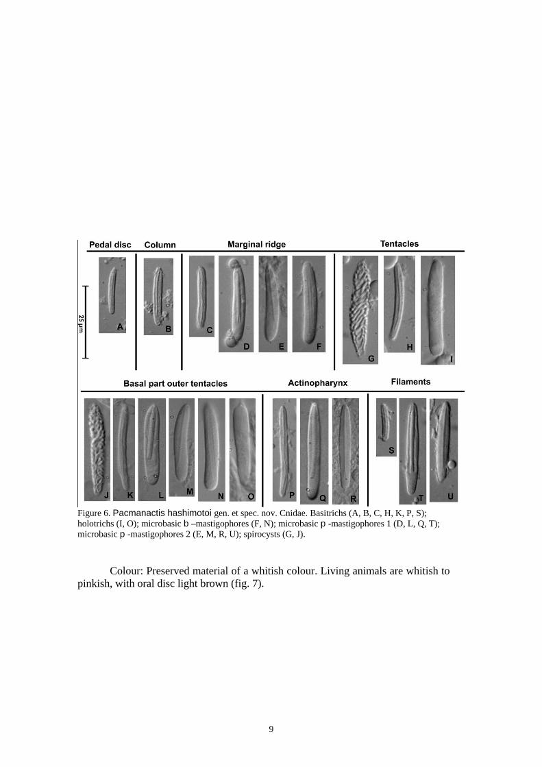
Figure 6. Pacmanactis hashimotoi gen. et spec. nov. Cnidae. Basitrichs (A, B, C, H, K, P, S); holotrichs (I, O); microbasic b –mastigophores (F, N); microbasic p -mastigophores 1 (D, L, Q, T); microbasic p -mastigophores 2 (E, M, R, U); spirocysts (G, J).
Colour: Preserved material of a whitish colour. Living animals are whitish to pinkish, with oral disc light brown (fig. 7).
9

Figure 7. Photographs taken in situ by the submarine Shinkai 2000 (BIOACCESS’98 cruise, 22 Nov. 1998) at Barnacle site (Field E, Manus Basin). (A) Specimens of Pacmanactis hashimotoi gen. et spec. nov. are visible in the foreground, among mytilid bivalves (Bathymodiolus brevior ), gastropods (Ifremeria nautilei ), and galatheid crabs (Munidopsis lauensis ). B, photograph of the same site, focus on a specimen of Pacmanactis hashimotoi gen. Et spec. nov., one specimen of the gastropod (Ifremeria nautilei ) and some juvenile mytilid bivalves, on basaltic substratum.
Habitat.— The new species was found in two sites at PACMANUS area: Barnacles site (Field E) and Kai Kai site (Field D), distant from ca 300 m. Barnacles site is located at the base of a small group of chimneys from 6 to 8 m height, either inactive or emitting a black fluid at 260-270°C. The vent fauna is established around these edifices, between cracks emitting a transparent fluid at measured at 5-40°C (Hashimoto et al., 1999). It consists of mussel (Bathymodiolus spp.) and gastropod beds (numerous Ifremeria nautilei Bouchet & Warén, 1991 and some Alviniconcha sp. 1?), galatheid crabs Munidopsis lauensis Baba & Saint Laurent, 1993, barnacles (Eochionelasmus ohtai manusensis Yamaguchi & Newman, 1997 and Leucolepas cf. longa Southward & Jones, 2003), alvinocaridid and hyppolytid shrimps (Alvinocaris longirostris Kikuchi & Ohta, 1995, and Lebbeus washingtonianus Rathbun, 1902); numerous crabs (Austinograea alayseae, Guinot, 1989), holothuroids (Chiridota hydrothermica
10

Smirnov et al., 2000), and zoarcid fish (Pyrolycus manusanus Machida & Hashimoto, 2002). A patch of these animals are observed in Fig. 7A. On the video-tapes, the sea anemones appear quite rarely with patches of 3-6 individuals. The new form described here lived in low numbers on live mussels or rocks, at 10 cm of a crack emitting a transparent fluid at ca 40°C. The temperaturewithin the mussel ranged from 7.3° to 14.7°C, and from 3.9 to 5.8°C (average, 4.6°C), 10 cm above the mussel (S. Tsuchida, pers. comm.). Isolated specimens are also present outside of the thermal vent influenced area, in surrounding water at 2.8°C, where isolated mytilid bivalves Bathymodiolus sp. - and siboglinid tube-worm (Arcovestia ivanovi Southward & Galkin, 1997). also occurred (MS pers. obs.), and where the seawater temperature was 2.88C. Galkin (1997) pointed out the abundance of holothuroid C. hydrothermica “at the periphery of vent fields, where actinostolid actiniaria were present”. Kai Kai site is one of the two large diffuse vent sites of the Field D where not chimneys occur, but where there are large diffuse vents with cracks emitting strong shimmering water at 30°C (Auzende et al., 1997). The fauna accompanying the sea anemone collected here is very similar to the previously described site, including vestimentiferan worms Alaysia sp., Escarpia sp. and Lamellibrachia sp. (Hashimoto et al., 1999).
According to in situ observations and harvests, several species of sea anemones occurred among the hydrothermal communities of PACMANUS. The most abundant species _/ ‘‘actinostolid sp. A’’ _/ is of small size (diameter up to 3.5 mm), generally fixed on the tubes of the vestimentiferan. Its histological study, made difficult by the presence of many mineral grains and internal preservation state, is deferred. Table I. Size ranges of the cnidae of Pacmanactis hashimotoi gen. et spec. nov. : mean. SD: standard deviation. Samples: the ratio indicates the number of polyps in which each cnidae was found out and the number of polyps of each species examined. N: indicates the total number of capsules measured. F: Frequency: +++ =very common, ++ =common, + = rather common, --- =/sporadic; two symbols indicate observed frequency range in different examined specimens. Abbreviation: Mc, Microbasic. See also Figure 6.
11

Etymology.— The specific name is given in honour of Jun Hashimoto (Nagasaki University, Japan) for his valuable contribution to the knowledge of chemoautotrophic ecosystems.
Geographic and depth distribution.— At present, Pacmanactis hashimotoi, is known only from the Manus Basin, SW Pacific, between 1620-1680 m in depth.
Discussion Taxonomic remarks.— According to Carlgren’s monograph (1949), Fautin & Hessler’s key (1989), and López-González et al. (2003), the present material shares the following set of characters with the actinostolid genera Anthosactis Danielssen, 1890, Marianactis Fautin & Barber, 1999, and Paranthosactis López-González, et al., 2003: (1) column smooth, not divisible in regions, (2) mesenteries not arrayed according to the Actinostola rule, (3) all stronger mesenteries fertile, (4) longitudinal muscles of tentacles ectodermal, (5) tentacular mastigophoresin the tentacles. This set of characters excludes from the following comparison several actinostolid genera such as: (1) Hormosoma Stephenson, 1918 and Cnidanthus Carlgren, 1927 by having a mesogloeal longitudinal tentacular muscle, (2) Tealidium Hertwig, 1882 and Hadalanthus Carlgren, 1956 by having a column divisible in scapus and scapulus, and (3) Bathydactylus Carlgren, 1928, Cnidanthea Carlgren, 1959, and Epiparactis Carlgren, 1921 by the absence of tentacular mastigophores, as well as the presence of only six pair of perfect mesenteries, being twelve pairs in Pacmanactis . The genus Paranthus Andres, 1883 was characterized in the last key to this set of actinostolid genera by the absence of tentacular mastigophores (see Fautin & Hessler 1989, p. 817). However, microbasic p-mastigophores are present in the tentacles of the type species, Paranthus rugosus Andres, 1880 (see Schmidt 1972, p. 58, Lopez-Gonzalez & Garcıa-Gomez 1994, p. 91). Nevertheless, Paranthus species have elongated bodies, with more mesenteries distally than proximally, while the body of Pacmanactis is clearly shorter, with equal number of proximal and distal mesenteries. The genus Anthosactis is clearly distinguishable from Pacmanactis by the dissimilar development of the ectodermal longitudinal musculature in the tentacles between oral and aboral sides, stronger on the oral being (see Hertwig, 1882; Riemann-Zürneck, 1997; White et al., 1999), by the relative thin body wall, and by the presence of parietobasilar muscles (distinctly marked but without forming a separate lamella). In Pacmanactis the longitudinal tentacle musculature is equally well developed on all sides, the body wall is relatively thick, and parietobasilar muscles are not developed. Furthermore, in Anthosactis, the outer tentacles are only a little shorter than the inner ones or all tentacles are of equal length, while in Pacmanactis ourter tentacles are distinctly shorter than inner ones. On the other hand, Anthosactis and Pacmanactic both present microbasic b-mastigophores at the base of the aboral side of the outer tentacles. However, Pacmanactis possesses tentacular microbasic p-mastigophores. The concentration of microbasic b-mastigophores and microbasic p-mastigophores in the tentacles of Pacmanactis clearly presents a continuation of the cnidae of the column, specifically from the oral side of the circumferential marginal ridge, running on the base of the aboral side of the outer tentacles (see fig. 5E, F of this paper). The genus Marianactis differs from Pacmanactis by its parietobasilar musculature wich differentiate as separate lamella, undifferentiated in Pacmanactis. In addition, in
12

Marianactis, the sphincter is not marked as a circumferential marginal ridge, there is only one cycle of perfect mesenteries, and there are three cycles of fertile mesenteries; while in Pacmanactis, the sphincter is marked as a circumferential marginal ridge, there are two cycles of perfect mesenteries and also two cycles of fertile mesenteries. On the other hand, Marianactis and Pacmanactis both present a concentration of nematocysts at the aboral basal part of the outer tentacles. These cnidae were identified as microbasic amastigophores by Fautin and Hessler (1989) in M. bythios , while microbasic b-mastigophores and microbasic p-mastigophores are present in the basal part of the outer tentacles of Pacmanactis The only known species in the genus Maractis, M. rimicarivora, can be differentiate from Pacmanactis hashimotoi by the column, much broader proximally in Maractis, while Pacmanactis is characterized by its broader oral disc. (not only in living specimens, but also observable in not-fully-retracted polyps) The body wall of Maractis is relatively thin, while that of Pacmanactis is relatively thick. Moreover, in Maractis the tentacles are relatively longer in living (see Lopez-Gonzalez et al. 2003: fig. 2F) and in preserved (see Fautin & Barber, 1999: fig. 1; López-González et al., 2003: fig. 14) state, giving a completely different general aspect (see for comparison Figure 2 of this paper for preserved material and Figure 7 for living specimens Pacmanactis hashimotoi). The marginal ridge produced by the distal part of the sphincter is distinct in Pacmanactis as well as the concentration of microbasic b-mastigophores and microbasic p-mastigophores at the oral side of this ridge and aboral part of outer tentacles basally, while all these characters are absent in Maractis. Both genera also differ in the number of perfect cycles of mesenteries (one in Maractis and two in Pacmanactis), the number of fertile cycles of mesenteries (4 in Maractis and only 2 in Pacmanactis), and in the presence of parietobasilar musculature only differentiate in the stronger cycles in Maractis, but not differentiate at all in Pacmanactis. Paranthosactis, the last actinostolid genus described, from hydrothermal vents, is in some features close to Pacmanactis, apart from other characteristics already enumerated for the group of actinostolid genera discussed here, they share the presence of marginal ridge and the absence of parietobasilar musculature.In Paranthosactis, the larger specimen examined (about 16 mm of column basal diameter excluding the characteristic expanded pedal disc)showed the fourth cycle incomplete, and (in all specimens) only the first cycle was perfect and the three older cycles were fertile, while in Pacmanactis (as already commented), the larger specimens (10 mm in maximum diameter) had the fourth cycle of mesenteries complete and all the specimens dissected showed the two older cycles perfect and fertile, the third and fourth being sterile. The body wall of Paranthosactis is relatively thin, while that of Pacmanactis is relatively thick (see López-González et al. 2003, fig. 4A and B, and fig. 4A and B of this paper). Finally, in Paranthosactis the pedal disc, while in Pacmanactis the oral disc is distinctly wider than the pedal disc. The observed minute ‘tubercle-like’ structures in Pacmanactis hashimotoi are not specifically discussed here because their presence has only been corroborated in the two larger specimens. Apart from this unclear structure, all the material here examined of P. hashimotoi do agree in external and internal anatomy, as well as cnidae measurements. The study of further collections preferably on larger specimens will provide more data on the histology of these structures and their possible taxonomic or functional
13

In table II, we present a tabular key comparing the selected group of actinostolid genera discussed above. Table II. Tabular key comparing the diagnostic characters of the set of actinostolid genera discussed in this paper. Abbreviations as follow: Column surface: S, smooth; P, with mesogloeal papillae. Number of mesenteries distally vs. proximally: D >/P, more mesenteries distally; D=/P, equal number of mesenteries distally that proximally; D<P, more mesenteries proximally than distally;?, no data. Basal battery of microbasic b-mastigophores on aboral side of outer tentacles: +/, present; -, absent. Microbasic b-mastigophores in tentacles: +/, present; -/, absent. Column regions: N, no distinct regions observed; SS, scapus and capulus distinctly present. Tentacular mastigophores: _/, present; -, absent. Oral disc vs. pedal disc: PD, pedal disc wider than oral disc; =/, pedal disc and oral disc similar in diameter; OD, oral disc wider than pedal disc. Column wall thickness: Tn, thin; Tc, thick. Development of longitudinal tentacles muscles: O, stronger developed on the oral sides than on the aboral sides; S, similar developed on oral and aboral sides. Sphincter: R, marked as a circumferencial marginal ridge; N, no marked as a circumferencial marginal ridge; nd, no data. Parietobasilar muscles: DL, differentiated as a separate lamella; DN, Distinctly marked but without forming a separate lamella; N, not differentiated as a separate lamella. Maximum number of cycles of mesenteries and mesenteries perfects: as indicated. Number of cycles of fertile mesenteries: as indicated. Tentacular cnidome: Sp, Spirocysts; Bs, Basitrichs, m-bm, Microbasic
14

Ecological remarks. The trophic behavior of the new species is not known. Maractis rimicarivora, associated with the Mid-Atlantic Ridge hydrothermal vents, can feed on shrimps (Rimicaris exoculata), which particularly abundant on these sites (Van Dover et al., 1997). The actinostolid Marianactis cf. bythios (Van Dover, 2002), wich is very abundant on the recently discovered site Kairei Field (Rodriguez Triple Junction, eastern Indian Ocean), might similarly feed on the shrimps Rimicaris kairei (Van Dover et al., 2001). It is unclear if shrimps are the only prey of the actinostolid anemones. Indeed, within the community of Mariana back-arc basin, where shrimps are not very abundant, the anemone Marianactis bythios was the dominant biomass species inhabiting the periphery of the vents. In comparison to the majority of the Mid-Atlantic Ridge and East Pacific Ridge hydrothermal sites, the Lau, N-Fiji and Manus back-arc basins shelter small sized and less abundant anemones. No anemones were observed at the Manus Basin sites. It is difficult to say if it is the consequence of a declining activity, as it was suggested for the Kai-Kai site, following two observations in 1996 and 1998 (Hashimoto et al., 1999). Pacmanactis hashimotoi has not been identified with certainty outside the areas studied in this paper. However, several ‘whitish’ sea anemones are often recorded by photographic and video devices in many other chemoautotrophic environments, as for example SEPR (South East Pacific Rise, BIOSPEEDO cruise). The detailed study of that material will be the goal of further contributions. Additional studies of the anemones collected by the French cruise BIOLAU and the French-Japanese cruise STARMER in 1989 (Desbruye`res et al. 1994) might give new insights into biogeographic relationships among anemones of the western Pacific back-arc basins, and, in general, in other Pacific vent sites. Acknowledgements The authors thank the Chief scientist Jun Hashimoto of the BIOACCESS’96 and BIOACCESS’98 (Biological Investigation Of A Chemosynthetic Community: Ecological and Systematic Survey) cruises, organized JAMSTEC, and the crew of RV Natsushima and submersible Shinkai 2000, for collecting specimens and making available them for study. Sincere thanks are extended to Shinji Tsuchida (JAMSTEC, Japan), for his improvements to the manuscript. We also thank V. Martin for preparing the map (fig. 1). Partial support was provided by a MCT-CSIC grant (I3P-BPD2001) to Estefanía Rodríguez. Tony Krupa is thanked for reviewing the final English version. The authors are also thankful for comments and suggestions given by Vreni Häussermann and an anonymous referee.
References Auzende, J.-M., J. Hashimoto, A. Fiala-Medioni, S. Ohta and E.d. BIOACCESS. –
1997. Etude géologique et biologique in situ de deux zones hydrothermales du bassin de Manus (Papouasie Nouvelle-Guinée). C. R. Acad. Sc. Paris, 325: 585-591.
Carlgren, O. – 1949. A survey of the Ptychodactiaria, Corallimorpharia and Actiniaria. Kungl. svenska Vetensk. Handl., ser. 4, 1(1): 1-121, pls 1-4.
Desbruyères, D. and M. Segonzac (eds). – 1997. Handbook of deep-sea hydrothermal vent fauna. Editions Ifremer, Plouzané, France.
Desbruyères, D., J. Hashimoto and M.-C. Fabri. – submitted. Composition and Biogeography of Hydrothermal Vent Communities in Western Pacific Back-Arc Basins. Geophys.-Monogr.
15

Desbruyères, D., A.-M. Alayse-Danet, S. Ohta, and the Scientific Parties of Biolau & Starmer cruises. – 1994. Deep-sea hydrothermal communities in southern Pacific back-arc basins (the North Fiji and Lau Basins): Composition, micro-distribution and food-web. Mar. Geol., 116, 227-242.
Doumenc, D. and M. Van-Praët. – 1988. Actinies abyssales d’un site hydrothermal du Pacifique oriental. Oceanol. Acta, spec. vol., 8: 61-68.
Fautin, D.G. and B.R. Barber. – 1999. Maractis rimicarivora, a new genus and species of sea anemone (Cnidaria: Anthozoa: Actiniaria: Actinostolidae) from an Atlantic hydrothermal vent. Proc. Biol. Soc. Wash., 112(3): 624-631.
Fautin, D.G. and R.R. Hessler. – 1989. Marianactis bythios, a new genus and species of Actinostolid sea anemone (Coelenterata: Actiniaria) from the Mariana vents. Proc. Biol. Soc. Wash., 102(4): 815-825.
Gabe, M. – 1968. Technique Histologique: i-iv, 1-1113. Masson et Cie (eds), Paris. Hashimoto, J., S. Ohta, A. Fiala-Médioni, J.-M. Auzende, S. Kojima, M. Segonzac, Y.
Fujiwara, J.C. Hunt, K. Gena, T. Miura, T. Kikuchi, T. Yamaguchi, T. Toda, H. Chiba, S. Tsuchida, J. Ishibashi, K. Henry, M. Zbinden, A. Pruski, A. Inoue, H. Kobayashi, J.-L. Birrien, J. Naka, T. Yamanaka, C. Laporte, K. Nishimura, C. Yeats, S. Malagun, P. Kia, M. Oyaizu and T. Katayama. – 1999. Hydrothermal vent communities in the Manus Basin, Papua New Guinea: Results of the BIOACCESS cruises’96 and ’98. InterRidge News, 8(2): 12-18.
Galkin, S.V. – 1997. Megafauna associated with hydrothermal vents in the Manus Back-Arc Basin (Bismarck Sea). Mar. Geol., 142: 197-206.
Hashimoto, J. – 2001. A New Species of Bathymodiolus (Bivalvia:Mytilidae) from Hydrothermal Vent Communities in the Indian Ocean. Venus, 60(3): 141-149.
Hashimoto, J., S. Ohta, T. Gamo, H. Chiba, T. Yamaguchi, S. Tsuchida, T. Okudaira, H. Watabe, T. Yamanaka & M. Kitazawa. – 2001. First Hydrothermal Vent Communities from the Indian Ocean Discovered. Zool. Sci., 18(5): 717-721.
Hertwig, R. – 1882. Report on the Actiniaria dredged by H.M.S. Challenger during the years 1873-1876. Sci. Res. Voy. H.M.S. Challenger, Zoology, 6(1): 1-136, pls 1-14.
Johansen, D.A. – 1940. Plant microtechniques. Mc Graw-Hill. New York & London. Kojima, S. S. Ohta, Y. Fujiwara and J. Hashimoto. – 1999. Speciation of gastropods of
the genus Alviniconcha. JAMSTEC J. Deep-Sea Res., 14: 501-506. López-González, P.J., E. Rodríguez, J.-M. Gili and M. Segonzac. – 2003. New records
of sea anemones (Anthozoa: Actiniaria) from hydrothermal vents and cold seeps. Zool. Verh., Leiden, 345: 215-243.
Riemann-Zürneck, K. – 1997. Anthosactis janmayeni Danielssen, 1890, a rare high-arctic sea anemone. Polar Biol., 17: 487-491.
Southward E.C. and S.V. Galkin. – 1997. A new vestimentiferan (Pogonophora: Obturara) from hydrothermal vent fields in the Manus Back-Arc Basin (southwest Pacific Ocean). J. of Nat. Hist., 31: 43-45.
Van Dover, C.L., M. Polz, J. Robinson, C. Cavanaugh, D. Kadko and J.P. Hickey. — 1997. Predatory anemones at TAG. Bridge Newsletter, 12: 33-34.
Van Dover, C.L. — 2002. Trophic relationships among invertebrates at the Kairei hydrothermal vent field (Central Indian Ridge). Mar. Biol., 141: 761-772.
White, T.R., A.K. Wakeefield Pagels and D.G. Fautin. – 1999. Abyssal sea anemones (Cnidaria, Anthozoa) of the northeast Pacific symbiotic with molluscs: Anthosactis nomados, a new species, and Monactis vestita (Gravier, 1918). Proc. Biol. Soc. Wash., 112(4): 637-651.
16
Copyright © 2022 FDOKUMEN