Potentiation of ATP-induced currents due to the activation of P2X receptors by ubiquitin...
12
Potentiation of ATP-induced currents due to the activation of P2X receptors by ubiquitin carboxy-terminal hydrolase L1 Yoshimasa Manago,* Yoshiko Kanahori,* Aki Shimada,* Ayumi Sato,* Taiju Amano,* Yae Sato-Sano,* , Rieko Setsuie,* , Mikako Sakurai,* , Shunsuke Aoki, Yu-Lai Wang, Hitoshi Osaka, , à Keiji Wada and Mami Nodaà *Laboratory of Pathophysiology, Graduate School of Pharmaceutical Sciences, Kyushu University, Fukuoka, Japan Department of Degenerative Neurological Diseases, National Institute of Neuroscience, National Center of Neurology and Psychiatry, Tokyo, Japan àInformation and Cellular function, PRESTO, Japan Science and Technology Corporation (JST), Kawaguchi, Saitama, Japan Abstract Mammalian neuronal cells abundantly express a de-ubiquiti- nating isozyme, ubiquitin carboxy-terminal hydrolase L1 (UCH L1). Loss of UCH L1 function causes dying-back type of axonal degeneration. However, the function of UCH L1 in neuronal cells remains elusive. Here we show that overexpression of UCH L1 potentiated ATP-induced currents due to the activa- tion of P2X receptors that are widely distributed in the brain and involved in various biological activities including neurosecre- tion. ATP-induced inward currents were measured in mock-, wild-type or mutant (C90S)-UCH L1-transfected PC12 cells under the conventional whole-cell patch clamp configuration. The amplitude of ATP-induced currents was significantly greater in both wild-type and C90S UCH L1-transfected cells, suggesting that hydrolase activity was not involved but in- creased level of mono-ubiquitin might play an important role. The increased currents were dependent on cAMP-dependent protein kinase (PKA) and Ca 2+ and calmodulin-dependent protein kinase (CaMKII) but not protein kinase C. In addition, ATP-induced currents were likely to be modified via dopamine and cyclic AMP-regulated phosphoprotein (DARPP-32) that is regulated by PKA and phosphatases. Our finding shows the first evidence that there is a relationship between UCH L1 and neurotransmitter receptor, suggesting that UCH L1 may play an important role in synaptic activity. Keywords: CaMKII, DARPP-32, PKA, patch-clamp, PC12, UCH L1. J. Neurochem. (2005) 92, 1061–1072. The ubiquitin-proteasome system is an evolutionarily con- served and energy-dependent proteolytic pathway that func- tions constitutively to degrade proteins. Recent studies indicate that ubiquitin-mediated proteolysis can also be regulated and is of widespread importance (Wilkinson 1995; Coux et al. 1996). Regulated proteolysis by the ubiquitin- Received August 24, 2004; accepted October 8, 2004. Address correspondence and reprint requests to Mami Noda, PhD, Laboratory of Pathophysiology, Graduate School of Pharmaceutical Sciences Kyushu University, 3-1-1 Maidashi, Higashi-ku, Fukuoka 812– 8582, Japan, Tel./Fax: + 81-92-642-6574. E-mail: [email protected] Abbreviations used: ATP, adenosine triphosphate; BSA, bovine serum albumin; CaMKII, Ca 2+ and calmodulin-dependent protein kinase; CDK, cyclin-dependent kinase; CHO, cells, Chinese hamster ovary cells; CREB, Ca 2+ -stimulated cAMP response element binding protein; DAPI, 4¢, 6-diamidino-2-phenylindole, dihydrochloride; EGTA, ethyleneglycol- bis-N, N, N¢,N¢-tetraacetic acid; ERK, extracellular signal-regulated kinase; DARPP-32, dopamine and cyclic AMP-regulated phosphopro- tein with molecular weight of about 32 000; 1,9-dideoxyforskolin, 7b-acetoxy-6b-hydroxy-8,13-epoxy-labd-14-en-11-one; FBS, fetal bovine serum; forskolin, 7b-acetoxy-8,13-epoxy-1a,6b,9a-trihydroxy-labd- 14-en-11-one; H-89, N-[2-(p-bromoocinamylamino)ethyl]-5-isoquino- linesulfonamide; HS, horse serum; KN-93, 2-[N-(2-hydroxyethyl)]- N-(4-methoxybenzenesulfonyl)]amino-N-(4-chlorocinnamyl)-N-methyl- benzylamine); HEPES, N-2-hydroxyethylpiperazine-N¢-2-ethansulfonic acid; MAPK, mitogen-activated protein kinase; NGF, nerve growth factor; PBS(–), Dulbecco’s Ca 2+ , Mg 2+ -free phosphate buffer saline; PC12, cells, rat phenochromocytoma cells; PD98059, 2¢-Amino-3¢- methoxyflavone; PGP9.5, protein gene product 9.5; PKA, cyclic AMP- dependent protein kinase; PKC, protein kinase C; PP1, protein phos- phatase 1; PP2, protein phosphatase 2; SDS-PAGE, sodium dodecyl sulfate-polyacrylamide gel electrophoresis; TBST, Tris buffer saline- Tween; Thr-34, threonine at 34; Thr-75, threonine at 75; Roscovitine, 2-(R)-(1-Ethyl-2-hydroxyethylamino)-6-benzylamino-9-isopropylpurine; UCH, L1, ubiquitin C-terminal hydrolase L1. Journal of Neurochemistry , 2005, 92, 1061–1072 doi:10.1111/j.1471-4159.2004.02963.x ȑ 2005 International Society for Neurochemistry, J. Neurochem. (2005) 92, 1061–1072 1061
-
Upload
independent -
Category
Documents
-
view
7 -
download
0
Transcript of Potentiation of ATP-induced currents due to the activation of P2X receptors by ubiquitin...

Potentiation of ATP-induced currents due to the activation of P2Xreceptors by ubiquitin carboxy-terminal hydrolase L1
Yoshimasa Manago,* Yoshiko Kanahori,* Aki Shimada,* Ayumi Sato,* Taiju Amano,*Yae Sato-Sano,*,� Rieko Setsuie,*,� Mikako Sakurai,*,� Shunsuke Aoki,� Yu-Lai Wang,�Hitoshi Osaka,�,� Keiji Wada� and Mami Noda�
*Laboratory of Pathophysiology, Graduate School of Pharmaceutical Sciences, Kyushu University, Fukuoka, Japan
�Department of Degenerative Neurological Diseases, National Institute of Neuroscience, National Center of Neurology and
Psychiatry, Tokyo, Japan
�Information and Cellular function, PRESTO, Japan Science and Technology Corporation (JST), Kawaguchi, Saitama, Japan
Abstract
Mammalian neuronal cells abundantly express a de-ubiquiti-
nating isozyme, ubiquitin carboxy-terminal hydrolase L1 (UCH
L1). Loss of UCH L1 function causes dying-back type of axonal
degeneration. However, the function of UCH L1 in neuronal
cells remains elusive. Here we show that overexpression of
UCH L1 potentiated ATP-induced currents due to the activa-
tion of P2X receptors that are widely distributed in the brain and
involved in various biological activities including neurosecre-
tion. ATP-induced inward currents were measured in mock-,
wild-type or mutant (C90S)-UCH L1-transfected PC12 cells
under the conventional whole-cell patch clamp configuration.
The amplitude of ATP-induced currents was significantly
greater in both wild-type and C90S UCH L1-transfected cells,
suggesting that hydrolase activity was not involved but in-
creased level of mono-ubiquitin might play an important role.
The increased currents were dependent on cAMP-dependent
protein kinase (PKA) and Ca2+ and calmodulin-dependent
protein kinase (CaMKII) but not protein kinase C. In addition,
ATP-induced currents were likely to be modified via dopamine
and cyclic AMP-regulated phosphoprotein (DARPP-32) that is
regulated by PKA and phosphatases. Our finding shows the
first evidence that there is a relationship between UCH L1 and
neurotransmitter receptor, suggesting that UCH L1 may play
an important role in synaptic activity.
Keywords: CaMKII, DARPP-32, PKA, patch-clamp, PC12,
UCH L1.
J. Neurochem. (2005) 92, 1061–1072.
The ubiquitin-proteasome system is an evolutionarily con-served and energy-dependent proteolytic pathway that func-tions constitutively to degrade proteins. Recent studies
indicate that ubiquitin-mediated proteolysis can also beregulated and is of widespread importance (Wilkinson 1995;Coux et al. 1996). Regulated proteolysis by the ubiquitin-
Received August 24, 2004; accepted October 8, 2004.Address correspondence and reprint requests to Mami Noda, PhD,
Laboratory of Pathophysiology, Graduate School of PharmaceuticalSciences Kyushu University, 3-1-1 Maidashi, Higashi-ku, Fukuoka 812–8582, Japan, Tel./Fax: + 81-92-642-6574.E-mail: [email protected] used: ATP, adenosine triphosphate; BSA, bovine serum
albumin; CaMKII, Ca2+ and calmodulin-dependent protein kinase; CDK,cyclin-dependent kinase; CHO, cells, Chinese hamster ovary cells;CREB, Ca2+-stimulated cAMP response element binding protein; DAPI,4¢, 6-diamidino-2-phenylindole, dihydrochloride; EGTA, ethyleneglycol-bis-N, N, N¢, N¢-tetraacetic acid; ERK, extracellular signal-regulatedkinase; DARPP-32, dopamine and cyclic AMP-regulated phosphopro-tein with molecular weight of about 32 000; 1,9-dideoxyforskolin,7b-acetoxy-6b-hydroxy-8,13-epoxy-labd-14-en-11-one; FBS, fetal bovine
serum; forskolin, 7b-acetoxy-8,13-epoxy-1a,6b,9a-trihydroxy-labd-14-en-11-one; H-89, N-[2-(p-bromoocinamylamino)ethyl]-5-isoquino-linesulfonamide; HS, horse serum; KN-93, 2-[N-(2-hydroxyethyl)]-N-(4-methoxybenzenesulfonyl)]amino-N-(4-chlorocinnamyl)-N-methyl-benzylamine); HEPES, N-2-hydroxyethylpiperazine-N¢-2-ethansulfonicacid; MAPK, mitogen-activated protein kinase; NGF, nerve growthfactor; PBS(–), Dulbecco’s Ca2+, Mg2+-free phosphate buffer saline;PC12, cells, rat phenochromocytoma cells; PD98059, 2¢-Amino-3¢-methoxyflavone; PGP9.5, protein gene product 9.5; PKA, cyclic AMP-dependent protein kinase; PKC, protein kinase C; PP1, protein phos-phatase 1; PP2, protein phosphatase 2; SDS-PAGE, sodium dodecylsulfate-polyacrylamide gel electrophoresis; TBST, Tris buffer saline-Tween; Thr-34, threonine at 34; Thr-75, threonine at 75; Roscovitine,2-(R)-(1-Ethyl-2-hydroxyethylamino)-6-benzylamino-9-isopropylpurine;UCH, L1, ubiquitin C-terminal hydrolase L1.
Journal of Neurochemistry, 2005, 92, 1061–1072 doi:10.1111/j.1471-4159.2004.02963.x
� 2005 International Society for Neurochemistry, J. Neurochem. (2005) 92, 1061–1072 1061

proteasome pathway has been implicated in the control ofcell cycle (King et al. 1996), transcription activation (Vermaet al. 1995), antigen presentation (Rock et al. 1994), cell fateand growth (Huang et al. 1995; Zhu et al. 1996), synapto-genesis (Muralidhar and Thomas 1993; Oh et al. 1994) andmemory (Hegde et al. 1997). Ubiquitination of proteins ismediated by specific enzymes (E1, E2, and E3) andpolyubiquitinated proteins are translocated to the 26Sproteasome and subsequently proteolytically degraded (Ciec-hanover et al. 2000). Conversely, deubiquitination is thoughtto be essential for the regulation of proteolysis and forrecycling of monoubiquitin from polyubiquitin chains.
Recently, one of the deubiquitinating enzymes, UCH L1(ubiquitin carboxy-terminal hydrolase L1), was reported to beessential for brain function.UCHL1 is selectively expressed inneuron and testis (Wilkinson et al. 1989; Wilkinson et al.1992). Loss of UCH L1 function was shown to cause neuronaldegeneration observed in the gracile axonal dystrophy (gad)mouse (Saigoh et al. 1999), andmissencemutation ofUCHL1was found in familial Parkinson disease (Leroy et al. 1998).Asphysiological functions of UCH L1, it is not limited tohydrolase activity; it has been shown that it associated withmono-ubiquitin and thus stabilized free ubiquitin by prevent-ing its degradation within lysosomes. UCH L1¢s affinity forubiquitin rather than hydrolase activity was required for theregulation of ubiquitin level (Osaka et al. 2003) and UCH L1even might work as ubiquitin ligase (Liu et al. 2002).Furthermore, UCH L1 has been reported to have an importantrole in apoptosis in germ cell and neuron (Harada et al. 2004;Kwon et al. 2004b) and different UCH isozymes have distinctfunction during spermatogenesis (Kwon et al. 2004a).
As for neural function of UCH L1, little is known yet. InAplysia, homologous UCH was shown to be important forlearning and memory (Hegde et al. 1997). It is not yet knownthat UCH L1 works in a similar way in mammalian cells, butthese results strongly suggest that UCH L1 plays an importantrole in synaptic function and morphology. In the present study,we investigated the neuronal function of UCH L1 on receptorchannels which affect neurotransmitter secretion.
PC12 cells are often used as a model for studying neuronalcell function. Among neurotransmitter receptors expressed inPC12 cells, ATP receptors induce dopamine release (Selaet al. 1991). ATP receptors are divided into two subtypes,P2X and P2Y receptors. P2X receptors are ionotropicreceptors and form cationic channels, while P2Y receptorsare G-protein-coupled receptors and P2Y1, 2, 4, 6, 11 causeintracellular Ca2+ mobilization via IP3 formation and activateCa2+-dependent K+ channels (Ikeuchi et al. 1996). AmongP2X receptors, P2X2 and P2X4 receptor mRNA have beendetected in PC 12 cells (Hur et al. 2001). P2X receptorsmediate fast ionic flow and are supposed to induce depolar-ization of the cells, hence contributing to the catecholaminerelease from PC12 cells. Therefore, we first analyzed P2Xreceptors and their modulation by UCH L1. This is the first
report to show a relationship between UCH L1 andneurotransmitter receptors and may help to understand thefunction of UCH L1 in the nervous system.
Materials and methods
Cell culture
PC12 Tet-off cells were grown in RPMI-1640 medium containing
5% fetal bovine serum (FBS) (Cell Culture Technologies, CANSER
INTERNATIONAL INC., Canada), 10% horse serum (HS) (Gibco/
BRL, Grand Island, NY, USA), 100 units/mL penicillin (Life
Technologies, Rockville, MD, USA) and 100 lg/mL streptomycin
(Life Technologies) in a humidified atmosphere with 10% CO2 at
37�C. To differentiate PC12 Tet-off cells, 100 ng/mL of nerve
growth factor (NGF) was added to the RPM1640 medium with 0.1%
HS, 0.05% FBS, 50 unit/mL penicillin and 100 lg/mL streptomycin
for 4 days.
CHO-AA8-Lucl cells were maintained in Minimum Essential
Medium Eagle a modification (Sigma, St Louis, MO, USA)
containing 10% FBS, 100 units/mL penicillin, 100 lg/mL strepto-
mycin, and 4 mML-glutamine (Gibco/BRL), with 10% CO2 at 37�C.
Transfection
Plasmids used for transfection were constructed using pBI-EGFP
Tet vector (Clontech). For electrophysiological recording, PC12 Tet-
Off cells were transfected with mock, wild-type or mutant (C90S)
human UCH L1 cDNA, using Lipofectamine 2000. After 24 h,
PC12 Tet-Off cells were treated with NGF and differentiated for
4–5 days. More precisely, 3.0 · �105/dish PC12 Tet-Off cells were
seeded in 35-mm dishes in RPMI with 10% HS and 5% FBS.
Twenty-four hours after seeding, the medium was replaced with
500 lL of serum-free RPMI1640 medium. Then, the transfection
mixture containing 4 lg of cDNA and 10 lL of Lipofectamine
2000 in 500 lL of RPMI-1640 was added to each dish and
incubated for 6 h in a humidified atmosphere with 10% CO2 at
37�C. One ml of complete RPMI-1640 supplemented with an
additional 10% HS and 5% FBS was then added to each dish. The
solution for transfection was discarded 18 h later and replaced with
RPMI-1640 medium for differentiation with added 100 ng/mL
NGF. For protein analysis, CHO-AA8-Lucl cells (7.5 · �105/well,
Clontech) were transfected in the same way. After 24 h, cells were
subjected to western blot analysis or immunocytochemical analysis.
Western blot analysis
Transfected CHO-AA8-Lucl cells were washed with PBS contained
protease inhibitor and after collecting lysates in solution containing
20 mM Tris-base, 0.1% SDS, 1% sodium deoxycholate, 1% Triton
X-100 and 0.001 g/5 mL protease inhibitor and then centrifuged at
15 000 r.p.m. for 30 min at 4�C. After collecting supernatant,
protein concentrations of lysates were determined using Bio-Rad
protein assay kits (Bio-Rad, Hercules, CA, USA). Lysates were
boiled for 10 min, resolved by 10–20% gradient SDS-PAGE, and
transferred to polyvinylidene difluoride membranes (Bio-Rad) with
a semidry electroblotter (Bio-Rad). The membrane was blocked by
incubation in 1% BSA/TBST for 1 h at room temperature. Anti-
PGP9.5 (UCH L1) antibody (1 : 100, Medac) was used as a primary
antibody in Western blotting. Anti-rabbit IgG conjugated with
1062 Y. Manago et al.
� 2005 International Society for Neurochemistry, J. Neurochem. (2005) 92, 1061–1072

horseradish peroxidase (1: 2000) (Dako, Carpinteria, CA, USA) was
used as secondary antibody. Immunoreactive bands were detected
using the supersignal substrate system (Pierce, Rockford, IL, USA)
according to manufacturer’s instructions.
Immunocytochemical analysis
After transfection, cells were fixed with 4% paraformaldehyde.
Immunocytochemistry on CHO-AA8-Lucl cells and PC12 Tet-Off
cells was performed as previously described (Osaka et al. 2003)using antibodies to ubiquitin that is predominantly reactive to free
ubiuitin in immuno-histochemistry (1 : 100, Sigma; polyclonal) and
UCH L1 (1 : 100, Medac; monoclonal). For immunofluorescence
studies, antirabbit IgG conjugated with Cy3 antibodies (1 : 200,
Jackson Immuno Research) and antirabbit IgG conjugated with
Alexa Fluor 568 antibodies (1 : 1000, Molecular Probes) were used
as secondary antibodies. Also, in PC12 Tet-Off cells, 300 lM DAPI
was applied to stain transfected and untransfected cell nuclei for
5 min, and then the cells were washed with PBS for 5 min at least
five times. Twenty confocal images with 0.5 lm width were
obtained and reconstructed using the confocal laser microscope
system (Radiance2100, Bio-Rad). To stain mono-ubiquitin, the same
laser strength was used in mock, wild-type and C90S UCH L1-
transfeacted cells under the confocal laser microscope system
(LSM510, Carl Zeiss, Germany).
Electrophysiological measurements
The cell with fluorescence was chosen under the fluorescence
microscope. Then, the patch pipette was applied to the cell to
obtain a giga-ohm seal under the phase bright mode. Whole-cell
recordings were made as reported previously (Noda et al. 1999,2000), using an Axopatch-200B amplifier (Axon Instruments,
Foster City, CA, USA), under voltage-clamp condition at the
holding potential of )70 mV. Membrane currents were measured
using a patch pipette containing (in mM): CsCl, 120; Mg2ATP3,
3; HEPES, 20; CaCl2, 1; MgCl2, 1; EGTA, 5. The pH of the
solution was adjusted to 7.2 with 1 N CsOH. The pipette
resistance was 5–9 MX. The external solution contained (mM):
NaCl, 132; KCl, 5; CaCl2, 2; MgCl2, 1; glucose, 10; and HEPES,
10. The pH was adjusted to 7.4 with 1 N NaOH. External ATP
or drugs were applied rapidly using the ‘Y tube’ technique (Min
et al. 1996), which allows the complete exchange of the external
solution surrounding a cell within 20 ms. Temperature monitored
in the recording dishes was 33–34�C.In the experiments using inhibitors except PD98059, ATP was
applied twice to ensure reproducibility of the ATP-induced current in
control experiments. The inhibitor solution was applied after first
application of ATP for the period according to the references for each
inhibitor until the end of second application of ATP. The current
amplitude obtained at the second application of ATP with or without
inhibitors normalized to the first ATP-induced current. All valueswere
presented asmeans ± SEM. Statistical analysis was done using ANOVA.
A value of p < 0.05 was considered to be the minimum level of
significance. Curve fitting was performed using Hill Equation (Igor
Pro 4.07; Wavemetrics, Lake Oswego, OR, USA).
SYBR Green-based Real-Time Quantitative RT–PCR
Total RNAs were prepared from 2 · 106 PC12 Tet-Off cells and
a rat brain with RNeasyTM. RNA purification kit (QIAGEN,
Valencia, CA, USA) according to the manufacturer’s protocol.
First-strand cDNA synthesized from 1 lg total RNA with random
hexamer primers was used as template for each reaction. SYBR
Green-based Real-time Quantitative RT-PCR was performed as
described (Wong et al. 2000; Aoki et al. 2002). Applied
Biosystems 7700 Sequence Detection System was used for the
signal detection and the PCR was performed in 1 · SYBR Green
Master mix (Applied Biosystems, Foster City, CA, USA) and
50 nM of each primer. For standardization and quantification, rat
b-actin or rat glyceraldehyde 3-phosphate dehydrogenase (GAP-
DH) was amplified simultaneously. Primer sequences were
designed with Primer ExpressTM. Software (Applied Biosystems).
The following primer pairs were employed: 5¢-TGCAGACCAT-CAGCAACCTG-3¢ (upper, 17–36) and 5¢-CTTGTGGATACCC-CAGCTCC-3¢ (lower, 103–84) for amplification of rat DARPP-32
(primer set A; GenBank accession No. AF281661): 5¢-CACCT-GCAGACCATCAGCAA-3¢ (upper, 13–32) and 5¢-CCTCTTGT-GGATACCCCAGC-3¢ (lower, 106–87) for amplification of rat
DARPP-32 (primer set B; AF281661): 5¢- ATCGCTGACAGGA-TGCAGAAG-3¢ (upper, 925–945) and 5¢-AGAGCCACCAAT-CCACACAGA-3¢ (lower, 1032–1012) for amplification of rat
b-actin: and 5¢-ACCACAGTCCATGCCATCAC-3¢ (upper, 586–
605) and 5¢- TCCACCACCCTGTTGCTGTA-3¢ (lower, 1037–
1018) for amplification of rat GAPDH. PCR conditions were:
95�C for 10 min, followed by 40 cycles at 95�C for 15 s and
60�C for 1 min. The threshold cycle of each gene was determined
as the PCR cycle at which an increase in fluorescence was
observed above the baseline signal in an amplification plot (Wada
et al. 2000). The ‘normalized expression level of target’ (dCt)
was calculated as the difference in threshold cycles for target and
reference (b-actin or GAPDH). Subtraction of dCt for PC12 Tet-
Off cells from dCt for the rat brain provided the ddCt value. The
formula, 2–ddCt, was used to calculate relative expression levels
for PC12 Tet-Off cells compared to the rat brain. To reduce
possible error, RT-PCR reaction was performed three times and
averaged 2–ddCt values were obtained. In addition, two gene-
specific primer sets (set A and B, see above) for DARPP-32 gene
and two independent RNA pools were examined to confirm
DARPP-32 gene expression.
Drugs and reagents
RPMI-1640 medium, ATP-2Na, H-89, chelerythrine, roscovitine,
PD98059, forskolin and 1,9-dideoxyforskolin were from SIGMA
(St. Louis, MO, USA). NGF and Lipofectamine 2000 were from
Gibco/BRL (Grand Island, NY, USA). KN-93 and okadaic acid was
from CALBIOCHEM (San Diego, CA, USA).
Results
Transfection of UCH L1 in PC12 Tet-Off cells
Expression activity of plasmid constructs was first examinedin CHO-AA8-Lucl cells that lack endogenous expression ofUCH L1 (Fig. 1). Confocal microscopic examinationrevealed that UCH L1 immunoreactivity colocalizes withGFP fluorescence (Fig. 1a). Western blot analysis showedbands immunostained by anti-UCH L1 antibodies were
Potentiation of ATP-induced currents by UCH L1 1063
� 2005 International Society for Neurochemistry, J. Neurochem. (2005) 92, 1061–1072

detected in cells transfected with pBI-EGFP-wild type UCHL1 or C90S UCH L1, but not mock plasmids (Fig. 1b).Figure 1(c) shows a PC12 Tet-Off representative cell withgreen fluorescence used for electrophysiological recording,although DAPI staining was not employed for the recording.The efficacy of the transfection was about 10% in PC12 Tet-Off cells.
Effects of overexpression of UCH L1 on ATP-induced
currents
ATP-activated inward currents due to the activation of P2Xreceptors at the negative holding potential in PC12 cells werereported (Nakazawa et al. 1994). In our experiments, PC12Tet-Off cells were voltage clamped at )70 mV and highconcentration of ATP were used to see whether or notoverexpression of UCH L1 affected maximum inwardcurrents. In UCH L1-transfected PC12 Tet-Off cells, ATP-induced inward currents were significantly larger than thosein mock-transfected cells. Unexpectedly the mutant (C90S)UCH L1, which lacks C-terminal hydrolase activity butretains ubiquitin binding affinity, had a similar effect to wild-type UCH L1 (Fig. 2a). The amplitude of peak inwardcurrents in mock-, wild-type UCH L1- and C90S UCHL1-transfected PC12 Tet-Off cells were 15.0 ± 1.6 pA/pF(n ¼ 5), 48.5 ± 6.0 pA/pF (n ¼ 6) and 47.6 ± 4.1 pA/pF(n ¼ 6), respectively (Fig. 2b).
The current-voltage relationships of ATP-induced inwardcurrents were analyzed by applying voltage steps of 10 mVincrements between )100 mV and + 50 mV with 50 msduration and 50 ms interval from the holding potential of)70 mV before and during the application of ATP (Fig. 3a).The current traces before and after application of ATP inwild-type UCH L1-transfected PC12 Tet-Off cells wereshown by applying voltage steps (Fig. 3b). Holding currentswere negligibly misaligned even during the application ofATP. The current levels at the end of each pulse before andduring ATP application were obtained in mock-, wild-typeUCH L1- or C90S UCH L1-transfected PC12 Tet-Off cells.
(a)
(b)
(c)
Fig. 1 Transfection efficacy of UCH L1 in CHO-AA8-Lucl cells and
PC12 Tet-Off cells. (a) Confocal image of CHO-AA8-Lucl cells 24 h
after transfection of pBI-EGFP-mock, wild type UCH L1 and C90S
UCH L1 with Lipofectamine 2000 were double stained with UCH L1
(red). Cells with green fluorescence (GFP) are the transfected cells.
(b) Western-blot analysis of CHO-AA8-Lucl cells 24 h after transfec-
tion of pBI-EGFP-mock, -wild type UCH L1 and -C90S UCH L1 with
Lipofectamine 2000. CHO-AA8-Lucl cells were lysed with TBS buffer
containing 0.1% Triton X. 10 lg of each protein was subjected to SDS-
PAGE and immunoblotted with anti-PGP9.5 antibody. (c) Confocal
image of overexpressed UCH L1 in PC12 Tet-Off cells were double
stained with GFP (green) and DAPI (blue).
Fig. 2 Current amplitudes of ATP-induced currents in mock-, wild-
type UCH L1- and C90S UCH L1-transfected PC12 Tet-Off cells. (a)
Inward membrane currents induced by 1 mM ATP at the holding
potential of ) 70 mV in mock-, wild-type (wt) and C90S UCH
L1-transfected PC12 Tet-Off cells. (b) Amplitudes of peak inward
currents induced by 1 mM ATP in mock-, wild-type and C90S UCH
L1-transfected PC12 Tet-Off cells. The bars represent the mean ±
SEM. ***:p < 0.001.
1064 Y. Manago et al.
� 2005 International Society for Neurochemistry, J. Neurochem. (2005) 92, 1061–1072

Then the amplitudes of ATP-induced currents at differentvoltages were obtained by subtracting the one beforeapplication of ATP from the one during application of ATP,and were plotted as in Fig. 3(c). In consideration ofdesensitization, the current-voltage relationships wereobtained by applying voltage steps in opposite direction,i.e. from + 50 to ) 100 mV, but there was almost no change(data not shown). The reversal potential was about 0 mV,suggesting that these currents were due to non-specificcationic channels.
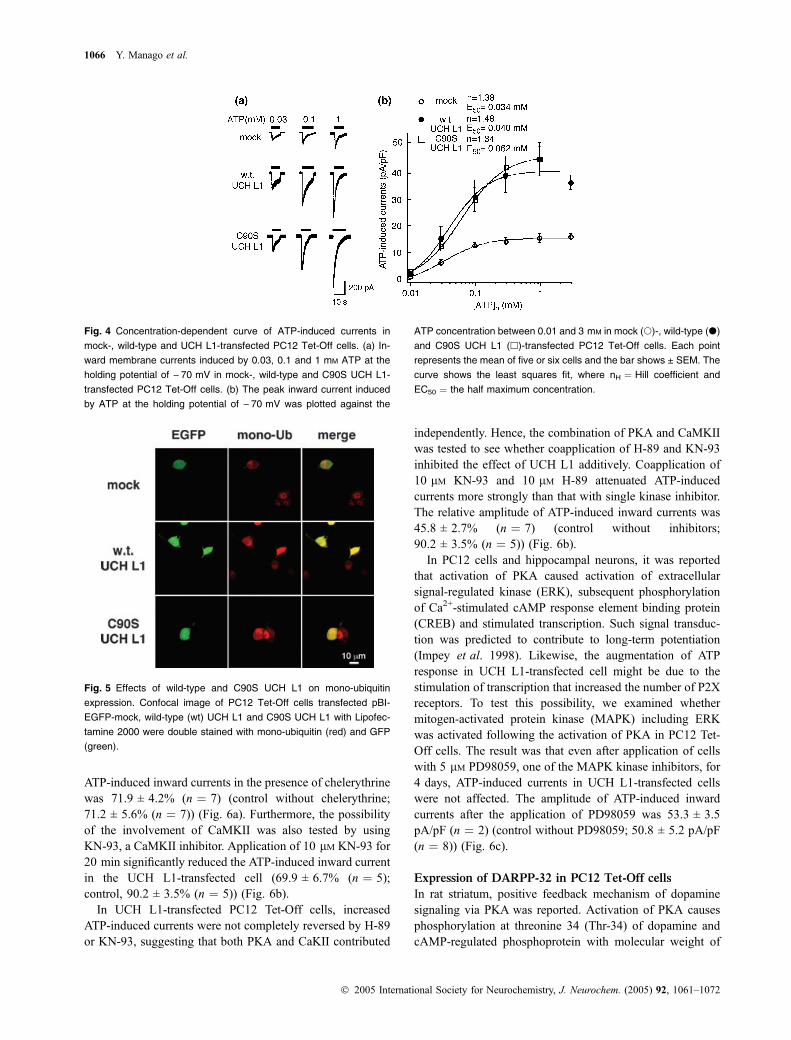
ATP-induced inward currents were concentration depend-ent and the sensitivity of ATP was not significantly changedby overexpression of either wild- or C90S UCH L1. EachEC50 was 34 lM, 40 lM and 62 lM and each Hill coefficient(nH) was 1.38, 1.48 and 1.34 in mock-, wild-type and C90SUCH L1-transfected cells, respectively (Fig. 4).
Effects of wild type and C90S UCH L1 on
mono-ubiquitin expression
It was reported that absence of UCH L1 reduced the mono-ubiquitin level in mouse brain and UCH L1 overexpressionincreased the level of mono-ubiquitin by alteration ofubiquitin metabolism in cultured cells. Therefore, UCHL1-mediated increases in ubiquitin levels are a function ofUCH L1 affinity for ubiquitin rather than hydrolase activity(Osaka et al. 2003). To clarify the effect of UCH L1 onubiquitin levels in PC12 Tet-Off cells, ubiquitin wasvisualized using confocal immunofluorescence microscopy(Fig. 5). Wild-type UCH L1-transfected cells showedstronger immunoreactivity for ubiquitin compared with those
in mock-transfected cells or non-transfected cells in the samefield. Increased ubiquitin immunoreactivity was also evidentin C90S UCH L1-transfected cells. These results wereconsistent with the previous report that ubiquitin were up-regulated by UCH L1 (Osaka et al. 2003).
Effects of Kinase inhibitors on ATP-induced currents
in UCH L1-transfected cells
The mechanism by which ATP-induced currents wereaugmented in UCH L1-transfected cells was investigated. Itwas reported that in Aplysia UCH activated PKA as a resultof the degradation of regulatory subunit of PKA, whichcontributed the long-term potentiation (Hegde et al. 1997).Therefore, the possibility of the involvement of the activatedPKA was tested by using H-89, a PKA inhibitor. Afterobtaining a large ATP-induced currents in the UCHL1-transfected cells, 10 lM H-89 was applied for 10 min.The amplitude of ATP-induced currents in the presence ofH-89 was 48.1 ± 3.51% (n ¼ 8) compared to the first ATP-induced current in the same cell (control without H-89;71.2 ± 5.6% (n ¼ 7)) (Fig. 6a). Also, it was reported that theintracellular carboxyl terminus of P2X receptor containsseveral consensus phosphorylation sites for PKC as well asPKA, suggesting that the function of the P2X receptor couldbe regulated by protein phosphorylation (Chow and Wang1998). Hence, the possibility of the involvement of theactivated PKC was tested by using chelerythrine, a PKCinhibitor. Application of 5 lM chelerythrine for 10 min hadno effect on the ATP-induced inward current in the UCHL1-transfected cell. The relative amplitude of second
Fig. 3 Voltage-dependency of ATP-induced currents in mock-, wild-
type UCH L1- and C90S UCH L1-transfected PC12 Tet-Off cells. A:
The voltage protocol shown in the upper panel was applied before and
during application of 1 mM ATP at the time indicated by (i) and (ii) in
the lower panel. (b) Cumulated current traces obtained in wild type
UCH L1-transfected cells before (i) and during (ii) application of ATP.
The subtracted current traces [(i)-(ii)] show the ATP-induced currents.
(c) The current–voltage relationships of ATP-induced currents. The
amplitudes of subtracted currents [(ii)-(I)] in (b) at the end of 50 ms
pulses were plotted against the pulse potentials in mock- (s), wild-
type (d) and C90S UCH L1-transfected cells (4).
Potentiation of ATP-induced currents by UCH L1 1065
� 2005 International Society for Neurochemistry, J. Neurochem. (2005) 92, 1061–1072
ATP-induced inward currents in the presence of chelerythrinewas 71.9 ± 4.2% (n ¼ 7) (control without chelerythrine;71.2 ± 5.6% (n ¼ 7)) (Fig. 6a). Furthermore, the possibilityof the involvement of CaMKII was also tested by usingKN-93, a CaMKII inhibitor. Application of 10 lM KN-93 for20 min significantly reduced the ATP-induced inward currentin the UCH L1-transfected cell (69.9 ± 6.7% (n ¼ 5);control, 90.2 ± 3.5% (n ¼ 5)) (Fig. 6b).
In UCH L1-transfected PC12 Tet-Off cells, increasedATP-induced currents were not completely reversed by H-89or KN-93, suggesting that both PKA and CaKII contributed
independently. Hence, the combination of PKA and CaMKIIwas tested to see whether coapplication of H-89 and KN-93inhibited the effect of UCH L1 additively. Coapplication of10 lM KN-93 and 10 lM H-89 attenuated ATP-inducedcurrents more strongly than that with single kinase inhibitor.The relative amplitude of ATP-induced inward currents was45.8 ± 2.7% (n ¼ 7) (control without inhibitors;90.2 ± 3.5% (n ¼ 5)) (Fig. 6b).
In PC12 cells and hippocampal neurons, it was reportedthat activation of PKA caused activation of extracellularsignal-regulated kinase (ERK), subsequent phosphorylationof Ca2+-stimulated cAMP response element binding protein(CREB) and stimulated transcription. Such signal transduc-tion was predicted to contribute to long-term potentiation(Impey et al. 1998). Likewise, the augmentation of ATPresponse in UCH L1-transfected cell might be due to thestimulation of transcription that increased the number of P2Xreceptors. To test this possibility, we examined whethermitogen-activated protein kinase (MAPK) including ERKwas activated following the activation of PKA in PC12 Tet-Off cells. The result was that even after application of cellswith 5 lM PD98059, one of the MAPK kinase inhibitors, for4 days, ATP-induced currents in UCH L1-transfected cellswere not affected. The amplitude of ATP-induced inwardcurrents after the application of PD98059 was 53.3 ± 3.5pA/pF (n ¼ 2) (control without PD98059; 50.8 ± 5.2 pA/pF(n ¼ 8)) (Fig. 6c).
Expression of DARPP-32 in PC12 Tet-Off cells
In rat striatum, positive feedback mechanism of dopaminesignaling via PKA was reported. Activation of PKA causesphosphorylation at threonine 34 (Thr-34) of dopamine andcAMP-regulated phosphoprotein with molecular weight of
Fig. 4 Concentration-dependent curve of ATP-induced currents in
mock-, wild-type and UCH L1-transfected PC12 Tet-Off cells. (a) In-
ward membrane currents induced by 0.03, 0.1 and 1 mM ATP at the
holding potential of ) 70 mV in mock-, wild-type and C90S UCH L1-
transfected PC12 Tet-Off cells. (b) The peak inward current induced
by ATP at the holding potential of ) 70 mV was plotted against the
ATP concentration between 0.01 and 3 mM in mock (s)-, wild-type (d)
and C90S UCH L1 (h)-transfected PC12 Tet-Off cells. Each point
represents the mean of five or six cells and the bar shows ± SEM. The
curve shows the least squares fit, where nH ¼ Hill coefficient and
EC50 ¼ the half maximum concentration.
Fig. 5 Effects of wild-type and C90S UCH L1 on mono-ubiquitin
expression. Confocal image of PC12 Tet-Off cells transfected pBI-
EGFP-mock, wild-type (wt) UCH L1 and C90S UCH L1 with Lipofec-
tamine 2000 were double stained with mono-ubiquitin (red) and GFP
(green).
1066 Y. Manago et al.
� 2005 International Society for Neurochemistry, J. Neurochem. (2005) 92, 1061–1072

about 32 000 (DARPP-32), which reduces PP1 activity andconsequently inhibits dephosphorylation of various sub-strates in the cell. On the other hand, activation of PKAstimulates PP2A activity and suppresses the phosphorylationat Thr-75 of DARPP-32. Since phosphorylation of DARPP-32 at Thr-75 has negative feedback regulation on PKAactivity, dephosphorylation at Thr-75 reduces the inhibitionof PKA activity (Nishi et al. 2000). To examine whether thesimilar mechanism exists in PC12 Tet-Off cells, we analyzedthe expression of DARPP-32 first. With RT-PCR methodusing two kinds of primer sets specific to DARPP-32 (primerset A and B), the relative expression levels of DARPP-32 inrat whole brain and PC12 Tet-Off cells under non-differen-
tiated and differentiated conditions were compared. Asshown in Fig. 7(a), DARPP-32 was expressed in PC12 Tet-Off cells and the expression level tended to increase afterdifferentiation of the cells with NGF. The possibility of theinvolvement of DARPP-32 and its phosphorylation wasfurther tested using okadaic acid, a PP1 and PP2A inhibitor.The relative amplitudes of ATP-induced inward currents afterthe application of 100 nM okadaic acid for 20 min wassignificantly inhibited to 49.9 ± 11.1% (n ¼ 5) (controlwithout okadaic acid; 90.2 ± 3.5% (n ¼ 5)), presumablydue to the inhibition of PP2A and subsequent dephospho-rylation of Thr-75, leading the release of negative feedbackon PKA (Fig. 7b). Since phosphorylation of Thr-75 was also
Fig. 6 Inhibition of ATP-induced currents by kinase inhibitors in wild
type UCH L1-transfected PC12 Tet-Off cells. (a) ATP-induced currents
were attenuated by pre-application of 10 lM H-89, a PKA inhibitor, but
not by 5 lM chelerythrine, a PKC inhibitor, for 10 min. (b) ATP-induced
currents were attenuated by pre-application of 10 lM KN-93, a CaMKII
inhibitor, for 20 min. ATP-induced currents were further attenuated by
copreapplication of 10 lM KN-93 and 10 lM H-89. C: ATP-induced
currents were not affected by application of 5 lM PD98059, a MAPKK
inhibitor, for four days. *p < 0.05, **p < 0.01, ***p < 0.001.
Fig. 7 Quantitative RT–PCR of DARPP-32
and effects of okadaic acid and roscovitine
on ATP-induced currents in wild-type UCH
L1-transfected PC12 Tet-Off cells. (a) The
expression level of DARPP-32 mRNA was
normalized to that of rat brain b-actin
mRNA. Using two kinds of primer (a and b),
PC12 Tet-Off cells were shown to express
DARPP-32 whose level was increased by
differentiation of the cells with NGF. B:
ATP-induced currents were attenuated by
preapplication of 100 nM okadaic acid, a
PP1 and PP2 inhibitor, for 20 min. (c) ATP-
induced currents were not affected by pre-
application 10 lM roscovitine, a CDK5
inhibitor, for 10 min **p < 0.01.
Potentiation of ATP-induced currents by UCH L1 1067
� 2005 International Society for Neurochemistry, J. Neurochem. (2005) 92, 1061–1072

mediated by cycline-dependent kinase (CDK), the effect of aCDK inhibitor was tested to see if ATP-induced currentswere more enhanced. However, application of 10 lM rosc-ovitine for 10 min did not have significant effect (74.4 ± 5.1%(n ¼ 4); control; 71.2 ± 5.6% (n ¼ 7)) (Fig. 7c).
ATP-induced currents in mock-transfected cells
We concluded that the increase of ATP-induced inwardcurrents in UCH L1-transfected cells was partly attributed tothe activation of PKA. Hence, we tested whether ATP-induced currents in the cells not transfected with UCH L1were increased by the application of forskolin, an adenylatecyclase activator that increases intracellular level of cAMP(Conn et al. 1989). The ATP-induced currents in mock-transfected cells were significantly increased after theapplication of 10 lM forskolin for 10 min (109.2 ± 2.2%(n ¼ 5); control without forskolin; 87.4 ± 3.8% (n ¼ 5))(Fig. 8a). It was confirmed that application of inactiveanalogue of forskolin, 10 lM 1,9-dideoxyforskolin, did nothave such effect (79.2 ± 2.2% (n ¼ 5); control; 87.4 ± 3.8%(n ¼ 5)) (Fig. 8a). Next, the effects of kinase inhibitors onATP-induced currents were tested in mock-transfected cells.In mock-transfected cells, application of 10 lM H-89 for10 min or 10 lM roscovitine for 10 min had no effect on theATP-induced inward current (H-89, 84.3 ± 3.0% (n ¼ 4);roscovitine, 93.4 ± 5.0% (n ¼ 4); control; 87.4 ± 3.8%(n ¼ 5)) (Fig. 8a). On the other hand, ATP-induced inwardcurrents were significantly increased after application of100 nM okadaic acid for 20 min (62.5 ± 6.7% (n ¼ 5)).However, application of 10 lM KN-93 had no effect(111.3 ± 7.1% (n ¼ 3); control; 79.0 ± 3.8% (n ¼ 5))(Fig. 8b).
ATP-induced currents in C90S UCH L1-transfected cells
Since ATP-induced currents in and C90S UCH L1-transfect-ed PC12 Tet-Off cells were significantly potentiated as well,the same pharmacological analyses were done to see whether
or not the mechanism was the same as in wild-type UCH L1-transfected cells. Application of 10 lM H-89 for 10 minsignificantly reduced ATP-induced currents in C90S UCHL1-transfected cells (61.9 ± 2.0% (n ¼ 8); control withoutH-89, 78.4 ± 3.3% (n ¼ 4)) (Fig. 9a). Also, applications of10 lM KN-93 or 100 nM okadaic acid for 20 min signifi-cantly reduced ATP-induced inward currents in C90S UCHL1-transfected cells (KN-93, 58.0 ± 3.7% (n ¼ 5); okadaicacid, 65.1 ± 4.9% (n ¼ 6); control; 88.0 ± 3.4% (n ¼ 5))(Fig. 9b). Furthermore, ATP-induced currents were moreattenuated by copreapplication of 10 lM KN-93 and 10 lMH-89 (44.2 ± 3.7% (n ¼ 6); control; 88.0 ± 3.4% (n ¼ 5))(Fig. 9b).
Discussion
To analyze the functional role of UCH L1 in the centralnervous system (CNS), it is important to know whether UCHL1 has any effects on ion channels and receptors that are thebasic elements of neurotransmission. There are many ways toanalyze them, but one of the most simple but efficient waysto start is to analyze the effects of exogenous UCH L1 inneuronal cultured cell line. To confirm that cells express thetransfected protein, we used CHO-AA8-Lucl cells because ofthe higher transfection efficacy and not having endogenousUCH L1 (Fig. 1). For functional analyses of UCH L1 innervous system, we used PC12 Tet-Off cells. The engineeredPC12 cells are constructed to have higher transfectionefficiency than wild-type PC12 cells which are one of thepopular neuronal cell line (unpublished data). Amongneurotransmitter receptors in PC12 cells, we analyzed ATP-activated receptors that are widely distributed in the brainand involved in various biological activities includingneurosecretion. In PC12 cells, P2X2 and P2X4 receptors(Hur et al. 2001) with lower level of P2X6 (unpublisheddata) are expressed and ATP-induced inward currents werewell characterized (Nakazawa et al. 1994). We recorded
Fig. 8 ATP-induced currents in mock-
transfected PC12 Tet-Off cells. (a) ATP-
induced currents were augmented by
preapplication of 10 lM forskolin for 10 min,
but were not affected by 10 lM H-89 and
10 lM roscovitine. (b) ATP-induced currents
were augmented by 100 nM okadaic acid,
but were not affected by 10 lM KN-93.
**p < 0.01.
1068 Y. Manago et al.
� 2005 International Society for Neurochemistry, J. Neurochem. (2005) 92, 1061–1072

ATP-induced inward currents due to the activation of P2Xreceptor channels at the holding potential of )70 mV underthe conventional whole-cell patch clamp configuration. Toanalyze the effects of overexpression of UCH L1 and forpharmacological characterization, high concentration of ATP(1 mM) was used to see the effects on the maximum responseto ATP. As to the possibility to analyze other receptorchannels expressed in PC12 cells, nicotinic acetylcholinereceptor (nAChR) was likely to be analyzed. However, nocurrents were recorded using ACh or nicotine (not shown),probably because nAChR required longer time to beexpressed after differentiation (Fukukawa et al. 1992).
The amplitude of ATP-induced inward currents wassignificantly greater in both wild-type and mutant (C90S)UCH L1-transfected PC12 Tet-Off cells (Fig. 2a). We foundthat the potentiation of ATP-induced currents was due to theactivation of PKA and CaMKII (Fig. 6). Though activation ofPKA by homologous UCH was reported in Aplysia (Hegdeet al. 1997), the mechanism of PKA activation in PC12 cellswould be different from the one in Aplysia. In Aplysia, UCHenhances the degradation of the regulatory subunit of PKAand consequently activate PKA. However, in our study, themutant (C90S) UCH L1, which lacks hydrolase activity, alsopotentiated ATP-induced currents, which was also attenuatedby H-89 and KN-93 (Figs 2 and 9). Therefore, function ofUCH L1 other than hydrolase activity should play afundamental role. Based on the previous reports that UCHL1 has multifunction, one of the plausible reason would be theability to increase free ubiquitin by both wild-type and C90Smutant UCH L1. It was reported that C90S UCH L1 lackshydrolase activity, but retains ubiquitin binding affinity andincreases free ubiquitin level in SH-SY5Y cells (Osaka et al.2003). Actually, immunostaining of mono-ubiquitin wasstronger in the cytoplasm of wild-type and C90S UCHL1-transfected PC12 Tet-Off cells (Fig. 5). The mechanismhow increased level of mono-ubiquitin activated PKA and
CaMKII were not yet known and should be investigatedfurther.
The mechanism how activated PKA and CaMKII poten-tiate ATP-induced currents also remains to be investigated.One possible mechanism would be phosphorylation of P2Xreceptors by these protein kinases, though it was reportedthat activation of PKA reduced the magnitude of the ATP-activated current in P2X2-transfected HEK293 cells (Chowand Wang 1998). The potentiation of ATP-induced currentsby PKA in our study might be similar to the one observed forCa2+ channels (Kamp and Hell 2000). Likewise, in mock-transfected cells, the amplitude of ATP-induced currents wassignificantly increased by forskolin, an adenylate cyclaseactivator that increases intracellular levels of cAMP andactivates PKA (Fig. 8). Therefore, it was suggested that atleast an activation of PKA contributed to the potentiation ofATP-induced currents. As for the involvement of CaMKII, ithas been recently reported that CaMKII potentiates ATPresponses by promoting trafficking of P2X receptors (Xu andHuang 2004), suggesting that phosphorylation of P2Xreceptors were not the sole mechanism of the potentiationof ATP-induced currents. Furthermore, since increased ATP-induced currents were not completely reduced by H-89,KN-93 nor coapplication of H-89 and KN-93 in wild-type orC90S UCH L1-transfected cells (Figs 6 and 9), it was alsosuggested that activation of PKA and CaMKII was not thesole mechanism of the potentiation of ATP-induced currentsbut there may be other components, too.
Another possible mechanism how PKA potentiates ATP-induced currents would be an increase in number of P2Xreceptors. In Aplysia, homologous UCH activates PKA andconsequently activates MAPK and subsequent transcription(Hegde et al. 1997). Such signal transduction is predicted tocontribute to a long-term potentiation (Impey et al. 1998). Ifthe similar signaling exists in mammalian cells, the numberof P2X receptors could increase during the differentiation
Fig. 9 ATP-induced currents in C90S UCH
L1-transfected PC12 Tet-Off cells were also
dependent on PKA and CaMKII. (a) ATP-
induced currents were attenuated by pre-
application of 10 lM H-89 for 10 min. (b)
ATP-induced currents were attenuated by
preapplication of 10 lM KN-93 and 100 nM
okadaic acid for 20 min. ATP-induced cur-
rents were further attenuated by copreap-
plication of 10 lM H-89 and 10 lM KN-93.
**p < 0.01, ***p < 0.001.
Potentiation of ATP-induced currents by UCH L1 1069
� 2005 International Society for Neurochemistry, J. Neurochem. (2005) 92, 1061–1072
after transfection of UCH L1 or C90S UCH L1. However, itwas unlikely because even after incubation with MAPKinhibitor during differentiation, the augmented ATP-inducedcurrents were still observed in wild-type (Fig. 6c) or C90SUCH L1-transfected cells (not shown).
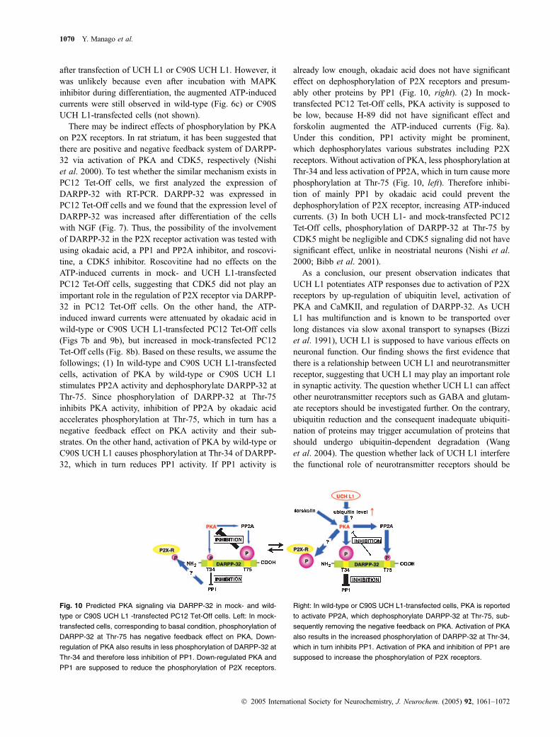
There may be indirect effects of phosphorylation by PKAon P2X receptors. In rat striatum, it has been suggested thatthere are positive and negative feedback system of DARPP-32 via activation of PKA and CDK5, respectively (Nishiet al. 2000). To test whether the similar mechanism exists inPC12 Tet-Off cells, we first analyzed the expression ofDARPP-32 with RT-PCR. DARPP-32 was expressed inPC12 Tet-Off cells and we found that the expression level ofDARPP-32 was increased after differentiation of the cellswith NGF (Fig. 7). Thus, the possibility of the involvementof DARPP-32 in the P2X receptor activation was tested withusing okadaic acid, a PP1 and PP2A inhibitor, and roscovi-tine, a CDK5 inhibitor. Roscovitine had no effects on theATP-induced currents in mock- and UCH L1-transfectedPC12 Tet-Off cells, suggesting that CDK5 did not play animportant role in the regulation of P2X receptor via DARPP-32 in PC12 Tet-Off cells. On the other hand, the ATP-induced inward currents were attenuated by okadaic acid inwild-type or C90S UCH L1-transfected PC12 Tet-Off cells(Figs 7b and 9b), but increased in mock-transfected PC12Tet-Off cells (Fig. 8b). Based on these results, we assume thefollowings; (1) In wild-type and C90S UCH L1-transfectedcells, activation of PKA by wild-type or C90S UCH L1stimulates PP2A activity and dephosphorylate DARPP-32 atThr-75. Since phosphorylation of DARPP-32 at Thr-75inhibits PKA activity, inhibition of PP2A by okadaic acidaccelerates phosphorylation at Thr-75, which in turn has anegative feedback effect on PKA activity and their sub-strates. On the other hand, activation of PKA by wild-type orC90S UCH L1 causes phosphorylation at Thr-34 of DARPP-32, which in turn reduces PP1 activity. If PP1 activity is
already low enough, okadaic acid does not have significanteffect on dephosphorylation of P2X receptors and presum-ably other proteins by PP1 (Fig. 10, right). (2) In mock-transfected PC12 Tet-Off cells, PKA activity is supposed tobe low, because H-89 did not have significant effect andforskolin augmented the ATP-induced currents (Fig. 8a).Under this condition, PP1 activity might be prominent,which dephosphorylates various substrates including P2Xreceptors. Without activation of PKA, less phosphorylation atThr-34 and less activation of PP2A, which in turn cause morephosphorylation at Thr-75 (Fig. 10, left). Therefore inhibi-tion of mainly PP1 by okadaic acid could prevent thedephosphorylation of P2X receptor, increasing ATP-inducedcurrents. (3) In both UCH L1- and mock-transfected PC12Tet-Off cells, phosphorylation of DARPP-32 at Thr-75 byCDK5 might be negligible and CDK5 signaling did not havesignificant effect, unlike in neostriatal neurons (Nishi et al.2000; Bibb et al. 2001).
As a conclusion, our present observation indicates thatUCH L1 potentiates ATP responses due to activation of P2Xreceptors by up-regulation of ubiquitin level, activation ofPKA and CaMKII, and regulation of DARPP-32. As UCHL1 has multifunction and is known to be transported overlong distances via slow axonal transport to synapses (Bizziet al. 1991), UCH L1 is supposed to have various effects onneuronal function. Our finding shows the first evidence thatthere is a relationship between UCH L1 and neurotransmitterreceptor, suggesting that UCH L1 may play an important rolein synaptic activity. The question whether UCH L1 can affectother neurotransmitter receptors such as GABA and glutam-ate receptors should be investigated further. On the contrary,ubiquitin reduction and the consequent inadequate ubiquiti-nation of proteins may trigger accumulation of proteins thatshould undergo ubiquitin-dependent degradation (Wanget al. 2004). The question whether lack of UCH L1 interferethe functional role of neurotransmitter receptors should be
Fig. 10 Predicted PKA signaling via DARPP-32 in mock- and wild-
type or C90S UCH L1 -transfected PC12 Tet-Off cells. Left: In mock-
transfected cells, corresponding to basal condition, phosphorylation of
DARPP-32 at Thr-75 has negative feedback effect on PKA, Down-
regulation of PKA also results in less phosphorylation of DARPP-32 at
Thr-34 and therefore less inhibition of PP1. Down-regulated PKA and
PP1 are supposed to reduce the phosphorylation of P2X receptors.
Right: In wild-type or C90S UCH L1-transfected cells, PKA is reported
to activate PP2A, which dephosphorylate DARPP-32 at Thr-75, sub-
sequently removing the negative feedback on PKA. Activation of PKA
also results in the increased phosphorylation of DARPP-32 at Thr-34,
which in turn inhibits PP1. Activation of PKA and inhibition of PP1 are
supposed to increase the phosphorylation of P2X receptors.
1070 Y. Manago et al.
� 2005 International Society for Neurochemistry, J. Neurochem. (2005) 92, 1061–1072

investigated next. These studies may help to understand howdysfunction of UCH L1 causes neurodegeneration.
Acknowledgements
We thank Ms. Yuki Kosai for technical assistance in gaining
confocal images. This work was supported by Grants-in Aid for
Scientific Research of Japan Society for Promotion of Science,
Grants-in Aid for Scientific Research in Priority Area Research of
the Ministry of Education, Culture, Sports, Science and Technology,
Japan, Kyushu University Foundation, Grants-in-Aid for Scientific
Research of the Ministry of Health, Labour and Welfare, Japan and a
grant from Pharmaceuticals and Medical Devices Agency, Japan.
References
Aoki K., Sun Y. J., Aoki S., Wada K. and Wada E. (2002) Cloning,expression, and mapping of a gene that is upregulated in adiposetissue of mice deficient in bombesin receptor subtype-3. Biochem.Biophys. Res. Commun. 290, 1282–1288.
Bibb J. A., Chen J., Taylor J. R., Svenningsson P., Nishi A., Snyder G.L., Yan Z., Sagawa Z. K., Ouimet C. C., Nairn A. C., Nestler E. J.and Greengard P. (2001) Effects of chronic exposure to cocaine areregulated by the neuronal protein Cdk5. Nature 410, 376–380.
Bizzi A., Schaetzle B., Patton A., Gambetti P. and Autilio-Gambetti L.(1991) Axonal transport of two major components of the ubiquitinsystem: free ubiquitin and ubiquitin carboxyl-terminal hydrolasePGP 9.5. Brain Res. 548, 292–299.
Chow Y. W. and Wang H. L. (1998) Functional modulation of P2X2receptors by cyclic AMP-dependent protein kinase. J. Neurochem.70, 2606–2612.
Ciechanover A., Orian A. and SchwartZ. A. L. (2000) The ubiquitin-mediated proteolytic pathway: Mode of action and clinical impli-cations. J. Cell Biochem. 77, 40–51.
Conn P. J., Strong J. A., Azhderian E. M., Nairn A. C., Greengard P. andKaczmarek L. K. (1989) Protein kinase inhibitors selectively blockphorbol ester- or forskolin-induced changes in excitability ofAplysia neurons. J. Neurosci. 9, 473–479.
Coux O., Tanaka K. and Goldberg A. L. (1996) Structure and function ofthe 20S and 26S proteasomes. Annu. Rev. Biochem. 65, 801–847.
Fukukawa K., Akaike M., Onodera H. and Kogure K. (1992) Expressionof 5-HT3 receptor in PC12 cells treated with NGF and8-Br.-cAMP. J. Neurophysiol. 67, 812–819.
Harada T., Harada C., Wang Y. L., Osaka H., Amanai K., Tanaka K.,Takizawa S., Setsuie R., Sakurai M., Sato Y., Noda M. and WadaK. (2004) Role of ubiquitin carboxy terminal hydrolase-l1 in neuralcell apoptosis induced by ischemic retinal injury in vivo. Am. J.Pathol. 164, 59–64.
Hegde A. N., Inokuchi K., Pei W., Casadio A., Ghirardi M., Chain D. G.,Martin K. C., Kandel E. R. and SchwartZ. J. H. (1997) UbiquitinC-terminal hydrolase is an immediate-early gene essential for long-term facilitation in Aplysia. Cell 89, 115–126.
Huang Y., Baker R. T. and Fischer-Vize J. A. (1995) Control of cell fateby a deubiquitinating enzyme encoded by fat facets gene. Science270, 1828–1831.
Hur E. M., Park T. J. and Kim K. T. (2001) Coupling of 1-type voltage-sensitive calcium channels to P2X (2) purinoceptors in PC-12 cells.Am. J. Physiol. Cell Physiol. 280, C1121–C1129.
Ikeuchi Y., Nishizaki T., Mori M. and Okada Y. (1996) Regulation of thepotassium current and cytosolic Ca2+ release induced by 2-meth-ylthio ATP in hippocampal neurons. Biochem. Biophys. Res.Commun. 218, 428–433.
Impey S., Obrietan K., Wong S. T., Poser S., Yano S., Wayman G.,Deloulme J. C., Chan G. and Storm D. R. (1998) Cross talkbetween ERK and PKA is required for Ca2+ stimulation of CREB-dependent transcription and ERK nuclear translocation. Neuron 21,869–883.
Kamp T. J. and Hell J. W. (2000) Regulation of cardiac 1-type calciumchannels by protein kinase A and protein kinase C. Circ Res. 87,1095–1102.
King R. W., Deshaies R. J., Peters J.-M. and Kirschner M. W. (1996)How proteolysis drives the cell cycle. Science 274, 1652–1659.
Kwon J., Wang Y. L., Setsuie R., Sekiguchi S., Sakurai M., Sato Y., LeeW. W., Ishii Y., Kyuwa S., Noda M., Wada K. and Yoshikawa Y.(2004a) Developmental regulation of ubiquitin C-terminal hy-drolase isozyme expression during spermatogenesis in mice. Biol.Reprod. 71, 515–521.
Kwon J., Wang Y. L., Setsuie R., Sekiguchi S., Sato Y., Sakurai M.,Noda M., Aoki S., Yoshikawa Y. and Wada K. (2004b) Two clo-sely related ubiquitin C-terminal hydrolase isozymes function asreciprocal modulators of germ cell apoptosis in cryptorchid testes.Am. J. Pathol. 165, 1367–1374.
Leroy E., Boyer R., Auburger G., Leube B., Ulm G., Mezey E., Harta G.,Brownstein M. J., Jonnalagada S., Chernova T., Dehejia A.,Lavedan C., Gasser T., Steinbach P. J., Wilkinson K. D. andPolymeropoulos M. H. (1998) The ubiquitin pathway in Parkin-son’s disease. Nature 395, 451–452.
Liu Y., Fallon L., Lashuel H. A., Liu Z. and Lansbury P. T. Jr (2002) TheUCH-L1 gene encodes two opposing enzymatic activities thataffect alpha-synuclein degradation and Parkinson’s disease sus-ceptibility. Cell 111, 209–218.
Min B. I., Kim C. J., Rhee J. S. and Akaike N. (1996) Modulation ofglycin-induced chloride current in acutely dissociated rat periaqu-eductal gray neurons by 1-opioid agonist DAGO. Brain Res. 734,72–78.
Muralidhar M. G. and Thomas J. B. (1993) The Drosophila bendlessgene encodes a neural protein related to ubiquitin-conjugatingenzymes. Neuron 11, 253–266.
Nakazawa K., Inoue K., Koizumi S. and Inoue K. (1994) Facilitation by5-hydroxytryptamine of ATP-activated current in rat pheochrom-ocytoma cells. Pflugers Arch. 427, 492–499.
Nishi A., Bibb J. A., Snyder G. L., Higashi H., Nairn A. C. andGreengard P. (2000) Amplification of dopaminergic signaling by apositive feedback loop. Proceedings Natl. Acad. Sci. USA 97,12 840–12 845.
Noda M., Nakanishi H. and Akaike N. (1999) Glutamate release frommicroglia via glutamate transporter is enhanced by amyloid-betapeptide. Neuroscience. 92, 1465–1474.
Noda M., Nakanishi H., Nabekura J. and Akaike N. (2000) AMPA-KAsubtypes of glutamate receptor in rat cerebral microglia. J. Neu-rosci. 20, 251–258.
Oh C. E., McMahon R., Benzer S. and Tanouye M. A. (1994) bendless, aDrosophila gene affecting neuronal connectivity, encodes a ubiqu-itin-conjugating enzyme homolog. J. Neurosci. 14, 3166–3179.
Osaka H., Wang Y. L., Takada K., Takizawa S., Setsuie R., Li H., SatoY., Nishikawa K., Sun Y. J., Sakurai M., Harada T., Hara Y.,Kimura I., Chiba S., Namikawa K., Kiyama H., Noda M., Aoki S.and Wada K. (2003) Ubiquitin carboxy-terminal hydrolase L1binds to and stabilizes monoubiquitin in neuron. Hum. Mol. Genet.12, 1945–1958.
Rock K. L., Gram C., Rothstein L., Clark K., Stein R., Dick L., HwangD. and Goldberg A. L. (1994) Inhibitors of the proteasome blockthe degradation of most cell proteins and the generation of peptidespresent on MHC class I molecules. Cell 78, 761–771.
Saigoh K., Wang Y. L., Suh J. G., Yamanishi T., Sakai Y., Kiyosawa H.,Harada T., Ichihara N., Wakana S., Kikuchi T. and Wada K. (1999)
Potentiation of ATP-induced currents by UCH L1 1071
� 2005 International Society for Neurochemistry, J. Neurochem. (2005) 92, 1061–1072

Intragenic deletion in the gene encoding ubiquitin carboxy-terminal hydrolase in gad mice. Nat. Genet. 23, 47–51.
Sela D., Ram E. and Atlas D. (1991) ATP receptor. A putative receptor-operated channel in PC-12 cells. J. Biol. Chem. 266, 17 990–17 994.
Verma I. M., Stevenson J. K., SchwartZ. E. M., Van Antwerp D. andMiyamoto S. (1995) Rel/NF-jB/I-jB family: intimate tales ofassociation and dissociation. Genes Dev. 9, 2723–2735.
Wada R., Tifft C. J. and Proia R. L. (2000) Microglial activation pre-cedes acute neurodegeneration in Sandhoff disease and is sup-pressed by bone marrow transplantation. Proc. Natl. Acad. Sci.USA 97, 10 954–10 959.
Wang Y. L., Takeda A., Osaka H., Hara Y., Furuta A., Setsuie R., Sun Y.J., Kwon J., Sato Y., Sakurai M., Noda M., Yoshikawa Y. andWada K. (2004) Accumulation of beta- and gamma-synucleins inthe ubiquitin carboxyl-terminal hydrolase L1-deficient gad mouse.Brain Res. 1019, 1–9.
Wilkinson K. D. (1995) Roles of ubiquitinylation in proteolysis andcellular regulation. Annu. Rev. Nutr. 15, 161–189.
Wilkinson K. D., Deshpande S. and Larsen C. N. (1992) Comparisons ofneuronal (PGP 9.5) and non-neuronal ubiquitin C-terminalhydrolases. Biochem. Soc. Trans. 20, 631–637.
Wilkinson K. D., Lee K. M., Deshpande S., Duerksen-Hughes P., Boss J.M. and Pohl J. (1989) The neuron-specific protein PGP 9.5 is aubiquitin carboxyl-terminal hydrolase. Science 246, 670–673.
Wong M. H., Saam J. R., Stappenbeck T. S., Rexer C. H. and Gordon J.I. (2000) Genetic mosaic analysis based on Cre recombinase andnavigated laser capture microdissection. Proc. Natl. Acad. Sci. USA97, 12 601–12 606.
Xu G. Y. and Huang L. Y. (2004) Ca2+/calmodulin-dependent proteinkinase II potentiates ATP responses by promoting trafficking ofP2X receptors. Proc. Natl Acad. Sci. USA 101, 11 868–11 873.
Zhu Y., Carroll M., Papa F., Hochstrazin M. E. and D’Andrea A. D.(1996) DUB-1, a deubiquitinating enzyme with growth-suppres-sing activity. Proc. Natl. Acad. Sci. USA 93, 3275–3279.
1072 Y. Manago et al.
� 2005 International Society for Neurochemistry, J. Neurochem. (2005) 92, 1061–1072