Crystal Structure of a Complex between Amino and Carboxy Terminal Fragments of mDia1: Insights into...
11
Crystal Structure of a Complex between Amino and Carboxy Terminal Fragments of mDia1: Insights into Autoinhibition of Diaphanous-Related Formins Azin Nezami 1,2¤a , Florence Poy 1¤b , Angela Toms 1,2 , Wei Zheng 1,2 , Michael J. Eck 1,2 * 1 Department of Cancer Biology, Dana-Farber Cancer Institute, Boston, Massachusetts, United States of America, 2 Department of Biological Chemistry and Molecular Pharmacology, Harvard Medical School, Boston, Massachusetts, United States of America Abstract Formin proteins direct the nucleation and assembly of linear actin filaments in a variety of cellular processes using their conserved formin homology 2 (FH2) domain. Diaphanous-related formins (DRFs) are effectors of Rho-family GTPases, and in the absence of Rho activation they are maintained in an inactive state by intramolecular interactions between their regulatory N-terminal region and a C-terminal segment referred to as the DAD domain. Although structures are available for the isolated DAD segment in complex with the interacting region in the N-terminus, it remains unclear how this leads to inhibition of actin assembly by the FH2 domain. Here we describe the crystal structure of the N-terminal regulatory region of formin mDia1 in complex with a C-terminal fragment containing both the FH2 and DAD domains. In the crystal structure and in solution, these fragments form a tetrameric complex composed of two interlocking N+C dimers. Formation of the tetramer is likely a consequence of the particular N-terminal construct employed, as we show that a nearly full-length mDia1 protein is dimeric, as are other autoinhibited N+C complexes containing longer N-terminal fragments. The structure provides the first view of the intact C-terminus of a DRF, revealing the relationship of the DAD to the FH2 domain. Delineation of alternative dimeric N+C interactions within the tetramer provides two general models for autoinhibition in intact formins. In both models, engagement of the DAD by the N-terminus is incompatible with actin filament formation on the FH2, and in one model the actin binding surfaces of the FH2 domain are directly blocked by the N-terminus. Citation: Nezami A, Poy F, Toms A, Zheng W, Eck MJ (2010) Crystal Structure of a Complex between Amino and Carboxy Terminal Fragments of mDia1: Insights into Autoinhibition of Diaphanous-Related Formins. PLoS ONE 5(9): e12992. doi:10.1371/journal.pone.0012992 Editor: Petri Kursula, University of Oulu, Germany Received May 6, 2010; Accepted August 27, 2010; Published September 30, 2010 Copyright: ß 2010 Nezami et al. This is an open-access article distributed under the terms of the Creative Commons Attribution License, which permits unrestricted use, distribution, and reproduction in any medium, provided the original author and source are credited. Funding: This work was supported by National Institutes of Health (NIH) grant R01GM071834 (MJE). The funders had no role in study design, data collection and analysis, decision to publish, or preparation of the manuscript. Competing Interests: The authors have declared that no competing interests exist. * E-mail: [email protected] ¤a Current address: Vertex Pharmaceuticals, Cambridge, Massachusetts, United States of America ¤b Current address: Constellation Pharmaceuticals, Cambridge, Massachusetts, United States of America Introduction Formin family proteins direct actin assembly in an array of cellular functions including cytokinesis, cell migration, and the establishment and maintenance of cell polarity[1–3]. Formins are present in all eukaryotes and many species express multiple isoforms of the protein[4]. They have in common the presence of the ,400 residue formin homology 2 (FH2) domain, which directly mediates actin assembly[5–7]. Biochemically, the FH2 domain potently nucleates new, unbranched actin filaments and remains attached to the growing barbed end of the filament as additional actin subunits are incorporated. This activity is referred to as processive capping. The adjacent formin homology 1 (FH1) domain is a proline-rich segment that can accelerate actin assembly on the FH2 domain by recruitment of profilin-bound actin[8–10]. Diaphanous-related formins (DRFs) are a subfamily of formins that are effectors of Rho-family GTPases[11–14]. The mamma- lian DRF Dia1 is important for stress fiber assembly and cell migration. Mice deficient in mDia1 exhibit myeloproliferative defects[15,16] and impaired lymphocyte trafficking[17]. In the absence of interaction with GTP-bound Rho, mDia1 and other DRFs are maintained in an autoinhibited state by intramolecular interactions between their C-terminal diaphanous autoregulatory domain (DAD) and the interacting region in the N-terminus, termed the DAD interacting domain (DID). DRFs share a number of other functional and structural domains. The N-terminal GTPase binding domain (GBD) is formed from the helical G domain and a portion of the adjacent DID domain. These elements are followed by the dimerization domain (DD), a coiled- coil region, the FH1 and FH2 domains, and finally the C-terminal DAD domain (Figure 1A). Extensive study of formins has revealed structures of the FH2 domain[18–21] and its mode of binding with actin[22], as well as structures of the regulatory N-terminal domains alone[23] and in complexes with GTP-bound Rho[24]or cdc42[25]. Biochemical studies and comparison of the Rho-bound structure with those of the N-terminal domains determined in complex with the DAD segment explain how the autoinhibitory DID/DAD interaction can be released by competition with GTP-bound Rho[26,27]. Despite this relative wealth of biochemical and structural information, it remains unclear how the DID/DAD interaction inhibits actin assembly by the FH2 domain. PLoS ONE | www.plosone.org 1 September 2010 | Volume 5 | Issue 9 | e12992
-
Upload
hms-harvard -
Category
Documents
-
view
1 -
download
0
Transcript of Crystal Structure of a Complex between Amino and Carboxy Terminal Fragments of mDia1: Insights into...

Crystal Structure of a Complex between Amino andCarboxy Terminal Fragments of mDia1: Insights intoAutoinhibition of Diaphanous-Related ForminsAzin Nezami1,2¤a, Florence Poy1¤b, Angela Toms1,2, Wei Zheng1,2, Michael J. Eck1,2*
1 Department of Cancer Biology, Dana-Farber Cancer Institute, Boston, Massachusetts, United States of America, 2 Department of Biological Chemistry and Molecular
Pharmacology, Harvard Medical School, Boston, Massachusetts, United States of America
Abstract
Formin proteins direct the nucleation and assembly of linear actin filaments in a variety of cellular processes using theirconserved formin homology 2 (FH2) domain. Diaphanous-related formins (DRFs) are effectors of Rho-family GTPases, and inthe absence of Rho activation they are maintained in an inactive state by intramolecular interactions between theirregulatory N-terminal region and a C-terminal segment referred to as the DAD domain. Although structures are available forthe isolated DAD segment in complex with the interacting region in the N-terminus, it remains unclear how this leads toinhibition of actin assembly by the FH2 domain. Here we describe the crystal structure of the N-terminal regulatory regionof formin mDia1 in complex with a C-terminal fragment containing both the FH2 and DAD domains. In the crystal structureand in solution, these fragments form a tetrameric complex composed of two interlocking N+C dimers. Formation of thetetramer is likely a consequence of the particular N-terminal construct employed, as we show that a nearly full-length mDia1protein is dimeric, as are other autoinhibited N+C complexes containing longer N-terminal fragments. The structureprovides the first view of the intact C-terminus of a DRF, revealing the relationship of the DAD to the FH2 domain.Delineation of alternative dimeric N+C interactions within the tetramer provides two general models for autoinhibition inintact formins. In both models, engagement of the DAD by the N-terminus is incompatible with actin filament formation onthe FH2, and in one model the actin binding surfaces of the FH2 domain are directly blocked by the N-terminus.
Citation: Nezami A, Poy F, Toms A, Zheng W, Eck MJ (2010) Crystal Structure of a Complex between Amino and Carboxy Terminal Fragments of mDia1: Insightsinto Autoinhibition of Diaphanous-Related Formins. PLoS ONE 5(9): e12992. doi:10.1371/journal.pone.0012992
Editor: Petri Kursula, University of Oulu, Germany
Received May 6, 2010; Accepted August 27, 2010; Published September 30, 2010
Copyright: � 2010 Nezami et al. This is an open-access article distributed under the terms of the Creative Commons Attribution License, which permitsunrestricted use, distribution, and reproduction in any medium, provided the original author and source are credited.
Funding: This work was supported by National Institutes of Health (NIH) grant R01GM071834 (MJE). The funders had no role in study design, data collection andanalysis, decision to publish, or preparation of the manuscript.
Competing Interests: The authors have declared that no competing interests exist.
* E-mail: [email protected]
¤a Current address: Vertex Pharmaceuticals, Cambridge, Massachusetts, United States of America¤b Current address: Constellation Pharmaceuticals, Cambridge, Massachusetts, United States of America
Introduction
Formin family proteins direct actin assembly in an array of
cellular functions including cytokinesis, cell migration, and the
establishment and maintenance of cell polarity[1–3]. Formins are
present in all eukaryotes and many species express multiple isoforms
of the protein[4]. They have in common the presence of the ,400
residue formin homology 2 (FH2) domain, which directly mediates
actin assembly[5–7]. Biochemically, the FH2 domain potently
nucleates new, unbranched actin filaments and remains attached to
the growing barbed end of the filament as additional actin subunits
are incorporated. This activity is referred to as processive capping.
The adjacent formin homology 1 (FH1) domain is a proline-rich
segment that can accelerate actin assembly on the FH2 domain by
recruitment of profilin-bound actin[8–10].
Diaphanous-related formins (DRFs) are a subfamily of formins
that are effectors of Rho-family GTPases[11–14]. The mamma-
lian DRF Dia1 is important for stress fiber assembly and cell
migration. Mice deficient in mDia1 exhibit myeloproliferative
defects[15,16] and impaired lymphocyte trafficking[17]. In the
absence of interaction with GTP-bound Rho, mDia1 and other
DRFs are maintained in an autoinhibited state by intramolecular
interactions between their C-terminal diaphanous autoregulatory
domain (DAD) and the interacting region in the N-terminus,
termed the DAD interacting domain (DID). DRFs share a number
of other functional and structural domains. The N-terminal
GTPase binding domain (GBD) is formed from the helical G
domain and a portion of the adjacent DID domain. These
elements are followed by the dimerization domain (DD), a coiled-
coil region, the FH1 and FH2 domains, and finally the C-terminal
DAD domain (Figure 1A).
Extensive study of formins has revealed structures of the FH2
domain[18–21] and its mode of binding with actin[22], as well as
structures of the regulatory N-terminal domains alone[23] and in
complexes with GTP-bound Rho[24]or cdc42[25]. Biochemical
studies and comparison of the Rho-bound structure with those of
the N-terminal domains determined in complex with the DAD
segment explain how the autoinhibitory DID/DAD interaction
can be released by competition with GTP-bound Rho[26,27].
Despite this relative wealth of biochemical and structural
information, it remains unclear how the DID/DAD interaction
inhibits actin assembly by the FH2 domain.
PLoS ONE | www.plosone.org 1 September 2010 | Volume 5 | Issue 9 | e12992

Figure 1. Structure of an autoinhibited mDia1 N+C complex. A, Dimeric domain structure of diaphanous-related formins. Dimerization ismediated by the DD, CC and FH2 domains as indicated. Numbering corresponds to murine mDia1; black bars indicate the extent of the crystallized N-and C-terminal fragments. Domains are colored as in panel B; this assignment of domains in the dimer corresponds to the proposed ‘‘trans’’ model ofinhibition (see text, and Fig. 6A). Abbreviations: GBD, GTPase binding domain; G, GTPase binding subdomain; DID, DAD interacting domain; DD,dimerization domain; CC, coiled-coil domain; FH1, formin homology-1 domain; FH2, formin homology-2 domain; DAD, diaphanous autoregulatorydomain. B, Stereo diagram showing the crystal structure of the tetrameric N+C mDia1 complex. Selected domains and subdomains are labeled. Thebridge elements comprising the putative FH2 dimers are colored blue and light blue (‘‘upper’’ FH2 dimer), or red and pink (lower FH2 dimer). Thesubunits of the N-terminal dimers are colored yellow and orange (upper dimer) or green and tan (lower dimer). Note that the N-terminal DID domainsengage the DAD domains extending from helix aT of the more distal FH2 domain. See Figures 4 and S3 for additional representations of the tetramer.C, Stereo diagram depicting the interaction of the DID and DAD regions. The DID domain is shown in a surface representation and the interactingregion of the C-terminus, including the end of aT and the DAD domain, is shown in a ribbon representation (blue, with selected sidechains shown inwhite). The core amphipathic DAD helix makes extensive hydrophobic contact with the DID domain as previously observed (residues Val 1181- Phe1195). Note that the end of aT and the connecting loop also contribute hydrophobic interactions to the interface (Leu 1169, Ile 1170, and Met 1172).The basic portion of the DAD extends in an acidic groove between the DID and aT. The sidechains of Arg 1197 and Lys 1198 in this region approach,but do not make clearly defined salt-bridge interactions with acidic residues on the DID domain. The surface corresponding to acidic residues Glu358, Asp361, Glu 362 and Asp 366 is shaded salmon.doi:10.1371/journal.pone.0012992.g001
Autoinhibition of mDia1
PLoS ONE | www.plosone.org 2 September 2010 | Volume 5 | Issue 9 | e12992

In order to better understand the mechanism of autoinhibition
in mDia1 and other DRFs, we have reconstituted complexes
containing the N-terminal regulatory region of mDia1 bound to a
C-terminal fragment containing its FH2 and DAD domains for
structural analysis. The FH1 domain was omitted from the C-
terminal constructs because it is expected to be unstructured and
therefore to impede crystallization. We crystallized and deter-
mined the structure of an ‘‘N+C’’ complex containing the N-
terminal DID and DD domains (DID-DD) bound to the C-
terminal FH2-DAD construct. In the crystal structure and in
solution, these fragments form a tetrameric complex with the
stoichiometry (DID-DD)4/(FH2-DAD)4. This tetrameric structure
can be described as an interlocked complex of two N+C dimers.
Formation of the tetramer is likely a consequence of the particular
N-terminal construct employed, as we show that a nearly full-
length mDia1 protein is dimeric, as are other autoinhibited N+C
complexes containing longer N-terminal fragments that include a
portion of the adjacent coiled-coil domain. The structure provides
the first view of the intact C-terminus of a DRF, revealing the
relationship of the DAD to the FH2 domain. Delineation of
alternative dimeric N+C interactions within the tetramer provides
two general models for autoinhibition in intact formins. In the
‘‘cis’’ model the N-terminal DID-DD region directly occludes the
actin binding surface of the FH2 dimer while in the ‘‘trans’’ model
the DID-DD region is maintained at a distance from the surface of
the FH2 domain by the long aT helix, which extends from the
FH2 domain to present the DAD segment for binding. In both the
models, engagement of the DAD by the N-terminus is incompat-
ible with actin filament formation on the FH2 domain.
Results
Preparation and analysis of mDia1 proteins and N+Ccomplexes
We prepared amino-terminal fragments of murine Dia1
(mDia1) and reconstituted them into complexes with a carboxy-
terminal fragment spanning the FH2 and DAD regions (residues
736–1200) of mDia1 for crystallization. The N-terminal fragments
included the DID and DD domains, which are expected to be
required for full autoinhibition[28], with and without the adjacent
G domain and a portion of the coiled-coil region (Figure 1A,
Table 1). All formed complexes with the C-terminal fragment that
were stable on gel-filtration and/or ion exchange chromatogra-
phy, and representative N-terminal preparations potently inhibited
the activity of FH2-DAD in pyrene actin assembly assays (Figure
S1). We refer to these as ‘‘N+C complexes’’ as they contain N- and
C-terminal fragments of mDia1.
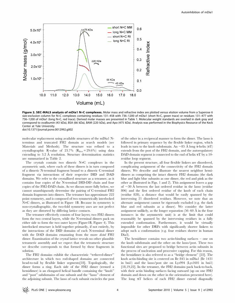
We analyzed selected N+C complexes using size-exclusion
chromatography and multi-angle light scattering (SEC-MALS) to
determine their approximate molar mass and thus their oligomeric
state (Table 1). The N- and C- fragments are both known to be
dimeric[23,24,29], and in the simplest model would be expected to
form a dimeric complex, i.e. one N-terminal dimer is presumed to
bind one C-terminal dimer via a dual DID/DAD interaction. This
was indeed the case for the N+C complex containing the N-
terminal fragment DID-DD-CC (residues 131–477). For this
complex, MALS analysis yielded a molar mass of 195 kDa,
consistent with a complex with the subunit composition (DID-DD-
CC)2/(FH2-DAD)2. However, for both the G-DID-DD and DID-
DD complexes, we observed molar masses most consistent with
formation of tetrameric complexes, i.e. (DID-DD)4/(FH2-DAD)4.
Despite their differences in subunit stoichiometry, the dimeric and
tetrameric species eluted from the size-exclusion column (Superose
6) at similar volumes (Figure 2), suggesting that the dimer has an
effective hydrodynamic radius similar to that of the tetramer. The
marked drop in the apparent molar mass of the tetrameric species
across its elution peak is suggestive of partial dissociation of the
complex or the presence of a small quantity of a lower molecular
weight complex (Figure 2, green trace). Analysis of these and other
N+C complexes by polyacrylamide gel electrophoresis under non-
denaturing conditions (NATIVE-PAGE) revealed that freshly
prepared complexes contained a faster migrating, presumably
dimeric species that partitioned with time into the more slowly
migrating tetrameric band (Figure S2). In contrast, a construct that
contained portions of the coiled-coil domain (G-DID-DD-CC,
residues 63–518) exhibited only the faster migrating, presumably
dimeric band (Figure S2).
We also expressed and purified a nearly full-length fragment of
mDia1 (mDia1-DID-C, residues 131–1255) for crystallization
trials. In this construct, we omitted the non-conserved N-terminal
region of the protein as well as the G domain, which is expected to
be disordered in the autoinhibited state[23]. The mDia1-DID-C
protein is dimeric, as judged by SEC-MALS analysis (Figure 3).
Collectively, our data support the hypothesis that autoinhibited
mDia1 is dimeric, and that truncation of the coiled-coil region in
the reconstituted N+C complexes leads to formation of tetrameric
species. We have been unable to crystallize the mDia1-DID-C
protein or of any of the dimeric N+C complexes, but the
tetrameric DID-DD/FH2-DAD complex crystallized readily, as
described below.
Crystal structure of an autoinhibited mDia1 complexWe obtained crystals of the DID-DD protein in complex with
FH2-DAD in space group P21 and determined the structure by
Table 1. SEC-MALS analysis of mDia1 proteins and complexes.
mDia1 Proteins and Complexes Mw Determined by MALS (kDa) Sequence Predicted Mw for monomer (kDa)
N+C N C
N: DID-DD, residues 131–458C: FH2-DAD, residues 736–1200
345 92 38 54
N: GBD-DID-DD, residues 72–458C: FH2-DAD, residues 736–1200
354 98 44 54
N: DID-DD-CC, residues 131–477C: FH2-DAD, residues 736–1200
195 94 40 54
C: FH2-DAD, residues 736–1200 109 - - 54
mDia1-DID-C: residues 131–1255 278 125 - -
doi:10.1371/journal.pone.0012992.t001
Autoinhibition of mDia1
PLoS ONE | www.plosone.org 3 September 2010 | Volume 5 | Issue 9 | e12992
molecular replacement using available structures of the mDia1 N-
terminus and truncated FH2 domain as search models (see
Materials and Methods). The structure was refined to a
crystallographic R-value of 23.7% (Rfree = 29.6%) using data
extending to 3.2 A resolution. Structure determination statistics
are summarized in Table 2.
The crystals contain two dimeric N+C complexes in the
asymmetric unit, where each of these dimers is in turn composed
of a dimeric N-terminal fragment bound to a dimeric C-terminal
fragment via interactions of their respective DID and DAD
domains. We refer to the crystallized structure as a tetramer, as it
contains four copies of the N-terminal DID-DD chain and four
copies of the FH2-DAD chain. As we discuss more fully below, we
cannot unambiguously determine the pairing of C-terminal FH2
domain fragments into dimers. The tetramer has approximate 222
point symmetry, and is composed of two symmetrically interlocked
N+C dimers, as illustrated in Figure 1B. Because its symmetry is
non-crystallographic, the two-fold symmetry axes are not perfect
as they are distorted by differing lattice contacts.
The tetramer effectively consists of four layers; two FH2 dimers
form the two central layers, while the N-terminal dimers pack on
either side to form the two outer layers (Figure1B, Figure 4). This
interlocked structure is held together primarily, if not entirely, by
the interactions of the DID domains of each N-terminal dimer
with the DAD domains emanating from the more distal FH2
dimer. Examination of the crystal lattice reveals no other plausible
tetrameric assembly and we expect that the tetrameric structure
we describe corresponds to that formed by these fragments in
solution.
The FH2 domains exhibit the characteristic ‘‘tethered-dimer’’
architecture in which two rod-shaped domains are connected
head-to-tail by flexible linker segments[18]. Topologically, the
dimer forms a ring. Each side of the FH2 dimer (termed a
hemidimer) is an elongated helical bundle containing the ‘‘knob’’
and ‘‘post’’ subdomains of one subunit and the ‘‘lasso’’ element of
the adjoining subunit. The lasso of each subunit encircles the post
of the other in a reciprocal manner to form the dimer. The lasso is
followed in primary sequence by the flexible linker region, which
leads in turn to the knob subdomain. An ,85 A long a-helix (aT)
extends from the post of the FH2 domain, and the autoregulatory
DAD domain segment is connected to the end of helix aT by a 10-
residue loop segment.
In the present structure, all four flexible linkers are disordered,
complicating assignment of the connectivity of the FH2 domain
dimers. We describe and illustrate the nearest neighbor hemi-
dimers as comprising the intact dimeric FH2 domains (the dark
blue and light blue subunits as one dimer, the red and pink as the
other as illustrated in Figs. 1 and 2). This assignment leaves a gap
of ,30 A between the last ordered residue in the lasso (residue
806) and the first ordered residue of the knob of each chain
(residue 828), a distance that would be easily spanned by the
intervening 21 disordered residues. However, we note that an
alternate assignment cannot be rigorously excluded (e.g. the dark
blue and red subunits as a dimer). We consider the latter
assignment unlikely, as the longer separation (56–60 A in the four
instances in the asymmetric unit) is at the limit that could
reasonably be spanned by the intervening residues in a fully
extended conformation. Furthermore, it would be virtually
impossible for other DRFs with significantly shorter linkers to
adopt such a conformation (e.g. four residues shorter in human
Dia3).
The hemidimer contains two actin-binding surfaces – one on
the knob subdomain and the other on the lasso/post. These two
functional sites are proposed to bridge between actin subunits in
the process of nucleation and processive capping. For this reason,
the hemidimer is also referred to as a ‘‘bridge element’’ [22]. The
knob actin-binding site is centered on Ile 845 in mDia1 (Ile 1431
in bni1) and the lasso/post site on Lys994 (Lys1601 in bni1)
[18,19,22]. In the tetramer, the FH2 domains pack back-to-back,
with their actin binding surfaces facing outward (up on one FH2
domain and down on the other in the orientation presented here).
The long aT helices of each FH2 dimer extend diagonally
Figure 2. SEC-MALS analysis of mDia1 N+C complexes. Molar mass and refractive index are plotted versus elution volume from a Superose 6size-exclusion column for N+C complexes containing residues 131–458 with 736–1200 of mDia1 (short N+C, green trace) or residues 131–477 with736–1200 of mDia1 (long N+C, red trace). Derived molar masses are presented in Table 1. Molecular weight standards are overlaid in dark gray andcorrespond to ovalbumin (43 kDa), BSA (66 kDa), BAM (220 kDa), and Apo (475 kDa). Analysis was performed in the Biophysics Resource of the KeckCenter at Yale University.doi:10.1371/journal.pone.0012992.g002
Autoinhibition of mDia1
PLoS ONE | www.plosone.org 4 September 2010 | Volume 5 | Issue 9 | e12992

between the bridge elements of the other FH2 dimer, thus
presenting the DAD for binding to the contralateral DID domain.
As previously described [23,24], the DID domain is composed
of a series of armadillo repeats, a structural motif consisting of a
repeating series of three helices arranged in a superhelical coil.
The DID domain contains five armadillo repeats; the atypical fifth
repeat leads to the dimerization domain via a long helix. The
dimerization domain (DD) is formed by a zig-zag of three
interdigitating helices from each subunit. The N-terminal DID-
DD dimers in the present structure are approximately two-fold
symmetric; the two halves of each DID-DD dimer are related by
178u–179u rotations. The isolated DID and DD domains
superimpose well on the corresponding regions of the structure
of the mDia1 N-terminus crystallized in complex with the isolated
DAD peptide[26]. The DID domains of these structures
superimpose with an RMSD of ,0.65 A, while the DD domains
superimpose with an RMSD of ,1.7 A. Despite the fact that both
structures are two-fold symmetric, the relative positions of their
DID and DD domains differ greatly. If the present structure is
superimposed on the mDia1 DAD complex (PDB ID 2BAP) based
on the alignment of the DD domain, rotations of 43u and
translations of approximately 15 A are required to bring the DID
domains into register. Considerable divergence in domain
orientation has been noted in comparisons of previous structures
of the DID-DD and G-DID-DD fragments of mDia1 [23,24,26].
The DAD is composed of a core helical region with the
sequence motif ‘‘MDXLLXL’’ and an adjacent basic segment with
the sequence ‘‘RRKR’’ in mDia1 [30]. The interactions of the
DAD domain with the DID are essentially the same as previously
described for structures containing the isolated DAD segment
[26,27], but additional interactions are observed for the basic C-
terminal portion of the DAD. The core ‘‘MDXLLXL’’ motif in
the DAD domain forms an amphipathic helix (residues 1180–
1192) that packs into a conserved groove on the concave surface of
the DID. Just beyond this helix, Phe 1195 packs into a
hydrophobic cleft, as previously described[26,27]. The basic
segment extends along the DID domain, in a channel formed by
the DID and helix aT (Fig 1C). This region has a highly acidic
character, with four negatively charged residues on the surface of
the DID and another contributed by aT. Electron density for the
basic segment is weak, and somewhat divergent among the four
copies in the tetramer. No interpretable density is present for the
last two residues in the crystallized protein. Helix aT and the loop
Figure 3. Purification and size analysis of mDia1-DID-C.Residues 131–1255 of murine mDia1 were expressed using an insectcell/baculovirus system, and purified by affinity and size-exclusionchromatography (see Materials and Methods). A, SEC-MALS analysis ofthe purified protein; molar mass (black trace, MM), refractive index (bluetrace, RI) and absorbance at 280 nM (green trace, UV) are plotted versuselution volume from a Superdex 200 size-exclusion column. Calculatedmolar mass is presented in Table 1, and was measured across thevolume indicated by the arrows. The earlier eluting peak contains asmall amount of polydisperse, high molecular weight aggregates ofmDia 131-1255 that were not completely removed in the prior size-exclusion chromatography step. B, Coomassie-stained SDS-PAGEanalysis of Superdex 200 elution fractions of the purified mDia1-DID-C protein.doi:10.1371/journal.pone.0012992.g003
Table 2. Data collection and refinement statistics.*
Data Collection
Space Group P21
Unit Cell Dimensions a = 95.76 A, b = 206.83 A,c = 131.06 A, b= 105.9u
Resolution (A) 25–3.2 (3.3–3.2)
Rmergea 9.7 (57.5)
I/sI 14.26 (2.7)
Completeness (%) 99.5 (100)
Redundancy 3.8 (3.8)
Refinement
Resolution (A) 25–3.2
Number of Reflections 75584
Rwork/Rfreeb 23.7/29.6
Number of Protein Atoms 24185
Number of Water Molecules 48
Average B-factor 82.9
R.M.S.D. Bond Lengths (A) 0.006
R.M.S.D. Bond Angles (u) 0.938
Ramachandran Analysis
Most Favored (%) 95.6
Allowed (%) 99.9
*Highest resolution shell is shown in parentheses.aRmerge = S|Ii-,I.|/SIi, where Ii is the ith measurement of the intensity of anindividual reflection or its symmetry-equivalent reflections and ,I. is theaverage intensity of that reflection and its symmetry-equivalent reflections.
bRwork = S||Fobs| 2 |Fcalc||/S|Fobs| for all reflections and Rfree = S||Fobs| 2
|Fcalc||/S|Fobs|, calculated on the 5% of data excluded from refinement.doi:10.1371/journal.pone.0012992.t002
Autoinhibition of mDia1
PLoS ONE | www.plosone.org 5 September 2010 | Volume 5 | Issue 9 | e12992

that connects it to the DAD also contribute to hydrophobic
contacts with the DID and DAD regions (Fig 1C).
Structural Comparison mDia1, Daam1 and Bni1 FH2domains
The structure of a monomeric fragment of the mDia1 FH2
domain has previously been described[19] and also compared with
Bni1 and Daam1[20,21]. However, the present structure provides
the first reported view of the functional mDia1 FH2 domain with
the lasso/post interface intact, affording additional insights into
conserved and divergent features of the FH2 domain. As expected,
the general mode of dimerization in which the lasso of one subunit
loops around the post region of the other is conserved among
mDia1, Daam1 and Bni1[18]. In particular, two highly conserved
tryptophan residues in the lasso insert into pockets in the post
domain as previously documented in Bni1 and Daam1 (Figure 5A).
However, there are a number of sites of divergence in the
interface. For example, Phe 986 in the post of the mDia1 FH2
domain replaces a much smaller serine in Daam1 (Figure 5B). This
change in the post is accommodated by a compensatory change in
the lasso – Pro765 in the mDia1 lasso replaces the phenylalanine
residue found in the corresponding position in Daam1. Similarly
Lys783 hydrogen bonds with Ser957 in the post in mDia1, but in
Daam1 the corresponding residues are Glu630 in the lasso and
Lys811 in the post (Figure 5B). These and other divergences in the
lasso/post interface may have evolved in part to preclude
heterodimerization of FH2 domains [31,32].
Although the knob and post regions of mDia1, Daam1 and Bni1
independently superimpose well, there is considerable variation in
the spacing between knob and post actin binding sites among these
formins (as much as 8 A, Figure 5C). This difference arises largely
from differences in the orientation of the knob subdomain with
respect to the rest of the bridge element[20]. This divergence is
unlikely to result entirely from flexibility in the bridge element; this
measurement differs by less than 1 A among crystallographically
independent copies of the FH2 hemidimer in the present structure.
Thus this variation in spacing suggests that precise positioning of
actin subunits by knob and post sites within the bridge element is
not critical for the mechanism of nucleation or processive capping.
Discussion
The autoinhibited tetramer structure suggests two general
models for autoinhibition in an intact, dimeric DRF (Figure 6A).
These are essentially the two ways in which an N+C dimer can be
dissected from the tetrameric structure, given our qualified
assignment of nearest-neighbor hemidimers as a connected FH2
domain dimer. The cis model consists of the upper half of the
tetramer, with the N-terminus packed against the adjacent FH2
domain. This model would require the aT Helix of each
hemidimer to break and/or fold back (as indicated by the dotted
lines) in order to allow binding of the DAD segment to the
adjacent N-terminus. If this model is correct, formation of the
tetramer represents a ‘‘domain swap’’ in which the DAD and C-
terminal portions of aT exchanged positions with the correspond-
Figure 4. Surface views of the autoinhibited mDia1 N+C tetramer. A, Side and top views of the tetramer, colored as in Figure 1B. Inset: topview of one FH2 dimer, with the residues expected to bind actin shaded magenta. Note that packing of the N-terminal DID-DD dimers onto the FH2domain blocks the actin-binding surface. B, ‘‘Exploded’’ view depicting construction of the tetramer.doi:10.1371/journal.pone.0012992.g004
Autoinhibition of mDia1
PLoS ONE | www.plosone.org 6 September 2010 | Volume 5 | Issue 9 | e12992

Figure 5. Comparison of FH2 domain structures. A, Lasso-post interface of mDia1 (left panel) and Daam1 (right panel). Both mDia1 and Daam1exhibit the same general dimerization mode first described for yeast formin Bni1, in which the lasso region of one subunit encircles the post of theother. Conserved tryptophan residues in the lasso insert into pockets in the post. B, Despite the generally conserved mode of interaction, distinctdifferences in the lasso-post interface in mDia1 (left) as compared with Daam1 (right) are likely to preclude heterodimerization of their FH2 domains.For example, Pro765 in the mDia1 lasso replaces Phe613 in Daam1. This change is accompanied by substitution of Phe896 in mDia1 for Ser839 in
Autoinhibition of mDia1
PLoS ONE | www.plosone.org 7 September 2010 | Volume 5 | Issue 9 | e12992
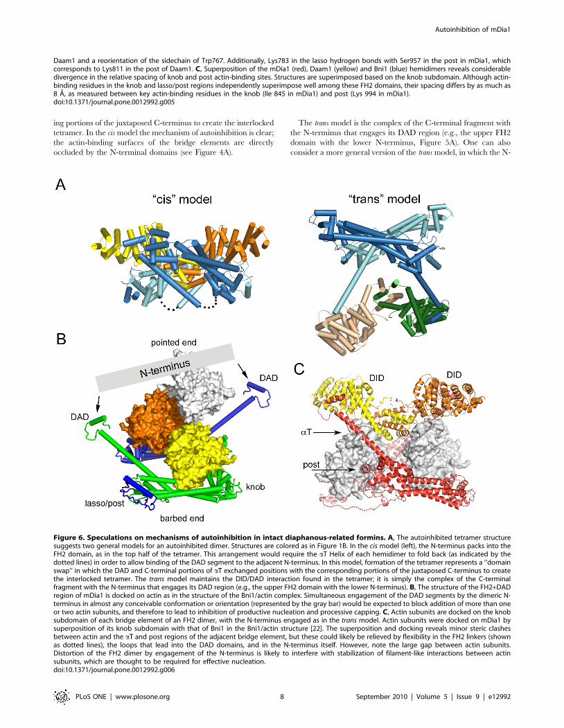
ing portions of the juxtaposed C-terminus to create the interlocked
tetramer. In the cis model the mechanism of autoinhibition is clear;
the actin-binding surfaces of the bridge elements are directly
occluded by the N-terminal domains (see Figure 4A).
The trans model is the complex of the C-terminal fragment with
the N-terminus that engages its DAD region (e.g., the upper FH2
domain with the lower N-terminus, Figure 5A). One can also
consider a more general version of the trans model, in which the N-
Figure 6. Speculations on mechanisms of autoinhibition in intact diaphanous-related formins. A, The autoinhibited tetramer structuresuggests two general models for an autoinhibited dimer. Structures are colored as in Figure 1B. In the cis model (left), the N-terminus packs into theFH2 domain, as in the top half of the tetramer. This arrangement would require the aT Helix of each hemidimer to fold back (as indicated by thedotted lines) in order to allow binding of the DAD segment to the adjacent N-terminus. In this model, formation of the tetramer represents a ‘‘domainswap’’ in which the DAD and C-terminal portions of aT exchanged positions with the corresponding portions of the juxtaposed C-terminus to createthe interlocked tetramer. The trans model maintains the DID/DAD interaction found in the tetramer; it is simply the complex of the C-terminalfragment with the N-terminus that engages its DAD region (e.g., the upper FH2 domain with the lower N-terminus). B, The structure of the FH2+DADregion of mDia1 is docked on actin as in the structure of the Bni1/actin complex. Simultaneous engagement of the DAD segments by the dimeric N-terminus in almost any conceivable conformation or orientation (represented by the gray bar) would be expected to block addition of more than oneor two actin subunits, and therefore to lead to inhibition of productive nucleation and processive capping. C, Actin subunits are docked on the knobsubdomain of each bridge element of an FH2 dimer, with the N-terminus engaged as in the trans model. Actin subunits were docked on mDia1 bysuperposition of its knob subdomain with that of Bni1 in the Bni1/actin structure [22]. The superposition and docking reveals minor steric clashesbetween actin and the aT and post regions of the adjacent bridge element, but these could likely be relieved by flexibility in the FH2 linkers (shownas dotted lines), the loops that lead into the DAD domains, and in the N-terminus itself. However, note the large gap between actin subunits.Distortion of the FH2 dimer by engagement of the N-terminus is likely to interfere with stabilization of filament-like interactions between actinsubunits, which are thought to be required for effective nucleation.doi:10.1371/journal.pone.0012992.g006
Daam1 and a reorientation of the sidechain of Trp767. Additionally, Lys783 in the lasso hydrogen bonds with Ser957 in the post in mDia1, whichcorresponds to Lys811 in the post of Daam1. C, Superposition of the mDia1 (red), Daam1 (yellow) and Bni1 (blue) hemidimers reveals considerabledivergence in the relative spacing of knob and post actin-binding sites. Structures are superimposed based on the knob subdomain. Although actin-binding residues in the knob and lasso/post regions independently superimpose well among these FH2 domains, their spacing differs by as much as8 A, as measured between key actin-binding residues in the knob (Ile 845 in mDia1) and post (Lys 994 in mDia1).doi:10.1371/journal.pone.0012992.g005
Autoinhibition of mDia1
PLoS ONE | www.plosone.org 8 September 2010 | Volume 5 | Issue 9 | e12992

and C-terminal fragments are not constrained to adopt precisely
the orientation found in the tetramer. This is particularly relevant
to the N-terminus; as discussed above, relative orientations of the
DID domains varies greatly between available crystal structures
and in the presence of the coiled-coil region they have been
observed to adopt a highly asymmetric configuration[23]. (Nota
Bene: In both cis and trans models, the interaction is intramolecular
in the context of the intact formin dimer. Cis and trans to refer to
the action and position of the N-terminus relative to the FH2
domain.)
How is actin assembly inhibited in the generalized trans model?
Although the actin binding surfaces of the bridge element are not
directly blocked in this model, the trans interaction could lead to
autoinhibition by at least two mechanisms. First, spanning of the
two DAD domains by the N-terminus, in essentially any plausible
conformation or orientation would block addition of more than
one or two actin subunits in a nascent filament or nucleus
(Figure 6B). Second, productive nucleation is thought to require
templating of two or three actin subunits by the FH2 domain in a
filament-like orientation[22]. Distortion and/or rigidification of
the FH2 dimer by the bound N-terminus are likely to preclude
productive engagement of actin. Docking of actin on the knob of
each bridge element in the dimer leaves the actin subunits
separated by several Angstroms, lacking the actin-actin contacts
thought to be critical for nucleation (Figure 6C). Finally, we note
that in both the cis and trans models, sequestration of the DAD
segment by binding with the DID domain may also contribute
directly to autoinhibition, as the DAD region also appears to play
a positive role in actin assembly (B. Goode, personal communi-
cation).
Because the same DID/DAD interactions stabilize the auto-
inhibited state in both the cis and trans models, it is not
straightforward to design a mutagenesis strategy to discriminate
between them. A more definitive understanding of the mechanism
of autoinhibition will require structural investigations of an intact,
dimeric autoinhibited formin.
Materials and Methods
Protein expression and purificationThe DNA sequence encoding residues 131–458 (N-terminus)
and 736–1200 (C-terminus) of mDia1 was amplified by PCR and
ligated into a modified pET vector containing a His6 tag with a
TEV cleavage site. Expression of the fusion protein in E. coli BL21
(DE3) was induced by IPTG and was allowed to proceed overnight
at 23uC. The cells were harvested and resuspended in 50 mM Tris
(pH 8), 150 mM NaCl, and 200 mg/ml lysozyme and were stored
at 280uC. Upon thawing and after the addition of 1 mM PMSF
and 2 mM TCEP, the cells were lysed by sonication and cleared
by centrifugation. The proteins were purified separately by metal-
affinity chromatography on a Ni-chelating column. To form the
N+C complex, excess C-terminal protein was added to N-terminal
protein and the affinity tag was removed by overnight incubation
at 4uC with TEV protease. The N+C complex was purified by
anion exchange chromatography (HiTrapQ) and buffer ex-
changed into 20 mM Tris (pH 8), 100 mM NaCl and 2 mM
TCEP (PD10 column), and concentrated to 5–7 mg/ml. Analo-
gous procedures were followed to prepare complexes containing
alternate N-terminal fragments listed in Table 1.
For insect cell expression and purification of mDia1-DID-C, a
PCR fragment encoding residues 131–1255 of murine mDia1 was
subcloned into a modified pTriEx transfer vector (Novagen) using
59-BglII and 39-XholI restriction sites. The modified transfer
vector (pTriExGST-mDia1-DID-C) drives expression of the
mDia1 protein as a GST-fusion with an intervening TEV protease
cleavage site. The pTriExGST-mDia1-DID-C plasmid was co-
transfected into Sf9 cells with linearized baculoviral DNA
(BacVector-3000, Novagen). The primary virus was harvested
after one week and was subsequently amplified, plaque purified
and used to prepare aliquots of Baculovirus-Infected Insect Cells
(BIICs)[33]. For protein production, five 800 mL shake flasks of
Sf9 cells were infected using BIICs aliquots when cells reached a
density of 2.0*106 cells/ml. Cells were harvested by centrifugation
72 h after infection, and were lysed with a detergent-containing
lysis buffer (20 mM Tris pH 8.0, 150 mM NaCl, 5% glycerol,
2 mM TCEP and 1% NP40). The lysate was clarified via
centrifugation at 40,000 g for 1 h, and the resulting supernatant
was incubated for 3 h with glutathione-Sepharose beads. The
bead-bound fusion protein was cleaved by overnight incubation
with TEV protease. TEV was removed by adsorption to an ion
exchange column (MonoS, GE Healthcare) and the mDia1
protein was further purified by a size-exclusion chromatography
(Superdex200, GE Healthcare) in storage buffer (20 mM Tris
pH 8.0, 150 mM NaCl, and 2 mM TCEP).
Light scattering analysisSize-exclusion chromatography Multi-angle Light Scattering
(SEC-MALS) experiments for mDia1 N+C and mDia1 C-terminal
only constructs were carried out at HHMI & W.M. Keck
Foundation Biotechnology Resource Laboratory, Yale University.
The N+C complexes were prepared for SEC-MALS as described
above for crystallization; 1:1 complexes were isolated by anion
exchange chromatography (HiTrapQ) prior to SEC-MALS
analysis. Protein samples (1 mg/ml, 0.3 ml) were injected onto a
Superose 6 size exclusion column at a flow rate of 0.3 ml/min in
20 mM HEPES pH 7.4, 150 mM NaCl, 1 mM EDTA and 2 mM
DTT. Data were evaluated using the Zimm model for static light
scattering data fitting. All proteins are monodisperse and their
experimental molecular weights and calculated molecular weights
for monomer are shown in Table 1.
The mDia1-DID-C protein sample (0.1 mg/ml, 0.3 ml) was
injected onto a Superdex 200 size exclusion column attached to a
GE AKTA purifier at a flow rate of 0.5 ml/min in 20 mM Tris
pH 8.0, 150 mM NaCl, 2 mM TCEP. The eluted peak was
analyzed using a Wyatt miniDAWN TREOS multi-angle light
scattering instrument and a Wyatt Optilab rEX differential
refractometer. Data were evaluated in ASTRA 5.3.4 software
using the Zimm model as above.
Crystallization and Structure DeterminationThe N+C complex was crystallized at 20uC by hanging-drop
vapor diffusion against 0.2 M sodium malonate (pH 7.0) and 10–
14% PEG 4000 as precipitant. Crystals grew overnight and were
cryo-protected with 20% glycerol in drop mother liquor prior to
flash freezing in liquid nitrogen. Diffraction data were collected at
beamline X29 at the NSLS (Table 2) and processed with XDS
[34]. Crystals belonged to the monoclinic space group P21 and
contained four N-terminal DID-DD chains and four C-terminal
FH2-DAD chains in the asymmetric unit. Phases were determined
by molecular replacement using the program Phaser[35,36] using
residues 133-371 of chain A of the mDia1 DID/DAD complex
(PDB:2F31) and residues 829–1150 of chain A of the mDia1 FH2
domain (PDB: 1V9D) as the search models. The correct solution
for the DID model yielded a log(likelihood) gain of 1092 in the 25–
3.5 A resolution range. With the N-terminal model in place a
correct solution for three of the four FH2 domains yielded a
log(likelihood) gain of 1550. The final FH2 domain could then be
placed into NCS averaged 2Fo-Fc maps. The programs Coot [37]
Autoinhibition of mDia1
PLoS ONE | www.plosone.org 9 September 2010 | Volume 5 | Issue 9 | e12992

and O [38] were used to build the model into the 2Fo-Fc and Fo-Fc
maps in iterative rounds of NCS refinement with Refmac5 [39]
and CNS v1.2 [40]. The final model contains residues 134–451 of
the N terminal portion of mDia1, residues 745–1198 of the C
terminal portion of mDia1, and 48 water molecules. Among the
four copies of each polypeptide in the asymmetric unit, there is a
variable break in the chain in the region of residues 192–200 in the
N-terminal fragment and between residues 806–828 in the C-
terminal fragment. The structure has been refined to an R value of
23.7% (Rfree = 29.6%) with good stereochemistry using data
extending to 3.2 A resolution.
Supporting Information
Figure S1 Pyrene actin assembly assays with N+C complexes. A,
The mDia1 FH2-DAD protein (residues 736–1200, blue traces)
potently nucleates actin assembly as previously described[41], but
the tetrameric N+C complex (residues 72–458 plus 736–1200,
green traces) shows little activity above that of actin alone (black
trace). B, Comparison of actin assembly activity of various N-
terminal constructs in complex with FH2-DAD. Note that the 72-
458 and 63-462 complexes form tetramers, while the 63-518
complex is dimeric (see Figure S2). N+C complexes were pre-
formed and purified (see Materials and Methods), and actin
filament assembly assays were performed using 1% pyrene-labeled
rabbit skeletal muscle actin as described [42]. Briefly, 2 mM G-
actin was mixed with F-buffer (10 mM Tris, pH 7.5, 0.7 mM
ATP, 0.2 mM CaCl2, 2 mM MgCl2, 50 mM KCl, 0.2 mM
DTT) alone or with the indicated mDia proteins. Pyrene
fluorescence was monitored using an excitation wavelength of
365 nm and an emission wavelength of 407 nm in a fluorescence
spectrophotometer.
Found at: doi:10.1371/journal.pone.0012992.s001 (0.37 MB TIF)
Figure S2 Native-Page analysis of N+C mDia1 complexes. The
indicated complexes of N- and C-terminal fragments of mDia1
were analyzed by polyacrylamide gel electrophoresis under non-
denaturing conditions using a 4–15% gradient gel on a
PhastSystem (Pharmacia). Complexes in lanes 1,2,5 and 6 were
co-purified as described (see Materials and Methods), while those
in lanes 3 and 4 were prepared by combining separately purified
N- and C-terminal fragments immediately prior to analysis on Day
0 (left panel). The same preparations were re-examined after five
days (right panel). Freshly prepared complexes containing N-
terminal residues 63-462 or 72-458 contained a faster migrating,
presumably dimeric species that partitioned with time into the
more slowly migrating tetrameric band (lanes 1 and 4, compare
Day 0 vs. Day 5). In contrast, a construct that contained portions
of the coiled-coil domain (residues 63–518) exhibited only the
faster migrating, presumably dimeric band (lane 2, Day 0 vs. Day
5). Aliquots of the 72-458/736-1200 and 131-458/736-1211
complexes that were used for crystallization (and were stored at
5 mg/ml) were mostly tetrameric (lanes 5, 6).
Found at: doi:10.1371/journal.pone.0012992.s002 (1.18 MB TIF)
Figure S3 Additional views of the tetrameric mDia1 complex.
A, Stereodiagram of the tetramer, colored as in Figure 1B. The
terminal residues of one N-terminal (DID-DD) and one C-
terminal (FH2-DAD) fragment are labeled to facilitate following
the path of the polypeptide chain. B, Subunits labeled in A are
shown in isolation (and in the same orientation). These N- and C-
terminal fragments could plausibly correspond to those of a
continuous polypeptide chain in the trans model of autoinhibition
(see text). Note the break in the chain between residues 811 and
828 in the C-terminal fragment; this corresponds to the flexible
linker in the FH2 domain that is disordered in the present
structure. C, Stereodiagrams of the tetramer in a ribbon
representation. The view in the lower panel is rotated by 90uabout the vertical axis.
Found at: doi:10.1371/journal.pone.0012992.s003 (5.16 MB TIF)
Acknowledgments
We thank Bruce Goode and Michael Rosen for helpful discussions and
Ewa Folta-Stogniew in the Keck Molecular Biophysics Facility at Yale
University for assistance with SEC-MALS analysis and interpretation.
Crystallographic coordinates and structure factors are available in the
Protein Data Bank with PDB ID 3O4X.
Author Contributions
Conceived and designed the experiments: AN FP AVT WZ MJE.
Performed the experiments: AN FP AVT WZ. Analyzed the data: AN FP
AVT WZ MJE. Wrote the paper: AN FP AVT WZ MJE.
References
1. Goode BL, Eck MJ (2007) Mechanism and function of formins in the control of
actin assembly. Annu Rev Biochem 76: 593–627.
2. Pollard TD (2007) Regulation of actin filament assembly by Arp2/3 complex
and formins. Annu Rev Biophys Biomol Struct 36: 451–477.
3. Chesarone MA, DuPage AG, Goode BL (2010) Unleashing formins to remodelthe actin and microtubule cytoskeletons. Nat Rev Mol Cell Biol 11: 62–74.
4. Higgs HN, Peterson KJ (2005) Phylogenetic analysis of the formin homology 2domain. Mol Biol Cell 16: 1–13.
5. Evangelista M, Pruyne D, Amberg DC, Boone C, Bretscher A (2002) Formins
direct Arp2/3-independent actin filament assembly to polarize cell growth inyeast. Nat Cell Biol 4: 32–41.
6. Pruyne D, Evangelista M, Yang C, Bi E, Zigmond S, et al. (2002) Role offormins in actin assembly: nucleation and barbed-end association. Science 297:
612–615.
7. Sagot I, Rodal AA, Moseley J, Goode BL, Pellman D (2002) An actin nucleation
mechanism mediated by Bni1 and profilin. Nat Cell Biol 4: 626–631.
8. Paul AS, Pollard TD (2008) The role of the FH1 domain and profilin in formin-mediated actin-filament elongation and nucleation. Curr Biol 18: 9–19.
9. Vavylonis D, Kovar DR, O’Shaughnessy B, Pollard TD (2006) Model of formin-associated actin filament elongation. Mol Cell 21: 455–466.
10. Kursula P, Kursula I, Massimi M, Song YH, Downer J, et al. (2008) High-
resolution structural analysis of mammalian profilin 2a complex formation withtwo physiological ligands: the formin homology 1 domain of mDia1 and the
proline-rich domain of VASP. J Mol Biol 375: 270–290.
11. Watanabe N, Madaule P, Reid T, Ishizaki T, Watanabe G, et al. (1997)
p140mDia, a mammalian homolog of Drosophila diaphanous, is a target protein
for Rho small GTPase and is a ligand for profilin. Embo J 16: 3044–3056.
12. Watanabe N, Kato T, Fujita A, Ishizaki T, Narumiya S (1999) Cooperation
between mDia1 and ROCK in Rho-induced actin reorganization. Nat Cell Biol
1: 136–143.
13. Ishizaki T, Morishima Y, Okamoto M, Furuyashiki T, Kato T, et al. (2001)
Coordination of microtubules and the actin cytoskeleton by the Rho effector
mDia1. Nat Cell Biol 3: 8–14.
14. Habas R, Kato Y, He X (2001) Wnt/Frizzled activation of Rho regulates
vertebrate gastrulation and requires a novel Formin homology protein Daam1.
Cell 107: 843–854.
15. Eisenmann KM, West RA, Hildebrand D, Kitchen SM, Peng J, et al. (2007) T
cell responses in mammalian diaphanous-related formin mDia1 knock-out mice.
J Biol Chem 282: 25152–25158.
16. Peng J, Kitchen SM, West RA, Sigler R, Eisenmann KM, et al. (2007)
Myeloproliferative defects following targeting of the Drf1 gene encoding the
mammalian diaphanous related formin mDia1. Cancer Res 67: 7565–7571.
17. Sakata D, Taniguchi H, Yasuda S, Adachi-Morishima A, Hamazaki Y, et al.
(2007) Impaired T lymphocyte trafficking in mice deficient in an actin-
nucleating protein, mDia1. J Exp Med 204: 2031–2038.
18. Xu Y, Moseley JB, Sagot I, Poy F, Pellman D, et al. (2004) Crystal structures of a
Formin Homology-2 domain reveal a tethered dimer architecture. Cell 116:
711–723.
19. Shimada A, Nyitrai M, Vetter IR, Kuhlmann D, Bugyi B, et al. (2004) The core
FH2 domain of diaphanous-related formins is an elongated actin binding protein
that inhibits polymerization. Mol Cell 13: 511–522.
20. Lu J, Meng W, Poy F, Maiti S, Goode BL, et al. (2007) Structure of the FH2
domain of Daam1: implications for formin regulation of actin assembly. J Mol
Biol 369: 1258–1269.
Autoinhibition of mDia1
PLoS ONE | www.plosone.org 10 September 2010 | Volume 5 | Issue 9 | e12992

21. Yamashita M, Higashi T, Suetsugu S, Sato Y, Ikeda T, et al. (2007) Crystal
structure of human DAAM1 formin homology 2 domain. Genes Cells 12:1255–1265.
22. Otomo T, Tomchick DR, Otomo C, Panchal SC, Machius M, et al. (2005)
Structural basis of actin filament nucleation and processive capping by a forminhomology 2 domain. Nature 433: 488–494.
23. Otomo T, Otomo C, Tomchick DR, Machius M, Rosen MK (2005) Structuralbasis of Rho GTPase-mediated activation of the formin mDia1. Mol Cell 18:
273–281.
24. Rose R, Weyand M, Lammers M, Ishizaki T, Ahmadian MR, et al. (2005)Structural and mechanistic insights into the interaction between Rho and
mammalian Dia. Nature 435: 513–518.25. Lammers M, Meyer S, Kuhlmann D, Wittinghofer A (2008) Specificity of
interactions between mDia isoforms and Rho proteins. J Biol Chem 283:35236–35246.
26. Lammers M, Rose R, Scrima A, Wittinghofer A (2005) The regulation of mDia1
by autoinhibition and its release by Rho*GTP. Embo J 24: 4176–4187.27. Nezami AG, Poy F, Eck MJ (2006) Structure of the autoinhibitory switch in
formin mDia1. Structure 14: 257–263.28. Li F, Higgs HN (2005) Dissecting requirements for auto-inhibition of actin
nucleation by the formin, mDia1. J Biol Chem 280: 6986–6992.
29. Moseley JB, Sagot I, Manning AL, Xu Y, Eck MJ, et al. (2004) A conservedmechanism for Bni1- and mDia1-induced actin assembly and dual regulation of
Bni1 by Bud6 and profilin. Mol Biol Cell 15: 896–907.30. Wallar BJ, Stropich BN, Schoenherr JA, Holman HA, Kitchen SM, et al. (2006)
The basic region of the diaphanous-autoregulatory domain (DAD) is requiredfor autoregulatory interactions with the diaphanous-related formin inhibitory
domain. J Biol Chem 281: 4300–4307.
31. Copeland JW, Copeland SJ, Treisman R (2004) Homo-oligomerization isessential for F-actin assembly by the formin family FH2 domain. J Biol Chem
279: 50250–50256.
32. Copeland SJ, Green BJ, Burchat S, Papalia GA, Banner D, et al. (2007) The
diaphanous inhibitory domain/diaphanous autoregulatory domain interaction isable to mediate heterodimerization between mDia1 and mDia2. J Biol Chem
282: 30120–30130.
33. Wasilko DJ, Lee SE, Stutzman-Engwall KJ, Reitz BA, Emmons TL, et al. (2009)The titerless infected-cells preservation and scale-up (TIPS) method for large-
scale production of NO-sensitive human soluble guanylate cyclase (sGC) frominsect cells infected with recombinant baculovirus. Protein Expr Purif 65:
122–132.
34. Kabsch W (1993) Automatic processing of rotation diffraction data from crystalsof initially unknown symmetry and cell constants. J Appl Crystallogr 26:
795–800.35. Storoni LC, McCoy AJ, Read RJ (2004) Likelihood-enhanced fast rotation
functions. Acta Crystallogr D Biol Crystallogr 60: 432–438.36. McCoy AJ, Grosse-Kunstleve RW, Storoni LC, Read RJ (2005) Likelihood-
enhanced fast translation functions. Acta Crystallogr D Biol Crystallogr 61:
458–464.37. Emsley P, Cowtan K (2004) Coot: model-building tools for molecular graphics.
Acta Crystallogr D Biol Crystallogr 60: 2126–2132.38. Kleywegt GJ, Jones TA (1996) Efficient rebuilding of protein structures. Acta
Crystallogr D Biol Crystallogr 52: 829–832.
39. Winn MD (2003) An overview of the CCP4 project in protein crystallography:an example of a collaborative project. J Synchrotron Radiat 10: 23–25.
40. Brunger AT, Adams PD, Clore GM, DeLano WL, Gros P, et al. (1998)Crystallography & NMR system: A new software suite for macromolecular
structure determination. Acta Crystallogr D Biol Crystallogr 54: 905–921.41. Li F, Higgs HN (2003) The mouse Formin mDia1 is a potent actin nucleation
factor regulated by autoinhibition. Curr Biol 13: 1335–1340.
42. Humphries CL, Balcer HI, D’Agostino JL, Winsor B, Drubin DG, et al. (2002)Direct regulation of Arp2/3 complex activity and function by the actin binding
protein coronin. J Cell Biol 159: 993–1004.
Autoinhibition of mDia1
PLoS ONE | www.plosone.org 11 September 2010 | Volume 5 | Issue 9 | e12992