Phosphorylation of STIM1 at ERK1/2 target sites modulates store-operated calcium entry
Upload
independentCategory
view
1download
0
Activation of ERK1/2 by extracellular nucleotides in macrophages
is mediated by multiple P2 receptors independently ofP2X7-associated pore or channel formation
1Cristiane Monteiro da Cruz, 2Ana Lucia Marques Ventura, 1Julieta Schachter, 1Helio MirandaCosta-Junior, 1Hercules Antonio da Silva Souza, 1Fernanda Ramos Gomes, 1Robson Coutinho-Silva, 3David M. Ojcius & *,1Pedro Muanis Persechini
1Laboratorio de Imunobiofısica, Instituto de Biofısica Carlos Chagas Filho, Universidade Federal do Rio de Janeiro, Rio deJaneiro, Brazil, 2Laboratorio de Neuroquımica, Instituto de Biologia, Universidade Federal Fluminense, Niteroi, RJ, Brazil and3School of Natural Sciences, University of California, Merced, CA, U.S.A.
1 Macrophages express several P2X and P2Y nucleotide receptors and display the phenomenon ofATP-induced P2X7-dependent membrane permeabilization, which occurs through a poorly under-stood mechanism. Several P2 receptors are known to be coupled to the activation of mitogen-activatedprotein kinases (MAPKs) and Ca2þ signaling.
2 Here, we use macrophages to investigate the phosphorylation of extracellular signal-regulatedkinases 1 and 2 (ERK1/2) by nucleotides and the involvement of MAPKs and intracellular Ca2þ
concentration in ATP-induced membrane permeabilization.
3 Short-term (5min) pre-exposure to oxidized ATP (oATP), a P2X7 antagonist that does not inhibitP2X7-associated inward currents or membrane permeabilization, inhibits the activation of ERK1/2 byATP, ADP, the P2X7 agonist 2
0-30-O-(4-benzoylbenzoyl)-ATP (BzATP), but not by UTP and UDP.We conclude that macrophages display several P2Y receptors coupled to the ERK1/2 pathway andthat oATP antagonizes the action of purine nucleotides, possibly binding to P2X7 and/or other purine-binding P2Y receptors.
4 We also show that BzATP and ATP activate ERK1/2 by two different pathways since ERK1/2activation by BzATP, but not by ATP, is blocked by the tryrosine kinase inhibitor, genistein, and theSrc protein kinase inhibitor, tyrphostin. However, the activation of ERK1/2 by ATP is blocked by theprotein kinase C (PKC) inhibitor, chelerythrine chloride. Under the same conditions, membranepermeabilization is not blocked by genistein, tyrphostin, or chelerythrine chloride, indicating thattyrosine kinase, Src protein kinase, and PKC are not required for pore opening.
5 Membrane permeabilization is independent of ERK1/2 activation since chelerythrine, or short-termexposure to oATP or PD98059, efficiently block ERK1/2 activation without inhibiting membranepermeabilization. In addition, membrane permeabilization is not inhibited by SB203580 and SB202190,two inhibitors of p38 MAPK, nor by intracellular BAPTA, which blocks ATP-induced Ca2þ signals.
6 These results suggest that multiple P2 receptors lead to ERK1/2 activation, that ligation of thesame receptors by agonists with different affinities can lead to differential stimulation of separatepathways, and that MAPKs and intracellular Ca2þ fluxes are independent of P2X7-associated poreformation.British Journal of Pharmacology (2006) 147, 324–334. doi:10.1038/sj.bjp.0706559;published online 12 December 2005
Keywords: P2 receptor; P2X7; ERK1/2; MAP kinase; macrophage; permeabilization; pore; ATP
Abbreviations: BzATP, 20-30-O-(4-benzoylbenzoyl)-ATP; DMSO, dimethyl sulfoxide; ERK1/2, extracellular signal-regulatedkinases 1 and 2; MAPK, mitogen-activated protein kinase; oATP, periodate-oxidized ATP; PKC, protein kinaseC; PVDF, polyvinylidene difluoride; SDS, sodium dodecyl sulfate
Introduction
Since 1929, the pharmacological and immunological properties
of extracellular nucleotides and nucleosides have attracted
much attention due to their ability to modulate proliferation,
differentiation, cell death, glioma formation, and cytokine
maturation and secretion (Ralevic & Burnstock, 1998). Many
of the effects of extracellular nucleotides are mediated by
P2 receptors. They comprise two groups: P2Y receptors, a
G-protein-coupled family of receptors comprised of nine
members (P2Y1,2,4,6,11–15); and P2X, a family of ligand-gated
cation channels comprised of seven members (P2X1–7) (Ralevic
& Burnstock, 1998; North, 2002; Burnstock & Knight, 2004).
Besides opening a small ion channel selective for Naþ , Kþ ,
and Ca2þ , P2X7 receptors are also associated with the opening
*Author for correspondence at: Laboratorio de Imunobiofısica,Instituto de Biofısica Carlos Chagas Filho – UFRJ, Bloco G doCCS – Ilha do Fundao, 21941-590 Rio de Janeiro, RJ, Brazil;E-mail: [email protected]
British Journal of Pharmacology (2006) 147, 324–334 & 2006 Nature Publishing Group All rights reserved 0007–1188/06 $30.00
www.nature.com/bjp
of a nonselective pore that allows the passage of molecules of
up to 900Da, induction of apoptosis and necrosis, cytokine
release, killing of intracellular pathogens, and membrane
blebbing (Steinberg et al., 1987; Coutinho-Silva & Persechini,
1997; Lammas et al., 1997; Coutinho-Silva et al., 1999; 2003;
Di Virgilio et al., 2001; Morelli et al., 2001; North, 2002;
Verhoef et al., 2003).
The amino-acid sequence of P2X7 receptors differs from
other members of the P2X family by the presence of a longer
C-terminal tail comprising approximately 240 amino acids
that is thought to be involved in signal transduction
(Surprenant et al., 1996; Kim et al., 2001; Saunders et al.,
2003). However, although different signaling pathways are
triggered by P2X7 receptors, the specific pathways that lead to
each of the P2X7-dependent responses are still poorly under-
stood (Kim et al., 2001; North, 2002). In particular, ATP-
induced pore formation has been described as a phenomenon
depending solely on the P2X7 molecule and/or as a conse-
quence of the engagement of other molecules coupled to P2X7
by a yet unknown signaling pathway (Surprenant et al., 1996;
Coutinho-Silva & Persechini, 1997; North, 2002). The latter
possibility is supported by results from patch-clamping
experiments suggesting that ATP-induced nonselective pore
opening in macrophages requires an intracellular signal
(Coutinho-Silva & Persechini, 1997; Persechini et al., 1998).
Kim et al. (2001) subsequently showed that dephosphorylation
of P2X7 residue 343Tyr is required for opening of the P2X7-
associated cation channel, raising the possibility that tyrosine
kinases may be involved in pore formation. Consistent with
this possibility, several studies have implicated tyrosine
kinases (Bronte et al., 1996; Kim et al., 2001; Hung & Sun,
2002), Rho-dependent kinase (Morelli et al., 2003; Verhoef
et al., 2003; Pfeiffer et al., 2004), and MAPKs (Hu et al., 1998;
Hide et al., 2000; Humphreys et al., 2000; Denlinger et al.,
2001; Panenka et al., 2001; Aga et al., 2002; Bradford &
Soltoff, 2002; Parvathenani et al., 2003; Gendron et al., 2003a;
Pfeiffer et al., 2004) in P2X7-depending signaling and/or cell
responses.
Identification of the signal transduction pathways and
cellular functions associated with P2X7 and other P2 receptors
have been hampered by the lack of specific pharmacological
tools available to study different members of this receptor
family (Ralevic & Burnstock, 1998; Jacobson et al., 2002;
North, 2002). In particular, most of published results on the
activation of mitogen-activated protein kinases (MAPKs) by
P2X7 receptors have relied on the use of 20-30-O-(4-benzoyl-benzoyl)-ATP (BzATP) (used as specific agonist) and/or
oxidized ATP (used as a specific antagonist), two drugs now
known to bind to other P2 receptors and to display other
effects independently of P2 receptors. Among these are the
non-P2-related inhibition of ATP-induced cytokine release by
oxidized ATP (Beigi et al., 2003) and the recently identified
effects of BzATP on all P2X receptors (Jacobson et al., 2002)
and some P2Y receptors such as P2Y1 and P2Y11 (Boyer et al.,
1996; Vigne et al., 1999; Vartian & Boehm, 2001; Jacobson
et al., 2002; White et al., 2003). These properties are
particularly relevant when studying primary cells such as
macrophages and monocytes that express several P2X and
P2Y receptor subtypes (Albuquerque et al., 1993; Dubyak &
El-Moatassim, 1993; Jin et al., 1998; Ralevic & Burnstock,
1998; Adrian et al., 2000; Di Virgilio et al., 2001; Santiago-
Perez et al., 2001; Warny et al., 2001; North, 2002; Bowler
et al., 2003; Coutinho-Silva et al., 2005), many of them coupled
to MAPK pathways (Dangelmaier et al., 2000; Lenz et al.,
2000; Santiago-Perez et al., 2001; Burnstock, 2002).
Nonetheless, recent studies using recombinant P2X7 recep-
tors transfected into cells that do not express other endogenous
P2 receptors have demonstrated that P2X7 receptors mediate
activation of extracellular signal-regulated kinases 1 and 2
(ERK1/2) (Gendron et al., 2003b; Amstrup & Novak, 2003).
However, the relevance of MAPKs for the induction of
membrane permeabilization still remains unclear. While
inhibitors of p38 but not of ERK1/2 activation block ATP-
induced dye uptake in the monocytic cell line THP-1
(Donnelly-Roberts et al., 2004), and ERK1/2 plays no role
in pore formation in primary thymocytes (Auger et al., 2005),
it was recently reported that inhibitors of either p38 or ERK1/
2 inhibit dye uptake and the formation of large pores in
intraperitoneal murine macrophages and 2BH4 cells (Faria
et al., 2004).
In order to reconcile these differences and to further
characterize the effects of different ligand on P2-dependent
signal transduction, we used intraperitoneal murine macro-
phages to study the activation of ERK1/2 by several agonists
of P2 receptors, the involvement of tyrosine kinases and
protein kinase C (PKC) in ERK1/2 activation, and the
involvement of these pathways together with intracellular
Ca2þ signaling in P2X7-associated ATP-induced membrane
permeabilization. The possible effect of BzATP and oxidized
ATP on ERK1/2 activation by P2 receptors other than P2X7
was also investigated.
Methods
Materials
Adenosine, ADP, ATP, UDP, UTP, BzATP, periodate-
oxidized ATP (oATP), genistein, tyrphostin, chelerythrine
chloride, 2-mercaptoethanol, Tween 20, ethidium bromide,
HEPES, dimethyl sulfoxide (DMSO), sodium dodecyl sulfate
(SDS), and RPMI 1640 medium were purchased from Sigma-
Aldrich (St Louis, MO, U.S.A.). PD98059 was obtained from
Biomol Research Laboratories (Plymouth Meeting, PA,
U.S.A.); SB203580 and SB202190 were from Calbiochem
(San Diego, CA, U.S.A.). Fetal bovine serum, penicillin, and
streptomycin were obtained from Gibco/BRL (Grand Island,
NY, U.S.A.). Thioglycollate medium was from Difco (Detroit,
MI, U.S.A.). NaCl, MgCl2, methanol, Trizma-base, glacial
acetic acid, formic acid, chloroform, glycine, and glycerol were
from Reagen (Rio de Janeiro, RJ, Brazil). Polyvinylidene
difluoride (PVDF) membranes and Enhanced Chemilumines-
cence (ECL-Plus) kit were from Amersham Pharmacia Biotech
(Sao Paulo, SP, Brazil). Kodak X-OMAT film was purchased
from Kodak (Rio de Janeiro, RJ, Brazil). Nonfat dry milk was
obtained from Molico (Rio de Janeiro, RJ, Brazil). Anti-p44/
p42 MAPK (Thr202/Tyr204) monoclonal antibody (anti-
phospho-ERK1/2 antibody) and anti-ERK antibody were
purchased from Cell Signaling Technology Inc. (Bervely, MA,
U.S.A.). Horseradish peroxidase-conjugated anti-mouse-IgG
was from Amersham Pharmacia Biosciences. Anti-rabbit IgG
was from Sigma; BAPTA, Fura-2, and probenicide were from
Molecular Probes (Eugene, OR, U.S.A.).
C.M. da Cruz et al P2 activation of macrophage ERK1/2 325
British Journal of Pharmacology vol 147 (3)
Animals
Female Swiss–Webster mice weighing 30 g, 8–12-week old,
were obtained from the animal facilities of the Instituto de
Microbiologia Paulo de Goes of the Federal University of Rio
de Janeiro (Rio de Janeiro, RJ, Brazil). Animal handling was
performed according to the guidelines for animal use in
scientific experiments of the Instituto de Biofisica Carlos
Chagas Filho of the Federal University of Rio de Janeiro.
Cell isolation and culture
Most experiments were performed with thioglycollate-elicited
macrophages obtained from the intraperitoneal cavity of
Swiss–Webster mice, collected 4 days after thioglycollate
injection, as described (Coutinho-Silva & Persechini, 1997).
In brief, cells were washed three times in PBS and kept on
ice at a concentration of 106 cellsml�1 until used either in the
flow cytometry-based membrane-permeabilization assays
(see below) or plated in 35mm culture dishes or glass
coverslips in order to obtain adherent macrophages for use
in ERK-activation assays, electrophysiological recordings,
intracellular calcium measurements, or fluorescence micro-
scopy-based permeabilization assays (see below). To obtain
adherent macrophages, cells were first added to the culture
dishes at a concentration of 6� 106 cells dish�1 in 2ml
(ERK-activation assays) or 2� 105 cells dish�1 in a central
spot with 200 ml (other assays) in RPMI 1640 medium
supplemented with 10% fetal bovine serum, 2 g l�1 sodium
bicarbonate, 0.3mg l�1L-glutamine, 100Uml�1 penicillin,
and 100mgml�1 streptomycin at 371C in a humidified
atmosphere containing 5% CO2. Nonadherent cells were
then removed after 2 h and the macrophages were kept for 4
days under the same culture conditions. Some experiments
were performed using the murine macrophage-like cell line
J774.A1 (American Type Culture Collection, Rockeville,
MD, U.S.A.), harvested on 60mm2 culture flasks in RPMI
1640 supplemented with 10% fetal bovine serum. The
cultures were expanded every 3–4 days under standard
culture conditions. Before the assay, J774.A1 cells were
detached by adding 1mgml�1 trypsin for 3min at 371C and
washed and kept in ice-cold PBS until use.
ERK-activation assays
Dishes containing 4-day-old macrophage cultures were gently
washed twice in prewarmed RPMI medium buffered with
20mM HEPES without serum (RPMI/HEPES) and kept for
15min at 371C. Nucleotides and/or other drugs were dissolved
in the same medium, prewarmed at the same temperature, and
then added to the culture dishes. Unless otherwise specified,
cells were kept at 371C for 5min in the presence of the
drugs. The medium was gently drained, 100 ml of lysis buffer
(1M Tris, 20% glycerol, 10% SDS, and 20% 2-mercapto-
ethanol, pH 6.8) was added and the cells were kept on ice for
10min. The cell lysates were then harvested by scraping, boiled
at 1001C for 10min, and centrifuged in a micro centrifuge for
10min at 14,000 r.p.m. The supernatant was collected and
stored at �201C until use.
Anti-ERK Western blots
Cell extracts (50 mg of protein per lane) were separated by
electrophoresis on a 9% SDS–PAGE, and transferred to a
PVDF membrane using standard protocols. Membranes were
then incubated with a blocking buffer consisting of TTBS
(50mM Tris, 200mM NaCl, 0.1% Tween 20, pH 7.6) contain-
ing 5% nonfat dry milk for 1 h at room temperature and
then incubated with anti-phospho-ERK1/2-specific antibody
(diluted 1:2000). After three rinses in TTBS for 5min each,
membranes were incubated for 1 h with horseradish perox-
idase-conjugated anti-IgG (1 : 5000) diluted in blocking buffer.
Membranes were then washed by gently shaking three times
for 5min in TTBS, and bound antibodies were detected by
ECL-plus detection kit according to the manufacturer’s
instructions. The protein concentration in the extracts was
determined by the Bradford assay (Bradford, 1976), using
bovine serum albumin as standard. Equal loading of gels was
confirmed by staining the gel immediately after electrophoresis
and again at the end of all immune-staining steps with Ponceau
Rouge S (Sigma) using a 0.2% solution in TCA. In selected
experiments, we have also confirmed total ERK1/2 loading by
striping the anti-phosfo-ERK and restaining with anti-ERK
antibody (Promega Corporation, 1 : 5000 dilution). Primary
antibody striping was performed by incubating membranes for
30min at 501C in 62.5mM Tris-HCl buffer, pH 6.8, containing
2% SDS and 0.78% 2-mercaptoethanol, followed by two
washes of 10min in Tris-Buffered Saline plus Tween-20 (TBST).
Permeabilization assays
Unless otherwise specified, ATP-induced membrane permea-
bilization was measured by detecting ethidium bromide uptake
using a flow cytometer (FACSCalibur cytometer, Becton,
Dickinson and Co., Franklin Lakes, NJ, U.S.A.). In brief,
freshly isolated macrophages or J774.A1 cells (106ml�1) were
removed from ice, prewarmed at 371C for 5min, treated with
the indicated drug for 5–30min at 371C, and then incubated
with ATP (0.1, 1, and 5mM) or BzATP (300mM or 1mM) for
10min in the presence of the drugs. Ethidium bromide (2 mMfinal concentration) was added during the last 5min of
incubation and the intensity of dye uptake was immediately
determined by flow cytometry using an excitation wavelength
of 488 nm and an emission wavelength of either 590 or 670 nm.
At least 5000 data points were collected for each sample. The
results were analyzed using the WinMDI program (Multiple
Document Interface Flow Cytometry Application, version 2.8,
by Joseph Trotter, The Scripps Research Institute, La Jolla,
CA, U.S.A.). In some experiments, adherent macrophages of
3–5 day-old cultures were used for the permeabilization assays.
In this case, dye uptake was determined using a fluorescence
microscope (Axiovert 100, Karl Zeiss, Oberkochen, Germany)
equipped with a HBO lamp and appropriate filters.
In permeabilization assays using BAPTA-loaded cells, freshly
isolated or adherent macrophages were prepared as described
below in ‘Intracellular calcium measurements’.
Electrophysiology
Thioglycollate-elicited macrophages were plated in 35mm
plastic culture dishes for 3–5 days as described above. Before
326 C.M. da Cruz et al P2 activation of macrophage ERK1/2
British Journal of Pharmacology vol 147 (3)
the experiment, the culture medium was exchanged for
an extracellular solution containing, inmM: 135 NaCl, 5 KCl,
1 MgCl2, and 10 Na-HEPES, pH 7.4. Ionic currents were
recorded in whole cell configuration, using an EPC-7 amplifier
(List Electronic, Darmstadt, Germany) according to standard
patch-clamping techniques (Hamill et al., 1981). Gigaohm
seals were formed after offset potential compensation, using
heat-polished micropipettes of 5–10MO filled with an
intrapipette solution (inmM: 135 KCl, 5 NaCl, 2 MgCl2, 0.1
K-EGTA, and 10 K-HEPES, pH 7.4). ATP was applied onto
the cell surface by pneumatic injection, using a second
micropipette filled with 5mM ATP dissolved in extracellular
solution and connected to a PPM-2 pneumatic pump
(NeuroPhore BH-2 system, List). Data were collected using
pClamp and Fetchex software, version 6.0, and a Digidata
1200 interface (Axon Instruments, U.S.A.) and plotted using
Origin software (Microcal Inc., U.S.A., version 4.0).
Intracellular calcium measurements
Thioglycollate-elicited intraperitoneal murine macrophages
plated on glass coverslips for 3–5 days were loaded with
5mM Fura 2-AM and/or 10mgml�1 BAPTA for 30min
at room temperature in HEPES-buffered culture medium
containing 2.5mM probenicide. The cells were then washed
and accommodated in a three-compartment superfusion cham-
ber whose base was formed by the coverslip containing the cells.
The central chamber containing the cells had a volume of 200ml,and was perfused at a rate of 1mlmin�1. The perfusion solution
contained, in mM: 135 NaCl, 5 KCl, 1 MgCl2, 10 Na-HEPES,
pH 7.4; and either 1mM CaCl2 or 1mM EGTA. This solution
was preheated to reach 371C at the perfusion chamber.
Intracellular calcium concentrations of groups of 20–40 cells
were monitored continuously at 371C with the use of a
fluorescence photometer (Photon Technology, Princeton, NJ,
U.S.A.). Fura-2 was excited alternately at 340 and 380nm, and
the emission at 510 nm was measured. The ratio measurement,
which is proportional to the intracellular calcium concentration,
was determined every 100ms. ATP application was via
continuous perfusion of the same solution containing the
indicated concentrations of the drug (Bisaggio et al., 2001).
Statistical analysis and controls
Each experiment was performed at least twice in triplicate with
a different group of mice. Data were analyzed using GraphPad
InsTat software (GraphPad Software Inc., version 3.0) by
Tukey test, considered significant at Po0.05.
Results
Activation of ERK1/2 by nucleotides and ATP-inducedmembrane permeabilization
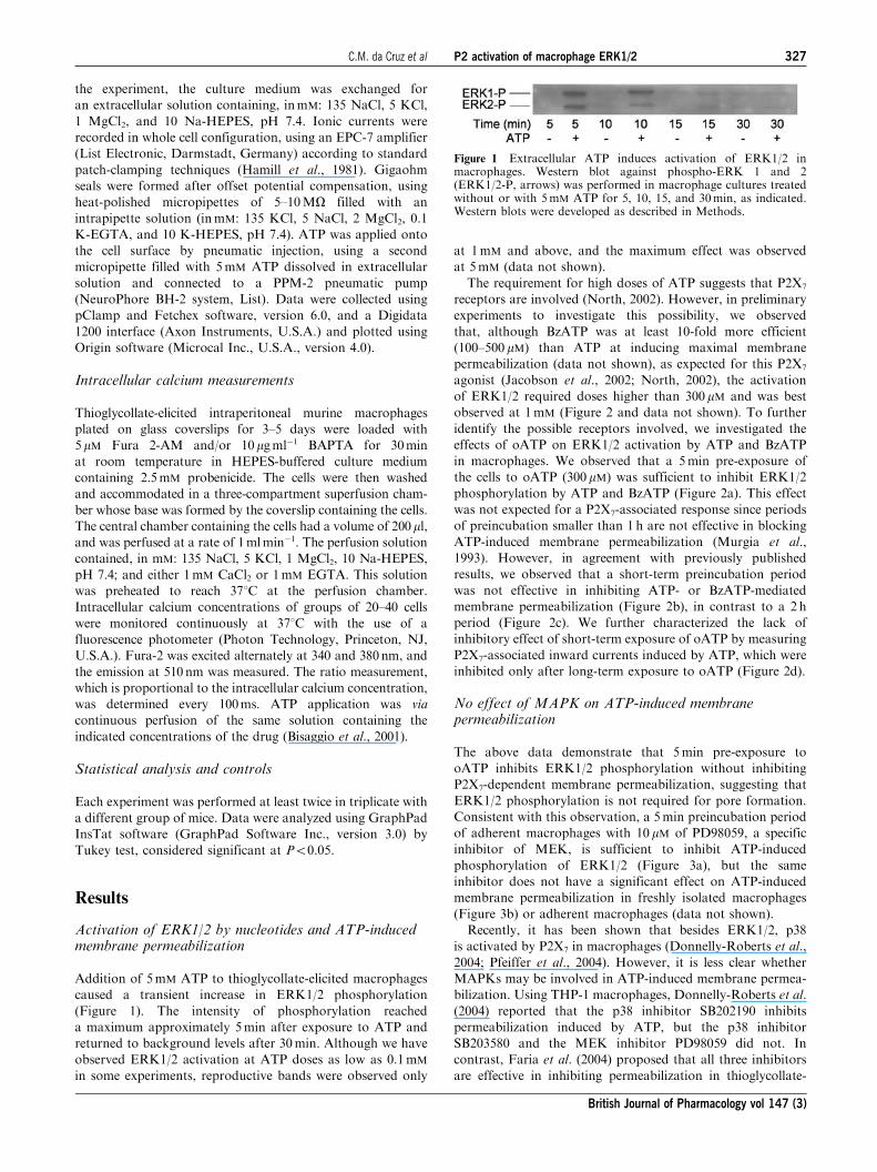
Addition of 5mM ATP to thioglycollate-elicited macrophages
caused a transient increase in ERK1/2 phosphorylation
(Figure 1). The intensity of phosphorylation reached
a maximum approximately 5min after exposure to ATP and
returned to background levels after 30min. Although we have
observed ERK1/2 activation at ATP doses as low as 0.1mM
in some experiments, reproductive bands were observed only
at 1mM and above, and the maximum effect was observed
at 5mM (data not shown).
The requirement for high doses of ATP suggests that P2X7
receptors are involved (North, 2002). However, in preliminary
experiments to investigate this possibility, we observed
that, although BzATP was at least 10-fold more efficient
(100–500 mM) than ATP at inducing maximal membrane
permeabilization (data not shown), as expected for this P2X7
agonist (Jacobson et al., 2002; North, 2002), the activation
of ERK1/2 required doses higher than 300mM and was best
observed at 1mM (Figure 2 and data not shown). To further
identify the possible receptors involved, we investigated the
effects of oATP on ERK1/2 activation by ATP and BzATP
in macrophages. We observed that a 5min pre-exposure of
the cells to oATP (300mM) was sufficient to inhibit ERK1/2
phosphorylation by ATP and BzATP (Figure 2a). This effect
was not expected for a P2X7-associated response since periods
of preincubation smaller than 1 h are not effective in blocking
ATP-induced membrane permeabilization (Murgia et al.,
1993). However, in agreement with previously published
results, we observed that a short-term preincubation period
was not effective in inhibiting ATP- or BzATP-mediated
membrane permeabilization (Figure 2b), in contrast to a 2 h
period (Figure 2c). We further characterized the lack of
inhibitory effect of short-term exposure of oATP by measuring
P2X7-associated inward currents induced by ATP, which were
inhibited only after long-term exposure to oATP (Figure 2d).
No effect of MAPK on ATP-induced membranepermeabilization
The above data demonstrate that 5min pre-exposure to
oATP inhibits ERK1/2 phosphorylation without inhibiting
P2X7-dependent membrane permeabilization, suggesting that
ERK1/2 phosphorylation is not required for pore formation.
Consistent with this observation, a 5min preincubation period
of adherent macrophages with 10 mM of PD98059, a specific
inhibitor of MEK, is sufficient to inhibit ATP-induced
phosphorylation of ERK1/2 (Figure 3a), but the same
inhibitor does not have a significant effect on ATP-induced
membrane permeabilization in freshly isolated macrophages
(Figure 3b) or adherent macrophages (data not shown).
Recently, it has been shown that besides ERK1/2, p38
is activated by P2X7 in macrophages (Donnelly-Roberts et al.,
2004; Pfeiffer et al., 2004). However, it is less clear whether
MAPKs may be involved in ATP-induced membrane permea-
bilization. Using THP-1 macrophages, Donnelly-Roberts et al.
(2004) reported that the p38 inhibitor SB202190 inhibits
permeabilization induced by ATP, but the p38 inhibitor
SB203580 and the MEK inhibitor PD98059 did not. In
contrast, Faria et al. (2004) proposed that all three inhibitors
are effective in inhibiting permeabilization in thioglycollate-
Figure 1 Extracellular ATP induces activation of ERK1/2 inmacrophages. Western blot against phospho-ERK 1 and 2(ERK1/2-P, arrows) was performed in macrophage cultures treatedwithout or with 5mM ATP for 5, 10, 15, and 30min, as indicated.Western blots were developed as described in Methods.
C.M. da Cruz et al P2 activation of macrophage ERK1/2 327
British Journal of Pharmacology vol 147 (3)

elicited murine macrophages. We therefore investigated the
possible effects of the three drugs using different doses and
preincubation periods. Using either freshly isolated or adherent
primary macrophages or the macrophage-like J774.A1 cell line,
we found no differences in ATP-induced dye uptake in the
presence of PD98059 (10, 30, and 100mM), SB203580 (1, 10,
and 100mM), or SB202190 (1 and 10mM) at pre-exposure times
of 5 or 30min (Figure 4 and data not shown).
Activation of macrophage ERK1/2 by different P2receptors
The above results prompted us to investigate whether other P2
and/or P1 receptors may be involved in the activation of ERK1/
2 in macrophages. Thus, we investigated the effects of UTP,
UDP, ADP, and adenosine on the phosphorylation of ERK1/2
in the presence or absence of oATP (Figure 5). UTP, UDP, and
ADP induced ERK activation, while adenosine had no effect
(Figure 5a). However, only the effects of ATP, BzATP, and
ADP were blocked by oATP (Figures 5b and 2a). These data
imply that several P2Y receptors (those that bind to UTP, UDP,
and ADP) and possibly one or more P2X receptors, but not P1
receptors, are coupled to ERK1/2 activation in macrophages.
Involvement of two different intracellular pathways inactivation of macrophage ERK1/2 by P2 receptors
We next investigated possible upstream mechanisms that could
link P2 ligation by ATP to ERK 1/2 activation. In most cases,
MAPK pathways require a tyrosine kinase receptor to initiate
the phosphorylation process (Dong et al., 2002). Alternatively,
ERK could be activated via a PKC-dependent pathway,
as already described for the ADP receptor, P2Y1 (Ralevic
& Burnstock, 1998). We thus pretreated macrophages with the
general tyrosine kinase inhibitor, genistein, for 5min before
exposure to either BzATP or ATP (Figure 6a). Unexpectedly,
we found that genistein inhibited BzATP-induced but not
ATP-induced ERK1/2 activation. Tyrphostin, an Src protein
kinase inhibitor, also inhibited BzATP, but not ATP-induced
ERK1/2 activation (Figure 6b). These data suggest that
BzATP activates MAPK through a classical pathway recruit-
ing tyrosine kinases, but ATP-induced activation may involve
other pathways. In line with this possibility, we observed that
chelerythrine chloride, an inhibitor of PKC, completely
blocked activation of ERK1/2 by ATP (Figure 6c).
We also investigated the possible effects of genistein (Figure 7a
and d), tyrphostin (Figure 7b), and chelerythrine (Figure 7c and
d) on the phenomenon of P2X7-associated membrane permea-
bilization and found no effect of these drugs on the uptake of
ethidium bromide by macrophages. These results further
demonstrate that ERK1/2 phosphorylation and ATP- and
BzATP-induced permeabilization are two independent events.
Intracellular Ca2þ signaling is not required forATP-induced membrane permeabilization
Most P2 receptors induce an increase in the free intracellular
Ca2þ concentration due to either influx from the extracellular
Figure 2 ERK1/2 activation by ATP and BzATP, but not membrane permeabilization, is blocked by short-term exposure tooATP. (a) Western blots were performed with antibodies against phospho-ERK1/2 using macrophage cultures preincubated in thepresence or absence of 300mM oATP for 5min, at 371C. Then, 1mM BzATP or 5mM ATP were added for an additional 10min, asindicated. Ethidium bromide was added during the last 5min of incubation. Control macrophages (CTRL) were exposed toethidium bromide in the absence of any drugs. (b, c) Permeabilization assay (ethidium bromide uptake) performed by flowcytometry in freshly isolated intraperitoneal macrophages. Cells were preincubated with or without 300 mM oATP for either 5min(b) or 2 h (c) and then exposed to different concentrations of either BzATP or ATP for 10min. Ethidium bromide was added 5minafter ATP. (d) Representative recordings of whole-cell inward currents obtained with three macrophages after pneumatic injectionsof ATP (arrows) before (upper recording) or 5min (middle recording) and 2 h (lower recording) after the addition of 300mM oATP.Data in b and c represent the mean %7s.e.m. of a representative experiment performed in triplicate. #P40.05; *Po0.001 representsthe significance of the differences between the permeabilization induced by ATP as compared to ATP plus drug.
328 C.M. da Cruz et al P2 activation of macrophage ERK1/2
British Journal of Pharmacology vol 147 (3)

medium or release of intracellular Ca2þ stocks (Greenberg
et al., 1988; Ralevic & Burnstock, 1998; North, 2002). Since
the increase in free intracellular Ca2þ concentration is
Figure 3 ERK1/2 phosphorylation is not required for ATP-induced membrane permeabilization in macrophages. (a) Westernblot was performed with antibodies against phospho-ERK1/2 usingmacrophage cultures preincubated in the presence or absence of10 mM PD98059 for 5min at 371C. Then, 1mM BzATP or 5mM ATPwas added for an additional 5min as indicated. Control macro-phages (CTRL) were not exposed to any drugs. (b) Permeabilizationassay (ethidium bromide uptake) performed by flow cytometry withfreshly isolated intraperitoneal macrophages. Cells were preincu-bated with or without 10 mM PD98059 for 5min at 371C and thenexposed to different concentrations of ATP for 10min. Ethidiumbromide was added 5min after ATP. Data represent the mean%7s.e.m. of a representative experiment performed in triplicate.#P40.05 represents the significance of the differences between thepermeabilization induced by ATP as compared to ATP plus drug.
Figure 4 ERK1/2 and p38 are not required for ATP-induced membrane permeabilization in macrophages. Permeabilization assays(ethidium bromide uptake) were performed by flow cytometry in freshly isolated intraperitoneal macrophages (a) and J774.A1 cells(b). Cells were preincubated with or without PD98059 (30 mM), SB20358 (10 mM), and SB202190 (10mM) for 30min at 371C and thenexposed to 1mM (a) or 5mM (b) ATP for 10min. Ethidium bromide was added 5min after ATP. Data represent the mean%7s.e.m. of a representative experiment performed in triplicate. #P40.05 represents the significance of the differences between thepermeabilization induced by ATP as compared to ATP plus drug.
Figure 5 ERK1/2 activation by different nucleotides and blockadeby oATP. (a) Western blot was performed with antibodies againstphospho-ERK1/2 (upper blot) and total ERK1/2 (lower blot) usingmacrophage cultures exposed to the indicated concentrations ofBzATP, ATP, adenosine, ADP, UTP, or UDP for 5min. (b)Densitometry analysis of a representative Western blot performedagainst phospho-ERK1/2 using macrophage cultures preincubatedin the presence or absence of 300 mM oATP for 5min at 371C andthen in the presence or absence of the indicated concentrations ofATP, UTP, UDP, or ADP for an additional 5min. Band densitiesare expressed in arbitrary units. Control macrophages (CTRL) werenot exposed to any drugs.
C.M. da Cruz et al P2 activation of macrophage ERK1/2 329
British Journal of Pharmacology vol 147 (3)

associated with the activation of a variety of signaling cascades,
and macrophages display several P2 receptors that can induce
intracellular Ca2þ signals (Coutinho-Silva et al., 2005), we
decided to investigate whether intracellular Ca2þ signaling
is required for membrane permeabilization in macrophages
(Figure 8). We first showed that the ATP-induced intracellular
Ca2þ concentration increase in adherent macrophages is
partially diminished in the absence of extracellular Ca2þ and
completely blocked by the intracellular calcium chelator,
BAPTA (Figure 8a). Under the same experimental conditions,
freshly isolated intraperitoneal macrophages (Figure 8b) and
adherent macrophages (data not shown) displayed no signifi-
cant alterations in ATP-induced membrane permeabilization.
Discussion
We had originally proposed that P2X7 activation may require
intracellular signals to induce the formation of large non-
selective pores based on results from electrophysiological
recordings of the pores by cell-attached patch clamping
(Coutinho-Silva & Persechini, 1997; Persechini et al., 1998).
However, despite evidence indicating that P2X7 receptors are
coupled to more than one signaling pathway, including
phospholipase D and MAPKs (El-Moatassim & Dubyak,
1992; Amstrup & Novak, 2003; Gendron et al., 2003b), neither
the intracellular mechanism of pore formation nor the
molecular components of the pore have been identified (North,
2002). This may be partially due to the expression of multiple
P2 receptor subtypes together with P2X7 in the cells studied,
Figure 6 Effect of inhibitors of protein kinases on ERK1/2activation by BzATP and ATP in macrophages. Western blots wereperformed with antibodies against phospho-ERK1/2 (a–c) and totalERK 1/2 (lower blot in a) using macrophage cultures preincubatedin the presence or absence of a kinase inhibitor for 5min at 371C andthen 1mM BzATP or 5mM ATP mM was added for an additional5min. Control macrophages (CTRL) were not exposed to any drugs.The inhibitors used were (a) 10 mM genistein, (b) 25 mM tyrphostin, or(c) 10 mM chelerythrine chloride.
Figure 7 Effect of inhibitors of protein kinase on ATP- and BzATP-induced membrane permeabilization in macrophages. Thepermeabilization assay (ethidium bromide uptake) was performed by flow cytometry in freshly isolated intraperitonealmacrophages. Cells were preincubated for 5min (a–c) or 30min (d) with or without 10 mM genistein (a and d), 25 mM tyrphostin(b), or 10 mM chelerythrine chloride (c and d), and then exposed to different concentrations of ATP (a–c) or 300mM BzATP for10min. Ethidium bromide was added 5min after ATP. Data represent the mean %7s.e.m. of a representative experimentperformed in triplicate. #P40.05 represents the significance of the differences between the permeabilization induced by nucleotide(ATP or BzATP) as compared to nucleotide plus drug.
330 C.M. da Cruz et al P2 activation of macrophage ERK1/2
British Journal of Pharmacology vol 147 (3)

and the use of drugs such as BzATP and oATP that can
stimulate other P2 receptors and non-P2 targets. In this study,
we therefore investigated the activation of ERK1/2 by
nucleotides and drugs targeting different receptors in intra-
peritoneal murine macrophages and J774.A1 cells and the
pathways involved in ATP-induced P2X7-dependent mem-
brane permeabilization.
Our results showed that ERK1/2 is activated by short-term
(5min) exposure to BzATP, ATP, UTP, ADP, and UDP,
implying that multiple P2 receptor subtypes are expressed in
macrophages and are coupled to this pathway. Moreover,
while trying to characterize the contribution of P2X7 receptors,
we observed that short-term (5min) pre-exposure to oATP,
a condition that does not inhibit P2X7-associated currents
or P2X7-associated membrane permeabilization, can block the
activation of ERK1/2 by ATP, BzATP, and ADP, but not by
UTP and UDP. The incubation time required for oATP to
inhibit ERK1/2 activation is surprisingly short, which has
consequences for our understanding of both the mechanism of
action of oATP and the signal transduction pathway leading
to pore formation. First, it is likely that, at short periods of
exposure, oATP acts as a competitive antagonist rather than as
a covalent ligand of a P2 receptor. However, we cannot
exclude the possibility that oATP may also enter the cell and
somehow block ERK activation by BzATP, ATP, and ADP,
but not UTP and UDP. Second, the observation that oATP
inhibits ERK1/2 activation by ATP and BzATP without
inhibiting either P2X7-associated currents or membrane
permeabilization reinforces the conclusion that ERK1/2
activation plays no role in pore formation.
Our results also demonstrate that genistein, tyrphostin,
chelerythrine, and oATP all block ERK1/2 activation but not
ATP-induced membrane permeabilization, suggesting that pore
formation does not require tyrosine kinase, Src protein kinase,
PKC, or ERK1/2 activation. The lack of involvement of
ERK1/2 was further confirmed by directly inhibiting ERK1/2
phosphorylation with PD98059, which does not affect pore
formation in freshly isolated macrophages, adherent macro-
phage cultures, or J774.A1 cells. Finally, we have extended
these results to other MAPKs by showing that neither
SB202190 nor SB203580, two inhibitors of p38, were able to
block ATP-mediated permeabilization. These findings also
raise new questions regarding the signal transduction pathways
involved in triggering P2X7-associated pore formation. Using
THP-1 macrophages, Donnelly-Roberts et al. (2004) showed
that the p38 inhibitor SB202190, but not the p38 inhibitor
SB203580 or the MEK inhibitor PD98059, inhibits permeabi-
lization. However, Faria et al. (2004) proposed that all three
inhibitors can inhibit permeabilization in thioglycollate-elicited
murine macrophages. In contrast, Auger et al. (2005) found
that ERK1/2 activation is not required for P2X7-associated
membrane permeabilization in thymocytes.
The reason for these discrepancies is currently not clear, but
our studies with ERK1/2 inhibitors, the P2X7 antagonist
oATP, and the differential effects of the P2X7 agonist ATP and
BzATP are all consistent with the lack of coupling between
ERK1/2 activation and P2X7-dependent pore formation. In
addition, it is now becoming clear that membrane permeabi-
lization is a more complicated phenomenon than initially
thought, as suggested by a recent report (Jiang et al., 2005)
showing that N-methyl-D-glucamine and YO-PRO-1 may
utilize different pathways to enter P2X7-transfected HEK cells.
While searching for signal transduction pathways that could
be involved in ATP-induced pore formation, we also demon-
strated that membrane permeabilization does not require
intracellular Ca2þ signaling. It was previously shown that
extracellular Ca2þ is not required for pore formation, since
P2X7-dependent membrane permeabilization is, on the con-
trary, potentiated by the absence of extracellular Mg2þ and
Ca2þ (Steinberg & Silverstein, 1987). Since the permeabiliza-
tion pore is large enough to permit passage of EGTA, EDTA,
and Ca2þ , it is reasonable to assume that the use of EGTA
to chelate extracellular Ca2þ could also lead to the depletion
of intracellular Ca2þ due to either efflux of Ca2þ or influx of
EGTA. In agreement with this hypothesis, only a brief peak of
intracellular Ca2þ is observed in macrophages exposed to ATP
in the absence of extracellular Ca2þ , as shown in Figure 8 and
by others (Steinberg & Silverstein, 1987; Greenberg et al.,
1988). The use of BAPTA allowed us to show that permeabi-
lization occurs even in the absence of the short intracellular
Ca2þ spike supplied by intracellular stocks of Ca2þ .
Our data demonstrated that oATP can inhibit ERK1/2
activation by ATP and BzATP under conditions that do not
block either P2X7-dependent pore formation or P2X7-asso-
ciated inward currents. However, the macrophage P2 recep-
tor(s) targeted by oATP during short-term exposure is (are)
not known. Since ERK1/2 activation by UTP and UDP is not
affected by oATP, none of the pyrimidine receptors (P2Y2,
P2Y4, and P2Y6) seem to be involved. BzATP and ATP are
agonists for all P2X receptors described to date (Jacobson
Figure 8 ATP-induced permeabilization in macrophages does notrequire intracellular Ca2þ signals. (a) The free intracellular Ca2þ
concentration (fura-2 fluorescence ratio) was measured in adherentmurine macrophages. ATP was added at a final concentration of5mM (arrow) and maintained in the medium for 10min bycontinuous perfusion (calibrated bar at the bottom). Upper trace:cells were not loaded with BAPTA and the extracellular mediumcontained 1mM CaCl2. Middle trace: cells were not loaded withBAPTA and the extracellular medium contained 1mM EGTA andno CaCl2. Lower trace: cells were loaded with BAPTA and theextracellular medium contained 1mM EGTA and no CaCl2. (b) Thepermeabilization assay (ethidium bromide uptake) was performedby flow cytometry in freshly isolated intraperitoneal macrophages.The concentrations of extracellular calcium and EGTA are indicatedon the x-axis. The fluorescence ratio is proportional to theintracellular calcium concentration and was measured as describedin Methods. Representative data of a single experiment are shown in(a). The experiments were repeated at least three times with similarresults. Means7s.e.m. from a representative experiment performedin triplicate are shown in (b). (#)P40.05 represents the significance ofthe differences between the permeabilization induced by ATP zeroCa2þ plus EGTA (second bar from the left) as compared to BAPTA(fourth bar from the left) and ATP 1mM Ca2þ (last bar in the right).
C.M. da Cruz et al P2 activation of macrophage ERK1/2 331
British Journal of Pharmacology vol 147 (3)
et al., 2002). However, the high doses of ATP and BzATP
required to activate ERK1/2 in macrophages indicates that
P2X1–6 are not coupled to ERK1/2 activation under our
experimental conditions. Instead, the high doses of agonist
required suggest that P2X7 receptors may be involved – a
possibility that cannot be excluded by the fact that short-term
treatment with oATP does not inhibit ATP-induced membrane
permeabilization, since ERK1/2 activation could be activated
and blocked independently of cation channel opening and pore
formation. In this context, it is worthwhile to note that
recombinant P2X7 receptors truncated at the N-terminal lose
their ability to activate ERK1/2 without decreasing Ca2þ
influx (Amstrup & Novak, 2003), while truncation at the
C-terminal, a condition known to abrogate membrane permea-
bilization but not cation channel opening (Kim et al., 2001),
does not affect ERK1/2 activation. Taken together, our data
suggest that oATP may inhibit ERK1/2 activation by
interfering with one or more P2 receptors that bind to ATP,
BzATP, and/or ADP. The best candidate receptors are P2X7
and the ADP-binding P2Y receptors (P2Y1, P2Y11, P2Y12).
The interaction of oATP with other mediators in the
pathway leading to ERK1/2 activation by purine nucleotides
should also be considered. Since oATP was first introduced to
study P2 receptors, it has been widely used as an inhibitor of
ATP-induced membrane permeabilization, presumably by
covalently binding to the P2X7 receptor (Murgia et al., 1993;
Di Virgilio et al., 2001). This reagent binds covalently to lysine
residues in ATP-binding proteins and its effect therefore
requires a preincubation period of over 1 h (Murgia et al.,
1993). However, the use of oATP is complicated by the recent
demonstration that it can inhibit the secretion of IL-8
stimulated by TNF-a independently of P2 receptors (Beigi
et al., 2003). Although oATP is no longer thought to have an
effect on the P2Y2 receptor (Murgia et al., 1993; Beigi et al.,
2003), blockade of P2Y1 after long periods of exposure to
oATP has been reported (Beigi et al., 2003). Our results indicate
that oATP can also have significant antagonistic effects after
short-term exposure, raising new possibilities for the use of this
drug in the study of signal transduction by P2 receptors.
Quite unexpectedly, our data showed that ATP and BzATP
are coupled to ERK1/2 activation through different pathways.
Both a blocker of tyrosine kinases and a blocker of Src protein
kinases inhibited the activation of ERK1/2 by BzATP, but not
by ATP. These results could be due to the different affinities of
BzATP and ATP for P2 receptors (Ralevic & Burnstock, 1998;
Jacobson et al., 2002; North, 2002), but are also consistent
with the possibility that BzATP and ATP may bind to
superimposing but not identical receptor subtypes and that
ATP may bind to more of these receptors than BzATP. In
agreement with the latter interpretation, our data show that
ERK1/2 phosphorylation is induced more efficiently by ATP
than by BzATP (e.g. Figures 2a and 5a). In addition, ecto-
nucleotidases present on the macrophage surface may also
produce ADP and activate ERK1/2 through ADP-binding
P2Y receptors (e.g. Figure 5). Moreover, chelerythrine
chloride, an inhibitor of PKC, strongly inhibits the activation
of ERK1/2 by ATP, indicating that PKC is possibly upstream
from all pathways linking P2 receptors to ERK1/2 activation,
in agreement with published results for P2X7 and other P2
receptors (Neary et al., 1999; Bradford & Soltoff, 2002; Hung
& Sun, 2002; Gendron et al., 2003b). However, it should be
noted that even at 100 mM, the intensity of the phopho-ERK1/
2 bands induced by ADP remained much smaller than the one
induced by ATP. Thus, although it is clear that an ADP-
sensitive P2Y receptor can induce ERK1/2 phosphorylation, it
seems unlikely that such a high dose of ADP could be induced
by ecto-nucleotidases in 5min and therefore it is improbable
that ATP effects could be due only to an ADP-sensitive P2Y
receptor. Further experiments are needed to clarify this point,
which could also be addressed through the use of phospho-
lipase C (PLC) inhibitors. Nonetheless, experiments with these
drugs should be interpreted carefully, since it has been shown
recently (Takenouchi et al., 2005) that U73122, a PLC
inhibitor, can block ATP-induced membrane permeabilization
in microglia through a PLC-independent mechanism.
In conclusion, multiple intracellular signaling pathways are
triggered by ATP, BzATP, and other nucleotides in macro-
phages. It is tempting to speculate that cell responses such as
proliferation, cell death, and cytokine secretion will depend on
the pattern of binding of each agonist to the different P2
receptors, as has been shown for serial binding of microbial
ligands to Toll-like receptors (Weiss et al., 2004; Napolitani
et al., 2005), and on the behavior of ecto-nucleotidases present
on the surface of these cells. Finally, our data suggest that,
although MAPKs are activated by multiple P2 receptors, the
protein kinases are not involved in ATP-induced P2X7-
dependent membrane permeabilization.
We thank Dr Rodrigo da Cunha Bisaggio for helpful discussions andVandir da Costa and Maria Leite Eduardo for technical assistance.This work was supported by Conselho Nacional de DesenvolvimentoCientıfico e Tecnologico (CNPq), Fundacao Carlos Chagas Filho deAmparo a Pesquisa do Estado do Rio de Janeiro (FAPERJ),Programa de Nucleos de Excelencia (PRONEX-CNPq), and Uni-versity of California.
References
ADRIAN, K., BERNHARD, M.K., BREITINGER, H. & OGILVIE, A.
(2000). Expression of purinergic receptors (ionotropic P2X1-7 andmetabotropic P2Y1-11) during myeloid differentiation of HL60cells. Biochim. Biophys. Acta, 1492, 127–138.
AGA, M., JOHNSON, C.J., HART, A.P., GUADARRAMA, A.G.,SURESH, M., SVAREN, J., BERTICS, P.J. & DARIEN, B.J. (2002).Modulation of monocyte signaling and pore formation in responseto agonists of the nucleotide receptor P2X(7). J. Leukocyte Biol., 72,
222–232.ALBUQUERQUE, C., OLIVEIRA, S.M., COUTINHO-SILVA, R.,
OLIVEIRA-CASTRO, G.M. & PERSECHINI, P.M. (1993). ATP-and UTP-induced currents in macrophages and macrophage-polykaryons. Am. J. Physiol., 265, C1663–C1673.
AMSTRUP, J. & NOVAK, I. (2003). P2X7 receptor activates extra-cellular signal-regulated kinases ERK1 and ERK2 independently ofCa2+ influx. Biochem. J., 374, 51–61.
AUGER, R., MOTTA, I., BENIHOUD, K., OJCIUS, D.M. & KANELLO-
POULOS, J.M. (2005). A role for mitogen-activated protein kinaseERK1/2 activation and non-selective pore formation in P2X7receptor-mediated thymocyte death. J. Biol. Chem., 280, 28142–28151.
BEIGI, R.D., KERTESY, S.B., AQUILINA, G. & DUBYAK, G.R. (2003).Oxidized ATP (oATP) attenuates proinflammatory signaling via P2receptor-independent mechanisms. Br. J. Pharmacol., 140, 507–519.
BISAGGIO, R.C., NIHEI, O.K., PERSECHINI, P.M., SAVINO, W. &ALVES, L.A. (2001). Characterization of P2 receptors in thymicepithelial cells. Cell. Mol. Biol., 47, 19–31.
332 C.M. da Cruz et al P2 activation of macrophage ERK1/2
British Journal of Pharmacology vol 147 (3)

BOWLER, J.W., BAILEY, R.J., NORTH, R.A. & SURPRENANT, A.
(2003). P2X4, P2Y1 and P2Y2 receptors on rat alveolar macro-phages. Br. J. Pharmacol., 140, 567–575.
BOYER, J.L., ROMERO-AVILA, T., SCHACHTER, J.B. & HARDEN,T.K. (1996). Identification of competitive antagonists of the P2Y1receptor. Mol. Pharmacol., 50, 1323–1329.
BRADFORD, M.D. & SOLTOFF, S.P. (2002). P2X7 receptorsactivate protein kinase D and p42/p44 mitogen-activated proteinkinase (MAPK) downstream of protein kinase C. Biochem. J., 366,
745–755.BRADFORD, M.M. (1976). A rapid and sensitive method for the
quantitation of microgram quantities of protein utilizing theprinciple of protein-dye binding. Anal. Biochem., 72, 248–254.
BRONTE, V., MACINO, B., ZAMBON, A., ROSATO, A., MANDRUZZATO,S., ZANOVELLO, P. & COLLAVO, D. (1996). Protein tyrosinekinases and phosphatases control apoptosis induced by extra-cellular adenosine 50-triphosphate. Biochem. Biophys. Res.Commun., 218, 344–351.
BURNSTOCK, G. (2002). Purinergic signaling and vascular cellproliferation and death. Arterioscler. Thromb. Vasc. Biol., 22,
364–373.BURNSTOCK, G. & KNIGHT, G.E. (2004). Cellular distribution and
functions of P2 receptor subtypes in different systems. Int. Rev.Citoles, 240, 31–304.
COUTINHO-SILVA, R., OJCIUS, D.M., GORECKI, D.C., PERSECHINI,P.M., BISAGGIO, R.C., MENDES, A.N., MARKES, J., BURN-
STOCK, G. & DUNN, P.M. (2005). Multiple P2X and P2Y receptorsubtypes in mouse J774, spleen and peritoneal macrophages.Biochem. Pharmacol., 69, 641–655.
COUTINHO-SILVA, R. & PERSECHINI, P.M. (1997). P2Z purinocep-tor-associated pores induced by extracellular ATP in macrophagesand J774 cells. Am. J. Physiol., 273, C1793–C1800.
COUTINHO-SILVA, R., PERSECHINI, P.M., BISAGGIO, R.C.,PERFETTINI, J.-L., SA-NETO, A.C.T., KANELLOPOULOS, J.M.,MOTTA-LY, I., DAUTRY-VARSAT, A. & OJCIUS, D.M. (1999).P2Z/P2X7 receptor-dependent apoptosis of dendritic cells. Am. J.Physiol., 276, C1139–C1147.
COUTINHO-SILVA, R., STAHL, L., RAYMOND, M.-N., JUNGAS, T.,VERBEKE, P., BURNSTOCK, G., DARVILLE, T. & OJCIUS, D.M.
(2003). Inhibition of chlamydial infectious activity due to P2X7R-dependent phospholipase D activation. Immunity, 19, 403–412.
DANGELMAIER, C., JIN, J., DANIEL, J.L., SMITH, J.B. & KUNAPULI,S.P. (2000). The P2Y1 receptor mediates ADP-induced p38 kinase-activating factor generation in human platelets. Eur. J. Biochem.,267, 2283–2289.
DENLINGER, L.C., FISETTE, P.L., SOMMER, J.A., WATTERS, J.J.,PRABHU, U., DUBYAK, G.R., PROCTOR, R.A. & BERTICS, P.J.
(2001). The nucleotide receptor P2X7 contains multiple protein-and lipid-interaction motifs including a potential binding site forbacterial lipopolysaccharide. J. Immunol., 167, 1871–1876.
DI VIRGILIO, F., CHIOZZI, P., FERRARI, D., FALZONI, S., SANZ,J.M., MORELLI, A., TORBOLI, M., BOLOGNESI, G. & BARICORDI,O.R. (2001). Nucleotide receptors: an emerging family of regulatorymolecules in blood cells. Blood, 97, 587–600.
DONG, C., DAVIS, R.J. & FLAVELL, R.A. (2002). MAP kinases in theimmune response. Annu. Rev. Immunol., 20, 55–72.
DONNELLY-ROBERTS, D.L., NAMOVIC, M.T., FALTYNEK, C.R. &JARVIS, M.F. (2004). Mitogen-activated protein kinase and caspasesignaling pathways are required for P2X7 receptor (P2X7R)-induced pore formation in human THP-1 cells. J. Pharmacol.Exp. Ther., 308, 1053–1061.
DUBYAK, G.R. & EL-MOATASSIM, C. (1993). Signal transductionvia P2-purinergic receptors for extracellular ATP and othernucleotides. Am. J. Physiol., 265, C577–C606.
EL-MOATASSIM, C. & DUBYAK, G.R. (1992). A novel pathway forthe activation of phospholipase-D by P(2z) purinergic receptors inBAC1.2F5 macrophages. J. Biol. Chem., 267, 23664–23673.
FARIA, R.X., DE FARIAS, F.P. & ALVES, L.A. (2004). Are secondmessengers crucial for opening the pore associated with P2X7receptor? Am. J. Physiol. Cell Physiol., 288, C260–C271.
GENDRON, F.P., CHALIMONIUK, M., STROSZNAJDER, J., SHEN, S.,GONZALEZ, F.A., WEISMAN, G.A. & SUN, G.Y. (2003a). P2X7nucleotide receptor activation enhances IFN gamma-inducedtype II nitric oxide synthase activity in BV-2 microglial cells.J. Neurochem., 87, 344–352.
GENDRON, F.P., NEARY, J.T., THEISS, P.M., SUN, G.Y., GONZALEZ,F.A. & WEISMAN, G.A. (2003b). Mechanisms of P2X7 receptor-mediated ERK1/2 phosphorylation in human astrocytoma cells.Am. J. Physiol. Cell Physiol., 284, C571–C581.
GREENBERG, S., DI VIRGILIO, F., STEINBERG, T.H. & SILVERSTEIN,S.C. (1988). Extracellular nucleotides mediate Ca2+ fluxes in J774 macro-phages by two distinct mechanisms. J. Biol. Chem., 263, 10337–10343.
HAMILL, O.P., MARTY, A., NEHER, E., SAKMANN, B. & SIGWORTH,F.J. (1981). Improved patch-clamp techniques for high-resolutioncurrent recording from cells and cell-free membrane patches. Eur. J.Physiol., 391, 85–100.
HIDE, I., TANAKA, M., INOUE, A., NAKAJIMA, K., KOHSAKA, S.,INOUE, K. & NAKATA, Y. (2000). Extracellular ATP triggerstumor necrosis factor-alpha release from rat microglia. J. Neuro-chem., 75, 965–972.
HU, Y., FISETTE, P.L., DENLINGER, L.C., GUADARRAMA, A.G.,SOMMER, J.A., PROCTOR, R.A. & BERTICS, P.J. (1998). Purinergicreceptor modulation of lipopolysaccharide signaling and indu-cible nitric-oxide synthase expression in RAW 264.7 macrophages.J. Biol. Chem., 273, 27170–27175.
HUMPHREYS, B.D., RICE, J., KERTESY, S.B. & DUBYAK, G.R.
(2000). Stress-activated protein kinase/JNK activation and apopto-tic induction by the macrophage P2X7 nucleotide receptor. J. Biol.Chem., 275, 26792–26798.
HUNG, A.C. & SUN, S.H. (2002). The P2X(7) receptor-mediatedphospholipase D activation is regulated by both PKC-dependentand PKC-independent pathways in a rat brain-derived type-2astrocyte cell line, RBA-2. Cell Signal., 14, 83–92.
JACOBSON, K.A., KING, B.F. & BURNSTOCK, G. (2002). Pharmacologicalcharacterization of P2 (nucleotide) receptors. Cell Transm., 16, 1–23.
JIANG, L.H., RASSENDREN, F., MACKENZIE, A.B., ZHANG, Y.H.,SURPRENANT, A. & NORTH, R.A. (2005). N-methyl-D-glucamineand propidium dyes utilize different permeation pathways at ratP2X7 receptors. Am. J. Physiol Cell Physiol., 289, C1295–C1302.
JIN, J.G., DASARI, V.R., SISTARE, F.D. & KUNAPULI, S.P. (1998).Distribution of P2Y receptor subtypes on haematopoietic cells.Br. J. Pharmacol., 123, 789–794.
KIM, M., JIANG, L.H., WILSON, H.L., NORTH, R.A. & SURPRENANT,A. (2001). Proteomic and functional evidence for a P2X(7) receptorsignalling complex. EMBO J., 20, 6347–6358.
LAMMAS, D.A., STOBER, C., HARVEY, C.J., KENDRICK, N.,PANCHALINGAM, S. & KUMARARATNE, D.S. (1997). ATP-induced killing of mycobacteria by human macrophages is mediatedby purinergic P2Z(P2X7) receptors. Immunity, 7, 433–444.
LENZ, G., GOTTFRIED, C., LUO, Z., AVRUCH, J., RODNIGHT, R.,NIE, W.J., KANG, Y. & NEARY, J.T. (2000). P(2Y) purinoceptorsubtypes recruit different MEK activators in astrocytes. Br. J.Pharmacol., 129, 927–936.
MORELLI, A., CHIOZZI, P., CHIESA, A., FERRARI, D., SANZ, J.M.,FALZONI, S., PINTON, P., RIZZUTO, R., OLSON, M.F. & DI VIRGILIO,F. (2003). Extracellular ATP causes ROCK I-dependent bleb formationin P2X7-transfected HEK293 cells. Mol. Biol. Cell, 14, 2655–2664.
MORELLI, A., FERRARI, D., BOLOGNESI, G., RIZZUTO, R. & DI
VIRGILIO, F. (2001). Proapoptotic plasma membrane pore:P2X7 receptor. Drug Dev. Res., 52, 571–578.
MURGIA, M., HANAU, S., PIZZO, P., RIPPA, M. & DI VIRGILIO, F.(1993). Oxidized ATP an irreversible inhibitor of macrophagepurinergic P2Z receptor. J. Biol. Chem., 268, 8199–8203.
NAPOLITANI, G., RINALDI, A., BERTONI, F., SALLUSTO, F. &LANZAVECCHIA, A. (2005). Selected toll-like receptor agonistcombinations synergistically trigger a T helper type 1-polarizingprogram in dendritic cells. Nat. Immunol., 6, 769–776.
NEARY, J.T., KANG, Y., BU, Y., YU, E., AKONG, K. & PETERS, C.M.
(1999). Mitogenic signaling by ATP/P2Y purinergic receptors inastrocytes: involvement of a calcium-independent protein kinase C,extracellular signal-regulated protein kinase pathway distinctfrom the phosphatidylinositol-specific phospholipase C/calciumpathway. J. Neurosci., 19, 4211–4220.
NORTH, R.A. (2002). Molecular physiology of P2X receptors. Physiol.Rev., 82, 1013–1067.
PANENKA, W., JIJON, H., HERX, L.M., ARMSTRONG, J.N., FEIGHAN,D., WEI, T., YONG, V.W., RANSOHOFF, R.M. & MACVICAR, B.A.
(2001). P2X7-like receptor activation in astrocytes increaseschemokine monocyte chemoattractant protein-1 expression viamitogen-activated protein kinase. J. Neurosci., 21, 7135–7142.
C.M. da Cruz et al P2 activation of macrophage ERK1/2 333
British Journal of Pharmacology vol 147 (3)

PARVATHENANI, L.K., TERTYSHNIKOVA, S., GRECO, C.R.,ROBERTS, S.B., ROBERTSON, B. & POSMANTUR, R. (2003).P2X7 mediates superoxide production in primary microglia andis up-regulated in a transgenic mouse model of Alzheimer’s disease.J. Biol. Chem., 278, 13309–13317.
PERSECHINI, P.M., BISAGGIO, R.C., ALVES-NETO, J.L. & COUTINHO-
SILVA, R. (1998). Extracellular ATP in the lymphohematopoieticsystem: P2Z purinoceptors and membrane permeabilization. Braz. J.Med. Biol. Res., 31, 25–34.
PFEIFFER, Z.A., AGA, M., PRABHU, U., WATTERS, J.J., HALL, D.J. &BERTICS, P.J. (2004). The nucleotide receptor P2X7 mediates actinreorganization and membrane blebbing in RAW 264.7 macrophagesvia p38 MAP kinase and Rho. J. Leukocyte Biol., 75, 1173–1182.
RALEVIC, V. & BURNSTOCK, G. (1998). Receptors for purines andpyrimidines. Pharmacol. Rev., 50, 413–492.
SANTIAGO-PEREZ, L.I., FLORES, R.V., SANTOS-BERRIOS, C.,CHORNA, N.E., KRUGH, B., GARRAD, R.C., ERB, L., WEISMAN,G.A. & GONZALEZ, F.A. (2001). P2Y(2) nucleotide receptorsignaling in human monocytic cells: activation, desensitizationand coupling to mitogen-activated protein kinases. J. Cell Physiol.,187, 196–208.
SAUNDERS, B.M., FERNANDO, S.L., SLUYTER, R., BRITTON, W.J. &WILEY, J.S. (2003). A loss-of-function polymorphism in the humanP2X7 receptor abolishes ATP-mediated killing of mycobacteria.J. Immunol., 171, 5442–5446.
STEINBERG, T.H., NEWMAN, A.S., SWANSON, J.A. & SILVERSTEIN,S.C. (1987). ATP4� permeabilizes the plasma membrane of mousemacrophages to fluorescent dyes. J. Biol. Chem., 262, 8884–8888.
STEINBERG, T.H. & SILVERSTEIN, S.C. (1987). Extracellular ATP4�
promotes cation fluxes in the J774 mouse macrophage cell line.J. Biol. Chem., 262, 3118–3122.
SURPRENANT, A., RASSENDREN, F., KAWASHIMA, E., NORTH, R.A.
& BUELL, G. (1996). The cytolytic P2Z receptor for extracellular ATPidentified as a P2X receptor (P2X7). Science, 272, 735–738.
TAKENOUCHI, T., OGIHARA, K., SATO, M. & KITANI, H. (2005).Inhibitory effects of U73122 and U73343 on Ca(2+) influx andpore formation induced by the activation of P2X7 nucleotidereceptors in mouse microglial cell line. Biochim. Biophys. Acta,1726, 177–186.
VARTIAN, N. & BOEHM, S. (2001). P2Y receptor-mediated inhibi-tion of voltage-activated Ca(2+) currents in PC12 cells. Eur. J.Neurosci., 13, 899–908.
VERHOEF, P.A., ESTACION, M., SCHILLING, W. & DUBYAK, G.R.
(2003). P2X7 receptor-dependent blebbing and the activation ofRho-effector kinases, caspases, and IL-1 beta release. J. Immunol.,170, 5728–5738.
VIGNE, P., HECHLER, B., GACHET, C., BREITTMAYER, J.P. &FRELIN, C. (1999). Benzoyl ATP is an antagonist of rat and humanP2Y1 receptors and of platelet aggregation. Biochem. Biophys. Res.Commun., 256, 94–97.
WARNY, M., ABOUDOLA, S., ROBSON, S.C., SEVIGNY, J., COMMUNI,D., SOLTOFF, S.P. & KELLY, C.P. (2001). P2Y(6) nucleotidereceptor mediates monocyte interleukin-8 production in response toUDP or lipopolysaccharide. J. Biol. Chem., 276, 26051–26056.
WEISS, D.S., RAUPACH, B., TAKEDA, K., AKIRA, S. & ZYCHLINSKY,A. (2004). Toll-like receptors are temporally involved in hostdefense. J. Immunol., 172, 4463–4469.
WHITE, P.J., WEBB, T.E. & BOARDER, M.R. (2003). Characterizationof a Ca2+ response to both UTP and ATP at human P2Y11receptors: evidence for agonist-specific signaling. Mol. Pharmacol.,63, 1356–1363.
(Received August 30, 2005Revised October 28, 2005
Accepted November 1, 2005Published online 12 December 2005)
334 C.M. da Cruz et al P2 activation of macrophage ERK1/2
British Journal of Pharmacology vol 147 (3)
Copyright © 2022 FDOKUMEN