Modulation of MCP-1 and iNOS by 50-Hz sinusoidal electromagnetic field
Upload
independentCategory
view
0download
0
B R A I N R E S E A R C H 1 2 4 5 ( 2 0 0 8 ) 2 6 – 3 5
ava i l ab l e a t www.sc i enced i rec t . com
www.e l sev i e r. com/ loca te /b ra in res
Research Report
Isoflurane preconditioning activates HIF-1α, iNOS and Erk1/2and protects against oxygen–glucose deprivationneuronal injury
Qi-fang Li, Ye-sen Zhu, Hong Jiang⁎
Department of Anesthesiology, Shanghai Ninth People's Hospital Affiliated to Shanghai Jiao Tong University School of Medicine, 639 ZhizaojuRoad, Shanghai 200011, China
A R T I C L E I N F O
⁎ Corresponding author. Fax: +86 21 5359 1385E-mail address: [email protected] (
0006-8993/$ – see front matter. Crown Copyrdoi:10.1016/j.brainres.2008.09.069
A B S T R A C T
Article history:Accepted 23 September 2008Available online 7 October 2008
Preconditioning neurons with isoflurane, a commonly used volatile anesthetic in clinicalpractice, improves tolerance of subsequent ischemia in both intact animal models and invitro preparations. To investigate the mechanisms of this protection, we primarily culturedrat hippocampal neurons and simulated ischemia in vitro by oxygen–glucose deprivation(OGD). Neuron viability was measured. Neuron injury was observed by inverted phasecontrast microscope and assessed by lactate dehydrogenase (LDH) release. Gene expressionwas examined by Western blot and reverse transcription-polymerase chain reaction (RT-PCR). Isoflurane exposure for 2 h at 24 h before a 2 h OGD dose-dependently reduced cellinjury. Isoflurane accumulated phosphorylation/activation of extracellular signal-relatedkinases 1 and 2 (Erk1/2) and hypoxia inducible factor (HIF)-1α, a transcription factor involvedin cell survival. Inhibition of the phospho-Erk1/2 partially abolished the isofluranepreconditioning-induced HIF-1α protein content accumulation and neuroprotection.Isoflurane also increased inducible nitric oxide synthase (iNOS) mRNA levels, adownstream gene of HIF-1α. Thus, the current results indicate that isofluranepreconditioning activates HIF-1α during protection against OGD neuronal injury and theactivation might be partly mediated by the Erk1/2 pathway.
Crown Copyright © 2008 Published by Elsevier B.V. All rights reserved.
Keywords:Isoflurane preconditioningHypoxia inducible factor-1αIschemiaPrimary hippocampal neuron
1. Introduction
It is documented that the fluorinated volatile anestheticisoflurane protects the brain when administered during orimmediated before an ischemic insult to the brain similar tothat achieved by ischemic preconditioning (Miura et al., 1998;Sakai et al., 2007). Although the mechanisms involved inisoflurane preconditioning are poorly understood, this con-ditioning has potential clinical prospects. Identifying the
.H. Jiang).
ight © 2008 Published by
mechanisms bywhich isoflurane and other volatile anestheticagents mediate their anti-ischemic actions may be of specialclinical significance in protection against the ischemic eventsthat frequently occur in neurosurgical patients in the perio-perative period.
Recently, we and others demonstrated that isofluranemodulates the expression of several genes concerned withcell survival including hypoxia inducible factor (HIF)-1, animportant DNA-binding complex (Semenza and Wang, 1992)
Elsevier B.V. All rights reserved.
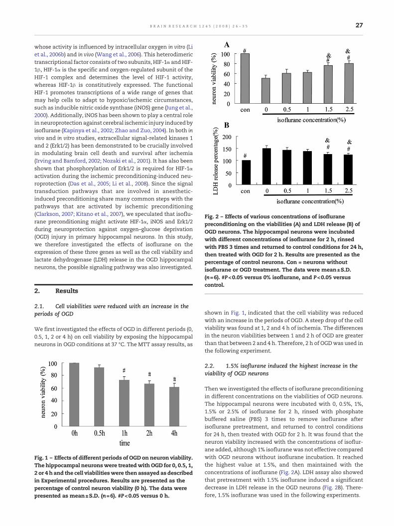
Fig. 2 – Effects of various concentrations of isofluranepreconditioning on the viabilities (A) and LDH release (B) ofOGD neurons. The hippocampal neurons were incubatedwith different concentrations of isoflurane for 2 h, rinsedwith PBS 3 times and returned to control conditions for 24 h,then treated with OGD for 2 h. Results are presented as thepercentage of control neurons. Con = neurons withoutisoflurane or OGD treatment. The data were mean±S.D.
27B R A I N R E S E A R C H 1 2 4 5 ( 2 0 0 8 ) 2 6 – 3 5
whose activity is influenced by intracellular oxygen in vitro (Liet al., 2006b) and in vivo (Wang et al., 2006). This heterodimerictranscriptional factor consists of two subunits, HIF-1α andHIF-1β, HIF-1α is the specific and oxygen-regulated subunit of theHIF-1 complex and determines the level of HIF-1 activity,whereas HIF-1β is constitutively expressed. The functionalHIF-1 promotes transcriptions of a wide range of genes thatmay help cells to adapt to hypoxic/ischemic circumstances,such as inducible nitric oxide synthase (iNOS) gene (Jung et al.,2000). Additionally, iNOS has been shown to play a central rolein neuroprotection against cerebral ischemic injury induced byisoflurane (Kapinya et al., 2002; Zhao and Zuo, 2004). In both invivo and in vitro studies, extracellular signal-related kinases 1and 2 (Erk1/2) has been demonstrated to be crucially involvedin modulating brain cell death and survival after ischemia(Irving and Bamford, 2002; Nozaki et al., 2001). It has also beenshown that phosphorylation of Erk1/2 is required for HIF-1αactivation during the ischemic preconditioning-induced neu-roprotection (Das et al., 2005; Li et al., 2008). Since the signaltransduction pathways that are involved in anesthetic-induced preconditioning share many common steps with thepathways that are activated by ischemic preconditioning(Clarkson, 2007; Kitano et al., 2007), we speculated that isoflu-rane preconditioning might activate HIF-1α, iNOS and Erk1/2during neuroprotection against oxygen–glucose deprivation(OGD) injury in primary hippocampal neurons. In this study,we therefore investigated the effects of isoflurane on theexpression of these three genes as well as the cell viability andlactate dehydrogenase (LDH) release in the OGD hippocampalneurons, the possible signaling pathway was also investigated.
(n=6). #P<0.05 versus 0% isoflurane, and P<0.05 versuscontrol.
2. Results
2.1. Cell viabilities were reduced with an increase in theperiods of OGD
We first investigated the effects of OGD in different periods (0,0.5, 1, 2 or 4 h) on cell viability by exposing the hippocampalneurons in OGD conditions at 37 °C. The MTT assay results, as
Fig. 1 – Effects of different periods of OGD on neuron viability.The hippocampal neuronswere treatedwith OGD for 0, 0.5, 1,2 or 4 h and the cell viabilitieswere then assayed as describedin Experimental procedures. Results are presented as thepercentage of control neuron viability (0 h). The data werepresented as mean±S.D. (n=6). #P<0.05 versus 0 h.
shown in Fig. 1, indicated that the cell viability was reducedwith an increase in the periods of OGD. A steep drop of the cellviability was found at 1, 2 and 4 h of ischemia. The differencesin the neuron viabilities between 1 and 2 h of OGD are greaterthan that between 2 and 4 h. Therefore, 2 h of OGDwas used inthe following experiment.
2.2. 1.5% isoflurane induced the highest increase in theviability of OGD neurons
Thenwe investigated the effects of isoflurane preconditioningin different concentrations on the viabilities of OGD neurons.The hippocampal neurons were incubated with 0, 0.5%, 1%,1.5% or 2.5% of isoflurane for 2 h, rinsed with phosphatebuffered saline (PBS) 3 times to remove isoflurane afterisoflurane pretreatment, and returned to control conditionsfor 24 h, then treated with OGD for 2 h. It was found that theneuron viability increased with the concentrations of isoflur-ane added, although 1% isofluranewas not effective comparedwith OGD neurons without isoflurane incubation. It reachedthe highest value at 1.5%, and then maintained with theconcentrations of isoflurane (Fig. 2A). LDH assay also showedthat pretreatment with 1.5% isoflurane induced a significantdecrease in LDH release in the OGD neurons (Fig. 2B). There-fore, 1.5% isoflurane was used in the following experiments.

28 B R A I N R E S E A R C H 1 2 4 5 ( 2 0 0 8 ) 2 6 – 3 5
2.3. Isoflurane preconditioning time-dependently protectedhippocampal neurons against OGD injury
To find out the protective effects of isoflurane precondition-ing duration against OGD injury, the hippocampal neuronswere incubated with 1.5% isoflurane for 0, 1, 2, 4 or 8 h, rinsedwith PBS 3 times and returned to control conditions for 24 hbefore the neurons were treated with OGD for 2 h. It wasfound that the viability of the OGD neurons pretreated withisoflurane (2, 4 or 8 h) was significantly higher than that inthe OGD neurons without isoflurane preconditioning (allP<0.05 versus ‘0h+2h’) (Fig. 3A). These data were in agree-ment with the corresponding morphological changes (Fig. 3B)and implied that 1.5% isoflurane preconditioning for 2, 4 and8 h could protect against OGD neuronal injury.
Fig. 3 – Effects of isoflurane preconditioning duration and OGD oThe hippocampal neurons were pretreated with 1.5% isofluraneto control conditions for 24 h, then treated with OGD for 2 h. Thechanges were observed. (A) The neuron viabilities (control = 0 hnormoxia+2 h OGD; 1 h+2 h=1 h isoflurane+24 h normoxia+2 h4 h+2 h=4 h isoflurane+24 h normoxia+2 h OGD and 8 h+2 h=8changes (a, control; b, 0 h+2 h; c, 1 h+2 h; d, 2 h+2 h; e, 4 h+2 h(n=6). #P<0.05 versus 0 h+2 h; *P<0.05 versus control. Scale bar
2.4. Isoflurane preconditioning induced a significantincrease in expression of HIF-1α protein and iNOS mRNA
To understand the potential mechanisms involved in the pro-tective effects of isoflurane preconditioning against OGDinjury, we investigated the effects of isoflurane precondition-ing on expression of HIF-1α. The expression of HIF-1α proteinandmRNAwas then determined byWestern blot and RT-PCR.The findings showed that incubation with OGD or isofluranealone for 2 h significantly induced the levels of the HIF-1αprotein in the neurons and a further accumulation wasachieved by ISO+OGD (neurons treated with 1.5% isofluranefor 2 h, then rinsed with PBS 3 times and returned to controlconditions for 24 h before treated with OGD for 2 h). Inaddition, expression of HIF-1α was higher after OGD than that
n the viability and morphology of hippocampal neurons.for 0, 1, 2, 4 or 8 h, rinsed with PBS 3 times and returnedneuron viabilities were then assayed and morphologicalhypoxia+24 h normoxia; 0 h+2 h=0 h isoflurane+24 hOGD; 2 h+2 h=2 h isoflurane+24 h normoxia+2 h OGD;
h isoflurane+24 h normoxia+2 h OGD), and (B) morphological; and f, 8 h+2 h). The data presented were mean±S.D.=100 μm.

29B R A I N R E S E A R C H 1 2 4 5 ( 2 0 0 8 ) 2 6 – 3 5
after isoflurane (Fig. 4A). RT-PCR results showed that levels ofHIF-1α mRNA in the hippocampal neuronal cultures wereunaltered in OGD, isoflurane neurons or OGD neurons pre-treated with isoflurane (Fig. 4B). These results indicated thatisoflurane preconditioning modulates HIF-1α via a post-transcriptional pathway and implied that the protective effectinduced by isoflurane pretreatment in hippocampal neurons
Fig. 4 – Effects of isoflurane preconditioning on HIF-1α mRNA anneurons. (A) Western blot analysis of HIF-1α protein from neuroisoflurane. RT-PCR analysis of HIF-1α mRNA (B) and iNOS mRNApretreated with isoflurane. The left panel is densitometric analysCON = neurons without isoflurane or OGD treatment, ISO = neurincubated with OGD for 2 h. ISO+OGD = neurons incubated withto control conditions for 24 h, then treated with OGD for 2 h. #P<
might be associated with the increased content of HIF-1αprotein.
iNOS has been shown to play an important role inneuroprotection induced by isoflurane preconditioning, andwhich is known to be regulated by HIF-1α activation underhypoxic/ischemic conditions (Jung et al., 2000). Fig. 4C showeda remarkable iNOSmRNA increase after OGD incubation and a
d protein expression and iNOS mRNA in hippocampalns treated with isoflurane, OGD or OGD pretreated with(C) from neurons treated with isoflurane, OGD or OGDis of the bands. Values were presented as mean±S.D. (n=3).ons incubated with isoflurane for 2 h, OGD = neuronsisoflurane for 2 h, rinsed with PBS 3 times and returned0.05 versus CON, & P<0.05 versus OGD, $P<0.05 versus ISO.

30 B R A I N R E S E A R C H 1 2 4 5 ( 2 0 0 8 ) 2 6 – 3 5
further accumulation in ISO+OGD neurons. Densitometricanalysis indicated that HIF-1α protein and iNOSmRNA furtherincreased in ISO+OGD group than that in OGD-only group (1.5-to 2-fold compared with the OGD-only group). Furthermore,dose–response analysis revealed that 1.5% and 1% isofluraneincubation for 2 h caused the maximal induction of HIF-1αprotein and iNOS mRNA respectively (Figs. 5A and B).
2.5. Isoflurane induces HIF-1α expression andneuroprotection via Erk1/2 phosphorylation
Previous studies have shown isoflurane leads to phosphoryla-tion of Erk1/2 in slice cultures and rat heart during protectionagainst hypoxic/ischemic injury (Gray et al., 2005; Wang et al.,2006), and this signaling cascade is involved in the inductionof HIF-1α protein by hypoxic or nonhypoxic stimuli (Sutton etal., 2007; Zhang et al., 2006). Then, we investigated theinhibition of Erk1/2 on expression of p-Erk (phosphorylatedErk, active Erk)/t-Erk (total Erk, phosphorylated and depho-sphorylated Erk) and HIF-1α in the neurons treated withisoflurane. PD98059 (the selective Erk1/2 inhibitor) was addedto the culture medium at a final concentration of 30 μM for 1 hprior to 1.5% isoflurane treatment (2 h) as a previouslydescribed application in hippocampal neurons (Rapoport andFerreira, 2000). Western blot analysis found that PD98059 sig-nificantly attenuated the level of p-ERK/t-ERK in the isofluraneneurons (Figs. 6A and B, samples were collected immediatelyafter isoflurane exposure). In the case of HIF-1α, isofluraneincubation increased HIF-1α protein content, and PD98059alleviated the induction of HIF-1α protein by isoflurane (Figs.6C and D). This implied that isoflurane induced the contents ofHIF-1α protein via Erk1/2 pathway. However, the contents ofHIF-1α in the neurons preincubated with PD98059 were stillhigher than those in the control group, indicating that the
Fig. 5 – Dose–response relationship for isoflurane and HIF-1α prprotein and (B) RT-PCR of iNOS mRNA from neurons treated withis densitometric analysis of the bands. Values were presented aversus 1% isoflurane.
inhibition of Erk1/2 could not completely inhibit the positiveeffects of isoflurane on the contents of HIF-1α. To exclude thepotential effects of PD98059 on the expression of p-ERK/t-ERKand HIF-1α, we also examined whether PD98059 can affectexpression of these proteins in the absence of isoflurane.Incubation with PD98059 did not induce any significantchange in the expression of these proteins as compared tothe neurons without PD98059 treatment (data not shown).
We thenaskedwhether blockade of Erk1/2 phosphorylationprevented isoflurane preconditioning in neuron cultures. Theneurons were preincubated with or without PD98059 for 1 hand then treatedwith 1.5% isoflurane for 2 h, rinsedwith PBS 3times and returned to control conditions for 24 h, then treatedwith OGD for 2 h. The LDH release assay revealed that PD98059prevented neuroprotection afforded by isoflurane precondi-tioning (138±7% versus 151±7% of control LDH release, n=10,P<0.05; Fig. 6E). MTT assay also ascertained this conclusion(75±6% versus 64±7% of control neuron viability, n=10,P<0.05; Fig. 6F). PD98059 alone did not alter LDH release orneuron viability.
3. Discussion
The major findings of our study were as follows: (1) isofluranepreconditioning protects against OGD neuronal injury. (2) Amaximum of protection is seen a concentration of 1.5% forisoflurane. (3) isoflurane preconditioning activates HIF-1αprotein content and subsequent iNOS mRNA expression inhippocampal neurons.(4) The increased content of HIF-1αprotein and decreases in OGD neuronal injury, which wereinduced by isoflurane preconditioning, could be significantlybut not completely inhibited by PD98059.
otein, iNOS mRNA expression. (A) Western blot of HIF-1αvarious concentrations of isoflurane for 2 h. The left panel
s mean±S.D. (n=3). #P<0.05 versus 0% isoflurane, *P<0.05
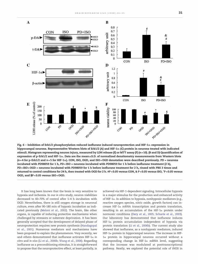
Fig. 6 – Inhibition of Erk1/2 phosphorylation reduced isoflurane induced neuroprotection and HIF-1α expression inhippocampal neurons. Representative Western blots of Erk1/2 (A) and HIF-1α (C) protein in neurons treated with indicatedstimuli. Histogram representing neuron injury, measured by LDH release (E) or MTT assay (F) (n=10). (B and D) Quantification ofexpression of p-Erk1/2 and HIF-1α. Data are the mean±S.D. of normalized densitometry measurements from Western blots(n=4 for p-Erk1/2 and n=5 for HIF-1α). CON, ISO, OGD, and ISO+OGD denotation were described previously. PD = neuronsincubated with PD98059 for 1 h, PD+ISO = neurons incubated with PD98059 for 1 h before isoflurane treatment (2 h),PD+ISO+OGD = neurons incubated with PD98059 for 1 h before isoflurane treatment for 2 h, rinsed with PBS 3 times andreturned to control conditions for 24 h, then treated with OGD for 2 h. #P<0.05 versus CON, & P<0.05 versus ISO, *P<0.05 versusOGD, and $P<0.05 versus ISO+OGD.
31B R A I N R E S E A R C H 1 2 4 5 ( 2 0 0 8 ) 2 6 – 3 5
It has long been known that the brain is very sensitive tohypoxia and ischemia. In our in vitro study, neuron viabilitiesdecreased to 60–70% of control after 1–4 h incubation withOGD. Nevertheless, there is still oxygen storage in neuronalculture, even after 90–180 min of hypoxic incubation as indi-cated previously (Meloni et al., 2002). The brain, like otherorgans, is capable of inducing protective mechanisms whenchallenged by stressors or substrate deprivation. It has beengenerally accepted that the development of delayed phase ofneuroprotection requires new protein synthesis (Nandagopalet al., 2001). Numerous mediators and mechanisms havebeen proposed to explain the phenomenon. Very recently, weand others demonstrated that isoflurane activates HIF-1α invitro and in vivo (Li et al., 2006b; Wang et al., 2006). Regardingisoflurane as a preconditioning stimulus, it is straightforwardto propose that the neuroprotective effect, at least partially, is
achieved via HIF-1-dependent signaling. Intracellular hypoxiais a major stimulus for the production and enhanced activityof HIF-1α. In addition to hypoxia, nonhypoxic mediators (e.g.,reactive oxygen species, nitric oxide, growth factors) can in-crease HIF-1α mRNA transcription and protein translation,resulting in an accumulation of the HIF-1α protein undernormoxic conditions (Dery et al., 2005; Scharte et al., 2003).Our laboratory has demonstrated that isoflurane inducesHIF-1α protein accumulation independent of hypoxia viaprotein translation (Li et al., 2006b). The current study alsoshowed that isoflurane, as a nonhypoxic mediators, inducedHIF-1α protein in hippocampal neurons. The increase in HIF-1α protein in hippocampal neurons occurred without acorresponding change in HIF-1α mRNA level, suggestingthat the increase was modulated at posttranscriptionalpathway. Nextly, we explored the potential role of iNOS in

32 B R A I N R E S E A R C H 1 2 4 5 ( 2 0 0 8 ) 2 6 – 3 5
the mechanisms of isoflurane neuroprotection. This notionwas derived from literature reporting that iNOS plays animportant role in both ischemic and isoflurane precondition-ing in rat brain (Kapinya et al., 2002; Luo et al., 2007; Zhao andZuo, 2004; Zhao et al., 2007). Our finding of simultaneouspeaks in HIF-1α protein and iNOS mRNA offers evidenceimplying that isoflurane preconditioning induces iNOS viaHIF-1α dependent pathway during protection against OGDinjury in hippocampal neurons. Nevertheless, additionalwork needs to be done to ascertain this conclusion. Althougha big increase of iNOS expression may be harmful (Anggard,1994), multiple studies have shown that a relatively smallincrease of iNOS expression may be beneficial and plays acritical role in neuroprotection induced by isoflurane pre-conditioning (Kapinya et al., 2002; Zhao and Zuo, 2004). Thedownstream events of iNOS to induce protection as shown inthe previous studies include activation of protein kinase C,heat shock proteins and manganese superoxide dismutase(Nandagopal et al., 2001; Zaugg et al., 2003). Furthermore,protein kinase C can also activate adenosine triphosphatesensitive potassium channels, an important effector for car-dioprotection induced by many preconditioning stimuli(Zaugg et al., 2003).
It is well documented that activation of the Erk1/2 pathwayleads to increased transcriptional activity of theHIF-1 complexand down-stream gene activation(Lee et al., 2002; Sutton et al.,2007). Our findings demonstrated that isoflurane precondi-tioning could significantly increase not only HIF-1α but also p-ERK/t-ERK. PD98059 significant reduced the expression of p-ERK/t-ERK and HIF-1α content as well as cell injury in the OGDneurons with isoflurane preconditioning. These suggestedthat the increased HIF-1α as well as the neuroprotective roleinduced by isoflurane might be partly mediated by theactivation of the Erk1/2 pathway. However, it should bepointed out that further studies are absolutely needed tofurther understandwhether the pathway has amajor or directrole in isoflurane induction of HIF-1α and iNOS. In addition, itwas noticed that although the increased contents of HIF-1αinduced by isoflurane could be significantly inhibited byPD98059, the contents of HIF-1α in the PD98059 neuronswere still higher than those in the control group. These factsmight indicate that some other pathways might also beinvolved in the isoflurane-induced neuroprotection byincreasing HIF-1α contents apart from Erk1/2 pathways.
Collectively, isoflurane is capable of preconditioning neu-rons against OGD injury. The activation of HIF-1α, iNOSexpression and the increased cell viability found in the OGDneurons pretreated with isoflurane suggest that HIF-1α mayplay a role in the neuroprotection processes. Isoflurane pre-conditioning induced HIF-1α protein content might be partlyvia the Erk1/2 pathways. Nevertheless, our study has limita-tions that may restrict extrapolation to clinical conditions.One significant limitation is that in vitro models do notreplicate the temporal loss of anesthetic neuroprotectionobserved in intact rodents (Sullivan et al., 2002). Anotherpossible limitation is that, at least in our study, it seemedweakly neuroprotective compared with conditions whenisoflurane is present during the simulated ischemia. Furtherstudies are needed to clarify the durability and power ofisoflurane preconditioning.
4. Experimental procedures
4.1. Animals
The experimental protocol used in this study was approved bythe Ethics Committee for Animal Experimentation and wasconducted according to the Guidelines for Animal Experimen-tation of Shanghai Jiao Tong University School of Medicine(Shanghai, China). The animals were studied at Shanghai JiaoTong University School of Medicine (Shanghai, China). New-born Wistar rats within 24 h, purchased from the LaboratoryAnimal Center, Shanghai Jiao Tong University School ofMedicine. All reagents were obtained from Sigma Chemical(St. Louis, MO) unless specified in the text.
4.2. Neuronal cell culture
Pure hippocampal cultures were prepared from NewbornWistar rats within 24 h and plated at a density of 4×10⁵ cellsper well in 24-well plates coated with poly-D-lysine inNeurobasal medium, serum-free B-27 supplement (Life Tech-nologies/BRL), 100 U/mL penicillin, 100 μg/mL streptomycin,and 2 mmol/L L-glutamine. Glutamate (25 μmol/L) was addedduring the initial 3 days in vitro (DIV). Cultures were kept at37 °C and 5% CO2 in a humidified incubator and fed beginningat 4 DIV with cultivating medium (as described above, butwithout glutamate) by replacing half of the medium twice aweek. The percentage of neuronal and astrocytic cells wasdetermined as follows: neuronal cultureswere fixed for 30minat 4 °C with 4% paraformaldehyde, 4% sucrose, and 0.1 M PBS,pH 7.4; cells were washed, permeabilized with 0.3% Triton X-100, blocked with 10% serum, and incubated overnight at 4 °Cin anti-NeuN monoclonal antibody (1:1000; Chemicon, Teme-cula, CA) and anti-GFAP polyclonal antibody (1:4000; Dako,Carpinteria, CA) with 3% serum; cells were then washed, andincubated with anti-rabbit Alexa Fluor 488 and anti-mouseAlexa Fluor 546 secondary fluorescent antibodies (MolecularProbes, Eugene, OR). The number of NeuN- and GFAP-positivecells was counted in eight separate fields over two wells, andthe percentage of NeuN-positive neurons and GFAP-positiveastrocytes was determined. Purity of neuronal cultures was94% neurons and 5% astrocytes.
4.3. Isoflurane preconditioning
Isoflurane preconditioning was performed as described pre-viously (Kapinya et al., 2002). Briefly, the neuron-enrichedcultures were kept after 8 days in vitro at 37 °C in a closedchamber in an atmosphere of 1.4% isoflurane, 5% CO2, 20% O2,and N2 (remainder) for a 2-hour period. Cells treated identi-cally but without the addition of isoflurane served as controls.
4.4. OGD treatment
To mimic cerebral ischemia in vitro, OGD was performed withthe pretreated or control cells 24 h later. For this purpose, weused a custom-made temperature-controlled anaerobic glovebox as previously described (Chavez et al., 2006), the glove-boxsystem was set up at 37 °C with an atmosphere of 5% CO2 and
33B R A I N R E S E A R C H 1 2 4 5 ( 2 0 0 8 ) 2 6 – 3 5
95% N2 (oxygen deprivation). All solutions were equilibratedfor at least 12 h. Cells were transferred into the chamber,washed with PBS, and incubated with a preequilibratedglucose-free balanced salt solution for up to 2 h. At the endof the procedure, cells were removed from the chamber, freshNeurobasal media were added (reoxygenation), and thecultures were returned to a regular incubator. Cell death wasassayed at 24 h after OGD measuring LDH released into themedia. For experimental controls, we used cultures that weresubjected to the same procedures but maintained withglucose-containing media at normoxia in a standard cell-culture incubator. The glove-box system used in this studywas equipped with an inverted microscope that allowed us tovisually inspect cell viability before terminating each experi-ment. All manipulations, including cell harvesting and celllysis, were performed within the chamber.
4.5. Assessment of cell survival
Neurons were maintained for a period of 24 h, and their sur-vival was assessed by phase-contrast microscopywith TrypanBlue exclusion assay and quantified with 3-(4,5-dimethyl-thiazol-2-yl)-2,5-diphenyl tetrazolium bromide (MTT) colori-metric assay as described previously (Wu et al., 2006). Briefly,after OGD in each group, MTT was added to all those assays ata final concentration of 0.5mg/mL for 4 h at 37 °C. The amountofMTT formazanwas dissolved by dimethyl sulfoxide (DMSO),quantified by determining calorimetrically its absorbance at570 nm using a microplate reader. Experiments were per-formed in triplicate, and repeated with at least three separatebatches of cultures.
4.6. Morphological observation
Morphological changes in the cells were observed using theinverted research microscope (Nikon) as described previously(Zhu et al., 1997).
4.7. Neuronal injury assay
Neuronal injury was quantitatively assessed by the measure-ment of LDH activity in the medium 24 h after the injury (Kohand Choi, 1987). In each experiment, the results of the LDHmeasurements from the controls (neurons without isofluraneor OGD treatment) were set as 100%. Isoflurane precondition-ing alone did not change the LDH release compared with the
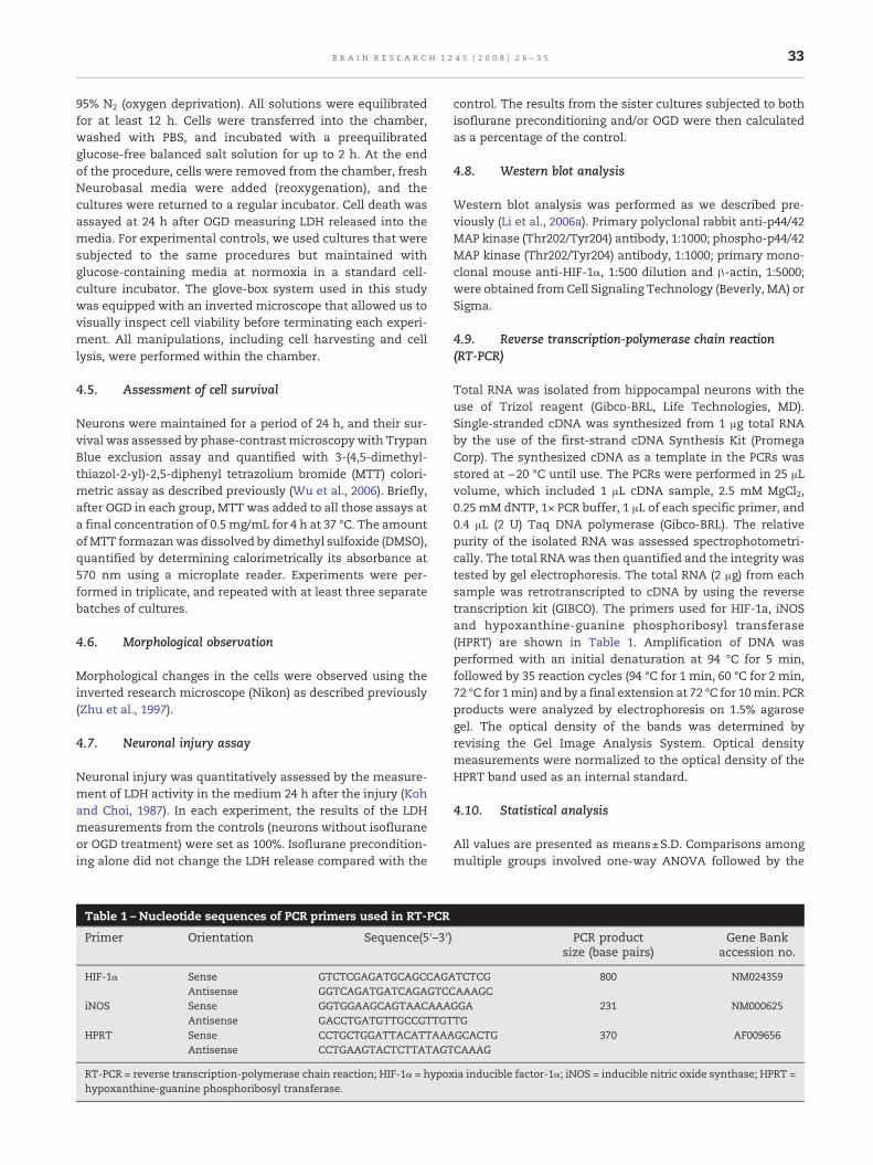
Table 1 – Nucleotide sequences of PCR primers used in RT-PCR
Primer Orientation Sequence(5′–3′)
HIF-1α Sense GTCTCGAGATGCAGCCAGAAntisense GGTCAGATGATCAGAGTCC
iNOS Sense GGTGGAAGCAGTAACAAAAntisense GACCTGATGTTGCCGTTGT
HPRT Sense CCTGCTGGATTACATTAAAAntisense CCTGAAGTACTCTTATAGT
RT-PCR = reverse transcription-polymerase chain reaction; HIF-1α = hypoxhypoxanthine-guanine phosphoribosyl transferase.
control. The results from the sister cultures subjected to bothisoflurane preconditioning and/or OGD were then calculatedas a percentage of the control.
4.8. Western blot analysis
Western blot analysis was performed as we described pre-viously (Li et al., 2006a). Primary polyclonal rabbit anti-p44/42MAP kinase (Thr202/Tyr204) antibody, 1:1000; phospho-p44/42MAP kinase (Thr202/Tyr204) antibody, 1:1000; primary mono-clonal mouse anti-HIF-1α, 1:500 dilution and β-actin, 1:5000;were obtained from Cell Signaling Technology (Beverly, MA) orSigma.
4.9. Reverse transcription-polymerase chain reaction(RT-PCR)
Total RNA was isolated from hippocampal neurons with theuse of Trizol reagent (Gibco-BRL, Life Technologies, MD).Single-stranded cDNA was synthesized from 1 μg total RNAby the use of the first-strand cDNA Synthesis Kit (PromegaCorp). The synthesized cDNA as a template in the PCRs wasstored at −20 °C until use. The PCRs were performed in 25 μLvolume, which included 1 μL cDNA sample, 2.5 mM MgCl2,0.25 mM dNTP, 1× PCR buffer, 1 μL of each specific primer, and0.4 μL (2 U) Taq DNA polymerase (Gibco-BRL). The relativepurity of the isolated RNA was assessed spectrophotometri-cally. The total RNAwas then quantified and the integrity wastested by gel electrophoresis. The total RNA (2 μg) from eachsample was retrotranscripted to cDNA by using the reversetranscription kit (GIBCO). The primers used for HIF-1a, iNOSand hypoxanthine-guanine phosphoribosyl transferase(HPRT) are shown in Table 1. Amplification of DNA wasperformed with an initial denaturation at 94 °C for 5 min,followed by 35 reaction cycles (94 °C for 1 min, 60 °C for 2 min,72 °C for 1min) and by a final extension at 72 °C for 10min. PCRproducts were analyzed by electrophoresis on 1.5% agarosegel. The optical density of the bands was determined byrevising the Gel Image Analysis System. Optical densitymeasurements were normalized to the optical density of theHPRT band used as an internal standard.
4.10. Statistical analysis
All values are presented as means±S.D. Comparisons amongmultiple groups involved one-way ANOVA followed by the
PCR productsize (base pairs)
Gene Bankaccession no.
TCTCG 800 NM024359AAAGCGGA 231 NM000625TGGCACTG 370 AF009656CAAAG
ia inducible factor-1α; iNOS = inducible nitric oxide synthase; HPRT =

34 B R A I N R E S E A R C H 1 2 4 5 ( 2 0 0 8 ) 2 6 – 3 5
Student–Newman–Keuls test and Dunnett's test (for compar-ison of multiple experimental treatments to a common con-trol value). All analyses were performed by using softwareSPSS 11.0. A value of P<0.05 was considered significant.
Acknowledgments
This work was supported by the National Nature ScienceFoundation of China (No. 30801079), Science Foundation ofShanghai Education Committee (No. 08YZ35), Youth ScienceFoundation of Shanghai Health Bureau (No. 2007Y34) andShanghai Rising-Star Program (No. 08QA14044).
R E F E R E N C E S
Anggard, E., 1994. Nitric oxide: mediator, murderer, and medicine.Lancet 343, 1199–1206.
Chavez, J.C., Baranova, O., Lin, J., Pichiule, P., 2006. Thetranscriptional activator hypoxia inducible factor 2(HIF-2/EPAS-1) regulates the oxygen-dependent expressionof erythropoietin in cortical astrocytes. J. Neurosci. 26,9471–9481.
Clarkson, A.N., 2007. Anesthetic-mediated protection/preconditioning during cerebral ischemia. Life Sci. 80,1157–1175.
Das, B., Yeger, H., Tsuchida, R., Torkin, R., Gee, M.F., Thorner, P.S.,Shibuya, M., Malkin, D., Baruchel, S., 2005. A hypoxia-drivenvascular endothelial growth factor/Flt1 autocrine loop inter-acts with hypoxia-inducible factor-1alpha throughmitogen-activated protein kinase/extracellularsignal-regulated kinase 1/2 pathway in neuroblastoma. CancerRes. 65, 7267–7275.
Dery, M.A., Michaud, M.D., Richard, D.E., 2005. Hypoxia-induciblefactor 1: regulation by hypoxic and non-hypoxic activators. Int.J. Biochem. Cell. Biol. 37, 535–540.
Gray, J.J., Bickler, P.E., Fahlman, C.S., Zhan, X., Schuyler, J.A., 2005.Isoflurane neuroprotection in hypoxic hippocampal slicecultures involves increases in intracellular Ca2+ andmitogen-activated protein kinases. Anesthesiology 102,606–615.
Irving, E.A., Bamford, M., 2002. Role of mitogen- andstress-activated kinases in ischemic injury. J. Cereb. Blood FlowMetab. 22, 631–647.
Jung, F., Palmer, L.A., Zhou, N., Johns, R.A., 2000. Hypoxicregulation of inducible nitric oxide synthase via hypoxiainducible factor-1 in cardiac myocytes. Circ. Res. 86, 319–325.
Kapinya, K.J., Lowl, D., Futterer, C., Maurer, M., Waschke, K.F.,Isaev, N.K., Dirnagl, U., 2002. Tolerance against ischemicneuronal injury can be induced by volatile anesthetics and isinducible NO synthase dependent. Stroke 33, 1889–1898.
Kitano, H., Kirsch, J.R., Hurn, P.D., Murphy, S.J., 2007. Inhalationalanesthetics as neuroprotectants or chemical preconditioningagents in ischemic brain. J. Cereb. Blood Flow Metab. 27,1108–1128.
Koh, J.Y., Choi, D.W., 1987. Quantitative determination ofglutamate mediated cortical neuronal injury in cell culture bylactate dehydrogenase efflux assay. J. Neurosci. Methods 20,83–90.
Lee, E., Yim, S., Lee, S.K., Park, H., 2002. Two transactivationdomains of hypoxia-inducible factor-1alpha regulated by theMEK-1/p42/p44 MAPK pathway. Mol. Cells. 14, 9–15.
Li, Q.F.,Wang, X.R., Yang, Y.W., Lin, H., 2006a. Hypoxia upregulateshypoxia inducible factor (HIF)-3alpha expression in lung
epithelial cells: characterization and comparison withHIF-1alpha. Cell. Res. 16, 548–558.
Li, Q.F., Wang, X.R., Yang, Y.W., Su, D.S., 2006b. Up-regulation ofhypoxia inducible factor 1alpha by isoflurane in Hep3B cells.Anesthesiology 105, 1211–1219.
Li, L., Xiong, Y., Qu, Y., Mao, M., Mu,W.,Wang, H., Mu, D., 2008. Therequirement of extracellular signal-related protein kinasepathway in the activation of hypoxia inducible factor 1 alpha inthe developing rat brain after hypoxia–ischemia. ActaNeuropathol. 115, 297–303.
Luo, C.X., Zhu, X.J., Zhou, Q.G., Wang, B., Wang, W., Cai, H.H., Sun,Y.J., Hu, M., Jiang, J., Hua, Y., Han, X., Zhu, D.Y., 2007. Reducedneuronal nitric oxide synthase is involved in ischemia-inducedhippocampal neurogenesis by up-regulating inducible nitricoxide synthase expression. J. Neurochem. 103, 1872–1882.
Meloni, B.P., Majda, B.T., Knuckey, N.W., 2002. Evaluation ofpreconditioning treatments to protect near-pure corticalneuronal cultures from in vitro ischemia induced acute anddelayed neuronal death. Brain Res. 928, 69–75.
Miura, Y., Grocott, H.P., Bart, R.D., Pearlstein, R.D., Dexter, F.,Warner, D.S., 1998. Differential effects of anesthetic agents onoutcome from near-complete but not incomplete globalischemia in the rat. Anesthesiology 89, 391–400.
Nandagopal, K., Dawson, T.M., Dawson, V.L., 2001. Critical role fornitric oxide signaling in cardiac and neuronal ischemicpreconditioning and tolerance. J. Pharmacol. Exp. Ther. 297,474–478.
Nozaki, K., Nishimura, M., Hashimoto, N., 2001. Mitogen-activatedprotein kinases and cerebral ischemia. Mol. Neurobiol. 23, 1–19.
Rapoport, M., Ferreira, A., 2000. PD98059 prevents neuritedegeneration induced by fibrillar beta-amyloid in maturehippocampal neurons. J. Neurochem. 74, 125–133.
Sakai, H., Sheng, H., Yates, R.B., Ishida, K., Pearlstein, R.D., Warner,D.S., 2007. Isoflurane provides long-term protection againstfocal cerebral ischemia in the rat. Anesthesiology 106, 92–99discussion 8–10.
Scharte, M., Han, X., Bertges, D.J., Fink, M.P., Delude, R.L., 2003.Cytokines induce HIF-1 DNA binding and the expression ofHIF-1-dependent genes in cultured rat enterocytes. Am. J.Physiol. Gastrointest. Liver Physiol. 284, G373–G384.
Semenza, G.L., Wang, G.L., 1992. A nuclear factor induced byhypoxia via de novo protein synthesis binds to the humanerythropoietin gene enhancer at a site required fortranscriptional activation. Mol. Cell. Biol. 12, 5447–5454.
Sullivan, B.L., Leu, D., Taylor, D.M., Fahlman, C.S., Bickler, P.E.,2002. Isoflurane prevents delayed cell death in an organotypicslice culture model of cerebral ischemia. Anesthesiology 96,189–195.
Sutton, K.M., Hayat, S., Chau, N.M., Cook, S., Pouyssegur, J., Ahmed,A., Perusinghe, N., Le Floch, R., Yang, J., Ashcroft, M., 2007.Selective inhibition of MEK1/2 reveals a differentialrequirement for ERK1/2 signalling in the regulation of HIF-1 inresponse to hypoxia and IGF-1. Oncogene 26, 3920–3929.
Wang, C., Weihrauch, D., Schwabe, D.A., Bienengraeber, M.,Warltier, D.C., Kersten, J.R., Pratt Jr., P.F., Pagel, P.S., 2006.Extracellular signal-regulated kinases trigger isofluranepreconditioning concomitant with upregulation ofhypoxia-inducible factor-1alpha and vascular endothelialgrowth factor expression in rats. Anesth. Analg. 103, 281–288table of contents.
Wu, B., Hu, S., Yang, M., Pan, H., Zhu, S., 2006. CART peptidepromotes the survival of hippocampal neurons byupregulating brain-derived neurotrophic factor. Biochem.Biophys. Res. Commun. 347, 656–661.
Zaugg, M., Lucchinetti, E., Uecker, M., Pasch, T., Schaub, M.C., 2003.Anaesthetics and cardiac preconditioning. Part I. Signallingand cytoprotective mechanisms. Br. J. Anaesth. 91, 551–565.
Zhang, Q., Oh, C.K., Messadi, D.V., Duong, H.S., Kelly, A.P., Soo, C.,Wang, L., Le, A.D., 2006. Hypoxia-induced HIF-1 alpha

35B R A I N R E S E A R C H 1 2 4 5 ( 2 0 0 8 ) 2 6 – 3 5
accumulation is augmented in a co-culture of keloid fibroblastsand human mast cells: involvement of ERK1/2 and PI-3K/Akt.Exp. Cell Res. 312, 145–155.
Zhao, P., Zuo, Z., 2004. Isoflurane preconditioning inducesneuroprotection that is inducible nitric oxidesynthase-dependent in neonatal rats. Anesthesiology 101,695–703.
Zhao, P., Peng, L., Li, L., Xu, X., Zuo, Z., 2007. Isofluranepreconditioning improves long-term neurologic outcome afterhypoxic–ischemic brain injury in neonatal rats. Anesthesiology107, 963–970.
Zhu, L., Wu, J., Liao, H., Gao, J., Zhao, X.N., Zhang, Z.X., 1997.Antagonistic effects of extract from leaves of Ginkgo biloba onglutamate neurotoxicity. Zhongguo Yao Li Xue Bao 18, 344–347.
Copyright © 2022 FDOKUMEN