iNOS-Producing Inflammatory Dendritic Cells Constitute the Major Infected Cell Type during the...
13
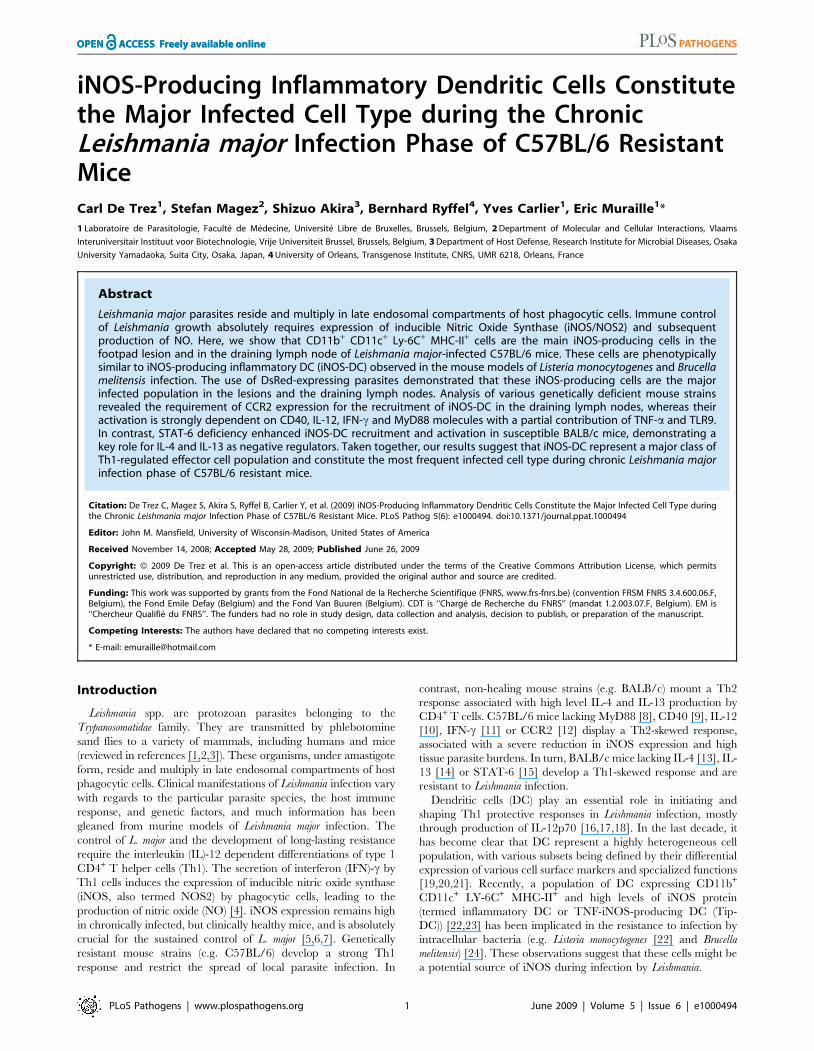
iNOS-Producing Inflammatory Dendritic Cells Constitute the Major Infected Cell Type during the Chronic Leishmania major Infection Phase of C57BL/6 Resistant Mice Carl De Trez 1 , Stefan Magez 2 , Shizuo Akira 3 , Bernhard Ryffel 4 , Yves Carlier 1 , Eric Muraille 1 * 1 Laboratoire de Parasitologie, Faculte ´ de Me ´ decine, Universite ´ Libre de Bruxelles, Brussels, Belgium, 2 Department of Molecular and Cellular Interactions, Vlaams Interuniversitair Instituut voor Biotechnologie, Vrije Universiteit Brussel, Brussels, Belgium, 3 Department of Host Defense, Research Institute for Microbial Diseases, Osaka University Yamadaoka, Suita City, Osaka, Japan, 4 University of Orleans, Transgenose Institute, CNRS, UMR 6218, Orleans, France Abstract Leishmania major parasites reside and multiply in late endosomal compartments of host phagocytic cells. Immune control of Leishmania growth absolutely requires expression of inducible Nitric Oxide Synthase (iNOS/NOS2) and subsequent production of NO. Here, we show that CD11b + CD11c + Ly-6C + MHC-II + cells are the main iNOS-producing cells in the footpad lesion and in the draining lymph node of Leishmania major-infected C57BL/6 mice. These cells are phenotypically similar to iNOS-producing inflammatory DC (iNOS-DC) observed in the mouse models of Listeria monocytogenes and Brucella melitensis infection. The use of DsRed-expressing parasites demonstrated that these iNOS-producing cells are the major infected population in the lesions and the draining lymph nodes. Analysis of various genetically deficient mouse strains revealed the requirement of CCR2 expression for the recruitment of iNOS-DC in the draining lymph nodes, whereas their activation is strongly dependent on CD40, IL-12, IFN-c and MyD88 molecules with a partial contribution of TNF-a and TLR9. In contrast, STAT-6 deficiency enhanced iNOS-DC recruitment and activation in susceptible BALB/c mice, demonstrating a key role for IL-4 and IL-13 as negative regulators. Taken together, our results suggest that iNOS-DC represent a major class of Th1-regulated effector cell population and constitute the most frequent infected cell type during chronic Leishmania major infection phase of C57BL/6 resistant mice. Citation: De Trez C, Magez S, Akira S, Ryffel B, Carlier Y, et al. (2009) iNOS-Producing Inflammatory Dendritic Cells Constitute the Major Infected Cell Type during the Chronic Leishmania major Infection Phase of C57BL/6 Resistant Mice. PLoS Pathog 5(6): e1000494. doi:10.1371/journal.ppat.1000494 Editor: John M. Mansfield, University of Wisconsin-Madison, United States of America Received November 14, 2008; Accepted May 28, 2009; Published June 26, 2009 Copyright: ß 2009 De Trez et al. This is an open-access article distributed under the terms of the Creative Commons Attribution License, which permits unrestricted use, distribution, and reproduction in any medium, provided the original author and source are credited. Funding: This work was supported by grants from the Fond National de la Recherche Scientifique (FNRS, www.frs-fnrs.be) (convention FRSM FNRS 3.4.600.06.F, Belgium), the Fond Emile Defay (Belgium) and the Fond Van Buuren (Belgium). CDT is ‘‘Charge ´ de Recherche du FNRS’’ (mandat 1.2.003.07.F, Belgium). EM is ‘‘Chercheur Qualifie ´ du FNRS’’. The funders had no role in study design, data collection and analysis, decision to publish, or preparation of the manuscript. Competing Interests: The authors have declared that no competing interests exist. * E-mail: [email protected] Introduction Leishmania spp. are protozoan parasites belonging to the Trypanosomatidae family. They are transmitted by phlebotomine sand flies to a variety of mammals, including humans and mice (reviewed in references [1,2,3]). These organisms, under amastigote form, reside and multiply in late endosomal compartments of host phagocytic cells. Clinical manifestations of Leishmania infection vary with regards to the particular parasite species, the host immune response, and genetic factors, and much information has been gleaned from murine models of Leishmania major infection. The control of L. major and the development of long-lasting resistance require the interleukin (IL)-12 dependent differentiations of type 1 CD4 + T helper cells (Th1). The secretion of interferon (IFN)-c by Th1 cells induces the expression of inducible nitric oxide synthase (iNOS, also termed NOS2) by phagocytic cells, leading to the production of nitric oxide (NO) [4]. iNOS expression remains high in chronically infected, but clinically healthy mice, and is absolutely crucial for the sustained control of L. major [5,6,7]. Genetically resistant mouse strains (e.g. C57BL/6) develop a strong Th1 response and restrict the spread of local parasite infection. In contrast, non-healing mouse strains (e.g. BALB/c) mount a Th2 response associated with high level IL-4 and IL-13 production by CD4 + T cells. C57BL/6 mice lacking MyD88 [8], CD40 [9], IL-12 [10], IFN-c [11] or CCR2 [12] display a Th2-skewed response, associated with a severe reduction in iNOS expression and high tissue parasite burdens. In turn, BALB/c mice lacking IL-4 [13], IL- 13 [14] or STAT-6 [15] develop a Th1-skewed response and are resistant to Leishmania infection. Dendritic cells (DC) play an essential role in initiating and shaping Th1 protective responses in Leishmania infection, mostly through production of IL-12p70 [16,17,18]. In the last decade, it has become clear that DC represent a highly heterogeneous cell population, with various subsets being defined by their differential expression of various cell surface markers and specialized functions [19,20,21]. Recently, a population of DC expressing CD11b + CD11c + LY-6C + MHC-II + and high levels of iNOS protein (termed inflammatory DC or TNF-iNOS-producing DC (Tip- DC)) [22,23] has been implicated in the resistance to infection by intracellular bacteria (e.g. Listeria monocytogenes [22] and Brucella melitensis) [24]. These observations suggest that these cells might be a potential source of iNOS during infection by Leishmania. PLoS Pathogens | www.plospathogens.org 1 June 2009 | Volume 5 | Issue 6 | e1000494
-
Upload
independent -
Category
Documents
-
view
8 -
download
0
Transcript of iNOS-Producing Inflammatory Dendritic Cells Constitute the Major Infected Cell Type during the...
iNOS-Producing Inflammatory Dendritic Cells Constitutethe Major Infected Cell Type during the ChronicLeishmania major Infection Phase of C57BL/6 ResistantMiceCarl De Trez1, Stefan Magez2, Shizuo Akira3, Bernhard Ryffel4, Yves Carlier1, Eric Muraille1*
1 Laboratoire de Parasitologie, Faculte de Medecine, Universite Libre de Bruxelles, Brussels, Belgium, 2 Department of Molecular and Cellular Interactions, Vlaams
Interuniversitair Instituut voor Biotechnologie, Vrije Universiteit Brussel, Brussels, Belgium, 3 Department of Host Defense, Research Institute for Microbial Diseases, Osaka
University Yamadaoka, Suita City, Osaka, Japan, 4 University of Orleans, Transgenose Institute, CNRS, UMR 6218, Orleans, France
Abstract
Leishmania major parasites reside and multiply in late endosomal compartments of host phagocytic cells. Immune controlof Leishmania growth absolutely requires expression of inducible Nitric Oxide Synthase (iNOS/NOS2) and subsequentproduction of NO. Here, we show that CD11b+ CD11c+ Ly-6C+ MHC-II+ cells are the main iNOS-producing cells in thefootpad lesion and in the draining lymph node of Leishmania major-infected C57BL/6 mice. These cells are phenotypicallysimilar to iNOS-producing inflammatory DC (iNOS-DC) observed in the mouse models of Listeria monocytogenes and Brucellamelitensis infection. The use of DsRed-expressing parasites demonstrated that these iNOS-producing cells are the majorinfected population in the lesions and the draining lymph nodes. Analysis of various genetically deficient mouse strainsrevealed the requirement of CCR2 expression for the recruitment of iNOS-DC in the draining lymph nodes, whereas theiractivation is strongly dependent on CD40, IL-12, IFN-c and MyD88 molecules with a partial contribution of TNF-a and TLR9.In contrast, STAT-6 deficiency enhanced iNOS-DC recruitment and activation in susceptible BALB/c mice, demonstrating akey role for IL-4 and IL-13 as negative regulators. Taken together, our results suggest that iNOS-DC represent a major class ofTh1-regulated effector cell population and constitute the most frequent infected cell type during chronic Leishmania majorinfection phase of C57BL/6 resistant mice.
Citation: De Trez C, Magez S, Akira S, Ryffel B, Carlier Y, et al. (2009) iNOS-Producing Inflammatory Dendritic Cells Constitute the Major Infected Cell Type duringthe Chronic Leishmania major Infection Phase of C57BL/6 Resistant Mice. PLoS Pathog 5(6): e1000494. doi:10.1371/journal.ppat.1000494
Editor: John M. Mansfield, University of Wisconsin-Madison, United States of America
Received November 14, 2008; Accepted May 28, 2009; Published June 26, 2009
Copyright: � 2009 De Trez et al. This is an open-access article distributed under the terms of the Creative Commons Attribution License, which permitsunrestricted use, distribution, and reproduction in any medium, provided the original author and source are credited.
Funding: This work was supported by grants from the Fond National de la Recherche Scientifique (FNRS, www.frs-fnrs.be) (convention FRSM FNRS 3.4.600.06.F,Belgium), the Fond Emile Defay (Belgium) and the Fond Van Buuren (Belgium). CDT is ‘‘Charge de Recherche du FNRS’’ (mandat 1.2.003.07.F, Belgium). EM is‘‘Chercheur Qualifie du FNRS’’. The funders had no role in study design, data collection and analysis, decision to publish, or preparation of the manuscript.
Competing Interests: The authors have declared that no competing interests exist.
* E-mail: [email protected]
Introduction
Leishmania spp. are protozoan parasites belonging to the
Trypanosomatidae family. They are transmitted by phlebotomine
sand flies to a variety of mammals, including humans and mice
(reviewed in references [1,2,3]). These organisms, under amastigote
form, reside and multiply in late endosomal compartments of host
phagocytic cells. Clinical manifestations of Leishmania infection vary
with regards to the particular parasite species, the host immune
response, and genetic factors, and much information has been
gleaned from murine models of Leishmania major infection. The
control of L. major and the development of long-lasting resistance
require the interleukin (IL)-12 dependent differentiations of type 1
CD4+ T helper cells (Th1). The secretion of interferon (IFN)-c by
Th1 cells induces the expression of inducible nitric oxide synthase
(iNOS, also termed NOS2) by phagocytic cells, leading to the
production of nitric oxide (NO) [4]. iNOS expression remains high
in chronically infected, but clinically healthy mice, and is absolutely
crucial for the sustained control of L. major [5,6,7]. Genetically
resistant mouse strains (e.g. C57BL/6) develop a strong Th1
response and restrict the spread of local parasite infection. In
contrast, non-healing mouse strains (e.g. BALB/c) mount a Th2
response associated with high level IL-4 and IL-13 production by
CD4+ T cells. C57BL/6 mice lacking MyD88 [8], CD40 [9], IL-12
[10], IFN-c [11] or CCR2 [12] display a Th2-skewed response,
associated with a severe reduction in iNOS expression and high
tissue parasite burdens. In turn, BALB/c mice lacking IL-4 [13], IL-
13 [14] or STAT-6 [15] develop a Th1-skewed response and are
resistant to Leishmania infection.
Dendritic cells (DC) play an essential role in initiating and
shaping Th1 protective responses in Leishmania infection, mostly
through production of IL-12p70 [16,17,18]. In the last decade, it
has become clear that DC represent a highly heterogeneous cell
population, with various subsets being defined by their differential
expression of various cell surface markers and specialized functions
[19,20,21]. Recently, a population of DC expressing CD11b+
CD11c+ LY-6C+ MHC-II+ and high levels of iNOS protein
(termed inflammatory DC or TNF-iNOS-producing DC (Tip-
DC)) [22,23] has been implicated in the resistance to infection by
intracellular bacteria (e.g. Listeria monocytogenes [22] and Brucella
melitensis) [24]. These observations suggest that these cells might be
a potential source of iNOS during infection by Leishmania.
PLoS Pathogens | www.plospathogens.org 1 June 2009 | Volume 5 | Issue 6 | e1000494
In the present study, using immunofluorescent microscopy and
ex vivo flow-cytometric analysis, we demonstrated that inflamma-
tory DCs are the main producers of iNOS in vivo during the course
of L. major infection. The recruitment of inflammatory DC was
dependent upon CCR2 expression, and the induction of iNOS
expression in these cells required the development of a local Th1
microenvironment, as demonstrated by the reduced frequency of
iNOS+ inflammatory DC in MyD882/2, CD402/2, IL-122/2
and IFN-c2/2 mice. In contrast, a Th2 environment inhibited the
local differentiation of iNOS+ inflammatory DC, as an enhanced
frequency of iNOS+ inflammatory DC was observed in STAT-62/2
BALB/c mice.
Results
Recruitment of iNOS-producing inflammatory DC duringthe course of L. major infection
Monitoring of L. major-induced lesion size in wild-type C57BL/6
(B6.WT), B6.iNOS, B6.TNF-a mice and wild-type BALB/C
(BC.WT) mice confirmed the previous results of our group and
others, showing an important contribution of both iNOS
enzymatic activity [5,6,7] and TNF-a [25] in the resistance of
B6.WT mice to L. major infection (Figure S1.A). However, the
severity of the lesions in the footpads four weeks post-infection (p.i.)
highlighted a more crucial role for iNOS compared to TNF-a in
this pathogenic model (Figure S1.A and S1.B). To this point, the
exact nature of the cell type(s) responsible for the production of
iNOS and TNF-a in vivo has remained unclear, and we therefore
attempted to directly identify them in the draining lymph node
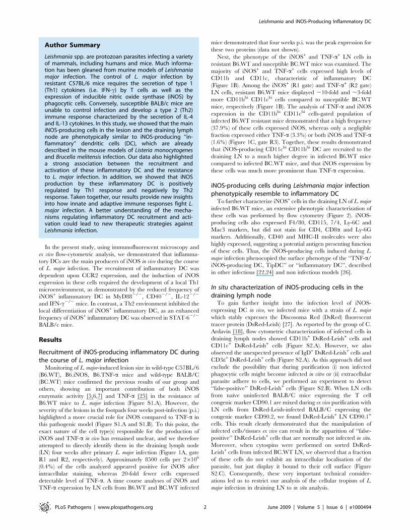
(LN) four weeks after primary L. major infection (Figure 1A, gate
R1 and R2, respectively). Approximately 8500 cells per 26106
(0.4%) of the cells analyzed appeared positive for iNOS after
intracellular staining, whereas 20-fold fewer cells expressed
detectable level of TNF-a. A time course analyses of iNOS and
TNF-a expression by LN cells from B6.WT and BC.WT infected
mice demonstrated that four weeks p.i. was the peak expression for
these two proteins (data not shown).
Next, the phenotype of the iNOS+ and TNF-a+ LN cells in
resistant B6.WT and susceptible BC.WT mice was examined. The
majority of iNOS+ and TNF-a+ cells expressed high levels of
CD11b and CD11c, characteristic of inflammatory DC
(Figure 1B). Among the iNOS+ (R1 gate) and TNF-a+ (R2 gate)
LN cells, resistant B6.WT mice displayed ,10-fold and ,3-fold
more CD11bhi CD11chi cells compared to susceptible BC.WT
mice, respectively (Figure 1B). The analysis of TNF-a and iNOS
expression in the CD11bhi CD11chi cells-gated population of
infected B6.WT resistant mice demonstrated that a high frequency
(37.9%) of these cells expressed iNOS, whereas only a negligible
fraction expressed either TNF-a (3.3%) or both iNOS and TNF-a(1.6%) (Figure 1C, gate R3). Together, these results demonstrated
that iNOS-producing CD11chi CD11bhi DC are recruited to the
draining LN to a much higher degree in infected B6.WT mice
compared to infected BC.WT mice, and that iNOS expression by
these cells was much more prominent than TNF-a expression.
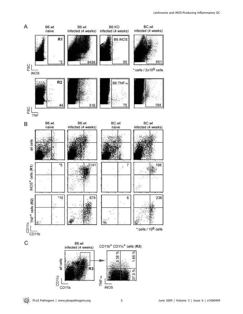
iNOS-producing cells during Leishmania major infectionphenotypically resemble to inflammatory DC
To further characterize iNOS+ cells in the draining LN of L. major
infected B6.WT mice, an extensive phenotypic characterization of
these cells was performed by flow cytometry (Figure 2). iNOS-
producing cells also expressed F4/80, CD115, 7/4, Ly-6C and
Mac3 markers, but did not stain for CD4, CD8a and Ly-6G
markers. Additionally, CD40 and MHC-II molecules were also
highly expressed, suggesting a potential antigen presenting function
of these cells. Thus, the iNOS-producing cells induced during L.
major infection phenocopied the surface phenotype of the ‘‘TNF-a/
iNOS-producing DC, TipDC’’ or ‘‘inflammatory DC’’, described
in other infectious [22,24] and non infectious models [26].
In situ characterization of iNOS-producing cells in thedraining lymph node
To gain further insight into the infection level of iNOS-
expressing DC in vivo, we infected mice with a strain of L. major
which stably expresses the Discosoma Red (DsRed) fluorescent
tracer protein (DsRed-Leish) [27]. As reported by the group of C.
Ardavin [18], flow cytometric characterization of infected cells in
draining lymph nodes showed CD11b+ DsRed-Leish+ cells and
CD11c+ DsRed-Leish+ cells (Figure S2.A). However, we also
observed the unexpected presence of IgD+ DsRed-Leish+ cells and
CD3e+ DsRed-Leish+ cells (Figure S2.A). As this approach did not
exclude the possibility that during purification (i) non infected
phagocytic cells might become infected in vitro or (ii) extracellular
parasite adhere to cells, we performed an experiment to detect
‘‘false-positive’’ DsRed-Leish+ cells (Figure S2.B). When LN cells
from naıve uninfected BALB/C mice expressing the T cell
congenic marker CD90.1 are mixed during ex vivo purification with
LN cells from DsRed-Leish-infected BALB/C expressing the
congenic marker CD90.2, we found DsRed-Leish+ LN CD90.1+
cells. This result clearly demonstrated that the manipulation of
infected cells/tissues ex vivo can result in the apparition of ‘‘false-
positive’’ DsRed-Leish+ cells that are normally not infected in situ.
Moreover, when cytospins were performed on sorted DsRed-
Leish+ cells from infected BC.WT LN, we observed that a fraction
of these cells do not exhibit an intracellular localisation of the
parasite, but just display it bound to their cell surface (Figure
S2.C). Consequently, these very important technical consider-
ations led us to restrict our analysis of the cellular tropism of L.
major infection in draining LN to in situ analysis.
Author Summary
Leishmania spp. are protozoan parasites infecting a varietyof mammals, including humans and mice. Much informa-tion has been gleaned from murine models of Leishmaniamajor infection. The control of L. major infection byresistant C57BL/6 mice requires the secretion of type 1(Th1) cytokines (i.e. IFN-c) by T cells as well as theexpression of inducible nitric oxide synthase (iNOS) byphagocytic cells. Conversely, susceptible BALB/c mice areunable to control infection and develop a type 2 (Th2)immune response characterized by the secretion of IL-4and IL-13 cytokines. In this study, we showed that the mainiNOS-producing cells in the lesion and the draining lymphnode are phenotypically similar to iNOS-producing ‘‘in-flammatory’’ dendritic cells (DC), which are alreadydescribed in the mouse models of Listeria monocytogenesand Brucella melitensis infection. Our data also highlighteda strong association between the recruitment andactivation of these inflammatory DC and the resistanceto L. major infection. In addition, we showed that iNOSproduction by these inflammatory DC is positivelyregulated by Th1 response and negatively by Th2response. Taken together, our results provide new insightsinto how innate and adaptive immune responses fight L.major infection. A better understanding of the mecha-nisms regulating inflammatory DC recruitment and acti-vation could lead to new therapeutic strategies againstLeishmania infection.
Leishmania and iNOS-Producing Inflammatory DC
PLoS Pathogens | www.plospathogens.org 2 June 2009 | Volume 5 | Issue 6 | e1000494
Leishmania and iNOS-Producing Inflammatory DC
PLoS Pathogens | www.plospathogens.org 3 June 2009 | Volume 5 | Issue 6 | e1000494
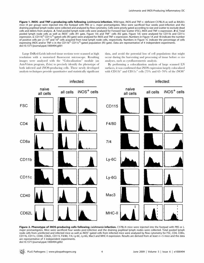
Large DsRed-Leish infected tissue sections were scanned at high
resolution with a motorized fluorescent microscope. Resulting
images were analyzed with the ‘‘Colocalization’’ module (an
AxioVision program, Zeiss) to precisely identify the phenotype of
both infected and iNOS-producing cells. These newly developed
analysis techniques provide quantitative and statistically significant
data, and avoid the potential loss of cell populations that might
occur during the harvesting and processing of tissue before ex vivo
analyses, such as cytofluorometric analysis.
By performing a colocalization analysis of large scanned LN
surfaces, it was confirmed that iNOS expression largely colocalized
with CD11b+ and CD11c+ cells (75% and 65–70% of the iNOS+
Figure 2. Phenotype of iNOS-producing cells following Leishmania infection. C57BL/6 mice were injected into the footpad with PBS or L.major promastigotes. Mice were sacrificed four weeks post-infection and the draining popliteal lymph nodes were collected. Total pooled lymphnode cells from uninfected and infected mice as well as iNOS+-gated cells from infected mice were analyzed by flow cytometry for FSC, CD4, CD8a,CD11b, CD11c, CD40, CD62L, CD115, F4/80, 7/4, Ly-6C, Ly-6G, Mac3 and MHC-II expression. Results are derived from at least n = 5 mice and the dataare representative of 3 independent experiments.doi:10.1371/journal.ppat.1000494.g002
Figure 1. iNOS- and TNF-a-producing cells following Leishmania infection. Wild-type, iNOS-and TNF-a -deficient C57BL/6 as well as BALB/cmice (4 per group) were injected into the footpad with PBS or L. major promastigotes. Mice were sacrificed four weeks post-infection and thedraining popliteal lymph nodes were collected and analyzed by flow cytometry. Cells were priorly gated according to size and scatter to exclude deadcells and debris from analysis. A, Total pooled lymph node cells were analyzed for Forward Size Scatter (FSC), iNOS and TNF-a expression. B–C, Totalpooled lymph node cells as well as iNOS+ cells (R1 gate, Figure 1A) and TNF+ cells (R2 gate, Figure 1A) were analyzed for CD11b and CD11cexpression. C, CD11bhi CD11chi-gated cells (R3 gate) were analyzed for iNOS and TNF-a expression. Numbers in Figure 1A and 1B indicate the numberof positive cells per 26106 and 106 cells acquired from total lymph node cells, respectively. Numbers in Figure 1C indicate the percentage of cellsexpressing iNOS and/or TNF-a in the CD11bhi CD11chi-gated population (R3 gate). Data are representative of 4 independent experiments.doi:10.1371/journal.ppat.1000494.g001
Leishmania and iNOS-Producing Inflammatory DC
PLoS Pathogens | www.plospathogens.org 4 June 2009 | Volume 5 | Issue 6 | e1000494
surface, respectively), with approximately 40% and 35% also
exhibiting DsRed signal and high expression levels of MHC-II,
respectively (Figure 3A). Using the same analysis technique, ,75%
of the DsRed-Leish+ surface colocalized with iNOS+, CD11b+ and
CD11c+ cells, but ,30% did with Ly-6G and MHC-IIhi
(Figure 3B). The Figures 3C–E depicted representative examples
of colocalization between CD11c+, iNOS+ and DsRed-Leish+ in
infected draining LN sections. They also illustrated the aggregated
distribution of these cells within the draining LN. CD45R/B220
expression (largely a B-cell marker) displayed very little colocaliza-
tion with iNOS+ and DsRed-Leish+ surface (,5%, Figure 3A and
3B), and likely represented the general degree of non-specific
colocalization (due to cell superposition in the section) when
analyzing tissue sections averaging 10 mm thickness. We did not
observe any iNOS staining in B6.iNOS2/2 mice (data not shown).
Analyses of tissue sections showed that approximately 70% of
iNOS+ cell surface expressed CD11b and CD11c markers, yet
only ,35% colocalized with MHC-IIhi+ surface. This correlated
with the flow cytometric analysis where MHC-II expression is
decreased by ,50% in the CD11b+ LN cells from infected B6.WT
mice compared to uninfected mice (Figure S3A). MHC-II
downregulation was observed in both resistant and susceptible
mice (data not shown). Figure S3.B illustrated representative serial
sections where colocalization is seen for CD11b+, iNOS+, MHC-
IIhi+ and DsRed+ LN surfaces. Altogether, these results demon-
strated that CD11b+ CD11c+ cells are by far the most abundant
cells expressing iNOS, and are the most highly infected population
of cells infected with L. major, within the LN at four weeks p.i. of
B6.WT mice.
In situ characterization of iNOS-producing cells in thecutaneous lesion
Few studies have investigated the phenotype of iNOS+ cells [7]
or infected cells [28] in the L. major-induced cutaneous lesion.
Using the same immunofluorescence microscopy technique
developed for the analysis of the LN, we investigated which cells
were infected in the cutaneous lesion of the B6.WT footpad four
weeks p.i. As in the draining LN, the majority of the iNOS staining
Figure 3. Characterization of iNOS- and DsRed-expressing cells in infected LN. Wild type C57BL/6 mice were injected into the footpad withPBS or DsRed expressing L. major promastigotes (DsRed-Leish). Mice were sacrificed four weeks post-infection, the draining popliteal lymph nodeswere collected and examined by immunohistofluorescence. A, The percentage of iNOS+ cells that colocalize with MHC-IIhi-, Ly-6G-, CD45R/B220-,CD11c-, CD11b-expressing cells and DsRed-Leish. B, The percentage of DsRed-Leish that colocalize with MHC-IIhi-, Ly-6G-, iNOS-, F4/80-, CD45R/B220-,CD11c- and CD11b-expressing cells. The bars are the mean6SD from at least 3 LN sections per LN from 8 mice. C–E, Immunofluorescence analysis ofCD11c, actin, DsRed and iNOS expression. Panels are color-coded with the text for the antigen or DsRed-Leish examined as well as the colocalization.D–E, Higher magnification view of Figure 3C, as indicated. Numbers in Figure 3D indicate the percentage of colocalizing cells in the upper panel.Scale bar = 500, 200 and 50 mm, as indicated. Data are representative of 3 independent experiments.doi:10.1371/journal.ppat.1000494.g003
Leishmania and iNOS-Producing Inflammatory DC
PLoS Pathogens | www.plospathogens.org 5 June 2009 | Volume 5 | Issue 6 | e1000494
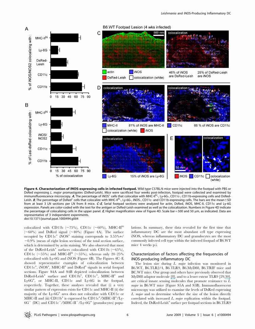
colocalized with CD11b (,75%), CD11c (,60%), MHC-IIhi
(,60%) and DsRed signal (,40%) (Figure 4A). The surface
occupied by CD11c+ iNOS+ staining corresponds to 3.55%+/
20.9% (mean of eight lesion sections) of the total section surface,
which is determined by actin staining. We also observed that most
of the DsRed-Leish+ surfaces colocalized with CD11b (,65%),
CD11c (,55%) and MHC-IIhi (,55%), whereas only 20–25%
colocalized with Ly-6G and iNOS (Figure 4B). The Figures 4C–E
showed representative examples of colocalization between
CD11c+, iNOS+, MHC-II+ and DsRed+ signals in serial footpad
sections. Figure S4A and S4B depicted colocalization between
DsRed-Leish+ surface and CD11b+, CD11c+, MHC-II+ and
Ly6G+, or MHC-II, CD11c and Ly-6G in the footpad,
respectively. Together, these analyses revealed that (i) a very
similar pattern of expression exists for CD11c and MHC-II (ii) the
majority of the Ly-6G+ area does not colocalize with CD11c or
MHC-II and (iii) CD11b+ is expressed by CD11c+/MHC-II+/Ly-
6G2 (DC) and CD11c2/MHC-II2/Ly-6G+ (granulocytes) popu-
lations. In summary, these data revealed for the first time that
inflammatory DC are the most abundant cell type expressing
iNOS, whereas inflammatory DC and granulocytes are the most
commonly infected cell type within the infected footpad of B6.WT
mice 4 weeks p.i.
Characterization of factors affecting the frequencies ofiNOS-producing inflammatory DC
The lesion size during L. major infection was monitored in
B6.WT, B6.TLR2/4, B6.TLR9, B6.MyD88, B6.TRIF mice and
BC.WT mice. Our group and others have previously observed that
MyD88 adaptor molecule [8], and to a lesser extent TLR9 [29,30],
are critical innate sensing molecules that promote resistance to L.
major in B6.WT mice (Figure S5A and S5B). Immunofluorescent
microscopy was utilized to examine the levels of DsRed expressing
L. major and to determine whether the size of the lesion directly
correlated with increased L. major replication within the footpad.
Indeed, the DsRed-Leish+ surface per footpad sections in B6.TLR9
Figure 4. Characterization of iNOS-expressing cells in infected footpad. Wild type C57BL/6 mice were injected into the footpad with PBS orDsRed expressing L. major promastigotes (DsRed-Leish). Mice were sacrificed four weeks post-infection, footpad were collected and examined byimmunofluorescence microscopy. A, The percentage of iNOS+ cells that colocalize with MHC-IIhi-, Ly-6G-, CD11c-, CD11b-expressing cells and DsRed-Leish. B, The percentage of DsRed+ cells that colocalize with MHC-IIhi-, Ly-6G-, iNOS-, CD11c- and CD11b-expressing cells. The bars are the mean6SDfrom at least 3 LN sections per LN from 8 mice. C–E, Serial footpad sections were analyzed for actin, DsRed, iNOS, MHC-II, CD11c and Ly-6Gexpression. Panels are color-coded with the text for the antigen or DsRed-Leish examined as well as the colocalization. Numbers in Figure 4D indicatethe percentage of colocalizing cells in the upper panel. E, Higher magnification view of Figure 4D. Scale bar = 500 and 50 mm, as indicated. Data arerepresentative of 3 independent experiments.doi:10.1371/journal.ppat.1000494.g004
Leishmania and iNOS-Producing Inflammatory DC
PLoS Pathogens | www.plospathogens.org 6 June 2009 | Volume 5 | Issue 6 | e1000494
(19.52%) and B6.MyD88 (24.91%) mice was increased when
compared to B6.WT (8.81%) mice (Figure S5C). Statistical analysis
of these sections further confirmed this correlation (Figure S5C) and
also suggested a negative role of TRIF in controlling L. major growth.
In turn, the same analysis performed on the infected draining LN
also highlighted a key role for MyD88 and TLR9 as well as a minor
contribution of TLR2/4 in the control of L. major burden (Figure
S5.E). Similar results were obtained when we determined the
number of parasites per infected LN using a limit dilution assay
(Figure S6). MyD882/2 and to a lesser extent TLR92/2 mice
showed the higher level of living parasites. TLR2/42/2 mice
displayed a slightly higher, but significant, level of living parasite per
LN when compared to WT mice. TLR22/2 and TLR42/2 mice
did not present any enhanced parasite count (data not shown). We
also excluded a possible contribution of the MyD88-dependent
inflammatory pathway as infected IL-18, IL-1beta-converting
enzyme (ICE)-deficient mice did not exhibit increased parasite
count compared to infected B6.WT.
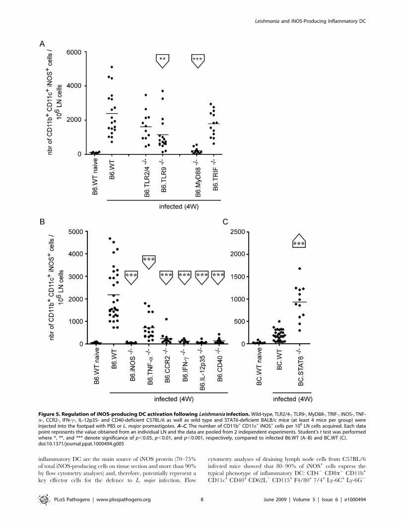
Next, we investigated whether TLR9-MyD88 signaling was
linked to iNOS production by inflammatory DCs. B6.TLR9 and
B6.MyD88 mice, but not B6.TLR2/4 and B6.TRIF mice, showed
statistically significant reductions in the number of iNOS+ CD11b+
CD11c+ cells when compared to B6.WT mice, suggesting that L.
major-derived PAMPs detected by this innate-sensing pathway lead
directly to iNOS production by inflammatory DC (Figure 5A).
For decades, immune control of L. major infection has been
associated with the development of a Th1-mediated response in
B6.WT mice, and the production of IFN-c by CD4+ T cells [1,2].
In contrast, the Th2 cytokine profile (i.e. IL-4 and -13) observed in
L major infected, susceptible BC.WT is promoted through a
STAT6-dependent signaling pathway [15]. Therefore, we exam-
ined the importance of various factors, i.e. cytokines and
chemokines, implicated in the establishment of protective Th1
responses and playing a role in cellular recruitment, to determine
which pathways might regulate the recruitment of iNOS+ CD11b+
CD11c+ cells to the infected draining LN. We found that IL-
12p35, CD40, IFN-c and CCR2 expression were absolutely
required for recruitment/development of iNOS-expressing in-
flammatory DC in the infected LN, while TNF-a played a less
important role in this process (Figure 5B). Consistent with these
results, STAT6-deficient BALB/C (BC.STAT6) mice, which are
defective in IL-4 and IL-13 signaling, showed higher levels of
iNOS+ CD11b+ CD11c+ cells, when compared to infected
BC.WT mice (Figure 5C).
These observations suggest that the presence of iNOS+
inflammatory DC in the infected LN is largely dependent upon
the development of an IFN-c-mediated Th1 protective response
against L. major infection. In agreement with this hypothesis, we
observed a statistical reduction in the frequency of IFN-c+ TCRb+
CD4+ LN T cells in B6.TLR2/4, B6.TLR9, B6.MyD88,
B6.CCR2, B6.IL-12p35 and B6.CD40 mice when compared to
B6.WT mice (Figure S7A–C). Moreover, BC.STAT6 mice
displayed an increased number of IFN-c-producing CD4+ T cells
compared to BC.WT mice (Figure S7.D).
In summary, these observations strongly suggest that the
resistance to L. major infection is closely associated with the
presence of iNOS-producing inflammatory DC, which seems
dependent on the development of a Th1 microenvironment by
IFN-c-producing CD4+ T cells.
Identification of factors regulating the recruitment ofinflammatory DC to infected draining lymph node
To further dissect the mechanism for iNOS-mediated control of
L. major infection, we examined whether the factors that were
required to promote increased frequencies of iNOS-producing
inflammatory DC in the draining LN functioned at the level of
recruitment only, or whether they might directly induce iNOS
production once the cells were present in the infected LN. We
found that only CCR2 deficiency decreased the frequency of the
CD11b+ CD11c+ cells in infected LN (Figure 6A), while in turn
STAT6 deficiency in susceptible BC.WT favored their recruit-
ment (Figure 6B). Figure 6C depicts representative flow cytometric
analyses, summarizing the role of specific factors implicated in the
recruitment of CD11c+ CD11b+ DC (R1 gate).
Discussion
Reactive oxygen intermediates (ROI) and reactive nitrogen
intermediates (RNI) can damage DNA and several chemical
moieties necessary for the replication/division of both host cells
and pathogens, and their production constitutes an essential arm
of the immune response to microbial infections. High level
production of ROI and RNI is typical of infected phagocyte cells,
including granulocyte and monocyte-derived cells [4]. ROI and
RNI production seems to be largely redundant, as illustrated by
the fact that gp91phox2/2 (ROI-deficient) and iNOS/NOS22/2
(RNI-deficient) mice are viable in normal housing conditions [4].
However, in the case of Leishmania infection, gp91phox2/2 C57BL/
6 mice largely control Leishmania growth, whereas iNOS2/2
C57BL/6 mice display a quite dramatic phenotype during the first
weeks of infection [5,6,7]. Lesions in iNOS2/2 mice appear highly
necrotic 3–4 weeks after L. major infection ([5,6,7] and Figure S1)
and mutilation is observed before 5 weeks. In comparison, TNF-
a2/2 [25,28], MyD882/2 [8], IL-122/2 [10] or IFN-c2/2 [11]
C57BL/6 mice, normally thought of as highly susceptible mice,
display necrotic lesions only after 4 or 6 weeks of infection and
mutilation is observed only after 8–10 weeks. These observations
demonstrate that among Th1 effectors, RNI constitutes a non
redundant and crucial immune mechanism for control of L. major
growth. This is also substantiated by the fact that L. major infection
can be reactivated in chronically infected healthy C57BL/6 mice
following iNOS inhibitor treatment [7]. To this point, cells
expressing iNOS in vivo during L. major infection have been only
generally characterized [7] as macrophages and dendritic cells
(DCs) based on their expression of F4/80 and NLDC-145
markers, respectively.
DCs were originally described as the population of splenocytes
which were responsible for promoting the mixed lymphocyte
reaction. Such splenic DC, known as ‘‘conventional’’ DC, cDC,
are present in all lymphoid organs and are essential for the
induction of immunity [19,20]. However, the term ‘‘DC’’ now
refers to a group of several cell populations in addition to cDCs
that differ in their cellular origin, their localization and their role in
immune response [23]. The antigen-presenting cell (APC) function
of nearly all DC populations seems to remain their main general
characteristic. Among DC subsets, inflammatory DCs (also termed
TNF-a iNOS-producing DC, TipDC) produce TNF-a, nitric
oxide (NO), IL-12 and can stimulate T cells [18,22]. They are
mainly defined by the expression of CD11b, CD11c, CD115,
MHC-II and Ly-6C markers and most likely derived from
CD11b+ CD11c2 CD115+ Ly-6C+ ‘‘inflammatory’’ monocytes
that are recruited to inflamed tissues, spleen and lymph nodes
[23]. They represent the major source of iNOS in the spleen from
Listeria monocytogenes [22] and Brucella melitensis [24] infected mice. A
recent work by the group of C. Ardavin [18] has reported that cells
expressing the cell surface phenotype of inflammatory DC were
recruited in skin lesions and draining lymph nodes of L. major
infected mice. In our study, we formally demonstrated that these
Leishmania and iNOS-Producing Inflammatory DC
PLoS Pathogens | www.plospathogens.org 7 June 2009 | Volume 5 | Issue 6 | e1000494
inflammatory DC are the main source of iNOS protein (70–75%
of total iNOS-producing cells on tissue section and more than 90%
by flow cytometry analyses) and, therefore, potentially represent a
key effector cells for the defence to L. major infection. Flow
cytometry analyses of draining lymph node cells from C57BL/6
infected mice showed that 80–90% of iNOS+ cells express the
typical phenotype of inflammatory DC: CD42 CD8a2 CD11b+
CD11c+ CD40+ CD62L2 CD115+ F4/80+ 7/4+ Ly-6C+ Ly-6G2
Figure 5. Regulation of iNOS-producing DC activation following Leishmania infection. Wild-type, TLR2/4-, TLR9-, MyD88-, TRIF-, iNOS-, TNF-a-, CCR2-, IFN-c-, IL-12p35- and CD40-deficient C57BL/6 as well as wild type and STAT6-deficient BALB/c mice (at least 4 mice per group) wereinjected into the footpad with PBS or L. major promastigotes. A–C, The number of CD11b+ CD11c+ iNOS+ cells per 106 LN cells acquired. Each datapoint represents the value obtained from an individual LN and the data are pooled from 2 independent experiments. Student’s t test was performedwhere *, **, and *** denote significance of p,0.05, p,0.01, and p,0.001, respectively, compared to infected B6.WT (A–B) and BC.WT (C).doi:10.1371/journal.ppat.1000494.g005
Leishmania and iNOS-Producing Inflammatory DC
PLoS Pathogens | www.plospathogens.org 8 June 2009 | Volume 5 | Issue 6 | e1000494

Figure 6. CCR2-dependent recruitment of iNOS-producing DC recruitment following Leishmania infection. Wild-type, TLR2/4-, TLR9-,MyD88-, TRIF-, iNOS-, TNF-a-, CCR2-, IFN-c-, IL-12p35- and CD40-deficient C57BL/6 as well as wild type and STAT6-deficient BALB/c mice (4 mice pergroup) were injected into the footpad with PBS or L. major promastigotes. Mice were sacrificed four weeks post-infection and the draining popliteallymph nodes were collected and analyzed by flow cytometry. A–B, Bars show the mean6SD of the number of CD11b+ CD11c+ iNOS+ cells per 106 LNcells acquired from at least 4 mice per group, and the data are pooled from 2 independent experiments. Student’s t test was performed where * and*** denote significance of p,0.05 and p,0.001, respectively, between the indicated groups. C, Total pooled lymph node cells iNOS+-gated cells fromuninfected and infected mice were analyzed for CD11b and CD11c expression. Results are derived from at least n = 4 mice and the data arerepresentative of 3 independent experiments. R1 and R2 associated values correspond to the numbers of CD11bhi CD11chi cells per 106 cells amongtotal LN and iNOS+ cells in the upper panels, respectively.doi:10.1371/journal.ppat.1000494.g006
Leishmania and iNOS-Producing Inflammatory DC
PLoS Pathogens | www.plospathogens.org 9 June 2009 | Volume 5 | Issue 6 | e1000494
Mac3+ and MHC-II+. In agreement with this previous result, we
confirmed that 60 to 70% of iNOS+ cells detected in tissue sections
express CD11c+ and CD11b+ using immunohistofluorescence
techniques and colocalization analyses. In comparison, granulo-
cytes identified by Ly-6G expression represent only 10–15% of
iNOS+ cells. Interestingly, the frequency of inflammatory DC
expressing detectable level of TNF-a protein appears very small
when compared to the frequency of iNOS+ cells, 2–6% and 30–
40%, respectively (Figure 1.C). Among iNOS-producing inflam-
matory DC, the frequency of TNF-a+ cells is 0.5–2%, suggesting
that in L. major model, like in B. melitensis model [24], inflammatory
DC can be mainly characterized by their iNOS production.
Despite advances made in mouse models of L. major infection,
many parameters regarding the nature and cell surface phenotype
of infected cells remain poorly characterized. Initially, L. major-
infected cells were largely thought to be macrophages. However,
we reported that cells expressing high level of CD11c, a DC
specific characteristic, are the most frequently infected cells in the
draining lymph nodes of infected mice [28]. In the work from the
group of C. Ardavin [18], DsRed-expressing L. major has been
used for flow cytometric characterization of infected cells in the
lesions and draining lymph nodes. Monocytes (CD11b+ CD11c2
F4/80int Ly-6Chigh), macrophages (CD11b+ CD11c2 F4/80high
Ly-6Cint) and inflammatory DC (CD11b+ CD11c+ F4/80int Ly-
6Chigh) were found infected. However, this approach did not
exclude the possibility that during purification (i) non infected
phagocytic cells might become infected in vitro, (ii) extracellular
parasite adheres to cells or (iii) that cells infected in vivo might be
lost ex vivo. Using flow cytometric analyses, we have detected the
presence of ‘‘false-positive’’ DsRed-expressing cells in LN cells
isolated from infected mice. This demonstrated that the manip-
ulation of infected cells/tissues ex vivo can result in the infection of
cells or adherence of parasite to cells that are normally not infected
in situ. In agreement, when cytospins were performed on sorted
DsRed-Leish+ cells from infected mice, we observed that a fraction
of these cells do not show intracellular presence of parasite, but just
display the parasite bound to their cell surface. Consequently,
these very important technical considerations led us to restrict our
analysis about the cellular tropism of L. major infection in tissues
and draining LN to in situ analysis. We observed that 70–80% of
DsRed signal colocalized with CD11b and CD11c inflammatory
DC markers in tissue section from draining lymph node of
C57BL/6 mice while only 20–30% costained with Ly-6G
granulocyte marker and less than 5% with B220 marker (used as
negative control). More interestingly, 30–40% of iNOS+ signal
overlap with DsRed and 70–80% of DsRed signal with iNOS. In
lesion tissue sections, the phenotype of DsRed cells appeared
similar to that observed in the draining lymph node with the
exception of the iNOS marker. Only 20–30% of DsRed signal
colocalized with iNOS. This difference could be explained by the
very high level of DsRed signal found in lesion (5–10% of DsRed+
surface among lesion surface) when compared to draining lymph
node (0.01–0.1% of DsRed+ surface among lymph node surface).
In total, these data demonstrate that inflammatory DCs are
oftentimes infected in vivo by L. major, the iNOS+ subset being the
most frequent of these and constituting the major infected cell
population in draining lymph node.
Recent studies in L. monocytogenes [31] and T. gondii [32] model
have shown that Ly-6Chigh inflammatory monocyte recruitment to
sites of infection involved CCR2-mediated emigration of mono-
cytes from the bone marrow into the bloodstream. In agreement,
we also observed a drastic inhibition of inflammatory DC
recruitment into the draining lymph node of CCR22/2
C57BL/6 mice in the L. major model. Factors regulating the
activation of effector functions of inflammatory DCs in vivo remain
largely undetermined. In vitro studies have shown that regulation of
iNOS gene expression is very complex. The murine iNOS gene
promoter contains nearly 30 consensus binding sites for known
transcriptional factors [33,34]. In the L. monocytogenes model, iNOS
production by inflammatory DCs appeared MyD88 dependent
[35]. In our L. major model, we observed a close association
between susceptibility to infection and reduced iNOS production
by inflammatory DCs. BALB/c susceptible mice displayed
decreased recruitment and activation of inflammatory DCs when
compared to resistant C57BL/6 mice. We took advantage of this
model to try to identify important factors for regulating iNOS
expression by inflammatory DCs. A defect in iNOS production,
but not in recruitment, for inflammatory DC was observed in
C57BL/6 mice deficient for MyD88, TLR9, CD40, IL-12, IFN-cand TNF-a. In contrast, in STAT-62/2 BALB/c mice, that are
defective for IL-4 and IL-13 signal transduction, the frequency of
iNOS-producing inflammatory DCs is clearly enhanced when
compared to wild-type BALB/c mice. In summary, these results
demonstrated that Th1 and Th2 responses have opposite effect on
effector function of inflammatory DC. Deficiencies in IFN-c or
factors affecting its production (e.g. CD40, IL-12, and MyD88) in
C57BL/6 mice negatively affect the frequency of iNOS-producing
DC. As IFN-c is mainly produced in our model by CD4+ T cells,
this suggests that these cells have an important role in the
regulation of inflammatory DC. Interestingly, the study from C.
Ardavin group suggests that inflammatory DC could be
responsible to the Th1 differentiation of CD4+ T cells during L.
major infection because they produce IL-12 and display L. major-
derived antigens associated to MHC-II molecules [18]. Thus, their
data as well as ours suggest a positive cross-regulation between
inflammatory DCs and CD4+ T cells during L. major infection.
iNOS production by inflammatory DCs also required TNF-a as
demonstrated by the fact that TNF-a2/2 C57BL/6 mice display
reduced frequency of iNOS-producing inflammatory DCs, despite
of an extremely high frequency of IFN-c-producing CD4+ T cells.
On the contrary, neutralisation of Th2 responses enhances iNOS-
expressing inflammatory DC frequency in BALB/c mice. These
observations are supported by several in vitro studies on established
cell lines showing that iNOS gene expression is positively regulated
by IFN-c [36] and TNF-a [37] and negatively regulated by IL-4
[36] and IL-13 [38].
In summary, our study showed a strong association between the
recruitment and activation of inflammatory DC and the resistance
to L. major. In addition, we showed that iNOS production by
inflammatory DCs is positively regulated by Th1 response and
negatively by Th2 response. Taken together, our results provide
new insight into how innate and adaptive immune responses fight
L. major infection. A better understanding of the mechanisms
regulating inflammatory DC recruitment and activation could lead
to new therapeutic strategies against Leishmania infection.
Materials and Methods
Mice and parasitesGenetically deficient mice in C57BL/6 background: TLR2/42/2
mice from Dr. T. van der Poll (Academic Medical Center, The
Netherlands), TLR92/2 [39] and MyD882/2 [40] were obtained
from Dr. S. Akira (Osaka University, Japan). TRIF2/2 mice [41]
were a kind gift from Dr. B. Beutler (The Scripps Research Institute,
CA), TNF-a2/2 mice [42] from Dr. S. Magez (Vrije Universiteit
Brussel, Belgium), IL-12p352/2 mice [10] from Dr. B. Ryffel
(University of Orleans, France), iNOS mice [6] from Dr. G. Lauvau
(Universite de Nice-Sophia Antipolis, France), IFN-c2/2 mice [43]
Leishmania and iNOS-Producing Inflammatory DC
PLoS Pathogens | www.plospathogens.org 10 June 2009 | Volume 5 | Issue 6 | e1000494
from Dr. M. Moser (Universite Libre de Bruxelles, Belgium),
CCR22/2 mice [44] from Dr. G. Brusselle (Universitair Ziekenhuis
Gent, Belgium). STAT-62/2 BALB/c mice [45] were obtained
from The Jackson Laboratory (Bar Harbor, ME). Wild type
C57BL/6 mice and BALB/c mice, purchased from Harlan
(Bicester, UK), were used as control. All mice used in this study
were bred in the animal facility of the Free University of Brussels
(ULB, Belgium). The maintenance and care of mice complied with
the guidelines of the ULB Ethic Committee for the use of laboratory
animals.
Leishmania major promastigotes (World Health Organization
strain WHOM/IR/-/173) were grown in M199 medium
containing 20% FCS. Discosoma Red (DsRed) Protein expressing
promastigotes [27] were selected as previously described [46].
Mice infectionLeishmania major parasites were harvested in stationary phase
after 6 to 8 days of culture growth, centrifuged (2,500 rpm,
10 min, 20uC) and washed in PBS (buffer). Promastigotes were
purified by 10% Polysucrose (Sigma) gradient and washed three
times in PBS before being used for infection. Mice were infected
s.c. in the hind footpad with 106 promastigotes in a final volume of
25 ml. The thickness of infected footpads was weekly monitored
with a metric caliper (in mm; Kroeplin, Schluchtern, Germany).
Mice were killed at indicated times by cervical dislocation.
Footpad lesions (cut tangentially to the bone ground) and popliteal
draining lymph nodes were collected for cytofluorometric and
microscopic analyses. Tissue parasite burden was determined by
limiting dilution analysis
Cytofluorometric analysisPopliteal draining lymph nodes were harvested and digested
with a cocktail of DNAse I fraction IX (Sigma-Aldrich Chimie
SARL, Lyon, France) (100 mg/ml) and 1.6 mg/ml of collagenase
(400 Mandl U/ml) at 37uC for 30 min. After washing, lymph node
cells were filtered and first incubated in saturating doses of purified
2.4G2 (anti-mouse Fc receptor, ATCC) in 200 ml PBS 0.5% BSA
0.02% NaN3 (FACS buffer) for 10 minutes on ice to prevent
antibody binding to Fc receptor. 3–56106 cells were stained on ice
with various fluorescent mAbs combinations in FACS buffer and
further collected on a FACScalibur cytofluorometer (Becton
Dickinson, BD). We purchased the following mAbs from BD
Biosciences: Biotin-coupled 53-2.1 (anti-CD90.2), AFS98 (anti-
CD115), AL-21 (anti-Ly-6C), M5/114.15.2 (anti- IA/IE), 3/23
(anti-CD40), Fluorescein (FITC)-coupled OX-7 (anti-CD90.1),
1A8 (anti-Ly-6G), RM4-5 (anti-CD4), 53-6.7 (anti-CD8a), M1/70
(anti-CD11b), Phycoerythrin (PE)-coupled HL3 (anti-CD11c).
Allophycocyanin (APC)-coupled BM8 (anti F4/80). Biotin-coupled
7/4 (anti-neutrophil) was obtained from Caltag Laboratories.
Biotin-coupled mAbs were stained with FITC or PE-coupled
streptavidin from BD Biosciences. The cells were analyzed on a
FACScalibur cytofluorometer. Cells were gated according to size
and scatter to eliminate dead cells and debris from analysis.
Intracellular cytokine stainingLymph node cells were treated as previously described [24].
Lymph node cells were incubated for 4 h in RPMI 1640 5%FCS
with 1 ml/ml Golgi Plug (BD Pharmingen) at 37uC, 5%CO2. The
cells were washed with FACS buffer and stained for cell surface
markers before fixation in PBS/1% PFA for 15–20 min on ice.
These cells were then permeabilized for 30 min using a saponin-
based buffer (16 Perm/Wash, BD Pharmingen in FACS buffer)
and stained with one or a combination of the following
intracellular mAbs: Phycoerythrin-coupled M3/84 (anti-Mac3;
BD Biosciences), Phycoerythrin-coupled MP6-XT22 (anti-TNF-a;
eBioscience), allophycocyanin-coupled MP6-XT22 (anti-TNF-a;
BD Biosciences), allophycocyanin-coupled XMG1.2 (anti-IFN-c;
BD Biosciences), purified M-19 (rabbit polyclonal IgG anti-NOS2;
Santa Cruz Biotechnology) stained with Alexa Fluor 647 goat anti-
rabbit (Molecular Probes). After final fixation in PBS/1% PFA,
cells were analyzed on a FACScalibur cytofluorometer. No signal
was detectable with control isotypes.
Histochemical staining on cytospinDraining lymph node cells from four weeks infected mice were
washed 3 times in PBS, and spun down onto glass slides. Slides
were air-dried overnight, fixed in acetone, stained with hematox-
ylin/eosin (Vector Laboratories Inc., Burlingame, CA) and
dehydrated in ethanol series. Slides were mounted and digitized
image were captured using Zeiss inverted microscope (Axiovert
200) equipped with high resolution monochrome camera (Ax-
ioCam HR, Zeiss).
Immunofluorescence microscopyFootpad lesions and lymph nodes were fixed for 3 h at 4uC in
1% paraformaldehyde (pH 7.4), washed in PBS, incubated
overnight at 4uC in a 20% PBS-sucrose solution under agitation,
and washed again in PBS. Tissues were embedded in the Tissue-
Tek OCT compound (Sakura), frozen at 280uC, and cryostat
sections (10 mm) were prepared. Tissues sections were rehydrated
in PBS, then incubated successively in a PBS solution containing
1% blocking reagent (Boeringer) (PBS-BR 1%) and in PBS-BR 1%
containing Alexa Fluor 488 phalloidin (Molecular Probes) and any
of the following mAbs: purified 1A8 (anti-Ly-6G), or rabbit
polyclonal antibodies anti-NOS2 (Calbiochem) (note that M-19
anti-NOS2; used for cytofluorometric analysis is not use for
immunofluorescence microscopy), biotin-coupled M1/70, HL3
and RA3-6B2 (anti-CD45R/B220, BD Biosciences) as well as
APC-coupled BM8 and M5/114.15.2 Uncoupled 1A8 mAb and
anti-NOS2 polyclonal antibodies were detected using biotin-
coupled R67/1.30 (mouse anti-rat IgG2a, BD Biosciences) and
Alexa Fluor 647-coupled goat anti-rabbit IgG (Molecular Probes)
in PBS-BR 1%, respectively. Biotin-coupled mAbs were amplified
using Alexa Fluor 350 or Alexa Fluor 647 Streptavidin (Molecular
Probes) in PBS-BR 1%. When two biotin-coupled mAbs were
used, free biotin sites were saturated with an avidin-biotin blocking
kit (Vector). Slides were mounted in Fluoro-Gel medium (Electron
Microscopy Sciences, Hatfield, PA). Labeled tissues sections were
visualized under a Zeiss fluorescent inverted microscope (Axiovert
200) equipped with high resolution monochrome camera (Ax-
ioCam HR, Zeiss). All images were acquired with 636objective at
maximal camera resolution. Acquisition of entire tissue section
surface by automatic scanning and measurement of colocalization
between two staining was realized using MosaiX module and
Colocalization module, respectively, from AxioVision program
(Zeiss). When images were treated with The Colocalization
module, double positive surface was stained in white (as indicated
in Figures).
Statistical analysisWe have used a (Wilcoxon-) Mann-Whitney test provided by
GraphPad Prism program to statistically analyze our results. Each
group of deficient mice was compared to wild type mice. We also
compared each group to each other and displayed the result when
it is required. Values of p,0.05 were considered to represent a
significant difference. *, **, *** denote p,0.05, p,0.01, p,0.001,
respectively.
Leishmania and iNOS-Producing Inflammatory DC
PLoS Pathogens | www.plospathogens.org 11 June 2009 | Volume 5 | Issue 6 | e1000494
Supporting Information
Figure S1 iNOS-deficient mice are highly susceptible to L. major
infection. A, Visualisation of the footpad from wild type, iNOS-,
TNF-a-deficient C57BL/6 and wild type BALB/c mice injected
with PBS or L. major promastigotes. B, Size of footpad during the
course of L. major infection in same groups of mice. Results are
expressed as means6SD from at least n = 6 mice per group and
the data are representative of 3 independent experiments.
Found at: doi:10.1371/journal.ppat.1000494.s001 (8.37 MB TIF)
Figure S2 False positive signals generated after DsRed Leishmania
infection using cytofluorometric analyses. CD90.1 and CD90.2
congenic wild-type BALB/c mice were injected into the footpad with
PBS or DsRed-expressing L. major promastigotes, respectively. Mice
were sacrificed four weeks post-infection and the draining popliteal
lymph nodes were collected. A, Total pooled lymph node cells from
uninfected and infected CD90.2 BALB/c mice as well as DsRed-
Leish+ cells (R1 gate) were analyzed by flow cytometry for FSC,
CD3e, IgD, CD11b and CD11c expression. B, Uninfected CD90.1,
infected CD90.2 and a mix of uninfected CD90.1 and infected
CD90.2 LN cells were analyzed for DsRed signal by flow cytometry.
Total lymph node cells as well as DsRed-Leish+-gated cells (R1) were
analyzed for CD90.1 and CD90.2 expression. C, Draining lymph
node cells from four weeks infected mice were washed 3 times in PBS,
spun down onto glass slides and stained with hematoxylin/eosin.
Pictures represent the resulting cytospins of infected LN cells showing
various forms and degree of infection. Red arrow represented a
parasite bound to the extracellular membrane of a non infected cells
purified from draining LN of infected mice.
Found at: doi:10.1371/journal.ppat.1000494.s002 (0.80 MB TIF)
Figure S3 Downregulation of MHC-II expression following L.
major infection. Wild-type C57BL/6 mice were injected into the
footpad with PBS or DsRed-expressing L. major promastigotes. Mice
were sacrificed four weeks post-infection and the draining popliteal
lymph nodes were collected. A, Total lymph node cells were analyzed
for MHC-II, iNOS and CD11b expression by flow cytometry. B,
Serial LN sections were analyzed for CD11b, actin, DsRed and
iNOS expression by immunofluorescence. Panels are color-coded
with the text for the antigen or fluorescent L. major parasite examined
as well as the colocalization. Numbers indicate the percentage of
colocalizing cells in the upper panel. Scale bar = 400 mm. Data are
representative of 3 independent experiments.
Found at: doi:10.1371/journal.ppat.1000494.s003 (2.66 MB TIF)
Figure S4 Characterization of infected cells in footpad lesion.
C57BL/6 mice were injected into the footpad with PBS or DsRed-
expressing L. major amastigotes. Mice were sacrificed four weeks post-
infection, footpad were collected and examined by immunohisto-
chemistry. A–B, Serial footpad sections were analyzed for actin,
DsRed, CD11b, CD11c, MHC-II and Ly-6G expression. Panels are
color-coded within the text for the antigen or fluorescent L. major
parasite examined as well as the colocalization. Numbers indicate the
percentage of colocalizing cells in the upper panel. Scale
bar = 500 mm. Data are representative of 3 independent experiments.
Found at: doi:10.1371/journal.ppat.1000494.s004 (2.24 MB TIF)
Figure S5 TLR-associated signalling pathways and susceptibility
to L. major infection. Wild-type, TLR2/4-, TLR9-, MyD88- and
TRIF-deficient C57BL/6 mice as well as wild type BALB/c mice
(at least 8 per group) were injected into the footpad with PBS or
DsRed-expressing L. major promastigotes (DsRed-Leish). A–B, Size
of primary footpad lesions was analyzed during the course of L.
major infection for each group of mice. Results illustrate one
representative experiment performed with 8 animals of each strain
and expressed as means6SD. 3 independent experiments have
been performed. C, Naıve and infected wild type C57BL/6 mice
as well as infected TLR9 and MyD88-deficient C57BL/6 mice
were sacrificed four weeks post-infection, footpad (C–D) and LN
(E) were collected and examined by immunofluorescence. Footpad
(C–D) and LN (E) sections were analyzed for actin and DsRed
expression. C, Panels are color-coded within the text for actin or
DsRed-Leish. Numbers indicate the percentage of DsRed-Leish
positive surface per footpad surface in the upper panel. D–E, Each
data point represents the percentage of DsRed-Leish positive
surface among footpad surface obtained from an individual
footpad (D) or LN (E) and the data are pooled from two analyses.
Student’s t test was performed where *, **, and *** denote
significance of p,0.05, p,0.01, and p,0.001, respectively,
compared to infected wild type C57BL/6 mice.
Found at: doi:10.1371/journal.ppat.1000494.s005 (5.16 MB TIF)
Figure S6 TLR-associated signalling pathways and susceptibility
to L. major infection. Wild-type, TLR2/4-, TLR9-, MyD88- and
TRIF-deficient C57BL/6 as well as wild type BALB/c mice (at
least 4 per group) were injected into the footpad L. major parasites.
Each data point represents the number of parasites obtained from
an individual LN and the data are pooled from two independent
experiments. Student’s t test was performed where ** and ***
denote significance of p,0.01 and p,0.001, respectively, com-
pared to infected wild type C57BL/6 mice.
Found at: doi:10.1371/journal.ppat.1000494.s006 (0.13 MB TIF)
Figure S7 Characterization of IFN-c-producing cells following
Leishmania infection. Wild-type, TLR2/4-, TLR9-, MyD88-,
TRIF-, iNOS-, TNF-a-, CCR2-, IFN-c-, IL-12p35- and CD40-
deficient C57BL/6 as well as wild-type and STAT6-deficient
BALB/c mice (at least 4 mice per group) were injected into the
footpad with PBS or L. major promastigotes parasites. Mice were
sacrificed four weeks post-infection and the draining popliteal
lymph nodes were collected and analyzed by flow cytometry. A,
Total lymph node cells were analyzed for FSC and IFN-cexpression. Total lymph node cells as well as IFN-c-gated cells
(R1) were analyzed for TCR-b and CD4 expression. Numbers in
box indicate the number of positive cells per 106 cells acquired
total cells. B–D, Each data point represents the number of TCRb+
CD4+ IFN-c+ cells per 106 LN cells acquired obtained from an
individual LN and the data are pooled from two (B) or three (C–D)
independent experiments. Student’s t test was performed where *,
**, and *** denote significance of p,0.05, p,0.01, and p,0.001,
respectively, compared to infected B6.WT (B–C) or BC.WT (D).
Found at: doi:10.1371/journal.ppat.1000494.s007 (0.38 MB TIF)
Acknowledgments
We thank Dr. B Beutler (Scripps Research Institute, USA), Dr. S
Schoenberger (LA Jolla Institue for Allergy and Immunology, CA) and Dr.
WA Kuziel (Protein Design Labs, Fremont, CA) for providing us the
authorization to use the TRIF2/2, CD402/2 and CCR22/2 mice,
respectively. We thank Dr. T Aebischer (Max Planck Institute for Infection
Biology, Germany) for giving us DsRed-expressing Leishmania major. We
thank Dr. G Lauvau (Universite de Nice-Sophia, Antipolis, France), T van
der Poll (Academic Medical Center, Amsterdam, The Netherlands), and
Guy Brusselle (Universitair Ziekenhuis, Gent, Belgium) for providing us
iNOS2/2, TLR2/42/2 and CCR22/2 mice, respectively. We thank Dr.
Nicolas Coltel and Chris Benedict for their critical review of the
manuscript.
Author Contributions
Conceived and designed the experiments: CDT EM. Performed the
experiments: CDT EM. Analyzed the data: CDT EM. Contributed
reagents/materials/analysis tools: SM SA BR YC EM. Wrote the paper:
CDT EM.
Leishmania and iNOS-Producing Inflammatory DC
PLoS Pathogens | www.plospathogens.org 12 June 2009 | Volume 5 | Issue 6 | e1000494
References
1. Reiner SL, Locksley RM (1995) The Regulation of Immunity to Leishmania-
Major. Annual Review of Immunology 13: 151–177.2. Sacks D, Noben-Trauth N (2002) The immunology of susceptibility and
resistance to Leishmania major in mice. Nature Reviews Immunology 2:845–858.
3. Bogdan C (2008) Mechanisms and consequences of persistence of intracellular
pathogens: leishmaniasis as an example. Cellular Microbiology 10: 1221–1234.4. Nathan C, Shiloh MU (2000) Reactive oxygen and nitrogen intermediates in the
relationship between mammalian hosts and microbial pathogens. Proceedings ofthe National Academy of Sciences of the United States of America 97:
8841–8848.
5. Stenger S, Thuring H, Rollinghoff M, Bogdan C (1994) Tissue Expression ofInducible Nitric-Oxide Synthase Is Closely Associated with Resistance to
Leishmania-Major. Journal of Experimental Medicine 180: 783–793.6. Wei XQ, Charles IG, Smith A, Ure J, Feng CJ, et al. (1995) Altered Immune-
Responses in Mice Lacking Inducible Nitric-Oxide Synthase. Nature 375:408–411.
7. Stenger S, Donhauser N, Thuring H, Rollinghof M, Bogdan C (1996)
Reactivation of latent leishmaniasis by inhibition of inducible nitric oxidesynthase. Journal of Experimental Medicine 183: 1501–1514.
8. Muraille E, De Trez C, Brait M, De Baetselier P, Leo O, et al. (2003)Genetically resistant mice lacking MyD88-adapter protein display a high
susceptibility to Leishmania major infection associated with a polarized Th2
response. Journal of Immunology 170: 4237–4241.9. Kamanaka M, Yu P, Yasui T, Yoshida K, Kawabe T, et al. (1996) Protective
role of CD40 in Leishmania major infection at two distinct phases of cell-mediated immunity. Immunity 4: 275–281.
10. Mattner F, Magram J, Ferrante J, Launois P, DiPadova K, et al. (1996)Genetically resistant mice lacking interleukin-12 are susceptible to infection with
Leishmania major and mount a polarized Th2 cell response. European Journal
of Immunology 26: 1553–1559.11. Wang ZE, Reiner SL, Zheng SC, Dalton DK, Locksley RM (1994) Cd4+
Effector-Cells Default to the Th2 Pathway in Interferon Gamma-Deficient MiceInfected with Leishmania-Major. Journal of Experimental Medicine 179:
1367–1371.
12. Sato N, Ahuja SK, Quinones M, Kostecki V, Reddick RL, et al. (2000) CCchemokine receptor (CCR)2 is required for Langerhans cell migration and
localization of T helper cell type 1 (Th1)-inducing dendritic cells: Absence ofCCR2 shifts the Leishmania major-resistant phenotype to a susceptible state
dominated by Th2 cytokines, B cell outgrowth, and sustained neutrophilicinflammation. Journal of Experimental Medicine 192: 205–218.
13. NobenTrauth N, Kropf P, Muller I (1996) Susceptibility to Leishmania major
infection in interleukin-4-deficient mice. Science 271: 987–990.14. Matthews DJ, Emson CL, McKenzie GJ, Jolin HE, Blackwell JM, et al. (2000)
IL-13 is a susceptibility factor for Leishmania major infection. Journal ofImmunology 164: 1458–1462.
15. Rosas LE, Keiser T, Barbi J, Satoskar AA, Septer A, et al. (2005) Genetic
background influences immune responses and disease outcome of cutaneous L.mexicana infection in mice. International Immunology 17: 1347–1357.
16. Gorak PMA, Engwerda CR, Kaye PM (1998) Dendritic cells, but notmacrophages, produce IL-12 immediately following Leishmania donovani
infection. European Journal of Immunology 28: 687–695.17. von Stebut E, Belkaid Y, Jakob T, Sacks DL, Udey MC (1998) Uptake of
Leishmania major amastigotes results in activation and interleukin 12 release
from murine skin-derived dendritic cells: Implications for the initiation of anti-Leishmania immunity. Journal of Experimental Medicine 188: 1547–1552.
18. Leon B, Lopez-Bravo M, Ardavin C (2007) Monocyte-derived dendritic cellsformed at the infection site control the induction of protective T helper 1
responses against Leishmania. Immunity 26: 519–531.
19. Banchereau J, Steinman RM (1998) Dendritic cells and the control of immunity.Nature 392: 245–252.
20. Mellman I, Steinman RM (2001) Dendritic cells: Specialized and regulatedantigen processing machines. Cell 106: 255–258.
21. Shortman K, Naik SH (2007) Steady-state and inflammatory dendritic-cell
development. Nature Reviews Immunology 7: 19–30.22. Serbina NV, Salazar-Mather TP, Biron CA, Kuziel WA, Pamer EG (2003) TN/
iNOS-producing dendritic cells mediate innate immune defense against bacterialinfection. Immunity 19: 59–70.
23. Geissmann F, Auffray C, Palframan R, Wirrig C, Ciocca A, et al. (2008) Bloodmonocytes: distinct subsets, how they relate to dendritic cells, and their possible
roles in the regulation of T-cell responses. Immunology and Cell Biology 86:
398–408.24. Copin R, De Baetselier P, Carlier Y, Letesson JJ, Muraille E (2007) MyD88-
dependent activation of B220(2)CD11b(+)LY-6C(+) dendritic cells duringBrucella melitensis infection. Journal of Immunology 178: 5182–5191.
25. Wilhelm P, Ritter U, Labbow S, Donhauser N, Rollinghoff M, et al. (2001)
Rapidly fatal Leishmaniasis in resistant C57BL/6 mice lacking TNF. Journal of
Immunology 166: 4012–4019.
26. Lowes MA, Chamian F, Abello MV, Fuentes-Duculan J, Lin SL, et al. (2005)
Increase in TNF-alpha and inducible nitric oxide synthase-expressing dendritic
cells in psoriasis and reduction with efalizumab (anti-CD11a). Proceedings of the
National Academy of Sciences of the United States of America 102:
19057–19062.
27. Sorensen M, Lippuner C, Kaiser T, Misslitz A, Aebischer T, et al. (2003)
Rapidly maturing red fluorescent protein variants with strongly enhanced
brightness in bacteria. Febs Letters 552: 110–114.
28. Muraille E, De Trez C, Pajak B, Torrentera FA, De Baetselier P, et al. (2003)
Amastigote load and cell surface phenotype of infected cells from lesions and
lymph nodes of susceptible and resistant mice infected with Leishmania major.
Infection and Immunity 71: 2704–2715.
29. Schleicher U, Liese J, Knippertz I, Kurzmann C, Hesse A, et al. (2007) NK cell
activation in visceral leishmaniasis requires TLR9, myeloid DCs, and IL-12, but
is independent of plasmacytoid DCs. Journal of Experimental Medicine 204:
893–906.
30. Liese J, Schleicher U, Bogdan C (2007) TLR9 signaling is essential for the innate
NK cell response in murine cutaneous leishmaniasis. European Journal of
Immunology 37: 3424–3434.
31. Serbina NV, Pamer EG (2006) Monocyte emigration from bone marrow during
bacterial infection requires signals mediated by chemokine receptor CCR2.
Nature Immunology 7: 311–317.
32. Dunay IR, DaMatta RA, Fux B, Presti R, Greco S, et al. (2008) Gr1(+)
inflammatory monocytes are required for mucosal resistance to the pathogen
Toxoplasma gondii. Immunity 29: 306–317.
33. Xie QW, Whisnant R, Nathan C (1993) Promoter of the Mouse Gene Encoding
Calcium-Independent Nitric-Oxide Synthase Confers Inducibility by Interferon-
Gamma and Bacterial Lipopolysaccharide. Journal of Experimental Medicine
177: 1779–1784.
34. Lowenstein CJ, Alley EW, Raval P, Snowman AM, Snyder SH, et al. (1993)
Macrophage Nitric-Oxide Synthase Gene - 2 Upstream Regions Mediate
Induction by Interferon-Gamma and Lipopolysaccharide. Proceedings of the
National Academy of Sciences of the United States of America 90: 9730–9734.
35. Serbina NV, Kuziel W, Flavell R, Akira S, Rollins B, et al. (2003) Sequential
MyD88-independent and -dependent activation of innate immune responses to
intracellular bacterial infection. Immunity 19: 891–901.
36. Coccia EM, Stellacci E, Marziali G, Weiss G, Battistini A (2000) IFN-gamma
and IL-4 differently regulate inducible NO synthase gene expression through
IRF-1 modulation. International Immunology 12: 977–985.
37. Medeiros R, Prediger RDS, Passos GF, Pandolfo P, Duarte FS, et al. (2007)
Connecting TNF-alpha signaling pathways to iNOS expression in a mouse
model of Alzheimer’s disease: Relevance for the Behavioral and synaptic deficits
induced by amyloid beta protein. Journal of Neuroscience 27: 5394–5404.
38. El Gayar S, Thuring-Nahler H, Pfeilschifter J, Rollinghoff M, Bogdan C (2003)
Translational control of inducible nitric oxide synthase by IL-13 and arginine
availability in inflammatory macrophages. Journal of Immunology 171:
4561–4568.
39. Hemmi H, Takeuchi O, Kawai T, Kaisho T, Sato S, et al. (2000) A Toll-like
receptor recognizes bacterial DNA. Nature 408: 740–745.
40. Kawai T, Adachi O, Ogawa T, Takeda K, Akira S (1999) Unresponsiveness of
MyD88-deficient mice to endotoxin. Immunity 11: 115–122.
41. Hoebe K, Du X, Georgel P, Janssen E, Tabeta K, et al. (2003) Identification of
Lps2 as a key transducer of MyD88-independent TIR signalling. Nature 424:
743–748.
42. Taniguchi T, Takata M, Ikeda A, Momotani E, Sekikawa K (1997) Failure of
germinal center formation and impairment of response to endotoxin in tumor
necrosis factor alpha-deficient mice. Laboratory Investigation 77: 647–658.
43. Dalton DK, Pittsmeek S, Keshav S, Figari IS, Bradley A, et al. (1993) Multiple
Defects of Immune Cell-Function in Mice with Disrupted Interferon-Gamma
Genes. Science 259: 1739–1742.
44. Kuziel WA, Morgan SJ, Dawson TC, Griffin S, Smithies O, et al. (1997) Severe
reduction in leukocyte adhesion and monocyte extravasation in mice deficient in
CC chemokine receptor 2. Proceedings of the National Academy of Sciences of
the United States of America 94: 12053–12058.
45. Kaplan MH, Schindler U, Smiley ST, Grusby MJ (1996) Stat6 is required for
mediating responses to IL-4 and for the development of Th2 cells. Immunity 4:
313–319.
46. Misslitz A, Mottram JC, Overath P, Aebischer T (2000) Targeted integration
into a rRNA locus results in uniform and high level expression of transgenes in
Leishmania amastigotes. Molecular and Biochemical Parasitology 107: 251–261.
Leishmania and iNOS-Producing Inflammatory DC
PLoS Pathogens | www.plospathogens.org 13 June 2009 | Volume 5 | Issue 6 | e1000494