Conventional and Novel Gγ Protein Families Constitute the Heterotrimeric G-Protein Signaling...
12
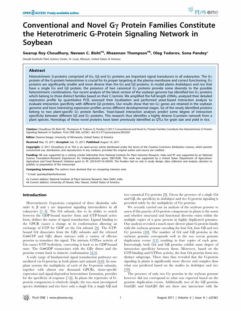
Conventional and Novel Gc Protein Families Constitute the Heterotrimeric G-Protein Signaling Network in Soybean Swarup Roy Choudhury, Naveen C. Bisht ¤a , Rheannon Thompson ¤b , Oleg Todorov, Sona Pandey* Donald Danforth Plant Science Center, St. Louis, Missouri, United States of America Abstract Heterotrimeric G-proteins comprised of Ga,Gb and Gc proteins are important signal transducers in all eukaryotes. The Gc protein of the G-protein heterotrimer is crucial for its proper targeting at the plasma membrane and correct functioning. Gc proteins are significantly smaller and more diverse than the Ga and Gb proteins. In model plants Arabidopsis and rice that have a single Ga and Gb protein, the presence of two canonical Gc proteins provide some diversity to the possible heterotrimeric combinations. Our recent analysis of the latest version of the soybean genome has identified ten Gc proteins which belong to three distinct families based on their C-termini. We amplified the full length cDNAs, analyzed their detailed expression profile by quantitative PCR, assessed their localization and performed yeast-based interaction analysis to evaluate interaction specificity with different Gb proteins. Our results show that ten Gc genes are retained in the soybean genome and have interesting expression profiles across different developmental stages. Six of the newly identified proteins belong to two plant-specific Gc protein families. Yeast-based interaction analyses predict some degree of interaction specificity between different Gb and Gc proteins. This research thus identifies a highly diverse G-protein network from a plant species. Homologs of these novel proteins have been previously identified as QTLs for grain size and yield in rice. Citation: Choudhury SR, Bisht NC, Thompson R, Todorov O, Pandey S (2011) Conventional and Novel Gc Protein Families Constitute the Heterotrimeric G-Protein Signaling Network in Soybean. PLoS ONE 6(8): e23361. doi:10.1371/journal.pone.0023361 Editor: Deanna Koepp, University of Minnesota, United States of America Received May 19, 2011; Accepted July 13, 2011; Published August 10, 2011 Copyright: ß 2011 Choudhury et al. This is an open-access article distributed under the terms of the Creative Commons Attribution License, which permits unrestricted use, distribution, and reproduction in any medium, provided the original author and source are credited. Funding: NB was supported by a visiting scholar fellowship from National Institute for Plant Genome Research, India and RT was supported by an National Science Foundation-Research Experiences for Undergraduates grant, (0851838). This work was supported by a United States Department of Agriculture/ Agriculture and Food Research Initiative grant to SP, (2010-65116-20454). The funders had no role in study design, data collection and analysis, decision to publish, or preparation of the manuscript. Competing Interests: The authors have declared that no competing interests exist. * E-mail: spandey@danforthcenter ¤a Current address: National Institute of Plant Genome Research, New Delhi, India ¤b Current address: University of Hawaii, Hilo, Hawaii, United States of America Introduction Heterotrimeric G-proteins comprised of three dissimilar sub- units a, b and c are important signaling intermediates in all eukaryotes [1–3]. The Ga subunit, due to its ability to switch between the GDP-bound inactive form and GTP-bound active form, defines the status of signal transduction. Ligand binding to the GPCR causes a change in its conformation allowing an exchange of GTP for GDP on the Ga subunit [4]. The GTP- bound Ga dissociates from the Gbc subunits and the released GaNGTP and Gbc dimer interact with a variety of effector proteins to transduce the signal. The intrinsic GTPase activity of Ga causes GTP hydrolysis, converting it back to its GDP-bound state. The GaNGDP reassociates with the Gbc dimer and the proteins return back to trimeric conformation [4,5]. A wide range of fundamental signal transduction pathways are mediated via G-proteins in both plants and animals [4,6]. In non- plant systems the multiplicity of each of the G-protein subunits, together with almost one thousand GPCRs, tissue-specific expression and signal-dependent heterotrimer formation, provides for the specificity of response [7,8]. In plants the repertoire of G- protein components is relatively simple; the two most investigated species Arabidopsis and rice have only a single Ga, a single Gb and two canonical Gc proteins [9]. Given the presence of a single Ga and Gb, the specificity in Arabidopsis and rice G-protein signaling is provided solely by the multiplicity of Gc proteins. We recently carried out an analysis of the soybean genome to assess if this paucity of G-protein components in plants is the norm and whether structural and functional diversity exists within the multiple copies of a gene present in highly duplicated genomes. Our analysis revealed a much more diverse plant G-protein family with the soybean genome encoding for four Ga, four Gb and two Gc proteins [10]. The number of Ga and Gb proteins in the soybean genome corresponds well to the two recent genome duplication events [11] resulting in four copies of each gene. Interestingly both Ga and Gb proteins exhibit some degree of interaction specificity between them. Moreover, based on the GTP-binding and GTPase activity, the four Ga proteins form two distinct subgroups. These data thus revealed that the G-protein signaling in plants is significantly more diverse and complex than what was predicted based on the studies in Arabidopsis and rice [10]. The presence of only two Gc proteins in the soybean genome however did not correspond to what was expected based on the genome duplication events. Additionally two of the Gb proteins GmGb1 and GmGb3 did not show any interaction with the PLoS ONE | www.plosone.org 1 August 2011 | Volume 6 | Issue 8 | e23361
-
Upload
danforthcenter -
Category
Documents
-
view
0 -
download
0
Transcript of Conventional and Novel Gγ Protein Families Constitute the Heterotrimeric G-Protein Signaling...
Conventional and Novel Gc Protein Families Constitutethe Heterotrimeric G-Protein Signaling Network inSoybeanSwarup Roy Choudhury, Naveen C. Bisht¤a, Rheannon Thompson¤b, Oleg Todorov, Sona Pandey*
Donald Danforth Plant Science Center, St. Louis, Missouri, United States of America
Abstract
Heterotrimeric G-proteins comprised of Ga, Gb and Gc proteins are important signal transducers in all eukaryotes. The Gcprotein of the G-protein heterotrimer is crucial for its proper targeting at the plasma membrane and correct functioning. Gcproteins are significantly smaller and more diverse than the Ga and Gb proteins. In model plants Arabidopsis and rice thathave a single Ga and Gb protein, the presence of two canonical Gc proteins provide some diversity to the possibleheterotrimeric combinations. Our recent analysis of the latest version of the soybean genome has identified ten Gc proteinswhich belong to three distinct families based on their C-termini. We amplified the full length cDNAs, analyzed their detailedexpression profile by quantitative PCR, assessed their localization and performed yeast-based interaction analysis toevaluate interaction specificity with different Gb proteins. Our results show that ten Gc genes are retained in the soybeangenome and have interesting expression profiles across different developmental stages. Six of the newly identified proteinsbelong to two plant-specific Gc protein families. Yeast-based interaction analyses predict some degree of interactionspecificity between different Gb and Gc proteins. This research thus identifies a highly diverse G-protein network from aplant species. Homologs of these novel proteins have been previously identified as QTLs for grain size and yield in rice.
Citation: Choudhury SR, Bisht NC, Thompson R, Todorov O, Pandey S (2011) Conventional and Novel Gc Protein Families Constitute the Heterotrimeric G-ProteinSignaling Network in Soybean. PLoS ONE 6(8): e23361. doi:10.1371/journal.pone.0023361
Editor: Deanna Koepp, University of Minnesota, United States of America
Received May 19, 2011; Accepted July 13, 2011; Published August 10, 2011
Copyright: � 2011 Choudhury et al. This is an open-access article distributed under the terms of the Creative Commons Attribution License, which permitsunrestricted use, distribution, and reproduction in any medium, provided the original author and source are credited.
Funding: NB was supported by a visiting scholar fellowship from National Institute for Plant Genome Research, India and RT was supported by an NationalScience Foundation-Research Experiences for Undergraduates grant, (0851838). This work was supported by a United States Department of Agriculture/Agriculture and Food Research Initiative grant to SP, (2010-65116-20454). The funders had no role in study design, data collection and analysis, decision topublish, or preparation of the manuscript.
Competing Interests: The authors have declared that no competing interests exist.
* E-mail: spandey@danforthcenter
¤a Current address: National Institute of Plant Genome Research, New Delhi, India¤b Current address: University of Hawaii, Hilo, Hawaii, United States of America
Introduction
Heterotrimeric G-proteins comprised of three dissimilar sub-
units a, b and c are important signaling intermediates in all
eukaryotes [1–3]. The Ga subunit, due to its ability to switch
between the GDP-bound inactive form and GTP-bound active
form, defines the status of signal transduction. Ligand binding to
the GPCR causes a change in its conformation allowing an
exchange of GTP for GDP on the Ga subunit [4]. The GTP-
bound Ga dissociates from the Gbc subunits and the released
GaNGTP and Gbc dimer interact with a variety of effector
proteins to transduce the signal. The intrinsic GTPase activity of
Ga causes GTP hydrolysis, converting it back to its GDP-bound
state. The GaNGDP reassociates with the Gbc dimer and the
proteins return back to trimeric conformation [4,5].
A wide range of fundamental signal transduction pathways are
mediated via G-proteins in both plants and animals [4,6]. In non-
plant systems the multiplicity of each of the G-protein subunits,
together with almost one thousand GPCRs, tissue-specific
expression and signal-dependent heterotrimer formation, provides
for the specificity of response [7,8]. In plants the repertoire of G-
protein components is relatively simple; the two most investigated
species Arabidopsis and rice have only a single Ga, a single Gb and
two canonical Gc proteins [9]. Given the presence of a single Gaand Gb, the specificity in Arabidopsis and rice G-protein signaling is
provided solely by the multiplicity of Gc proteins.
We recently carried out an analysis of the soybean genome to
assess if this paucity of G-protein components in plants is the norm
and whether structural and functional diversity exists within the
multiple copies of a gene present in highly duplicated genomes.
Our analysis revealed a much more diverse plant G-protein family
with the soybean genome encoding for four Ga, four Gb and two
Gc proteins [10]. The number of Ga and Gb proteins in the
soybean genome corresponds well to the two recent genome
duplication events [11] resulting in four copies of each gene.
Interestingly both Ga and Gb proteins exhibit some degree of
interaction specificity between them. Moreover, based on the
GTP-binding and GTPase activity, the four Ga proteins form two
distinct subgroups. These data thus revealed that the G-protein
signaling in plants is significantly more diverse and complex than
what was predicted based on the studies in Arabidopsis and rice
[10].
The presence of only two Gc proteins in the soybean genome
however did not correspond to what was expected based on the
genome duplication events. Additionally two of the Gb proteins
GmGb1 and GmGb3 did not show any interaction with the
PLoS ONE | www.plosone.org 1 August 2011 | Volume 6 | Issue 8 | e23361
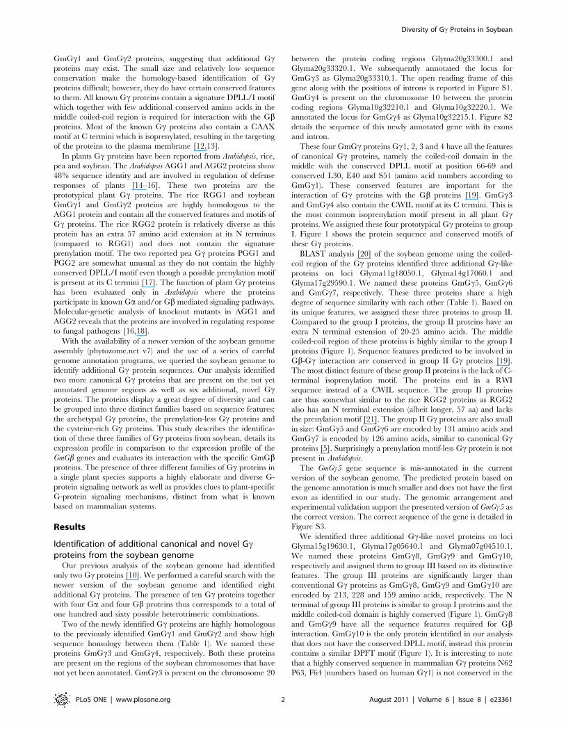
GmGc1 and GmGc2 proteins, suggesting that additional Gcproteins may exist. The small size and relatively low sequence
conservation make the homology-based identification of Gcproteins difficult; however, they do have certain conserved features
to them. All known Gc proteins contain a signature DPLL/I motif
which together with few additional conserved amino acids in the
middle coiled-coil region is required for interaction with the Gbproteins. Most of the known Gc proteins also contain a CAAX
motif at C termini which is isoprenylated, resulting in the targeting
of the proteins to the plasma membrane [12,13].
In plants Gc proteins have been reported from Arabidopsis, rice,
pea and soybean. The Arabidopsis AGG1 and AGG2 proteins show
48% sequence identity and are involved in regulation of defense
responses of plants [14–16]. These two proteins are the
prototypical plant Gc proteins. The rice RGG1 and soybean
GmGc1 and GmGc2 proteins are highly homologous to the
AGG1 protein and contain all the conserved features and motifs of
Gc proteins. The rice RGG2 protein is relatively diverse as this
protein has an extra 57 amino acid extension at its N terminus
(compared to RGG1) and does not contain the signature
prenylation motif. The two reported pea Gc proteins PGG1 and
PGG2 are somewhat unusual as they do not contain the highly
conserved DPLL/I motif even though a possible prenylation motif
is present at its C termini [17]. The function of plant Gc proteins
has been evaluated only in Arabidopsis where the proteins
participate in known Ga and/or Gb mediated signaling pathways.
Molecular-genetic analysis of knockout mutants in AGG1 and
AGG2 reveals that the proteins are involved in regulating response
to fungal pathogens [16,18].
With the availability of a newer version of the soybean genome
assembly (phytozome.net v7) and the use of a series of careful
genome annotation programs, we queried the soybean genome to
identify additional Gc protein sequences. Our analysis identified
two more canonical Gc proteins that are present on the not yet
annotated genome regions as well as six additional, novel Gcproteins. The proteins display a great degree of diversity and can
be grouped into three distinct families based on sequence features:
the archetypal Gc proteins, the prenylation-less Gc proteins and
the cysteine-rich Gc proteins. This study describes the identifica-
tion of these three families of Gc proteins from soybean, details its
expression profile in comparison to the expression profile of the
GmGb genes and evaluates its interaction with the specific GmGbproteins. The presence of three different families of Gc proteins in
a single plant species supports a highly elaborate and diverse G-
protein signaling network as well as provides clues to plant-specific
G-protein signaling mechanisms, distinct from what is known
based on mammalian systems.
Results
Identification of additional canonical and novel Gcproteins from the soybean genome
Our previous analysis of the soybean genome had identified
only two Gc proteins [10]. We performed a careful search with the
newer version of the soybean genome and identified eight
additional Gc proteins. The presence of ten Gc proteins together
with four Ga and four Gb proteins thus corresponds to a total of
one hundred and sixty possible heterotrimeric combinations.
Two of the newly identified Gc proteins are highly homologous
to the previously identified GmGc1 and GmGc2 and show high
sequence homology between them (Table 1). We named these
proteins GmGc3 and GmGc4, respectively. Both these proteins
are present on the regions of the soybean chromosomes that have
not yet been annotated. GmGc3 is present on the chromosome 20
between the protein coding regions Glyma20g33300.1 and
Glyma20g33320.1. We subsequently annotated the locus for
GmGc3 as Glyma20g33310.1. The open reading frame of this
gene along with the positions of introns is reported in Figure S1.
GmGc4 is present on the chromosome 10 between the protein
coding regions Glyma10g32210.1 and Glyma10g32220.1. We
annotated the locus for GmGc4 as Glyma10g32215.1. Figure S2
details the sequence of this newly annotated gene with its exons
and intron.
These four GmGc proteins Gc1, 2, 3 and 4 have all the features
of canonical Gc proteins, namely the coiled-coil domain in the
middle with the conserved DPLL motif at position 66-69 and
conserved L30, E40 and S51 (amino acid numbers according to
GmGc1). These conserved features are important for the
interaction of Gc proteins with the Gb proteins [19]. GmGc3
and GmGc4 also contain the CWIL motif at its C termini. This is
the most common isoprenylation motif present in all plant Gcproteins. We assigned these four prototypical Gc proteins to group
I. Figure 1 shows the protein sequence and conserved motifs of
these Gc proteins.
BLAST analysis [20] of the soybean genome using the coiled-
coil region of the Gc proteins identified three additional Gc-like
proteins on loci Glyma11g18050.1, Glyma14g17060.1 and
Glyma17g29590.1. We named these proteins GmGc5, GmGc6
and GmGc7, respectively. These three proteins share a high
degree of sequence similarity with each other (Table 1). Based on
its unique features, we assigned these three proteins to group II.
Compared to the group I proteins, the group II proteins have an
extra N terminal extension of 20-25 amino acids. The middle
coiled-coil region of these proteins is highly similar to the group I
proteins (Figure 1). Sequence features predicted to be involved in
Gb-Gc interaction are conserved in group II Gc proteins [19].
The most distinct feature of these group II proteins is the lack of C-
terminal isoprenylation motif. The proteins end in a RWI
sequence instead of a CWIL sequence. The group II proteins
are thus somewhat similar to the rice RGG2 proteins as RGG2
also has an N terminal extension (albeit longer, 57 aa) and lacks
the prenylation motif [21]. The group II Gc proteins are also small
in size: GmGc5 and GmGc6 are encoded by 131 amino acids and
GmGc7 is encoded by 126 amino acids, similar to canonical Gcproteins [5]. Surprisingly a prenylation motif-less Gc protein is not
present in Arabidopsis.
The GmGc5 gene sequence is mis-annotated in the current
version of the soybean genome. The predicted protein based on
the genome annotation is much smaller and does not have the first
exon as identified in our study. The genomic arrangement and
experimental validation support the presented version of GmGc5 as
the correct version. The correct sequence of the gene is detailed in
Figure S3.
We identified three additional Gc-like novel proteins on loci
Glyma15g19630.1, Glyma17g05640.1 and Glyma07g04510.1.
We named these proteins GmGc8, GmGc9 and GmGc10,
respectively and assigned them to group III based on its distinctive
features. The group III proteins are significantly larger than
conventional Gc proteins as GmGc8, GmGc9 and GmGc10 are
encoded by 213, 228 and 159 amino acids, respectively. The N
terminal of group III proteins is similar to group I proteins and the
middle coiled-coil domain is highly conserved (Figure 1). GmGc8
and GmGc9 have all the sequence features required for Gbinteraction. GmGc10 is the only protein identified in our analysis
that does not have the conserved DPLL motif, instead this protein
contains a similar DPFT motif (Figure 1). It is interesting to note
that a highly conserved sequence in mammalian Gc proteins N62
P63, F64 (numbers based on human Gc1) is not conserved in the
Diversity of Gc Proteins in Soybean
PLoS ONE | www.plosone.org 2 August 2011 | Volume 6 | Issue 8 | e23361
plant Gc proteins. This region is required for the GPCR-
dependent conformational change in Gc [22].
In addition to large size, there are other features that are unique
to group III Gc proteins. The proteins are predicted to have a
TNFR (tumor necrosis growth factor receptor) signature and a
long cysteine rich C-terminal region which is not found in any
other known Gc proteins to date. The C terminus of these proteins
is quite variable. Counting from the conserved DPLL (DPFT in
case of GmGc10) motif, the C terminal of GmGc8, GmGc9 and
GmGc10 consists of 130, 145 and 74 amino acids, respectively.
This variable region of group III proteins is unusually high in
cysteine content: GmGc8 contains 30% Cys (39 out of 130 amino
acids), GmGc9 contains 33% Cys (49 out of 145 amino acids) and
GmGc10 contains 26% Cys (19 out of 74 amino acids).
The sequence of GmGc8 and GmGc10 is mis-annotated in the
current version of the soybean genome assembly. The predicted
genes could never be amplified in our analysis. We manually
annotated these genes and amplified the full length product. Based
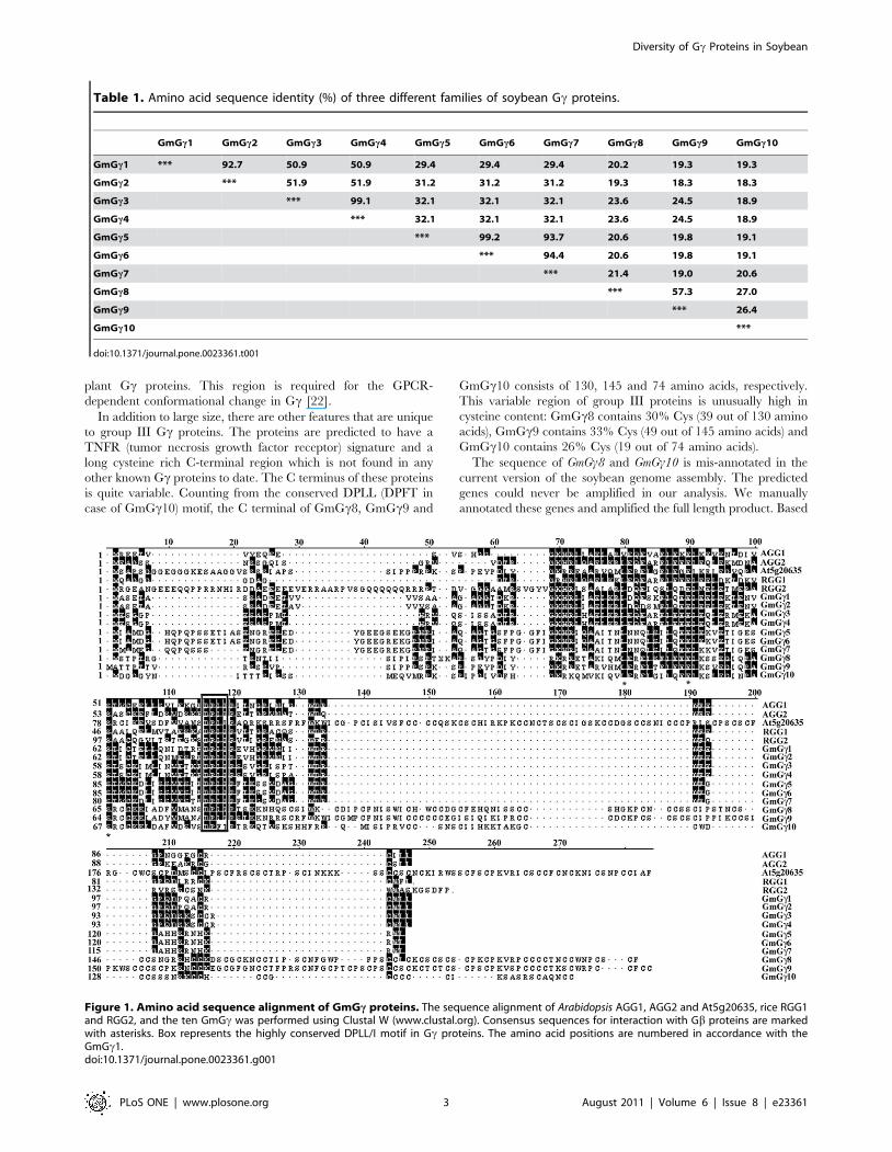
Table 1. Amino acid sequence identity (%) of three different families of soybean Gc proteins.
GmGc1 GmGc2 GmGc3 GmGc4 GmGc5 GmGc6 GmGc7 GmGc8 GmGc9 GmGc10
GmGc1 *** 92.7 50.9 50.9 29.4 29.4 29.4 20.2 19.3 19.3
GmGc2 *** 51.9 51.9 31.2 31.2 31.2 19.3 18.3 18.3
GmGc3 *** 99.1 32.1 32.1 32.1 23.6 24.5 18.9
GmGc4 *** 32.1 32.1 32.1 23.6 24.5 18.9
GmGc5 *** 99.2 93.7 20.6 19.8 19.1
GmGc6 *** 94.4 20.6 19.8 19.1
GmGc7 *** 21.4 19.0 20.6
GmGc8 *** 57.3 27.0
GmGc9 *** 26.4
GmGc10 ***
doi:10.1371/journal.pone.0023361.t001
Figure 1. Amino acid sequence alignment of GmGc proteins. The sequence alignment of Arabidopsis AGG1, AGG2 and At5g20635, rice RGG1and RGG2, and the ten GmGc was performed using Clustal W (www.clustal.org). Consensus sequences for interaction with Gb proteins are markedwith asterisks. Box represents the highly conserved DPLL/I motif in Gc proteins. The amino acid positions are numbered in accordance with theGmGc1.doi:10.1371/journal.pone.0023361.g001
Diversity of Gc Proteins in Soybean
PLoS ONE | www.plosone.org 3 August 2011 | Volume 6 | Issue 8 | e23361
on the experimentally obtained cDNAs, we marked the correct
exon-intron boundaries of GmGc8 (Figure S4) and GmGc10 (Figure
S5).
We also identified a homolog of GmGc9 in the Arabidopsis
genome at locus At5g20635. This gene has been recently
described as a Gc protein in Arabidopsis [23]. Homologs of group
III proteins are present in all plant species. The proteins also show
some homology to a keratin associated protein present in
mammals. Interestingly the homologs of group III proteins in rice
which are named DEP1 and GS3 have been recently identified as
major QTLs for grain size and yield determination [24,25].
Genome organization and phylogenetic relationshipanalysis of soybean Gc proteins
The availability of multiple Gc protein sequences in soybean with
seemingly variable sequence features raised the question whether
these protein families originated from the duplication of a single
gene. We analyzed the chromosomal location of all ten GmGc genes
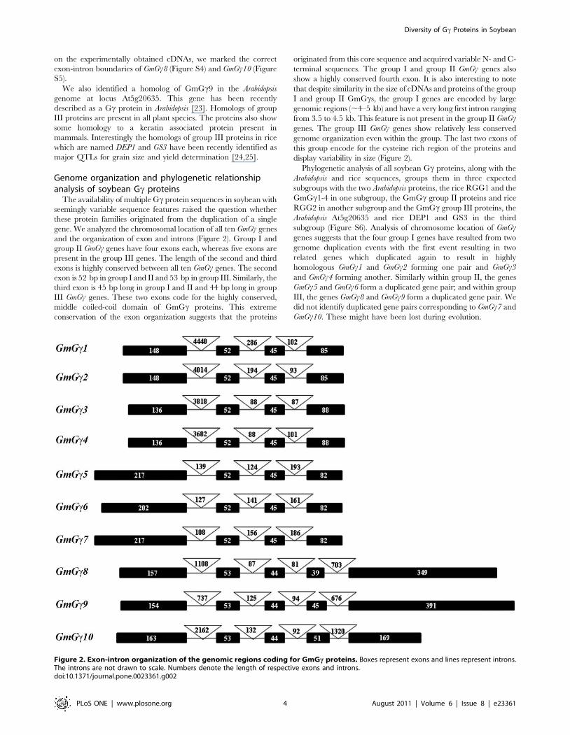
and the organization of exon and introns (Figure 2). Group I and
group II GmGc genes have four exons each, whereas five exons are
present in the group III genes. The length of the second and third
exons is highly conserved between all ten GmGc genes. The second
exon is 52 bp in group I and II and 53 bp in group III. Similarly, the
third exon is 45 bp long in group I and II and 44 bp long in group
III GmGc genes. These two exons code for the highly conserved,
middle coiled-coil domain of GmGc proteins. This extreme
conservation of the exon organization suggests that the proteins
originated from this core sequence and acquired variable N- and C-
terminal sequences. The group I and group II GmGc genes also
show a highly conserved fourth exon. It is also interesting to note
that despite similarity in the size of cDNAs and proteins of the group
I and group II GmGcs, the group I genes are encoded by large
genomic regions (,4–5 kb) and have a very long first intron ranging
from 3.5 to 4.5 kb. This feature is not present in the group II GmGcgenes. The group III GmGc genes show relatively less conserved
genome organization even within the group. The last two exons of
this group encode for the cysteine rich region of the proteins and
display variability in size (Figure 2).
Phylogenetic analysis of all soybean Gc proteins, along with the
Arabidopsis and rice sequences, groups them in three expected
subgroups with the two Arabidopsis proteins, the rice RGG1 and the
GmGc1-4 in one subgroup, the GmGc group II proteins and rice
RGG2 in another subgroup and the GmGc group III proteins, the
Arabidopsis At5g20635 and rice DEP1 and GS3 in the third
subgroup (Figure S6). Analysis of chromosome location of GmGcgenes suggests that the four group I genes have resulted from two
genome duplication events with the first event resulting in two
related genes which duplicated again to result in highly
homologous GmGc1 and GmGc2 forming one pair and GmGc3
and GmGc4 forming another. Similarly within group II, the genes
GmGc5 and GmGc6 form a duplicated gene pair; and within group
III, the genes GmGc8 and GmGc9 form a duplicated gene pair. We
did not identify duplicated gene pairs corresponding to GmGc7 and
GmGc10. These might have been lost during evolution.
Figure 2. Exon-intron organization of the genomic regions coding for GmGc proteins. Boxes represent exons and lines represent introns.The introns are not drawn to scale. Numbers denote the length of respective exons and introns.doi:10.1371/journal.pone.0023361.g002
Diversity of Gc Proteins in Soybean
PLoS ONE | www.plosone.org 4 August 2011 | Volume 6 | Issue 8 | e23361
Tissue-dependent expression analysis of soybean Gcgenes
In mammalian systems where multiple isoforms of Gc proteins
are present, a high degree of tissue specificity is observed for
expression. Similarly the Arabidopsis Gc proteins also show distinct
expression patterns [16]. We assessed the expression profile of the
ten GmGc genes by real-time quantitative PCR to evaluate
whether all ten GmGc genes are expressed and the comparative
expression levels. We quantified the absolute expression of each
gene by a 100 fold serial dilution of cloned plasmid DNA and
ascertained the specificity and efficiency of the individual primer
pairs (Table S2, Figure S7). A linear correlation coefficient (R2) of
0.98-0.99 was observed over a 100,000 fold dilution. Interestingly
unlike Ga and Gb genes that are expressed at relatively similar
levels in different tissue types [10], a range of variable expression
levels were observed for the GmGc genes.
We analyzed the tissue specific expression of the three families
of GmGc genes in vegetative tissues and reproductive tissues.
Additionally given the possible role of G-protein dependent
signaling during nodulation [26–30], we also analyzed the
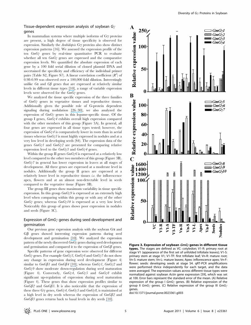
expression of GmGc genes in this legume-specific tissue. Of the
group I genes, GmGc4 exhibits overall high expression compared
with the other members of this group (Figure 3A). In general, all
four genes are expressed in all tissue types tested; however, the
expression of GmGc4 is comparatively lower in roots than in aerial
tissues whereas GmGc3 is most highly expressed in nodules and at a
very low level in developing seeds (S4). The expression data of the
genes GmGc1 and GmGc2 are presented for comparing relative
expression level to the GmGc3 and GmGc4 genes.
Within the group II genes GmGc6 is expressed at a relatively low
level compared to the other two members of this group (Figure 3B).
GmGc7 in general has lower expression in leaves at all stages of
development. All three genes are expressed at a moderate level in
nodules. Additionally the group II genes are expressed at a
relatively lower level in reproductive tissues i.e. the inflorescence
apex, flowers and at an almost non-detectable level in seeds
compared to the vegetative tissue (Figure 3B).
The group III genes show maximum variability in tissue specific
expression. In this group GmGc9 is expressed at an extremely high
level when comparing within this group or with any of the other
GmGc genes; whereas GmGc10 is expressed at a very low level.
Noticeably this group of genes shows poor expression in nodules
and seeds (Figure 3C).
Expression of GmGc genes during seed development andgermination
Our previous gene expression analysis with the soybean Ga and
Gb genes showed interesting expression patterns during seed
development and germination [10]. We analyzed the expression
pattern of the newly discovered GmGc genes during seed development
and germination and compared it to the expression of GmGb genes.
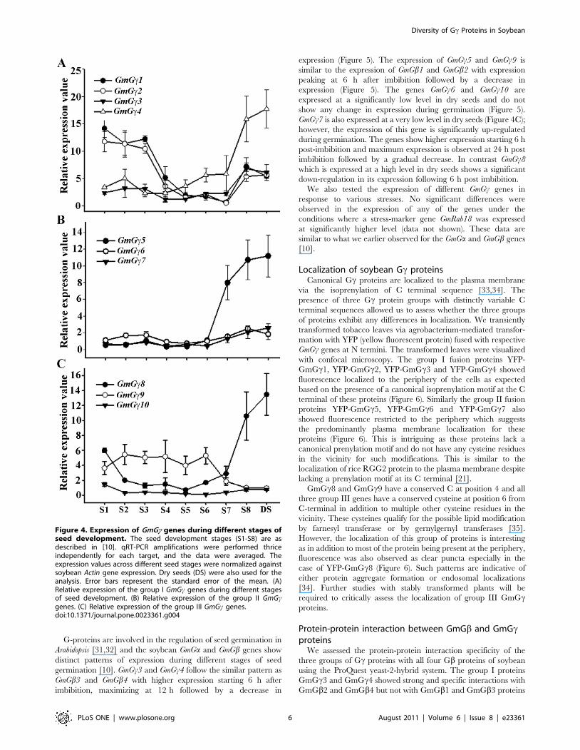
Specific patterns of gene expression were observed for different
GmGc genes. For example GmGc3, GmGc6 and GmGc7 do not show
any change in expression during seed development (Figure 4)
similar to GmGb1 and GmGb4 [10]; whereas GmGc1, GmGc2 and
GmGc9 show moderate down-regulation during seed maturation
(Figure 4). Conversely, GmGc4, GmGc5 and GmGc8 exhibit
significant up-regulation of expression during seed maturation
(Figure 4). These genes thus show expression profiles similar to
GmGb2 and GmGb3. It is also noticeable that the expression of
these three Gc genes, GmGc4, GmGc5 and GmGc8, is maintained at
a high level in dry seeds whereas the expression of GmGb2 and
GmGb3 genes returns back to basal levels in dry seeds [10].
Figure 3. Expression of soybean GmGc genes in different tissuetypes. The stages are defined as VC: cotyledon; V1-R: primary root atstage V1 (appearance of the first set of unfolded trifoliate leaves); V1-S:primary stem at stage V1; V1-TF: first trifoliate leaf; Vn-R: mature root;Vn-S: mature stem; Vn-L: mature leaves; Apex: inflorescence apex; Vn-F:flower; seeds: developing seeds at stage S4. qRT-PCR amplificationswere performed thrice independently for each target, and the datawere averaged. The expression values across different tissue types werenormalized against soybean Actin gene expression [59], which was setat 100. Error bars represent the standard error of the mean. (A) Relativeexpression of the group I GmGc genes. (B) Relative expression of thegroup II GmGc genes. (C) Relative expression of the group III GmGcgenes.doi:10.1371/journal.pone.0023361.g003
Diversity of Gc Proteins in Soybean
PLoS ONE | www.plosone.org 5 August 2011 | Volume 6 | Issue 8 | e23361
G-proteins are involved in the regulation of seed germination in
Arabidopsis [31,32] and the soybean GmGa and GmGb genes show
distinct patterns of expression during different stages of seed
germination [10]. GmGc3 and GmGc4 follow the similar pattern as
GmGb3 and GmGb4 with higher expression starting 6 h after
imbibition, maximizing at 12 h followed by a decrease in
expression (Figure 5). The expression of GmGc5 and GmGc9 is
similar to the expression of GmGb1 and GmGb2 with expression
peaking at 6 h after imbibition followed by a decrease in
expression (Figure 5). The genes GmGc6 and GmGc10 are
expressed at a significantly low level in dry seeds and do not
show any change in expression during germination (Figure 5).
GmGc7 is also expressed at a very low level in dry seeds (Figure 4C);
however, the expression of this gene is significantly up-regulated
during germination. The genes show higher expression starting 6 h
post-imbibition and maximum expression is observed at 24 h post
imbibition followed by a gradual decrease. In contrast GmGc8
which is expressed at a high level in dry seeds shows a significant
down-regulation in its expression following 6 h post imbibition.
We also tested the expression of different GmGc genes in
response to various stresses. No significant differences were
observed in the expression of any of the genes under the
conditions where a stress-marker gene GmRab18 was expressed
at significantly higher level (data not shown). These data are
similar to what we earlier observed for the GmGa and GmGb genes
[10].
Localization of soybean Gc proteinsCanonical Gc proteins are localized to the plasma membrane
via the isoprenylation of C terminal sequence [33,34]. The
presence of three Gc protein groups with distinctly variable C
terminal sequences allowed us to assess whether the three groups
of proteins exhibit any differences in localization. We transiently
transformed tobacco leaves via agrobacterium-mediated transfor-
mation with YFP (yellow fluorescent protein) fused with respective
GmGc genes at N termini. The transformed leaves were visualized
with confocal microscopy. The group I fusion proteins YFP-
GmGc1, YFP-GmGc2, YFP-GmGc3 and YFP-GmGc4 showed
fluorescence localized to the periphery of the cells as expected
based on the presence of a canonical isoprenylation motif at the C
terminal of these proteins (Figure 6). Similarly the group II fusion
proteins YFP-GmGc5, YFP-GmGc6 and YFP-GmGc7 also
showed fluorescence restricted to the periphery which suggests
the predominantly plasma membrane localization for these
proteins (Figure 6). This is intriguing as these proteins lack a
canonical prenylation motif and do not have any cysteine residues
in the vicinity for such modifications. This is similar to the
localization of rice RGG2 protein to the plasma membrane despite
lacking a prenylation motif at its C terminal [21].
GmGc8 and GmGc9 have a conserved C at position 4 and all
three group III genes have a conserved cysteine at position 6 from
C-terminal in addition to multiple other cysteine residues in the
vicinity. These cysteines qualify for the possible lipid modification
by farnesyl transferase or by gernylgernyl transferases [35].
However, the localization of this group of proteins is interesting
as in addition to most of the protein being present at the periphery,
fluorescence was also observed as clear puncta especially in the
case of YFP-GmGc8 (Figure 6). Such patterns are indicative of
either protein aggregate formation or endosomal localizations
[34]. Further studies with stably transformed plants will be
required to critically assess the localization of group III GmGcproteins.
Protein-protein interaction between GmGb and GmGcproteins
We assessed the protein-protein interaction specificity of the
three groups of Gc proteins with all four Gb proteins of soybean
using the ProQuest yeast-2-hybrid system. The group I proteins
GmGc3 and GmGc4 showed strong and specific interactions with
GmGb2 and GmGb4 but not with GmGb1 and GmGb3 proteins
Figure 4. Expression of GmGc genes during different stages ofseed development. The seed development stages (S1-S8) are asdescribed in [10]. qRT-PCR amplifications were performed thriceindependently for each target, and the data were averaged. Theexpression values across different seed stages were normalized againstsoybean Actin gene expression. Dry seeds (DS) were also used for theanalysis. Error bars represent the standard error of the mean. (A)Relative expression of the group I GmGc genes during different stagesof seed development. (B) Relative expression of the group II GmGcgenes. (C) Relative expression of the group III GmGc genes.doi:10.1371/journal.pone.0023361.g004
Diversity of Gc Proteins in Soybean
PLoS ONE | www.plosone.org 6 August 2011 | Volume 6 | Issue 8 | e23361
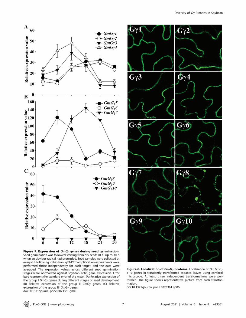
Figure 5. Expression of GmGc genes during seed germination.Seed germination was followed starting from dry seeds (0 h) up to 30 hwhen an obvious radical had protruded. Seed samples were collected atevery 6 h following imbibition. qRT-PCR amplification experiments wereperformed thrice independently for each target, and the data wereaveraged. The expression values across different seed germinationstages were normalized against soybean Actin gene expression. Errorbars represent the standard error of the mean. (A) Relative expression ofthe group I GmGc genes during different stages of seed development.(B) Relative expression of the group II GmGc genes. (C) Relativeexpression of the group III GmGc genes.doi:10.1371/journal.pone.0023361.g005
Figure 6. Localization of GmGc proteins. Localization of YFP:GmGc1-10 genes in transiently transformed tobacco leaves using confocalmicroscopy. At least three independent transformations were per-formed. The figure shows representative picture from each transfor-mation.doi:10.1371/journal.pone.0023361.g006
Diversity of Gc Proteins in Soybean
PLoS ONE | www.plosone.org 7 August 2011 | Volume 6 | Issue 8 | e23361
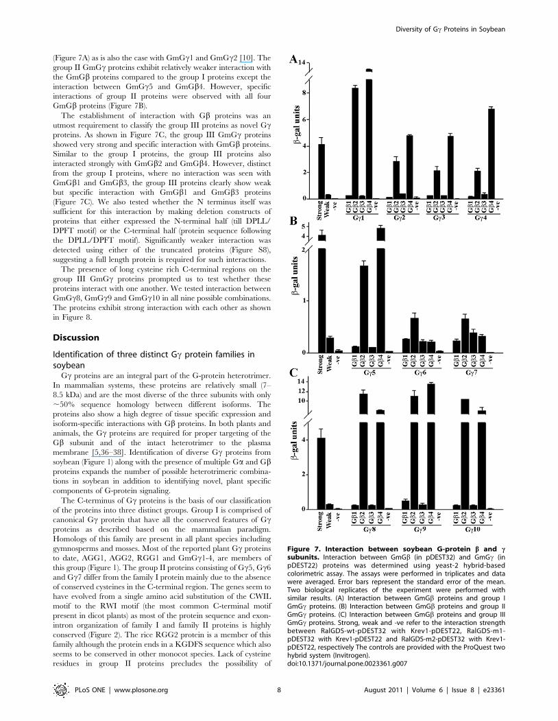
(Figure 7A) as is also the case with GmGc1 and GmGc2 [10]. The
group II GmGc proteins exhibit relatively weaker interaction with
the GmGb proteins compared to the group I proteins except the
interaction between GmGc5 and GmGb4. However, specific
interactions of group II proteins were observed with all four
GmGb proteins (Figure 7B).
The establishment of interaction with Gb proteins was an
utmost requirement to classify the group III proteins as novel Gcproteins. As shown in Figure 7C, the group III GmGc proteins
showed very strong and specific interaction with GmGb proteins.
Similar to the group I proteins, the group III proteins also
interacted strongly with GmGb2 and GmGb4. However, distinct
from the group I proteins, where no interaction was seen with
GmGb1 and GmGb3, the group III proteins clearly show weak
but specific interaction with GmGb1 and GmGb3 proteins
(Figure 7C). We also tested whether the N terminus itself was
sufficient for this interaction by making deletion constructs of
proteins that either expressed the N-terminal half (till DPLL/
DPFT motif) or the C-terminal half (protein sequence following
the DPLL/DPFT motif). Significantly weaker interaction was
detected using either of the truncated proteins (Figure S8),
suggesting a full length protein is required for such interactions.
The presence of long cysteine rich C-terminal regions on the
group III GmGc proteins prompted us to test whether these
proteins interact with one another. We tested interaction between
GmGc8, GmGc9 and GmGc10 in all nine possible combinations.
The proteins exhibit strong interaction with each other as shown
in Figure 8.
Discussion
Identification of three distinct Gc protein families insoybean
Gc proteins are an integral part of the G-protein heterotrimer.
In mammalian systems, these proteins are relatively small (7–
8.5 kDa) and are the most diverse of the three subunits with only
,50% sequence homology between different isoforms. The
proteins also show a high degree of tissue specific expression and
isoform-specific interactions with Gb proteins. In both plants and
animals, the Gc proteins are required for proper targeting of the
Gb subunit and of the intact heterotrimer to the plasma
membrane [5,36–38]. Identification of diverse Gc proteins from
soybean (Figure 1) along with the presence of multiple Ga and Gbproteins expands the number of possible heterotrimeric combina-
tions in soybean in addition to identifying novel, plant specific
components of G-protein signaling.
The C-terminus of Gc proteins is the basis of our classification
of the proteins into three distinct groups. Group I is comprised of
canonical Gc protein that have all the conserved features of Gcproteins as described based on the mammalian paradigm.
Homologs of this family are present in all plant species including
gymnosperms and mosses. Most of the reported plant Gc proteins
to date, AGG1, AGG2, RGG1 and GmGc1-4, are members of
this group (Figure 1). The group II proteins consisting of Gc5, Gc6
and Gc7 differ from the family I protein mainly due to the absence
of conserved cysteines in the C-terminal region. The genes seem to
have evolved from a single amino acid substitution of the CWIL
motif to the RWI motif (the most common C-terminal motif
present in dicot plants) as most of the protein sequence and exon-
intron organization of family I and family II proteins is highly
conserved (Figure 2). The rice RGG2 protein is a member of this
family although the protein ends in a KGDFS sequence which also
seems to be conserved in other monocot species. Lack of cysteine
residues in group II proteins precludes the possibility of
Figure 7. Interaction between soybean G-protein b and csubunits. Interaction between GmGb (in pDEST32) and GmGc (inpDEST22) proteins was determined using yeast-2 hybrid-basedcolorimetric assay. The assays were performed in triplicates and datawere averaged. Error bars represent the standard error of the mean.Two biological replicates of the experiment were performed withsimilar results. (A) Interaction between GmGb proteins and group IGmGc proteins. (B) Interaction between GmGb proteins and group IIGmGc proteins. (C) Interaction between GmGb proteins and group IIIGmGc proteins. Strong, weak and -ve refer to the interaction strengthbetween RalGDS-wt-pDEST32 with Krev1-pDEST22, RalGDS-m1-pDEST32 with Krev1-pDEST22 and RalGDS-m2-pDEST32 with Krev1-pDEST22, respectively The controls are provided with the ProQuest twohybrid system (Invitrogen).doi:10.1371/journal.pone.0023361.g007
Diversity of Gc Proteins in Soybean
PLoS ONE | www.plosone.org 8 August 2011 | Volume 6 | Issue 8 | e23361

prenylation; however, the proteins do seem to localize at the
plasma membrane (Figure 6) as has also been reported for the rice
RGG2 (21). A single cysteine present in the middle of these
proteins could be a potential target for palmotylation which might
assist in its anchoring to the plasma membrane [21]. Additionally
the proteins have a high number of positively charged and
aromatic amino acids at the C-terminus (7 out of 10) which may
target them to the plasma membrane by the formation of an ahelix [39]. In mammals, lack of prenylation either by mutation of
the conserved cysteine residue or by chemical inhibition has been
shown to result in localization of specific Gbc proteins to the
nucleus of the cells and its possible role in regulating transcription,
a function not typically associated with G-proteins [40]. Lack of a
prenylation-less gene in the Arabidopsis genome has limited the
functional characterization of this family of Gc in plants. The
availability of an insertional mutant line in RGG2 might be able to
resolve the issue of whether such proteins play any unconventional
roles in plants.
The group III proteins constitute a novel Gc family, specific to
plants. Homologs of these proteins are present in both angiosperm
and gymnosperm families. We applied the following criteria to
establish the group III proteins as authentic Gc proteins. The
coiled-coil domain of group III Gc proteins is highly similar to the
conventional Gc proteins with full conservation of amino acid
residues involved in the interaction with Gb proteins (Figure 1).
The size of the second and third exons of these proteins is very
similar to group I and group II Gc proteins (Figure 2). Additionally
a homology modeling-based analysis of three-dimensional protein
structures using a fold recognition server Phyre (Protein
Homology/analogY Recognition Engine, http://www.sbg.bio.ic.
ac.uk/ phyre/) predicted these proteins to be Gc proteins with
40–55% precision. Finally the proteins showed strong and specific
interaction with the GmGb proteins. The Arabidopsis homolog of
group III family protein At5g20635 has recently been identified as
a novel Gc protein [23].
Rice has two proteins that show homology to group III proteins,
DEP1 (dense and erect panicle 1) and GS3 (grain size 3), that have
been isolated as major QTLs for seed size and yield [24,25].
Interestingly, the rice Ga protein RGA1 is also involved in
regulation of seed size [41]. The rice DEP1 and GS3 proteins have
been described as novel proteins containing a TNFR motif, a
transmembrane domain and proteins with homology to human
keratin-associated protein. We also identified a TNFR motif in the
soybean group III family proteins; however, using multiple
transmembrane prediction programs including TMHMM
(http://www.cbs.dtu.dk/services/TMHMM/), HMMTOP
(http://www.enzim.hu/hmmtop/) and DAS (http://www.sbc.su.
se/ miklos/DAS/), we did not identify any transmembrane
domains in the GmGc8, GmGc9 or GmGc10 proteins. Rice
and Arabidopsis group III proteins are predicted to have a single
transmembrane domain using DAS, but not with TMHMM or
HMMTOP. Experimental verification of the presence of a
transmembrane domain and any possible role it might play in
localization and/or positioning of these at the plasma membrane
will be needed to evaluate its importance. Interestingly, YFP-fused
group III GmGc proteins, in addition to the peripheral YFP
fluorescence, also showed small vesicle like structures which were
very evident in GmGc8. These proteins might be localized to
endosomes structures in addition to the plasma membrane.
However, since these proteins are highly cysteine rich such
structures could also be due to protein aggregate formation or self-
interaction (Figure 8). Our data at this time cannot differentiate
between these possibilities. Expression of proteins with native
promoters in a protein null background will help decipher correct
localization.
Expression profile of soybean Gc proteins and possiblecorrelation with Gb proteins
The analysis of the complete repertoire of the GmGc genes and
its comparison with the expression pattern of GmGa and GmGbgenes began to display specific expression patterns related to
particular genes or gene families. Moreover, when comparing the
absolute expression levels within different subunits, a wide range of
expression levels were observed for the GmGc genes (e.g. Figure 3,
GmGc9 versus GmGc6), whereas all GmGa and GmGb genes were
expressed at a relatively similar level to each other. Additionally
the duplicated gene pairs of GmGa or GmGb typically showed
similar expression patterns, a trend not observed between
duplicated gene pairs of GmGc genes. GmGc9 was the most highly
expressed gene, whereas its duplicated gene GmGc8 was expressed
at the moderate level. Likewise, GmGc4 was a highly expressed
gene but its duplicated gene pair GmGc3 was relatively poorly
expressed.
Some tissue specificity of gene expression was also evident while
comparing the expression of multiple Gc genes such as low
expression of group II genes in reproductive organs or lower
expression of group III genes in nodules (Figure 3). Additionally,
during seed development and germination, specific expression
profiles were observed for individual genes which in some cases
corresponded well to the expression of GmGb genes. These
observations suggest that developmental stage-specific or tissue-
specific expression of particular genes may lead to specific bccombinations, similar to what is observed in the mammalian
systems [42,43].
Since the two rice homologs of group III genes DEP1 and GS3
are involved in grain size determination and yield, we focused on
the expression pattern of soybean group III GmGc genes during
Figure 8. Interaction between group III GmGc proteins.Interaction between different members of group III GmGc proteinswas determined using yeast-2 hybrid-based growth and colorimetricassay. The assays were performed in triplicates and data were averaged.Error bars represent the standard error of the mean. Two biologicalreplicates of the experiment were performed with similar results. Insetshows growth of yeast colonies on media lacking Leu and Trp butcontaining 50 mM 3-AT. Strong, weak and -ve refer to the interactionstrength between RalGDS-wt-pDEST32 with Krev1-pDEST22, RalGDS-m1-pDEST32 with Krev1-pDEST22 and RalGDS-m2-pDEST32 with Krev1-pDEST22, respectively.doi:10.1371/journal.pone.0023361.g008
Diversity of Gc Proteins in Soybean
PLoS ONE | www.plosone.org 9 August 2011 | Volume 6 | Issue 8 | e23361
seed development. Our data showed that of the three group III
genes in soybean, GmGc8 shows the most interesting expression
pattern during seed development and germination (Figure 4, 5).
The expression of this gene was highly up-regulated when seed is
undergoing maturation (stages S7 onwards), whereas a sharp
decrease was observed during seed germination. This gene could
be a true functional homolog of the rice DEP1 or GS3 gene.
Additionally the expression of this gene in seeds could be
dependent on the endogenous ABA and/or GA concentration as
the levels of both these hormones change significantly during seed
maturation and germination. Interestingly, GmGc5 also followed a
similar expression profile where the expression was up-regulated
during seed maturation and generally down-regulated during
germination. These genes could be potential targets for manipu-
lation to regulate soybean seed development. It was also obvious
that this group of genes is highly variable functionally as GmGc10
shows very little expression in seeds at all developmental stages and
exhibits no change in expression profile during germination.
Interaction specificity of GmGb and GmGc proteinsSpecific mammalian Gb and Gc proteins form non-dissociable
dimers and interact very strongly under a variety of in vitro and in
vivo conditions. The data presented in this study suggest that there
is specificity of interaction between different GmGb and GmGcproteins. It is especially intriguing that the GmGb1 and GmGb3
are in general weaker interactors compared to GmGb2 and
GmGb4 even though they have more than 90% sequence
similarity at the protein level (Figure 7). Additionally the group
II GmGc proteins also exhibited weak interactions compared to
the group I and group III proteins even though they do interact
with similar strengths with all four GmGb proteins. Similar
differences in the interaction between mammalian Gb and Gcproteins have also been observed. The human Gb1-4 share 80–
90% sequence identity; however, Gb1 in general interacts with
multiple Gc isoforms, Gb2 is more restricted in its interaction
partners and Gb3 displays significantly weaker interactions [44–
46]. In most cases, however, the interaction data were based on in
vitro assays and its relevance in the context of a specific cell type or
a signal remains to be evaluated in both mammalian and plant
systems.
An intriguing observation in our studies is the strong interaction
between different members of the group III proteins themselves
(Figure 8). It would be interesting to assess how the oligomeriza-
tion of these proteins might affect interaction with GmGb proteins
or other possible interactors. The unusual nature of these proteins
does not preclude the possibility of its involvement in some plant-
specific signaling mechanisms which are different from what is
known from studies in mammalian systems. Plants do have several
unconventional G-protein components such as the extra-large G-
proteins that have a Ga domain [47–50]; the GTG proteins that
have GTP-binding and hydrolysis activity of their own and are
regulated by GPA1 [51]; and the RGS1 protein that has a 7TM
GPCR-like structure fused to RGS domain [52]. Likewise, most of
the known effector proteins of G-protein signaling in plants are
also distinct from the conventional effector proteins of mammalian
systems. For example, PRN1 (Pirin1) which is a member of an
iron-containing subgroup of the cupin superfamily, PD1 (pre-
phenate dehydratase1), a protein involved in phenylalanine
biosynthesis, and a NF-Y family transcription factor form a
signaling complex during G-protein mediated light and ABA
signaling pathways during early growth and development in
Arabidopsis [53–55]. Similarly, a chloroplast-localized protein
THF1 (thylakoid formation 1) is a GPA1 effector protein during
sugar signaling [56]. Detailed study of specific pathways mediated
by these unconventional proteins in the context of canonical
heterotrimeric G-protein signaling is only in its infancy and future
work may divulge additional signaling mechanisms specifically
evolved in plants.
ConclusionWe have identified three distinct families of Gc proteins
including a novel, plant-specific Gc protein family in the soybean
genome. The elucidation of this complete repertoire of different G-
protein subunits in soybean reveals a highly elaborate G-protein
signaling network in plants. Our data also suggest the presence of
subunit-specific and tissue-type or developmental stage-specific
heterotrimeric combinations. Additionally the homologs of the
group III Gc protein have been identified as major QTLs for grain
size and yield in rice. Further work with the generation of RNAi
and overexpression lines of soybean G-protein genes will help us
decipher its signaling mechanisms as well as its use as potential
targets for biotechnological applications.
Materials and Methods
Plant material and growth conditionsSoybean (Glycine max L.) cv. Jack seeds were grown in growth
chamber (26/20uC day/night temperature, photoperiod of 14/
10 h, 800 mmol m22 s21 light intensity, and 60% humidity).
Different developmental stages of soybean plants were collected,
immediately frozen in liquid nitrogen and stored at 280uC. Tissue
for germination and stress-related experiments was prepared as
described in [10].
Cloning of soybean G-protein genesSoybean Gc genes were identified by analysis of the latest the
soybean genome assembly (www.phytozome.net/soybean) with
Arabidopsis and rice full length and middle coiled-coil region Gcprotein sequences as queries. Full-length Gc genes were amplified
from soybean seedling cDNA using gene-specific primers (Table
S1). The eight newly identified genes were cloned into the
pENTR/D-TOPO vector and confirmed by sequencing.
RNA isolation and qRT-PCRTotal RNA was isolated from different tissues of soybean plants
using Trizol reagent (Invitrogen) and qRT-PCR experiments were
performed essentially according to [10]. The real-time PCR
amplification was repeated three times and data were averaged.
Sequencing and melt curve analysis of amplicons confirmed
specificity. Standard curves for each of the genes were generated
using the cloned plasmid DNA of each gene.
Localization of GmGc proteinsThe ten GmGc genes were cloned into the pEarleyGate 104 [57]
destination vector using LR clonase mix (Invitrogen). Sequence-
confirmed recombinant plasmids containing the YFP::GmGc1-10
were transformed into A. tumefaciens strain GV3101 for subsequent
plant transformation.
Abaxial surface of tobacco leaves were infiltrated with a log-
phase culture of A. tumefaciens containing either the gene of interest
or an empty vector control according to [58]. Infiltrated plants
were incubated in darkness for 36 h followed by 24 h in light. The
leaves were imaged with the Zeiss LSM 510 laser scanning
confocal microscope (Carl Zeiss) using a 40x water-immersion, 1.2
numerical aperture, C-Apochromat objective. The yellow fluores-
cent protein (YFP) was excited with the 458-nm line of the argon
laser. At least three independent infiltrations were performed for
each construct.
Diversity of Gc Proteins in Soybean
PLoS ONE | www.plosone.org 10 August 2011 | Volume 6 | Issue 8 | e23361
Protein-protein interaction assaysTo determine the interaction between specific GmGb and
GmGc proteins, GATEWAY-based yeast-two-hybrid assay was
performed (ProQuest Two Hybrid System, Invitrogen). Briefly,
GmGb1-4 genes were cloned into pDEST32 bait vector (containing
DNA-binding domain) and GmGc1-10 genes (full length, N-
terminal and C terminal parts) were cloned into pDEST22 prey
vector (containing DNA-activating domain). The constructs were
co-transformed in yeast host strain MaV203 (Invitrogen) in specific
combinations. Interaction was determined by growth of diploid
yeast colonies on minimal media lacking leucine and tryptophan,
but containing 50 mM 3AT (3-Amino-1,2,4-triazole). The quan-
titative strength of interaction was determined by b-galactosidase
(b-gal) expression assay using ONPG (o-nitrophenyl-b-D-galacto-
pyranoside) as a substrate per manufacturer’s instructions.
Supporting Information
Table S1 Gene-specific primers used for expressionanalysis of GmGc genes.(DOCX)
Table S2 Absolute copy number quantification andprimer amplification efficiency test of GmGc genes.(DOCX)
Figure S1 Genomic sequence of the newly annotatedGmGc3 on chromosome 20.(DOC)
Figure S2 Genomic sequence of the newly annotatedGmGc4 on chromosome 10.(DOC)
Figure S3 Correct genomic sequence of GmGc5 asexperimentally verified.(DOC)
Figure S4 Correct genomic sequence of GmGc8 asexperimentally verified.(DOC)
Figure S5 Correct genomic sequence of GmGc10 asexperimentally verified.(DOC)
Figure S6 Evolutionary relationships of GmGc proteins.
(PPT)
Figure S7 PCR efficiency of GmGc genes over 100,000fold dilution.(PPT)
Figure S8 Test of interaction between the N-terminaland C-terminal parts of GmGc 8, 9 and 10 proteins withdifferent GmGb proteins.
(PPT)
Acknowledgments
We thank Drs. Jan A. Meirnyk, Christopher Taylor, Senthil Subramanian
and Manjula Govindarajulu for their help with various soybean-related
resources and Ms. Christine Ehret for critical editing of the manuscript.
Author Contributions
Conceived and designed the experiments: SC NB SP. Performed the
experiments: SC NB RT OT SP. Analyzed the data: SC NB SP. Wrote the
paper: SP.
References
1. Hamm HE (1998) The many faces of G protein signaling. J Biol Chem 273:
669–672.
2. Cabrera-Vera TM, Vanhauwe J, Thomas TO, Medkova M, Preininger A, et al.(2003) Insights into G protein structure, function, and regulation. Endocr Rev
24: 765–781.
3. Jones AM, Assmann SM (2004) Plants: the latest model system for G-protein
research. EMBO Rep 5: 572–578.
4. Offermanns S (2003) G-proteins as transducers in transmembrane signaling.Prog Biophys Mol Biol 83: 101–130.
5. McIntire WE (2009) Structural determinants involved in the formation and
activation of G protein bc dimer. Neurosignals 17: 82–99.
6. Chen JG (2008) Heterotrimeric G-proteins in plant development. Front Biosci13: 3321–3333.
7. Williamson CM, Ball ST, Nottingham WT, Skinner JA, Plagge A, et al. (2004) A
cis-acting control region is required exclusively for the tissue-specific imprintingof Gnas. Nat Genet 36: 894–899.
8. Liu J, Chen M, Deng C, Bourc’his D, Nealon JG, et al. (2005) Identification of
the control region for tissue-specific imprinting of the stimulatory G protein a-subunit. Proc Natl Acad Sci U S A 102: 5513–5518.
9. Perfus-Barbeoch L, Jones AM, Assmann SM (2004) Plant heterotrimeric G
protein function: insights from Arabidopsis and rice mutants. Curr Opin PlantBiol 7: 719–731.
10. Bisht NC, Jez JM, Pandey S (2011) An elaborate heterotrimeric G-protein family
from soybean expands the diversity of plant G-protein networks. New Phytol
190: 35–48.
11. Schmutz J, Cannon SB, Schlueter J, Ma J, Mitros T, et al. (2010) Genome
sequence of the palaeopolyploid soybean. Nature 463: 178–183.
12. Fukada Y (1995) Prenylation and carboxylmethylation of G-protein c subunit.
Methods Enzymol 250: 91–105.
13. Clapham DE, Neer EJ (1997) G protein bc subunits. Annu Rev PharmacolToxicol 37: 167–203.
14. Mason MG, Botella JR (2000) Completing the heterotrimer: isolation and
characterization of an Arabidopsis thaliana G protein c-subunit cDNA. Proc NatlAcad Sci U S A 97: 14784–14788.
15. Mason MG, Botella JR (2001) Isolation of a novel G-protein c-subunit from
Arabidopsis thaliana and its interaction with Gb. Biochim Biophys Acta 1520:147–153.
16. Trusov Y, Rookes JE, Tilbrook K, Chakravorty D, Mason MG, et al. (2007)
Heterotrimeric G protein c subunits provide functional selectivity in Gbc dimersignaling in Arabidopsis. Plant Cell 19: 1235–1250.
17. Misra S, Wu Y, Venkataraman G, Sopory SK, Tuteja N (2007) Heterotrimeric
G-protein complex and G-protein-coupled receptor from a legume Pisum sativum:
role in salinity and heat stress and cross-talk with phospholipase C. Plant J 51:656–669.
18. Trusov Y, Zhang W, Assmann SM, Botella JR (2008) Gc1+Gc2 ? Gb:
Heterotrimeric G protein Gc-deficient mutants do not recapitulate all
phenotypes of Gb-deficient mutants. Plant Physiol 147: 636–649.
19. Temple BR, Jones AM (2007) The plant heterotrimeric G-protein complex.Annu Rev Plant Biol 58: 249–266.
20. Altschul SF, Gish W, Miller W, Myers EW, Lipman DJ (1990) Basic local
alignment search tool. J Mol Biol 215: 403–410.
21. Kato C, Mizutani T, Tamaki H, Kumagai H, Kamiya T, et al. (2004)
Characterization of heterotrimeric G protein complexes in rice plasmamembrane. Plant J 38: 320–331.
22. Gautam N (2003) A conformational switch regulates receptor-G protein
interaction. Structure 11: 359–360.
23. Chakravorty D, Trusov Y, Zhang W, Acharya BR, Sheahan MB, et al. (2011)
An atypical heterotrimeric G protein c subunit is involved in guard cell K (+)channel regulation and morphological development in Arabidopsis thaliana. Plant J
(in press);doi: 10.1111/j.1365-313X.2011.04638.x.
24. Huang X, Qian Q, Liu Z, Sun H, He S, et al. (2009) Natural variation at the
DEP1 locus enhances grain yield in rice. Nat Genet 41: 494–497.
25. Mao H, Sun S, Yao J, Wang C, Yu S, et al. (2010) Linking differential domainfunctions of the GS3 protein to natural variation of grain size in rice. Proc Natl
Acad Sci U S A 107: 19579–19584.
26. Pingret JL, Journet EP, Barker DG (1998) Rhizobium nod factor signaling:
Evidence for a G protein-mediated transduction mechanism. Plant Cell 10:659–672.
27. den Hartog M, Musgrave A, Munnik T (2001) Nod factor-induced phosphatidic
acid and diacylglycerol pyrophosphate formation: a role for phospholipase C
and D in root hair deformation. Plant J 25: 55–65.
28. Kelly MN, Irving HR (2003) Nod factors activate both heterotrimeric andmonomeric G-proteins in Vigna unguiculata L Walp. Planta 216: 674–685.
29. Sun J, Miwa H, Downie JA, Oldroyd GE (2007) Mastoparan activates calcium
spiking analogous to Nod factor-induced responses in Medicago truncatula root hair
cells. Plant Physiol 144: 695–702.
30. Santos-Briones CDL, Cardenas L, Estrada-Navarrete G, Santana O, Minero-Garcıa Y, et al. (2009) GTPcS antagonizes the mastoparan-induced in vitro
activity of PIP2-phospholipase C from symbiotic root nodules of Phaseolus vulgaris.
Physiol Plantarum 135: 237–245.
Diversity of Gc Proteins in Soybean
PLoS ONE | www.plosone.org 11 August 2011 | Volume 6 | Issue 8 | e23361
31. Ullah H, Chen JG, Wang S, Jones AM (2002) Role of a heterotrimeric G protein
in regulation of Arabidopsis seed germination. Plant Physiol 129: 897–907.
32. Pandey S, Chen JG, Jones AM, Assmann SM (2006) G-protein complex mutants
are hypersensitive to abscisic acid regulation of germination and postgermina-
tion development. Plant Physiol 141: 243–256.
33. Simonds WF, Butrynski JE, Gautam N, Unson CG, Spiegel AM (1991) G-
protein bc dimers. Membrane targeting requires subunit coexpression and intact
c C-A-A-X domain. J Biol Chem 266: 5363–5366.
34. Takida S, Wedegaertner PB (2003) Heterotrimer formation, together with
isoprenylation, is required for plasma membrane targeting of Gbc. J Biol Chem
278: 17284–17290.
35. Crowell DN, Huizinga DH (2009) Protein isoprenylation: the fat of the matter.
Trends Plant Sci 14: 163–170.
36. Adjobo-Hermans MJ, Goedhart J, Gadella TW (2006) Plant G protein
heterotrimers require dual lipidation motifs of Ga and Gc and do not dissociate
upon activation. J Cell Sci 119: 5087–5097.
37. Zeng Q, Wang X, Running MP (2007) Dual lipid modification of Arabidopsis
Gc-subunits is required for efficient plasma membrane targeting. Plant Physiol
143: 1119–1131.
38. Smrcka AV (2008) G protein bc subunits: central mediators of G protein-
coupled receptor signaling. Cell Mol Life Sci 65: 2191–2214.
39. Prinz WA, Hinshaw JE (2009) Membrane-bending proteins. Crit Rev Biochem
Mol Biol 44: 278–291.
40. Kino T, Kozasa T, Chrousos GP (2005) Statin-induced blockade of prenylation
alters nucleocytoplasmic shuttling of GTP-binding proteins c2 and b2 and
enhances their suppressive effect on glucocorticoid receptor transcriptional
activity. Eur J Clin Invest 35: 508–513.
41. Ueguchi-Tanaka M, Fujisawa Y, Kobayashi M, Ashikari M, Iwasaki Y, et al.
(2000) Rice dwarf mutant d1, which is defective in the a subunit of the
heterotrimeric G protein, affects gibberellin signal transduction. Proc Natl Acad
Sci U S A 97: 11638–11643.
42. Wilcox MD, Dingus J, Balcueva EA, McIntire WE, Mehta ND, et al. (1995)
Bovine brain GO isoforms have distinct gamma subunit compositions. J Biol
Chem 270: 4189–4192.
43. Oldham WM, Hamm HE (2008) Heterotrimeric G protein activation by G-
protein-coupled receptors. Nat Rev Mol Cell Biol 9: 60–71.
44. Pronin AN, Gautam N (1992) Interaction between G-protein b and c subunit
types is selective. Proc Natl Acad Sci U S A 89: 6220–6224.
45. Schmidt CJ, Thomas TC, Levine MA, Neer EJ (1992) Specificity of G protein band c subunit interactions. J Biol Chem 267: 13807–13810.
46. Dingus J, Wells CA, Campbell L, Cleator JH, Robinson K, et al. (2005) G
Protein bc dimer formation: Gb and Gc differentially determine efficiency of invitro dimer formation. Biochemistry 44: 11882–11890.
47. Assmann SM (2002) Heterotrimeric and unconventional GTP binding proteins
in plant cell signaling. Plant Cell 14: S355–S373.48. Lee YR, Assmann SM (1999) Arabidopsis thaliana ‘extra-large GTP-binding
protein’ (AtXLG1): a new class of G-protein. Plant Mol Biol 40: 55–64.49. Ding L, Pandey S, Assmann SM (2008) Arabidopsis extra-large G proteins
(XLGs) regulate root morphogenesis. Plant J 53: 248–263.
50. Pandey S, Monshausen GB, Ding L, Assmann SM (2008) Regulation of root-wave response by extra large and conventional G proteins in Arabidopsis thaliana.
Plant J 55: 311–322.51. Pandey S, Nelson D, Assmann SM (2009) Two novel GPCR-type G-proteins are
abscisic acid receptors in Arabidopsis. Cell 136: 136–148.52. Chen JG, Willard FS, Huang J, Liang J, Chasse SA, et al. (2003) A seven-
transmembrane RGS protein that modulates plant cell proliferation. Science
301: 1728–1731.53. Warpeha KM, Lateef SS, Lapik Y, Anderson M, Lee BS, et al. (2006) G-protein-
coupled receptor 1, G-protein Galpha-subunit 1, and prephenate dehydratase 1are required for blue light-induced production of phenylalanine in etiolated
Arabidopsis. Plant Physiol 140: 844–855.
54. Warpeha KM, Upadhyay S, Yeh J, Adamiak J, Hawkins SI, et al. (2007) TheGCR1, GPA1, PRN1, NF-Y signal chain mediates both blue light and abscisic
acid responses in Arabidopsis. Plant Physiol 143: 1590–1600.55. Warpeha KM, Gibbons J, Carol A, Slusser J, Tree R, et al. (2008) Adequate
phenylalanine synthesis mediated by G protein is critical for protection from UVradiation damage in young etiolated Arabidopsis thaliana seedlings. Plant Cell
Environ 31: 1756–1770.
56. Huang J, Taylor JP, Chen JG, Uhrig JF, Schnell DJ, et al. (2006) The plastidprotein THYLAKOID FORMATION1 and the plasma membrane G-protein
GPA1 interact in a novel sugar-signaling mechanism in Arabidopsis. Plant Cell18: 1226–1238.
57. Earley KW, Haag JR, Pontes O, Opper K, Juehne T, et al. (2006) Gateway-
compatible vectors for plant functional genomics and proteomics. Plant J 45:616–629.
58. Voinnet O, Rivas S, Mestre P, Baulcombe D (2003) An enhanced transientexpression system in plants based on suppression of gene silencing by the p19
protein of tomato bushy stunt virus. Plant J 33: 949–956.59. Subramanian S, Xu L, Lu G, Odell J, Yu O (2004) The promoters of the
isoflavone synthase genes respond differentially to nodulation and defense signals
in transgenic soybean roots. Plant Mol Biol 54: 623–639.
Diversity of Gc Proteins in Soybean
PLoS ONE | www.plosone.org 12 August 2011 | Volume 6 | Issue 8 | e23361