A mutation affecting polycystin-1 mediated heterotrimeric G ...
12
3313 GENERAL ARTICLE A mutation affecting polycystin-1 mediated heterotrimeric G-protein signaling causes PKD Stephen C. Parnell 1,2, *, Brenda S. Magenheimer 1,2 , Robin L. Maser 1,2,3 , Tengis S. Pavlov 4 , Mallory A. Havens 5 , Michelle L. Hastings 6 , Stephen F. Jackson 2 , Christopher J. Ward 2 , Kenneth R. Peterson 1,2 , Alexander Staruschenko 7 and James P. Calvet 1,2, * 1 Department of Biochemistry and Molecular Biology, 2 The Jared Grantham Kidney Institute, 3 Department of Clinical Laboratory Sciences, University of Kansas Medical Center, Kansas City, KS 66160, USA, 4 Division of Hypertension and Vascular Research, Henry Ford Hospital, Detroit, MI 48202, USA, 5 Department of Biology, Lewis University, Romeoville, IL 60446, USA, 6 Department of Cell Biology and Anatomy, Chicago Medical School, Rosalind Franklin University of Medicine and Science, North Chicago, IL 60064, USA and 7 Department of Physiology, Medical College of Wisconsin, Milwaukee, WI 53226, USA *To whom correspondence should be addressed at: Department of Biochemistry and Molecular Biology and The Jared Grantham Kidney Institute, University of Kansas Medical Center, 3901 Rainbow Blvd MS3018, Kansas City, KS 66160, USA. Tel: þ1 9135880705; Fax: þ1 9135889251; Email: sparnell@- kumc.edu (S.C.P.); Department of Biochemistry and Molecular Biology and The Jared Grantham Kidney Institute, University of Kansas Medical Center, 3901 Rainbow Blvd MS3018, Kansas City, KS 66160, USA. Tel: þ1 9135887424; Fax: þ1 9135889251; Email: [email protected] (J.P.C.) Abstract Autosomal dominant polycystic kidney disease (ADPKD) is characterized by the growth of renal cysts that ultimately destroy kidney function. Mutations in the PKD1 and PKD2 genes cause ADPKD. Their protein products, polycystin-1 (PC1) and polycystin-2 (PC2) have been proposed to form a calcium-permeable receptor-channel complex; however the mechanisms by which they function are almost completely unknown. Most mutations in PKD1 are truncating loss-of-function mutations or affect protein biogenesis, traffick- ing or stability and reveal very little about the intrinsic biochemical properties or cellular functions of PC1. An ADPKD patient muta- tion (L4132D or DL), resulting in a single amino acid deletion in a putative G-protein binding region of the PC1 C-terminal cytosolic tail, was found to significantly decrease PC1-stimulated, G-protein-dependent signaling in transient transfection assays. Pkd1 DL/DL mice were embryo-lethal suggesting that DL is a functionally null mutation. Kidney-specific Pkd1 DL/cond mice were born but developed severe, postnatal cystic disease. PC1 DL protein expression levels and maturation were comparable to those of wild type PC1, and PC1 DL protein showed cell surface localization. Expression of PC1 DL and PC2 complexes in transfected CHO cells failed to support PC2 channel activity, suggesting that the role of PC1 is to activate G-protein signaling to regulate the PC1/PC2 calcium channel. Introduction ADPKD cyst growth leads to massive kidney enlargement and ultimately to renal failure. Mutations in the PKD1 gene cause 85% of ADPKD cases and those in the PKD2 gene 15% of ADPKD cases (1–3). PKD1 and PKD2 encode PC1 and PC2, respectively. Received: January 16, 2018. Revised: June 5, 2018. Accepted: June 5, 2018 V C The Author(s) 2018. Published by Oxford University Press. This is an Open Access article distributed under the terms of the Creative Commons Attribution Non-Commercial License (http://creativecommons.org/ licenses/by-nc/4.0/), which permits non-commercial re-use, distribution, and reproduction in any medium, provided the original work is properly cited. For commercial re-use, please contact [email protected] Human Molecular Genetics, 2018, Vol. 27, No. 19 3313–3324 doi: 10.1093/hmg/ddy223 Advance Access Publication Date: 20 June 2018 General Article Downloaded from https://academic.oup.com/hmg/article/27/19/3313/5040778 by guest on 15 January 2022
-
Upload
khangminh22 -
Category
Documents
-
view
0 -
download
0
Transcript of A mutation affecting polycystin-1 mediated heterotrimeric G ...

3313
G E N E R A L A R T I C L E
A mutation affecting polycystin-1 mediated
heterotrimeric G-protein signaling causes PKDStephen C. Parnell1,2,*, Brenda S. Magenheimer1,2, Robin L. Maser1,2,3,Tengis S. Pavlov4, Mallory A. Havens5, Michelle L. Hastings6,Stephen F. Jackson2, Christopher J. Ward2, Kenneth R. Peterson1,2,Alexander Staruschenko7 and James P. Calvet1,2,*1Department of Biochemistry and Molecular Biology, 2The Jared Grantham Kidney Institute, 3Department ofClinical Laboratory Sciences, University of Kansas Medical Center, Kansas City, KS 66160, USA, 4Division ofHypertension and Vascular Research, Henry Ford Hospital, Detroit, MI 48202, USA, 5Department of Biology,Lewis University, Romeoville, IL 60446, USA, 6Department of Cell Biology and Anatomy, Chicago MedicalSchool, Rosalind Franklin University of Medicine and Science, North Chicago, IL 60064, USA and 7Department ofPhysiology, Medical College of Wisconsin, Milwaukee, WI 53226, USA
*To whom correspondence should be addressed at: Department of Biochemistry and Molecular Biology and The Jared Grantham Kidney Institute,University of Kansas Medical Center, 3901 Rainbow Blvd MS3018, Kansas City, KS 66160, USA. Tel: þ1 9135880705; Fax: þ1 9135889251; Email: [email protected] (S.C.P.); Department of Biochemistry and Molecular Biology and The Jared Grantham Kidney Institute, University of Kansas Medical Center,3901 Rainbow Blvd MS3018, Kansas City, KS 66160, USA. Tel: þ1 9135887424; Fax: þ1 9135889251; Email: [email protected] (J.P.C.)
AbstractAutosomal dominant polycystic kidney disease (ADPKD) is characterized by the growth of renal cysts that ultimately destroy kidneyfunction. Mutations in the PKD1 and PKD2 genes cause ADPKD. Their protein products, polycystin-1 (PC1) and polycystin-2 (PC2)have been proposed to form a calcium-permeable receptor-channel complex; however the mechanisms by which they function arealmost completely unknown. Most mutations in PKD1 are truncating loss-of-function mutations or affect protein biogenesis, traffick-ing or stability and reveal very little about the intrinsic biochemical properties or cellular functions of PC1. An ADPKD patient muta-tion (L4132D or DL), resulting in a single amino acid deletion in a putative G-protein binding region of the PC1 C-terminal cytosolictail, was found to significantly decrease PC1-stimulated, G-protein-dependent signaling in transient transfection assays. Pkd1DL/DL
mice were embryo-lethal suggesting that DL is a functionally null mutation. Kidney-specific Pkd1DL/cond mice were born but developedsevere, postnatal cystic disease. PC1DL protein expression levels and maturation were comparable to those of wild type PC1, andPC1DL protein showed cell surface localization. Expression of PC1DL and PC2 complexes in transfected CHO cells failed to support PC2channel activity, suggesting that the role of PC1 is to activate G-protein signaling to regulate the PC1/PC2 calcium channel.
IntroductionADPKD cyst growth leads to massive kidney enlargement andultimately to renal failure. Mutations in the PKD1 gene cause
�85% of ADPKD cases and those in the PKD2 gene �15% ofADPKD cases (1–3). PKD1 and PKD2 encode PC1 and PC2,respectively.
Received: January 16, 2018. Revised: June 5, 2018. Accepted: June 5, 2018
VC The Author(s) 2018. Published by Oxford University Press.This is an Open Access article distributed under the terms of the Creative Commons Attribution Non-Commercial License (http://creativecommons.org/licenses/by-nc/4.0/), which permits non-commercial re-use, distribution, and reproduction in any medium, provided the original work is properly cited.For commercial re-use, please contact [email protected]
Human Molecular Genetics, 2018, Vol. 27, No. 19 3313–3324
doi: 10.1093/hmg/ddy223Advance Access Publication Date: 20 June 2018General Article
Dow
nloaded from https://academ
ic.oup.com/hm
g/article/27/19/3313/5040778 by guest on 15 January 2022

PC1 is a large (4 303 aa) integral protein with a> 3 000 aa N-terminal extracellular domain, 11 membrane-spanningdomains and a smaller C-terminal cytosolic domain of about200 aa (4). PC1 is related to the adhesion-GPCRs and hasstructural features consistent with it being a membrane receptor(3,5). PC2 (TRPP2) is a member of the transient receptor potential(TRP) family of membrane channels (6). PC2 has nonspecificcation channel activity and acts as a Ca2þ-regulated Ca2þ channel(7,8). PC2 has also been shown to be an ER Ca2þ release channelthat functions in an IP3 receptor-dependent fashion (9). TheC-tails of PC1 and PC2 directly interact via a coiled-coil interaction(10), and the complex has been shown to generate a unique Ca2þ
signal in transfected cells (11). PC1 and PC2 are widely expressedin many embryonic and adult tissues and organs making it likelythat they function together in most tissues.
Expression of the cytosolic C-tail of PC1 stimulates a numberof signaling pathways in transfected cells, leading to the activa-tion of promoter reporters such as AP-1 and NFAT (12–15). Themembrane-proximal region of the PC1 C-tail contains a hetero-trimeric G-protein binding and activation domain (16) that ini-tiates signaling by activating Gi/o, Gq/11 and G12/13 (14), and wheninjected into neuronal cells, PC1 has been shown to activate Gi/o
and release Gbc subunits that modulate ion channel activity(17). Ciliary PC1 and PC2 have also been shown to mediate fluid-flow mechanosensory, transient elevations in intracellular Ca2þ
in a ryanodine receptor-dependent fashion (18). While itappears that PC1/PC2 form a signaling-responsive Ca2þ channel,their ciliary mechanosensory actions and their biochemical andcellular functions are unclear (19–21).
Most mutations in PKD1 are loss-of-function, including dele-tions, splicing, frameshift and nonsense mutations, whichwould be expected to significantly alter PC1 protein levels (22).Missense mutations have also been described that affect func-tional protein levels (23); however, other than revealing that PC1biogenesis and protein levels are critical, these mutations donot provide insight into the biochemical functions of PC1. Thereare a number of C-tail single aa mutations associated withADPKD including a cluster of mutations in the G-protein bindingand activation region of the PC1 C-tail that might affect G-pro-tein signaling without affecting other properties of PC1. Onesuch mutation is a three base pair deletion resulting in the dele-tion of a single conserved leucine residue (DL) within the PC1 C-tail (24).
In this study, we show that the DL mutation is one of severalsingle aa mutations that significantly impairs G-protein signal-ing to AP-1 in transient transfection assays. Generation of aknock-in mutant mouse model (Pkd1DL) showed that this muta-tion causes a severe PKD phenotype. Homozygous Pkd1DL/DL
mice are embryo lethal, similar to truncating loss-of-functionmutations, and combination of Pkd1DL with a conditional Pkd1deletion allele during embryonic development causes a severecystic phenotype in pups. Although Pkd1DL behaves like a null
allele, normal levels of full-length PC1DL protein product aremade and produce GPS-cleaved and mature, glycosylated pro-tein. The PC1DL protein also forms complexes with PC2, yet isunable to support PC2 channel activity. As such, these experi-ments suggest that the G-protein binding and activation regionof the C-tail is critical to PC1 function by regulating PC2 Ca2þ
channel activity.
ResultsPKD1 patient mutations within the G-protein bindingregion affect PC1 signaling
We had previously demonstrated that the C-terminal tail of PC1binds and activates heterotrimeric G-proteins (16), and thatPC1-mediated activation of JNK and AP-1 (12) is mediated byboth Ga and Gbc subunits (14). We also identified a polybasicG-protein activation domain (Fig. 1) that promotes nucleotideexchange, located within a highly conserved region of thePC1 C-tail required for stable binding of G-proteins (16). Todetermine whether mutations within this region affect PC1-mediated G-protein dependent signaling, we conducted a litera-ture and database (pkdb.mayo.edu) (22) search to identifyADPKD patient mutations for further study. Our analysis identi-fied a cluster of in-frame deletion or missense variants within
Figure 1. PC1 structure and C-tail sequence. The G-protein activation domain
(G-activation, black cylinder) is a peptide region capable of activating nucleotide
exchange of heterotrimeric G-proteins (abc). The site of ADPKD-associated hu-
man mutation L4132D (DL) is indicated by an arrow in the C-tail cytosolic do-
main. The coiled-coil (coiled-coil, hatched cylinder) mediates interactions with
PC2. PC1 is cleaved at a G-protein coupled receptor proteolytic site (GPS cleav-
age, star) to produce N-and C-terminal fragments (NTF, CTF; respectively).
Significance Statement
ADPKD is caused by mutations in either of two genes, PKD1 or PKD2, whose protein products, polycystin-1 (PC1) and polycys-tin-2 (PC2), are proposed to form a calcium-permeable receptor-channel complex. A single leucine deletion (DL) in the cyto-solic C-tail of PC1 was found to cause severe ADPKD in a knock-in mouse model. Most mutations in PKD1 affect PC1 proteinlevels, biogenesis, trafficking, stability or interactions with PC2 and reveal little about the biochemical function of PC1. In con-trast, DL appears to specifically impair PC1-dependent heterotrimeric G-protein signaling and activation of the PC1/PC2 recep-tor-channel complex, suggesting that the function of PC1 is to regulate PC2 through heterotrimeric G-proteins.
3314 | Human Molecular Genetics, 2018, Vol. 27, No. 19
Dow
nloaded from https://academ
ic.oup.com/hm
g/article/27/19/3313/5040778 by guest on 15 January 2022

the G-protein binding domain in human PC1 (SupplementaryMaterial, Fig. S1). To screen for function, corresponding muta-tions were introduced into a mouse PC1 C-tail fusion construct(sIg-PKD222), and their effect on AP-1 activation was determinedin transient transfection assays in HEK293T cells (14).
AP-1 activity was significantly stimulated by the WT sIg-PKD222 construct but was reduced in cells expressing sIg-PKD222
with patient mutations corresponding to L4132D, R4136T,R4136G (24–26) and engineered mutation R4135L (Fig. 2). An ala-nine substitution (L4132A) at the same site as L4132D did nothave an effect on AP-1 activity (Supplementary Material,Fig. S2). These results show that mutations L4132D, R4135L,R4136T and R4136G interfere with AP-1 activation by the PC1 C-tail.
Previous work had shown that co-transfection of Ga
subunits augmented AP-1 activation by WT sIg-PKD222 (14,15).To determine if the patient mutations would have an effect onGa augmentation of AP-1 activation, HEK293T cells wereco-transfected with Ga12 or Gaq and WT or mutant sIg-PKD222
expression constructs. As previously observed, PC1-dependentAP-1 activity was augmented in cells co-expressing WT sIg-PKD222 and Ga12 (Fig. 2). Ga12-augmented activity was reduced incells expressing R4136T or R4136G. Activity was further reducedin cells expressing the R4135L mutation, and was eliminatedin cells expressing the L4132D mutant. Similar results wereobserved with co-expression of Gaq (Supplementary Material,Fig. S3A). Despite these pronounced effects on AP-1 activation,the mutant fusion proteins were able to interact and co-immunoprecipitate (IP) with co-transfected PC2 (SupplementaryMaterial, Fig. S3B), consistent with these mutations not disrupt-ing the coiled-coil region of PC1. These results suggest that themutations specifically impair the ability of PC1 to activate heter-otrimeric G-protein signaling. Of all the mutations studied,L4132D had the strongest effect on PC1-dependent AP-1 activa-tion and was selected for further analysis.
Generation of Pkd1NEO DL and Pkd1DL mice
The human mutation L4132D (DL) was identified in an ADPKDfamily. The mutation was found in all affected family members,
but not the unaffected parent, suggesting that L4132D was thecausative mutation in that family (24). However, it could not becertain if this mutation was pathogenic, as the 50 coding regionof the PKD1 gene was not examined for mutations in thatanalysis.
To determine whether the corresponding mouse DL muta-tion (L4122D) is sufficient to cause PKD, a targeting vector wasconstructed to introduce this mutation into mice using stan-dard ES cell technology. Germline transmission of the Pkd1NEO DL
allele was observed in six founders, which were normal and via-ble. Proper integration of the targeting construct was verified bySouthern blotting (Supplementary Material, Fig. S4).
To generate the Pkd1DL allele, heterozygous Pkd1NEO DL/þ micewere crossed to a Cre-deleter strain (EIIa-Cre) to excise thefloxed NEO cassette leaving a single, remnant loxP site withinintron 45 (27). Excision was verified by genotyping using PCR pri-mers that span the excised region and by sequence analysis(Supplementary Material, Fig. S4).
Characterization of Pkd1NEO DL and Pkd1DL transcripts
We reasoned that the presence of the floxed NEO cassettewithin intron 45 would interfere with proper splicing of thePkd1NEO DL transcript, effectively generating a null allele, andthat Cre-mediated excision would restore splicing and propertranscription of Pkd1DL message. To test this, RNA was isolatedfrom mouse embryos and used as templates for RT-PCR usingprimers designed to selectively amplify Pkd1þ, Pkd1NEO DL andPkd1DL transcripts. The results indicated that the Pkd1NEO DL al-lele is most likely non-functional due to lack of intron 45 splic-ing. In contrast, the Pkd1DL allele is properly spliced. As such,phenotypes arising from this allele are likely to be specific tothe DL deletion (Supplementary Material, Fig. S5).
Characterization of Pkd1NEO DL and Pkd1DL mice
Pkd1NEO DL/þ mice were crossed with mice heterozygous for thePkd1m1Bei allele, which is essentially null (28), to determine
Figure 2. Effect of mutations in the G-protein activation region on AP-1 activation and G-protein augmented AP-1 activity. Expression of a WT C-terminal PC1 construct
(sIg-PKD222) activates an AP-1 promoter-reporter as compared to that in control- (sIg-0) transfected HEK293T cells. Single aa mutations L4132D, R4135L, R4136T and
R4136G significantly reduce basal (#P<0.001) and Ga12-augmented (†P<0.001) AP-1 activity compared to WT-expressing cells. Protein expression was confirmed by im-
munoblot (IB). n¼3. Data are means 6 SE.
3315|Human Molecular Genetics, 2018, Vol. 27, No. 19
Dow
nloaded from https://academ
ic.oup.com/hm
g/article/27/19/3313/5040778 by guest on 15 January 2022
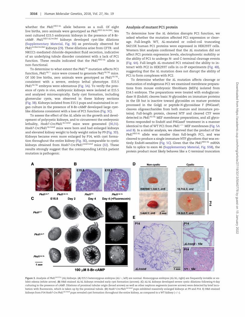
whether the Pkd1NEO DL allele behaves as a null. Of eightlive births, zero animals were genotyped as Pkd1NEO DL/m1Bei. Wenext cultured E15.5 embryonic kidneys in the presence of 8-Br-cAMP. Pkd1NEO DL/m1Bei kidneys developed cyst-like dilations(Supplementary Material, Fig. S6) as previously observed forPkd1m1Bei/m1Bei kidneys (29). These dilations arise from CFTR- andNKCC1-mediated chloride-dependent fluid secretion, indicativeof an underlying tubule disorder consistent with a lack of PC1function. These results indicated that the Pkd1NEO DL allele isnon-functional.
To determine to what extent the Pkd1DL mutation affects PC1function, Pkd1DL/þ mice were crossed to generate Pkd1DL/DL mice.Of 100 live births, zero animals were genotyped as Pkd1DL/DL,consistent with a severe, embryo lethal phenotype. E15.5Pkd1DL/DL embryos were edematous (Fig. 3A). To verify the pres-ence of cysts in vivo, embryonic kidneys were isolated at E15.5and analyzed microscopically. Early cyst formation, includingglomerular cysts, was observed in these kidney sections(Fig. 3B). Kidneys isolated from E15.5 pups and maintained in or-gan culture in the presence of 8-Br-cAMP developed large cyst-like dilations consistent with a loss of PC1 function (Fig. 3C).
To assess the effect of the DL allele on the growth and devel-opment of polycystic kidneys, and to circumvent the embryoniclethality, Hoxb7-Cre:Pkd1DL/cond mice were generated (30,31).Hoxb7-Cre:Pkd1DL/cond mice were born and had enlarged kidneysand elevated kidney weight to body weight ratios by P9 (Fig. 3D).Kidneys became even more enlarged by P14, with cyst forma-tion throughout the entire kidney (Fig. 3E), comparable to cystickidneys obtained from Hoxb7-Cre:Pkd1cond/cond mice (32). Theseresults strongly suggest that the corresponding L4132D patientmutation is pathogenic.
Analysis of mutant PC1 protein
To determine how the DL deletion disrupts PC1 function, weasked whether the mutation affected PC1 expression or cleav-age. Full-length WT, DL-mutated or coiled-coil truncatingS4213X human PC1 proteins were expressed in HEK293T cells.Western blot analysis confirmed that the DL mutation did notaffect PC1 protein expression levels, electrophoretic mobility orthe ability of PC1 to undergo N- and C-terminal cleavage events(Fig. 4A). Full-length DL-mutated PC1 retained the ability to in-teract with PC2 in HEK293T cells in co-IP experiments (Fig. 4B),suggesting that the DL mutation does not disrupt the ability ofPC1 to form complexes with PC2.
To determine whether the DL mutation affects cleavage ormaturation of endogenous PC1 we examined membrane prepara-tions from mouse embryonic fibroblasts (MEFs) isolated fromE14.5 embryos. The preparations were treated with endoglycosi-dase H (EndoH; cleaves basic N-glycosides on immature proteinsin the ER but is inactive toward glycosides on mature proteinsprocessed in the Golgi) or peptide-N-glycosidase F (PNGaseF;cleaves oligosaccharides from both mature and immature pro-teins). Full-length protein, cleaved NTF and cleaved CTF weredetected in Pkd1DL/DL MEF membrane preparations, and all glyco-forms responded to EndoH and PNGaseF treatment in a manneridentical to that of WT PC1 from Pkd1þ/þ MEF membranes (Fig. 5Aand B). In a similar analysis, we observed that the product of thePkd1NEODL allele was smaller than full-length PC1, and wascleaved to produce a single immature NTF glycoform that was en-tirely EndoH-sensitive (Fig. 5C). Given that the Pkd1NEODL mRNAfails to splice to exon 46 (Supplementary Material, Fig. S5B), theprotein product most likely behaves like a C-terminal truncation
Figure 3. Analysis of Pkd1L4122D (DL) kidneys. (A) E15.5 heterozygous embryos (DL/þ; left) are normal. Homozygous embryos (DL/DL; right) are frequently inviable or ex-
hibit edema (white arrow). (B) H&E-stained DL/DL kidneys revealed early cyst formation (arrows). (C) DL/DL kidneys developed severe cystic dilations following 4-day
culturing in the presence of cAMP. Dilations of proximal tubular origin (broad arrows) as well as other nephron segments (narrow arrows) were detected by brief incu-
bation with fluorescein, which is taken up by the proximal tubule. (D) Hoxb7-Cre:Pkd1DL/cond pups exhibited massively enlarged kidneys at P9 and P14. E) H&E-stained
kidneys from P14 Hoxb7-Cre:Pkd1DL/cond pups revealed cyst formation throughout the entire kidney, as compared to a WT kidney (þ/þ).
3316 | Human Molecular Genetics, 2018, Vol. 27, No. 19
Dow
nloaded from https://academ
ic.oup.com/hm
g/article/27/19/3313/5040778 by guest on 15 January 2022

mutant. These truncation mutants are known to undergo GPScleavage but fail to mature beyond the ER due to an inability tointeract with PC2, which is required for full maturation of PC1(33,34). This is in marked contrast to the mature, full-length DLprotein.
To determine whether the DL mutation affects PC1 proteinlevels, we quantified the amount of the PC1 glycoforms (full-length, mature and immature cleaved NTF) in membranes pre-pared from three independent WT and Pkd1DL/DL MEF cultures.The DL mutation did not affect the ratio of mature to immatureNTF or the ratio of full-length PC1 relative to a membranemarker protein, ALIX, and there was only a small non-significant reduction in the amount of mature NTF relative toALIX (Fig. 5C, and see discussion).
To determine whether the DL mutation affects PC1 intracel-lular membrane localization we analyzed protein from E14.5
whole embryo membrane preparations following fractionationover a heavy water/5–30% sucrose gradient (35) to separatemembrane compartments. Immunoblot analysis of the totalmembrane preparations revealed comparable levels of full-length, and mature and immature NTF glycoforms of PC1 fromWT and Pkd1DL/DL embryos (Fig. 6 and Supplementary Material,Fig. S7A). As observed with MEF cultures, mature NTF couldnot be detected in the total membrane preparation fromPkd1NEO DL/NEO DL embryos. Following centrifugation, gradientfractions were collected and analyzed by immunoblot. Full-length protein, mature NTF and immature NTF from both WTand PC1DL protein had the same fractionation pattern across theentire spectrum of the gradient (Fig. 6 and SupplementaryMaterial, Fig. S7A). In contrast, PC1NEO DL protein had a qualita-tively different fractionation pattern marked by decreased lev-els of mature NTF in peak fractions, increased levels of
Figure 4. Expression of human PC1DL and its interaction with PC2. (A) Full-length (FL) 3xHA-tagged (C-terminal) human PC1 proteins were expressed in HEK293T cells
and detected with anti-HA (detects FL PC1, cleavage products CTF and p100, but not NTF) and N-terminal PC1 antibody 7E12 (detects FL and cleaved NTF, but not CTF
and p100). PC1S4213X is truncated just upstream of the coiled-coil domain (but also contains a C-terminal 3xHA tag), leading to faster migration of its CTF and p100 prod-
ucts. Dashed arrows indicate crop lines where separate portions of the same image are spliced together. (B) 3xHA-tagged PC1 and myc-tagged PC2 were expressed in
HEK293T cells. Tagged PC1 was immunoprecipitated (IP’d) with anti-HA antibody and proteins were detected by IB with 7E12 for PC1 and anti-myc for PC2. The blot
shows that PC1DL is as capable as WT PC1 to interact with and IP PC2.
3317|Human Molecular Genetics, 2018, Vol. 27, No. 19
Dow
nloaded from https://academ
ic.oup.com/hm
g/article/27/19/3313/5040778 by guest on 15 January 2022

immature NTF, and the persistent presence of immature NTFtoward the bottom of the gradient. These results suggest thatthe expression and membrane localization of PC1DL is funda-mentally identical to that of WT PC1. We also analyzed E14.5embryonic kidneys for PC1 protein by immunoblotting. Full-length and mature NTF were easily detected in membrane prep-arations (Fig. 6 and Supplementary Material, Fig. S7A), but not inwhole-cell kidney lysates where immature NTF was the onlyPC1 glycoform detected (Supplementary Material, Fig. S7B).
To confirm that PC1DL protein was able to localize to theplasma membrane/cell surface, porcine LLC-PK1 cells were elec-troporated with constructs encoding GFP-tagged PC2 and eitherN-terminal mCherry-tagged WT PC1 or PC1DL proteins (Fig. 7A)(33), and protein expression was confirmed in whole-cell lysatesby immunoblot (Supplementary Material, Fig. S8A and B). PC1was detected by immunofluorescence using antibody directedagainst the mCherry epitope tag under both non-permeabilizedand permeabilized conditions (33). Under non-permeabilized
Figure 5. Expression and glycosylation of endogenous PC1DL protein. (A) Mouse embryonic fibroblast (MEF) cultures were prepared from Pkd1DL/DL and Pkd1þ/þ E14.5
embryos and treated with endoglycosidase H (EndoH) or peptide-N-glycosidase F (PNGaseF) to detect the maturation status of endogenous FL PC1 and GPS-cleaved
NTF. PC1 was detected with 7E12. NTFs from Pkd1þ/þ and Pkd1DL/DL cells resolve into two distinct glycoforms (see key at right). The faster migrating band represents the
immature (IM) glycoform of NTF which is sensitive to EndoH and the slower migrating band represents the mature (M) glycoform of NTF which is insensitive to EndoH.
The uncleaved (FL) forms of PC1DL and WT PC1 are also EndoH sensitive and their bands co-migrate with the mature NTF bands following digestion. The patterns for
full-length and NTF glycoforms of PC1DL and WT PC1 are identical. (B) Similar analysis was conducted to analyze maturation of the GPS-cleaved CTF using C-terminal
PC1 antibody E8, which detects FL and CTF. Following EndoH, the CTF resolves into mature, EndoH-resistant (upper CTF band) and immature, EndoH-sensitive (lower
CTF band) bands. Again, the digestion patterns for PC1DL and WT PC1 are identical. The E8 antibody detects a nonspecific band (*ns). (C) Membrane preparations from
Pkd1NEO DL/NEO DL (NEO/NEO) were also analyzed with EndoH. Note the position of the full-length PC1NEO DL protein (*) which is truncated due to the NEO insertion, and
PC1NEO DL-derived, immature NTF before (**) and after (***) EndoH treatment. Also note the absence of mature NTF (arrow) in the untreated Pkd1NEO DL/NEO DL lane. (D)
Endogenous PC1 levels were determined in MEF cultures isolated from three Pkd1þ/þ and three Pkd1DL/DL embryo littermates. The DL mutation did not affect the ratio of
mature to immature NTF or the ratio of full-length PC1 relative to a membrane marker protein, ALIX, and there was only a small, non-significant (Student’s t-test) re-
duction in the amount of mature NTF relative to ALIX.
Figure 6. Cellular membrane fractionation of endogenous PC1DL protein. Membrane preparations from Pkd1NEODL/NEODL (NEO DL, open circle), Pkd1DL/DL (DL, closed
square) and Pkd1þ/þ (WT, open diamond) whole embryos were fractionated by centrifugation over heavy water/sucrose gradients. PC1 was detected in a total mem-
brane aliquot (Input) and in gradient fractions by IB with 7E12. Localization of PC1 in plasma membrane fractions was confirmed by overlap with Naþ/Kþ-ATPase
(ATPase). The refractive index and complete gradient profile of PC1 from an independent experiment are shown in Supplementary Material, Figure S7A.
3318 | Human Molecular Genetics, 2018, Vol. 27, No. 19
Dow
nloaded from https://academ
ic.oup.com/hm
g/article/27/19/3313/5040778 by guest on 15 January 2022

conditions, both WT and DL-mutated PC1 could be detectedacross the entire plasma membrane (Fig. 7B). Incubation with-out primary antibody to mCherry showed no PC1 detection(Supplementary Material, Fig. S8C). Under permeabilized condi-tions, both WT and DL-mutated PC1 were readily detectedthroughout the cell, with reduced immunofluorescence in thenuclear region (Fig. 7B). To determine whether PC1DL traffickedas efficiently as WT PC1, we counted the number of cells thatwere positive for GFPPC2, mCherryPC1 or both in 5–10 fields fromthe non-permeabilized condition of three independent transfec-tion experiments. For both WT PC1 and PC1DL, the majority ofcells were positive for GFPPC2 only, and the percentage of cellspositive for both GFPPC2 and mCherryPC1 was greater than formCherryPC1 alone. Importantly, the total percentage of cellsexhibiting surface expression of mCherry-PC1DL (both with andwithout PC2) was essentially the same as for WT PC1 (Fig. 7C).These results confirm that the PC1DL protein is capable oftrafficking to the cell-surface in a manner comparable to thatof WT PC1.
To determine if the DL mutation affects the ability of PC1 toregulate PC1/PC2 channel activity, we co-expressed human PC2with either full-length WT PC1, PC1DL or C-terminally truncated
mutant PC1S4213X in CHO cells and measured whole-cell macro-scopic currents under voltage-clamp conditions (Fig. 8).Compared to cells expressing WT PC1, channel activity in cellsexpressing PC1DL was reduced to levels comparable to that ofcells expressing truncated PC1S4213X, which cannot interact withPC2 (Figs 8A and 4B). To verify that the channel activity in CHOcells is PC2 dependent, we showed that a PC2 channel mutant,D511V (9), blocked activity to the same extent as both the PC1truncation mutant and PC1DL (Fig. 8B). Thus, it appears that theDL mutation inhibits PC1/PC2 channel activity without affectingthe PC1/PC2 binding interaction.
DiscussionThe majority of PKD1 mutations analyzed has been shown to af-fect PC1 protein biogenesis, trafficking or stability and haverevealed little about the intrinsic biochemical or cellular func-tions of PC1. In theory, analysis of missense Pkd1 alleles has thepotential to identify critical signaling domains and functionalproperties of PC1. We previously identified a highly conservedregion within the C-terminal, cytosolic tail of PC1 that consti-tutes a minimal binding domain for heterotrimeric G-proteins
Figure 7. Surface expression of PC1DL in porcine kidney epithelial cells. (A) LLC-PK1 cells were electroporated with constructs encoding GFPPC2 and mCherryPC1 (WT or
DL), and analyzed by immunofluorescence. PC2 was detected using the intrinsic GFP fluorescence, and PC1 was detected using mCherry (mCh) antibodies. (B) Non-per-
meabilized conditions were used to detect surface PC1 expression, and permeabilized conditions were used to detect internal PC1 expression. (C) To determine the effi-
ciency of PC1DL trafficking to the cell surface (relative to WT PC1), GFP-positive cells in 5–10 random fields (non-permeabilized) were counted, followed by counting of
mCherry-positive cells in the same fields. The percentage of all counted cells positive for GFP alone, mCherry alone or both GFP and mCherry is shown in the pie charts.
Comparable transfection efficiency of the WT and DL constructs was confirmed by counting the number of cells positive for GFP, mCherry and GFP/mCherry signal as
detected using permeabilized conditions (data not shown).
3319|Human Molecular Genetics, 2018, Vol. 27, No. 19
Dow
nloaded from https://academ
ic.oup.com/hm
g/article/27/19/3313/5040778 by guest on 15 January 2022

and contains a 20 aa peptide sequence capable of activating het-erotrimeric G-protein nucleotide exchange (16). To determinewhether disruption of PC1-mediated activation of heterotri-meric G-protein signaling might be relevant to PKD, we ana-lyzed ADPKD-associated missense mutations that lie in andaround the G-protein activation domain using AP-1 activationas a surrogate for PC1-mediated heterotrimeric G-protein activ-ity (14).
Human substitution mutations (R4135L, R4136T and R4136G)that removed the positive charge from the first two basic resi-dues of the previously identified, polybasic G-protein activationpeptide reduced PC1-mediated basal and Ga-augmented AP-1activation without affecting the ability of PC1 to co-IP with PC2.Similar results were obtained for the single aa deletion L4132D
(DL). Secondary structure algorithms (NPS@ and PORTER) (36,37)predict that L4132 lies within an amphipathic helix contiguouswith and including the G-protein activation peptide. As such,deletion of L4132 would likely offset the distribution of multipleside-chain residues that could be important for PC1-mediatedactivation of heterotrimeric G-proteins (SupplementaryMaterial, Fig. S9). An L4132A substitution at the same location,which we predicted would not alter the overall side-chaincharge distribution, had no effect on PC1-mediated AP-1activity.
We also noted the frequency of ADPKD-associated muta-tions affecting the G-protein activation domain (SupplementaryMaterial, Fig. S10). While it may appear that there are no strongmutational hotspots when all loss-of-function mutationsthroughout the PKD1 gene are considered (38), our analysis indi-cates that within the C-tail region there is a clustering of patientmutations within and around the conserved G-protein activa-tion domain. Of the mutations studied here, human mutationL4132D (24) had the largest impact in an AP-1 transcriptional as-say, blocking both basal and Ga-augmented activity. We se-lected this mutation for further analysis to determinepathogenicity and to gain insight into the importance and rele-vance of PC1-mediated G-protein signaling to PKD.
In all aspects tested, the Pkd1DL allele behaves as a null allele.Pkd1DL/DL mice are embryo lethal, and Hoxb7-Cre:Pkd1DL/cond micedevelop severely cystic kidneys within the first 2 weeks
following birth. A striking feature of the Pkd1DL allele is that thePC1DL protein shows normal expression, cleavage and matura-tion. In this regard, it is helpful to compare the properties of thePkd1DL allele to those of other missense Pkd1 alleles such asPkd1RC, Pkd1V and Pkd1m1Bei. Among these alleles, Pkd1RC has theleast severe phenotype. Pkd1RC/RC mice have obvious renaldefects by 3 months of age, but can survive up to a year of age(23). The PC1RC protein appears to be a temperature-sensitivefolding mutant, with a �65% reduction in PC1RC protein levelsrelative to WT PC1 (33). Pkd1V is more severe, with Pkd1V/V miceexhibiting cystic defects by P4 and dying by about 1 month ofage. Full-length PC1V is expressed, but fails to undergo GPScleavage to produce NTF and CTF (39). Pkd1m1Bei is the most se-vere, with in utero cyst formation and embryonic lethality inPkd1m1Bei/m1Bei animals (28). Analysis of the PC1m1Bei1 proteinrevealed that it is expressed and undergoes GPS cleavage.However, the resulting cleavage products are sensitive toEndoH, indicating that it is unable to egress from the ER (34). Assuch, all three of these missense Pkd1 alleles have PKD pheno-types because of decreased levels of mature, cleaved protein.
While the disease severity of the Pkd1DL allele is comparableto that of Pkd1m1Bei and to Pkd1 null alleles, our analysis of thePC1DL protein reveals that there is at most only a small andnon-significant decrease in the overall level of mature NTF.While this small reduction in mature NTF could in principle in-fluence cystogenesis, it cannot account for the lethal pheno-type. In a comparable experiment (33), MEFs isolated fromPkd1þ/� embryos, which do not develop cystic disease, exhibit agreater reduction (�50%) in mature NTF, establishing this asa threshold for sufficient PC1 function. MEFs isolated fromPkd1RC/RC embryos, which will develop only mild cystic diseaseas adults, exhibit a �65% reduction in mature NTF. In contrast,MEFs derived from severely cystic (embryo lethal) Pkd1DL/DL em-bryos have mature NTF levels that are well above these thresh-old levels, indicating that the embryonic lethality caused by theDL mutation cannot be explained by a reduction in PC1DL ex-pression relative to WT PC1.
Further evidence suggesting that PC1DL protein expressionand localization are comparable to those of WT PC1 wasobtained by analyzing native protein in whole embryo
Figure 8. The PC1 DL mutation decreases PC1/PC2 channel activity. (A) La3þ-sensitive current density at �80 mV for voltage-clamped CHO cells expressing WT or mu-
tant PC1 together with PC2 (*, P<0.05, n¼10, 8 and 9, respectively from at least 4 independent transfections). (B) La3þ-sensitive current density at –80 mV for voltage-
clamped CHO cells expressing WT or channel-defective (D511V) PC2 together with WT PC1 (*, P< 0.05, n¼ 5 and 7, respectively). (C) Representative current traces
recorded from an individual cell overexpressing WT PC1/PC2 before and after superfusion with 0.5 mM La3þ and from an untransfected cell. (D) Current-voltage rela-
tionship in CHO cells expressing WT or DL mutant of PC1 and PC2 (Em-voltage, I-density of La3þ-sensitive current).
3320 | Human Molecular Genetics, 2018, Vol. 27, No. 19
Dow
nloaded from https://academ
ic.oup.com/hm
g/article/27/19/3313/5040778 by guest on 15 January 2022

membrane preparations and in embryonic kidney lysates; andby analyzing proteins transiently expressed in porcine kidneyepithelial cells. In all of these assays, the levels of PC1DL expres-sion, its fractionation profile, and its cellular localization werecomparable to that of WT PC1. This was in marked contrast tothe behavior of the protein produced from the Pkd1DL precursorallele, Pkd1NEO DL, which does not produce appreciable levels ofmature NTF, and has a different membrane fractionation pro-file. As such, our results indicate that the DL mutation disruptsa critical biochemical function of PC1.
In the current study, we sought to determine whether the DLmutation would affect the channel activity of PC2. Several linesof evidence suggest that PC2 channel activity might be affectedby alterations in the ability of PC1 to activate heterotrimeric G-protein signaling (17,40). CHO cells expressing full-length PC1and PC2 exhibited significantly higher channel activity thancells expressing PC2 and a C-terminally truncated PC1S4213X thatcannot interact with PC2, or cells expressing PC1 and PC2 chan-nel mutant, D511V (9), indicating that this channel activityrequires the PC1/PC2 complex. Cells expressing full-lengthPC1DL and PC2 had current levels comparable to that ofchannel-inactive PC1S4213X/PC2 or PC1/PC2D511V cells. We believethat in these experiments the loss of PC2 channel activityoccurs from an inability of the PC1DL mutant to initiate G-pro-tein signaling.
A disruption in PC1-mediated heterotrimeric G-protein sig-naling could potentially alter PC2 channel activity by severaldifferent mechanisms. For instance, PC2 localization and chan-nel activity are regulated by phosphorylation (7,41–43), IP3-in-duced Ca2þ release (44), and membrane lipid composition andintracellular Ca2þ (45–47). Each of these regulatory mechanismsinvolves second-messenger-dependent signaling that may bedownstream of PC1-mediated G-protein signaling. PC2 couldalso be regulated through direct interactions with G-proteinsubunits activated by PC1. There are numerous examples of G-protein subunits directly regulating channel protein activity(48,49), a possibility that remains to be explored for PC2. Giventhese potential mechanisms, we speculate that the inability ofthe PC1DL mutant to initiate G-protein signaling may result inan otherwise structurally normal PC1/PC2 complex that is func-tionally impaired.
Additional questions remain concerning the role of hetero-trimeric G-protein signaling in PKD. Wu et al. demonstrated thatin mice, Pkd1 knockout leads to increased Ga12 activation, andgenetic deletion of Gna12 blocks cystogenesis induced by dele-tion of Pkd1. In this model, PC1 is hypothesized to antagonizecystogenesis via its ability to sequester Ga12 subunits in an inac-tive state (50). In a more recent study, Zhang et al. demonstratea PKD phenotype in the Xenopus pronephric kidney followingloss of the G-protein a subunit Gnas. However, this phenotypeis antagonized by pharmacological and genetic inhibition of Gbc
signaling, suggesting that loss of Gnas leads to increased, cysto-genic Gbc signaling (51). Both of these results are in contrastwith our model of PC1 signaling, which predicts that loss of PC1signaling results in cystogenesis due to decreased heterotri-meric G-protein signaling. At this point it is difficult to reconcilethese seemingly disparate models of PC1 function, but a com-mon theme between Wu et al., Zhang et al. and our present find-ings is the central importance of the PC1 G-protein bindingdomain to regulate heterotrimeric G-protein signaling (14,16).
In conclusion, our results have implications for treatment ofPKD, as therapies designed to mimic or restore G-protein-medi-ated activation of PC2 may be sufficient to counteract cystogen-esis in cases where PC1 is not expressed or incapable of
activating G-proteins. Likewise, signaling mutants such asPC1DL may be exquisitely responsive to small-molecule therapydesigned to act specifically on inactive PC1/PC2 receptor-channel complexes. Future efforts should focus on revealing themolecular dynamics of the PC1/PC2 complex and the mecha-nisms by which PC1-activated G-protein subunits may regulatePC2 channel activity.
Materials and MethodsDetailed methods are available as supplementary material.
Plasmids
Plasmid sIg-PKD222 and sIg-0 were described previously (14).Mutations were introduced as previously described (52). 7xAP-1Firefly luciferase reporter and pBlueScript were fromStratagene. Ga expression constructs were from the MissouriS&T cDNA Resource Center (www.cdna.org; date last accessedMay 6, 2018). pRL-null was from Promega. PC2 expression plas-mid pcDNA3 PKD2myc was provided by Leonidas Tsiokas (11);pTag GFPPC2 was described previously (33). The D511V mutationwas introduced via PCR. Human PC1 expression plasmids werederived by subcloning PKD1 sequences from AF20 and AF203xHA (provided by the Baltimore Polycystic Kidney DiseaseResearch and Clinical Core Center) into pcDNA5/FRT/TO(Invitrogen) or mCherryPC1 (33). The Pkd1 targeting vector was de-rived from a fragment containing mouse Pkd1 exons 39–46 anda portion of the 30 end of Tsc2 isolated from a k phage FIX II(Stratagene) library clone provided by Marianna Rodova (27). Afloxed NEO cassette from pNTL (53) was inserted into intron 45,and the DL mutation was introduced with a QuikChangeII Site-Directed Mutagenesis Kit (Stratagene). All cloning manipula-tions were verified by DNA sequencing.
Cell culture, AP-1 assays, IP and Western blotting
HEK293T (ATCC) cells were transfected as described previously(14,15). AP-1 activity was determined with the dual luciferaseassay kit (Promega) using an EG&G Berthold 9507 Luminometer.For co-IP following dual luciferase, sIg proteins were precipi-tated with EZ-View Protein A beads (Sigma) and proteinsdetected as described previously (14,52). For co-IP of full-lengthPC1 and PC2, HA-tagged PC1 was precipitated with agarose-conjugated HA-probe (F-7; Santa Cruz Biotech).
ES cell production
The targeting vector was linearized and electroporated into 129/Ola parental cells by the University of Kansas Medical CenterTransgenic and Gene-Targeting Institutional Facility. Neomycinresistant clones were screened by long-range PCR. Positiveclones were expanded and karyotyped, and proper recombina-tion was verified by Southern blotting.
Southern blotting
ES cell or mouse tail DNA was isolated with the WizardGenomic DNA Purification Kit (Promega), digested with BglII,and electrophoresed and detected by Southern blotting. ProbeDNA was generated by PCR amplification of WT ES cell DNA.The product was cloned into pGEM-T (Promega), and an �800 bp
3321|Human Molecular Genetics, 2018, Vol. 27, No. 19
Dow
nloaded from https://academ
ic.oup.com/hm
g/article/27/19/3313/5040778 by guest on 15 January 2022

NcoI fragment was labeled using the AlkPhos DIRECT kit (GEHealthcare).
Mice
Super-ovulated C57BL/6 females (The Jackson Laboratory)were mated to C57BL/6 males. Blastocysts were injected withPkd1NEO DL targeted ES cells and transferred to recipient CD-1females (Charles River Laboratories). Chimeric mice were identi-fied by agouti coloration and mated to C57BL/6 mice to obtaingermline transmission. Agouti pups were genotyped by PCR andthe presence of the Pkd1NEO DL allele was verified by Southern.Pkd1DL mice were generated by crossing F1 Pkd1NEO DL heterozy-gotes to EIIa-Cre mice. The rearranged Pkd1DL allele wasdetected by PCR and verified by DNA sequencing. Hoxb7-Cre:Pkd1DL/cond mice were acquired by crossing Pkd1DL/þ andHoxb7-Cre:Pkd1cond/þ (30,31) mice. Phenotypic analysis was per-formed on mice of both sexes over a range of 2–4 backcrosses toC57BL/6. Total body weight and kidney weight were determined.Kidneys were fixed in formalin, paraffin embedded and stainedwith hematoxylin and eosin. All animal methods were ap-proved by the University of Kansas Medical Center IACUC.
RNA isolation and RT-PCR
Kidney or brain tissue was homogenized in TRIzol (Invitrogen)and RNA was isolated per manufacturer’s instructions. RNAwas reverse transcribed with GoScript (Promega) and PCRwas performed with GoTaq (Promega). Restriction digest withEcoRI (New England Biolabs) was used to distinguish WT fromPkd1NEO DL or Pkd1DL transcripts.
Organ culture
Metanephroi were dissected from embryonic mice and lysed inSDS-PAGE sample buffer or PC1 lysis buffer (Supplementarymethods) for immunoblot analysis, or cultured 6100 mM 8-Br-cAMP and analyzed as described previously (29).
Mouse embryonic fibroblasts (MEFs)
Genotyped embryo carcasses were minced and digested twicewith trypsin. Cells were maintained in DMEM (Cellgro) contain-ing additional L-glutamine (2 mM) and 100 000 units penicillin/100 mg streptomycin per liter supplemented with 10% heat-inactivated fetal calf serum. Genotypes were verified after thefirst passage.
Deglycosylation assays
Total cellular membranes were purified from hyper-confluentMEF cultures as previously described (33) with minor modifica-tions. Membrane fractions were treated with EndoH or PNGaseF(Promega) according to manufacturer’s instructions with minorrevisions.
Membrane preparation and sucrose gradientcentrifugation
E14.5 embryos in lysis buffer were disrupted by polytron,Dounce homogenization and sonication. Membranes were clari-fied by gentle centrifugation, equilibrated and applied to aheavy water/5–30% sucrose gradient (35) and centrifuged at
�274 000g for 16 h in a Sorvall TH-641 rotor. Gradient fractionswere collected from the top down using a BioComp fraction col-lector. The refractive index of each fraction was determined,and proteins were detected by immunoblotting.
Immunofluorescence
Porcine LLC-PK1 cells were electroporated (Nucleofector II;Amaxa with Basic Nucleofector Kit for Primary MammalianEpithelial cells; Lonza) with pTAG GFPPC2 and mCherryPC1 con-structs, and surface and intracellular expression of WT andDL-mutated PC1 was detected by immunofluorescence using an-tibody directed against the mCherry epitope (33). Images wereobtained with an Eclipse TE2000-U microscope (Nikon) using a60� oil objective and Image-Pro Premier (MediaCybernetics) andPhotoshop (Adobe) software. For quantification, images weretaken of 5–10 non-overlapping fields (20� objective) that con-tained GFP-positive cells. The same fields were subsequently im-aged for Texas Red, and the number of cells positive for GFP,Texas Red or both was determined. Quantification was from threeindependent transfection experiments.
Electrophysiology
CHO cells (ATCC) were transfected using Polyfect reagent(Qiagen) as described previously (40,54) with green fluorescentprotein (pEGFP-F; TaKaRa Clontech) and full-length PC1/PC2 ex-pression constructs. Whole-cell macroscopic recordings of PC1/PC2 channel activity were made under voltage-clamp condi-tions on EGFP-positive cells 48–72 h after transfection as previ-ously described (11,55) with an Axopatch 200B amplifier(Molecular Devices) interfaced via a Digidata 1440 (MolecularDevices) to a PC running the pClamp 10.3 software suite(Molecular Devices).
Secondary structure prediction analysis
Predictions were performed using NPS@ (Network ProteinSequence Analysis; https://npsa-prabi.ibcp.fr; date last accessedMay 6, 2018) (36) and PORTER (37). Helical wheel projectionanalysis and hydrophobic moment calculations were performedat http://rzlab.ucr.edu/scripts/wheel/wheel.cgi; date lastaccessed May 6, 2018 (56).
Statistics
Data are means 6 SE. Statistical significance was determined byone-way ANOVA and Student–Newman–Keuls (S–N–K) posttestfor multiple comparisons.
Supplementary MaterialSupplementary Material is available at HMG online.
Acknowledgements
The authors thank Marianna Rodova, Terry Peterson, JoshuaAnderson and Lance Brandenburgh for generation of plasmids,Lynn Magenheimer for assistance maintaining mouse colonies,Darren Wallace for assistance with the manuscript, DonArmstrong for assistance with helical wheel predictions,Vladimir Gainullin for advice on PC1 deglycosylation assays,and the Baltimore Polycystic Kidney Disease Research and
3322 | Human Molecular Genetics, 2018, Vol. 27, No. 19
Dow
nloaded from https://academ
ic.oup.com/hm
g/article/27/19/3313/5040778 by guest on 15 January 2022

Clinical Core Center, P30 DK090868, for plasmids and E8antibody.
Conflict of Interest statement. None declared.
FundingThe study was supported by National Institutes of HealthNS069759 (M.L.H.), 1F31NS076237 (M.A.H.), R35 HL135749 (A.S.),K99/R00 HL116603 (T.S.P.), P50 DK057301 (J.P.C.), P30 DK106912(J.P.C.), American Heart Association 16EIA26720006 (A.S.) and aUniversity of Kansas Medical Center Jared Grantham KidneyInstitute Pilot and Feasibility award (S.C.P.). Funding to pay theOpen Access publication charges for this article was provided byThe Jared Grantham Kidney Institute PKD Rodent Model andDrug-Testing Core.
References1. Cornec-Le Gall, E., Audrezet, M.P., Meur, Y.L., Chen, J.M. and
Ferec, C. (2014) Genetics and pathogenesis of autosomaldominant polycystic kidney disease: 20 years on. Hum.Mutat., 35, 1393–1406.
2. Harris, P.C. and Torres, V.E. (2014) Genetic mechanisms andsignaling pathways in autosomal dominant polycystic kid-ney disease. J. Clin. Invest., 124, 2315–2324.
3. Paul, B.M. and Vanden Heuvel, G.B. (2014) Kidney: polycystickidney disease. Wiley Interdiscip. Rev. Dev Biol, 3, 465–487.
4. Nims, N., Vassmer, D. and Maser, R.L. (2003)Transmembrane domain analysis of polycystin-1, the prod-uct of the polycystic kidney disease-1 (PKD1) gene: evidencefor 11 membrane-spanning domains. Biochemistry, 42,13035–13048.
5. Promel, S., Langenhan, T. and Arac, D. (2013) Matching struc-ture with function: the GAIN domain of adhesion-GPCR andPKD1-like proteins. Trends Pharmacol. Sci., 34, 470–478.
6. Hofherr, A. and Kottgen, M. (2011) TRPP channels and poly-cystins. Adv. Exp. Med. Biol., 704, 287–313.
7. Cai, Y., Anyatonwu, G., Okuhara, D., Lee, K.-B., Yu, Z., Onoe,T., Mei, C.-L., Qian, Q., Geng, L. and Wiztgall, R. (2004)Calcium dependence of polycystin-2 channel activity ismodulated by phosphorylation at Ser812. J. Biol. Chem., 279,19987–19995.
8. Celi�c, A.S., Petri, E.T., Benbow, J., Hodsdon, M.E., Ehrlich, B.E.and Boggon, T.J. (2012) Calcium-induced conformationalchanges in C-terminal tail of polycystin-2 are necessary forchannel gating. J. Biol. Chem., 287, 17232–17240.
9. Koulen, P., Cai, Y., Geng, L., Maeda, Y., Nishimura, S.,Witzgall, R., Ehrlich, B.E. and Somlo, S. (2002) Polycystin-2 isan intracellular calcium release channel. Nat. Cell Biol., 4,191–197.
10. Qian, F., Germino, F.J., Cai, Y., Zhang, X., Somlo, S. andGermino, G.G. (1997) PKD1 interacts with PKD2 through aprobable coiled-coil domain. Nat. Genet., 16, 179–183.
11. Hanaoka, K., Qian, F., Boletta, A., Bhunia, A.K., Piontek, K.,Tsiokas, L., Sukhatme, V.P., Guggino, W.B. and Germino, G.G.(2000) Co-assembly of polycystin-1 and -2 produces uniquecation-permeable currents. Nature, 408, 990–994.
12. Arnould, T., Kim, E., Tsiokas, L., Jochimsen, F., Gruning, W.,Chang, J.D. and Walz, G. (1998) The polycystic kidney dis-ease 1 gene product mediates protein kinase Calpha-dependent and c-Jun N-terminal kinase-dependentactivation of the transcription factor AP-1. J. Biol. Chem., 273,6013–6018.
13. Le, N.H., van der Bent, P., Huls, G., van de Wetering, M.,Loghman-Adham, M., Ong, A.C.M., Calvet, J.P., Clevers, H.,Breuning, M.H., van Dam, H. et al. (2004) Aberrantpolycystin-1 expression results in modification of activatorprotein-1 activity, whereas Wnt signaling remains unaf-fected. J. Biol. Chem., 279, 27472–27481.
14. Parnell, S.C., Magenheimer, B.S., Maser, R.L., Zien, C.A.,Frischauf, A.-M. and Calvet, J.P. (2002) Polycystin-1 activationof c-Jun N-terminal kinase and AP-1 is mediated by hetero-trimeric G proteins. J. Biol. Chem., 277, 19566–19572.
15. Puri, S., Magenheimer, B.S., Maser, R.L., Ryan, E.M., Zien,C.A., Walker, D.D., Wallace, D.P., Hempson, S.J. and Calvet,J.P. (2004) Polycystin-1 activates the calcineurin/NFAT (nu-clear factor of activated T-cells) signaling pathway. J. Biol.Chem., 279, 55455–55464.
16. Parnell, S.C., Magenheimer, B.S., Maser, R.L., Rankin, C.A.,Smine, A., Okamoto, T. and Calvet, J.P. (1998) The polycystickidney disease-1 protein, polycystin-1, binds and activatesheterotrimeric G-proteins in vitro. Biochem. Biophys. Res.Commun., 251, 625–631.
17. Delmas, P., Nomura, H., Li, X., Lakkis, M., Luo, Y., Segal, Y.,Fernandez-Fernandez, J.M., Harris, P., Frischauf, A.-M.,Brown, D.A. et al. (2002) Constitutive activation of G-proteinsby polycystin-1 is antagonized by polycystin-2. J. Biol. Chem.,277, 11276–11283.
18. Nauli, S.M., Alenghat, F.J., Luo, Y., Williams, E., Vassilev, P.,Li, X., Elia, A.E.H., Lu, W., Brown, E.M., Quinn, S.J. et al. (2003)Polycystins 1 and 2 mediate mechanosensation in the pri-mary cilium of kidney cells. Nat. Genet., 33, 129–137.
19. DeCaen, P.G., Delling, M., Vien, T.N. and Clapham, D.E. (2013)Direct recording and molecular identification of the calciumchannel of primary cilia. Nature, 504, 315–318.
20. Delling, M., DeCaen, P.G., Doerner, J.F., Febvay, S. andClapham, D.E. (2013) Primary cilia are specialized calciumsignalling organelles. Nature, 504, 311–314.
21. Jin, X., Mohieldin, A.M., Muntean, B.S., Green, J.A., Shah, J.V.,Mykytyn, K. and Nauli, S.M. (2014) Cilioplasm is a cellularcompartment for calcium signaling in response to mechani-cal and chemical stimuli. Cell. Mol. Life Sci. 71, 2165–2178.
22. Autosomal Dominant Polycystic Kidney Disease: MutationDatabase. http://pkdb.mayo.edu: Updated 2014; date lastaccessed May 6, 2018.
23. Hopp, K., Ward, C.J., Hommerding, C.J., Nasr, S.H., Tuan, H.-F., Gainullin, V.G., Rossetti, S., Torres, V.E. and Harris, P.C.(2012) Functional polycystin-1 dosage governs autosomaldominant polycystic kidney disease severity. J. Clin. Invest.,122, 4257–4273.
24. Afzal, A.R., Hand, M., Ternes-Pereira, E., Saggar-Malik, A.,Taylor, R. and Jeffery, S. (1999) Novel mutations in the 3 re-gion of the polycystic kidney disease 1 (PKD1) gene. Hum.Genet., 105, 648–653.
25. Gagnon, J.-S., Hupe, P., Arthus, M.-F., Lonergan, M., Nawar,T., Morgan, K., Fujiwara, T.M. and Bichet, D.G. (1998) PKD1mutation and polymorphisms in French-Canadian families.J. Am. Soc. Nephrol., 9, 373A.
26. Perrichot, R.A., Mercier, B., Simon, P.M., Whebe, B., Cledes, J.and Ferec, C. (1999) DGGE screening of PKD1 gene revealsnovel mutations in a large cohort of 146 unrelated patients.Hum. Genet., 105, 231–239.
27. Rodova, M., Islam, M.R., Peterson, K.R. and Calvet, J.P. (2003)Remarkable sequence conservation of the last intron in thePKD1 gene. Mol. Biol. Evol., 20, 1669–1674.
28. Herron, B.J., Lu, W., Rao, C., Liu, S., Peters, H., Bronson, R.T.,Justice, M.J., McDonald, J.D. and Beier, D.R. (2002) Efficient
3323|Human Molecular Genetics, 2018, Vol. 27, No. 19
Dow
nloaded from https://academ
ic.oup.com/hm
g/article/27/19/3313/5040778 by guest on 15 January 2022

generation and mapping of recessive developmental muta-tions using ENU mutagenesis. Nat. Genet., 30, 185–189.
29. Magenheimer, B.S., St John, P.L., Isom, K.S., Abrahamson, D.R.,De Lisle, R.C., Wallace, D.P., Maser, R.L., Grantham, J.J. andCalvet, J.P. (2006) Early embryonic renal tubules of wild-typeand polycystic kidney disease kidneys respond to cAMP stimu-lation with cystic fibrosis transmembrane conductanceregulator/Na(þ), K(þ), 2Cl(-) Co-transporter-dependent cysticdilation. J. Am. Soc. Nephrol., 17, 3424–3437.
30. Piontek, K.B., Huso, D.L., Grinberg, A., Liu, L., Bedja, D., Zhao,H., Gabrielson, K., Qian, F., Mei, C., Westphal, H. et al. (2004) Afunctional floxed allele of Pkd1 that can be conditionallyinactivated in vivo. J. Am. Soc. Nephrol., 15, 3035–3043.
31. Yu, J., Carroll, T.J. and McMahon, A.P. (2002) Sonic hedgehogregulates proliferation and differentiation of mesenchymalcells in the mouse metanephric kidney. Development, 129,5301–5312.
32. Paul, B.M., Vassmer, D., Taylor, A., Magenheimer, L., Carlton,C.G., Piontek, K.B., Germino, G.G. and Vanden Heuvel, G.B.(2011) Ectopic expression of Cux1 is associated with reducedp27 expression and increased apoptosis during late stagecyst progression upon inactivation of Pkd1 in collectingducts. Dev. Dyn., 240, 1493–1501.
33. Gainullin, V.G., Hopp, K., Ward, C.J., Hommerding, C.J. andHarris, P.C. (2015) Polycystin-1 maturation requirespolycystin-2 in a dose-dependent manner. J. Clin. Invest.,125, 607–620.
34. Kurbegovic, A., Kim, H., Xu, H., Yu, S., Cruanes, J., Maser, R.L.,Boletta, A., Trudel, M. and Qian, F. (2014) Novel functionalcomplexity of polycystin-1 by GPS cleavage in vivo: role inpolycystic kidney disease. Mol. Cell. Biol., 34, 3341–3353.
35. Hogan, M.C., Bakeberg, J.L., Gainullin, V.G., Irazabel, M.V.,Harmon, A.J., Lieske, J.C., Charlesworth, M.C., Johnson, K.L.,Madden, B.J., Zenka, R.M. et al. (2014) Identification of bio-markers for PKD1 using urinary exosomes. J. Am. Soc.Nephrol., 26: 1661–70.
36. Combet, C., Blanchet, C., Geourjon, C. and Deleage, G. (2000)NPS@: network protein sequence analysis. Trends Biochem.Sci., 25, 147–150.
37. Pollastri, G. and McLysaght, A. (2005) Porter: a new, accurateserver for protein secondary structure prediction.Bioinformatics, 21, 1719–1720.
38. Rossetti, S., Strmecki, L., Gamble, V., Burton, S., Sneddon, V.,Peral, B., Roy, S., Bakkaloglu, A., Komel, R., Winearls, C.G.et al. (2001) Mutation analysis of the entire PKD1 gene: ge-netic and diagnostic implications. Am. J. Hum. Genet., 68,46–63.
39. Yu, S., Hackmann, K., Gao, J., Gao, J., He, X., Piontek, K.,Garcıa-Gonzalez, M.A., Garcıa Gonzalez, M.A., Menezes, L.F.,Xu, H. et al. (2007) Essential role of cleavage of Polycystin-1 atG protein-coupled receptor proteolytic site for kidney tubu-lar structure. Proc. Natl. Acad. Sci. U. S. A., 104, 18688–18693.
40. Kwon, M., Pavlov, T.S., Nozu, K., Rasmussen, S.A.,Ilatovskaya, D.V., Lerch-Gaggl, A., North, L.M., Kim, H., Qian,F., Sweeney, W.E. et al. (2012) G-protein signaling modulator1 deficiency accelerates cystic disease in an orthologousmouse model of autosomal dominant polycystic kidney dis-ease. Proc. Natl. Acad. Sci. U. S. A., 109, 21462–21467.
41. Cantero, M. d R., Velazquez, I.F., Streets, A.J., Ong, A.C.M. andCantiello, H.F. (2015) The cAMP signaling pathway and direct
protein kinase a phosphorylation regulate polycystin-2(TRPP2) channel function. J. Biol. Chem., 290, 23888–23896.
42. Streets, A.J., Needham, A.J., Gill, S.K. and Ong, A.C. (2010)Protein kinase D-mediated phosphorylation of polycystin-2(TRPP2) is essential for its effects on cell growth and calciumchannel activity. Mol. Biol. Cell, 21, 3853–3865.
43. Streets, A.J., Wessely, O., Peters, D.J. and Ong, A.C. (2013)Hyperphosphorylation of polycystin-2 at a critical residue indisease reveals an essential role for polycystin-1-regulateddephosphorylation. Hum. Mol. Genet., 22, 1924–1939.
44. Sammels, E., Devogelaere, B., Mekahli, D., Bultynck, G.,Missiaen, L., Parys, J.B., Cai, Y., Somlo, S. and De Smedt, H.(2010) Polycystin-2 activation by inositol 1, 4, 5-trisphospha-te-induced Ca2þ release requires its direct association withthe inositol 1, 4, 5-trisphosphate receptor in a signalingmicrodomain. J. Biol. Chem., 285, 18794–18805.
45. Grieben, M., Pike, A.C.W., Shintre, C.A., Venturi, E., El-Ajouz,S., Tessitore, A., Shrestha, L., Mukhopadhyay, S., Mahajan,P., Chalk, R. et al. (2017) Structure of the polycystic kidneydisease TRP channel Polycystin-2 (PC2). Nat. Struct. Mol. Biol.,24, 114–122.
46. Shen, P.S., Yang, X., DeCaen, P.G., Liu, X., Bulkley, D.,Clapham, D.E. and Cao, E. (2016) The structure of the poly-cystic kidney disease channel PKD2 in lipid nanodiscs. Cell,167, 763–773.
47. Wilkes, M., Madej, M.G., Kreuter, L., Rhinow, D., Heinz, V., DeSanctis, S., Ruppel, S., Richter, R.M., Joos, F., Grieben, M. et al.(2017) Molecular insights into lipid-assisted Ca(2þ) regulation ofthe TRP channel Polycystin-2. Nat. Struct. Mol. Biol., 24, 123–130.
48. Wettschureck, N. and Offermanns, S. (2005) Mammalian Gproteins and their cell type specific functions. Physiol. Rev.,85, 1159–1204.
49. Zhang, X., Mak, S., Li, L., Parra, A., Denlinger, B., Belmonte, C.and McNaughton, P.A. (2012) Direct inhibition of thecold-activated TRPM8 ion channel by Galphaq. Nat. Cell Biol.,14, 851–858.
50. Wu, Y., Xu, J.X., El-Jouni, W., Lu, T., Li, S., Wang, Q., Tran, M.,Yu, W., Wu, M., Barrera, I.E. et al. (2016) Galpha12 is requiredfor renal cystogenesis induced by Pkd1 inactivation. J. CellSci., 129, 3675–3684.
51. Zhang, B., Tran, U. and Wessely, O. (2018) Polycystin 1 loss offunction is directly linked to an imbalance in G-protein sig-naling in the kidney. Development, 145, . pii: dev158931.
52. Parnell, S.C., Puri, S., Wallace, D.P. and Calvet, J.P. (2012)Protein phosphatase-1alpha interacts with and dephosphor-ylates polycystin-1. PLoS One, 7, e36798.
53. Abuin, A. and Bradley, A. (1996) Recycling selectablemarkers in mouse embryonic stem cells. Mol. Cell. Biol., 16,1851–1856.
54. Staruschenko, A., Booth, R.E., Pochynyuk, O., Stockand, J.D.and Tong, Q. (2006) Functional reconstitution of the humanepithelial Naþ channel in a mammalian expression system.Methods Mol. Biol., 337, 3–13.
55. Babich, V., Zeng, W.-Z., Yeh, B.-I., Ibraghimov-Beskrovnaya,O., Cai, Y., Somlo, S. and Huang, C.-L. (2004) The N-terminalextracellular domain is required for polycystin-1-dependentchannel activity. J. Biol. Chem., 279, 25582–25589.
56. Zidovetzki, R., Rost, B., Armstrong, D.L. and Pecht, I. (2003)Transmembrane domains in the functions of Fc receptors.Biophys. Chem., 100, 555–575.
3324 | Human Molecular Genetics, 2018, Vol. 27, No. 19
Dow
nloaded from https://academ
ic.oup.com/hm
g/article/27/19/3313/5040778 by guest on 15 January 2022