A Mitochondrial Kinase Complex Is Essential to Mediate an ERK1/2Dependent Phosphorylation of a Key...
11
A Mitochondrial Kinase Complex Is Essential to Mediate an ERK1/2-Dependent Phosphorylation of a Key Regulatory Protein in Steroid Biosynthesis Cecilia Poderoso 1 , Daniela P. Converso 2 , Paula Maloberti 1 , Alejandra Duarte 1 , Isabel Neuman 1 , Soledad Galli 2 , Fabiana Cornejo Maciel 1 , Cristina Paz 1 , Marı ´a C. Carreras 2,3 , Juan J. Poderoso 2 , Ernesto J. Podesta ´ 1 * 1 Instituto de Investigaciones Moleculares de Enfermedades Hormonales, Neurodegenerativas y Oncolo ´ gicas (IIMHNO), Department of Human Biochemistry, School of Medicine, University of Buenos Aires, Buenos Aires, Argentina, 2 Laboratory of Oxygen Metabolism, University Hospital, University of Buenos Aires, Buenos Aires, Argentina, 3 Department of Clinical Biochemistry, University Hospital, University of Buenos Aires, Buenos Aires, Argentina ERK1/2 is known to be involved in hormone-stimulated steroid synthesis, but its exact roles and the underlying mechanisms remain elusive. Both ERK1/2 phosphorylation and steroidogenesis may be triggered by cAMP/cAMP-dependent protein kinase (PKA)-dependent and-independent mechanisms; however, ERK1/2 activation by cAMP results in a maximal steroidogenic rate, whereas canonical activation by epidermal growth factor (EGF) does not. We demonstrate herein by Western blot analysis and confocal studies that temporal mitochondrial ERK1/2 activation is obligatory for PKA-mediated steroidogenesis in the Leydig- transformed MA-10 cell line. PKA activity leads to the phosphorylation of a constitutive mitochondrial MEK1/2 pool with a lower effect in cytosolic MEKs, while EGF allows predominant cytosolic MEK activation and nuclear pERK1/2 localization. These results would explain why PKA favors a more durable ERK1/2 activation in mitochondria than does EGF. By means of ex vivo experiments, we showed that mitochondrial maximal steroidogenesis occurred as a result of the mutual action of steroidogenic acute regulatory (StAR) protein –a key regulatory component in steroid biosynthesis-, active ERK1/2 and PKA. Our results indicate that there is an interaction between mitochondrial StAR and ERK1/2, involving a D domain with sequential basic-hydrophobic motifs similar to ERK substrates. As a result of this binding and only in the presence of cholesterol, ERK1/2 phosphorylates StAR at Ser 232 . Directed mutagenesis of Ser 232 to a non-phosphorylable amino acid such as Ala (StAR S232A) inhibited in vitro StAR phosphorylation by active ERK1/2. Transient transfection of MA-10 cells with StAR S232A markedly reduced the yield of progesterone production. In summary, here we show that StAR is a novel substrate of ERK1/2, and that mitochondrial ERK1/2 is part of a multimeric protein kinase complex that regulates cholesterol transport. The role of MAPKs in mitochondrial function is underlined. Citation: Poderoso C, Converso DP, Maloberti P, Duarte A, Neuman I, et al (2008) A Mitochondrial Kinase Complex Is Essential to Mediate an ERK1/2- Dependent Phosphorylation of a Key Regulatory Protein in Steroid Biosynthesis. PLoS ONE 3(1): e1443. doi:10.1371/journal.pone.0001443 INTRODUCTION In the complex process of steroidogenesis, the mitochondria is the site where the rate limiting step -cholesterol transport across the mitochondrial membranes- occurs [1,2]. Cholesterol transport requires specific interactions at the mitochondria between several proteins including the voltage- dependent anion channel (VDAC) [3], the peripheral benzodiaz- epine receptor (PBR, currently named translocator protein or TSPO) [4], the PBR-associated protein (PAP7) [5], and the steroidogenic acute regulatory protein (StAR) [5–8]. The StAR protein, which has the mitochondrial target sequence at the N terminus, is synthesized as a 37 kDa precursor protein in the cytosol, which is cleaved in the mitochondrial matrix to form a 30 kDa protein [7,9–11]. The N terminal 47 or 62 aminoacid- truncated murine or human forms of StAR protein stimulates cholesterol transport outside the mitochondria, indicating that the 30 kDa form is active at the outer mitochondrial membrane [12,13]. According to the results described above, the active form of StAR should be re-exported after the processing of the precursor in the matrix. This hypothesis is not proved yet. Nevertheless, it is known that the re-export process exists for other proteins such as HSP60 [14]. Additionally, it has also been shown that the inhibition of the processing of the 37 kDa form of StAR protein inhibits steroidogenesis in MA-10 Leydig and Y1 adrenocortical cells [9,11,15–18]. Thus, the relation between the processing efficiency of StAR protein in mitochondria and the steroidogenic activity in cells of the active form of StAR has not been fully clarified. The transcription of the StAR gene increases in a cAMP- dependent protein kinase (PKA)-dependent manner [6,19]. In addition, the non-genomic post-translational effects of PKA have been reported in relationship to StAR. PKA phosphorylates murine and human StAR at specific motifs like Ser 56-57 and Ser 194-195 , respectively [20]. This event is required for StAR function [21]. Although it is certain that PKA activation is important for trophic hormone-stimulated steroid biosynthesis [22–26], it is also known that ERK1/2 and its upstream activator MEK1/2 participate in the regulation of steroidogenesis by genomic and non-genomic effects [27–32]. However, little is Academic Editor: Wenqing Xu, University of Washington, United States of America Received September 12, 2007; Accepted December 13, 2007; Published January 16, 2008 Copyright: ß 2008 Poderoso et al. This is an open-access article distributed under the terms of the Creative Commons Attribution License, which permits unrestricted use, distribution, and reproduction in any medium, provided the original author and source are credited. Funding: National Research Council (CONICET) University of Buenos Aires (UBA) National Agency for Promotion of Science and Technology (ANPCyT) Sigma Xi, the Scientific Research Society Competing Interests: The authors have declared that no competing interests exist. * To whom correspondence should be addressed. E-mail: [email protected] PLoS ONE | www.plosone.org 1 January 2008 | Issue 1 | e1443
-
Upload
independent -
Category
Documents
-
view
1 -
download
0
Transcript of A Mitochondrial Kinase Complex Is Essential to Mediate an ERK1/2Dependent Phosphorylation of a Key...
A Mitochondrial Kinase Complex Is Essential to Mediatean ERK1/2-Dependent Phosphorylation of a KeyRegulatory Protein in Steroid BiosynthesisCecilia Poderoso1, Daniela P. Converso2, Paula Maloberti1, Alejandra Duarte1, Isabel Neuman1, Soledad Galli2, Fabiana Cornejo Maciel1, CristinaPaz1, Marıa C. Carreras2,3, Juan J. Poderoso2, Ernesto J. Podesta1*
1 Instituto de Investigaciones Moleculares de Enfermedades Hormonales, Neurodegenerativas y Oncologicas (IIMHNO), Department of HumanBiochemistry, School of Medicine, University of Buenos Aires, Buenos Aires, Argentina, 2 Laboratory of Oxygen Metabolism, University Hospital,University of Buenos Aires, Buenos Aires, Argentina, 3 Department of Clinical Biochemistry, University Hospital, University of Buenos Aires, BuenosAires, Argentina
ERK1/2 is known to be involved in hormone-stimulated steroid synthesis, but its exact roles and the underlying mechanismsremain elusive. Both ERK1/2 phosphorylation and steroidogenesis may be triggered by cAMP/cAMP-dependent protein kinase(PKA)-dependent and-independent mechanisms; however, ERK1/2 activation by cAMP results in a maximal steroidogenic rate,whereas canonical activation by epidermal growth factor (EGF) does not. We demonstrate herein by Western blot analysis andconfocal studies that temporal mitochondrial ERK1/2 activation is obligatory for PKA-mediated steroidogenesis in the Leydig-transformed MA-10 cell line. PKA activity leads to the phosphorylation of a constitutive mitochondrial MEK1/2 pool with alower effect in cytosolic MEKs, while EGF allows predominant cytosolic MEK activation and nuclear pERK1/2 localization. Theseresults would explain why PKA favors a more durable ERK1/2 activation in mitochondria than does EGF. By means of ex vivoexperiments, we showed that mitochondrial maximal steroidogenesis occurred as a result of the mutual action ofsteroidogenic acute regulatory (StAR) protein –a key regulatory component in steroid biosynthesis-, active ERK1/2 and PKA.Our results indicate that there is an interaction between mitochondrial StAR and ERK1/2, involving a D domain with sequentialbasic-hydrophobic motifs similar to ERK substrates. As a result of this binding and only in the presence of cholesterol, ERK1/2phosphorylates StAR at Ser232. Directed mutagenesis of Ser232 to a non-phosphorylable amino acid such as Ala (StAR S232A)inhibited in vitro StAR phosphorylation by active ERK1/2. Transient transfection of MA-10 cells with StAR S232A markedlyreduced the yield of progesterone production. In summary, here we show that StAR is a novel substrate of ERK1/2, and thatmitochondrial ERK1/2 is part of a multimeric protein kinase complex that regulates cholesterol transport. The role of MAPKs inmitochondrial function is underlined.
Citation: Poderoso C, Converso DP, Maloberti P, Duarte A, Neuman I, et al (2008) A Mitochondrial Kinase Complex Is Essential to Mediate an ERK1/2-Dependent Phosphorylation of a Key Regulatory Protein in Steroid Biosynthesis. PLoS ONE 3(1): e1443. doi:10.1371/journal.pone.0001443
INTRODUCTIONIn the complex process of steroidogenesis, the mitochondria is the
site where the rate limiting step -cholesterol transport across the
mitochondrial membranes- occurs [1,2].
Cholesterol transport requires specific interactions at the
mitochondria between several proteins including the voltage-
dependent anion channel (VDAC) [3], the peripheral benzodiaz-
epine receptor (PBR, currently named translocator protein or
TSPO) [4], the PBR-associated protein (PAP7) [5], and the
steroidogenic acute regulatory protein (StAR) [5–8]. The StAR
protein, which has the mitochondrial target sequence at the N
terminus, is synthesized as a 37 kDa precursor protein in the
cytosol, which is cleaved in the mitochondrial matrix to form a
30 kDa protein [7,9–11]. The N terminal 47 or 62 aminoacid-
truncated murine or human forms of StAR protein stimulates
cholesterol transport outside the mitochondria, indicating that the
30 kDa form is active at the outer mitochondrial membrane
[12,13]. According to the results described above, the active form of
StAR should be re-exported after the processing of the precursor in
the matrix. This hypothesis is not proved yet. Nevertheless, it is
known that the re-export process exists for other proteins such as
HSP60 [14]. Additionally, it has also been shown that the inhibition
of the processing of the 37 kDa form of StAR protein inhibits
steroidogenesis in MA-10 Leydig and Y1 adrenocortical cells
[9,11,15–18]. Thus, the relation between the processing efficiency
of StAR protein in mitochondria and the steroidogenic activity in
cells of the active form of StAR has not been fully clarified.
The transcription of the StAR gene increases in a cAMP-
dependent protein kinase (PKA)-dependent manner [6,19]. In
addition, the non-genomic post-translational effects of PKA have
been reported in relationship to StAR. PKA phosphorylates
murine and human StAR at specific motifs like Ser56-57 and
Ser194-195, respectively [20]. This event is required for StAR
function [21]. Although it is certain that PKA activation is
important for trophic hormone-stimulated steroid biosynthesis
[22–26], it is also known that ERK1/2 and its upstream activator
MEK1/2 participate in the regulation of steroidogenesis by
genomic and non-genomic effects [27–32]. However, little is
Academic Editor: Wenqing Xu, University of Washington, United States ofAmerica
Received September 12, 2007; Accepted December 13, 2007; Published January16, 2008
Copyright: � 2008 Poderoso et al. This is an open-access article distributedunder the terms of the Creative Commons Attribution License, which permitsunrestricted use, distribution, and reproduction in any medium, provided theoriginal author and source are credited.
Funding: National Research Council (CONICET) University of Buenos Aires (UBA)National Agency for Promotion of Science and Technology (ANPCyT) Sigma Xi,the Scientific Research Society
Competing Interests: The authors have declared that no competing interestsexist.
* To whom correspondence should be addressed. E-mail: [email protected]
PLoS ONE | www.plosone.org 1 January 2008 | Issue 1 | e1443
known about their role in the non-genomic effects. Several groups,
including ours, have shown that ERK1/2 and MEK1/2 are
targeted to mitochondria in different tissues [33,34]. Given that
mitochondria are the main location for acute regulation of
steroidogenesis, the aim of this study was to evaluate the role of
ERK1/2 in the mitochondria of steroidogenic cells. Herein we
demonstrated a PKA-dependent activation of mitochondrial
ERK1/2, which in turn interacts with and phosphorylates StAR
in a cholesterol-dependent fashion.
In summary, we have demonstrated that mitochondrial ERK1/
2 is a regulator of cholesterol transport, emphasizing that the
subcellular localization of MAPKs is an important mechanism in
the regulation of specific cell functions.
RESULTS
MEK1/2 activation and active ERK1/2 are strictly
required for steroidogenesis and are dependent on
PKA activationFig. 1A shows that treatment with U0126 or PD98095 (inhibitors
of the activation of MEK1/2) inhibited production of progesterone
(P4) by MA-10 Leydig cells stimulated with 8Br-cAMP during
15 minutes. These findings confirm those by Gyles et al. and
Manna and Stocco [27,29]. However, the results do not agree with
those by Seger et al. which show that ERK signaling cascade
inhibited gonadotropin-stimulated steroidogenesis [31]. It is worth
to mention that in this paper, there are several methodological
differences, being the main of them that the effect of MEK/ERK
inhibitors are tested after 24 and 48 hs of stimulation of rLHR4
granulosa cells with gonadotropin [31].
The effects we observed with U0126 and PD98095 were not
mediated by inhibition of PKA, since this enzyme remained fully
active in the presence of both inhibitors (data not shown). Again in
line with previously published results [30,35,36], U0126 and
PD98095 had no effects on the cytochrome P450 cholesterol side
chain cleavage enzyme because 22(R)-OH-cholesterol, a freely
diffusible analogue of cholesterol, initiated steroid production even
when the inhibitors were in the culture media (data not shown).
Active ERK1/2 was confirmed to be necessary for steroido-
genesis. The overexpression of a wild-type form of ERK2 in MA-
10 cells produced an increase in steroid production stimulated by
submaximal concentrations of 8Br-cAMP (Fig. 1A). Overexpres-
sion of wild-type ERK2 resulted in an increase of about 45% in
steroid production and comprised about 70% of maximal
steroidogenesis (data not shown). The 45% increase is explained
by the abundance of active ERK2 in the transfected cells. It is
worth to mention here that the cell transfection efficiency in these
experiments is about 35–40%, as determined by green fluores-
cence protein transfection control, and it is in accordance with the
bibliography [37,38]. An inactive form of ERK2, the H230R
variant, which fails to interact with MEK1, but retains the ability
to interact with MEK2 in a weakened fashion [39] did not
produce the effect of wild-type ERK2 (Fig. 1A).
Next, we exposed MA-10 cells to differential stimulation with
epidermal growth factor (EGF) or with cAMP. Physiologically,
whereas cAMP analogues such as 8Br-cAMP maximally stimulate
steroid production in Leydig cells, EGF only produces 10–20% of
the maximal steroid production compared to cAMP [40].
Regarding ERK1/2 phosphorylation, we found that both stimuli
display distinct temporal courses. 8Br-cAMP (Fig 1B) and EGF
(Fig 1C) induced maximal ERK1/2 phosphorylation at 15 and
5 min, respectively, followed by a fast decay up to the basal level in
the EGF-treated cells. Instead, 8Br-cAMP sustained prolonged
ERK1/2 activation, which was still evident at one hour of
stimulation. In both conditions, ERK1/2 phosphorylation was
almost completely inhibited by MEK1/2 inhibitor U0126 (Fig. 1B
and C).
As shown previously [28], ERK1/2 phosphorylation by human
gonadotropin (hCG) or 8Br-cAMP was inhibited when the cells
were treated with H89, a PKA inhibitor (data not shown).
Localization of pERK1/2 after stimulation by cAMP
or EGFThe immunoblots showed that after stimulation with 8Br-cAMP,
phospho-ERK1/2 (pERK1/2) was located in the cytosol and
mitochondria and, to a much lesser extent, in the nuclear fraction
(Fig. 2, upper panels of A, B and C). In both the mitochondria and
cytosol, an early peak of ERK1/2 phosphorylation was followed
by a slow progressive decrease of the signal during the first hour of
8Br-cAMP action. On the contrary, after EGF stimulation,
pERK1/2 is mainly localized in the cytosol and nucleus (Fig. 2,
middle panels of A, B, and C). In mitochondria, activation peaked
early (5 minutes) and then decayed. hCG stimulation led to
ERK1/2 activation with a similar profile to that induced by 8Br-
cAMP (Fig. 2, lower panels of A, B, and C). This later result
validates the use of 8Br-cAMP to replace the hormone.
Confocal microscopy corroborated two distinct chronologies for
mitochondrial ERK1/2 activation by 8Br-cAMP and EGF
(Fig. 2E). As in immunoblots, pERK1/2 colocalized with
mitochondria after 8Br-cAMP action, an effect observable as
early as five minutes (Fig. 2E) and still evident 30 minutes later
(Fig. 2E). In contrast, after EGF supplementation colocalization of
pERK1/2 and a mitochondrial marker was brief (5 minutes,
Fig. 2E) and not evident at 30 minutes (Fig. 2E).
We found two different pools of MEK1/2 and phospho-
MEK1/2 (pMEK1/2) constitutively present in the cytosol and
mitochondria of MA-10 cells (Fig. 3). Interestingly, MEK1/2
responded differently to stimulation depending on distribution
(Fig. 3A). 8Br-cAMP clearly induced prolonged MEK1/2
phosphorylation in mitochondria, but had a less significant effect
on the cytosolic kinases. Conversely, EGF induced a sustained and
robust MEK1/2 activation in the cytosol but only a modest
phosphorylation in mitochondria. Interestingly, although both
EGF and 8Br-cAMP increased total cytosolic MEK1/2, only EGF
promoted its phosphorylation in this subcellular fraction. Due to
the fact that total MEK1/2 changed during the treatments, in the
experiments depicted in Fig. 3, we also evaluated the contents of
an acyl-CoA thioesterase (Acot2), 39 kDa subunit of the NADH-
cytochrome c reductase (complex I) and b-tubulin in the
corresponding blots. In cells stimulated with 8Br-cAMP, Acot2
was used as mitochondrial loading control [41]. In cells treated
with EGF, Acot2 detection was replaced by the 39 kDa subunit of
the NADH-cytochrome c reductase (complex I) since the content
of the thioesterase changes with EGF treatment (unpublished
observation). In both treatments, b-tubulin detection was used as
cytosolic loading control. These loading controls indicated that the
changes in total MEK1/2 signal are really due to a biological
variation and not to different amount of proteins in each lane (15,
30 and 60 minutes of cAMP treatment in the cytosolic fraction
and 60 minutes in the mitochondrial fraction; 30 minutes of EGF
treatment in the cytosolic fraction). Therefore, we expressed
pMEK1/2 relative to the loading control levels instead of relative
to total MEK1/2 levels.
The increase of mitochondrial pERK1/2 and pMEK1/2 due to
cAMP action was abolished by treatment of the cells with the PKA
inhibitor H89 (Fig. 3B) and by PKA knockdown experiments
(Fig. 3C and D). Accordingly, PKA activity in mitochondria
ERK and Mitochondrial Function
PLoS ONE | www.plosone.org 2 January 2008 | Issue 1 | e1443

showed a clear increase after 5 minutes of 8Br-cAMP action
(Fig. 3E), in parallel with the appearance of the phosphorylated
forms of MEK1/2 and ERK1/2 in the organelles.
The efficacy of the silence interference RNA (siRNA) treatment
on the expression of the a isoform of the PKA catalytic subunit is
demonstrated by the reduction in the cellular content of this
isoform detected by western blot (Fig. 3C). As expected, the
decrease in PKA protein levels results also in the reduction of
cAMP-stimulated P4 production (P4 concentrations in the
incubation media in ng/ml: control: 1.6060.01; 8Br-cAMP
15 min: 3.760.6; siRNA+8Br-cAMP 15 min: 2.260.2, p,0.01).
The reduction in progesterone production by PKA knockdown is
less extensive than with the use of H89 (data not shown) since, as
described above, the efficiency of the transfection is not 100% as it
is the uptake of H89 by the cells.
MEK phosphorylation via PKA together with StAR
and pERK1/2 increase ex vivo cholesterol transport
and mitochondrial synthesis of progesteroneTo test the direct effect of active ERK1/2 on cholesterol import
and progesterone synthesis, we performed a cell-free assay [22],
determining progesterone production by isolated mitochondria.
To enrich non-stimulated mitochondria with StAR, we transfected
StAR cDNA to MA-10 cells. Immunoblot showed that StAR
expression was efficiently increased and processed in mitochondria
(Fig. 4A). Transfection of full-length StAR cDNA in the sense
orientation produced a small increase of P4 by mitochondria, as
compared to the transfection with the empty vector or the
antisense sequence (Fig. 4B, bar a vs. antisense and pRc/CMVi
alone). After supplementation of isolated mitochondria with
Figure 1. MEK1/2 activation and active ERK1/2 are strictly required for steroidogenesis. (A) MA-10 cells were pre-incubated for 30 minutes with50 mM of PD98059 or 10 mM of U0126. Then, they were treated with or without 0.5 mM of 8Br-cAMP for 15 minutes. Progesterone (P4) was measuredby radioimmunoassay (RIA) as previously described [41,60] (a, p,0.05 vs. control; b, p,0.05 vs. 8Br-cAMP alone). MA-10 cells were also transfected byelectroporation using two plasmids, bearing the sequence of wild-type or a mutated form of ERK2 (H230R). Data are expressed as means6SD of threeindependent experiments. c, p,0.05 vs. control transfection pRc/CMVi-GFP; d, p,0.05 vs. transfected cells+8Br-cAMP; e, p,0.05 vs. wild-type ERK2).After the incubation with or without 10 mM of U0126, MA-10 cells were stimulated with 0.5 mM of 8Br-cAMP (B) or 10 ng/ml of EGF (C) for theindicated times. Cell lysates were subjected to SDS-PAGE and Western blot as previously described [41], with specific antibodies against pERK1/2 andtotal ERK1/2 sequentially. The immunoblots show a representative result of three independent experiments. The intensity of the bands wasquantitated using total ERK1/2 as loading control. Bars denote relative levels of pERK1/2 presence in arbitrary units. Data are expressed as means6SDof three independent experiments. **, p,0.01 vs. 0 min.doi:10.1371/journal.pone.0001443.g001
ERK and Mitochondrial Function
PLoS ONE | www.plosone.org 3 January 2008 | Issue 1 | e1443

recombinant ERK1, the P4 yield increased (Fig. 4B, bar b vs. a).
Addition of an inactive ERK1 mutant (K71A) did not affect
steroid production (Fig. 4B, bar c vs. a). K71A lacks the capacity of
autophosphorylation and has a reduced phosphotransferase
activity [42]. Production of P4 increased more when the isolated
mitochondria were supplemented with the recombinant PKA
catalytic subunit and wild-type ERK1 than when they were
supplemented with wild-type ERK1 alone (Fig. 4B, bar d vs. b).
Again, this effect was lost when ERK1 was replaced by its K71A
inactive form (Fig. 4B, bar e vs. d). Supplementation with wild-
type ERK1, K71A ERK1, with or without the PKA catalytic
subunit, did not affect StAR mitochondrial content (Fig. 4A). Also
shown in Figure 4A is the mitochondrial content of PKA,
pMEK1/2, pERK1/2 and total ERK1/2 after the corresponding
incubation. It is worth to mention that in the mitochondrial
samples incubated with ERK1-GST (Fig. 4A, lanes b, c, d and e)
there is a strong signal of the 44 kDa ERK1, probably due to a
cleavage of the fusion protein by mitochondrial proteases
rendering the free protein [43]. The use of protease inhibitors is
not indicated in this kind of experiment since mitochondrial
steroidogenesis is dependent on StAR processing by these
proteases [10,44]. As already demonstrated in the in vivo
experiments shown in Figure 3, the addition of PKA increased
mitochondrial pMEK1/2 in vitro (Fig. 4A). These results support
the possible formation of a large mitochondrial multi-kinase
complex which leads to activation in steroid production.
ERK1 interacts with StAR in mitochondriaInitially, we examined StAR structure to identify consensus
sequences that would allow docking to ERK1/2. A typical
docking site known as the D domain (KTKLTWLLSI) was found
between amino acids 235 and 244. This site is conserved among
Figure 2. Comparison of subcellular distribution of hormone- or EGF-stimulated pERK1/2 activation. MA-10 cells were incubated with or without0.5 mM 8Br-cAMP (upper panels of A, B and C), 10 ng/ml of EGF (middle panels of A, B and C) or 20 ng/ml of hCG (lower panels of A, B and C) for theindicated times. Next, subcellular fractions were obtained and 40 mg of the total protein of each fraction were subjected to SDS-PAGE and Westernblot to detect pERK1/2 (indicated with pERK1/2 in A, B, and C). After stripping, total ERK1/2 (indicated with ERK1/2 in A, B, and C) was detected in thesame membrane. The Western blots show the results of a representative experiment performed three times. D shows the quantification, performedas described in Figure 1. **, p,0.01, *, p,0.05 vs. time 0. (E) Immunofluorescent staining for pERK1/2 (green) and mitochondria (red) in MA-10 cellsafter treatment with or without 0.5 mM of 8Br-cAMP or 10 ng/ml of EGF for the indicated times. Merged images are shown in the right panel.doi:10.1371/journal.pone.0001443.g002
ERK and Mitochondrial Function
PLoS ONE | www.plosone.org 4 January 2008 | Issue 1 | e1443

ERK1/2 upstream kinases, MAPK phosphatases and ERKs
substrates [45]. This finding led us to test protein-protein
interactions in MA-10 subcellular fractions. To work with
mitochondria enriched in StAR, we isolated this subcellular
fraction from cells that had been stimulated for 3 hours with 8Br-
cAMP. After this stimulation, the 30-kDa isoform of StAR was
detected in the cytosolic and mitochondrial fractions (Fig. 4C).
Treatment of the subcellular fractions with pERK1-GST showed
Figure 3. MEK1/2 activation in mitochondria and cytosol is entirely dependent on stimulus type and on PKA activity. (A) MA-10 cells werestimulated with 0.5 mM 8Br-cAMP or 10 ng/ml of EGF for the indicated times. Cytosolic and mitochondrial pMEK1/2 contents were analyzed byWestern blot. A mitochondrial acyl-CoA thioesterase (Acot2), the 39 kDa subunit of the NADH-cytochrome c reductase (complex I) and cytosolic b-tubulin detection were used as loading control. The immunoblots show a representative result of three independent experiments. Bars denote levelsof pMEK1/2 (black bars) and total MEK1/2 (grey bars) relative to b-tubulin (cytosolic fractions), Acot2 (mitochondrial fractions- 8Br-cAMP treatments)or complex I (mitochondrial fractions – EGF treatments) in arbitrary units. Data are expressed as means6SD of three independent experiments. *p,0.05 vs. 0 min; **, p,0.01 vs. 0 min. (B) Mitochondrial pERK1/2 and pMEK1/2 contents were analyzed in mitochondria obtained from MA-10 cellsstimulated with or without 0.5 mM 8Br-cAMP for varying times, in the presence or absence of 20 mM of H-89, an inhibitor of PKA activity. Acot2detection was used as loading control in pMEK1/2 and total MEK1/2 western blots. The panel shows a representative immunoblot from threeindependent experiments. (C) MA-10 cells were transiently transfected with 100 nM siRNA against the a isoform of the PKA catalytic subunit, usingLipofectamine 2000 reagent. After transfection, the cells were incubated with or without 0.5 mM of 8Br-cAMP for varying times and the contents ofmitochondrial pMEK1/2, total MEK1/2, a isoform of the PKA catalytic subunit and the 39 kDa subunit of the NADH-cytochrome c reductase (complexI) were analyzed by western blot. This is a representative experiment from three separate experiments. (D) MA-10 cells were transiently transfected asdescribed in (C) and stimulated with 8Br-cAMP for 15 and 30 minutes. pERK1/2 activity was measured using a pERK1/2 (pThr185/pTyr187) ELISA kit(Sigma Chemical Company, St. Louis, MO, USA), following the manufacturer’s instructions. Bars represent the pERK1/2 activity as means6SD (n = 3). a,p,0.05, mock-transfected and 8Br-cAMP-treated cells (15 min) vs. mock-transfected and non-8Br-cAMP-treated cells (0 min); b, p,0.05, PKA catalyticsubunit siRNA–transfected and 8Br-cAMP-treated (15 min) cells vs. mock-transfected and 8Br-cAMP-treated cells (15 min). (E) PKA activity in cytosolicand mitochondrial fractions isolated from MA-10 cells incubated with 0.5 mM 8Br-cAMP for varying times. Bars represent radioactivity incorporatedinto the kemptide-specific PKA synthetic substrate. The PKA assay was performed as previously described [61]. Data are expressed as means6SD(n = 3). **, p,0.01, * p,0.05 vs. 0 min.doi:10.1371/journal.pone.0001443.g003
ERK and Mitochondrial Function
PLoS ONE | www.plosone.org 5 January 2008 | Issue 1 | e1443
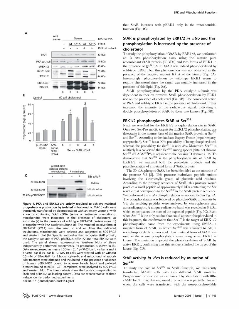
that StAR interacts with pERK1 only in the mitochondrial
fraction (Fig. 4C).
StAR is phosphorylated by ERK1/2 in vitro and this
phosphorylation is increased by the presence of
cholesterolTo study the phosphorylation of StAR by ERK1/2, we performed
an in vitro phosphorylation assay using the mature pure
recombinant StAR protein (30 kDa) and two forms of ERK1 in
the presence of [c-32P]ATP. StAR was indeed phosphorylated by
wild-type ERK1, but this phenomenon was not observed in the
presence of the inactive mutant K71A of the kinase (Fig. 5A).
Interestingly, phosphorylation by wild-type ERK1 seems to
require cholesterol since the signal was notably increased in the
presence of this lipid (Fig. 5A).
StAR phosphorylation by the PKA catalytic subunit was
dependent neither on previous StAR phosphorylation by ERK1
nor on the presence of cholesterol (Fig. 5B). The combined action
of PKA and wild-type ERK1 in the presence of cholesterol further
increased the intensity of the radioactive signal, indicating a
double phosphorylation of StAR by these two kinases (Fig. 5B).
ERK1/2 phosphorylates StAR at Ser232
Next, we searched for the ERK1/2 phosphorylation site in StAR.
Only two Ser-Pro motifs, targets for ERK1/2 phosphorylation, are
detectable in the mature form of the murine StAR protein at Ser232
and Ser277. According to the database Expasy Prosite (http://expasy.
org/prosite/), Ser232 has a 90% probability of being phosphorylated,
whereas the probability for Ser277 is only 5%. Moreover, Ser277 is
relatively less conserved than Ser232 among species (data not shown).
Ser232 (PLAGS232PS) is adjacent to the docking D domain (22). To
demonstrate that Ser232 is the phosphorylation site of StAR by
ERK1/2, we analyzed both the proteolytic products and the
phosphorylation of a mutated form of StAR protein.
The 30 kDa phospho-StAR has been identified as the substrate of
the protease V8 [8]. This protease hydrolyzes peptidic unions
involving the a-carboxylic group of glutamic acid residues.
According to the primary sequence of StAR, this protease would
produce a small peptide of approximately 6 kDa containing the Ser
residue that corresponds to the Ser232 in the StAR protein sequence.
We performed the in vitro phosphorylation assay described in Fig. 5A.
The phosphorylation was followed by phospho-StAR proteolysis by
V8; the resulting peptides were analyzed by electrophoresis and
autoradiography. A unique radioactive band was revealed at 6 kDa,
which encompasses the mass of the expected peptide (Fig. 5C). Even
when Ser232 is the only residue that could appear phosphorylated in
this fragment, the confirmation that Ser232 is the target of ERK1/2
phosphorylation came from the experiments using S232A, a
mutated form of StAR, in which Ser232 was changed to Ala, a
non-phosphorylable amino acid. This mutated form of StAR was
used in the in vitro phosphorylation assay using active ERK1 as
kinase. The mutation impeded the phosphorylation of StAR by
active ERK1, confirming that this residue is indeed the target of the
kinase (Fig. 5D).
StAR activity in vivo is reduced by mutation of
Ser232
To study the role of Ser232 in StAR function, we transiently
transfected MA-10 cells with two different StAR mutants.
Progesterone production was enhanced by stimulation with 8Br-
cAMP for 30 min; that enhanced production was partially blocked
when the cells were transfected with the non-phosphorylable
Figure 4. PKA and ERK1/2 are strictly required to achieve maximalprogesterone production by isolated mitochondria. MA-10 cells weretransiently transfected by electroporation with an empty vector or witha vector containing StAR cDNA (sense or antisense orientations).Mitochondria were incubated in the presence of cholesterol assubstrate (a) in the presence of wild type ERK1-GST protein alone (b)or together with PKA catalytic subunit (d). The mutated inactive form ofERK1-GST (K71A) was also used (c and e). After the indicatedincubations, mitochondria were pelleted and subjected to SDS-PAGEand Western blot (A). Specific antibodies that recognize StAR protein,the catalytic subunit of PKA, pMEK1/2, pERK1/2 and total ERK1/2 wereused. The panel shows representative Western blots of threeindependently performed experiments. P4 production is shown in (B).Data are expressed as means6SD (n = 3). * p,0.05 bar b vs. bar a and 1p,0.05 bar d vs. bar b. (C) MA-10 cells were treated with or without0.5 mM of 8Br-cAMP for 3 hours; cytosolic and mitochondrial subcel-lular fractions were obtained and incubated in the presence or absenceof human pERK1-GST bound to agarose beads. Input and pelletedproteins bound to pERK1-GST (complexes) were analyzed by SDS-PAGEand Western blot. The immunoblots show the bands corresponding toStAR and pERK1/2, as loading control. Data are representative of threeindependently performed experiments.doi:10.1371/journal.pone.0001443.g004
ERK and Mitochondrial Function
PLoS ONE | www.plosone.org 6 January 2008 | Issue 1 | e1443
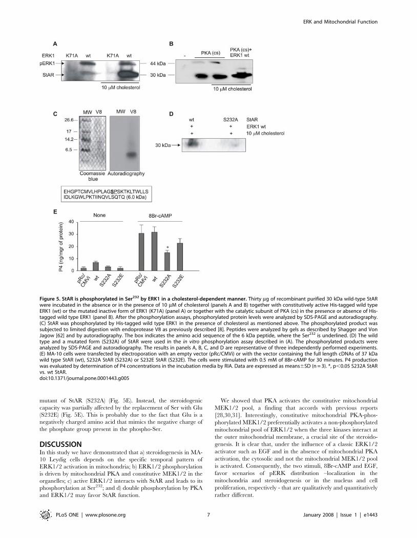
mutant of StAR (S232A) (Fig. 5E). Instead, the steroidogenic
capacity was partially affected by the replacement of Ser with Glu
(S232E) (Fig. 5E). This is probably due to the fact that Glu is a
negatively charged amino acid that mimics the negative charge of
the phosphate group present in the phospho-Ser.
DISCUSSIONIn this study we have demonstrated that a) steroidogenesis in MA-
10 Leydig cells depends on the specific temporal pattern of
ERK1/2 activation in mitochondria; b) ERK1/2 phosphorylation
is driven by mitochondrial PKA and constitutive MEK1/2 in the
organelles; c) active ERK1/2 interacts with StAR and leads to its
phosphorylation at Ser232; and d) double phosphorylation by PKA
and ERK1/2 may favor StAR function.
We showed that PKA activates the constitutive mitochondrial
MEK1/2 pool, a finding that accords with previous reports
[28,30,31]. Interestingly, constitutive mitochondrial PKA-phos-
phorylated MEK1/2 preferentially activates a non-phosphorylated
mitochondrial pool of ERK1/2 when the three kinases interact at
the outer mitochondrial membrane, a crucial site of the steroido-
genesis. It is clear that, under the influence of a classic ERK1/2
activator such as EGF and in the absence of mitochondrial PKA
activation, the cytosolic and not the mitochondrial MEK1/2 pool
is activated. Consequently, the two stimuli, 8Br-cAMP and EGF,
favor scenarios of pERK distribution –localization in the
mitochondria and steroidogenesis or in the nucleus and cell
proliferation, respectively - that are qualitatively and quantitatively
rather different.
Figure 5. StAR is phosphorylated in Ser232 by ERK1 in a cholesterol-dependent manner. Thirty mg of recombinant purified 30 kDa wild-type StARwere incubated in the absence or in the presence of 10 mM of cholesterol (panels A and B) together with constitutively active His-tagged wild typeERK1 (wt) or the mutated inactive form of ERK1 (K71A) (panel A) or together with the catalytic subunit of PKA (cs) in the presence or absence of His-tagged wild type ERK1 (panel B). After the phosphorylation assays, phosphorylated protein levels were analyzed by SDS-PAGE and autoradiography.(C) StAR was phosphorylated by His-tagged wild type ERK1 in the presence of cholesterol as mentioned above. The phosphorylated product wassubjected to limited digestion with endoprotease V8 as previously described [8]. Peptides were analyzed by gels as described by Shagger and VonJagow [62] and by autoradiography. The box indicates the amino acid sequence of the 6 kDa peptide, where the Ser232 is underlined. (D) The wildtype and a mutated form (S232A) of StAR were used in the in vitro phosphorylation assay described in (A). The phosphorylated products wereanalyzed by SDS-PAGE and autoradiography. The results in panels A, B, C, and D are representative of three independently performed experiments.(E) MA-10 cells were transfected by electroporation with an empty vector (pRc/CMVi) or with the vector containing the full length cDNAs of 37 kDawild type StAR (wt), S232A StAR (S232A) or S232E StAR (S232E). The cells were stimulated with 0.5 mM of 8Br-cAMP for 30 minutes. P4 productionwas evaluated by determination of P4 concentrations in the incubation media by RIA. Data are expressed as means6SD (n = 3). *, p,0.05 S232A StARvs. wt StAR.doi:10.1371/journal.pone.0001443.g005
ERK and Mitochondrial Function
PLoS ONE | www.plosone.org 7 January 2008 | Issue 1 | e1443

It was confirmed here that pERK1/2 in mitochondria have a
functional interaction with StAR, MEK1/2 and PKA, thus
forming a mitochondrial multi-complex. On the basis of
crystallographic analysis, acidic and hydrophobic patches in the
ERK1/2 structure (the CD domain) were described [46]. For
instance, in the model of interaction between a peptide that
belongs to MKP3 (MAP kinase phosphatase 3) and ERK2 [45],
Asp316 and Asp319 of the ERK2 CD domain were observed to
interact with Arg20 and Arg21 of the representative MKP3 peptide
(R20R21GSNVALML, the D domain), an interaction putatively
attributed in the present case to Lys235 and Lys237 of StAR
(K235TK237LTWLLSI). Based on a computerized model of the
ERK2-StAR complex (Fig. 6), we found a possible interaction
between the e-amine group of Lys235 in StAR structure and the
carboxylic group of Asp319 in ERK2 structure, separated by 11 A.
As in MKP3, other interactions of the hydrophobic motifs,
underlined in the partial sequences, are expected to stabilize the
StAR binding to the ERK docking groove [45].
We also confirmed that ERK1/2-StAR binding conducts to
StAR phosphorylation at Ser232. This residue integrates the classic
SP motif for phosphorylation of substrates by ERK1/2 [47]. In
StAR, the SP motif is adjacent to the binding domain to ERK1/2,
considerably augmenting the phosphorylating efficiency of the
kinase [48]. This case is similar to that of the classical ERK1/2
substrate Elk-1 [49], although the SP position is inverted in respect
to the docking domain. In addition, SP motifs susceptible to
phosphorylation by ERK1/2 have a Pro residue in position 21 or
22, whereas StAR has a conserved Pro at position 23. However,
as in other substrates, Pro is followed by Leu and there are no
acidic residues in the motif [47].
StAR phosphorylation by ERK1/2 is not dependent on
previous StAR phosphorylation by PKA but requires the presence
of cholesterol. Some have suggested that StAR is a molten globule
that changes its carboxyl-terminal helix when cholesterol
approaches the hydrophobic surface [50,51]. Cholesterol has
been shown to act as an allosteric modulator, facilitating further
binding of StAR to ligand [51]. In this case, the displacement of
the C termini of StAR by cholesterol may increase the exposure
of its docking domain to the ERK groove. The docking motif
containing Lys235 in the StAR sequence and Asp319 in the ERK
sequence constitute the closest interaction, separated by a short
distance, approximately 11 A (Fig. 6). This distance is not
enough; then, the conformational changes that cholesterol and
the disorganization of the carboxi terminal may provoke
would contribute to a closer localization between Ser232 and the
catalytic site of ERK, producing a more efficient phosphorylation.
It is known that after the approximation of the docking site, there
is a conformational change in the kinase that provokes the
approximation of the phosphorylable site to the catalytic center
[52].
Regarding the form of StAR protein that was subjected to in vitro
phosphorylation, several reasons explain the election of the
30 kDa protein. First, the bibliography is controversial, since
there is not a full explanation of StAR protein mitochondrial
import, maturation/activation and function. In this regard, there
is not conclusive evidence to assign the activity to either of the two
isoforms, 30 or 37 kDa. Second, in our pull down experiments,
described above, ERK1 specifically co-precipitates with the
30 kDa mitochondrial form of StAR protein, indicating that
interaction between ERK and this isoform is probably after StAR
protein cleavage. Thus, these facts support the concept that the
30 kDa form of StAR could be the substrate of ERK1/2.
Nevertheless, the transfection experiments, performed with a
mutated form of the full-length form of StAR (37 kDa), indicate
that being the 37 or the 30 kDa forms the substrate for ERK1/2,
the phosphorylation of StAR at Ser232 is necessary for its
steroidogenic activity.
The presence of a multi-protein complex in the outer
mitochondrial membrane has functional repercussions for ste-
roidogenesis. In this complex, phosphorylation of StAR by
ERK1/2 is a key process for cholesterol transport. It is known
that StAR works together with PBR which in turn interacts with
VDAC. However, little is known about how StAR interacts with
these proteins. In previous unpublished observations we detected
that StAR could interact with VDAC. It will be interesting to
further analyze the role of Ser232-StAR phosphorylation in the
assembly of this multiprotein complex and in its function. The
additional negative charges in the StAR (30 kDa) molecule due to
phosphorylation at Ser232 may contribute to the retention of the
mature form of 30 kDa StAR in the outer mitochondrial
membrane and to the formation of the multiprotein complex.
MATERIALS AND METHODS
Antibodies and reagentsPurified hCG was a kindly provided by Dr. Parlow (National
Hormone and Pituitary Program, National Institute of Diabetes &
Digestive & Kidney Diseases (NIDDK, NIH, Bethesda, MD,
USA). Anti-phospho-ERK1/2, anti-phospho-MEK1/2, anti-total
MEK1/2 antibodies and U0126 were obtained from Cell
Signaling (Beverly, MA, USA); anti-total ERK1/2 and anti-b-
tubulin were purchased from Upstate Biotechnology (Lake Placid,
NY, USA). Anti- 39 kDa subunit of the NADH-cytochorme c
reductase was obtained from Molecular Probes, Inc. (Eugene, OR,
USA). Anti-StAR was generously provided by Dr. Douglas Stocco
(Texas Tech University Health Sciences Center, Lubbock, TX,
USA). The antibody that recognizes the PKA a catalytic subunit
(Santa Cruz Biotechnology, CA, USA) was a generous gift from
Dr. Tellez Inon (INGEBI, Buenos Aires, Argentina). The antibody
against Acot2 was generated at our laboratory [53]. Wild-type and
Figure 6. Predictive model of the molecular interaction betweenERK1/2 and StAR. The reconstruction of molecular interaction of ERK2and StAR was performed using PyMOL (DeLano Scientific, USA;www.delanoscientific.com). ERK2 (Protein Data Bank code 2GPH) isrepresented in blue and StAR (START domain in StartD4 from Musmusculus) in green. In this model, StAR is located in the docking grooveof ERK2. The active center of ERK2 is in dark red. The CD domain ofERK2, represented in yellow, includes Asp316 and Asp319 in contact withthe D domain of StAR. Lys174 and Lys176, corresponding to Lys235 andLys237 of StAR sequence in Mus musculus are represented in orange, andSer171, corresponding to Ser232 of murine StAR, in dark pink.doi:10.1371/journal.pone.0001443.g006
ERK and Mitochondrial Function
PLoS ONE | www.plosone.org 8 January 2008 | Issue 1 | e1443
a catalytically inactive variant (K71A) of human ERK1 fused to
GST were purchased from Stressgen (Ann Arbor, MI, USA).
Wild-type ERK1-GST was activated in vitro in accordance with the
manufacturer’s instructions. Constitutively active His-tagged
ERK1 and PD98059 were purchased from Calbiochem (San
Diego, CA, USA). The PKA catalytic subunit was obtained from
New England Biolabs (Beverly, MA, USA). Site-directed muta-
genesis on StAR wild-type cDNA was performed on a pRc/CMVi
construction by GenScript (GenScript Corporation, Piscataway,
NJ, USA). The siRNA against the PKA a catalytic subunit was
obtained from Santa Cruz Biotechnology (Santa Cruz Biotech-
nology, CA, USA). The 36flag-CMV7-ERK2 plasmid containing
the wild-type or a mutated (H230R) variant of ERK2 was kindly
provided by Dr. Melanie H. Cobb (Department of Pharmacology,
University of Texas Southwestern Medical Center, Dallas, TX,
USA). All others reagents were commercial products of the highest
grade available.
Cell CultureThe MA-10 cell line is a clonal strain of mouse Leydig tumor cells
that produces progesterone (P4) rather than testosterone as the
major steroid [54]. MA-10 cells were generously provided by Dr.
Mario Ascoli, University of Iowa, College of Medicine (Iowa City,
IA, USA) and were handled as originally described [41,54].
Cell transfection and constructionsCell transfections were performed by electroporation or using a
cationic lipid reagent. MA-10 cells were transiently transfected by
electroporation in accordance with already published procedures
[41] with an empty vector or with a vector containing StAR cDNA
(sense or antisense orientations). The incorporation of the siRNA
of the a isoform of the catalytic subunit of PKA into the cells was
performed using Lipofectamine 2000 reagent (Invitrogen, Carls-
bad, CA, USA), according to previously used procedures [37].
Full length StAR cDNA (Gen Bank accession nu BC082283)
was obtained by PCR from MA-10 cells as described in Maloberti
et al. [37]. Primers were designed according to the published
sequence of mouse StAR. The forward (59-GGACCTT-
GAAAGGCTCAGGAAGAACAACCC-39) and the reverse (59-
GGATTAGTAGGGAAGTCGGCACAATGATGG-39) primers
were used to amplify a 1440-bp fragment. The sequence was
subcloned in the eukaryotic expression plasmid, pRc/CMVi, a gift
from Ingo Leibiger from Karolinska Institut, Stockholm, Sweden.
The ligation provided both sense and antisense orientations that
were used for transformation of XL-1 E. coli. Then, several clones
were screened and sense and antisense plasmids were obtained by
midipreparations with the WizardH system (Promega, Madison,
WI, USA); afterwards used for transfections of MA-10 cells.
Recombinant 30 kDA StAR protein was obtained as described
before [15]. Briefly, StAR cDNA for the mature form (30 kDa)
was obtained by PCR from MA-10 cells with the following
primers: the forward 59-GGATCCGCAGGGTGGATGGGT-
CAA-39 and the reverse 59-GGATTAGTAGGGAAGTCGGCA-
CAATGATGG-39 that amplify a 1200-pb fragment. Then, the
StAR cDNA was cloned in a pGEX-4T-3 plasmid (Promega,
Madison, WI, USA). The construction was used to transform BL-
21 E. coli, the recombinant protein GST-StAR was purified by
glutathione affinity chromatography (GST Purification Module,
Amersham Biosciences, Sweden) and subjected to thrombin
proteolytic cleavage. Then, in vitro phosphorylation experiments
were performed with 30 kDa StAR. The same protocol was
followed to obtain a mutated form of the StAR protein (S232A)
using the site-directed mutated StAR cDNA.
Subcellular fractionationSubcellular fractionation was performed as described previously;
nuclear, cytosolic and mitochondrial fractions from MA-10 cells
were isolated by differential centrifugation [22,34,55]. A detailed
description of the procedure is the following. After the corre-
sponding treatments, culture media was removed, MA-10 cells
were washed with PBS supplemented with protease and
phosphatase inhibitors and collected by centrifugation at 8006g
for 10 minutes. MA-10 cells were resuspended in MSHE buffer
(219 mM D-mannitol, 70 mM sucrose, 0.02% EGTA, 0,1% BSA,
1.8 mM Hepes pH 7.4) and subjected to mechanical disruption
with 60 strokes with an insulin syringe. Then, nuclear fraction was
obtained by centrifugation at 50006g for 10 minutes. The
supernatant is centrifuged at 150006g for 30 minutes. Then, the
pellet corresponding to the mitochondrial fraction was washed
once and resuspended in MSHE buffer and supernatant
corresponds to cytosolic fraction. Fractions were subjected to
enzymatic analysis to assess their purity (according to [34]). The
purity of each fraction was at least 90%, value similar to previous
publications [56].
Cell-free assay for mitochondrial steroidogenesisAfter transfections, MA-10 cells were subjected to subcellular
fractionation in order to obtain mitochondria. The organelles were
resuspended in 10 mM of malate, 1 mM of Mg2Cl, 50 mM of
ATP, 40 mM of b-Glicerophosphate, 1 mM of DTT. Stimulation
of mitochondrial steroidogenesis was performed according to
published procedures [22,55]. Mitochondria were incubated in the
absence or presence of 50 mM of cholesterol as substrate for
20 minutes at 30uC. Cholesterol supplementation was performed
in the presence or absence of 1 mg of wild- type ERK1-GST
protein alone or together with 1 IU of PKA catalytic subunit. The
mutated inactive form of ERK1 (K71A) was also used. The K71A
mutant of ERK1 was described by Charest et al. [42]. The change
in one aminoacidic residue abolishes the autophosphorylation
activity of the kinase and the myelin basic protein phosphotrans-
ferase activity. While this mutated form is able to be phosphor-
ylated by MEK1/2 to approximately 80% of the level achieved
with its non-mutated form, the result is a partial activation of the
myelin basic protein phosphotransferase activity (20%). After the
indicated incubations, P4 production in the incubation media was
determined by radioimmunoassay [41] while mitochondrial
protein content was analyzed by SDS-PAGE and Western blot.
Pull-down assayTo study interaction between StAR protein and pERK, pull-down
assays were carried out as indicated in Hirakawa and Ascoli [28].
Briefly, cytosolic or mitochondrial fractions (500 mg or 300 mg,
respectively) were incubated in the presence of human recombi-
nant ERK1-GST coupled to agarose beads, previously activated
as described by the manufacturer. The incubations were
performed in pull down buffer; 50 mM Tris pH 7.4, 150 mM
NaCl, 1 mM EDTA, 1 mM EGTA, 10% glycerol, 0.5% Nonidet
P-40, 1 mM MgCl2 supplemented with protease and phosphatases
inhibitors overnight at 4uC. Afterwards, proteins bound to ERK1-
GST contained in the agarose-protein pellet (complexes) were
washed three times with the same buffer, boiled for 5 minutes,
subjected to SDS-PAGE and immunoblot analysis.
In vitro phosphorylation of recombinant StARPhosphorylation assays of wild-type or S232A StAR were
performed with wild-type His-tagged ERK1 or the catalytically
inactive form of ERK1-GST (K71A) and PKA catalytic subunit.
ERK and Mitochondrial Function
PLoS ONE | www.plosone.org 9 January 2008 | Issue 1 | e1443
The assays were performed with 10 mCi of [c-32P]ATP for
30 minutes at 30uC as described previously [49,57].
SDS-PAGE and immunoblot assayProteins were separated by SDS-PAGE and electrotransferred to
poly-(vinylidine difluoride) (PDVF) membranes as previously
described [58]. Immunodetection was performed using the specific
antibodies described in the figure legends. Antibodies recognizing
pERK and total ERK were used sequentially in the same
membrane after treatment of the blots with stripping buffer
(62.5 mM Tris, pH 6.8, 100 mM 2-mercaptoethanol and 2%
SDS) for 30 minutes at 60uC. The same statement is valid for
pMEK and total MEK recognition. In the western blots evaluating
pMEK1/2 content, total MEK1/2 and other proteins were used
as loading control: a mitochondrial acyl-CoA thioesterase for 8Br-
cAMP treatment as previously described [41], the mitochondrial
39 kDa subunit of the NADH-cytochrome c reductase (complex I)
for EGF treatment, and cytosolic b-tubulin in both cases. Proteins
were visualized using horseradish peroxidase-coupled secondary
antibodies followed by the ECL chemiluminescence detection
system (Amersham Pharmacia Biotech, Piscataway, NJ, USA) and
X-ray film exposure. For quantitative analysis, densitometry was
performed using a Storm Phosphorimager scanner (Amersham
Biosciences, Sweden) and band intensities were analyzed using
ImageQuant 5.2 software.
Immunofluorescence analysisMA-10 cells were grown to approximately 80% confluence on poly-
L-lysine glass coverslips. After treatments, cells were incubated for
45 minutes at 36.5uC with 300 nM Mitotracker RedH (Molecular
Probes, Inc., Eugene, OR, USA), a specific mitochondrial dye.
Then, cells were fixed with 4% paraformaldehyde in PBS for
10 minutes at room temperature and permeabilized with blocking
solution (0.3% Triton X-100 and 1% BSA in PBS) for 60 minutes at
room temperature. The detailed procedure was described previously
[59]. Cells were incubated with anti-pERK1/2 cy2-conjugated
antibody (1:250) overnight at 4uC. After several washes with PBS,
cells were incubated for 1h at room temperature with a goat anti-
rabbit antibody (1:400). Coverslips were mounted onto the slides
using Fluorsave antifade reagent (Calbiochem), followed by confocal
analysis using a Nikon C1 microscope (IByME, UBA, Argentina).
Statistical analysisData were analyzed by ANOVA followed by the Dunnet test.
ACKNOWLEDGMENTS
Author Contributions
Conceived and designed the experiments: EP JP. Performed the
experiments: CP DC PM AD IN SG FC MC. Analyzed the data: EP
CP DC PM AD IN SG FC CP MC JP. Contributed reagents/materials/
analysis tools: EP JP. Wrote the paper: EP CP FC MC JP.
REFERENCES1. Crivello JF, Jefcoate CR (1980) Intracellular movement of cholesterol in rat
adrenal cells. Kinetics and effects of inhibitors. J Biol Chem 255: 8144–8151.
2. Privalle CT, Crivello JF, Jefcoate CR (1983) Regulation of intramitochondrial
cholesterol transfer to side-chain cleavage cytochrome P-450 in rat adrenal
gland. Proc Natl Acad Sci U S A 80: 702–706.
3. McEnery MW, Snowman AM, Trifiletti RR, Snyder SH (1992) Isolation of the
mitochondrial benzodiazepine receptor: association with the voltage-dependent
anion channel and the adenine nucleotide carrier. Proc Natl Acad Sci U S A 89:
3170–3174.
4. Papadopoulos V, Baraldi M, Guilarte TR, Knudsen TB, Lacapere JJ, et al.
(2006) Translocator protein (18 kDa): new nomenclature for the peripheral-type
benzodiazepine receptor based on its structure and molecular function. Trends
Pharmacol Sci 27: 402–409.
5. Liu J, Rone MB, Papadopoulos V (2006) Protein-Protein Interactions Mediate
Mitochondrial Cholesterol Transport and Steroid Biosynthesis. J Biol Chem
281: 38879–38893.
6. Clark BJ, Wells J, King SR, Stocco DM (1994) The purification, cloning, and
expression of a novel luteinizing hormone-induced mitochondrial protein in
MA-10 mouse Leydig tumor cells. Characterization of the steroidogenic acute
regulatory protein (StAR). J Biol Chem 269: 28314–28322.
7. Bose H, Lingappa VR, Miller WL (2002) Rapid regulation of steroidogenesis by
mitochondrial protein import. Nature 417: 87–91.
8. Krueger RJ, Orme-Johnson NR (1983) Acute adrenocorticotropic hormone
stimulation of adrenal corticosteroidogenesis. Discovery of a rapidly induced
protein. J Biol Chem 258: 10159–10167.
9. Stocco DM, Sodeman TC (1991) The 30-kDa mitochondrial proteins induced
by hormone stimulation in MA-10 mouse Leydig tumor cells are processed from
larger precursors. J Biol Chem 266: 19731–19738.
10. Yamazaki T, Matsuoka C, Gendou M, Izumi S, Zhao D, et al. (2006)
Mitochondrial processing of bovine adrenal steroidogenic acute regulatory
protein. Biochim Biophys Acta 1764: 1561–1567.
11. Epstein LF, Orme-Johnson NR (1991) Regulation of steroid hormone
biosynthesis. Identification of precursors of a phosphoprotein targeted to the
mitochondrion in stimulated rat adrenal cortex cells. J Biol Chem 266:
19739–19745.
12. Miller WL (2007) Mechanism of StAR’s regulation of mitochondrial cholesterol
import. Mol Cell Endocrinol 265–266: 46–50.
13. Arakane F, Sugawara T, Nishino H, Liu Z, Holt JA, et al. (1996) Steroidogenic
acute regulatory protein (StAR) retains activity in the absence of its
mitochondrial import sequence: implications for the mechanism of StAR action.
Proc Natl Acad Sci U S A 93: 13731–13736.
14. Soltys BJ, Gupta RS (1999) Mitochondrial-matrix proteins at unexpected
locations: are they exported? Trends Biochem Sci 24: 174–177.
15. Artemenko IP, Zhao D, Hales DB, Hales KH, Jefcoate CR (2001)
Mitochondrial processing of newly synthesized steroidogenic acute regulatory
protein (StAR), but not total StAR, mediates cholesterol transfer to cytochrome
P450 side chain cleavage enzyme in adrenal cells. J Biol Chem 276:
46583–46596.
16. Allen JA, Shankara T, Janus P, Buck S, Diemer T, et al. (2006) Energized,
Polarized, and Actively Respiring Mitochondria Are Required for Acute Leydig
Cell Steroidogenesis. Endocrinology 147: 3924–3935.
17. Duarte A, Castillo AF, Castilla R, Maloberti P, Paz C, et al. (2007) An
arachidonic acid generation/export system involved in the regulation of
cholesterol transport in mitochondria of steroidogenic cells. Febs Letters 581:
4023–4028.
18. Wang X, Liu Z, Eimerl S, Timberg R, Weiss AM, et al. (1998) Effect of
truncated forms of the steroidogenic acute regulatory protein on intramitochon-
drial cholesterol transfer. Endocrinology 139: 3903–3912.
19. Stocco DM, Clark BJ (1996) Regulation of the acute production of steroids in
steroidogenic cells. Endocr Rev 17: 221–244.
20. Stocco DM, Wang X, Jo Y, Manna PR (2005) Multiple signaling pathways
regulating steroidogenesis and steroidogenic acute regulatory protein expression:
more complicated than we thought. Mol Endocrinol 19: 2647–2659.
21. Fleury A, Mathieu AP, Ducharme L, Hales DB, LeHoux JG (2004)
Phosphorylation and function of the hamster adrenal steroidogenic acute
regulatory protein (StAR). J Steroid Biochem Mol Biol 91: 259–271.
22. Neher R, Milani A, Solano AR, Podesta EJ (1982) Compartmentalization of
corticotropin-dependent steroidogenic factors in adrenal cortex: evidence for a
post-translational cascade in stimulation of the cholesterol side-chain split. Proc
Natl Acad Sci U S A 79: 1727–1731.
23. Podesta EJ, Dufau ML, Catt KJ (1976) Adenosine 39,59-monophosphate-
dependent protein kinase of Leydig cells: in vitro activation and relationship to
gonadotropin action upon cyclic AMP and steroidogenesis. FEBS Lett 70:
212–216.
24. Sala GB, Hayashi K, Catt KJ, Dufau ML (1979) Adrenocorticotropin action in
isolated adrenal cells. The intermediate role of cyclic AMP in stimulation of
corticosterone synthesis. J Biol Chem 254: 3861–3865.
25. Rae PA, Gutmann NS, Tsao J, Schimmer BP (1979) Mutations in cyclic AMP-
dependent protein kinase and corticotropin (ACTH)-sensitive adenylate cyclase
affect adrenal steroidogenesis. Proc Natl Acad Sci U S A 76: 1896–1900.
26. Cooke BA, Lindh ML, Janszen FH (1976) Correlation of protein kinase
activation and testosterone production after stimulation of Leydig cells with
luteinizing hormone. Biochem J 160: 439–446.
27. Gyles SL, Burns CJ, Whitehouse BJ, Sugden D, Marsh PJ, et al. (2001) ERKs
regulate cyclic AMP-induced steroid synthesis through transcription of the
steroidogenic acute regulatory (StAR) gene. J Biol Chem 276: 34888–34895.
ERK and Mitochondrial Function
PLoS ONE | www.plosone.org 10 January 2008 | Issue 1 | e1443
28. Hirakawa T, Ascoli M (2003) The lutropin/choriogonadotropin receptor-
induced phosphorylation of the extracellular signal-regulated kinases in leydig
cells is mediated by a protein kinase a-dependent activation of ras. Mol
Endocrinol 17: 2189–2200.
29. Manna PR, Jo Y, Stocco DM (2007) Regulation of Leydig cell steroidogenesis by
extracellular signal-regulated kinase 1/2: role of protein kinase A and protein
kinase C signaling. J Endocrinol 193: 53–63.
30. Martinelle N, Holst M, Soder O, Svechnikov K (2004) Extracellular Signal-
Regulated Kinases Are Involved in the Acute Activation of Steroidogenesis in
Immature Rat Leydig Cells by Human Chorionic Gonadotropin. Endocrinology
145: 4629–4634.
31. Seger R, Hanoch T, Rosenberg R, Dantes A, Merz WE, et al. (2001) The ERK
Signaling Cascade Inhibits Gonadotropin-stimulated Steroidogenesis. J Biol
Chem 276: 13957–13964.
32. Otis M, Gallo-Payet N (2007) Role of MAPKs in angiotensin II-induced
steroidogenesis in rat glomerulosa cells. Mol Cell Endocrinol 265–266: 126–130.
33. Baines CP, Zhang J, Wang GW, Zheng YT, Xiu JX, et al. (2002) Mitochondrial
PKCepsilon and MAPK form signaling modules in the murine heart: enhanced
mitochondrial PKCepsilon-MAPK interactions and differential MAPK activa-
tion in PKCepsilon-induced cardioprotection. Circ Res 90: 390–397.
34. Alonso M, Melani M, Converso D, Jaitovich A, Paz C, et al. (2004)
Mitochondrial extracellular signal-regulated kinases 1/2 (ERK1/2) are modu-
lated during brain development. J Neurochem 89: 248–256.
35. Gyles SL, Burns CJ, Persaud SJ, Jones PM, Whitehouse BJ (2000) A role for the
p42/44 isoforms of MAPK in the regulation of steroid secretion from Y1 mouse
adrenocortical cells. Endocr Res 26: 579–581.
36. Renlund N, Jo Y, Svechnikova I, Holst M, Stocco DM, et al. (2006) Induction of
steroidogenesis in immature rat Leydig cells by interleukin-1alpha is dependent
on extracellular signal-regulated kinases. J Mol Endocrinol 36: 327–336.
37. Maloberti P, Castilla R, Castillo F, Maciel FC, Mendez CF, et al. (2005)
Silencing the expression of mitochondrial acyl-CoA thioesterase I and acyl-CoA
synthetase 4 inhibits hormone-induced steroidogenesis. Febs J 272: 1804–1814.
38. Shiraishi K, Ascoli M (2006) Activation of the lutropin/choriogonadotropin
receptor in MA-10 cells stimulates tyrosine kinase cascades that activate ras and
the extracellular signal regulated kinases (ERK1/2). Endocrinology 147:
3419–3427.
39. Robinson FL, Whitehurst AW, Raman M, Cobb MH (2002) Identification of
novel point mutations in ERK2 that selectively disrupt binding to MEK1. J Biol
Chem 277: 14844–14852.
40. Ascoli M, Euffa J, Segaloff DL (1987) Epidermal growth factor activates steroid
biosynthesis in cultured Leydig tumor cells without affecting the levels of cAMP
and potentiates the activation of steroid biosynthesis by choriogonadotropin and
cAMP. J Biol Chem 262: 9196–9203.
41. Castillo F, Cornejo Maciel F, Castilla R, Duarte A, Maloberti P, et al. (2006)
cAMP increases mitochondrial cholesterol transport through the induction of
arachidonic acid release inside this organelle in MA-10 Leydig cells. Febs J 273:
5011–5021.
42. Charest DL, Mordret G, Harder KW, Jirik F, Pelech SL (1993) Molecular
cloning, expression, and characterization of the human mitogen-activated
protein kinase p44erk1. Mol Cell Biol 13: 4679–4690.
43. Hengen PN (1996) Methods and reagents. Purification of GST fusion proteins.
Trends Biochem Sci 21: 400–401.
44. Granot Z, Melamed-Book N, Bahat A, Orly J (2007) Turnover of StAR protein:
Roles for the proteasome and mitochondrial proteases. Molecular and Cellular
Endocrinology 265–266: 51–58.
45. Zhou T, Sun L, Humphreys J, Goldsmith EJ (2006) Docking interactions induce
exposure of activation loop in the MAP kinase ERK2. Structure 14: 1011–1019.46. Xu B, Stippec S, Robinson FL, Cobb MH (2001) Hydrophobic as well as
charged residues in both MEK1 and ERK2 are important for their proper
docking. J Biol Chem 276: 26509–26515.47. Gonzalez FA, Raden DL, Davis RJ (1991) Identification of substrate recognition
determinants for human ERK1 and ERK2 protein kinases. J Biol Chem 266:22159–22163.
48. Bardwell AJ, Abdollahi M, Bardwell L (2003) Docking sites on mitogen-activated
protein kinase (MAPK) kinases, MAPK phosphatases and the Elk-1 transcriptionfactor compete for MAPK binding and are crucial for enzymic activity.
Biochem J 370: 1077–1085.49. Fantz DA, Jacobs D, Glossip D, Kornfeld K (2001) Docking sites on substrate
proteins direct extracellular signal-regulated kinase to phosphorylate specificresidues. J Biol Chem 276: 27256–27265.
50. Miller WL (2007) StAR search–what we know about how the steroidogenic
acute regulatory protein mediates mitochondrial cholesterol import. MolEndocrinol 21: 589–601.
51. Petrescu AD, Gallegos AM, Okamura Y, Strauss JF 3rd, Schroeder F (2001)Steroidogenic acute regulatory protein binds cholesterol and modulates
mitochondrial membrane sterol domain dynamics. J Biol Chem 276:
36970–36982.52. Chen Z, Gibson TB, Robinson F, Silvestro L, Pearson G, et al. (2001) MAP
kinases. Chem Rev 101: 2449–2476.53. Maloberti P, Lozano RC, Mele PG, Cano F, Colonna C, et al. (2002) Concerted
regulation of free arachidonic acid and hormone-induced steroid synthesis byacyl-CoA thioesterases and acyl-CoA synthetases in adrenal cells. Eur J Biochem
269: 5599–5607.
54. Ascoli M (1981) Characterization of several clonal lines of cultured Leydig tumorcells: gonadotropin receptors and steroidogenic responses. Endocrinology 108:
88–95.55. Podesta EJ, Milani A, Steffen H, Neher R (1979) Adrenocorticotropin (ACTH)
induces phosphorylation of a cytoplasmic protein in intact isolated adrenocor-
tical cells. Proc Natl Acad Sci U S A 76: 5187–5191.56. Stocco DM (1983) Rapid, quantitative isolation of mitochondria from rat liver
using Ficoll gradients in vertical rotors. Anal Biochem 131: 453–457.57. Arakane F, King SR, Du Y, Kallen CB, Walsh LP, et al. (1997) Phosphorylation
of steroidogenic acute regulatory protein (StAR) modulates its steroidogenicactivity. J Biol Chem 272: 32656–32662.
58. Towbin H, Staehelin T, Gordon J (1979) Electrophoretic transfer of proteins
from polyacrylamide gels to nitrocellulose sheets: procedure and someapplications. Proc Natl Acad Sci U S A 76: 4350–4354.
59. Colonna C, Podesta EJ (2005) ACTH-induced caveolin-1 tyrosine phosphor-ylation is related to podosome assembly in Y1 adrenal cells. Exp Cell Res 304:
432–442.
60. Cornejo Maciel F, Poderoso C, Gorostizaga A, Paz C, Podesta EJ (2001) LH/chorionic gonadotropin signaling pathway involves protein tyrosine phosphatase
activity downstream of protein kinase A activation: evidence of an obligatorystep in steroid production by Leydig cells. J Endocrinol 170: 403–411.
61. Paz C, Cornejo Maciel F, Maloberti P, Walsh LP, Stocco DM, et al. (2002)Protein tyrosine phosphatases are involved in LH/chorionic gonadotropin and
8Br-cAMP regulation of steroidogenesis and StAR protein levels in MA-10
Leydig cells. J Endocrinol 175: 793–801.62. Schagger H, von Jagow G (1987) Tricine-sodium dodecyl sulfate-polyacrylamide
gel electrophoresis for the separation of proteins in the range from 1 to 100 kDa.Anal Biochem 166: 368–379.
ERK and Mitochondrial Function
PLoS ONE | www.plosone.org 11 January 2008 | Issue 1 | e1443