Effects of smooth and rough Pasteurella haemolytica lipopolysaccharides on plasma cyclic-nucleotides...
14
Veterinary Microbiology, 15 (1987) 279-292 279 Elsevier Science Publishers B.V., Amsterdam -- Printed in The Netherlands Effects of Smooth and Rough PasteureUa haemolytica Lipopolysaccharides on Plasma Cyclic-Nucleotides and Free Fatty Acids in Calves PETER EMAU l, SHRI N. GIRI"" and MICHAEL L. BRUSS ~ Departments of ~ Veterinary Pharmacology and Toxicology and "2physiological Sciences, School of Veterinary Medicine, University of California, Davis, CA 95616 (U.S.A.) (Accepted for publication 18 May 1987) ABSTRACT Emau, P., Giri, S.N. and Bruss, M.L., 1987. Effects of smooth and rough Pasteurella haemolytica lipopolysaccharides on plasma cyclic-nucleotides and free fatty acids in calves. Vet. Microbiol., 15: 279-292. The present study examined the potency of smooth or rough PasteureUa haemolytica lipopo- lysaccharide infusion (LPS, 24 ng kg-1 min-~ for 500 min) on plasma cyclic-nucleotides and several free fatty acids (FFA) in calves. Both smooth or rough LPS increased plasma cAMP immediately to its maximum at 1 h of infusion, whereas plasma cGMP levels rose slowly and peaked 12 h later. The increases in cAMP levels were more prolonged for smooth LPS than rough LPS. The maximum plasma cAMP rise coincided with increases of several plasma FFA. Rough LPS increased plasma oleic > palmitic > stearic > linoleic acids in the second hour and reached their steady state levels between 2 h of infusion and 5 h post-infusion. Thereafter, oleic acid remained maximally elevated, while stearic acid decreased and other FFA returned to baseline. Smooth LPS had no effects on palmitic and stearic acids, but elevated oleic acid in an essentially similar manner to rough LPS and increased linoleic acid initially at 5 h, followed by decreases throughout post-infusion. These results demonstrate that endotoxemia produces early marked elevations in plasma cAMP, a gradual rise in plasma cGMP and disproportionate increases in several plasma FFA. The data also demonstrate that smooth and rough LPS differ in their abilities to increase plasma cAMP and FFA and these may be attributed to differences in their in vivo mechanisms of action. The study suggests that cAMP and cGMP may mediate actions of endo- toxin at the cellular level and that differences exist in release and/or utilization of each FFA at different stages of endotoxemia. INTRODUCTION Endotoxins from PasteureUa haemolytica have been implicated in the path- ogenesis of bovine pneumonic pasteurellosis {shipping fever pneumonia) "Author to whom all correspondence should be addressed. 0378-1135/87/$03.50 © 1987 Elsevier Science Publishers B.V.
-
Upload
independent -
Category
Documents
-
view
6 -
download
0
Transcript of Effects of smooth and rough Pasteurella haemolytica lipopolysaccharides on plasma cyclic-nucleotides...
Veterinary Microbiology, 15 (1987) 279-292 279 Elsevier Science Publishers B.V., Amsterdam - - Printed in The Netherlands
Effects of Smooth and Rough PasteureUa haemolytica Lipopolysaccharides on Plasma Cycl ic-Nucleot ides and Free Fatty Acids in Calves
PETER EMAU l, SHRI N. GIRI"" and MICHAEL L. BRUSS ~
Departments of ~ Veterinary Pharmacology and Toxicology and "2physiological Sciences, School of Veterinary Medicine, University of California, Davis, CA 95616 (U.S.A.)
(Accepted for publication 18 May 1987)
ABSTRACT
Emau, P., Giri, S.N. and Bruss, M.L., 1987. Effects of smooth and rough Pasteurella haemolytica lipopolysaccharides on plasma cyclic-nucleotides and free fatty acids in calves. Vet. Microbiol., 15: 279-292.
The present study examined the potency of smooth or rough PasteureUa haemolytica lipopo- lysaccharide infusion (LPS, 24 ng kg-1 min-~ for 500 min) on plasma cyclic-nucleotides and several free fatty acids (FFA) in calves. Both smooth or rough LPS increased plasma cAMP immediately to its maximum at 1 h of infusion, whereas plasma cGMP levels rose slowly and peaked 12 h later. The increases in cAMP levels were more prolonged for smooth LPS than rough LPS. The maximum plasma cAMP rise coincided with increases of several plasma FFA. Rough LPS increased plasma oleic > palmitic > stearic > linoleic acids in the second hour and reached their steady state levels between 2 h of infusion and 5 h post-infusion. Thereafter, oleic acid remained maximally elevated, while stearic acid decreased and other FFA returned to baseline. Smooth LPS had no effects on palmitic and stearic acids, but elevated oleic acid in an essentially similar manner to rough LPS and increased linoleic acid initially at 5 h, followed by decreases throughout post-infusion. These results demonstrate that endotoxemia produces early marked elevations in plasma cAMP, a gradual rise in plasma cGMP and disproportionate increases in several plasma FFA. The data also demonstrate that smooth and rough LPS differ in their abilities to increase plasma cAMP and FFA and these may be attributed to differences in their in vivo mechanisms of action. The study suggests that cAMP and cGMP may mediate actions of endo- toxin at the cellular level and that differences exist in release and/or utilization of each FFA at different stages of endotoxemia.
INTRODUCTION
Endotoxins from PasteureUa haemolytica have been implicated in the path- ogenesis of bovine pneumonic pasteurellosis {shipping fever pneumonia)
"Author to whom all correspondence should be addressed.
0378-1135/87/$03.50 © 1987 Elsevier Science Publishers B.V.
280
(Lillie, 1974; Yates, 1982). Endotoxins, the lipopolysaccharide (LPS) of gram- negative bacterial cell walls, produce a variety of adverse reactions such as cardiopulmonary hemodynamic dysfunctions and lethal shock (Rietschel et al., 1982 ). The LPS also exert a marked influence on host metabolism, includ- ing altered lipid metabolism { Spitzer and Spitzer, 1982). Lipid metabolism is important in gram-negative sepsis and endotoxemia because lipids, predomi- nantly in the form of plasma free fatty acids (FFA), constitute the most im- portant energy source since carbohydrate reserves are limited (Daniel et al., 1983). In addition, fatty acids form structural and functional components of cell membranes (Grossman et al., 1980, Nordenstrom et al., 1983).
In many pathological conditions involving stress and decreased food intake, FFA mobilization is stimulated (Vernon, 1981 ). However, endotoxemia causes an increase followed by a decrease in lipolytic patterns of adipocytes (Spitzer and Fish, 1986) and produces increases and decreases in total plasma FFA {Kaufman et al., 1976; Spitzer and Spitzer, 1982; Gaal et al., 1984). Differ- ences are known to exist in the adipose tissue release and tissue utilization of different FFA under various pathological conditions (Thompson and Darling, 1975; Vernon, 1981; Colombo and Pinelli, 1982). There is a need, therefore, to examine the effects of endotoxin on individual plasma FFA in order to gain further understanding of fat metabolism in endotoxemia and bacterial sepsis.
The major source for plasma FFA is adipose tissue from which their release is regulated by the cAMP-dependent activation of hormone-sensitive lipase (Vernon, 1981). Endotoxin induces intracellular accumulation of cAMP in adipocytes and enhances catecholamine-FFA release from adipocytes ( Spitzer and Fish, 1986). Cyclic-nucleotides are also involved in other effects due to endotoxin, such as fever (Lin et al., 1982), abortion (Shaw and Cameron, 1979), pulmonary injury (Snapper et al., 1983) and regulation of activities of macrophages (Wahl et al., 1979), lymphocytes { Watson, 1975) and of hema- topoietic stem cells (Graber and Clancey, 1985). Cyclic-nucleotides regulate a variety of cellular processes, including mediation of the actions of several hormones and eicosanoids which are released during endotoxemia (Hamet et al., 1984). It is likely that many effects of endotoxin, including alterations in lipid metabolism, are mediated by cyclic-nucleotides.
The LPS molecule consists of three parts: Lipid A, core polysaccharide and long-chain polysaccharide {O-antigen). Considerable variation exists in the polysaccharide contents of LPS found on gram-negative bacteria (Rietschel et al., 1982). Some LPS have no O-antigen, but are made of Lipid A and core polysaccharide (KDO + ) { i.e., rough LPS); others have abundant O-antigen polysaccharide { i.e., smooth LPS) (Luderitz et al., 1966; Goldman and Leive, 1980; Rimsay et al., 1981). A single strain and species of gram-negative bac- teria may contain different proportions of both smooth and rough LPS (Gold- man and Leive, 1980); in P. haemolytica, rough LPS constitutes 1.5% and
281
smooth LPS 8% of dry cells ( Rimsay et al., 1981 ). Although Lipid A is consid- ered responsible for almost all the biological actions of LPS, variation in the ratio of Lipid A to polysaccharide in LPS has a significant influence on the cellular mechanisms and subsequent toxicity of endotoxins and gram-negative bacterial sepsis (Nakano, 1962; Luderitz et al., 1966; Morrison, 1983).
A partially purified P. haemolytica endotoxin was found to produce severe cardiopulmonary hemodynamic toxicity in sheep, comparable to endotoxins from other gram-negative bacteria (Keiss et al., 1964). Recently, we demon- strated that smooth LPS from P. haemolytica elevated circulating prostanoids (Emau et al., 1984) and produced marked changes in carbohydrate metabo- lism in sheep (Bruss et al., 1983 ). To examine further the effects of endotoxins from P. haemolytica, the slow intravenous infusions of the same dose of smooth and rough P. haemolytica LPS was carried out in calves for two purposes: first, to characterize more precisely the changes in plasma cyclic nucleotides and individual FFA during different stages of endotoxemia; second, to find out whether or not differences exist in the effects of smooth and rough LPS on plasma cyclic-nucleotides and FFA.
MATERIALS AND METHODS
Calves
Eleven male Holstein calves 100-200 kg (2-6 months old) were prepared for endotoxin infusion experiments as follows: 1 day before each experiment a calf was secured in a stanchion and using aseptic techniques, the right and left jugular veins were catheterized with vinyl catheters: one for endotoxin infusion and the other for blood sampling. The calves were able to stand up or lie down freely and had access to food and water ad libitum. Food consisted of standard alfalfa pellets provided by Animal Resource Services, University of California, Davis, to meet daily nutritional requirements.
Pasteurella haemolytica lipopolysaccharides
The smooth and rough LPS used in the present study were prepared from P. hemolytica type A1 using the procedures described by Rimsay et al. (1981) and were provided for us by Dr. P. Squire (Colorado State University, Fort Collins, CO). Rough LPS was extracted and purified using a mixture of phenol:chloroform:petroleum ether (2:5:8), a procedure that is specific for the isolation of LPS from rough mutants of bacteria (Galanos et al., 1969). The LPS obtained is rich in Lipid A (i.e., > 70%). Smooth LPS was purified by the method of Westphal and Jann (1965) which utilizes hot phenol-water mixture. The LPS extracted by this procedure has long-chain polysaccharide bound to Lipid A. The purity of the extracted LPS was checked by gel diffusion.
282
The extracts of LPS from P. haemolytica and commercial pure rough and smooth LPS from Salmonella sp. and Escherichia coli were run on gel electro- phoresis, so that the mobility and the number of bands were used to indicate puri ty of LPS. In the present studies, smooth LPS was reconsti tuted in sterile pyrogen-free 0.9% NaC1 while rough LPS was prepared in 0.9% NaC1 contain- ing 0.5% sodium oleate the night before use.
Infusions of LPS and blood sampling
At least 18-24 h after the preparatory procedures, smooth LPS was infused intravenously to calves in Group I ( N = 5) and rough LPS was administered by the same route to calves in Group II ( N - - 6 ) . In each case, the rate of in- fusion was 24 ng kg -1 min -1 for 500 min, to provide a total dose 12 ]~g kg -~ The rationale for the dosage was to create a continuous non-lethal endotoxi- cosis in order to simulate endotoxin release during bacterial sepsis. Previous experiments had est imated that 12 ttg kg-~ is the approximate L D~ in calves and sheep for smooth and rough LPS from P. haemolytica (unpublished). Blood ( 35 ml ) was drawn from the jugular vein into heparinized syringes at 60 min,
just prior to LPS infusions and at various intervals during and after the infu- sions. For determinat ion of FFA, blood (8 ml) was immediately distributed into ice-cold tubes. Another aliquot (3 ml) of blood was transferred into an ice-cold tube containing theophylline sulfate (10 mg) and used for analysis of cyclic-nucleotides. The blood samples were centrifuged at 4 ° C, 1100 X g for 20 min and plasma was harvested carefully, avoiding the buffy coat. Plasma (3 ml) samples for assays of FFA were immediately treated with a gentle stream of nitrogen. The plasma samples (1.0 ml) for analysis of cyclic-nucleotides were acidified with an equal volume of 20% trichloroacetic acid (TCA). Soon after t reatment , all plasma samples were stored frozen ( - 20 ° C) until assays were run.
Determination of free fatty acids
Plasma FFA were separated and quant i ta ted by gas chromatography using a modification of the method described by Hagenfeldt (1966). According to this method, plasma (3 ml) was added to 15 ml of an extraction mixture of isopropanol:heptane:l N sulfuric acid, (4:1:0.1) and shaken vigorously for 10 min. Then, 6 ml water, 4 ml heptadecanoic acid in heptane (internal standard, 30 mg l - 1 ) and 5 ml heptane were added to the extraction mixture and shaken for 10 min. The FFA extracted in heptane were dried under a stream of nitro- gen and methylated immediately using diazomethane, as described by Schlenk and Gellerman (1960). Diazo-methane was prepared immediately prior to use according to the method described by Fales (1983). The methylated samples were reconsti tuted in 50/A iso-octane and 5 Bl of each sample were injected
283
into a gas chromatograph (Sigma 2000 Perkin Elmer) equipped with glass column and flame ionization detector. The glass column (180 cm long and 30 mm internal diameter) was packed with GP, 10% Sp-2330 on 100/120 chro- mosorb W (SUPELCO, U.S.A.) and condit ioned for 72 h at 200°C. GP- in- dicates that the packing material was pre-tested for fatty acid analysis by SUPELCO. The following gas chromatographic conditions were employed: in- jection port temperature 240 ° C, oven temperature 200 ° C and flow rate of car- rier gas (helium) 40 ml min-1. Under these conditions, the methyl esters of oleic, linoleic, stearic, palmitic and heptadecanoic acids gave an identical area per weight response. Standards of these fatty acids were methylated and in- jected into the gas chromatograph. The retention times of their peaks were used to identify individual fat ty acids in the samples. Using Chromatographics Intelligent Terminal 3600 (Perkin Elmer) and 660 data recorder (Perkin El- mer) , peak areas of each fatty acid were measured by triangulation. The peak areas were compared with that of heptadecanoic acid (internal s tandard) for quanti tat ion of individual plasma FFA. The minimum detectable amount of FFA by this assay was 2 nmol ml-1 plasma.
Determination of cyclic-nucleotides in plasma
The acidified plasma samples for cyclic-nucleotide determination were thawed at room temperature and centrifuged at 1100 x g for 20 min. The su- pernatants were washed several times with hydrated ethyl ether (10 ml) until pH 6-7 was obtained. After heating the supernatants for 30 min at 45°C to evaporate ether, samples were stored frozen ( - 2 0 °C) until radioimmunoas- say. An appropriate volume of 10% TCA was extracted in a similar way and used as reagent blank. The cAMP and cGMP levels in the supernatants were determined by radioimmunoassay, as described by Frandsen and Krishna (1976).
Analysis of data
The calves were used as their own controls. The results are reported as mean _+ standard error of mean. The values for each substance obtained prior to endotoxin infusions were averaged in individual calves and used as control (zero t ime). Because of large inter-animal variations in the plasma levels of FFA, the data obtained after the onset of infusions were expressed as percent- age of their controls. One-way analysis of variance with the least significant differences was employed to evaluate whether or not there were differences in the means of each substance within a group. Unpaired t-test was used to de- termine the significant differences in the means between the two groups. An error probabili ty of ~< 0.05 was considered significant (Finney, 1980).
284
RESULTS
Cyclic- nucleotides
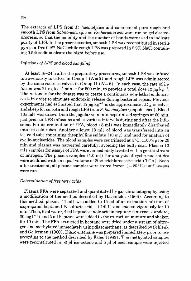
The changes in plasma cAMP concentrations in response to slow intrave- nous infusions of smooth or rough PasteureUa hemolytica LPS in calves are summarized in Fig. 1. For each type of LPS, the results represent the average values of plasma cAMP from five calves. The baseline plasma cAMP levels prior to treatments with smooth and rough LPS were 2.6_+0.6 and 3.1_+0.5 pmol ml- 1, respectively. Immediately after the onset of infusion, both types of LPS caused identical marked increases in plasma cAMP to a 5-fold peak level at 50 min of infusion. Thereafter, plasma cAMP levels returned towards con- trol, but with a difference between smooth and rough LPS. While in rough LPS-treated calves, plasma cAMP returned to control levels at 300 min of infusion, plasma cAMP remained significantly elevated in smooth LPS-treated calves until 60 min post-infusion.
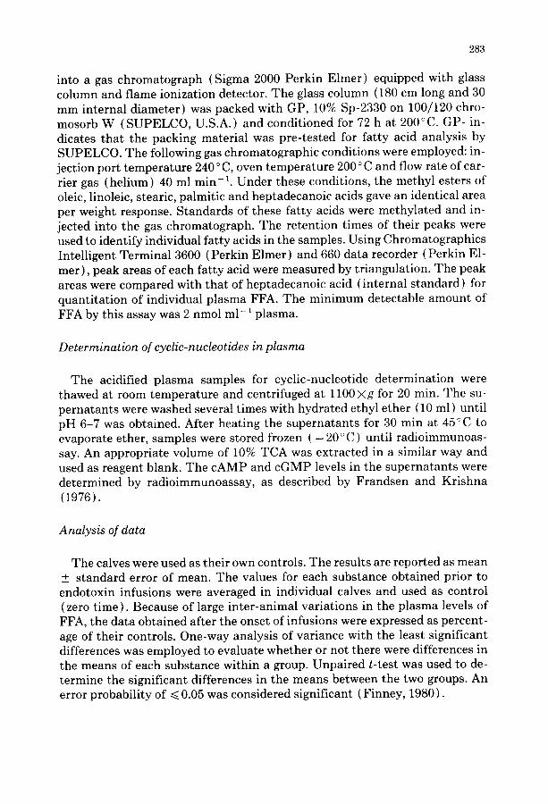
Smooth and rough LPS also caused marked increases in plasma cGMP ( Fig. 2 ). However, the pattern of change was different from that observed for cAMP. In the calves that received smooth LPS, control plasma cGMP averaged 1.3 _+ 0.1 pmol ml-1; in those given rough LPS, the control levels were 1.9 _+ 0.1pmol ml-1. Both types of LPS increased plasma cGMP in two stages. After a lag period of ~ 100 min, plasma cGMP increased gradually in the second hour of infusion to 150-170% of control and then increased further to 200-270% at 5 h post-infusion before returning to control.
Plasma free fatty acids
There was considerable inter-animal variation in the plasma FFA levels. Therefore, the results were expressed as percentage of control, with each ani- mal serving as its own control.
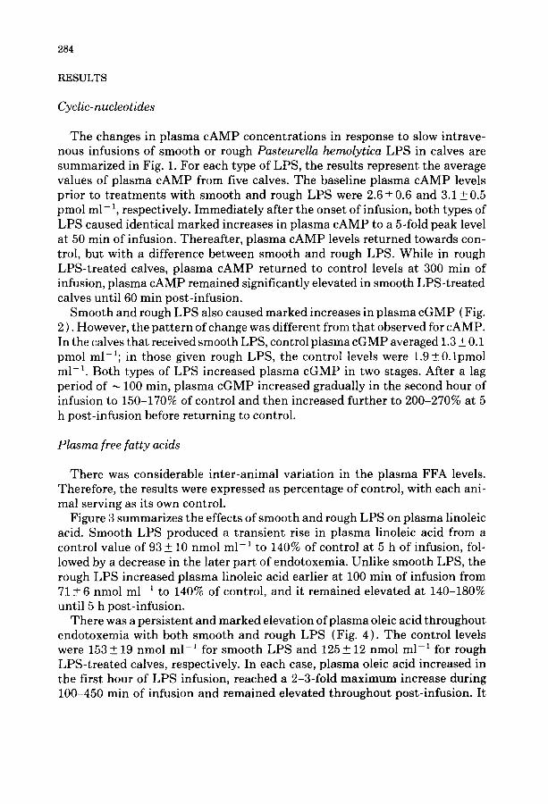
Figure 3 summarizes the effects of smooth and rough LPS on plasma linoleic acid. Smooth LPS produced a transient rise in plasma linoleic acid from a control value of 93 + 10 nmol ml- 1 to 140% of control at 5 h of infusion, fol- lowed by a decrease in the later part of endotoxemia. Unlike smooth LPS, the rough LPS increased plasma linoleic acid earlier at 100 min of infusion from 71 +6 nmol m1-1 to 140% of control, and it remained elevated at 140-180% until 5 h post-infusion.
There was a persistent and marked elevation of plasma oleic acid throughout endotoxemia with both smooth and rough LPS (Fig. 4). The control levels were 153 + 19 nmol m1-1 for smooth LPS and 125+ 12 nmol m1-1 for rough LPS-treated calves, respectively. In each case, plasma oleic acid increased in the first hour of LPS infusion, reached a 2-3-fold maximum increase during 100-450 min of infusion and remained elevated throughout post-infusion. It
285
16
1 4 SS-LPS
~12 R-LP S
o _
~ 1 0 - *
~'4 ~ '
2 .f. -_~ "~- , z ; s i I 1 z l 1 i L i ~ I i ; I ; I I
0 I 0 0 3 0 0 5 0 0 3 0 0 9 0 0 1 5 0 0
TIME IN MINUTES I<--INFUSION >~< POST-INFUSION )I
Fig. 1. Effects of smooth lipopolysaccharide (SS-LPS) or rough lipopolysaccharide (R-LPS) on plasma cAMP levels in calves. "denotes P ~< 0.05 as compared to control (zero time) values within the same group (analysis of variance). At 450 min of infusion to 60 rain post-infusion, plasma cAMP was greater in SS-LPS than R-LPS groups (P~<0.05, t-test).
¢.5
- R - L P S
I l/ / I X ~ 2.0
0_ I.S
f'O l l I z | I I t , I , L i I , I i I ' ' ' •
0 I00 300 500 300 900 1500 TIME IN MINUTES
I'<---I NFUSION ~ ' , c POST- INFUSION ~
Fig. 2. Effects of smooth lipopolysaccharide ( SS-LPS ) or rough lipopolysaccharide ( R-LPS ) on plasma cGMP in calves. "denotes P ~< 0.05 as compared to control (zero time) values of the same group (analysis of variance ). No differences between plasma cGMP levels in SS-LPS and R-LPS groups were detected (t-test).
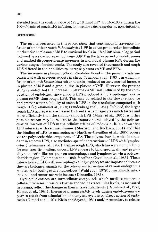
was apparent, however, that rough LPS had a significantly greater ability than smooth LPS to increase plasma oleic acid. Smooth LPS caused little increase in plasma palmitic acid ( Fig. 5 ) and stearic acids ( Fig. 6) from their control

286
2OO
t ,60
3 140 <~ ~ mo
Ioo
8 0
6C ! i i , , , i 0 I 0 0 : 3 0 0 :.500
I"~--I NFUSION x', :
I l ~ S S - L P S
//', /// \ R - L P S
i ' T \ \ \
\ \ . / /
\ / \ j f
\ \ ~ / / /
, 1 1 A t i i i i ~ i i i I 500 900 1500
TIME IN MINUTES POST-INFUSION )1
Fig. 3. Effects of smooth lipopolysaccharide ( SS-LPS ) or rough lipopolysaccharide ( R-LPS ) on plasma linoleic acid. *denotes P ~ 0.05 as compared to control (zero time) values of the same group using analysis of variance. At 300-1500 min post-infusion, plasma linoleic acid was greater in R-LPS than SS-LPS groups (P~< 0.05, t-test).
42O
~ C
:3 54C
o 8 / ' , , ,
; }J ,
S S - L P S
. . . . . . R - L P S
U / \ \ \
o "l'ff ) " • I "" ~ -
il
I 500 5 0 3 0 900 Ib00 TIME IN MINUTES
I~---rNFUSION ~ POST-INFUSION ~
Fig. 4. Effects of smooth lipopolysaccharide ( SS-LPS ) or rough lipopolysaccharide ( R-LPS ) on plasma oleic acid in calves. *denotes P~<0.05 as compared to control (zero time) values using analysis of variance. At 50 and 450 rain of infusion and 300 rain post-infusion, plasma oleic acid was greater in R-LPS than in SS-LPS groups (P~< 0.05, t-test).
values of 103 _+ 15 and 156 + 22 nmol ml-1, respectively. However, the rough LPS increased plasma palmitic acid from a control value of 125 _+ 10 nmol m l - 1 by 2-3-fold during 50 rain of infusion to 5 h post-infusion. Stearic acid was
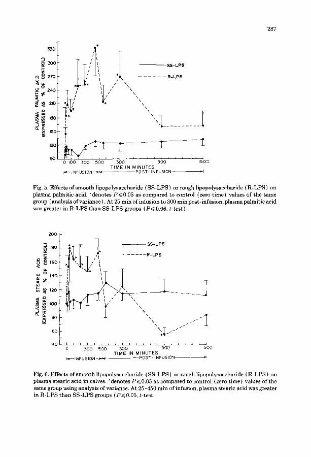
287
33O
~O0 I - Z
882~o 0
_~ 240 ~E
~ 2,o
Q. el 15C
120
90
T I/IItll *~I - . . . o ~ ~ " \ %., , ,
I I I
I I
0 I 0 0 300 5 0 0
~<----/N F USION
S S - L P S
. . . . . . R - L P S
N \ \ N X
\\'\x~ D ..........
3"00 ' ~ ' ~ h , ~ , , 9 0 0 1500 T I M E IN M I N U T E S
POST- INFUSION >I
Fig. 5. Effects of smooth lipopolysaccharide (SS-LPS) or rough lipopolysaccharide (R-LPS) on plasma palmitic acid. "denotes P~<0.05 as compared to control (zero time) values of the same group ( analysis of variance). At 25 min of infusion to 300 min post-infusion, plasma palmitic acid was greater in R-LPS than SS-LPS groups (P~< 0.06, t-test).
2o0
~ ~ ,2o_- ] , , , ,
~ so
6 O
4 0 I I , , , I I ~ l I
0 300 500 300
,~"~I NFU S ION ~ <
~ S S - L P S
. . . . . . R - L P S
\ \
\
\ / /
i i , , , ~ , i , , J
9 0 0 1500 T I M E IN M I N U T E S
P O S T - INFUSION >1
Fig. 6. Effects of smooth lipopolysaccharide (SS-LPS) or rough lipopolysaccharide (R-LPS) on plasma stearic acid in calves. "denotes P~<0.05 as compared to control (zero time) values of the same group using analysis of variance. At 25-450 min of infusion, plasma stearic acid was greater in R-LPS than SS-LPS groups (P~<0.05, t-test.
288
elevated from the control value of 179 + 10 nmol ml- 1 by 150-180% during the 100-450 min of rough LPS infusion, followed by a decrease during post-infusion.
DISCUSSION
The results presented in this report show that continuous intravenous in- fusion of smooth or rough P. haemolytica LPS in calves produced an immediate marked rise in plasma cAMP to maximal levels in 1 h of infusion, a lag period followed by a slow increase in plasma cGMP in the later period of endotoxemia and marked disproportionate increases in individual plasma FFA during the various stages of endotoxemia. The study also revealed that smooth and rough LPS differed in their abilities to increase plasma cAMP and FFA.
The increases in plasma cyclic-nucleotides found in the present study are consistent with previous reports in sheep (Snapper et al., 1983), in which in- fusion of smooth Escherichia coli endotoxin produced an early marked increase in plasma cAMP and a gradual rise in plasma cGMP. However, the present study revealed that the increase in plasma cAMP was influenced by the com- position of endotoxin, since smooth LPS produced a more prolonged rise in plasma cAMP than rough LPS. This may be related to the slower clearance and greater water solubility of smooth LPS in the circulation compared with rough LPS ( Galanos et al., 1969; Freudenberg et al., 1984 ). In blood, the larger rough LPS aggregates are cleared by fixed tissue phagocytes much faster and more efficiently than the smaller smooth LPS (Maier et al., 1981). Another possible reason may be related to the important role played by the polysac- charide fraction of LPS in the cellular effects of endotoxin. It is known that LPS interacts with cell membranes (Morrison and Rudbach, 1981 ) and that the binding of LPS to macrophages (Haeffner-Cavaillon et al., 1985) occurs via the polysaccharide component of LPS. The polysaccharide, which is abun- dant in smooth LPS, also mediates specific interactions of LPS with lympho- cytes ( Lehmann et al., 1980 ). Unlike rough LPS, which has a greater tendency for non-specific binding, smooth LPS appears to bind specifically and prefer- ably to a lectin-like receptor on macrophages and lymphocytes via a polysac- charide region (Lehmann et al., 1980; Haeffner-Cavaillon et al., 1985). These interactions of LPS with macrophages and lymphocytes are important because they are biological signals for the release and formation of various endogenous mediators including cyclic-nucleotides (Wahl et al., 1979), prostanoids, inter- leukin-1 and tumor-necrosis factors ( Dinarello, 1983).
Cyclic-nucleotides are intracellular compounds which mediate numerous cellular processes in various tissues and their extracellular levels, as measured in plasma, reflect the changes in their intracellular levels ( Broadus et al., 1971; Hamet et al., 1984). Increased plasma cAMP levels during endotoxemia ap- pear to result from stimulation of adenylate cyclase by direct action of endo- toxin (Gimpel et al., 1974; Klein and Sayeed, 1980) and/or secondary to release
289
of endogenous substances such as catecholamines and prostaglandins (Broadus et al., 1971; Hamet et al., 1984). The slow rise in plasma cGMP appears to be due to the synthesis of intermediary proteins and nucleic acids necessary for cGMP synthesis (Graber and Clancey, 1985) stimulated by Lipid A.
The release of FFA from adipose tissues occurs via activation of hormone- sensitive lipase via cAMP protein kinase activation (Vernon, 1981). In the present study, the maximal plasma cAMP increase coincided with the rise in several plasma FFA, which suggests that cAMP is involved in the mobilization of plasma FAA in endotoxemia. During the course of endotoxin effects, adipose tissue mobilization is most likely stimulated by several hormones, including glucagon, catecholamines, corticosteroids and somatotropin. These hormones are released in large amounts during endotoxemia and are known to release FFA from adipose tissues through the reactions of cAMP ( Spitzer et al., 1981 ). However, FFA release in endotoxemia can be impaired due to the severely decreased blood flow to adipose tissues ( Spitzer and Spitzer, 1982 ). Thus, the concentration of FFA in circulation may change with stages of endotoxic shock.
We observed uneven increases in plasma FFA: plasma oleic acid was consis- tently elevated to a greater extent than palmitic, stearic and linoleic acids. Similar disparity in increases of plasma FFA have been reported in bacterial infections (Colombo and Pinelli, 1982). This suggests that differences may exist either in the rates of release and/or tissue utilization of individual FFA at various stages of endotoxemia. Previous studies have reported preferential tissue uptake of oleic acid in sheep following norepinephrine infusion (Thompson and Darling, 1975) and a greater release of oleic and palmitic acids from adipose tissues than that of stearic acid (Vernon, 1981 ). Whenever there is an active FFA output from fat depots, the composition of FFA in blood is likely to reflect that of adipose tissue and of the FFA released from the de- pots. For example, increased lipolysis of subcutaneous adipose tissue produces a higher proportion of oleic acid than other FFA, when compared to abdominal adipose tissues (Vernon, 1981 ). In endotoxemia, the maldistribution of blood flow to several organs provides a basis for assuming that lipolysis in some fat pads may be affected more than others, resulting in greater and/or preferential release of particular FFA. In addition, there may be preferential tissue metab- olism of different FFA. Furthermore, enhanced FFA re-esterification of some FFA in adipose tissue may occur in endotoxemia because of increased availa- bility of lactate and a-glycerophosphate as well as lowered pH (Spitzer and Spitzer, 1982). These factors may contribute to the increases and decreases in individual FFA during the different stages of endotoxemia. The fact that plasma oleic acid was increased markedly for a prolonged period may be significant in endotoxin-induced pulmonary endotoxemia in view of the potential role of this FFA in producing lung injury. Oleic acid has been found to cause pulmonary damage which resembles adult respiratory distress syndrome (Gemer et al., 1975; Grossman et al., 1980).
290
The reason for the apparent greater increase in plasma FFA caused by rough LPS compared to smooth LPS is not clear. However, it is consistent with the previous findings by Gaal et al. (1984) that Lipid A is required for lipid met- abolic disturbances in endotoxemia. Thus, one possible explanation for the differential potency of the two LPS on FFA may be related to the greater pro- portion of Lipid A in rough LPS than smooth LPS. These results are also consistent with a previous suggestion that lipid metabolism during bacterial sepsis varies with the nature of the invading bacteria as well as the stage and duration of the infectious illness (Kaufman et al., 1976; Colombo and Pinelli, 1982; Spitzer et al., 1981). In endotoxemia, changes in lipid metabolism are also influenced by the alterations in the hormonal environment (Spitzer and Fish, 1986).
Besides its role in FFA mobilization, cAMP is involved in induction of other effects of endotoxin, including fever (Lin et al., 1982), abortion (Shaw and Cameron, 1979), macrophage activation (Wahl et al., 1979) and pulmonary injury (Snapper et al., 1983). Although the significance of the slow rise in plasma cGMP in endotoxemia is largely unknown, it appears to mediate pul- monary injury (Snapper et al., 1983), lymphocyte activation and antibody synthesis (Watson, 1975) and regulate growth and differentiation of hema- topoietic stem cells ( Graber and Clancey, 1985). The data in the present study suggest that the actions of endotoxin may be mediated by cAMP and cGMP. Although the role of cyclic-nucleotides in endotoxemia is not clear, it appears from the present study that cAMP is involved in the early events of endotoxin action including FFA mobilization, whereas cGMP mediates the later effects of endotoxin.
ACKNOWLEDGMENTS
We thank Dr. P. Squire for providing the P. hemolytica lipopolysaccharides. This work represents a portion of Peter Emau's dissertation in partial fulfil- ment of the requirements for a Ph.D. degree in Comparative Pathology (Vet- erinary Pharmacology and Toxicology) from the University of California, Davis. Part of this paper was presented at the 69th Annual Meeting of the Federation of American Societies for Experimental Biology, Anaheim, CA, 1985 and was published as an Abstract in Fed. Proc., 44:902 (1985).
This work was supported by USDA Grants nos. 83-CRSR-2-2287 and 84- CRSR-2-2414 and USDA Formula Funds (AH-39).
REFERENCES
Broadus, A.E., Hardman, J.G., Kaminsky, N.I., Ball, J.H., Sutherland, E.W. and Liddle, G.W., 1971. Extracellular cyclic nucleotides. Ann. New York Acad. Sci., 185: 50-60.
291
Bruss, M.L., Squire, P.G., Emau, P. and Giri, S.N., 1983. Metabolic effects of purified endotoxin from PasteureUa hemolytica in sheep. Circ. Shock, 10:269-270 (Abstract).
Colombo, R. and Pinelli, A., 1982. Effects of in vivo increase of free fatty acids on human plasma protein binding of furosemide. Pharmacology, 25: 73-81.
Daniel, A.M., Kapadia, B. and McClean, L.D., 1983. Fatty acid supply and organ phospholipid turn-over in two canine shock models. J. Surg. Res., 35: 218-226.
Dinarello, C.A., 1983. Molecular mechanisms in endotoxin fever. Agents Actions, 13: 470-486. Emau, P., Giri, S.N. and Bruss, M.L., 1984. Role of prostaglandins, histamine and serotonin in
the pathophysiology induced by PasteureUa hemolytica endotoxin in sheep. Circ. Shock., 12: 47-59.
Fales, H.M., Jaouni, T.M. and Babashak, J.F., 1973. Simple device for preparing ethereal diazo- methane without resorting to codistillation. Anal. Chem., 45: 2302-2304.
Finney, D.J., 1980. Statistics for Biologists. Chapman and Hall, New York, London. Frandsen, E.K. and Krishna, G., 1976. A simple ultrasensitive method for the assay of cyclic AMP
and cyclic GMP in tissues. Life Sci., 18: 529-542. Freudenberg, M.A., Kleine, B., Freudenberg, N. and Galanos, C., 1984. Distribution, degradation,
and elimination of intravenously applied lipopolysaccharides in rats. In: J.Y. Homma, S. Ka- negasaki, 0. Luderitz, T. Shiba and 0. Westphal (Editors), Bacterial Endotoxin, Chemical, Biological and Clinical Aspects. Verlag Chemie, Florida, pp. 295-304.
Gaal, D., Kremmer, T., Balint, Z., Holczinger, L., Bertok, L. and Nowotny, A., 1984. Effects of bacterial endotoxins and their detoxified derivatives on serum and liver lipids in mice. Toxicol. Appl. Pharmacol., 75: 437-443.
Galanos, C., Luderitz, O. and Westphal, 0., 1969. A new method for the extraction of R-Lipopo- lysaccharides. Eur. J. Biochem., 9: 245-249.
Gemer, M., Dunegan, L.J., Lehr, J.L., Brunet, J.D., Stetz, C.W., Don, H.F., Hayes, J.A. and Drinker, P.A., 1975. Pulmonary insufficiency induced by oleic acid in the sheep. J. Thorac. Cardiovasc. Surg., 69: 793-799.
Gimpel, L., Hodgins, D.S. and Jacobson, E.D., 1974. Effect of endotoxin on hepatic adenylate cyclase activity. Circ. Shock, 1: 31-37.
Goldman, R.C. and Leive, L., 1980. Heterogeneity of antigenic-side-chain length in lipopolysac- charide from Escherichia coli 0111 and Salmonella typhimurium LT2. Eur. J. Biochem., 107: 145-153.
Graber, S.E. and Clancey, M.A., 1985. Further characterization of the effect of bacterial lipopo- lysaccharide preparations on cyclic GMP levels: The importance of macromolecular synthesis. Proc. Soc. Exp. Biol. Med., 180: 163-169.
Grossman, R.F., Jones, J.G. and Murray, J.F., 1980. Effects of oleic acid-induced pulmonary edema on lung mechanics. J. Appl. Physiol., 48: 1045-1051.
Haeffner-Cavaillon, N.J., Cavaillon, J.M., Etievant, M., Lebbar, S. and Szabo, L., 1985. Specific binding of endotoxin to human monocytes and mouse macrophages: serum requirement. Cell. Immun., 91: 119-131.
Hagenfeldt, L., 1966. A gas chromatographic method for the determination of individual free fatty acids in plasma. Clin. Chim. Acta, 13: 266-268.
Hamet, P., Tremblay, J. and Pangs, C., 1984. Cyclic nucleotides in pathophysiology. Adv. Cyclic Nucleotide and Protein Phosphorylation Res., 17: 651-659.
Kaufmann, R.L., Matson, C.F. and Beisel, W.R., 1976. Hypertriglyceridemia produced by endo- toxin: role of impaired disposal mechanisms. J. Infect. Dis., 133: 548-555.
Keiss, R.E., Will, D.H. and Collier, J.R., 1964. Skin toxicity and hemodynamic properties of endo- toxin derived from Pasteurella hemolytica. Am. J. Vet. Res., 25: 935-942.
Klein, D.M. and Sayeed, M.M., 1980. Lung cyclic 3' ,-5' -adenosine monophosphate and adenylate cyclase in endotoxic shock. Exp. Lung. Res., 1: 191-199.
Lehmann, V., Streck, H., Minner, I., Krammer, P.H. and Ruschman, E., 1980. Selection of bac-
292
terial mutants from Salmonella specifically recognizing determinants on the cell surface of activated T. lymphocytes. Eur. J. Immunol., 10: 685-693.
Lillie, L.E., 1974. The bovine respiratory disease complex. Can. Vet. J., 15: 233-242. Lin, M.T., Wu, J.J., Chandra, A. and Tsay, B.L., 1982. A nor-epinephrine cyclic AMP link in the
hypothalamic pathways which mediate fever induced by endotoxin and prostaglandin Ee in the rat. J. Exp. Pharm. Ther., 222: 251-257.
Luderitz, 0., Galanos, C., Risse, H.J., Ruschman, E., Schlecht, S., Schmidt, G., Schulte-Holthau- sen, H. Wheat, R. and Westphal, 0., 1966. Structural relationships of Salmonella 0 and R antigens. Ann. N.Y. Acad. Sci., 133: 349-374.
Maier, R.V., Mathison, J.C. and Ulevitch, R.J., 1981. Interactions of bacterial lipopolysaccharides with tissue macrophages and plasma lipoproteins. Prog. Clin. Biol. Res., 62: 133-155.
Morrison, D.C., 1983. Bacterial endotoxins and pathogenesis. Rev. Infect. Dis., supp. S 733-747. Morrison, D.C. and Rudbach, J.A., 1981. Endotoxin-cell-membrane interactions leading to trans-
membrane signalling. In: F.P. Inman and W.J. Mandy (Editors), Contemporary Topics in Molecular Immunology: Vol. 8, Plenum Press, New York, pp. 187-218.
Nakano, M., 1962. Mutants of Salmonella with unusually low toxicity in mice. Nature, 196: 1118-1119.
Nordenstrom, J., Carpenter, Y.A. and Askanazi, J., Robin, A.P., Elwijn, D.H., Hensle, T.W. and Kinney, J.M., 1983. Free fatty acid mobilization and oxidation during total parenteral nutri- tion in trauma and infection. Ann. Surg., 198: 725-735.
Rietschel, E.T., Schade, U., Jensen, M., Wollenweber, H.W., Luderitz, O. and Greisman, S.G., 1982. Bacterial endotoxins: chemical structure, biological activity and role in septicaemia. Scand. J. Infect. Dis. (Suppl.), 31: 8-21.
Rimsay, R.L., Coyle-Dennis, J.E., Lauerman, L.H. and Squire, P.G., 1981. Purification and bio- logical characterization of endotoxin fractions from PasteureUa haemolytica. Am. J. Vet. Res., 42: 2134-2138.
Schlenk, H. and Gellerman, J.L., 1960. Esterification of fatty acids with diazomethane on a small scale. Anal. Chem., 32: 1412-1414.
Shaw, R.Jr., and Cameron, J.A., 1979. Effects of cyclic AMP altering drugs on endotoxin-induced termination of pregnancy. Res. Commun. Chem. Pathol. Pharmacol., 24: 49-56.
Snapper, J.R., Brigham, K.L., Heflin, A.C., Bomboy, J.D.Jr., and Graber,S.E., 1983. Effects of endotoxemia on cyclic nucleotides in unanesthetised sheep. J. Lab. Clin. Med., 102: 240-249.
Spitzer, J.A. and Fish, R.E., 1986. Lipolytic patterns in isolated adipocytes of continuously en- dotoxemic rats. Circ. Shock, 18: 21-29.
Spitzer, J.J. and Spitzer, J.A., 1982. Alterations in carbohydrate and lipid metabolism following administration of endotoxin. Klin. Wochenschr., 60: 717-719.
Spitzer, J.A., Leach, G.J. and Reeves, J.A., 1981. Metabolic and endocrine alterations induced by endotoxin and sepsis at cellular level. Prog. Clin. Biol. Res., 62: 123-130.
Thompson, G.E. and Darling, K.F., 1975. The hepatic uptake of individual free fatty acids in sheep during norepinephrine infusion. Res. Vet. Sci., 18: 325-327.
Vernon, R.G., 1981. Lipid metabolism in the adipose tissue of ruminant animals. In: W.W. Chris- tie { Editor), Lipid Metabolism in Ruminant Animals. Pergamon Press, New York, pp. 279-362.
Wahl, L.M., Olsen, C.E., Wahl, S.M., McCarthy, J.B., Sandberg, A .L. and Mergenhagen, S.E., 1979. Prostaglandin and cyclic AMP regulation of macrophage involvement in connective tis- sue destruction. Ann. New York Acad. Sci., 332: 271-278.
Watson, J., 1975. The influence of intracellular levels of cyclic nucleotides on cell proliferation and the induction of antibody synthesis. J. Exp. Med., 141: 97-111.
Westphal, O. and Jann, K., 1965. Bacterial lipopolysaccharides: Extraction with phenol-water and further applications of the procedure. In: R.L. Whistler J.N. BeMiller and M.L. Wolf man (Editors), Methods in Carbohydrate Chemistry. Vol. 5. Academic Press, New York, p. 830.
Yates, W.D., 1982. A review of infectious bovine rhinotracheitis, shipping fever pneumonia, and viral-bacterial synergism in respiratory disease of cattle. Can. J. Comp. Med., 46: 225-263.


![Rearing Healthy Calves Manual 2nd ed (1)[2] copy](https://static.fdokumen.com/doc/165x107/6326a762051fac18490ddddd/rearing-healthy-calves-manual-2nd-ed-12-copy.jpg)