Molecular characterisation of dicot-infecting mastreviruses from Australia
Upload
independentCategory
view
0download
0
Infection, Genetics and Evolution 20 (2013) 96–102
Contents lists available at SciVerse ScienceDirect
Infection, Genetics and Evolution
journal homepage: www.elsevier .com/locate /meegid
First molecular characterisation of Cryptosporidium and Giardia fromBubalus bubalis (water buffalo) in Victoria, Australia
1567-1348/$ - see front matter � 2013 Elsevier B.V. All rights reserved.http://dx.doi.org/10.1016/j.meegid.2013.07.019
⇑ Corresponding author. Tel.: +61 397312283; fax: +61 397312366.E-mail address: [email protected] (R.B. Gasser).
Harshanie Abeywardena a, Aaron R. Jex a, Georg von Samson-Himmelstjerna b, Shane R. Haydon c,Melita A. Stevens c, Robin B. Gasser a,⇑a Faculty of Veterinary Science, The University of Melbourne, Parkville, Victoria 3010, Australiab Institute of Parasitology and Tropical Veterinary Medicine, 14163 Berlin, Germanyc Melbourne Water Corporation, Victoria, Australia
a r t i c l e i n f o
Article history:Received 11 May 2013Received in revised form 14 July 2013Accepted 16 July 2013Available online 23 July 2013
Keywords:Bubalus bubalisCryptosporidiumGiardiaSingle-strand conformation polymorphism(SSCP) analysisRestriction endonuclease fingerprinting(REF)Zoonotic potential
a b s t r a c t
We conducted a molecular epidemiological survey of Cryptosporidium and Giardia from Bubalus bubalis(water buffalo) on two extensive farms (450 km apart) in Victoria, Australia. Faecal samples (n = 476)were collected from different age groups of water buffalo at two time points (six months apart) andtested using a PCR-based mutation scanning-targeted sequencing-phylogenetic approach, employingmarkers within the small subunit of ribosomal RNA (designated pSSU) and triose phosphate isomerase(ptpi) genes. Based on pSSU data, Cryptosporidium parvum, Cryptosporidium bovis and Cryptosporidiumgenotypes 1, 2 (each 99% similar genetically to Cryptosporidium ryanae) and 3 (99% similar to Cryptospo-ridium suis) were detected in two (0.4%), one (0.2%), 38 (8.0%), 16 (3.4%) and one (0.2%) of the 476 samplestested, respectively. Using ptpi, Giardia duodenalis assemblages A and E were detected in totals of 56(11.8%) and six (1.3%) of these samples, respectively. Cryptosporidium was detected on both farms,whereas Giardia was detected only on farm B, and both genera were detected in 1.5% of all samples tested.The study showed that water buffaloes on these farms excreted C. parvum and/or G. duodenalis assem-blage A, which are consistent with those found in humans, inferring that these particular pathogensare of zoonotic significance. Future work should focus on investigating, in a temporal and spatial manner,the prevalence and intensity of such infections in water buffaloes in various geographical regions in Aus-tralia and in other countries.
� 2013 Elsevier B.V. All rights reserved.
1. Introduction
Cryptosporidium and Giardia are common aetiological agents ofprotozoal enteritis of humans and animals, including livestock,companion animals and wildlife (Fayer, 2004; Thompson andMonis, 2004; Thompson et al., 2008; Jex et al., 2011). Species ofboth genera are faecal-orally transmitted through resilient, infec-tive stages (oocysts or cysts) (Korich et al., 1990; Carpenter et al.,1999; Betancourt and Rose, 2004), usually via water, food or directcontact (Cacciò et al., 2005; Smith et al., 2007). Clinical signs of dis-ease can range from self-limiting diarrhoea in immune-competentindividuals (O’Donoghue, 1995; Homan and Mank, 2001) tochronic and life-threatening infection in immune-compromisedor -suppressed individuals (Hunter and Nichols, 2002; Petri et al.,2008; Stark et al., 2009). Humans have been reported to becomeinfected with a range of species and genotypes of Cryptosporidium(Smith et al., 2006) or Giardia (Foronda et al., 2008). However,based on current molecular data (Cacciò et al., 2005; Xiao and
Fayer, 2008), Cryptosporidium hominis, Cryptosporidium parvumand Giardia duodenalis assemblages A and B are responsible forthe majority of known human disease cases. Of these species, C.hominis is recognised to be transmitted from human to human(Xiao et al., 2004), whereas C. parvum and G. duodenalis infectionsmay be acquired through human–human or animal–human trans-mission (Xiao and Ryan, 2004; Xiao and Feng, 2008).
Livestock animals have been implicated as a source of humancryptosporidiosis and giardiasis, based on the molecular epidemio-logical studies conducted in various countries (Hunter and Thomp-son, 2005; Budu-Amoako et al., 2011). There is a considerableamount of data demonstrating that dairy and beef cattle harbourzoonotic species and genotypes of Cryptosporidium and Giardia,representing a potentially significant reservoir of infection to hu-mans (e.g., Santín et al., 2004; Geurden et al., 2008; Xiao and Feng,2008; Feng and Xiao, 2011). Interestingly, there is an age-specificstratification for Cryptosporidium in cattle. Usually, C. parvum isfound principally in pre-weaned cattle, Cryptosporidium bovis andCryptosporidium ryanae are mostly found in weaned calves andyearlings, and Cryptosporidium andersoni is most common in adultcattle (Santín et al., 2004). In contrast to the situation in cattle, very

H. Abeywardena et al. / Infection, Genetics and Evolution 20 (2013) 96–102 97
little is known about the range of species and genotypes of Cryptos-poridium and Giardia in other members of the family Bovidae,including water buffalo (Bubalus bubalis). This species contributessubstantially to the agricultural economies in many developingcountries, and is widely used as a working animal (for transportand ploughing) and as a source of milk, meat and leather (Perera,2011). Generally, bovids produce large amounts of faecal wasteeach day, and, if infected, can excrete substantial numbers of infec-tive stages that contaminate the environment (Fayer et al., 2000;Budu-Amoako et al., 2011).
To date, molecular studies of Cryptosporidium of water buffaloeshave been conducted in countries including Egypt (Amer et al.,2013), Italy (Cacciò et al., 2007), Nepal (Feng et al., 2012) and Spain(Gomez-Couso et al., 2005). Unlike cattle, a genotype with 99%similarity to C. ryanae has been characterised from water buffaloesmore frequently than any other species of Cryptosporidium (Fenget al., 2012; Amer et al., 2013), while C. andersoni and C. bovis havenot yet been reported. One molecular study of Giardia from waterbuffaloes conducted in Italy reported assemblages A and E (Cacciòet al., 2007, 2010). These studies suggest that water buffalo, likecattle, might also serve as an important reservoir of Cryptosporidi-um and Giardia for transmission to humans, which warrants inves-tigations using molecular tools. In the present study, wegenetically characterized Cryptosporidium and Giardia from waterbuffaloes in Victoria, Australia, and assessed their zoonotic poten-tial. For this purpose, we utilized a PCR-based mutation scanning-targeted sequencing-phylogenetic approach, employing markerswithin the small subunit of ribosomal RNA (designated pSSU)and triose phosphate isomerase (ptpi) genes. As these two loci havebeen widely used for molecular characterisation of Cryptosporidiumand Giardia, there is a substantial amount of sequence data avail-able in public databases for comparative analysis.
2. Materials and methods
2.1. Sample collection, calves and management practices
Faecal samples (n = 476) were collected from water buffaloes(Bubalus bubalis; mixed sexes; various age groups), including 32,232 and 212 samples from animals of <6 months, 6 months to2 years, and >2 years, on extensive farms A and B, located450 km apart in Victoria, Australia. Samples were collected fromeach farm, at two time points, six months apart. In total, 100 and400 buffaloes were present on each of the two farms (Farm Aand B, respectively) at each visit. Freshly deposited faecal sampleswere collected from paddocks or enclosures. On both farms, waterbuffaloes of >3 months of age were maintained together. Calveswere born in situ on both farms and maintained with their damsup to 24 h to ensure that each calf had received colostrum. Then,calves were kept in pens, fed with whole milk, weaned at 3 monthsof age and released on to paddocks with cows. Farm A commencedfarming swamp buffaloes in 1992, and introduced riverine buffa-loes in 2002, whereas on farm B, all of the buffaloes were of theriverine type.
2.2. Isolation of genomic DNA from faecal samples, and PCRamplification
Genomic DNA was extracted from individual faecal samplesusing the PowerSoil DNA isolation kit (MoBIO, USA) (cf. Abeywar-dena et al., 2013), and then frozen at �20 �C until molecular test-ing. Each genomic DNA sample was tested specifically for thepresence of Cryptosporidium and Giardia DNA, employing geneticmarkers (designated pSSU and ptpi) in the small subunit (SSU) nu-clear ribosomal RNA gene and triose phosphate isomerase (tpi)
genes, respectively. For the amplification of pSSU, nested PCR wasperformed in a 50 ll volume containing 2.0 mM of MgCl2, 200 lMof each deoxynucleotide triphosphate (dNTP), 25 pmol of each oli-gonucleotide primer and 1.25 U of MangoTaq polymerase in a stan-dard PCR buffer (Bioline, USA). In the primary reaction of SSU forCryptosporidium, primers XF2 (forward: 50-GGAAGGGTTGTATTTAT-TAGATAAAG-30) and XR2 (reverse: 50-AAGGAGTAAGGAACAACCTC-CA-30) (Xiao et al., 1999) were employed, and primers pssu-f(forward: 50-AAAGCTCGTAGTTGGATTTCTGTT-30) and pssu-r (re-verse: 50- ACCTCTGACTGTTAAATACRAATGC-30) (cf. Nolan et al.,2010a) were utilised for the secondary reaction to amplify pSSU(�240 bp). The cycling protocol for the primary amplification in-cluded an initial cycle of 94 �C/5 min (initial denaturation), fol-lowed by 30 cycles of 94 �C/45 s (denaturation), 45 �C/2 min(annealing), and 72 �C/1.5 min (extension) and a final extensionof 72 �C/10 min. From 1 ll of primary amplicon, the nested ampli-fication was performed using a cycling protocol of 94 �C/5 min (ini-tial denaturation), followed by 35 cycles of 94 �C/30 s(denaturation), 55 �C/30 s (annealing) and 72 �C/30 s (extension),followed by a final extension of 72 �C/10 min.
For the amplification of ptpi, nested PCR was performed in a50 ll volume containing 3.0 mM of MgCl2, 200 lM of each deoxy-nucleotide triphosphate (dNTP), 25 pmol of each oligonucleotideprimer and 1.25 U of GoTaq polymerase in standard PCR buffer(Promega, USA). In the primary reaction for tpi of Giardia, primersAL3543 (forward: 50-AAATTATGCCTGCTCGTCG-30) and AL3546 (re-verse: 50-CAAACCTTTTCCGCAAACC-30) were employed, whereasprimers AL3544 (forward: 50-CCCTTCATCGGIGGTAACTT-30) andAL3545 (reverse: 50-GTGGCCACCACTCCCGTGCC-30) were utilisedin the secondary reaction to amplify ptpi (530 bp) (cf. Nolanet al., 2010b). The cycling protocol for primary amplification in-cluded a cycle of 94 �C/5 min (initial denaturation), followed by35 cycles of 94 �C/45 s (denaturation), 50 �C/45 s (annealing) and72 �C/60 s (extension), followed by a final extension of 72 �C/10min. The secondary PCR was identical, except that the annealingtemperature was 60 �C.
2.3. Mutation scanning, sequencing and phylogenetic analyses
For pSSU amplicons, single-strand conformation polymorphism(SSCP) analysis (Gasser et al., 2006) was carried out as describedpreviously (Jex et al., 2007). For ptpi amplicons, restriction endonu-clease fingerprinting (Orita et al., 1989; Zhu and Gasser, 1998) wasemployed, using the enzyme RsaI (Promega) (cf. Nolan et al.,2010b). Amplicons representing each banding profile were se-lected and treated with exonuclease I and shrimp alkaline phos-phatase (Fermentas), according to the manufacturer’sinstructions, and then sequenced in both directions by direct, auto-mated sequencing (BigDye Terminator v.3.1 chemistry, AppliedBiosystems, USA), using the same primers employed in secondaryPCR. The quality of each sequence was assessed based on the cor-responding electropherogram using the program BioEdit (Hall,1999), and the sequences determined were compared with knownreference sequences using the Basic Local Alignment Search Tool(BLAST; http://www.ncbi.nlm.nih.gov/BLAST).
Some pSSU amplicons were cloned prior to sequencing. In brief,amplicons were purified using minicolumns (Wizard PCR PrepsDNA Purification System, Promega) and ligated into the pGEM-T-Easy vector (Promega) and Escherichia coli (a-select chemicallycompetent cells, Bioline) as described previously (Abeywardenaet al., 2013). Then, the cells were plated on to agar plates and incu-bated overnight at 37 �C. Ten colonies were picked from each plateand grown at 37 �C overnight for plasmid purification (using Wiz-ard Plus SV Minipreps DNA Purification System, Promega). Thereaf-ter, inserts were PCR-amplified using the primers pssu-f and pssu-r(Section 2.2); following the SSCP analysis of all amplicons, some
98 H. Abeywardena et al. / Infection, Genetics and Evolution 20 (2013) 96–102
were selected, treated with exonuclease I and shrimp alkalinephosphatase, and sequenced using the same primers.
Phylogenetic analysis of sequence data was performed usingthe Bayesian Inference (BI) tree building method in MrBayes3.1.2 (Huelsenbeck and Ronquist, 2001; Ronquist and Huelsenbeck,2003). Posterior probabilities (pp) were calculated via 1,000,000(pSSU) or 2,000,000 generations (ptpi), utilizing four simultaneoustree-building chains, with every 100th tree being saved. At thispoint, the standard deviation of split frequencies was <0.01, andthe potential scale reduction factor (PSRF) approached one. A 50%majority rule consensus tree for each analysis was constructedbased on the final 75% of trees generated by BI.
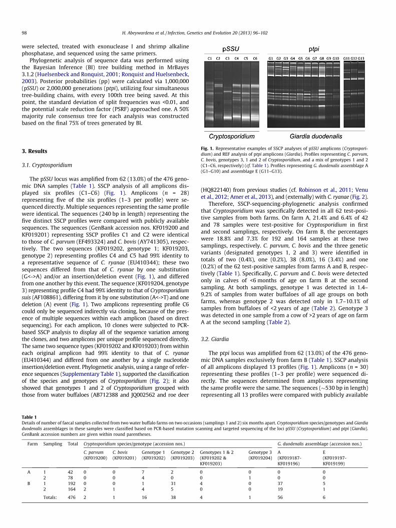
Fig. 1. Representative examples of SSCP analyses of pSSU amplicons (Cryptospori-dium) and REF analysis of ptpi amplicons (Giardia). Profiles representing C. parvum,C. bovis, genotypes 3, 1 and 2 of Cryptosporidium, and a mix of genotypes 1 and 2(C1–C6, respectively) (cf. Table 1). Profiles representing G. duodenalis assemblage A(G1–G10) and assemblage E (G11–G13).
3. Results
3.1. Cryptosporidium
The pSSU locus was amplified from 62 (13.0%) of the 476 geno-mic DNA samples (Table 1). SSCP analysis of all amplicons dis-played six profiles (C1–C6) (Fig. 1). Amplicons (n = 28)representing five of the six profiles (1–3 per profile) were se-quenced directly. Multiple sequences representing the same profilewere identical. The sequences (240 bp in length) representing thefive distinct SSCP profiles were compared with publicly availablesequences. The sequences (GenBank accession nos. KF019200 andKF019201) representing SSCP profiles C1 and C2 were identicalto those of C. parvum (EF493324) and C. bovis (AY741305), respec-tively. The two sequences (KF019202, genotype 1; KF019203,genotype 2) representing profiles C4 and C5 had 99% identity toa representative sequence of C. ryanae (EU410344); these twosequences differed from that of C. ryanae by one substitution(G<->A) and/or an insertion/deletion event (Fig. 1), and differedfrom one another by this event. The sequence (KF019204, genotype3) representing profile C4 had 99% identity to that of Cryptosporidiumsuis (AF108861), differing from it by one substitution (A<->T) and onedeletion (A) event (Fig. 1). Two amplicons representing profile C6could only be sequenced indirectly via cloning, because of the pres-ence of multiple sequences within each amplicon (based on directsequencing). For each amplicon, 10 clones were subjected to PCR-based SSCP analysis to display all of the sequence variation amongthe clones, and two amplicons per unique profile sequenced directly.The same two sequence types (KF019202 and KF019203) from withineach original amplicon had 99% identity to that of C. ryanae(EU410344) and differed from one another by a single nucleotideinsertion/deletion event. Phylogenetic analysis, using a range of refer-ence sequences (Supplementary Table 1), supported the classificationof the species and genotypes of Cryptosporidium (Fig. 2); it alsoshowed that genotypes 1 and 2 of Cryptosporidium grouped withthose from water buffaloes (AB712388 and JQ002562 and roe deer
Table 1Details of number of faecal samples collected from two water buffalo farms on two occasionduodenalis assemblages in these samples were classified based on PCR-based mutation scGenBank accession numbers are given within round parentheses.
Farm Sampling Total Cryptosporidium species/genotype (accession nos.)
C. parvum C. bovis Genotype 1 Genotype 2(KF019200) (KF019201) (KF019202) (KF019203)
A 1 42 0 0 7 22 78 0 0 4 0
B 1 192 0 0 1 312 164 2 1 4 5
Totals: 476 2 1 16 38
(HQ822140) from previous studies (cf. Robinson et al., 2011; Venuet al., 2012; Amer et al., 2013), and (externally) with C. ryanae (Fig. 2).
Therefore, SSCP-sequencing-phylogenetic analysis confirmedthat Cryptosporidium was specifically detected in all 62 test-posi-tive samples from both farms. On farm A, 21.4% and 6.4% of 42and 78 samples were test-positive for Cryptosporidium in firstand second samplings, respectively. On farm B, the percentageswere 18.8% and 7.3% for 192 and 164 samples at these twosamplings, respectively. C. parvum, C. bovis and the three geneticvariants (designated genotypes 1, 2 and 3) were identified intotals of two (0.4%), one (0.2%), 38 (8.0%), 16 (3.4%) and one(0.2%) of the 62 test-positive samples from farms A and B, respec-tively (Table 1). Specifically, C. parvum and C. bovis were detectedonly in calves of <6 months of age on farm B at the secondsampling. At both samplings, genotype 1 was detected in 1.4–9.2% of samples from water buffaloes of all age groups on bothfarms, whereas genotype 2 was detected only in 1.7–10.1% ofsamples from buffaloes of <2 years of age (Table 2). Genotype 3was detected in one sample from a cow of >2 years of age on farmA at the second sampling (Table 2).
3.2. Giardia
The ptpi locus was amplified from 62 (13.0%) of the 476 geno-mic DNA samples exclusively from farm B (Table 1). SSCP analysisof all amplicons displayed 13 profiles (Fig. 1). Amplicons (n = 30)representing these profiles (1–3 per profile) were sequenced di-rectly. The sequences determined from amplicons representingthe same profile were the same. The sequences (�530 bp in length)representing all 13 profiles were compared with publicly available
s (samplings 1 and 2) six months apart. Cryptosporidium species/genotypes and Giardiaanning and targeted sequencing of the loci pSSU (Cryptosporidium) and ptpi (Giardia).
G. duodenalis assemblage (accession nos.)
Genotypes 1 & 2 Genotype 3 A E(KF019202 &KF019203)
(KF019204) (KF019187-KF019196)
(KF019197-KF019199)
0 0 0 00 1 0 04 0 37 50 0 19 1
4 1 56 6

Fig. 2. Relationship among pSSU sequences of Cryptosporidium based on Bayesian inference (BI) analysis. The five distinct pSSU sequences determined in the present studyand 33 reference sequences representing Cryptosporidium (Supplementary Table 1) were included in the analysis. Sequences of Eimeria tenella and E. necatrix were used asoutgroups. Accession numbers of publicly available sequences are indicated. In bold-type are accession numbers of sequences determined in the present study, and numbersof samples with these sequences (in parentheses). Posterior probabilities (pp) are indicated at all major nodes.
Table 2Details of number of faecal samples collected from different age groups of water buffaloes (<6, 6–24 and >24 months) on farms A and B. Cryptosporidium species/genotypes andGiardia duodenalis assemblages in these samples were classified based on PCR-based mutation scanning and targeted sequencing of the loci pSSU (Cryptosporidium) and ptpi(Giardia). GenBank accession numbers are given within round parentheses.
Farm Age group(months)
No. ofsamples
Cryptosporidium species/genotype (accession nos.) Giardia duodenalis assemblage(accession nos.)
C. parvum C. bovis Genotype 1 Genotype 2 Genotypes 1 & 2 Genotype 3 A E(KF019200) (KF019201) (KF019202) (KF019203) (KF019202 &
KF019203)(KF019204) (KF019187-
KF019196)(KF019197-KF019199)
A <6 12 0 0 2 0 0 0 0 06–24 41 0 0 8 2 0 0 0 0>24 67 0 0 1 0 0 1 0 0
B <6 20 2 1 0 1 0 0 0 06–24 191 0 0 4 35 4 0 15 6>24 145 0 0 1 0 0 0 41 0
Totals: 476 2 1 16 38 4 1 56 6
H. Abeywardena et al. / Infection, Genetics and Evolution 20 (2013) 96–102 99
sequences. Of the 13 sequences determined, one (KF019187) andnine (KF019188-KF019196) sequences had 100% and 99% identi-ties, respectively, to a reference sequence (L02120) for G. duodenal-is assemblage A (G1-G10). One (KF019197) and two sequences(KF019198 and KF019199) had 100% and 99% identity to assem-blage E of G. duodenalis (G11-G13). According to a phylogeneticanalysis using a wide range of reference sequences (Supplemen-tary Table 2), ten and three sequences determined here clustered,with strong nodal support, with G. duodenalis assemblages A andE, respectively (Fig. 3), irrespective of the minor nucleotide varia-tion detected in the ptpi sequences determined and representingeach assemblage.
SSCP and sequencing analyses confirmed that G. duodenalis wasspecifically detected in all 62 test-positive samples from farm B.On this farm, 21.9% and 12.2% of 192 and 164 samples were
test-positive for G. duodenalis in first and second samplings, respec-tively. G. duodenalis assemblages A and E were detected in totals of56 (90.3%) and six (9.7%) of all 62 samples. Specifically, assemblageA was detected in samples from buffaloes of two age groups (0.5–2and >2 years), whereas assemblage E was detected only in the for-mer age group. At the first sampling, assemblage A was detected in37 of the 42 (88.1%) test-positive samples, and assemblage E in fivesamples (11.9%), respectively (Table 1). At the second sampling,assemblage A was detected in 19 of 20 (95.0%) test-positive sam-ples, and assemblage E in one sample (5.0%).
3.3. Cryptosporidium and Giardia
Both Cryptosporidium and Giardia (1.5%) were detected in onlyseven of the 476 samples tested (Tables 1 and 2): Cryptosporidium

Fig. 3. Phylogenetic relationship among ptpi sequences of Giardia duodenalis based on Bayesian Inference (BI) analysis. Thirteen sequences determined in the present study,and 38 reference sequences representing G. duodenalis assemblages A–G (Supplementary Table 2) were included in the analysis. Sequences representing G. ardeae, G. murisand G. microti were used as outgroups. In bold-type are accession numbers of sequences determined in the present study, and numbers of samples with these sequences (inparentheses). Posterior probabilities (pp) are indicated at all major nodes.
100 H. Abeywardena et al. / Infection, Genetics and Evolution 20 (2013) 96–102
genotype 1 and G. duodenalis assemblage E in five at the first sam-pling; genotype 1 and G. duodenalis assemblage A in one, and geno-type 2 and G. duodenalis assemblage A in another at the secondsampling.
4. Discussion
Cryptosporidium genotype 1 (accession no. KF019202) was mostfrequently detected (67.7%) among the 62 test-positive samples.This genotype was present in water buffalo of <2 years of age,whereas genotype 2 (KF019203) was detected in buffaloes of allage groups (<6 months, 6 months to 2 years and >2 years). Otherstudies have detected genotype 1 in water buffaloes in Egypt(Amer et al., 2013), India (Venu et al., 2012) and Nepal (Fenget al., 2012) and in cervids in Australia (Nolan et al., 2013), theUK (Robinson et al., 2011) and the USA (Jellison et al., 2009). Ithas also been isolated from storm water in the USA (Jiang et al.,2005). This genotype of Cryptosporidium has also been identifiedin two studies of cattle in India (Venu et al., 2012) and Nepal (Fenget al., 2012). For instance, in Nepal, this genotype had been isolatedfrom Bos indicus (zebu cattle) sharing the same habitat with waterbuffaloes, which suggested that genotype 1 might be cross trans-missible between these bovids. The study conducted in India (Venuet al., 2012) did not report whether cattle had contact with
buffaloes. Although previous studies of Cryptosporidium of waterbuffaloes using the pSSU locus have reported genotype 1, genotype2 is a new record. According to the phylogenetic tree (cf. Fig. 2),genotypes 1 and 2 characterised in the present study grouped withC. ryanae (which grouped separately; see Fig. 2), and they werewithin the same clade as sequences representing Cryptosporidiumfrom other water buffaloes from Egypt (AB712388) and India(JQ002562), and roe deer (Capreolus capreolus) from the UK(HQ822140) (pp = 0.99). Genotype 3 (KF019204) was identifiedonly in one faecal sample from an adult cow on farm A. Thisgenotype has been identified previously in cattle in Australia(Abeywardena et al., 2013) and other countries, including Denmark(DQ182599; Langkjaer et al., 2007), India (GQ345008; Khan et al.,2010) and the UK (HQ822134; Robinson et al., 2011). It has alsobeen linked to cryptosporidiosis cases in cattle in Canada(AY030084 and AY030085; Ong et al., 2002) and the UK(HQ822146; Robinson et al., 2011); the validity of this genotypehas been supported by sequence data for the heat shock protein70 (hsp70) and actin genes (Robinson et al., 2011). Nonetheless,there is still limited information on species and genotypes ofCryptosporidium from water buffaloes. Various studies havereported the detection of Cryptosporidium copro-antigens oroocysts in buffalo faeces (e.g., Rinaldi et al., 2007a,b; El-Khoderyand Osman, 2008), but, to date, only few molecular studies(Gomez-Couso et al., 2005; Cacciò et al., 2007; Feng et al., 2012;

H. Abeywardena et al. / Infection, Genetics and Evolution 20 (2013) 96–102 101
Amer et al., 2013) have been performed. Therefore, detailed studiesare required to assess the significance of species and genotypes ofCryptosporidium as pathogens in water buffaloes, and their zoo-notic potential.
Cryptosporidium parvum was detected in two of 62 Cryptospori-dium test-positive faecal samples. This species was detected only inthe second sampling from farm B and was found only in calves of<6 months of age. C. parvum is usually found in bovine calves of <1month of age, and has been detected previously in water buffaloes(Cacciò et al., 2007), but most of the faecal samples in the presentstudy originated from animals of >1 month of age. C. bovis was de-tected in one sample, and this is the first record of this species ofCryptosporidium in water buffalo. Given that C. bovis is a commonlyfound in cattle (Bos taurus and Bos indicus), and that water buffalo(Bubalus bubalis) belong to the same family (Bovidae) as cattle, thisfinding is not surprising. However, the detection of C. bovis in onlyone of the 476 samples tested might suggest a higher affinity of C.bovis for Bos taurus or Bos indicus than Bubalus bubalis. This hypoth-esis needs to be tested.
The predominance of G. duodenalis assemblage A in water buf-falo was unexpected, given that, in cattle, G. duodenalis assem-blage E has been reported to be more common than assemblageA (e.g., Appelbee et al., 2003; Becher et al., 2004; Trout et al.,2004; Geurden et al., 2008; Ng et al., 2011). Although assemblageA has been detected in water buffalo in Italy (Cacciò et al., 2007),it was detected in only 2 of 57 samples, whereas six test-positivesamples represented assemblage E. According to the presentstudy, assemblage A was >2.7 times more prevalent in animalsof >2 years of age than in younger animals at both samplingson farm B (Table 2). One explanation for not detecting Giardiaon farm A might be differences in management practices betweenthe two farms; another might be that all of the buffaloes on farmB were riverine, whereas farm A had a mixed population ofswamp and riverine buffaloes, suggesting that host or environ-mental factors might relate to the difference between thesefarms. Irrespective, the findings suggest that some adult waterbuffalo populations in Australia can represent significant reser-voirs of zoonotic G. duodenalis assemblage A.
5. Conclusion
This study explored species and genotypes of Cryptosporidiumand Giardia present in the faeces from water buffalo in Victoria,Australia. The findings revealed the presence of two genotypesof Cryptosporidium each with 99% genetic similarity to C. ryanaefrom cattle, one of which was a new record for Bubalus bubalis.The results also showed a dominance of G. duodenalis assemblageA over assemblage E and the presence of C. parvum, also known toinfect humans, suggesting that water buffaloes might serve asreservoirs for zoonotic transmission. This finding emphasizesthe need for further work on investigating, in a temporal and spa-tial way, the prevalence and intensity of such infections in vari-ous geographical regions in Australia and in other countries,particularly those in which there is a close relationship betweenhumans and the water buffalo, and a reliance on this animal inagriculture (e.g., for meat, milk, leather, transport and for plough-ing). Moreover, it will be particularly important to investigate lev-els of oocyst/cyst excretion and contamination in areassurrounding paddocks, in wastewater run-off or creeks and otherwater-ways flowing from the farms to water reservoirs and indrinking water. Furthermore, assessing whether the genotypesinfecting water buffalo occur in humans or other animal specieswill be important to study the epidemiology of cryptosporidiosisand giardiasis, particularly in endemic regions in developingcountries of the world.
Acknowledgements
Funding support from the Australian Research Council (ARC),Melbourne Water Corporation (MWC), National Health and Medi-cal Research Council (NHMRC) and the Alexander von HumboldtFoundation is gratefully acknowledged. Thanks to Dr Anson Koeh-ler for support. The authors have declared no conflict of interest.
Appendix A. Supplementary data
Supplementary data associated with this article can be found, inthe online version, at http://dx.doi.org/10.1016/j.meegid.2013.07.019.
References
Abeywardena, H., Jex, A.R., Firestone, S.M., McPhee, S., Driessen, N., Koehler, A.V.,Haydon, S.R., von Samson-Himmelstjerna, G., Stevens, M.A., Gasser, R.B., 2013.Assessing calves as carriers of Cryptosporidium and Giardia with zoonoticpotential on dairy and beef farms within a water catchment area by mutationscanning. Electrophoresis 34, 2259–2267.
Amer, S., Zidan, S., Feng, Y., Adamu, H., Li, N., Xiao, L., 2013. Identity and publichealth potential of Cryptosporidium spp. in water buffalo calves in Egypt. Vet.Parasitol. 191, 123–127.
Appelbee, A.J., Frederick, L.M., Heitman, T.L., Olson, M.E., 2003. Prevalence andgenotyping of Giardia duodenalis from beef calves in Alberta, Canada. Vet.Parasitol. 112, 289–294.
Becher, K.A., Robertson, I.D., Fraser, D.M., Palmer, D.G., Thompson, R.C., 2004.Molecular epidemiology of Giardia and Cryptosporidium infections in dairycalves originating from three sources in Western Australia. Vet. Parasitol. 123,1–9.
Betancourt, W.Q., Rose, J.B., 2004. Drinking water treatment processes for removalof Cryptosporidium and Giardia. Vet. Parasitol. 126, 219–234.
Budu-Amoako, E., Greenwood, S.J., Dixon, B.R., Barkema, H.W., McClure, J.T., 2011.Foodborne illness associated with Cryptosporidium and Giardia from livestock. J.Food Prot. 74, 1944–1955.
Cacciò, S.M., Thompson, R.C., McLauchlin, J., Smith, H.V., 2005. UnravellingCryptosporidium and Giardia epidemiology. Trends Parasitol. 21, 430–437.
Cacciò, S.M., Rinaldi, L., Cringoli, G., Condoleo, R., Pozio, E., 2007. Molecularidentification of Cryptosporidium parvum and Giardia duodenalis in the Italianwater buffalo (Bubalus bubalis). Vet. Parasitol. 150, 146–149.
Cacciò, S.M., Beck, R., Almeida, A., Bajer, A., Pozio, E., 2010. Identification of Giardiaspecies and Giardia duodenalis assemblages by sequence analysis of the 5.8SrDNA gene and internal transcribed spacers. Parasitology 137, 919–925.
Carpenter, C., Fayer, R., Trout, J., Beach, M.J., 1999. Chlorine disinfection ofrecreational water for Cryptosporidium parvum. Emerg. Infect. Dis. 5, 579–584.
El-Khodery, S.A., Osman, S.A., 2008. Cryptosporidiosis in buffalo calves (Bubalusbubalis): Prevalence and potential risk factors. Trop. Anim. Health Prod. 40,419–426.
Fayer, R., 2004. Cryptosporidium: a water-borne zoonotic parasite. Vet. Parasitol.126, 37–56.
Fayer, R., Trout, J.M., Graczyk, T.K., Lewis, E.J., 2000. Prevalence of Cryptosporidium,Giardia and Eimeria infections in post-weaned and adult cattle on threeMaryland farms. Vet. Parasitol. 93, 103–112.
Feng, Y., Xiao, L., 2011. Zoonotic potential and molecular epidemiology of Giardiaspecies and giardiasis. Clin. Microbiol. Rev. 24, 110–140.
Feng, Y., Raj Karna, S., Dearen, T.K., Singh, D.K., Adhikari, L.N., Shrestha, A., Xiao, L.,2012. Common occurrence of a unique Cryptosporidium ryanae variant in zebucattle and water buffaloes in the buffer zone of the Chitwan National Park,Nepal. Vet. Parasitol. 185, 309–314.
Foronda, P., Bargues, M.D., Abreu-Acosta, N., Periago, M.V., Valero, M.A., Valladares,B., Mas-Coma, S., 2008. Identification of genotypes of Giardia intestinalis ofhuman isolates in Egypt. Parasitol. Res. 103, 1177–1181.
Gasser, R., Hu, M., Chilton, N.B., Campbell, B.E., Jex, A.R., Otranto, D., Cafarchia, C.,Beveridge, I., Zhu, X.Q., 2006. Single-strand conformation polymorphism (SSCP)for the analysis of genetic variation. Nat. Protoc. 1, 3121–3128.
Geurden, T., Geldhof, P., Levecke, B., Martens, C., Berkvens, D., Casaert, S.,Vercruysse, J., Claerebout, E., 2008. Mixed Giardia duodenalis assemblage Aand E infections in calves. Int. J. Parasitol. 38, 259–264.
Gomez-Couso, H., Amar, C.F., McLauchlin, J., Ares-Mazas, E., 2005. Characterisationof a Cryptosporidium isolate from water buffalo (Bubalus bubalis) by sequencingof a fragment of the Cryptosporidium oocyst wall protein gene (COWP). Vet.Parasitol. 131, 139–144.
Hall, T.A., 1999. BioEdit: a user-friendly biological sequence alignment editor andanalysis program for Windows 95/98/NT. Nucleic Acids Symp. Ser. 41, 95–98.
Homan, W.L., Mank, T.G., 2001. Human giardiasis: genotype linked differences inclinical symptomatology. Int. J. Parasitol. 31, 822–826.
Huelsenbeck, J.P., Ronquist, F., 2001. MRBAYES: Bayesian inference of phylogenetictrees. Bioinformatics 17, 754–755.

102 H. Abeywardena et al. / Infection, Genetics and Evolution 20 (2013) 96–102
Hunter, P.R., Nichols, G., 2002. Epidemiology and clinical features ofCryptosporidium infection in immunocompromised patients. Clin. Microbiol.Rev. 15, 145–154.
Hunter, P.R., Thompson, R.C., 2005. The zoonotic transmission of Giardia andCryptosporidium. Int. J. Parasitol. 35, 1181–1190.
Jellison, K.L., Lynch, A.E., Ziemann, J.M., 2009. Source tracking identifies deer andgeese as vectors of human-infectious Cryptosporidium genotypes in an urban/suburban watershed. Environ. Sci. Technol. 43, 4267–4272.
Jex, A.R., Ryan, U.M., Ng, J., Campbell, B.E., Xiao, L., Stevens, M.A., Gasser, R.B., 2007.Specific and genotypic identification of Cryptosporidium from a broad range ofhost species by nonisotopic SSCP analysis of nuclear ribosomal DNA.Electrophoresis 28, 2818–2825.
Jex, A.R., Chalmers, R.M., Smith, H.V., Widmer, G., McDonald, V., B., Gasser, R.B.,Cryptosporidiosis. In: Palmer, S.R., Soulsby, E.J.L., Torgerson, P.R., Brown, D.W.G.(Eds.), Oxford Textbook of Zoonoses, Oxford University Press Inc., New York,2011 (Chapter 46).
Jiang, J., Alderisio, K.A., Xiao, L., 2005. Distribution of Cryptosporidium genotypes instorm event water samples from three watersheds in New York. Appl. Environ.Microbiol. 71, 4446–4454.
Khan, S.M., Debnath, C., Pramanik, A.K., Xiao, L., Nozaki, T., Ganguly, S., 2010.Molecular characterization and assessment of zoonotic transmission ofCryptosporidium from dairy cattle in West Bengal, India. Vet. Parasitol. 171,41–47.
Korich, D.G., Mead, J.R., Madore, M.S., Sinclair, N.A., Sterling, C.R., 1990. Effects ofozone, chlorine dioxide, chlorine, and monochloramine on Cryptosporidiumparvum oocyst viability. Appl. Environ. Microbiol. 56, 1423–1428.
Langkjaer, R.B., Vigre, H., Enemark, H.L., Maddox-Hyttel, C., 2007. Molecular andphylogenetic characterization of Cryptosporidium and Giardia from pigs andcattle in Denmark. Parasitology 134, 339–350.
Ng, J., Yang, R., McCarthy, S., Gordon, C., Hijjawi, N., Ryan, U.M., 2011. Molecularcharacterization of Cryptosporidium and Giardia in pre-weaned calves inWestern Australia and New South Wales. Vet. Parasitol. 176, 145–150.
Nolan, M.J., Jex, A.R., Haydon, S.R., Stevens, M.A., Gasser, R.B., 2010a. Moleculardetection of Cryptosporidium cuniculus in rabbits in Australia. Infect. Genet. Evol.10, 1179–1187.
Nolan, M.J., Jex, A.R., Pangasa, A., Young, N.D., Campbell, A.J., Stevens, M., Gasser,R.B., 2010b. Analysis of nucleotide variation within the triose-phosphateisomerase gene of Giardia duodenalis from sheep and its zoonoticimplications. Electrophoresis 31, 287–298.
Nolan, M.J., Jex, A.R., Koehler, A.V., Haydon, S.R., Stevens, M.A., Gasser, R.B., 2013.Molecular-based investigation of Cryptosporidium and Giardia from animalsin water catchments in southeastern Australia. Water Res. 47, 1726–1740.
O’Donoghue, P.J., 1995. Cryptosporidium and cryptosporidiosis in man and animals.Int. J. Parasitol. 25, 139–195.
Ong, C.S., Eisler, D.L., Alikhani, A., Fung, V.W., Tomblin, J., Bowie, W.R., Isaac-Renton,J.L., 2002. Novel Cryptosporidium genotypes in sporadic cryptosporidiosis cases:first report of human infections with a cervine genotype. Emerg. Infect. Dis. 8,263–268.
Orita, M., Iwahana, H., Kanazawa, H., Hayashi, K., Sekiya, T., 1989. Detection ofpolymorphisms of human DNA by gel electrophoresis as single-strandconformation polymorphisms. Proc. Natl. Acad. Sci. USA 86, 2766–2770.
Perera, B.M.A.O., 2011. Reproductive cycles of buffalo. Anim. Reprod. Sci. 124, 194–199.
Petri, W.A., Miller, M., Binder, H.J., Levine, M.M., Dillingham, R., Guerrant, R.L., 2008.Enteric infections, diarrhea, and their impact on function and development. J.Clin. Invest. 118, 1277–1290.
Rinaldi, L., Condoleo, R.U., Condoleo, R., Saralli, G., Bruni, G., Cringoli, G., 2007a.Cryptosporidium and Giardia in water buffaloes (Bubalus bubalis) of the ItalianMediterranean bred. Vet. Res. Commun. 31 (Suppl 1), 253–255.
Rinaldi, L., Musella, V., Condoleo, R., Saralli, G., Veneziano, V., Bruni, G., Condoleo,R.U., Cringoli, G., 2007b. Giardia and Cryptosporidium in water buffaloes (Bubalusbubalis). Parasitol. Res. 100, 1113–1118.
Robinson, G., Chalmers, R.M., Stapleton, C., Palmer, S.R., Watkins, J., Francis, C., Kay,D., 2011. A whole water catchment approach to investigating the origin anddistribution of Cryptosporidium species. J. Appl. Microbiol. 111, 717–730.
Ronquist, F., Huelsenbeck, J.P., 2003. MrBayes 3: Bayesian phylogenetic inferenceunder mixed models. Bioinformatics 19, 1572–1574.
Santín, M., Trout, J.M., Xiao, L., Zhou, L., Greiner, E., Fayer, R., 2004. Prevalence andage-related variation of Cryptosporidium species and genotypes in dairy calves.Vet. Parasitol. 122, 103–117.
Smith, H.V., Cacciò, S.M., Tait, A., McLauchlin, J., Thompson, R.C., 2006. Tools forinvestigating the environmental transmission of Cryptosporidium and Giardiainfections in humans. Trends. Parasitol. 22, 160–167.
Smith, H.V., Cacciò, S.M., Cook, N., Nichols, R.A., Tait, A., 2007. Cryptosporidium andGiardia as foodborne zoonoses. Vet. Parasitol. 149, 29–40.
Stark, D., Barratt, J.L.N., van Hal, S., Marriott, D., Harkness, J., Ellis, J.T., 2009. Clinicalsignificance of enteric protozoa in the immunosuppressed human population.Clin. Microbiol. Rev. 22, 634–650.
Thompson, R.C., Monis, P.T., 2004. Variation in Giardia: implications for taxonomyand epidemiology. Adv. Parasitol. 58, 69–137.
Thompson, R.C., Palmer, C.S., O’Handley, R., 2008. The public health and clinicalsignificance of Giardia and Cryptosporidium in domestic animals. Vet. J. 177, 18–25.
Trout, J.M., Santín, M., Greiner, E., Fayer, R., 2004. Prevalence of Giardia duodenalisgenotypes in pre-weaned dairy calves. Vet. Parasitol. 124, 179–186.
Venu, R., Latha, B.R., Basith, S.A., Raj, G.D., Sreekumar, C., Raman, M., 2012.Molecular prevalence of Cryptosporidium spp. in dairy calves in Southern statesof India. Vet. Parasitol. 188, 19–24.
Xiao, L., Fayer, R., 2008. Molecular characterisation of species and genotypes ofCryptosporidium and Giardia and assessment of zoonotic transmission. Int. J.Parasitol. 38, 1239–1255.
Xiao, L., Feng, Y., 2008. Zoonotic cryptosporidiosis. FEMS Immunol. Med. Microbiol.52, 309–323.
Xiao, L., Ryan, U.M., 2004. Cryptosporidiosis: an update in molecular epidemiology.Curr. Opin. Infect. Dis. 17, 483–490.
Xiao, L., Morgan, U.M., Limor, J., Escalante, A., Arrowood, M., Shulaw, W., Thompson,R.C., Fayer, R., Lal, A.A., 1999. Genetic diversity within Cryptosporidium parvumand related Cryptosporidium species. Appl. Environ. Microbiol. 65, 3386–3391.
Xiao, L., Fayer, R., Ryan, U.M., Upton, S.J., 2004. Cryptosporidium taxonomy: recentadvances and implications for public health. Clin. Microbiol. Rev. 17, 72–97.
Zhu, X.Q., Gasser, R.B., 1998. Single-strand conformation polymorphism (SSCP)-based mutation scanning approaches to fingerprint sequence variation inribosomal DNA of ascaridoid nematodes. Electrophoresis 19, 1366–1373.
Copyright © 2022 FDOKUMEN


![Rearing Healthy Calves Manual 2nd ed (1)[2] copy](https://static.fdokumen.com/doc/165x107/6326a762051fac18490ddddd/rearing-healthy-calves-manual-2nd-ed-12-copy.jpg)