Arg-Gly-Asp-Ser-Selective Adhesion and the Stabilization of Long Term Potentiation: Pharmacological...
10
Arg-Gly-Asp-Ser-Selective Adhesion and the Stabilization of Long- Term Potentiation: Pharmacological Studies and the Characterization of a Candidate Matrix Receptor Ben A. Bahr, 1 Ursula Staubli, 2 Peng Xiao, 1 Daniel Chun, 2 Zhan-Xin Ji, 2 Everard T. Esteban, 1 and Gary Lynch 1 1 Center for the Neurobiology of Learning and Memory, University of California, Irvine, California 92697, and 2 Center for Neural Science, New York University, New York, New York 10003 Peptides known to block the extracellular interactions of adhe- sion receptors belonging to a subclass of the integrin family were tested for their effects on the stabilization of long-term potentiation (LTP) in hippocampal slices. Theta burst stimula- tion delivered after infusions of Gly-Ala-Val-Ser-Thr-Ala (GAVSTA) resulted in a potentiation effect that decayed steadily over a period of 40 min; LTP elicited in the presence of inactive control peptides remained stable over this time period. GAVSTA had no detectible influence on baseline responses, induction processes, or the initial degree of potentiation. Infu- sions of integrin antagonists after application of theta bursts also resulted in the occurrence of a decremental form of LTP. Affinity chromatography was then used in an effort to identify targets of the structurally dissimilar integrin blockers that dis- rupt LTP stabilization. Both integrin antagonists Gly-Arg-Gly- Asp-Ser-Pro and GAVSTA eluted a major species of 55 kDa (synaptegrin-1) from GRGDSP-affinity columns that had been loaded with solubilized synaptic membranes; lesser concentra- tions of three polypeptides of ;20, 27, and 30 kDa were also collected. Synaptegrin-1 was labeled by antibodies to the RGDS-binding integrin a 5 b 1 . In addition, the synaptegrin, as well as the 27 kDa, protein was found to copurify with pre- and postsynaptic markers during the isolation of forebrain synap- tosomes. These results indicate that a matrix recognition event occurring several minutes after induction of LTP is a necessary step in the stabilization of potentiated synapses; they also identify an integrin-like matrix receptor of 55 kDa that may contribute to this event. Key words: GAVSTA; hippocampus; long-term potentiation; matrix receptors; RGDS-binding proteins; synaptic adhesion molecules; synaptegrin-1 Long-term potentiation (LTP) is characterized by its rapid induc- tion (Bliss and Lomo, 1973; Gustafsson et al., 1989) and remark- able stability (Bliss and Gardner-Medwin, 1973; Staubli and Lynch, 1987). Although much has been learned about the first of these properties, the cellular changes responsible for the second remain unclear. Electron microscopic analyses indicate that LTP is associated with alterations in the morphology of the postsynap- tic region (Lee et al., 1979, 1980; Desmond and Levy, 1983; Chang and Greenough, 1984; Wallace et al., 1991; Geinisman et al., 1993; Buchs and Muller, 1996), and effects of this kind might persist for very long periods. In addition, antagonists that block cell– cell and cell–matrix interactions prevent the stabilization of LTP (Staubli et al., 1990; Xiao et al., 1991; Lu ¨thi et al., 1994; Rønn et al., 1995; see also Muller et al., 1996; Nosten-Bertrand et al., 1996). The antagonists act on two separate classes of cell surface adhesion molecules: (1) neural cell adhesion molecules (NCAMs) of the immunoglobulin superfamily (see Persohn et al., 1989; Schachner and Martini, 1995), and (2) integrin-type matrix receptors that are assembled from a variety of subunits and are involved in signaling pathways (see Schwartz and Ingber, 1994; Clark and Brugge, 1995; Rosales et al., 1995). The anatomical and pharmacological results concerning LTP could be related; that is, adhesion receptors play important roles in the assembly of pericellular matrices (Akiyama et al., 1989; Fogerty et al., 1990; Wu et al., 1993, 1995ab) and in the organization and maintenance of contact morphology (Hor- witz et al., 1986; Burridge et al., 1988; Albelda and Buck, 1990; Otey et al., 1990; Hynes, 1992; Arregui et al., 1994). Thus, acti- vation or insertion of such receptors immediately after LTP in- duction could promote the stable rearrangement of the synaptic region, and in this way, convert the initial potentiation into a persistent and not readily disrupted state. Previous studies on the potential contributions of integrins to LTP (Staubli et al., 1990; Xiao et al., 1991) used peptides containing the amino acid sequence Arg-Gly-Asp-Ser (RGDS), which are known to compete with extracellular matrix proteins for the binding site of a subgroup of the adhesion receptors (Pierschbacher and Ruoslahti, 1984; Ruoslahti and Piersch- bacher, 1987). A second class of antagonists mimics the matrix binding domain of RGDS-binding integrins and blocks the adhesive interactions between these integrins and their targets (Brentani et al., 1988; Pasqualini et al., 1989). Accordingly, the first goal of the present experiments was to test whether one such antagonist, the peptide Gly-Ala-Val-Ser-Thr-Ala (GAVSTA), disrupts LTP stabilization as do RGDS peptides. A second objective was to determine whether involvement of adhesion chemistries is an early versus late stage event in the stabilization of LTP. This was tested by infusing RGDS pep- tides into slices immediately after or several minutes after LTP had been induced. The initial findings obtained with RGDS peptides in LTP Received Sept. 16, 1996; revised Dec. 2, 1996; accepted Dec. 3, 1996. This work was supported by Air Force Office of Scientific Research Grant 92-J0307 (G.L.), a University of California Committee of 1000 Young Investigator Award (B.A.B.), and a New York University Whitehead Fellowship (U.S.). Correspondence should be addressed to Dr. Gary Lynch, Center for the Neuro- biology of Learning and Memory, University of California, Irvine, CA 92697-3800. Dr Ziao’s present address: Department of Biology, South China Normal Univer- sity, Guangzhou, GD 510631 People’s Republic of China. Copyright q 1997 Society for Neuroscience 0270-6474/97/171320-10$05.00/0 The Journal of Neuroscience, February 15, 1997, 17(4):1320 –1329
-
Upload
independent -
Category
Documents
-
view
0 -
download
0
Transcript of Arg-Gly-Asp-Ser-Selective Adhesion and the Stabilization of Long Term Potentiation: Pharmacological...
Arg-Gly-Asp-Ser-Selective Adhesion and the Stabilization of Long-Term Potentiation: Pharmacological Studies and theCharacterization of a Candidate Matrix Receptor
Ben A. Bahr,1 Ursula Staubli,2 Peng Xiao,1 Daniel Chun,2 Zhan-Xin Ji,2 Everard T. Esteban,1 and Gary Lynch1
1Center for the Neurobiology of Learning and Memory, University of California, Irvine, California 92697, and 2Center forNeural Science, New York University, New York, New York 10003
Peptides known to block the extracellular interactions of adhe-sion receptors belonging to a subclass of the integrin familywere tested for their effects on the stabilization of long-termpotentiation (LTP) in hippocampal slices. Theta burst stimula-tion delivered after infusions of Gly-Ala-Val-Ser-Thr-Ala(GAVSTA) resulted in a potentiation effect that decayed steadilyover a period of 40 min; LTP elicited in the presence of inactivecontrol peptides remained stable over this time period.GAVSTA had no detectible influence on baseline responses,induction processes, or the initial degree of potentiation. Infu-sions of integrin antagonists after application of theta burstsalso resulted in the occurrence of a decremental form of LTP.Affinity chromatography was then used in an effort to identifytargets of the structurally dissimilar integrin blockers that dis-rupt LTP stabilization. Both integrin antagonists Gly-Arg-Gly-Asp-Ser-Pro and GAVSTA eluted a major species of 55 kDa
(synaptegrin-1) from GRGDSP-affinity columns that had beenloaded with solubilized synaptic membranes; lesser concentra-tions of three polypeptides of ;20, 27, and 30 kDa were alsocollected. Synaptegrin-1 was labeled by antibodies to theRGDS-binding integrin a5b1. In addition, the synaptegrin, aswell as the 27 kDa, protein was found to copurify with pre- andpostsynaptic markers during the isolation of forebrain synap-tosomes. These results indicate that a matrix recognition eventoccurring several minutes after induction of LTP is a necessarystep in the stabilization of potentiated synapses; they alsoidentify an integrin-like matrix receptor of 55 kDa that maycontribute to this event.
Key words: GAVSTA; hippocampus; long-term potentiation;matrix receptors; RGDS-binding proteins; synaptic adhesionmolecules; synaptegrin-1
Long-term potentiation (LTP) is characterized by its rapid induc-tion (Bliss and Lomo, 1973; Gustafsson et al., 1989) and remark-able stability (Bliss and Gardner-Medwin, 1973; Staubli andLynch, 1987). Although much has been learned about the first ofthese properties, the cellular changes responsible for the secondremain unclear. Electron microscopic analyses indicate that LTPis associated with alterations in the morphology of the postsynap-tic region (Lee et al., 1979, 1980; Desmond and Levy, 1983; Changand Greenough, 1984; Wallace et al., 1991; Geinisman et al., 1993;Buchs and Muller, 1996), and effects of this kind might persist forvery long periods. In addition, antagonists that block cell–cell andcell–matrix interactions prevent the stabilization of LTP (Staubliet al., 1990; Xiao et al., 1991; Luthi et al., 1994; Rønn et al., 1995;see also Muller et al., 1996; Nosten-Bertrand et al., 1996). Theantagonists act on two separate classes of cell surface adhesionmolecules: (1) neural cell adhesion molecules (NCAMs) of theimmunoglobulin superfamily (see Persohn et al., 1989; Schachnerand Martini, 1995), and (2) integrin-type matrix receptors that areassembled from a variety of subunits and are involved in signalingpathways (see Schwartz and Ingber, 1994; Clark and Brugge, 1995;Rosales et al., 1995). The anatomical and pharmacological results
concerning LTP could be related; that is, adhesion receptors playimportant roles in the assembly of pericellular matrices (Akiyamaet al., 1989; Fogerty et al., 1990; Wu et al., 1993, 1995ab) and inthe organization and maintenance of contact morphology (Hor-witz et al., 1986; Burridge et al., 1988; Albelda and Buck, 1990;Otey et al., 1990; Hynes, 1992; Arregui et al., 1994). Thus, acti-vation or insertion of such receptors immediately after LTP in-duction could promote the stable rearrangement of the synapticregion, and in this way, convert the initial potentiation into apersistent and not readily disrupted state.Previous studies on the potential contributions of integrins to
LTP (Staubli et al., 1990; Xiao et al., 1991) used peptidescontaining the amino acid sequence Arg-Gly-Asp-Ser (RGDS),which are known to compete with extracellular matrix proteinsfor the binding site of a subgroup of the adhesion receptors(Pierschbacher and Ruoslahti, 1984; Ruoslahti and Piersch-bacher, 1987). A second class of antagonists mimics the matrixbinding domain of RGDS-binding integrins and blocks theadhesive interactions between these integrins and their targets(Brentani et al., 1988; Pasqualini et al., 1989). Accordingly, thefirst goal of the present experiments was to test whether onesuch antagonist, the peptide Gly-Ala-Val-Ser-Thr-Ala(GAVSTA), disrupts LTP stabilization as do RGDS peptides.A second objective was to determine whether involvement ofadhesion chemistries is an early versus late stage event in thestabilization of LTP. This was tested by infusing RGDS pep-tides into slices immediately after or several minutes after LTPhad been induced.The initial findings obtained with RGDS peptides in LTP
Received Sept. 16, 1996; revised Dec. 2, 1996; accepted Dec. 3, 1996.This work was supported by Air Force Office of Scientific Research Grant
92-J0307 (G.L.), a University of California Committee of 1000 Young InvestigatorAward (B.A.B.), and a New York University Whitehead Fellowship (U.S.).Correspondence should be addressed to Dr. Gary Lynch, Center for the Neuro-
biology of Learning and Memory, University of California, Irvine, CA 92697-3800.Dr Ziao’s present address: Department of Biology, South China Normal Univer-
sity, Guangzhou, GD 510631 People’s Republic of China.Copyright q 1997 Society for Neuroscience 0270-6474/97/171320-10$05.00/0
The Journal of Neuroscience, February 15, 1997, 17(4):1320–1329
experiments prompted a search for integrins and /or integrin-likemolecules in synaptic membrane fractions. Somewhat surpris-ingly, immunochemical analyses using a panel of antibodies failedto detect proteins equivalent in size to integrin subunits (Bahr etal., 1991a,b). They did, however, identify smaller species (27, 40,and 55 kDa) that are concentrated in synaptosomal membranesand that possess integrin epitopes. Additional work confirmedthat two of these proteins exhibit integrin-like matrix recognitionand are brain-specific molecules (see Bahr et al., 1991b, 1997;Bahr and Lynch, 1992). Immunocytochemical studies using simi-lar antibodies found patterned staining in the adult hippocampusindicative of the presence of integrin epitopes in synapses(Grooms et al., 1993; Jones and Grooms, 1995; Einheber et al.,1996; Jones, 1996). These observations strongly suggest thatintegrin-like receptors targeted by the RGDS peptides used in theLTP experiments are present in hippocampal synapses. The thirdsection of the present study tested this conclusion by sampling forRGDS-binding proteins in synaptosomal membranes and assess-ing the extent to which such proteins comigrate with synapticmarkers across subcellular fractions.
MATERIALS AND METHODSAntibodies and chemicals. Polyclonal antibodies (goat anti-a5b1) to theChinese hamster ovarian fibronectin receptor (Pytela et al., 1985;Schreiner et al., 1989; Schwarz et al., 1989) were generously providedby Dr. R.L. Juliano (Department of Pharmacology, University of NorthCarolina School of Medicine, Chapel Hill, North Carolina). Rabbitantibodies to the AMPA receptor subunits GluR1 and GluR2/3 wereprepared by injecting rabbits with the respective C-terminal sequencesand then subjecting the antisera to affinity purification, as described(Wenthold et al., 1992). Antibodies against the cytoplasmic domain ofthe b1 protein (affinity-purified) and the avb3 vitronectin receptor fromhuman placenta were obtained from Chemicon International (Te-mecula, CA). The monoclonal antibody anti-synaptophysin andN-octyl-b-D-glucopyranoside were obtained from Sigma (St. Louis,MO). Nitrocellulose paper and alkaline phosphatase-conjugated anti-bodies to rabbit, mouse, and goat IgGs were obtained from BioRadLaboratories (Richmond, CA). Fibronectin–agarose was obtained fromCalbiochem (San Diego, CA). The peptides GAVSTA, RGDS, andAla-Ser-Gly (ASG) and tetraglycine were obtained from BACHEMBiosciences (King of Prussia, PA), Gly-Arg-Gly-Asp-Ser-Pro(GRGDSP) and Gly-Arg-Ala-Asp-Ser-Pro (GRADSP) from Calbio-chem, and Gly-Arg-Gly-Asp-Ser-Pro-Lys (GRGDSPK) from TeliosPharmaceuticals, (San Diego, CA). All other materials were from usualcommercial sources, unless otherwise noted.Hippocampal slice physiology. Male Sprague Dawley rats (60 to 90 d
old) in good condition were killed by metofane anesthesia and decapita-tion. The brains were rapidly removed and placed into artificial CSF(ACSF) containing (in mM): 124 NaCl, 10 glucose, 3 KCl, 1.25 KH2PO4,2.5 MgSO4, 3.4 CaCl2, 26 NaHCO3, 2 ascorbate, pH 7.4, at 08C. Hip-pocampi were immediately dissected free and 400 mm slices were cutperpendicular to the septo-temporal axis. The slices were then trans-ferred to an interface chamber where they were maintained at 358C whilebeing superfused with a humidified oxygen/carbon dioxide mixture (95:5;;1 l/min) and subfused with ACSF (0.5 ml/min) saturated with the samegas mixture for a 60 min equilibration period. A pulled-glass recordingelectrode containing 2 M NaCl (;5 MV) was subsequently positionedinto the stratum radiatum of CA1b, and field EPSPs were elicited every20 sec by delivering stimulation pulses (0.1 msec, 0.1 Hz) to two bipolarnichrome electrodes (25 mm, twisted) positioned equidistant from therecording electrode among the Schaffer commissural projections in CA1aand CA1c. The two pathways were deemed independent because of thelack of any heterosynaptic paired-pulse facilitation. The intensity ofstimulation was adjusted in each case to produce a dendritic field poten-tial that was 50–60% of the maximum spike-free response (1–1.5 mV),and recordings were collected until a stable baseline was present for10–20 min; if the latter condition did not occur, the slice was not includedin the study. The two pathways were routinely tested for overlap byapplying twice the normal current via the individual stimulation elec-trodes. Response parameters were measured during and after 50–90 minof infusion with integrin antagonists or control peptides. In some cases,
baseline responses were continually recorded in one pathway while LTPwas induced in the second pathway using a theta burst stimulation (TBS)paradigm (Larson et al., 1986; Larson and Lynch, 1988) consisting of 10high-frequency bursts (4 pulses at 100 Hz) delivered at five bursts/sec; theduration of the stimulation pulses was doubled during the train of bursts.TBS was delivered to the first pathway 40 min after being delivered to thesecond. In other experiments, RGDS peptides were infused immediatelyafter or 10 min after applying TBS to the second pathway. Potentiationwas tabulated as the percent increase above the baseline initial slope.Synaptic plasma membrane isolation. Sprague Dawley rats (groups of
3–5) were killed by metofane anesthesia and decapitation, and eachbrain was rapidly cooled, removed from the skull, and dissected inice-cold homogenization buffer (HB) consisting of 0.32 M sucrose, 5 mMHEPES, pH 7.4, 1 mM EDTA, 1 mM EGTA, and the following proteaseinhibitors: 4 mg/ml antipain, 2 mg/ml aprotinin, 2 mg/ml leupeptin, 2mg/ml pepstatin A, and 0.4 mM phenylmethylsulfonyl fluoride (addedfresh). The brainstem and cerebellum were first separated from theendbrain which, after removal of the olfactory bulbs, was dissected intoneocortex, hippocampus, striatum-rich material, and thalamus-rich ma-terial as follows. The frontal third of the endbrain was separated by acoronal cut, and the tissue under the callosal ring was collected asstriatum-rich tissue. The loosely attached core of the caudal two-thirdsof the endbrain containing mostly thalamus and hypothalamus wascalled thalamus-rich tissue. The neocortical tissue remaining afterfolding out and detaching the hippocampus was combined with thenonstriatal tissue from the frontal one-third of the endbrain. Thepooled regions or whole brains from simultaneous dissections werequickly homogenized in HB using a Potter-Elvehjem tissue grinder for30 sec at high speed and then subjected to the following procedure byDunkley et al. (1986) to isolate synaptosomes. Homogenates werecentrifuged at 1090 g for 10 min, and the resultant supernatantscentrifuged again at 14,600 g for 20 min. The P2 pellets were resus-pended in 1–2 ml HB and slowly layered over freshly prepared 4 3 2 mlgradients consisting of 3, 10, 15, and 25% (vol/vol) Percoll in HB (eachsolution was adjusted to pH 7.4 immediately before use). The densitygradients were centrifuged in a fixed-angled rotor at 45,700 g for exactly5 min (not including acceleration and deceleration time). The interfa-cial fractions designated as 3 and 4, which are concentrated in synapticmarkers (see Dunkley et al., 1986), were pooled and washed in HB bycentrifugation (40,000 g for 30 min). Alternatively, all interfacial frac-tions (1–5) were carefully removed and washed in HB. The resultingpellets were lysed in 2–10 ml of hyposmotic buffer containing 8 mMHEPES, pH 7.4, 1 mM EDTA, 0.3 mM EGTA, and the proteaseinhibitors mentioned above. Membranes were isolated and washed byrepeated centrifugation–resuspension cycling in 25 mM HEPES, 100mM NaCl, 0.5 mM EDTA, pH 7.4, with protease inhibitors. Proteincontent for soluble and membrane fractions was determined using theBioRad Protein Assay with a BSA standard. The fractions were finallydivided into aliquots and stored at 2808C.Affinity chromatography. Thawed synaptosomal membranes (SPMs; 3–4
mg protein at 0.8 mg/ml) in column buffer containing 35 mM Tris, pH7.74, 150 mM NaCl, 1 mM CaCl2, 1 mM MgCl2, 20 mg/ml antipain, 2 mg/mlaprotinin, 40 mg/ml calpain inhibitor I, 2 mg/ml leupeptin, 2 mg/mlpepstatin A, and 0.3 mM fresh phenylmethylsulfonyl fluoride were solu-bilized with 150 mM N-octyl-b-D-glucopyranoside or 1% (wt/vol) TritonX-100 (the latter included 10% glycerol and 0.01% phosphatidylcholine)on ice for 1 hr. The detergent–membrane suspensions were homogenizedin an etched-glass Potter-Elvehjem tissue grinder for 30 sec at high speed,and nonsoluble material was removed by centrifugation at 50,400 g for 30min at 48C. The clear supernatants were slowly applied to immobilizedGRGDSPK (8–10 mg per 4 ml column) on activated sepharose 4B(Pharmacia Biotech, Piscataway, NJ) or to fibronectin–agarose, overmany hours at 48C. Each column was preequilibrated in column bufferwith either 30 mM N-octyl-b-D-glucopyranoside or 0.1% Triton X-100 at48C and washed with ;10 column volumes of the same solution after theloading of soluble membranes. RGDS-binding proteins were eluted byadding integrin antagonists (3 mM) to the wash solution. Individual orpooled fractions of the eluant were concentrated to appropriate volumeswith Centricon-10 concentrators (Amicon, Danvers, MA) before electro-phoresis and immunoblotting.Electrophoresis and immunoblotting. Samples were treated with 2.5%
(wt/vol) SDS in the presence of 3% (vol/vol) 2-mercaptoethanol andincubated at 1008C for 5 min. The denatured proteins were then sepa-rated by electrophoresis on polyacrylamide linear gradient gels (3–17%wt/vol), after which the proteins were silver-stained, coomassie-stained,
Bahr et al. • LTP Stabilization and the Adhesion Molecule Synaptegrin-1 J. Neurosci., February 15, 1997, 17(4):1320–1329 1321
or transferred to nitrocellulose (0.2 mm pore size) for 1–2 hr or 24 hr withblotting systems from BioRad Laboratories. Immunodetection of trans-ferred proteins used the following antibodies diluted to the indicatedconcentration in 1.5% (wt/vol) nonfat dry milk: anti-a5b1 (diluted 500- to2000-fold), anti-avb3 (1:500), anti-b1 (5 mg/ml), anti-GluR1 (2 mg/ml),anti-GluR2/3 (4 mg/ml), and anti-synaptophysin (50 ng/ml). After a 12–16hr incubation at 48C with agitation, the blots were stained with anti-IgG-alkaline phosphatase conjugates, using the 5-bromo-chloro-3-indolylphosphate/nitro blue tetrazolium substrate system. Color development ofimmunoreactive bands was terminated before maximal intensity wasreached to avoid saturation and allow comparative studies within eachblot. In some cases, the optical densities and image areas of the labeledantigens were quantitatively compared within single immunoblots using acomputerized image analysis system; the total immunoreactivity [(opticaldensity 2 background) 3 image area)] for each band was determinedfrom these values. Calibration of immunoblots using prestained proteinmolecular weight standards allowed the determination of the Mr forpertinent species.
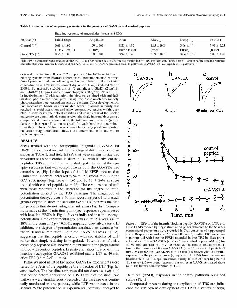
RESULTSSlices treated with the hexapeptide antagonist GAVSTA for50–90 min exhibited no evident physiological disturbances and, asshown in Table 1, had field EPSPs that were similar in size andwaveform to those recorded in slices infused with inactive controlpeptides. TBS resulted in an immediate potentiation of the syn-aptic responses that was comparable in both the GAVSTA andcontrol slices (Fig. 1); the slopes of the field EPSPs measured at2 min after TBS were increased by 54 6 22% (mean 6 SD) in theGAVSTA group (Fig. 1a; n 5 16) and by 66 6 26% in slicestreated with control peptide (n 5 16). These values accord wellwith those reported in the literature for the degree of initialpotentiation elicited by the TBS paradigm. The magnitude ofpotentiation decayed over a 40 min recording period to a muchgreater degree in slices infused with GAVSTA than was the casefor peptides that do not antagonize integrins (Fig. 1d). Compar-isons made at the 40 min time point (see responses superimposedwith baseline EPSPs in Fig. 1, b vs c) indicated that the averagepotentiation in the experimental group was 206 15% versus 49618% in the controls ( p , 0.0001; unpaired, two-tailed t test). Inaddition, the degree of potentiation continued to decrease be-tween 30 and 40 min after TBS in the GAVSTA slices (Fig. 1d),suggesting that the peptide interferes with the stability of LTPrather than simply reducing its magnitude. Potentiation of a sizecommonly reported was, however, maintained in the preparationsinfused with control peptides. In particular, slices treated with theinactive hexapeptide GRADSP exhibited stable LTP at 40 minafter TBS (46 6 24%, n 5 6).Pathways used in 10 of the above GAVSTA experiments were
tested for effects of the peptide before induction of LTP (Fig. 1d,open circles). The baseline responses did not decrease over a 40min period before application of TBS. In four of the slices, twopathways were simultaneously recorded; the baseline was contin-ually monitored in one pathway while LTP was induced in thesecond. While potentiation in experimental pathways decayed to
18 6 8% (6SD), responses in the control pathways remainedstable (Fig. 2).Compounds present during the application of TBS can influ-
ence the subsequent development of LTP in a variety of ways,
Table 1. Comparison of response parameters in the presence of GAVSTA and control peptides
Peptide (n)
Baseline response characteristics (mean 6 SEM)
Initial slope Amplitude Area Rise t1/2 Decay t1/2 1⁄2 width
Control (16) 0.60 6 0.02 1.29 6 0.04 8.25 6 0.37 1.95 6 0.06 3.96 6 0.14 5.91 6 0.23(2mV z ms21) (2mV) (mV z msec) (msec) (msec) (msec)
GAVSTA (16) 0.59 6 0.03 1.38 6 0.05 8.94 6 0.40 2.09 6 0.05 3.86 6 0.15 6.07 6 0.20
Field EPSP parameters were assessed during the 1–2 min period immediately before the application of TBS. Peptides were infused for 50–90 min before baseline responsecharacteristics were measured. Control: 2 mM ASG or 0.8 mM GRADSP, measured from 16 pathways. GAVSTA: 0.8 mM peptide in 16 pathways.
Figure 1. Effects of the integrin blocking peptide GAVSTA on LTP. a–c,Field EPSPs evoked by single stimulation pulses delivered to the Schaffercommissural projections were recorded in CA1 dendrites of hippocampalslices. Responses recorded at 2 (a) and 40 min (b, c) after TBS are shownsuperimposed with baseline EPSPs recorded before TBS in slices prein-cubated with 1 mM GAVSTA (a, b) or 2 mM control peptide ASG (c) for50–90 min (calibration: 1 mV, 10 msec). d, The time course of potentia-tion in the presence of 0.8 mM GAVSTA (n 5 16) or control peptide (2mM ASG or 0.8 mM GRADSP; n 5 16 total) is shown with the resultsexpressed as the percent change (group mean 6 SEM) from the averagebaseline field EPSP slope, measured during 15 min of recording beforeTBS (arrow). Open circles represent responses in GAVSTA-treated slices(n 5 10) before administration of TBS.
1322 J. Neurosci., February 15, 1997, 17(4):1320–1329 Bahr et al. • LTP Stabilization and the Adhesion Molecule Synaptegrin-1
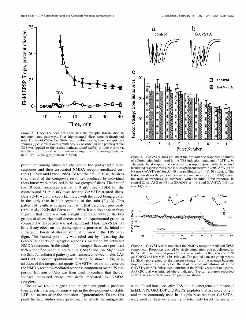
prominent among which are changes in the postsynaptic burstresponses and their associated NMDA receptor-mediated cur-rents (Larson and Lynch, 1988). To test the first of these, the sizes(i.e., areas) of the composite responses produced by individualtheta bursts were measured in the two groups of slices. The first ofthe 10 burst responses was 50 6 8 mVzmsec (6SD) for thecontrols and 51 6 6 mVzmsec for the GAVSTA-treated slices.Bursts 2–10 were markedly facilitated with the effect being greaterin the early than in later segments of the train (Fig. 3). Thispattern of results is in agreement with that described previously(Arai et al., 1990b; del Cerro et al., 1990). It can also be seen fromFigure 3 that there was only a slight difference between the twogroups of slices; the small decrease in the experimental group ascompared with controls was not significant. Thus, GAVSTA haslittle if any effect on the postsynaptic responses to the initial orsubsequent bursts of afferent stimulation used in the TBS para-digm. The second possibility was ruled out by measuring theGAVSTA effects on synaptic responses mediated by activatedNMDA receptors. In this study, hippocampal slices were perfusedwith a modified medium containing CNQX and low Mg21, andthe Schaffer collateral pathway was transected between fields CA3and CA1 to prevent spontaneous bursting. As shown in Figure 4,infusion of the integrin antagonist GAVSTA had no influence onthe NMDA receptor-mediated response component over a 75 minperiod. Infusion of AP5 was then used to confirm that the re-sponses measured were exclusively mediated by NMDAreceptors.The above results suggest that integrin antagonists produce
their effects by acting on some stage in the development of stableLTP that occurs after the induction of potentiation. To test thispoint further, studies were performed in which the antagonists
were infused into slices after TBS and the emergence of enhancedfield EPSPs. GRGDSP and RGDS, peptides that are more potentand more commonly used in integrin research than GAVSTA,were used in these experiments to selectively target the recogni-
Figure 2. GAVSTA does not affect baseline synaptic transmission innonpotentiated pathways. Four hippocampal slices were preincubatedwith 1 mM GAVSTA for 50–60 min. Subsequently, basal synaptic re-sponses (open circles) were simultaneously recorded in one pathway whileTBS was applied to the second pathway (solid circles) at time 0 (arrow).Results are expressed as the percent change from the average baselinefield EPSP slope (group mean 6 SEM). Figure 3. GAVSTA does not affect the postsynaptic responses to bursts
of afferent stimulation used in the TBS induction paradigm of LTP. a, b,The initial burst response of a series of 10 is superimposed with the secondfacilitated response measured in slices preincubated with 2 mM ASG (a) or0.8 mM GAVSTA (b) for 50–90 min (calibration: 1 mV, 20 msec). c, Thehistogram shows the percent increase in burst area (mean 6 SEM) acrossthe train of responses, as compared with the initial burst response, incontrol (2 mM ASG or 0.8 mM GRADSP; n 5 14) and GAVSTA (0.8 mM;n 5 14) slices.
Figure 4. GAVSTA does not affect the NMDA receptor-mediated EPSPcomponent. Responses elicited by single stimulation pulses delivered tothe Schaffer commissural projections were recorded in the presence of 20mM CNQX and low Mg21 (50–100 mM). The plotted data are group means(6 SEM) represented as the percent change from the average baselineslope measured 15 min before the start of constant infusion of 1 mMGAVSTA (n5 7). Subsequent infusion of the NMDA receptor antagonistAP5 (200 mM) was initiated where indicated. Typical responses recordedat the times indicated above the graph are shown.
Bahr et al. • LTP Stabilization and the Adhesion Molecule Synaptegrin-1 J. Neurosci., February 15, 1997, 17(4):1320–1329 1323
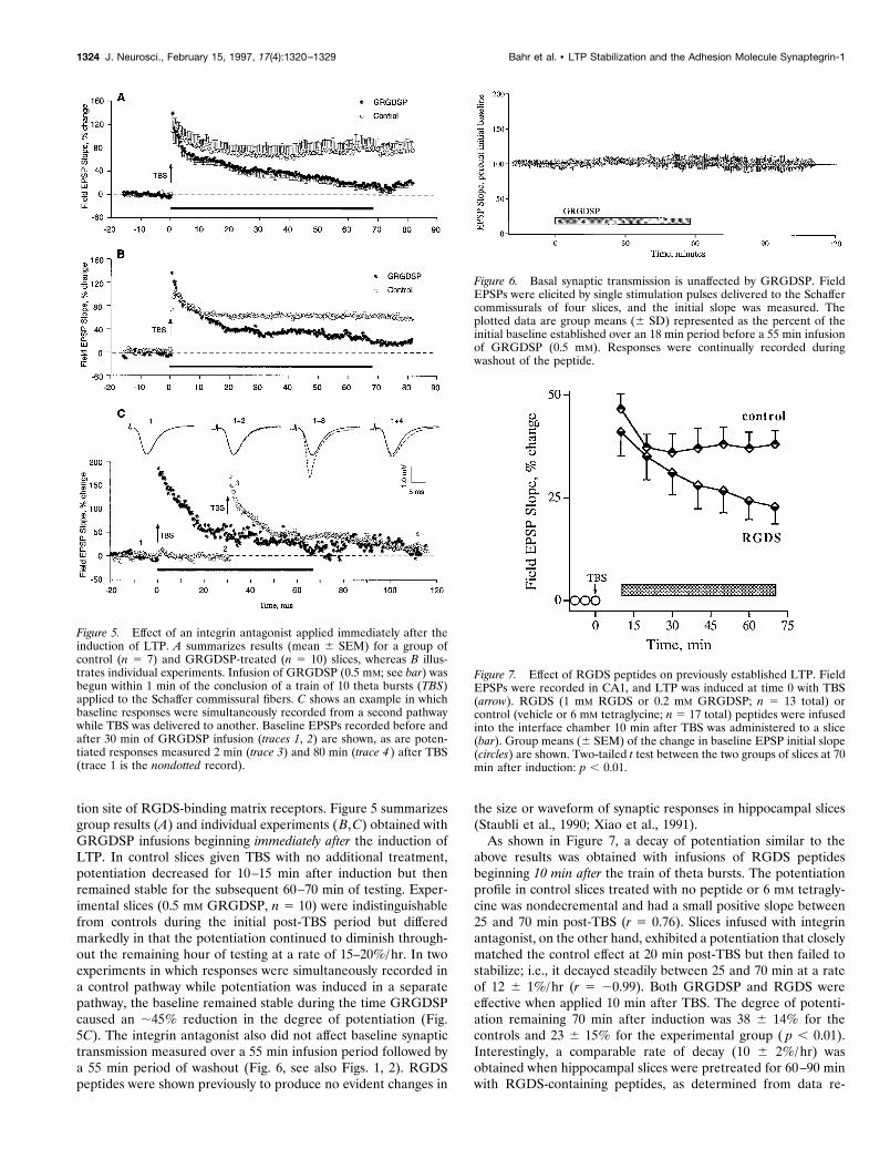
tion site of RGDS-binding matrix receptors. Figure 5 summarizesgroup results (A) and individual experiments (B,C) obtained withGRGDSP infusions beginning immediately after the induction ofLTP. In control slices given TBS with no additional treatment,potentiation decreased for 10–15 min after induction but thenremained stable for the subsequent 60–70 min of testing. Exper-imental slices (0.5 mM GRGDSP, n 5 10) were indistinguishablefrom controls during the initial post-TBS period but differedmarkedly in that the potentiation continued to diminish through-out the remaining hour of testing at a rate of 15–20%/hr. In twoexperiments in which responses were simultaneously recorded ina control pathway while potentiation was induced in a separatepathway, the baseline remained stable during the time GRGDSPcaused an ;45% reduction in the degree of potentiation (Fig.5C). The integrin antagonist also did not affect baseline synaptictransmission measured over a 55 min infusion period followed bya 55 min period of washout (Fig. 6, see also Figs. 1, 2). RGDSpeptides were shown previously to produce no evident changes in
the size or waveform of synaptic responses in hippocampal slices(Staubli et al., 1990; Xiao et al., 1991).As shown in Figure 7, a decay of potentiation similar to the
above results was obtained with infusions of RGDS peptidesbeginning 10 min after the train of theta bursts. The potentiationprofile in control slices treated with no peptide or 6 mM tetragly-cine was nondecremental and had a small positive slope between25 and 70 min post-TBS (r 5 0.76). Slices infused with integrinantagonist, on the other hand, exhibited a potentiation that closelymatched the control effect at 20 min post-TBS but then failed tostabilize; i.e., it decayed steadily between 25 and 70 min at a rateof 12 6 1%/hr (r 5 20.99). Both GRGDSP and RGDS wereeffective when applied 10 min after TBS. The degree of potenti-ation remaining 70 min after induction was 38 6 14% for thecontrols and 23 6 15% for the experimental group ( p , 0.01).Interestingly, a comparable rate of decay (10 6 2%/hr) wasobtained when hippocampal slices were pretreated for 60–90 minwith RGDS-containing peptides, as determined from data re-
Figure 5. Effect of an integrin antagonist applied immediately after theinduction of LTP. A summarizes results (mean 6 SEM) for a group ofcontrol (n 5 7) and GRGDSP-treated (n 5 10) slices, whereas B illus-trates individual experiments. Infusion of GRGDSP (0.5 mM; see bar) wasbegun within 1 min of the conclusion of a train of 10 theta bursts (TBS)applied to the Schaffer commissural fibers. C shows an example in whichbaseline responses were simultaneously recorded from a second pathwaywhile TBS was delivered to another. Baseline EPSPs recorded before andafter 30 min of GRGDSP infusion (traces 1, 2) are shown, as are poten-tiated responses measured 2 min (trace 3) and 80 min (trace 4 ) after TBS(trace 1 is the nondotted record).
Figure 6. Basal synaptic transmission is unaffected by GRGDSP. FieldEPSPs were elicited by single stimulation pulses delivered to the Schaffercommissurals of four slices, and the initial slope was measured. Theplotted data are group means (6 SD) represented as the percent of theinitial baseline established over an 18 min period before a 55 min infusionof GRGDSP (0.5 mM). Responses were continually recorded duringwashout of the peptide.
Figure 7. Effect of RGDS peptides on previously established LTP. FieldEPSPs were recorded in CA1, and LTP was induced at time 0 with TBS(arrow). RGDS (1 mM RGDS or 0.2 mM GRGDSP; n 5 13 total) orcontrol (vehicle or 6 mM tetraglycine; n 5 17 total) peptides were infusedinto the interface chamber 10 min after TBS was administered to a slice(bar). Group means (6 SEM) of the change in baseline EPSP initial slope(circles) are shown. Two-tailed t test between the two groups of slices at 70min after induction: p , 0.01.
1324 J. Neurosci., February 15, 1997, 17(4):1320–1329 Bahr et al. • LTP Stabilization and the Adhesion Molecule Synaptegrin-1
ported by Xiao et al. (1991). Pretreatment with the antagonistGAVSTA also caused a similar decay rate of 11 6 1%/hr starting25 min after TBS (see Fig. 1d). As was the case for control slicesin Figure 7, the controls in Figure 1d exhibited nondecrementalLTP during the latter part of the test period. It appears, then, thatblocking integrin adhesion sites before or after LTP inductionproduces nearly equal rates of decline in the magnitude ofpotentiation.In an attempt to identify targets of the integrin antagonists used
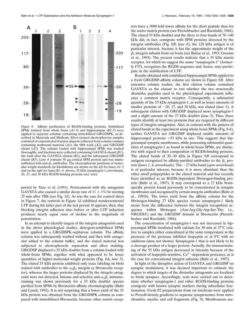
in the above physiological studies, detergent-solubilized SPMswere applied to a GRGDSPK–sepharose column. The affinitycolumn was subsequently washed without and then with antago-nist added to the column buffer, and the eluted material wassubjected to electrophoretic separation and silver staining.GRGDSP displaced a 55 kDa doublet from a column loaded withwhole-brain SPMs, together with what appeared to be lesserquantities of higher-molecular-weight proteins (Fig. 8A, lane 3).The eluted 55 kDa protein exhibited only trace labeling on blotsstained with antibodies to the a5b1 integrin (a fibronectin recep-tor), whereas the larger proteins displaced by the integrin antag-onist were not detected. Intense and selective anti-a5b1 immuno-staining was shown previously for a 55 kDa doublet speciespurified from SPMs by fibronectin-affinity chromatography (Bahrand Lynch, 1992). It is not surprising that a lower yield of the 55kDa protein was obtained from the GRGDSPK column as com-pared with immobilized fibronectin, because other matrix recep-
tors have a 3000-fold lower affinity for the short peptide than forthe native matrix protein (see Pierschbacher and Ruoslahti, 1984).The eluted 55 kDa doublet and the three to four bands of 70–140kDa did, in fact, comigrate with SPM proteins detected by theintegrin antibodies (Fig. 8B, lane 4); the 120 kDa antigen is ofparticular interest, because it has the approximate weight of theb1 integrin subunit from rat brain (see Balzac et al., 1993; Groomset al., 1993). The present results indicate that a 55 kDa matrixreceptor, for which we suggest the name “synaptegrin-1” (former-ly F55), recognizes the RGDS sequence and, hence, may partici-pate in the stabilization of LTP.Results obtained with solubilized hippocampal SPMs applied to
a fresh GRGDSP-affinity column are shown in Figure 8B. Afterextensive column washes, the first elution volume containedGAVSTA as the elutant to test whether the two structurallydissimilar peptides used in the physiological experiments influ-ence a common matrix receptor. Consequently, a substantialquantity of the 55 kDa synaptegrin-1, as well as lesser amounts ofsmaller proteins of ;20, 27, and 30 kDa, was eluted (lane 1). Asubsequent elution with GRGDSP displaced more synaptegrin-1and a slight amount of the 27 kDa doublet (lane 3). Thus, theseresults identify at least two proteins that are targeted by differenttypes of integrin antagonists. Also, in contrast with the pattern ofeluted bands in the experiment using whole-brain SPMs (Fig. 8A),neither GAVSTA nor GRGDSP displaced sizable amounts ofhippocampal proteins .55 kDa in size. This suggests that hip-pocampal synaptic membranes, while possessing substantial quan-tities of synaptegrin-1 as found in whole-brain SPMs, are distinc-tive with regard to their composition of RGDS-binding proteins.The eluted bands of 20–30 kDa in Figure 8B correspond toantigens recognized by affinity-purified antibodies to the b1 pro-tein (lanes 1, 4, arrowheads). The;27 kDa band (open arrowhead)is of particular interest, because it is more abundant than theother small polypeptides in the eluted material and has recentlybeen identified as an RGDS-dependent fibrinogen-binding pro-tein (Bahr et al., 1997). It may correspond to a 27 kDa brain-specific protein found previously to be concentrated in synapticmembranes and recognized by certain integrin antibodies (Bahr etal., 1991b). The lower yield from the affinity column for thefibrinogen-binding 27 kDa species versus synaptegrin-1 likelystems from the difference between the integrin recognition se-quence within fibrinogen (Asn-Arg-Gly-Asp-Ser-Thr; i.e.,NRGDST) and the GRGDSP domain in fibronectin (Piersch-bacher and Ruoslahti, 1984).The concentration of synaptegrin-1 was not increased in hip-
pocampal SPMs incubated with calcium for 30 min at 378C rela-tive to samples either coincubated at the same temperature in thepresence of the protease inhibitor leupeptin or at 08C with noadditions (data not shown). Synaptegrin-1 thus is not likely to bea cleavage product of a larger protein. Actually, the immunostain-ing of the 55 kDa antigen decreased in intensity because of theactivation of leupeptin-sensitive, Ca21-dependent proteases, as isthe case for conventional integrin subunits (Bahr et al., 1997).In light of the disruptive action of GAVSTA and GRGDSP on
synaptic modulation, it was deemed important to estimate thedegree to which targets of the dissimilar antagonists are localizedto brain synapses. Accordingly, tests were carried out to deter-mine whether synaptegrin-1 and other RGDS-binding proteinscomigrate with known synaptic markers during subcellular frac-tionation. Fresh P2 preparations from rat forebrain were appliedto Percoll-density gradients to separate synaptosomes from mito-chondria, myelin, and cell fragments (Fig. 9). Membranous ma-
Figure 8. Affinity purification of RGDS-binding proteins. SolubilizedSPMs isolated from whole brain (A1-3) and hippocampus (B1-3) wereapplied to separate columns containing immobilized GRGDSPK, as de-scribed in Materials and Methods. Silver-stained electrophoresis samplesconsisted of concentrated fraction aliquots collected from column volumescontaining nonbound material (A1), the fifth wash (A2), and GRGDSPeluant (A3). The column loaded with hippocampal SPMs was washedthoroughly, and fractions were collected containing GAVSTA eluant (B1),the wash after the GAVSTA elution (B2), and the subsequent GRGDSPeluant (B3). Lane 4 contains 50 mg cortical SPM protein and was immu-noblotted with anti-b1 antibodies. The electrophoretic positions of molec-ular weight standards (in kilodaltons) are shown on the left for lanes A1–3and on the right for lanes B1–4. Arrows, 55 kDa synaptegrin-1; arrowheads,20, 27, and 30 kDa RGDS-binding proteins (see text).
Bahr et al. • LTP Stabilization and the Adhesion Molecule Synaptegrin-1 J. Neurosci., February 15, 1997, 17(4):1320–1329 1325
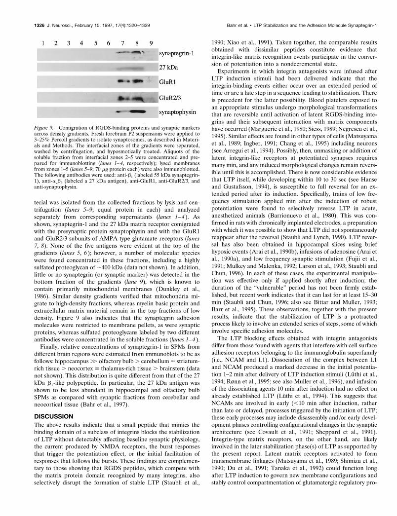
terial was isolated from the collected fractions by lysis and cen-trifugation (lanes 5–9; equal protein in each) and analyzedseparately from corresponding supernatants (lanes 1–4). Asshown, synaptegrin-1 and the 27 kDa matrix receptor comigratedwith the presynaptic protein synaptophysin and with the GluR1and GluR2/3 subunits of AMPA-type glutamate receptors (lanes7, 8). None of the five antigens were evident at the top of thegradients (lanes 5, 6); however, a number of molecular specieswere found concentrated in these fractions, including a highlysulfated proteoglycan of ;400 kDa (data not shown). In addition,little or no synaptegrin (or synaptic marker) was detected in thebottom fraction of the gradients (lane 9), which is known tocontain primarily mitochondrial membranes (Dunkley et al.,1986). Similar density gradients verified that mitochondria mi-grate to high-density fractions, whereas myelin basic protein andextracellular matrix material remain in the top fractions of lowdensity. Figure 9 also indicates that the synaptegrin adhesionmolecules were restricted to membrane pellets, as were synapticproteins, whereas sulfated proteoglycans labeled by two differentantibodies were concentrated in the soluble fractions (lanes 1–4).Finally, relative concentrations of synaptegrin-1 in SPMs from
different brain regions were estimated from immunoblots to be asfollows: hippocampus.. olfactory bulb. cerebellum' striatum-rich tissue . neocortex $ thalamus-rich tissue . brainstem (datanot shown). This distribution is quite different from that of the 27kDa b1-like polypeptide. In particular, the 27 kDa antigen wasshown to be less abundant in hippocampal and olfactory bulbSPMs as compared with synaptic fractions from cerebellar andneocortical tissue (Bahr et al., 1997).
DISCUSSIONThe above results indicate that a small peptide that mimics thebinding domain of a subclass of integrins blocks the stabilizationof LTP without detectably affecting baseline synaptic physiology,the current produced by NMDA receptors, the burst responsesthat trigger the potentiation effect, or the initial facilitation ofresponses that follows the bursts. These findings are complemen-tary to those showing that RGDS peptides, which compete withthe matrix protein domain recognized by many integrins, alsoselectively disrupt the formation of stable LTP (Staubli et al.,
1990; Xiao et al., 1991). Taken together, the comparable resultsobtained with dissimilar peptides constitute evidence thatintegrin-like matrix recognition events participate in the conver-sion of potentiation into a nondecremental state.Experiments in which integrin antagonists were infused after
LTP induction stimuli had been delivered indicate that theintegrin-binding events either occur over an extended period oftime or are a late step in a sequence leading to stabilization. Thereis precedent for the latter possibility. Blood platelets exposed toan appropriate stimulus undergo morphological transformationsthat are reversible until activation of latent RGDS-binding inte-grins and their subsequent interaction with matrix componentshave occurred (Marguerie et al., 1980; Siess, 1989; Negrescu et al.,1995). Similar effects are found in other types of cells (Matsuyamaet al., 1989; Ingber, 1991; Chang et al., 1995) including neurons(see Arregui et al., 1994). Possibly, then, unmasking or addition oflatent integrin-like receptors at potentiated synapses requiresmany min, and any induced morphological changes remain revers-ible until this is accomplished. There is now considerable evidencethat LTP itself, while developing within 10 to 30 sec (see Hanseand Gustafsson, 1994), is susceptible to full reversal for an ex-tended period after its induction. Specifically, trains of low fre-quency stimulation applied min after the induction of robustpotentiation were found to selectively reverse LTP in acute,anesthetized animals (Barrionuevo et al., 1980). This was con-firmed in rats with chronically implanted electrodes, a preparationwith which it was possible to show that LTP did not spontaneouslyreappear after the reversal (Staubli and Lynch, 1990). LTP rever-sal has also been obtained in hippocampal slices using briefhypoxic events (Arai et al., 1990b), infusions of adenosine (Arai etal., 1990a), and low frequency synaptic stimulation (Fujii et al.,1991; Mulkey and Malenka, 1992; Larson et al., 1993; Staubli andChun, 1996). In each of these cases, the experimental manipula-tion was effective only if applied shortly after induction; theduration of the “vulnerable” period has not been firmly estab-lished, but recent work indicates that it can last for at least 15–30min (Staubli and Chun, 1996; also see Bittar and Muller, 1993;Barr et al., 1995). These observations, together with the presentresults, indicate that the stabilization of LTP is a protractedprocess likely to involve an extended series of steps, some of whichinvolve specific adhesion molecules.The LTP blocking effects obtained with integrin antagonists
differ from those found with agents that interfere with cell surfaceadhesion receptors belonging to the immunoglobulin superfamily(i.e., NCAM and L1). Dissociation of the complex between L1and NCAM produced a marked decrease in the initial potentia-tion 1–2 min after delivery of LTP induction stimuli (Luthi et al.,1994; Rønn et al., 1995; see also Muller et al., 1996), and infusionof the dissociating agents 10 min after induction had no effect onalready established LTP (Luthi et al., 1994). This suggests thatNCAMs are involved in early (,10 min after induction, ratherthan late or delayed, processes triggered by the initiation of LTP;these early processes may include disassembly and /or early devel-opment phases controlling configurational changes in the synapticarchitecture (see Covault et al., 1991; Sheppard et al., 1991).Integrin-type matrix receptors, on the other hand, are likelyinvolved in the later stabilization phase(s) of LTP as supported bythe present report. Latent matrix receptors activated to formtransmembrane linkages (Matsuyama et al., 1989; Shimizu et al.,1990; Du et al., 1991; Tanaka et al., 1992) could function longafter LTP induction to govern new membrane configurations andstably control compartmentation of glutamatergic regulatory pro-
Figure 9. Comigration of RGDS-binding proteins and synaptic markersacross density gradients. Fresh forebrain P2 suspensions were applied to3–25% Percoll gradients to isolate synaptosomes, as described in Materi-als and Methods. The interfacial zones of the gradients were separated,washed by centrifugation, and hyposmotically treated. Aliquots of thesoluble fraction from interfacial zones 2–5 were concentrated and pre-pared for immunoblotting (lanes 1–4, respectively); lysed membranesfrom zones 1–5 (lanes 5–9; 70 mg protein each) were also immunoblotted.The following antibodies were used: anti-b1 (labeled 55 kDa synaptegrin-1), anti-avb3 (labeled a 27 kDa antigen), anti-GluR1, anti-GluR2/3, andanti-synaptophysin.
1326 J. Neurosci., February 15, 1997, 17(4):1320–1329 Bahr et al. • LTP Stabilization and the Adhesion Molecule Synaptegrin-1

teins (Greengard et al., 1991; Wang et al., 1991; Rosenmund andWestbrook, 1993; Rosenmund et al., 1994; Yakel et al., 1995; seealso Sheng, 1996). Thus, distinct phases of LTP consolidation aredistinguished by separate classes of adhesion molecules. Accord-ing to this idea, the two types of adhesion molecules constitute asystem that first stabilizes a potentiated state of excitatory syn-apses and then establishes an appropriate environment for main-taining the new functional state. Note that the stabilization phaseoccurs between induction and the end of the “vulnerable” perioddescribed above. Beyond this period, LTP reversing stimuli are nolonger effective and no active processes appear to be involved inthe expression of the potentiated state (see Larson and Vanderk-lish, 1997). One plausible scenario is that the synaptic structurehas been redefined by strategically localized focal adhesions, andthis new structure is maintained by normal cellular processes.Experiments using affinity chromatography confirmed that hip-
pocampal synapses are enriched in a small set of proteins thatbind the RGDS sequence and hence are likely to be functionallydisrupted by the integrin antagonists used to prevent LTP stabi-lization. One of these proteins with a molecular weight of 55 kDais particularly well suited to serve as a synaptic, integrin-likereceptor (i.e., a synaptegrin), because it (1) binds to a matrixprotein in a selective manner, (2) recognizes the RGDS sequence,(3) is labeled by antibodies to specific integrin receptors andsubunits, (4) does not appear to be found outside the brain, (5) isgreatly enriched in synaptic membrane fractions, and (6) comi-grates with synaptic marker proteins during the isolation of syn-aptosomes. This matrix receptor, referred to here assynaptegrin-1, is substantially smaller than the subunits of knownintegrins, thus raising the possibility that it is a cleavage product ofperhaps the b1 integrin subunit. However, this was not evident inproteolyzed samples and, moreover, would require the pertinentprotease to be (1) extremely active (the b1 protein does notco-occur in sizable concentrations with synaptegrin-1 in any tissuefraction examined thus far), (2) resistant to a variety of broad-spectrum inhibitors (such compounds are added to all dissectionand fractionation buffers), and (3) brain-specific [b1 is foundthroughout the body (Balzac et al., 1993) but synaptegrin-1 is not(Bahr and Lynch, 1992)]. The size of the synaptegrin adhesionmolecule is also similar to that of the major species phosphory-lated in cultured neuronal cells in response to anandamide, anagent that regulates a focal adhesion kinase found concentrated inhippocampal neurons (Derkinderen et al., 1996; see also Burgayaet al., 1995). Anandamide is an endogenous ligand for centralcannabinoid receptors, is released after neuronal depolarization,and initiates a signal transduction pathway implicated in cytoskel-etal modulation. Given the characteristics of synaptegrin-1, it islikely that the matrix receptor is an integrin variant with proper-ties that are specialized for functional regulation of synapses.
REFERENCESAkiyama SK, Yamada SS, Chen WT, Yamada KM (1989) Analysis offibronectin receptor function with monoclonal antibodies: roles in celladhesion, migration, matrix assembly, and cytoskeletal organization.J Cell Biol 109:863–875.
Albelda SM, Buck CA (1990) Integrins and other cell adhesion mole-cules. FASEB J 4:2868–2880.
Arai A, Kessler M, Lynch G (1990a) The effects of adenosine on thedevelopment of long-term potentiation. Neurosci Lett 119:41–44.
Arai A, Larson J, Lynch G (1990b) Anoxia reveals a vulnerable period inthe development of long-term potentiation. Brain Res 511:353–357.
Arregui CO, Carbonetto S, McKerracher L (1994) Characterization ofneural cell adhesion sites: point contacts are the sites of interaction
between integrins and the cytoskeleton in PC12 cells. J Neuroscience14:6967–6977.
Bahr BA, Lynch G (1992) Purification of an Arg-Gly-Asp selective ma-trix receptor from brain synaptic plasma membranes. Biochem J281:137–142.
Bahr BA, Sheppard A, Lynch G (1991a) Fibronectin binding by brainsynaptosomal membranes may not involve conventional integrins. Neu-roReport 2:13–16.
Bahr BA, Sheppard A, Vanderklish PW, Bakus BL, Capaldi D, Lynch G(1991b) Antibodies to the avb3 integrin label a protein concentrated inbrain synaptosomal membrane. NeuroReport 2:321–324.
Bahr BA, Capaldi D, Rosario R, Esteban ET, Lynch G (1997) A 27 kDamatrix receptor from rat brain synaptosomes: selective recognition ofthe Arg-Gly-Asp-Ser domain and resistance to calcium-dependent pro-teolysis, in press.
Balzac F, Belkin AM, Koteliansky VE, Balabanov YV, Altruda F,Silengo L, Tarone G (1993) Expression and functional analysis of acytoplasmic domain variant of the b1 integrin subunit. J Cell Biol121:171–178.
Barr DS, Lambert NA, Hoyt KL, Moore SD, Wilson WA (1995) Induc-tion and reversal of long-term potentiation by low- and high-intensitytheta pattern stimulation. J Neurosci 15:5402–5410.
Barrionuevo G, Schottler F, Lynch G (1980) The effects of repetitive lowfrequency stimulation on control and “potentiated” synaptic responsesin the hippocampus. Life Sci 27:2385–2391.
Bittar P, Muller D (1993) Time-dependent reversal of long-term poten-tiation by brief cooling shocks in rat hippocampal slices. Brain Res620:181–188.
Bliss TVP, Gardner-Medwin AR (1973) Long-lasting potentiation ofsynaptic transmission in the dentate area of the unanesthetized rabbitfollowing stimulation of the perforant path. J Physiol (Lond)232:357–374.
Bliss TVP, Lomo T (1973) Long-lasting potentiation of synaptic trans-mission in the dentate area of the anaesthetized rabbit following stim-ulation of the perforant path. J Physiol (Lond) 232:334–356.
Brentani RR, Ribeiro SF, Potocnjak P, Pasqualini R, Lopes JD, NakaieCR (1988) Characterization of the cellular receptor for fibronectinthrough a hydropathic complementary approach. Proc Natl Acad SciUSA 85:364–367.
Buchs P-A, Muller D (1996) Induction of long-term potentiation is as-sociated with major ultrastructural changes of activated synapses. ProcNatl Acad Sci USA 93:8040–8045.
Burgaya F, Menegon A, Menegoz M, Valtorta F, Girault JA (1995)Focal adhesion kinase in rat central nervous system. Eur J Neurosci7:1810–1821.
Burridge K, Fath K, Kelly T, Nuckolls G, Turner C (1988) Focal adhe-sions: transmembrane junctions between the extracellular matrix andthe cytoskeleton. Annu Rev Cell Biol 4:478–525.
Chang F-LF, Greenough WT (1984) Transient and enduring morpholog-ical correlates of synaptic activity and efficacy change in the rat hip-pocampal slice. Brain Res 309:35–46.
Chang MC, Jeng JH, Cheong TC, Huang TF (1995) The morphologicchange of endothelial cells by ancrod-generated fibrin is triggered byavb3 integrin binding and the subsequent activation of a G-proteincoupled phospholipase C. Biochim Biophys Acta 1269:115–121.
Clark EA, Brugge JS (1995) Integrins and signal transduction pathways:the road taken. Science 268:233–239.
Covault J, Liu Q, El-Deeb S (1991) Calcium-activated proteolysis ofintracellular domains in the cell adhesion molecules NCAM andN-cadherin. Mol Brain Res 11:11–16.
del Cerro S, Larson J, Oliver MW, Lynch G (1990) Development ofhippocampal long-term potentiation is reduced by recently introducedcalpain inhibitors. Brain Res 530:91–95.
Derkinderen P, Toutant M, Burgaya F, Le Bert M, Siciliano JC, deFranciscis V, Gelman M, Girault J-A (1996) Regulation of a neuronalform of focal adhesion kinase by anandamide. Science 273:1719–1722.
Desmond NL, Levy WB (1983) Synaptic correlates of associative poten-tiation /depression: an ultrastructural study in the hippocampus. BrainRes 265:21–30.
Du XP, Plow EF, Frelinger AL, O’Toole TE, Loftus JC, Ginsberg MH(1991) Ligands “activate” integrin aIIbb3 (platelet GPIIb-IIIa). Cell65:409–416.
Dunkley PR, Jarvie PE, Heath JW, Kidd GJ, Rostas JAP (1986) A rapidmethod for isolation of synaptosomes on percoll gradients. Brain Res372:115–129.
Bahr et al. • LTP Stabilization and the Adhesion Molecule Synaptegrin-1 J. Neurosci., February 15, 1997, 17(4):1320–1329 1327
Einheber S, Schnapp LM, Salzer JL, Cappiello ZB, Milner TA (1996)Regional and ultrastructural distribution of the a8 integrin subunit indeveloping and adult rat brain suggests a role in synaptic function.J Comp Neurol 370:105–134.
Fogerty FJ, Akiyama SK, Yamada KM, Mosher DF (1990) Inhibition ofbinding of fibronectin to matrix assembly sites by anti-integrin (alpha 5beta 1) antibodies. J Cell Biol 111:699–708.
Fujii S, Saito K, Miyakawa H, Ito K, Kato H (1991) Reversal of long-term potentiation (depotentiation) induced by tetanus stimulation ofthe input to CA1 neurons of guinea pig hippocampal slices. Brain Res555:112–122.
Geinisman Y, de Toledo-Morrell L, Morrell F, Heller RE, Rossi M,Parshall RF (1993) Structural synaptic correlates of long-term poten-tiation: formation of axospinous synapses with multiple, completelypartitioned transmission zones. Hippocampus 3:435–445.
Greengard P, Jen J, Nairn AC, Stevens CF (1991) Enhancement of theglutamate response by cAMP-dependent protein kinase in hippocampalneurons. Science 253:1135–1138.
Grooms SY, Terracio L, Jones LS (1993) Anatomical localization of b1integrin-like immunoreactivity in rat brain. Exp Neurol 122:253–259.
Gustafsson B, Asztely F, Hanse E, Wigstrom H (1989) Onset character-istics of long-term potentiation in the guinea-pig hippocampal CA1region in vitro. Eur J Neurosci 1:382–394.
Hanse E, Gustafsson B (1994) Onset and stabilization of NMDAreceptor-dependent hippocampal long-term potentiation. Neurosci Res20:15–25.
Horwitz A, Duggan E, Buck C, Beckerle MC, Burridge K (1986) Inter-action of plasma membrane fibronectin receptor with talin: a transmem-brane linkage. Nature 320:531–533.
Hynes RO (1992) Integrins: versatility, modulation, and signaling in celladhesion. Cell 69:11–25.
Ingber D (1991) Extracellular matrix and cell shape: potential controlpoints for inhibition of angiogenesis. J Cell Biochem 47:236–241.
Jones LS (1996) Integrins: possible functions in the adult CNS. TrendsNeurosci 19:68–72.
Jones LS, Grooms S (1995) Localization of the a5 integrin-like immu-nofluorescence in the adult rat hippocampus. Soc Neurosci Abstr21:1313.
Larson J, Lynch G (1988) Role of N-methyl-D-aspartate receptors in theinduction of synaptic potentiation by burst stimulation patterned afterthe hippocampal theta rhythm. Brain Res 441:111–118.
Larson J, Vanderklish PW (1997) Involvement of AMPA receptors inLTP mechanisms and memory. In: Long-term potentiation, Vol 3(Baudry M, Davis JL, eds), pp 73–103. Cambridge: MIT.
Larson J, Wong D, Lynch G (1986) Patterned stimulation at the thetafrequency is optimal for induction of hippocampal long-term potentia-tion. Brain Res 368:347–350.
Larson J, Xiao P, Lynch G (1993) Reversal of LTP by theta frequencystimulation. Brain Res 600:97–102.
Lee K, Oliver M, Schottler F, Creager R, Lynch G (1979) Ultra-structural effects of repetitive synaptic stimulation in the hippocampalslice preparation: a preliminary report. Exp Neurol 65:478–480.
Lee K, Schottler F, Oliver M, Lynch G (1980) Brief bursts of high-frequency stimulation produce two types of structural changes in rathippocampus. J Neurophysiol 44:247–258.
Luthi A, Parent J-P, Figurov D, Muller D, Schachner M (1994) Hip-pocampal long-term potentiation and neural cell adhesion molecules L1and NCAM. Nature 372:777–779.
Marguerie GA, Edgington TS, Plow EF (1980) Interaction of fibrinogenwith its platelet receptor as part of a multistep reaction in ADP-inducedplatelet aggregation. J Biol Chem 255:154–161.
Matsuyama T, Yamada A, Kay J, Yamada KM, Akiyama SK, SchlossmanSF, Morimoto C (1989) Activation of CD4 cells by fibronectin andanti-CD3 antibody. A synergistic effect mediated by the VLA-5 fi-bronectin receptor complex. J Exp Med 170:1133–1148.
Mulkey RM, Malenka RC (1992) Mechanisms underlying induction ofhomosynaptic long-term depression in area CA1 of the hippocampus.Neuron 9:967–975.
Muller D, Wang C, Skibo G, Toni N, Cremer H, Calaora V, Rougon G,Kiss JZ (1996) PSA-NCAM is required for activity-induced synapticplasticity. Neuron 17:413–422.
Negrescu EV, de Quintana KL, Siess W (1995) Platelet shape changeinduced by thrombin receptor activation. Rapid stimulation of ty-rosine phosphorylation of novel protein substrates through an
integrin- and Ca21-independent mechanism. J Biol Chem270:1057–1061.
Nosten-Bertrand M, Errington ML, Murphy KPSJ, Tokugawa Y, Bar-boni E, Kozlova E, Michalovich D, Morris RGM, Silva J, Stewart CL,Bliss TVP, Morris RJ (1996) Normal spatial learning despiteregional inhibition of LTP in mice lacking Thy-1. Nature379:826–829.
Otey CA, Pavalko FM, Burridge K (1990) An interaction betweena-actinin and the b1 integrin subunit in vitro. J Cell Biol111:721–729.
Pasqualini R, Chamone DF, Brentani RR (1989) Determination of theputative binding site for fibronectin on platelet glycoprotein IIb-IIIacomplex through a hydropathic complementarity approach. J BiolChem 264:14566–14570.
Pierschbacher MD, Ruoslahti E (1984) Cell attachment activity of fi-bronectin can be duplicated by small synthetic fragments of the mole-cule. Nature 309:30–33.
Persohn E, Pollerberg E, Schachner M (1989) Immunoelectron-microscopic localization of the 180 kDa component of the neural celladhesion molecule NCAM in postsynaptic membranes. J Comp Neurol288:92–100.
Pytela R, Pierschbacher MD, Ruoslahti E (1985) Identification and iso-lation of a 140 kd cell surface glycoprotein with properties expected ofa fibronectin receptor. Cell 40:191–198.
Rønn LCB, Bock E, Linnemann D, Jahnsen H (1995) NCAM-antibodiesmodulate induction of long-term potentiation in rat hippocampal CA1.Brain Res 677:145–151.
Rosales C, O’Brien V, Kornberg L, Juliano R (1995) Signal transductionby cell adhesion receptors. Biochim Biophys Acta 1242:77–98.
Rosenmund C, Carr DW, Bergeson SE, Nilaver G, Scott JD, WestbrookGL (1994) Anchoring of protein kinase A is required for modulationof AMPA/kainate receptors on hippocampal neurons. Nature368:853–856.
Rosenmund C, Westbrook GL (1993) Calcium-induced actin depolymer-ization reduces NMDA channel activity. Neuron 10:805–814.
Ruoslahti E, Pierschbacher MD (1987) New perspectives in cell adhe-sion: RGD and integrins. Science 238:491–497.
Schachner M, Martini R (1995) Glycans and the modulation of neural-recognition molecule function. Trends Neurosci 18:183–191.
Schreiner CL, Bauer JS, Danilov YN, Hussein S, Sczekan MM, JulianoRL (1989) Isolation and characterization of Chinese hamster ovarycell variants deficient in the expression of fibronectin receptor. J CellBiol 109:3157–3167.
Schwartz MA, Ingber DE (1994) Integrating with integrins. Mol Biol Cell5:389–393.
Schwarz MA, Brown PJ, Eveleth DD, Bradshaw RA (1989) Modulationof growth factor induced fiber outgrowth in rat pheochromocytoma(PC12) cells by a fibronectin receptor antibody. J Cell Physiol138:121–128.
Sheng M (1996) PDZs and receptor/channel clustering: rounding up thelatest suspects. Neuron 17:575–578.
Sheppard A, Wu J, Rutishauser U, Lynch G (1991) Proteolytic modifi-cation of neural cell adhesion molecule (NCAM) by the intracellularproteinase calpain. Biochim Biophys Acta 1076:156–160.
Shimizu Y, Van Seventer GA, Horgan KJ, Shaw S (1990) Regulatedexpression and binding of three VLA (b1) integrin receptors on T cells.Nature 345:250–253.
Siess W (1989) Molecular mechanisms of platelet activation. Physiol Rev69:58–178.
Staubli U, Chun D (1996) Factors regulating the reversibility of long-term potentiation. J Neurosci 16:853–860.
Staubli U, Lynch G (1987) Stable hippocampal long-term potentiationelicited by “theta” pattern stimulation. Brain Res 435:227–234.
Staubli U, Lynch G (1990) Stable depression of potentiated synapticresponses in the hippocampus induced by low frequency stimulation.Brain Res 513:113–118.
Staubli U, Vanderklish P, Lynch G (1990) An inhibitor of integrin re-ceptors blocks long-term potentiation. Behav Neural Biol 53:1–5.
Tanaka Y, Albelda SM, Horgan KJ, van Seventer GA, Shimizu Y, New-man W, Hallam J, Newman PJ, Buck CA, Shaw S (1992) CD31 ex-pressed on distinctive T cell subsets is a preferential amplifier of b1integrin-mediated adhesion. J Exp Med 176:245–253.
Wallace CS, Hawrylak N, Greenough WT (1991) Studies of synapticstructural modifications after long-term potentiation and kindling: con-text for a molecular morphology. In: Long-term potentiation: a debate
1328 J. Neurosci., February 15, 1997, 17(4):1320–1329 Bahr et al. • LTP Stabilization and the Adhesion Molecule Synaptegrin-1

of current issues (Baudry M, Davis JL, eds), pp 189–232. Cambridge:MIT.
Wang L-Y, Salter MW, MacDonald JF (1991) Regulation of kainatereceptors by cAMP-dependent protein kinase and phosphatases. Sci-ence 253:1132–1135.
Wenthold RJ, Yokotani N, Doi K, Wada K (1992) Immunochemicalcharacterization of the non-NMDA glutamate receptor using subunit-specific antibodies. J Biol Chem 267:501–507.
Wu C, Bauer JS, Juliano RL, McDonald JA (1993) The a5b1 integrinfibronectin receptor, but not the a5 cytoplasmic domain, functions in anearly and essential step in fibronectin matrix assembly. J Biol Chem268:21883–21888.
Wu C, Chung AE, McDonald JA (1995a) A novel role for a3b1 integrinsin extracellular matrix assembly. J Cell Sci 108:2511–2523.
Wu C, Keivens VM, O’Toole TE, McDonald JA, Ginsberg MH (1995b)Integrin activation and cytoskeletal interaction are essential for theassembly of a fibronectin matrix. Cell 83:715–724.
Xiao P, Bahr BA, Staubli U, Vanderklish PW, Lynch G (1991) Evidencethat matrix recognition contributes to the stabilization but not inductionof LTP. NeuroReport 2:461–464.
Yakel JL, Vissavajjhala P, Derkach VA, Brickey DA, Soderling TR(1995) Identification of a Ca21/calmodulin-dependent protein kinase IIregulatory phosphorylation site in non-N-methyl-D-aspartate glutamatereceptors. Proc Natl Acad Sci USA 92:1376–1380.
Bahr et al. • LTP Stabilization and the Adhesion Molecule Synaptegrin-1 J. Neurosci., February 15, 1997, 17(4):1320–1329 1329