Increased Glucose Metabolism and Glycerolipid Formation by Fatty Acids and GPR40 Receptor Signaling...
15
Charles F. Burant Mary K. Treutelaar, Robert T. Kennedy and Mahmoud El-Azzouny, Charles R. Evans, Secretion Fatty Acid Potentiation of Insulin GPR40 Receptor Signaling Underlies the Glycerolipid Formation by Fatty Acids and Increased Glucose Metabolism and Metabolism: doi: 10.1074/jbc.M113.531970 originally published online March 27, 2014 2014, 289:13575-13588. J. Biol. Chem. 10.1074/jbc.M113.531970 Access the most updated version of this article at doi: . JBC Affinity Sites Find articles, minireviews, Reflections and Classics on similar topics on the Alerts: When a correction for this article is posted • When this article is cited • to choose from all of JBC's e-mail alerts Click here http://www.jbc.org/content/289/19/13575.full.html#ref-list-1 This article cites 42 references, 20 of which can be accessed free at at NYU School of Medicine Library on November 3, 2014 http://www.jbc.org/ Downloaded from at NYU School of Medicine Library on November 3, 2014 http://www.jbc.org/ Downloaded from
Transcript of Increased Glucose Metabolism and Glycerolipid Formation by Fatty Acids and GPR40 Receptor Signaling...

Charles F. BurantMary K. Treutelaar, Robert T. Kennedy and Mahmoud El-Azzouny, Charles R. Evans,
SecretionFatty Acid Potentiation of Insulin GPR40 Receptor Signaling Underlies theGlycerolipid Formation by Fatty Acids and Increased Glucose Metabolism andMetabolism:
doi: 10.1074/jbc.M113.531970 originally published online March 27, 20142014, 289:13575-13588.J. Biol. Chem.
10.1074/jbc.M113.531970Access the most updated version of this article at doi:
.JBC Affinity SitesFind articles, minireviews, Reflections and Classics on similar topics on the
Alerts:
When a correction for this article is posted•
When this article is cited•
to choose from all of JBC's e-mail alertsClick here
http://www.jbc.org/content/289/19/13575.full.html#ref-list-1
This article cites 42 references, 20 of which can be accessed free at
at NY
U School of M
edicine Library on N
ovember 3, 2014
http://ww
w.jbc.org/
Dow
nloaded from
at NY
U School of M
edicine Library on N
ovember 3, 2014
http://ww
w.jbc.org/
Dow
nloaded from

Increased Glucose Metabolism and Glycerolipid Formation byFatty Acids and GPR40 Receptor Signaling Underlies theFatty Acid Potentiation of Insulin Secretion*
Received for publication, November 8, 2013, and in revised form, March 24, 2014 Published, JBC Papers in Press, March 27, 2014, DOI 10.1074/jbc.M113.531970
Mahmoud El-Azzouny‡§, Charles R. Evans‡, Mary K. Treutelaar‡, Robert T. Kennedy§¶, and Charles F. Burant‡1
From the Departments of ‡Internal Medicine, §Chemistry, and ¶Pharmacology, The University of Michigan,Ann Arbor, Michigan 48105
Background: Pathways underlying fatty acid potentiation of glucose-stimulated insulin secretion have not been fullyelucidated.Results: In INS-1 cells, fatty acids increase de novo production of glycerolipids and simultaneously increase glucose utilization.GPR40 receptor activation increases these activities.Conclusion: Fatty acids enhance the production of multiple signals supporting glucose-stimulated insulin secretion.Significance: The studies clarify the effects of fatty acids and GPR40 activity in � cell insulin secretion.
Acute fatty acid (FA) exposure potentiates glucose-stimulatedinsulin secretion in � cells through metabolic and receptor-me-diated effects. We assessed the effect of fatty acids on thedynamics of the metabolome in INS-1 cells following exposureto [U-13C]glucose to assess flux through metabolic pathways.Metabolite profiling showed a fatty acid-induced increase inlong chain acyl-CoAs that were rapidly esterified with glucose-derived glycerol-3-phosphate to form lysophosphatidic acid,mono- and diacylglycerols, and other glycerolipids, some impli-cated in augmenting insulin secretion. Glucose utilization andglycolytic flux increased, along with a reduction in the NADH/NAD� ratio, presumably by an increase in conversion of dihy-droxyacetone phosphate to glycerol-3-phosphate. The fattyacid-induced increase in glycolysis also resulted in increases intricarboxylic cycle flux and oxygen consumption. Inhibition offatty acid activation of FFAR1/GPR40 by an antagonist decreasedglycerolipid formation, attenuated fatty acid increases in glu-cose oxidation, and increased mitochondrial FA flux, as evi-denced by increased acylcarnitine levels. Conversely, FFAR1/GPR40 activation in the presence of low FA increased flux intoglycerolipids and enhanced glucose oxidation. These resultssuggest that, by remodeling glucose and lipid metabolism, fattyacid significantly increases the formation of both lipid- and TCAcycle-derived intermediates that augment insulin secretion,increasing our understanding of mechanisms underlying � cellinsulin secretion.
Glucose-stimulated insulin secretion (GSIS)2 is mediated bya series of events that depend upon increases in glycolytic andtricarboxylic cycle (TCA) flux and the generation of metaboliccoupling factors (1). Glycolytically derived ATP likely plays arole on the closure of KATP channels during the early phase ofinsulin secretion (1), and efficient glycolysis is dependent uponregeneration of NAD�, at least in part via malate-aspartate andglycerol-3-phosphate shuttles (2). The TCA cycle is thought toparticipate in substrate cycling, which allows the generation ofNADPH and other proposed intermediates that participate inthe augmentation of insulin secretion (3).
Free fatty acids play an important role in regulating � cellfunction under physiological and pathological conditions.Exposure to fatty acid is known to amplify GSIS, with optimalpotentiation dependent upon both fatty acid metabolismwithin the � cell (4, 5) and activation of the FFAR1/GPR40receptor. Activation of this surface G protein-coupled receptorhas been shown to be responsible for �50% of the fatty acidpotentiation effect of the second phase of GSIS in rat islets (6,7). Both lipogenesis and lipolysis generate glycerolipids, such asDAG, which provides additional signals for potentiating GSIS(8). Fatty acids must be esterified to acyl-CoAs to function inGSIS potentiation. Inhibition of lipogenesis by triascin C, anacyl-CoA synthase inhibitor, inhibited FFA potentiation ofinsulin secretion in rat islets (9). Long chain acyl-CoA can act asa lipid signal for insulin exocytosis (10, 11), increase intracellu-lar free calcium, and modulate KATP (12) and calcium channels(13). Increased formation of malonyl-CoA via mitochondriallyderived citrate reduces fatty acid oxidation and enhances lipo-genesis (5), increasing the availability of acyl-CoAs for metab-olism. In addition, inhibition of lipase decreased phase 2 insulinsecretion in rat islets (14), indicating a role for lipolysis in the
* This work was supported, in whole or in part, by National Institutes of HealthGrants DK046960 (to R. T. K.), DK079084 (to C. F. B. and R. T. K.), andK25DK092558 (to C. R. E.). This work was also supported by Michigan Nutri-tion Obesity Research Center Grant P30 DK089503, by Michigan DiabetesResearch and Training Center Grant P60 DK20572, by Michigan RegionalComprehensive Metabolomics Resource Core Grant U24 DK097153, by theRobert C. and Veronica Atkins Foundation (to C. F. B.), and by the A. AlfredTaubman Institute (to C. F. B).
1 To whom correspondence should be addressed: Dept. of Internal Medicine,The University of Michigan, 6309 Brehm Tower, 1000 Wall St., Ann Arbor, MI,48105-5714. Tel.: 734-615-3481; Fax: 734-232-8175; E-mail: [email protected].
2 The abbreviations used are: GSIS, glucose-stimulated insulin secretion; TCA,tricarboxylic acid; DAG, diacylglycerol; ACC, acetyl-CoA carboxylase;AMPK, AMP-activated protein kinase; PG, phosphatidylglycerol; Go3P,glycerol-3-phosphate; OCR, oxygen consumption rate(s); FFAR, FFA recep-tor; LPA, lysophosphatidic acid; 2PG, 2-phosphoglycerate; 3PG, 3-phos-phoglycerate; ZMP, 5-aminoimidazole-4-carboxamide ribotide; G3P,glycerol-3-phosphate.
THE JOURNAL OF BIOLOGICAL CHEMISTRY VOL. 289, NO. 19, pp. 13575–13588, May 9, 2014© 2014 by The American Society for Biochemistry and Molecular Biology, Inc. Published in the U.S.A.
MAY 9, 2014 • VOLUME 289 • NUMBER 19 JOURNAL OF BIOLOGICAL CHEMISTRY 13575
at NY
U School of M
edicine Library on N
ovember 3, 2014
http://ww
w.jbc.org/
Dow
nloaded from

potentiation of insulin secretion. The decrease was reversed bypalmitic acid, suggesting that lipolysis can be substituted by denovo generation of signaling lipids.
To our knowledge, there are no large scale assessments ofmetabolite changes in response to acute fatty acid exposure in �cells. In previous papers (15, 16), we have shown that INS-1cells provide a tractable model for studying GSIS and assess-ments of flux through multiple metabolic pathways. Usingthese techniques, we assessed the changes in the INS-1 cellmetabolome associated with fatty acid potentiation of GSIS. Inaddition to temporal changes in metabolites, we used both[U-13C]glucose and [U-13C]palmitate to follow remodeling ofINS-1 cell metabolism in response to fatty acids and to syn-thetic FFAR1/GPR40 agonists and antagonists. The studies sug-gest that fatty acids enhance the flux of dihydroxyacetone phos-phate to glycerol-3-phosphate, regenerating NAD�, resulting inenhanced glycolytic and TCA cycle flux. We also provide evidencethat FFAR1/GPR40 signaling enhances this flux, providing addi-tional mechanisms for the positive pharmacological action of theFFAR1/GPR40 agonist (17, 18).
EXPERIMENTAL PROCEDURES
Materials—INS-1 832/3 cells (hereafter called INS-1 cells)were provided by Dr. Christopher Newgard (Duke University).All chemicals were purchased from Sigma-Aldrich unlessnoted otherwise.
Cell Culture and Media—INS-1 cells were cultured in RPMImedium as described previously (16). Cells were incubated inRPMI medium supplemented with 3 mM glucose for �6 h priorto experimentation. Free fatty acid solution was prepared byfinely grinding fatty acid (sodium palmitate) using a mortar andpestle and adding it to a warm solution of BSA (fatty acid-free)dissolved in Krebs-Ringer-HEPES buffer (20 mM HEPES, 118mM NaCl, 5.4 mM KCl, 2.4 mM CaCl2, 1.2 mM MgSO4, and 1.2mM KH2PO4 adjusted to pH 7.4 using sodium hydroxide)(16). Leaving the palmitate solution to stir using a magneticstirrer for several hours at high speed yielded a homogenoussuspension.
Glucose Stimulation—Palmitic or oleic acid (0.5 mM) wascomplexed with a 0.5% BSA (fatty acid-free) solution in KRHBbuffer to achieve a 1:6 molar ratio of BSA to fatty acid. Cellswere incubated for 30 min in glucose-free medium with BSA orpalmitate or oleate before stimulation with 16.6 mM [12C]glu-cose or [U-13C]glucose for time periods ranging from 5– 60min. BSA or fatty acids were present throughout the incubationwith glucose. At each time point, cells were snap-frozen usingliquid nitrogen and kept at �80 °C until metabolite extraction,as described previously (15). At each time point, the mediumwas also collected for insulin measurement.
Fatty Acid Metabolism—Cells were incubated with 500 �M
[U-13C]palmitic acid (Sigma), 500 �M [U-13C]oleic acid (Sigma),or 0.5% BSA (fatty acid-free) for 30 min in KRHB buffer with 0mM glucose before stimulation with 16.7 mM [12C]glucose for60 min.
GPR40 Activation and Inhibition—Cells were incubated for30 min with or without palmitate in the presence of either theFFAR1/GPR40 antagonist GW1100 (5 �M) or the FFAR1/GPR40 agonist Cay 10587 (10 �M) (Cayman Chemical, Ann
Arbor, MI) or TAK 875 (5 �M) (Selleck Chemicals, Houston,TX). Drugs were dissolved in dimethyl sulfoxide, which wasadded alone for control studies.
Oxygen Consumption and Extracellular AcidificationRate Measurement—Measurements were performed using aSeaHorse XF24 extracellular flux analyzer (SeaHorse Biosci-ence) using 5� Krebs-Henseleit buffer as described by the man-ufacturer (555 mM NaCl, 23.5 mM KCl, 10 mM MgSO4, and 6mM Na2HPO4). Briefly, cells were seeded in 24 a Seahorse platein full RPMI medium. RPMI medium was replaced with low-glucose RPMI medium 6 h before the experiment. The cellswere incubated at 37 °C in the absence of CO2 for 1 h, afterwhich the medium was changed to KHB containing fatty acidor BSA before the assay. After equilibration, measurementswere taken for 10 min, followed by the addition of 16.7 mM
glucose for an additional 40 min for assessment of oxygenconsumption.
Glucose Utilization—Cells were cultured in either 24- or6-well plates and starved for 6 h in RPMI medium with 3 mM
glucose before changing the medium to KRHB with no glucosefor 30 min. This was followed by the addition of 16.7 mM glu-cose containing D-[5-3H]glucose tracer. Cells were quenchedand extracted as described in Ref. 19.
Insulin Measurement and Western Blot Analysis—For theinsulin assay, a 100-�l aliquot of supernatant was assayed usinga rat/mouse insulin ELISA kit (Millipore, Billerica, MA) afterdilution with 1% BSA. Western blot analysis for phospho-ACC,phospho-AMPK, or �-actin (all from Cell Signaling Technol-ogy, Danvers, MA) was performed as described previously (16).The blot was quantified using ImageJ software (20).
Metabolite Measurement—Metabolites were extracted using90% (9:1 methanol: chloroform)/10% water. Polar metaboliteswere separated using Luna NH2, and lipids were separatedusing an Xbridge BEH C18 XP or Capcell C18 column (2 � 150mm). Metabolites were detected by time of flight mass spec-trometry in negative and positive mode as described previously(16). Untargeted analysis was performed using XCMS online(21), and metabolites were identified using accurate mass orauthentic standards (if available) (16). Relative peak areas wereused for the relative quantification of identified metabolites(16).
Statistics—Data are expressed as mean � S.E. Statistical sig-nificance was determined, when appropriate, using unpaired,two-tailed Student’s t test, assuming equal variance or analysisof variance using Tukey’s post hoc analysis using SPSS. p � 0.05was considered significant.
RESULTS
Metabolic Changes Associated with Fatty Acid-inducedPotentiation of GSIS—INS-1 cells were preincubated with BSAor 500 �M palmitate complexed to BSA (“palmitate”) for 30 minin 0 mM glucose, followed by stimulation with 16.6 mM glucose.Medium was collected for measurement of insulin secretion0 – 60 min after the addition of glucose. Cell extracts were col-lected for determination of 69 metabolites by LC/MS prior tothe addition of BSA or palmitate (�30 min (30 minutes beforethe stimulation with glucose)) or from the same cultures used toassess insulin secretion (0 – 60 min) (Fig. 1). As demonstrated
Fatty Acid Remodeling of �-Cell Glycerolipid and Glucose Utilization
13576 JOURNAL OF BIOLOGICAL CHEMISTRY VOLUME 289 • NUMBER 19 • MAY 9, 2014
at NY
U School of M
edicine Library on N
ovember 3, 2014
http://ww
w.jbc.org/
Dow
nloaded from

FIGURE 1. Temporal metabolites profile of INS-1/832/3 cells upon glucose stimulation in the presence or absence of palmitic acid. The heat maps (centerand right columns) are showing the metabolites levels expressed as fold change of the (�30 min) time point. INS-1 cells were incubated in RPMI with 3 mM
glucose for 6 h (�30 time point) before incubation in KRHB for 30 min with no glucose in the presence or absence of 500 �M palmitate (0 time point). Cells werestimulated with 16.6 mM [12C]glucose for different time points (5– 60 min). *, p � 0.05 in the peak areas, or their ratios, versus t � �30 min by analysis of varianceusing Tukey’s post hoc analysis. The heat map in the left column shows the ratio of palmitate/BSA. *, p � 0.05 in the peak areas, or their ratios, between palmitate(Palm) or BSA at each time point by analysis of variance using Tukey’s post hoc analysis. PPP, pentose phosphate pathway; LC-CoA, long chain-CoA; HILIC, lipidsor phospholipids separated using hydrophilic interaction chromatography (HILIC).
Fatty Acid Remodeling of �-Cell Glycerolipid and Glucose Utilization
MAY 9, 2014 • VOLUME 289 • NUMBER 19 JOURNAL OF BIOLOGICAL CHEMISTRY 13577
at NY
U School of M
edicine Library on N
ovember 3, 2014
http://ww
w.jbc.org/
Dow
nloaded from

previously in islets (4), preincubation of INS-1 cells with fattyacids potentiated GSIS �2-fold at each time point comparedwith BSA controls (Fig. 2A). Heat maps of metabolite profiles(Fig. 1) showed that 30 min of preincubation with either BSA orpalmitate in glucose-free medium resulted in lowering of theconcentrations of most metabolites relative to preincubationlevels, with the exception of expected rises in nucleotide mono-phosphate levels. As we (16) and others (22) have observed pre-viously, palmitoyl-CoA decreased rapidly (�50%) following theaddition of glucose in both BSA- and palmitate-pretreated cells(Fig. 2B). This observation is despite the marked increase inpalmitoyl-CoA concentration following preincubation with
palmitate (Fig. 2B). In addition, the concentration of palmitoyl-carnitine declined rapidly (Fig. 2C), likely representing a reduc-tion in the entry of palmitate into the mitochondria. The reduc-tion in fatty acid entry into the mitochondria in � cells has beenascribed to elevations in malonyl-CoA, which inhibits CPT1activity on the outer mitochondrial membrane (for a review, seeRef. 23). Indeed, in the absence of fatty acids, malonyl-CoAlevels increase following glucose addition to INS-1 cells. How-ever, in the presence of palmitate, the malonyl-CoA rise wasblunted (Figs. 1 and 2D).
AMP-activated protein kinase (AMPK) is a known regulatorof the glycerolipids/free fatty acid cycle (24, 25) and inhibits the
FIGURE 2. Palmitic acid incubation effect on GSIS, AMPK, and related metabolites. A, insulin levels after stimulation with 16.6 mM glucose for different timeintervals in the presence or absence of 500 �M palmitate. Also shown are levels of palmitoyl-CoA (B), palmitoyl-carnitine (C), malonyl-CoA (D), AMP (G), and ZMP(H) before and after 30-min incubation with 500 �M palmitate (time 0) and after stimulation with 16.6 mM glucose for different time points. A Western blotanalysis for p-AMPK (E) and p-ACC (F) before glucose addition (time 0) and after 30 min of glucose stimulation (30 min) is shown. *, p � 0.05 between BSA andpalmitate at each time point. Points represent mean � S.E. n � 3– 4/time point.
Fatty Acid Remodeling of �-Cell Glycerolipid and Glucose Utilization
13578 JOURNAL OF BIOLOGICAL CHEMISTRY VOLUME 289 • NUMBER 19 • MAY 9, 2014
at NY
U School of M
edicine Library on N
ovember 3, 2014
http://ww
w.jbc.org/
Dow
nloaded from

formation of malonyl-CoA by phosphorylating ACC. We foundthat fatty acid addition increased the phosphorylation of AMPKand ACC (Fig. 2, E and F), increased AMP levels �30% (Fig.2G), and resulted in a small but persistent increase in p-ACCfollowing glucose addition (Fig. 2F). Interestingly, ZMP, anAMP analog known to activate AMPK, also increased after glu-cose addition with both BSA and fatty acid. However, ZMPremained elevated through the time course of insulin secretionin cells exposed to palmitate (Fig. 2H), which may also contrib-ute to the increased AMPK and ACC phosphorylation.
Fatty Acid Caused an Increase in de Novo-synthesized Glycero-lipids—To assess the effect of fatty acids on the formation ofglycerolipids, we used LC/MS to quantify a series of lipid-asso-ciated metabolites in INS-1 during GSIS using [U-13C]glucoseto track the de novo generation of glycerolipids containingpalmitate. In the absence of added palmitate, a small rise wasseen in the M�3 isotopologues of LPA, palmitic acid, DAG,triglycerides, phosphatidylglycerol (PG) and phosphatidyli-nositol, and monoacylglycerol (MAG) (Figs. 1 and 3, A–H). Incontrast, total levels and M�3 isotopologues rose significantlyin cells preincubated with palmitate (Fig. 3, A–H) with the risein LPA preceding that of the other lipids, demonstrating asignificant increase in the esterification of [U-13C]glycerol-3-phosphate (Go3P) (Fig. 3, A–H). In INS-1 cells, theincrease in DAG, suggested previously to play an importantrole in GSIS (6, 9), was primarily due to increases in M�3
species, suggesting that the bulk of DAG is generated de novoduring GSIS in INS-1 cells. We also observed increases in thelabeling of phosphatidylcholine and phosphatidylethano-lamine and a parallel, rapid decrease in their precursor moi-eties, CDP-choline and CDP-ethanolamine, respectively(Fig. 3, I–L). A mass shift of M�3 is detected for all thesemetabolites, except for PG, which shows M�6 mass shifts,presumably because of the incorporation of two [13C]Go3Pmolecules into the lipid.
Fatty Acid Exposure Increases Sphingosine-1-Phosphate andN-acyl Amide Levels—To further identify metabolites thatcould be implicated in fatty acid potentiation of GSIS, we useduntargeted metabolomic profiling. We found significant increasesin sphingosine-1-phosphate and two acylamides, palmitoyltaurine and palmitoyl glycine (Fig. 4, A–C), whose identitieswere confirmed by accurate mass and retention time match-ing with standards. Addition of taurine or glycine increasedthe levels of palmitoyl taurine and palmitoyl glycine, respec-tively, in the presence of palmitic acid (Fig. 4, D and E).Although significant increases in the palmitoylated specieswere observed, a minimal effect on basal or GSIS was found(Fig. 4F). Despite recent studies that suggest that acyltaurinespecies added exogenously to � cells enhanced GSIS (26), wedid not detect changes in insulin secretion following theaddition of taurine (data not shown) and, similarly, minimaleffects of glycine.
FIGURE 3. Palmitic acid effect on carnitines and glycerolipids metabolites. Glycerolipids levels before and after stimulation with 16.6 mM [U-13C]glucosewith and without preincubation with 500 �M palmitate for 30 min (time 0) and after 5, 15, or 60 min of glucose stimulation (A–I and K). CDP-choline (J) andCDP-ethanolamine (CDP-eth) (L) are measured after stimulation with [12C]glucose. *, p � 0.05 between BSA and palmitate at each time point. Error barsrepresent mean � S.E. n � 3– 4.
Fatty Acid Remodeling of �-Cell Glycerolipid and Glucose Utilization
MAY 9, 2014 • VOLUME 289 • NUMBER 19 JOURNAL OF BIOLOGICAL CHEMISTRY 13579
at NY
U School of M
edicine Library on N
ovember 3, 2014
http://ww
w.jbc.org/
Dow
nloaded from

Fatty Acids Increase Glycolytic and TCA Cycle Carbon Flux—Following the addition of glucose, glycolytic and pentose phos-phate intermediates rose as expected in both BSA- and palmi-tate-treated cells (Fig. 1). However, palmitate blunted this rise.By using [U-13C]glucose, we found that the levels of fructose-bisphosphate fell (Fig. 5A) because of a reduction in 13C-labeledintermediates (Fig. 5B). Similar findings were observed forother glycolytic intermediates (data not shown) as well as in thepentose phosphate intermediates such as 6-phsophoglucanateand ribose and ribulose-5P (not chromatographically resolved)(Fig. 5, C–F). Similar changes were seen in Go3P levels (Fig. 5, G
and H). Palmitate did not affect the glucose-induced rise in2PG � 3PG 2-phosphoglycerate � 3-phosphoglycerate (2PG �3PG), which are not chromatographically resolved, or TCAcycle intermediates (Figs. 1 and 5, I–K).
Because we found an increase in the flux of glucose into glycer-olipids in the presence of palmitate, we hypothesized that thereduction in the glycolytic intermediates was due to their rapidconsumption. The conversion of dihydroxyacetone phosphate toGo3P utilizes NADH, and we observed a significant reduction ofthe NADH levels in palmitate-treated cells following the additionof glucose (Fig. 5L) as well as reduction in NADH/NAD� (Fig. 1).
FIGURE 4. Alterations in additional fatty acid identified by untargeted metabolomic profiling and effects on insulin secretion. A–C, levels of palmitoylglycine, palmitoyl taurine, and sphingosine phosphate after incubation with fatty acid for 30 min, followed by stimulation with 16.6 mM [U-13C]glucose for 5,15,or 60 min. D and E, levels of palmitoyl taurine and palmitoyl glycine after the addition of increasing concentrations of their precursor amino acids in thepresence or absence of palmitate. F, insulin levels after incubation of cells with BSA or palmitic acid in the presence or absence of glycine. Medium was collectedbefore addition of glucose (0 min) or after stimulation with 16.6 mM glucose for 20 or 60 min. Error bars represent mean � S.E. n � 3– 4. *, p � 0.05 between BSAand palmitate at each time point unless described otherwise on the graph.
Fatty Acid Remodeling of �-Cell Glycerolipid and Glucose Utilization
13580 JOURNAL OF BIOLOGICAL CHEMISTRY VOLUME 289 • NUMBER 19 • MAY 9, 2014
at NY
U School of M
edicine Library on N
ovember 3, 2014
http://ww
w.jbc.org/
Dow
nloaded from

FIGURE 5. Palmitic acid effect on glycolysis, pentose phosphate metabolites, and the TCA cycle. Shown are changes in levels of fructose bisphosphate (A)phosphogluconate (C), ribose and ribulose-P (E), glycerol-3-phosphpate (G), 2PG � 3PG (I), citrate (J), malate (K), and NADH (L) after stimulation with 16.6 mM
[12C]glucose in the presence or absence of 500 �M palmitate. Also shown are changes in total mass and 13C isotopologues after stimulation with 16.6 mM
[U-13C]glucose for fructose bisphosphate (B), phosphogluconate (D), ribose and ribulose-P (F), and glycerol-3-phosphate (H). Error bars represent mean � S.E.n � 3– 4, except for control at 60 min using [U-13C]glucose, where n � 2. The values were confirmed with n � 4 in a separate experiment. *, p � 0.05 betweenBSA and palmitate at each time point.
FIGURE 6. Oleate and palmitate effect on different metabolites. INS-1 832/13 cells were incubated in RPMI medium with low glucose for 6 h beforeincubation with either 500 �M palmitate, 500 �M oleate, or 0.5% BSA for 30 min. Preincubation was followed by stimulation with 16.6 mM [U-13C]glucose for 60min. Changes in the 13C isotopologues are shown for glycerol-3-phosphate (A), 2PG � 3PG (B), NADH/NAD� (C), malonyl-CoA (D), long chain CoA (E),lysophosphatidic acid (F), diacylglycerol (G), and triglycerides (H). The relative peak area � S.E. of the indicated metabolites was assessed. n � 3– 4 for eachmetabolite.
Fatty Acid Remodeling of �-Cell Glycerolipid and Glucose Utilization
MAY 9, 2014 • VOLUME 289 • NUMBER 19 JOURNAL OF BIOLOGICAL CHEMISTRY 13581
at NY
U School of M
edicine Library on N
ovember 3, 2014
http://ww
w.jbc.org/
Dow
nloaded from

To confirm that these metabolic changes are not restricted tosaturated fatty acids, we repeated the same experiment usingINS-1 832/13 cells and pretreated cells with BSA, palmitate,or oleate. Oleate- and palmitate-treated cells showed similarreductions in M�3 [13C]Go3P following the addition of[U-13C]glucose (Fig. 6A) without changing 2PG � 3PG levels
(Fig. 6B). NADH/NAD� (Fig. 6C) and malonyl-CoA (Fig. 6D)were also reduced. Oleate increased the levels of oleoyl-CoA(Fig. 6E) and oleoyl-LPA (Fig. 6F), dioleoyl DAG (Fig. 6G),and trioleol triglyceride (Fig. 6H), whereas palmitate formedthe palmitoylated analogues of these lipid species (Fig. 6,E–H).
FIGURE 7. Palmitic acid effect on glycolytic flux, fatty acid oxidation, oxygen consumption, and glucose utilization. A, for the pulse-chase experiment,cells were stimulated with 16.7 mM [U-13C]glucose for 15 min (pulse) before the KRHB was replaced with the same medium containing 16.7 mM [12C]glucose for2 min, after which the cells were quenched and the rate of consumption of labeled Go3P and the rate of formation of unlabeled Go3P in 2 min was plotted. Band C, the OCR and extracellular acidification rate were measured before and after the addition of 16.6 mM glucose. D, the OCR of cells incubated with 500 �M
palmitate before the addition of 0.2 mM etomoxir followed by 16.7 mM glucose stimulation. E, the percentage labeling of citrate and isocitrate (IsoCit) afterincubation of INS-1 cells with either BSA or [U-13C]palmitate or [U-13C]oleate for 30 min with no glucose (time 0) and after stimulation with 16.6 mM [12C]glucosefor 60 min (time 60). Citr, citrate. F, the percentage labeling of ultimate labeling of citrate, aspartate, and malate after 1-h treatment with [U-13C]glucose. G andH, glucose utilization using 5-[3H]glucose in the presence or absence of 100 or 500 �M palmitate. Error bars represent mean � S.E. with n � 3– 4 for metabolitesanalysis and n � 10 for SeaHorse experiments and n � 3–10 for glucose utilization. *, p � 0.05 between BSA and palmitate at each time point.
Fatty Acid Remodeling of �-Cell Glycerolipid and Glucose Utilization
13582 JOURNAL OF BIOLOGICAL CHEMISTRY VOLUME 289 • NUMBER 19 • MAY 9, 2014
at NY
U School of M
edicine Library on N
ovember 3, 2014
http://ww
w.jbc.org/
Dow
nloaded from

To confirm increased glucose flux to Go3P by fatty acid treat-ment, we performed a pulse-chase experiment in which a15-min pulse of [U-13C]glucose was chased for 2 min with unla-beled glucose in INS-1 cells pretreated with BSA or palmitate(Fig. 7A). The consumption of 13C-labeled Go3P as well as thegeneration of unlabeled Go3P was faster in the presence of fattyacid (Fig. 7A), confirming a rapid increase of Go3P consump-tion following palmitate treatment.
As a consequence of the regeneration of NAD� followingpalmitate treatment, an increase glucose flux into the TCAcycle would be expected. Thus, we measured oxygen consump-tion in INS-1 cells pretreated with BSA or palmitate for 30 min.As shown in Fig. 7B, the addition of palmitate produced a small,statistically insignificant increase in oxygen consumption rates(OCR) prior to the addition of glucose. In contrast, followingthe addition of glucose, palmitate preincubation increased theOCR by �66%, demonstrating an increase in glucose oxidation.Extracellular acidification rose following glucose addition butwas not affected by palmitate pretreatment (Fig. 7C). The rise inextra cellular acidification rate, a measure of lactate production,was small, consistent with the low levels of lactate dehydrogen-ase in these cells (27). The OCR in INS-1 cells incubated with
500 �M palmitate was not changed by the addition of 0.2 mM
etomoxir and did not prevent the rise in the OCR followingglucose addition (Fig. 7D). The minimal contribution of palmi-tate to oxygen consumption in INS-1 cells was confirmed by thelow levels of 13C carbon incorporation into citrate followingexposure of cells to [U-13C]palmitate or [U-13C]oleate in thepresence or absence of glucose (Fig. 7E).
To further assess flux through the TCA cycle, cells were prein-cubated with BSA or palmitate and then, for 60 min, in the pres-ence of [U-13C]glucose. Measurement of the ratio of 13C isotopo-logues of TCA intermediates revealed that fully labeled citrate(M�6), malate (M�4), and aspartate (M�4) were increased in thepalmitate-pretreated cells by �35% (Fig. 7F), supporting anincrease of flux of glucose into the TCA cycle following palmitatetreatment. Finally, to directly confirm that glucose utilization isincreased with fatty acid treatment, we measured 5-[3H]glucoseutilization in the presence or absence of palmitate (Fig. 7G–H).Preincubation of palmitate at 100 and 500 �M palmitate increasedglucose utilization by �12 and 40%, respectively.
The GPR40 Receptor Modulates FFA Potentiation of GSIS andFatty Acid Esterification—We next examined the role of the freefatty acid receptor FFAR1/GPR40 on metabolic flux in INS-1 cells.
FIGURE 8. GPR40 role in palmitic acid-induced metabolic changes. INS-1 cells were incubated with 250 �M palmitate or 0.25% BSA in the presence orabsence of 5 �M GPR40 antagonist (GW1100) for 30 min, followed by stimulation with 16.6 mM [U-13C]glucose for 60 min. Using these conditions, the followingwere measured: insulin levels (A), changes in total mass and 13C isotopologues of glycerol-3-phosphate (B), hexose phosphates (C), LPA (16:0) and DAG(32:0)(E), DAG (34:1) (F), and CDP-ethanolamine (L). INS-1 cells were incubated with 50 �M palmitate or BSA in the presence or absence of 10 �M Cay 10587 for 30 min,followed by stimulation with 16.6 mM [U-13C]glucose for 60 min. G, accumulation of DAG (34:1). INS-1 cells were incubated with 50 �M palmitate or BSA in thepresence or absence of 5 �M TAK 875 for 30 min, followed by stimulation with 16.6 mM [U-13C]glucose for 30 min. Under these conditions, the following weremeasured: fructose bisphosphate (FBP) (I), NADH/NAD� ratio (J), DAG (32:0) (K), and CDP-ethanolamine (L). Error bars represent mean � S.E. n � 3– 4.
Fatty Acid Remodeling of �-Cell Glycerolipid and Glucose Utilization
MAY 9, 2014 • VOLUME 289 • NUMBER 19 JOURNAL OF BIOLOGICAL CHEMISTRY 13583
at NY
U School of M
edicine Library on N
ovember 3, 2014
http://ww
w.jbc.org/
Dow
nloaded from
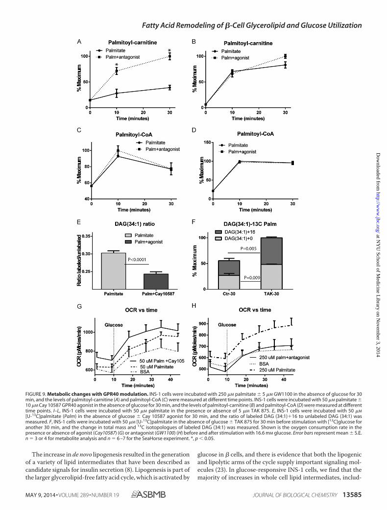
We first preincubated cells with the FFAR1/GPR40 antagonistGW1100 in the presence or absence of 250 �M palmitate andmeasured insulin secretion and metabolite levels in response to theaddition of 16.6 mM [U-13C]glucose for 30 min. GSIS wasdecreased significantly by GW1100 inhibition of the GPR40 recep-tor, both in the absence and presence of palmitate (Fig. 8A), asdescribed previously (7, 28). Metabolite analysis showed thatpalmitate reduced the concentration of M�6 hexose phos-phates as well as M�3 Go3P, whereas GW1100-treated sam-ples showed increased levels of M�6 hexoses (Fig. 8B) as well asM�3 Go3P isotopologues (Fig. 8C) following preincubationwith palmitate. There was a parallel decrease in the accumula-tion of M�3 isotopologues of LPA (Fig. 8D) and various DAGspecies (Fig. 8, E and F) in antagonist-treated cells followingpalmitate addition. Conversely, the FFAR1/GPR40 agonist Cay10587 increased the M�3 isotopologues of DAG, but only inthe presence of 50 �M palmitate (Fig. 8G). CDP-ethanolamineincreased in the presence of GPR40 antagonist (Fig. 8H), whichis in agreement with our finding that an increase in CDP-etha-nolamine may be a marker for decreased activity of the glycer-olipids cycle.3 We repeated these studies with TAK-875, whichhas shown clinical efficacy in phase II clinical trials (18). Wefound that, in the presence of [U-13C]glucose and 50 �M palmi-tate, TAK-875 reduced the levels of fructose bisphosphate (Fig.8I), and NADH/NAD� ratio (Fig. 8J), whereas it increased theM�3 isotopologues of DAG (Fig. 8K) and further reduced thelevels of CDP-ethanolamine (Fig. 8L), indicative of increasedactivity of glycerolipid cycle and opposite that seen in the pres-ence of the inhibitor.
Preincubation of INS-1 cells with GW1100 resulted in an�2-fold increase in palmitoyl-carnitine levels, suggesting anincrease in flux of palmitate into the mitochondria (Fig. 9A).The FFAR1/GPR40 agonist had no effect on palmitoyl-carni-tine levels when cells were incubated with a low level (50 �M) ofpalmitate (Fig. 9B). These results suggest that flux into glycer-olipids may be a primary determinant of fatty acid entry intomitochondria rather than modulation of CPT-1 by malonyl-CoA. Neither antagonist (Fig. 9C) nor agonist (Fig. 9D) affectedpalmitoyl-CoA accumulation in the absence of glucose, sug-gesting no effect of FFAR1/GPR40 receptors on fatty aciduptake or activation of the lipids by CoA synthase. To assess theeffect of FFAR1/GPR40 activation on lipolysis, we incubatedINS-1cells with [U-13C]palmitate with or without agonist for 20min in the absence of glucose. In the presence of agonist, therewas an increase in the unlabeled DAG (34:1) and a decrease inthe M�16/M�0 labeling ratio (Fig. 9E), consistent with previ-ous reports of phospholipase C activation by the receptor (29).Similarly, using [U-13C]palmitate and unlabeled glucose, TAK-875 increased the levels of both labeled and unlabeled DAG,also consistent with an increase in both lipogenesis and lipolysis(Fig. 9F).
Finally, we assessed the effect of FFAR1/GPR40 receptormanipulation on palmitate-stimulated glucose oxidation inINS-1 cells. In the presence of agonist, there was a furtherincrease in glucose oxidation in the presence of palmitate and
Cay 10587 (Fig. 9G). Conversely, the augmentation of glucoseoxidation by palmitate was reduced by the FFAR1/GPR40receptor antagonist (Fig. 9H). This suggests that FFAR1/GPR40activation enhances the intrinsic metabolic remodeling of glu-cose metabolism by fatty acids.
DISCUSSION
The secretion of insulin is primarily regulated by the entry ofglucose into the � cell but is modulated by the nervous systemand hormonal and nutritive inputs (30). Fatty acids augmentinsulin release in vivo and in vitro, and its effects have beenshown to be mediated via intracellular metabolism and throughsurface receptors (8). In this report, we show that a 30-minincubation of INS-1 cells with palmitate prior to the addition ofglucose results in a doubling of insulin release, as seen previ-ously in intact islets (4). Prior to glucose addition, palmitateexposure results in an �7-fold increase in intracellular palmi-toyl-CoA and a small increase in oxygen consumption but littleeffect on basal insulin secretion and minimal changes in themajority of intracellular metabolites, with the exception of asignificant increase in the formation of palmitoyl-carnitine,likely generated by the flux of fatty acid into the mitochondria.This suggests that, despite a small increase in TCA cycle activ-ity, there is a minimal generation of factors necessary for insulinsecretion. Following the addition of [U-13C]glucose, palmitoyl-CoA rapidly declines with the formation M�3 LPA and DAGisotopologues containing palmitate, directly demonstratingthat acyl-CoAs are rapidly esterified to Go3P derived fromextracellular glucose. This effect is not limited to saturated fattyacids because identical changes are seen following preincuba-tion of INS-1 cells with oleic acid (Fig. 6).
The esterification of fatty acids with Go3P has several effectsthat appear to contribute to their ability to augment insulinsecretion. We identified an increased glycolytic flux towardGo3P (Fig. 7A), reduced NADH levels, and a decrease in theNADH/NAD� ratio following fatty acid addition (Figs. 1 and 4).As suggested previously (31), we show directly that fatty acidaddition increases glycolytic (Fig. 7A) and TCA cycle (Fig. 7F)flux, glucose utilization (Fig. 7, G and H), and glucose oxidation(Fig. 7B). Each should result in the generation of additionalstimulus-secreting coupling factors that have been suggested toarise from the mitochondrial metabolism in � cells (1).Although fatty acids increased glucose oxidation, glucose addi-tion reduced fatty acid entry and oxidation in mitochondria, asindicated by the fall in C16 acylcarnitine levels (Fig. 2C). Thereduction in fatty acid mitochondrial uptake has been attrib-uted to increases in malonyl-CoA inhibition of CPT-1.Although malonyl-CoA levels rise with glucose, fatty acid addi-tion results in a significant blunting of the rise in malonyl-CoAin INS-1 cells (Fig. 2D). The reason for the reduction is notclear, but decreased export of citrate/isocitrate from the mito-chondria or an increase in consumption for de novo lipogenesismay play a role. These results also suggest that the decrease infatty acid oxidation following glucose addition may be primarilydue to the redirection of acyl-CoA toward esterification ratherthan blocking of the influx to the mitochondria via CPT1 inhi-bition by malonyl-CoA.
3 M. El-Azzouny, C. R. Evans, M. K. Treutelaar, R. T. Kennedy, and C. F. Burant,unpublished data.
Fatty Acid Remodeling of �-Cell Glycerolipid and Glucose Utilization
13584 JOURNAL OF BIOLOGICAL CHEMISTRY VOLUME 289 • NUMBER 19 • MAY 9, 2014
at NY
U School of M
edicine Library on N
ovember 3, 2014
http://ww
w.jbc.org/
Dow
nloaded from
The increase in de novo lipogenesis resulted in the generationof a variety of lipid intermediates that have been described ascandidate signals for insulin secretion (8). Lipogenesis is part ofthe larger glycerolipid-free fatty acid cycle, which is activated by
glucose in � cells, and there is evidence that both the lipogenicand lipolytic arms of the cycle supply important signaling mol-ecules (23). In glucose-responsive INS-1 cells, we find that themajority of increases in whole cell lipid intermediates, includ-
FIGURE 9. Metabolic changes with GPR40 modulation. INS-1 cells were incubated with 250 �M palmitate � 5 �M GW1100 in the absence of glucose for 30min, and the levels of palmitoyl-carnitine (A) and palmitoyl-CoA (C) were measured at different time points. INS-1 cells were incubated with 50 �M palmitate �10 �M Cay 10587 GPR40 agonist in the absence of glucose for 30 min, and the levels of palmitoyl-carnitine (B) and palmitoyl-CoA (D) were measured at differenttime points. I–L, INS-1 cells were incubated with 50 �M palmitate in the presence or absence of 5 �M TAK 875. E, INS-1 cells were incubated with 50 �M
[U-13C]palmitate (Palm) in the absence of glucose � Cay 10587 agonist for 30 min, and the ratio of labeled DAG (34:1)�16 to unlabeled DAG (34:1) wasmeasured. F, INS-1 cells were incubated with 50 �M [U-13C]palmitate in the absence of glucose � TAK 875 for 30 min before stimulation with [12C]glucose foranother 30 min, and the change in total mass and 13C isotopologues of labeled DAG (34:1) was measured. Shown is the oxygen consumption rate in thepresence or absence of agonist (Cay10587) (G) or antagonist (GW1100) (H) before and after stimulation with 16.6 mM glucose. Error bars represent mean � S.E.n � 3 or 4 for metabolite analysis and n � 6 –7 for the SeaHorse experiment. *, p � 0.05.
Fatty Acid Remodeling of �-Cell Glycerolipid and Glucose Utilization
MAY 9, 2014 • VOLUME 289 • NUMBER 19 JOURNAL OF BIOLOGICAL CHEMISTRY 13585
at NY
U School of M
edicine Library on N
ovember 3, 2014
http://ww
w.jbc.org/
Dow
nloaded from

ing LPA, palmitic acid, DAG, PG, phosphatidylinositol, andmonoacylglycerol, arise from the lipogenesis pathway becauselittle increases in M�0 isotopologues are found, even after 60min of glucose stimulation. DAG has multiple potential roles ininsulin secretion, including activation of Munc13-1, a receptorknown to amplify insulin exocytosis (32, 33) and activation ofprotein kinase D1 (PKD1), which modulates the reorganizationof the cortical actin network, perhaps playing a role in the sec-ond phase of insulin secretion (6). Recently, Prentki et al. (23)have suggested that increases in monoacylglycerol as well asDAG in � cells can activate Munc13-1.
In addition to lipid species identified previously, we foundmarked increases in cellular content of various acylamides.These fatty acid derivatives have not been reported previouslyin � cells, and their effect is unknown. Acylamides have beenfound in neuronal tissue and reportedly possess various func-tions (34, 35), including modulation of calcium flux, nitric oxidegeneration (36), and transient receptor potential (TRP) calciumchannels (37). Although we were able to increase the intracel-lular concentration of those metabolites by increasing the levelsof their amino acid precursors, this did not result in alterationsin insulin secretion. Interestingly, taurine has been suggested torestore � cell insulin secretion following chronic fatty acidexposure. However, this beneficial effect was attributed to itsantioxidant ability (38).
The activation of the FFAR1/GPR40 receptor mediatesabout 50% of the augmentation of insulin secretion by fatty acid(7). As indicated above, FFAR1/GPR40 has been proposed toincrease � cell calcium and activate phospholipase C-generat-ing diacylglycerols, which may tether PKD1 to the plasmamembrane increasing its activity (6), although the latter has notbeen demonstrated directly in � cells. We sought to determinewhether acute modulation of FFAR1/GPR40 had an effect onthe � cell metabolome. Inhibiting FFAR1/GPR40 receptorsresulted in attenuation of the free fatty acid augmentation of denovo DAG synthesis. It also decreased oxygen consumption byINS-1 cells and decreased insulin secretion. In contrast,
FFAR1/GPR40 agonists increased the flux of glucose andpalmitate into DAG, increased lipolysis, and increased oxygenconsumption. The increase in palmitoyl carnitine levels follow-ing disruption of GPR40 signaling reflects increased fatty acidinflux into mitochondria. The redirection of fatty acids into themitochondria for oxidation by overexpression of CPT-1 hasbeen shown to be associated with an attenuation of insulinsecretion (39, 40), and this redirection is associated withdecreased glycerolipid formation (39). Thus, it appears thatGPR40 activation is important for augmenting the partitioningof fatty acids to the glycerolipid pathway and fatty acidenhancement of GSIS.
These data are also in agreement with studies that show that theGPR40 agonist TAK-875 is more effective in the presence of fattyacids to increase insulin secretion (41), although they attribute theaugmentation to enhanced binding of the agonist to the receptor.However, our data are somewhat at odds with a previous study byAlquier et al. (42) examining glucose and fatty acid utilization inislets isolated from GPR40�/� mice. Consistent with our data, theauthors saw no effect of GPR40 disruption on glucose utilization inthe absence of fatty acids, although glucose utilization was notassessed in the presence of fatty acids. However, they saw noreduction in the incorporation of palmitate into total lipids. Thismay reflect our direct assessment of 13C label flux into specificglycerolipids or might indicate intrinsic differences between isletsand INS-1 cells or the potential for adaptation of islets followingdisruption of GPR40 in mice. The development of a specific meth-odology to estimate metabolite flux in intact rodent islets will helpclarify these potential differences.
In summary, our data suggest that FFAR1/GPR40 enhancesthe intrinsic effects of fatty acids to remodel the intermediarymetabolism to augment insulin secretion. This effect does notappear to be due to increased uptake or activation of fatty acidbecause disruption of FFAR1/GPR40 did not alter the levels ofpalmitoyl-CoA in INS-1 cells. At present, it is unclear how theactivity of the pathways leading to de novo lipogenesis occursfollowing FFAR1/GPR40 activation, but modulation of G3P
FIGURE 10. Proposed pathways for fatty acid metabolism during the fed and fasting state and its role in potentiating insulin secretion. In � cells, fattyacids increase glucose flux to glycerol-3-phosphate (Glycerol-3-P) to increase formation of lysophosphatidic acid (LPA) and downstream signaling molecules.The increased conversion of dihydroxyacetone phosphate regenerates NAD�, which increases glycolytic flux and increases TCA cycle activity. FFAR1/GPR40receptor activation appears to enhance the formation of LPA and increase glycerolipid cycling. LC-CoA, long chain CoA.
Fatty Acid Remodeling of �-Cell Glycerolipid and Glucose Utilization
13586 JOURNAL OF BIOLOGICAL CHEMISTRY VOLUME 289 • NUMBER 19 • MAY 9, 2014
at NY
U School of M
edicine Library on N
ovember 3, 2014
http://ww
w.jbc.org/
Dow
nloaded from
acyltransferase (GPAT) activity may be a target, given theincreased flux into the early steps of the lipogenic pathway. Ourdata are also consistent with the idea that, in the fasting state,plasma fatty acids increase, leading to increased long chainacyl-CoA levels in � cells. Although glucose and fatty acid oxi-dation is occurring, there is minimal generation of couplingfactors to increase insulin secretion (Fig. 10A). As glucose levelsrise after feeding, the presence of fatty acids increases esterifi-cation with glycerol-3-phosphate, resulting in an increased fluxto glycolipids and the formation of lipid-derived factors impor-tant in insulin secretion (Fig. 10B). The increased flux increasesregeneration of NAD�, accelerating glycolysis and flux into theTCA cycle above that which would occur following exposure toglucose alone. Our data also suggests that, in addition to theactivation of the PLC-PKD1 pathway described previously (6),FFAR1/GPR40 activation also enhances flux through the glyc-erolipid pathway, augmenting the formation of coupling fac-tors. This may be important because fatty acid entry into the �cell likely falls as plasma fatty acids decrease following a mealbecause of insulin-mediated reduction in lipolysis in fat cells.Thus, activation of the FFAR1/GPR40 receptor by fatty acids orreceptor agonists (18) may ensure continued fatty acid esterifi-cation and the provision of signaling lipids for insulin secretion.
Acknowledgments—We thank Katherine Overmyer for discussions,Dr. Heidi Iglay Reger for assistance with statistical analysis, SydneyBridges for performing the SeaHorse analysis, and Dr. Ian Sweet forproviding unpublished data for review.
REFERENCES1. Jitrapakdee, S., Wutthisathapornchai, A., Wallace, J. C., and MacDonald,
M. J. (2010) Regulation of insulin secretion: role of mitochondrial signal-ling. Diabetologia 53, 1019 –1032
2. Bender, K., Newsholme, P., Brennan, L., and Maechler, P. (2006) Theimportance of redox shuttles to pancreatic �-cell energy metabolism andfunction. Biochem. Soc. Trans. 34, 811– 814
3. Jensen, M. V., Joseph, J. W., Ronnebaum, S. M., Burgess, S. C., Sherry,A. D., and Newgard, C. B. (2008) Metabolic cycling in control of glucose-stimulated insulin secretion. Am. J. Physiol. Endocrinol. Metab. 295,E1287–97
4. Parker, S. M., Moore, P. C., Johnson, L. M., and Poitout, V. (2003) Palmi-tate potentiation of glucose-induced insulin release: a study using 2-bro-mopalmitate. Metabolism 52, 1367–1371
5. Nolan, C. J., Madiraju, M. S., Delghingaro-Augusto, V., Peyot, M. L., andPrentki, M. (2006) Fatty acid signaling in the �-cell and insulin secretion.Diabetes 55, S16 –23
6. Ferdaoussi, M., Bergeron, V., Zarrouki, B., Kolic, J., Cantley, J., Fielitz, J.,Olson, E. N., Prentki, M., Biden, T., MacDonald, P. E., and Poitout, V.(2012) G protein-coupled receptor (GPR)40-dependent potentiation ofinsulin secretion in mouse islets is mediated by protein kinase D1. Diabe-tologia 55, 2682–2692
7. Latour, M. G., Alquier, T., Oseid, E., Tremblay, C., Jetton, T. L., Luo, J., Lin,D. C., and Poitout, V. (2007) GPR40 is necessary but not sufficient for fattyacid stimulation of insulin secretion in vivo. Diabetes 56, 1087–1094
8. Prentki, M., and Madiraju, S. R. (2012) Glycerolipid/free fatty acid cycleand islet �-cell function in health, obesity and diabetes. Mol. Cell Endo-crinol. 353, 88 –100
9. Roduit, R., Nolan, C., Alarcon, C., Moore, P., Barbeau, A., Delghingaro-Augusto, V., Przybykowski, E., Morin, J., Massé, F., Massie, B., Ruderman,N., Rhodes, C., Poitout, V., and Prentki, M. (2004) A role for the malonyl-CoA/long-chain acyl-CoA pathway of lipid signaling in the regulation ofinsulin secretion in response to both fuel and nonfuel stimuli. Diabetes 53,
1007–101910. Deeney, J. T., Gromada, J., Høy, M., Olsen, H. L., Rhodes, C. J., Prentki, M.,
Berggren, P. O., and Corkey, B. E. (2000) Acute stimulation with longchain acyl-CoA enhances exocytosis in insulin-secreting cells (HIT T-15and NMRI �-cells). J. Biol. Chem. 275, 9363–9368
11. Corkey, B. E., Deeney, J. T., Yaney, G. C., Tornheim, K., and Prentki, M.(2000) The role of long-chain fatty acyl-CoA esters in �-cell signal trans-duction. J. Nutr. 130, 299S-304S
12. Bränström, R., Aspinwall, C. A., Välimäki, S., Ostensson, C. G., Tibell, A.,Eckhard, M., Brandhorst, H., Corkey, B. E., Berggren, P. O., and Larsson,O. (2004) Long-chain CoA esters activate human pancreatic �-cell KATPchannels: potential role in type 2 diabetes. Diabetologia 47, 277–283
13. Tian, Y., Corkey, R. F., Yaney, G. C., Goforth, P. B., Satin, L. S., and Moitosode Vargas, L. (2008) Differential modulation of L-type calcium channelsubunits by oleate. Am. J. Physiol. Endocrinol. Metab. 294, E1178 – 86
14. Mulder, H., Yang, S., Winzell, M. S., Holm, C., and Ahrén, B. (2004) Inhi-bition of lipase activity and lipolysis in rat islets reduces insulin secretion.Diabetes 53, 122–128
15. Lorenz, M. A., Burant, C. F., and Kennedy, R. T. (2011) Reducing time andincreasing sensitivity in sample preparation for adherent mammalian cellmetabolomics. Anal. Chem. 83, 3406 –3414
16. Lorenz, M. A., El Azzouny, M. A., Kennedy, R. T., and Burant, C. F. (2013)Metabolome response to glucose in the �-cell line INS-1 832/13. J. Biol.Chem. 288, 10923–10935
17. Burant, C. F. (2013) Activation of GPR40 as a therapeutic target for thetreatment of type 2 diabetes. Diabetes Care 36, S175–9
18. Burant, C. F., Viswanathan, P., Marcinak, J., Cao, C., Vakilynejad, M., Xie,B., and Leifke, E. (2012) TAK-875 versus placebo or glimepiride in type 2diabetes mellitus: a phase 2, randomised, double-blind, placebo-con-trolled trial. Lancet 379, 1403–1411
19. Malmgren, S., Nicholls, D. G., Taneera, J., Bacos, K., Koeck, T., Tamaddon,A., Wibom, R., Groop, L., Ling, C., Mulder, H., and Sharoyko, V. V. (2009)Tight coupling between glucose and mitochondrial metabolism in clonal�-cells is required for robust insulin secretion. J. Biol. Chem. 284,32395–32404
20. Schneider, C. A., Rasband, W. S., and Eliceiri, K. W. (2012) NIH Image toImageJ: 25 years of image analysis. Nat. Methods 9, 671– 675
21. Tautenhahn, R., Patti, G. J., Rinehart, D., and Siuzdak, G. (2012) XCMSonline: a web-based platform to process untargeted metabolomic data.Anal. Chem. 84, 5035–5039
22. Prentki, M., Vischer, S., Glennon, M. C., Regazzi, R., Deeney, J. T., andCorkey, B. E. (1992) Malonyl-CoA and long chain acyl-CoA esters as met-abolic coupling factors in nutrient-induced insulin secretion. J. Biol.Chem. 267, 5802–5810
23. Prentki, M., Matschinsky, F. M., and Madiraju, S. R. (2013) Metabolicsignaling in fuel-induced insulin secretion. Cell Metabolism 18, 162–185
24. Hardie, D. G. (2011) Sensing of energy and nutrients by AMP-activatedprotein kinase. Am. J. Clin. Nutr. 93, 891S-6
25. Hardie, D. G. (2008) AMPK: a key regulator of energy balance in the singlecell and the whole organism. Int. J. Obes. 32, S7–12
26. Waluk, D. P., Vielfort, K., Derakhshan, S., Aro, H., and Hunt, M. C. (2013)N-Acyl taurines trigger insulin secretion by increasing calcium flux inpancreatic �-cells. Biochem. Biophys. Res. Commun. 430, 54 –59
27. Sekine, N., Cirulli, V., Regazzi, R., Brown, L. J., Gine, E., Tamarit-Rodri-guez, J., Girotti, M., Marie, S., MacDonald, M. J., and Wollheim, C. B.(1994) Low lactate dehydrogenase and high mitochondrial glycerol phos-phate dehydrogenase in pancreatic �-cells. Potential role in nutrient sens-ing. J. Biol. Chem. 269, 4895– 4902
28. Briscoe, C. P., Peat, A. J., McKeown, S. C., Corbett, D. F., Goetz, A. S.,Littleton, T. R., McCoy, D. C., Kenakin, T. P., Andrews, J. L., Ammala, C.,Fornwald, J. A., Ignar, D. M., and Jenkinson, S. (2006) Pharmacologicalregulation of insulin secretion in MIN6 cells through the fatty acid recep-tor GPR40: identification of agonist and antagonist small molecules. Br. J.Pharmacol. 148, 619 – 628
29. Mancini, A. D., and Poitout, V. (2013) The fatty acid receptor FFA1/GPR40 a decade later: how much do we know? Trends Endocrinol. Metab.24, 398 – 407
30. Rorsman, P., and Braun, M. (2013) Regulation of insulin secretion in hu-
Fatty Acid Remodeling of �-Cell Glycerolipid and Glucose Utilization
MAY 9, 2014 • VOLUME 289 • NUMBER 19 JOURNAL OF BIOLOGICAL CHEMISTRY 13587
at NY
U School of M
edicine Library on N
ovember 3, 2014
http://ww
w.jbc.org/
Dow
nloaded from

man pancreatic islets. Annu. Rev. Physiol. 75, 155–17931. Prentki, M., and Madiraju, S. R. (2008) Glycerolipid metabolism and sig-
naling in health and disease. Endocr. Rev. 29, 647– 67632. Kwan, E. P., Xie, L., Sheu, L., Nolan, C. J., Prentki, M., Betz, A., Brose, N.,
and Gaisano, H. Y. (2006) Munc13–1 deficiency reduces insulin secretionand causes abnormal glucose tolerance. Diabetes 55, 1421–1429
33. Sheu, L., Pasyk, E. A., Ji, J., Huang, X., Gao, X., Varoqueaux, F., Brose, N.,and Gaisano, H. Y. (2003) Regulation of insulin exocytosis by Munc13–1.J. Biol. Chem. 278, 27556 –27563
34. Tan, B., O’Dell, D. K., Yu, Y. W., Monn, M. F., Hughes, H. V., Burstein,S., and Walker, J. M. (2010) Identification of endogenous acyl aminoacids based on a targeted lipidomics approach. J. Lipid Res. 51,112–119
35. Tan, B., Yu, Y. W., Monn, M. F., Hughes, H. V., O’Dell, D. K., and Walker,J. M. (2009) Targeted lipidomics approach for endogenous N-acyl aminoacids in rat brain tissue. J. Chromatogr. B. Analyt. Technol. Biomed. Life.Sci. 877, 2890 –2894
36. Rimmerman, N., Bradshaw, H. B., Hughes, H. V., Chen, J. S., Hu, S. S.,McHugh, D., Vefring, E., Jahnsen, J. A., Thompson, E. L., Masuda, K.,Cravatt, B. F., Burstein, S., Vasko, M. R., Prieto, A. L., O’Dell, D. K., andWalker, J. M. (2008) N-palmitoyl glycine, a novel endogenous lipid thatacts as a modulator of calcium influx and nitric oxide production in sen-sory neurons. Mol. Pharmacol. 74, 213–224
37. Saghatelian, A., McKinney, M. K., Bandell, M., Patapoutian, A., and Cra-vatt, B. F. (2006) A FAAH-regulated class of N-acyl taurines that activates
TRP ion channels. Biochemistry 45, 9007–901538. Oprescu, A. I., Bikopoulos, G., Naassan, A., Allister, E. M., Tang, C., Park,
E., Uchino, H., Lewis, G. F., Fantus, I. G., Rozakis-Adcock, M., Wheeler,M. B., and Giacca, A. (2007) Free fatty acid-induced reduction in glucose-stimulated insulin secretion: evidence for a role of oxidative stress in vitroand in vivo. Diabetes 56, 2927–2937
39. Rubí, B., Antinozzi, P. A., Herrero, L., Ishihara, H., Asins, G., Serra, D.,Wollheim, C. B., Maechler, P., and Hegardt, F. G. (2002) Adenovirus-mediated overexpression of liver carnitine palmitoyltransferase I in INS1Ecells: effects on cell metabolism and insulin secretion. Biochem. J. 364,219 –226
40. Herrero, L., Rubí, B., Sebastián, D., Serra, D., Asins, G., Maechler, P.,Prentki, M., and Hegardt, F. G. (2005) Alteration of the malonyl-CoA/carnitine palmitoyltransferase I interaction in the �-cell impairs glucose-induced insulin secretion. Diabetes 54, 462– 471
41. Yabuki, C., Komatsu, H., Tsujihata, Y., Maeda, R., Ito, R., Matsuda-Naga-sumi, K., Sakuma, K., Miyawaki, K., Kikuchi, N., Takeuchi, K., Habata, Y.,and Mori, M. (2013) A novel antidiabetic drug, fasiglifam/TAK-875, actsas an ago-allosteric modulator of FFAR1. PLoS ONE 8, e76280
42. Alquier, T., Peyot, M. L., Latour, M. G., Kebede, M., Sorensen, C. M.,Gesta, S., Ronald Kahn, C., Smith, R. D., Jetton, T. L., Metz, T. O., Prentki,M., and Poitout, V. (2009) Deletion of GPR40 impairs glucose-inducedinsulin secretion in vivo in mice without affecting intracellular fuel metab-olism in islets. Diabetes 58, 2607–2615
Fatty Acid Remodeling of �-Cell Glycerolipid and Glucose Utilization
13588 JOURNAL OF BIOLOGICAL CHEMISTRY VOLUME 289 • NUMBER 19 • MAY 9, 2014
at NY
U School of M
edicine Library on N
ovember 3, 2014
http://ww
w.jbc.org/
Dow
nloaded from