Fatty Acid-Mediated Quorum Sensing Systems in ...
173
DOCTORAL THESIS Fatty Acid-Mediated Quorum Sensing Systems in Stenotrophomonas maltophilia Pol Huedo Moreno September 2014
-
Upload
khangminh22 -
Category
Documents
-
view
0 -
download
0
Transcript of Fatty Acid-Mediated Quorum Sensing Systems in ...

DOCTORAL THESIS
Fatty Acid-Mediated Quorum Sensing Systems
in Stenotrophomonas maltophilia
Pol Huedo Moreno September 2014

ii
� � � � �
Parc de Recerca UAB
Institut de Biotecnologia i Biomedicina
Departament de Genètica i Microbiologia
Fatty Acid-Mediated Quorum Sensing Systems in
Stenotrophomonas maltophilia A thesis submitted by Pol Huedo Moreno in partial fulfilment of the requirements for
the Doctor of Philosophy degree in Biotechnology in the Universitat Autònoma de
Barcelona.�
Approval of the directors,
Dr. Isidre Gibert Gonzàlez Dr. Daniel Yero Corona Bellaterra, September 2014.

iv

v
Als meus pares, a en Lluís i a la Txell.

vi

vii
AKNOWLEDGEMENTS En primer lloc m’agradaria agrair al Dr. Isidre Gibert per donar-me la oportunitat de
desenvolupar aquesta tesi al seu laboratori. Gràcies de tot cor per confiar en mi dia rere
dia i permetre’m dur a terme les meves esbogerrades idees (algunes més que d’altres),
sense cap mena d’entrebanc.
Aquest segon paràgraf li dedico als meus codirectors. Gràcies Raquel per guiar-me en
les meves etapes inicials i, tot i que els esdeveniments han fet que finalment et
desvinculis de la direcció, considero que tu també n’ets responsable d’aquest treball.
Daniel, m’és difícil expressar el que ha significat per mi la teva arribada al laboratori.
Vas incorporar-te en aquest treball quan més ho necessitava. Gràcies a la teva
personalitat (exigent però sempre optimista) i als teus magistrals consells, has fet que
això remunti. Els pocs fruits (però dolços) que hem recollit aquests darrers anys són
teus. Sempre t’admiraré i et recordaré com el meu mentor.
A tots els col·legues de laboratori amb qui he tingut la fortuna d’aprendre i compartir
experiències al llarg d’aquests anys: Mario, Celeste, Elías, Gerard, Sònia i Iratxe. A
banda d’il·lustrar-me en molts aspectes científics, m’heu ajudat a superar el dia a dia
amb un somriure. A la Paula, gràcies per compartir amb mi la teva vitalitat i alegria.
No puc fugir de l’IBB sense recordar el suport que m’han brindat molts companys de
l’institut. Xavi Daura, encara que no signis aquesta tesi, per mi has sigut el meu cap a
l’ombra. Infinites gràcies per participar en tot moment de l’esdevenir d’aquesta tesi.
Nerea i Àngels, gràcies per suportar i dur a terme les meves reiterades propostes, ha
sigut un plaer treballar i aprendre de vosaltres. Xino, tu també has pringat centenars de
reunions de grup i “pitis”, mil gràcies per tot. Per acabar, si hi ha algú que ha hagut de
sofrir les meves improductives demandes durant molt de temps, aquests són en Sebas i
la Sílvia. Gràcies per la generositat que m’heu mostrat sempre, tot el que sé de
proteòmica (nivell sobretaula) us ho dec a vosaltres dos, i a en Mario. Sóc conscient que
us dec 100 pastissos, espero que quan llegiu això us en degui només 99.
Impossible oblidar-se del col·lectiu de “menjadors de tuppers anònims”. Gràcies David,
Rita, Gyo, Marín, Albert, Font, Jofre, Gisela, Laia i Javi, per fer teràpia de grup.
Igualment, a l’Albert, la Sílvia i tots els químics per tantes tardes de divendres. A tota la
colla de Sant Cugat per acollir-me i fer-me sentir a casa. Als meus amics de tota la vida,
ja sabeu qui sou i us estimo. Especial menció a Aliment, La Castanya i tot el seu entorn.
Infinites gràcies per ajudar-me a realitzar dos somnis en paral·lel.

viii
A en Miquel, la Teresa i la Marina per tota l’ajuda i estima que m’han lliurat al llarg
d’aquests anys.
A la meva família, pel seu suport i amor incondicional que he rebut i rebré sempre.
A la persona més important de la meva vida, la Txell, pel seu amor sincer i infinit.

ix
PRESENTATIONS AND PUBLICATIONS
Publications Two Different rpf Clusters Distributed among a Population of Stenotrophomonas maltophilia Clinical Strains Display Differential Diffusible Signal Factor Production and Virulence Regulation. Huedo P, Yero D, Martínez-Servat S, Estibariz I, Planell R, Martínez P, Ruyra A, Roher N, Roca I, Vila J, Daura X, Gibert I. J Bacteriol. 2014 Jul 1;196(13):2431-2442. Epub 2014 Apr 25. Draft Genome Sequence of Stenotrophomonas maltophilia Strain M30, Isolated from a Chronic Pressure Ulcer in an Elderly Patient. Huedo P, Conchillo-Solé O, Yero D, Martínez-Servat S, Daura X, Gibert I. Genome Announc. 2014 Jun 12;2(3). pii: e00576-14. doi: 10.1128/genomeA.00576-14. Abundance of the Quorum-Sensing Factor Ax21 in Four Strains of Stenotrophomonas maltophilia Correlates with Mortality Rate in a New Zebrafish Model of Infection. Ferrer-Navarro M, Planell R, Yero D, Mongiardini E, Torrent G, Huedo P, Martínez P, Roher N, Mackenzie S, Gibert I, Daura X. PLoS One. 2013 Jun 26;8(6):e67207. Print 2013. Oral presentations Huedo P, Yero D, Martínez-Servat S, Daura X and Gibert I. Decoding the DSF-Quorum Sensing System of Stenotrophomonas maltophilia. Sociedad Española de Microbiología (SEM), X Reunión Microbiología Molecular, Segovia, Spain, 9-11 Jun 2014. Huedo P. Quorum Sensing, the Bacterial Social Networks. 2ª Jornada Científica, Dept. de Genética y Microbiologia, UAB, Barcelona, Spain, 29 Nov 2013. Posters Huedo P, Planell R, Ferrer-Navarro M, Martínez P, Yero D, Daura X, Gibert I. DSF-mediated Quorum Sensing in S. maltophilia. Sociedad Española de Microbiología (SEM), IX Reunión Microbiología Molecular, Mallorca, Spain, 14-16 Nov 2012. Huedo P, Yero D, Martínez-Servat S, Daura X and Gibert I. Decoding the DSF-Quorum Sensing System of Stenotrophomonas maltophilia. Sociedad Española de Microbiología (SEM), X Reunión Microbiología Molecular, Segovia, Spain, 9-11 Jun 2014.

x

xi
ABBREVIATIONS
2-DE 2-Dimensional Electrophoresis
aa Amino Acid
Ap Ampicillin
Ara Arabinose
ATP Adenosil Triphosphate
BHI Brain Heart Infusion
BLAST Basic Local Alignment Search Tool
BM2 Basal Medium 2
cdi-GMP Cyclic Diguanosine Monophosphate
CF Cystic Fibrosis
CFTR CF transmembrane conductance regulator
cfu Colony Formation Units
Cm Chloramphenicol
CMC Carboxymethylcellulose
COPD Chronic Obstructive Pulmonary Disease
DSF Diffusible Signal Factor
DSF-QS DSF-Quorum Sensing
Erm Erythromycin
g grams
GC/MS Gas Chromatography/Mass Spectrometry
GMP Guanosine Monophosphate
h Hours
HATP Histidine ATPase
xii
HisKA Histidine Kinase
hpi Hours Post Injection
HPT Histidine Phosphotransferase
ICUs Intensive Care Units
Kan Kanamycin
kDa Kilodaltons
LB Luria-Bertani
M Molar
M9 Medium 9
MDR Multi Drug Resistant
MDRO Multi Drug Resistant Organism
MIC Minimum Inhibitory Concentration
min Minutes
ml Milliliters
mM Milimolar
N-ter Amino-Terminus
nm Nanometers
NYG Nutrient Broth, Yeast Extract, Glucose
OD Optical Density
ORF Open Reading Frame
PAA Peracetic Acid
PBS Phosphate Buffer Saline
PCR Polymerase Chain Reaction
PQS Pseudomonas Quinolone System
PVD Pyoverdine

xiii
QS Quorum Sensing
RBS Ribosome Binding Site
REC Receiver Domain
Rf Ratio to Front
rpf Regulation of Pathogenicity Factors
rpm Revolutions Per Minute
RR Response Regulator
SDS Sodium Dodecyl Sulfate
SMX Sulfamethoxazole
Tc Tetracycline
TEM Transmission Electron Microscopy
TFP Type IV Pili
TMP Trimethoprim
TMP-SMX Trimethoprim- Sulfamethoxazole
TMR Trans Membrane Region
wt Wild Type
YEB Yeast Extract Broth
µL Microliters
µM Micromolar

xiv
xv
TABLE OF CONTENTS
AKNOWLEDGEMENTS .............................................................................................. vii PRESENTATIONS AND PUBLICATIONS .................................................................. ix
ABBREVIATIONS ......................................................................................................... xi TABLE OF CONTENTS ............................................................................................... xv
LIST OF TABLES ....................................................................................................... xviii LIST OF FIGURES ....................................................................................................... xix
ABSTRACT/RESUM ................................................................................................... xxi 1. INTRODUCTION ...................................................................................................... 2
1.1 Microbiology of Stenotrophomonas maltophilia ................................................. 2 1.1.1 Historical aspects ............................................................................................. 2 1.1.2 Physiology and metabolism ............................................................................. 2 1.1.3 Genome ............................................................................................................ 2 1.1.4 Ecology ............................................................................................................ 3
1.2. Clinical Significance of Stenotrophomonas maltophilia .................................... 4 1.2.1 Hospital and community-acquired infections .................................................. 4 1.2.2 Cystic fibrosis .................................................................................................. 4
1.3. Molecular Mechanisms Involved in Pathogenesis ............................................ 5 1.3.1 Exoproducts ..................................................................................................... 5 1.3.2 Antibiotic resistance ........................................................................................ 5 1.3.3 Biofilm formation ............................................................................................ 6 1.3.4 Bacterial motility ............................................................................................. 8
1.4 Quorum Sensing ................................................................................................... 9 1.4.1 Generalities ...................................................................................................... 9 1.4.2 Generalities of DSF quorum sensing system ................................................. 11 1.4.3 DSF-QS in Xanthomonas campestris pv. campestris .................................... 12 1.4.4 DSF-QS in Xylella fastidiosa ........................................................................ 15 1.4.5 DSF-QS in Burkholderia spp. ....................................................................... 16 1.4.6 DSF-QS in Stenotrophomonas maltophila .................................................... 16
1.5 Pseudomonas aeruginosa, an overview ............................................................. 17 1.5.1 Microbiology of P. aeruginosa ..................................................................... 17 1.5.2 Mechanisms involved in pathogenesis .......................................................... 18 1.5.3 Quorum sensing regulatory network in P. aeruginosa .................................. 18
2. OBJECTIVES ........................................................................................................... 22
3. MATERIALS AND METHODS ............................................................................. 24 3.1 Bacterial strains and growth conditions. .......................................................... 24
3.1.1 S. maltophilia and other Xanthomonads ........................................................ 24 3.1.2 P. aeruginosa ................................................................................................. 24 3.1.3 E. coli ............................................................................................................. 26 3.1.4 Bacterial preservation .................................................................................... 27
3.2 Oligonucleotides and plasmid vectors ............................................................... 27 3.3 Chemical reagents ............................................................................................... 28

xvi
3.4 Molecular biology techniques ............................................................................ 29 3.4.1 General guidelines ......................................................................................... 29 3.4.2 Polymerase Chain Reaction (PCR) and quantitative reverse transcription-PCR (qRT-PCR). .................................................................................................... 29
3.5 DNA Sequencing ................................................................................................. 30 3.5.1 rpfF sequence determination and analysis ..................................................... 30 3.5.2 Genome sequencing ....................................................................................... 31
3.6 Mutants generation and complementation ....................................................... 32 3.6.1 Generation and complementation of S. maltophilia ∆rpfF and ∆rpfC mutants ................................................................................................................................ 32 3.6.2 Generation and complementation of S. maltophilia M30 ∆smlt0266, ∆smlt0267 and ∆smlt0266-0267 mutants ............................................................... 35 3.6.3 Arabinose-induction of pBADSMdsp ........................................................... 36 3.6.4 Complementation of P. aeruginosa ∆dspI and ∆dspII mutants .................... 36
3.7 General analytical tools ...................................................................................... 38 3.7.1 SDS-PAGE .................................................................................................... 38 3.7.2 Protein Identification by Mass Spectrometry ................................................ 38 3.7.3 Lipid extraction from culture supernatants .................................................... 38 3.7.4 Lipid separation by Thin-Layer Chromatography (TLC) .............................. 39 3.7.5 Lipid identification from culture supernatants by GC/MS ............................ 39 3.7.6 Purification of fatty acids by High-Liquid Pressure Chromatography (HPLC) ................................................................................................................................ 39 3.7.7 Extraction and analysis of total cellular fatty acids ....................................... 39
3.8 DSF extraction, detection and quantification. ................................................. 40 3.8.1 Colony and supernatant DSF bioassay .......................................................... 40 3.8.2 TLC coupled to DSF bioassay ....................................................................... 40 3.8.3 Liquid DSF bioassay ..................................................................................... 41 3.8.4 DSF detection and quantification from infected animal tissues .................... 41
3.9 Phenotypic analysis of mutant, complemented and wild type strains. .......... 41 3.9.1 Biofilm formation .......................................................................................... 41 3.9.2 Swimming motility ........................................................................................ 42 3.9.3 Twitching motility ......................................................................................... 42 3.9.4 Swarming motility ......................................................................................... 42 3.9.5 Surfactant imaging ......................................................................................... 42 3.9.6 TEM microscopy imaging ............................................................................. 43 3.9.7 Determination of antibiotic susceptibility ..................................................... 43 3.9.8 Congo red binding assay ................................................................................ 43 3.9.9 Determination of virulence in the Caenorhabditis elegans model ................ 43 3.9.10 Determination of virulence in the adult zebrafish infection model ............. 44
3.10 Bioinformatic Tools .......................................................................................... 44 3.11 Statistical Analysis ............................................................................................ 45 3.12 Ethics Statements .............................................................................................. 45
4. RESULTS AND DISCUSSION ............................................................................... 48 4.1 Molecular Basis of DSF-QS System in S. maltophilia ...................................... 48
4.1.1 Two variants of rpf cluster with main differences in rpfF and rpfC genes are distributed among S. maltophilia population. ......................................................... 48 4.1.2 RpfC-1 but not RpfC-2 contains a transmembrane sensor input domain highly related to the Xcc RpfH-RpfC complex ...................................................... 51 4.1.3 Only strains carrying RpfC/F-1 combination produce detectable levels of DSF. ........................................................................................................................ 52

xvii
4.1.4 Both RpfF variants are able to synthesize DSF ............................................. 53 4.1.5 RpfF-2 is permanently repressed by RpfC-2 ................................................. 57 4.1.6 An improved liquid bioassay for accurately quantification of DSF molecules. ................................................................................................................................ 58 4.1.7 DSF production is temperature and media-dependent .................................. 59 4.1.8 13-methyl-tetradecanoic acid (C:15 iso), a possible DSF precursor, is the most abundant fatty acid in S. maltophilia ............................................................. 60 4.1.9 Unspecific medium-length fatty acids modulate DSF production in RpfC/F-1 variant strains .......................................................................................................... 62 4.1.10 S. maltophilia RpfC/F-1 and RpfC/F-2 variant strains cross-talk each other, producing DSF in a positive feedback-manner. ..................................................... 63 4.1.11 Discussion .................................................................................................... 64
4.2 Phenotypic implications of DSF-QS in S. maltophilia ..................................... 69 4.2.1 Optimization of swarming motility assay for S. maltophilia ......................... 69 4.2.2 ∆rpfF-1 but not ∆rpfF-2 mutant shows alteration in swarming motility ...... 70 4.2.3 ∆rpfF-1 but not ∆rpfF-2 mutant shows alteration in biofilm formation ....... 72 4.2.4 Only the ΔrpfF-1 mutant shows attenuation in C. elegans ........................... 74 4.2.5 Both ∆rpfF mutants show attenuation in the adult Zebrafish infection model ................................................................................................................................ 75 4.2.6 Attenuation of ∆rpfF-1 mutant is due to its incapacity to disseminate throw the fish tissues ......................................................................................................... 75 4.2.7 RpfF-1 and RpfF-2 strains act synergistically in virulence ability in the zebrafish infection model. ...................................................................................... 76 4.2.8 The full virulence capacity observed in the mixed inoculum correlates with in-vivo DSF production .......................................................................................... 77 4.2.9 Discussion ...................................................................................................... 78
4.3 cis-DA-mediated Quorum Sensing System in S. maltophilia and P. aeruginosa .................................................................................................................................... 82
4.3.1 smlt0266 is the dspI orthologous in S. maltophilia ....................................... 82 4.3.2 Smlt0266 and Smlt0267 inversely regulate biofilm formation in S. maltophilia. ............................................................................................................. 83 4.3.3 Preliminary identification of cis-DA-like fatty acid produced by the DspI orthologous Smlt0266 ............................................................................................. 84 4.3.4 Smlt0267 is involved in antibiotic resistance and virulence regulation. ....... 86 4.3.6 P. aeruginosa DspI and DspII inversely regulate biofilm formation as occurs in S. maltophilia ...................................................................................................... 89 4.3.7 P. aeruginosa DspI and DspII regulate virulence-related phenotypes similarly to S. maltophilia Smlt0266 and Smlt0267 .............................................................. 90 4.3.8 P. aeruginosa ∆dspI and ∆dspII showed attenuation in the C. elegans model ................................................................................................................................ 92 4.3.9 Discussion ...................................................................................................... 93
5. GENERAL DISCUSSION AND FUTURE PERSPECTIVES ............................. 98 6. CONCLUSIONS ..................................................................................................... 106
7. REFERENCES ....................................................................................................... 110 8. ANNEX .................................................................................................................... 124
8.1 PUBLICATIONS .............................................................................................. 124
xviii
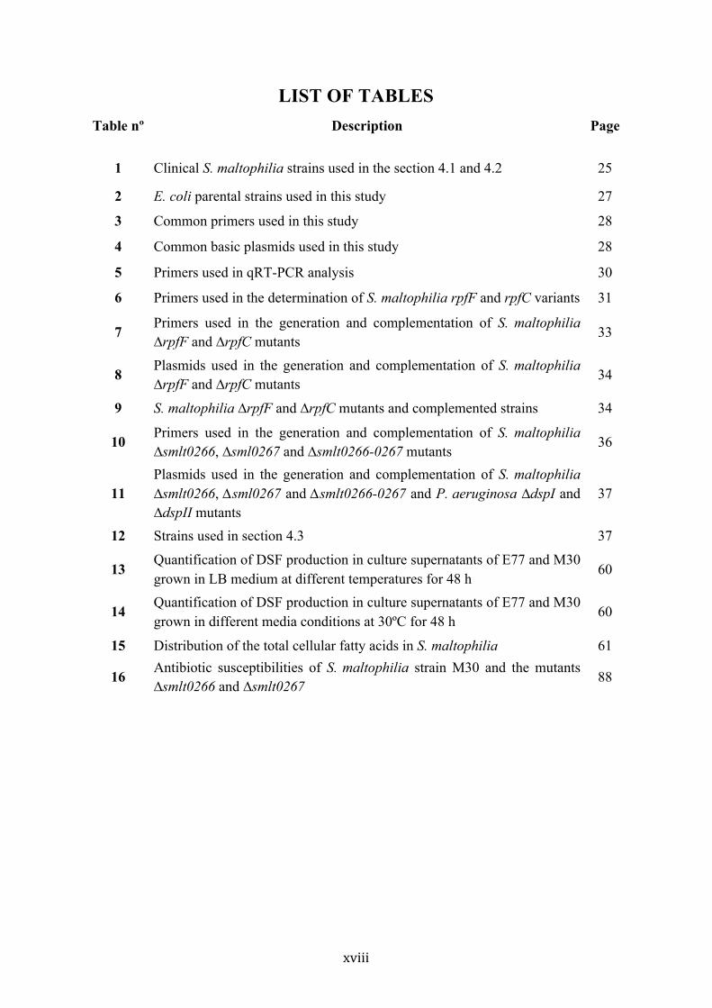
LIST OF TABLES Table nº Description Page
1 Clinical S. maltophilia strains used in the section 4.1 and 4.2 25
2 E. coli parental strains used in this study 27
3 Common primers used in this study 28
4 Common basic plasmids used in this study 28
5 Primers used in qRT-PCR analysis 30
6 Primers used in the determination of S. maltophilia rpfF and rpfC variants 31
7 Primers used in the generation and complementation of S. maltophilia ∆rpfF and ∆rpfC mutants
33
8 Plasmids used in the generation and complementation of S. maltophilia ∆rpfF and ∆rpfC mutants
34
9 S. maltophilia ∆rpfF and ∆rpfC mutants and complemented strains 34
10 Primers used in the generation and complementation of S. maltophilia ∆smlt0266, ∆sml0267 and ∆smlt0266-0267 mutants
36
11 Plasmids used in the generation and complementation of S. maltophilia ∆smlt0266, ∆sml0267 and ∆smlt0266-0267 and P. aeruginosa ∆dspI and ∆dspII mutants
37
12 Strains used in section 4.3 37
13 Quantification of DSF production in culture supernatants of E77 and M30 grown in LB medium at different temperatures for 48 h
60
14 Quantification of DSF production in culture supernatants of E77 and M30 grown in different media conditions at 30ºC for 48 h
60
15 Distribution of the total cellular fatty acids in S. maltophilia 61
16 Antibiotic susceptibilities of S. maltophilia strain M30 and the mutants ∆smlt0266 and ∆smlt0267
88

xix
LIST OF FIGURES
Fig. Nº Description Page
i Schematic representation of a bacterial biofilm development 7
ii QS signalling molecules in gram-negative and gram-positive bacteria 10
iii Gene organization in the rpf cluster in Xcc 12
iv Schematic representation of the DSF-QS system in Xcc at low and high cell density
15
1 Comparison and alignment of the rpf cluster in Xcc and two S. maltophilia strains K279a and D457.
49
2 Phylogenetic analysis of 82 S. maltophilia clinical strains based on the first 108 aminoacids of RpfF
50
3 SMART-software analysis of RpfC and RpfH from Xcc and RpfC from S. maltophilia K279a and D457
51
4 Colony DSF bioassay of 78 S. maltophilia strains 52
5 DSF bioassay of E77 and M30 with their respective ∆rpfF mutants and the homologous and heterologous complemented strains
53
6 TLC coupled to DSF bioassay of culture supernatants from E77 and M30, their respective ∆rpfF mutants and complemented strains
54
7 Gas Chromatography analysis of culture supernatants of E77 and M30, their respective ∆rpfF mutants and complemented strains
55
8 Mass spectra of the gas chromatography peaks with DSF activity 56
9 DSF bioassay of E77, E77 complemented with vectors prpfGCE77 and prpfGCM30, M30 wt, M30 ∆rpfC mutant and M30 ∆rpfC mutant complemented with vectors prpfGCE77 and prpfGCM30
57
10 Quantification of the gene expression of rpfF and rpfC from strains E77 and M30 by qRT-PCR
58
11 An Improved liquid DSF bioassay 59
12 Illustration of the fatty acids 13-methyl-tetradecanoic (15:0 iso) and 11-cis-methyl-dodecenoic (DSF)
61
13 DSF quantification of E77 supernatant supplemented with different fatty acids
62
14 Colony-based DSF bioassay of E77, M30 and their respective ∆rpfF strains, seeded at different distances one from the other
63
15 DSF production in cultures of axenic E77, axenic M30, a mixed culture of E77 and M30 and a culture of M30 supplemented with 0,1 µM DSF
64
16 Optimization of swarming motility in S. maltophilia E77 70
17 Swarming motility assay of E77, M30, their ∆rpfF mutants and the homologous and heterologous complemented strains
71

xx
18 Surfactant imaging and TEM micrographs of E77, E77 ∆rpfF mutant and the complemented ∆rpfF mutant
72
19 Biofilm formation of E77, M30 and their respective ∆rpfF mutants and the homologous and heterologous complemented strains
73
20 Virulence determination of S. maltophilia E77, M30, their respective ∆rpfF mutants and the complemented strains in the C. elegans CF512 model
74
21 Virulence determination of S. maltophilia E77, M30, their respective ∆rpfF mutants and the complemented strains in the adult zebrafish
75
22 Bacteria recovery from different regions of the body of sacrificed fishes 76
23 Virulence determination of E77, M30, and a mixed inoculum of both wt strains and both ∆rpfF mutants
77
24 Bioassay of DSF extraction from sacrificed fishes after 48 hpi with E77, M30 and the mixed inoculum (E77:M30, 1:1)
78
25 Comparison of the dsp cluster between P. aeruginosa and S. maltophilia 83
26 Biofilm formation of M30, the mutants ∆smlt0266, ∆smlt0266-0267, ∆smlt0267 and M30f pBADSMdsp
84
27 HPLC chromatogram of purified M30 wt supernatant, M30 pBADSMdsp supernatant and synthetic cis-DA
85
28 Analysis of exopolysaccharide production and colony morphology of S. maltophilia strains M30, ∆smlt0266 and ∆smlt0267 mutants, seeded in Congo Red plates
86
29 Swimming motility of S. maltophilia M30 and the mutants ∆smlt0266 and ∆smlt0267
87
30 Twitching motility of S. maltophilia strains M30 wt and the mutants ∆smlt0266 and ∆smlt0267
87
31 Virulence determination of S. maltophilia M30 and the mutants ∆smlt0266 and ∆smlt0267 in the C. elegans CF512 model
89
32 Biofilm formation of P. aeruginosa MPAO1, the ∆dspI and ∆dspII mutants and the complemented strains
90
33 Swarming motility of P. aeruginosa MPAO1 and the mutants ∆dspI and ∆dspII
91
34 Twitching motility of P. aeruginosa MPAO1 and the mutants ∆dspI and ∆dspII
91
35 Exopolysaccharide production and colony morphology of P. aeruginosa strains MPAO1 and the mutants ∆dspI and ∆dspII, seeded in Congo Red plates
92
36 Virulence determination of P. aeruginosa wild type strain MPAO1 and the mutants ∆dspI and ∆dspII in the C. elegans CF512 model
93
v Schematic model of DSF-QS network proposed for both S. maltophilia rpf-1 and rpf-2 variant strains
102
xxi
ABSTRACT
Fatty-acid mediated Quorum Sensing (QS) systems have aroused considerably interest
in the last years since it has been reported that many important bacterial pathogens use
these communication systems to regulate virulence-related functions. It is known that
Stenotrophomonas maltophilia presents the DSF (Diffusible Signal Factor) QS system,
which is controlled by components that are encoded in the rpf cluster (Regulation of
Pathogenicity Factors). However, the mechanisms by which S. maltophilia synthesize
and sense as well as the biological functions that are under control of DSF-QS remain
unclear. Here, we have first demonstrated that two populations of S. maltophilia can be
distinguished depending on the rpf cluster (rpf-1 or rpf-2) they harbour. Each variant
cluster differs basically in the genes that encode for the synthase RpfF and the sensor
RpfC. Moreover, we have observed that there exist a full association between both
components, existing the pair RpfF-1/RpfC-1 for the rpf-1 variant and RpfF-2/RpfC-2
for the rpf-2 variant. In addition, we have demonstrated that only strains harbouring the
rpf-1 variant produce detectable levels of DSF and it seems to regulate bacterial
motility, biofilm development and virulence. On the other hand, strains harbouring the
rpf-2 variant need extra copies of rpfF-2 or the absence of rpfC-2 to achieve detectable
levels of DSF. In this case, DSF-QS seems to control only some virulence-related
phenotypes in very specific environments (e.g., zebrafish infection). We also have
shown that DSF is produced in a positive feedback-manner in S. maltophilia, and also,
that both rpf-variant groups act synergistically in the DSF production and virulence
ability of the whole population. In addition, we have observed that while RpfC-1 is a
promiscuous sensor that liberates free active-RpfF-1 -with the subsequent DSF
synthesis- upon detection not only DSF, but also saturated medium-length fatty acids,
the sensor RpfC-2 only allows activation of RpfF-2 upon detection of DSF-itself,
indicating that this sensor component is much more specific.
Here, we further report that the cis-DA (cis-decenoic acid) QS system recently
described in Pseudomonas aeruginosa is also present in S. maltophilia, and it regulates
various virulence factors. In this line, we have preliminary characterized two important
components in the biosynthesis of cis-DA, the enoyl-CoA hydratases (ECH) Smlt0266
and Smlt0267. We have observed that while the mutation in the putative synthase
smlt0266 lead to alteration basically in biofilm formation, the mutation of the

xxii
alternative ECH smlt0267 results in a drastic effect in many virulence-related
behaviours such as biofilm formation, bacterial motility, exopolysaccharide production,
antibiotic resistance and virulence. Similar results have been obtained for the mutants in
the orthologous P. aeruginosa genes ∆dspI and ∆dspII. These results further support the
significance of these two ECH, in addition to DSF-QS system, in virulence regulation
of S. maltophilia and provide new interesting targets for developing new antimicrobial
therapies against this potential human pathogen.

xxiii
RESUM
Els sistemes de comunicació bacteriana -coneguts com quorum sensing (QS)- a través
de molècules senyalitzadores del tipus àcid gras han despertat molt d’interès en els
darrers anys ja que s’ha vist que molts bacteris patògens els utilitzen per regular
funcions relacionades amb la virulència. Es coneix que Stenotrophomonas maltophilia
presenta el sistema de QS DSF (Diffusible Signal Factor) el qual és controlat pels gens
que conformen el clúster rpf (Regulation of Pathogenecity Factors). No obstant, no està
clar els mecanismes pels quals S. maltophilia sintetitza i sensa les molècules senyal així
com quines funcions estan regulades per aquest sistema. En aquest treball hem
demostrat que existeixen dues poblacions de S. maltophilia les quals es diferencien en
base al clúster rpf (rpf-1 o rpf-2) que presenten. Cada variant difereix bàsicament en els
gens que codifiquen per la sintasa RpfF i el sensor RpfC. A més, hem observat que
existeix una associació entre ambdós components, generant-se la parella RpfF-1/RpfC-1
per les soques rpf-1 i RpfF-2/RpfC-2 per les soques rpf-2. Addicionalment, hem
demostrat que només aquelles soques que presenten la variant rpf-1 produeixen nivells
detectables de DSF i aquest regula motilitat bacteriana, formació de biofilm i virulència.
Per altra banda, les soques de la variant rpf-2 necessiten més còpies de la sintasa RpfF-2
o l’absència del repressor RpfC-2 per produir DSF. En aquest cas, el sistema de QS
DSF sembla només regular pocs fenotips relacionats amb virulència en situacions molt
específiques. També hem mostrat que existeix un feedback positiu en la síntesi de DSF i
que ambdós grups de soques actuen de manera sinèrgica en la producció de DSF i la
virulència de tota la població. Addicionalment, hem observat que, mentre la variant
RpfC-1 és un sensor promiscu el qual permet l’alliberació de la sintasa RpfF-1 tant punt
detecta no només DSF sinó també àcids grassos de cadena mitja, el sensor RpfC-2 és
molt més específic, alliberant RpfF-2 només quan detecta DSF.
A més a més, aquí també mostrem com el sistema de QS cis-DA (cis-decenoic) descrit
recentment a Pseudomonas aeruginosa és també present a S. maltophilia i regula un alt
nombre de factors de virulència. En aquesta línia, hem sigut capaços de caracteritzar
preliminarment dos components importants en la biosíntesi de l’àcid gras cis-DA: les
enoil coA hidratases (ECH) Smlt0266 i Smlt0267. Hem observat que, mentre la mutació
de la hipotètica sintasa Smlt0266 només condueix a l’increment de la formació de
biofilm, la mutació en el gen que codifica per la ECH alternativa Smlt0267 implica una

xxiv
reducció dràstica en la formació de biofilm, la motilitat bacteriana, la producció
d’exopolisacàrids, la resistència a antibiòtics i la virulència. Resultats similars s’han
obtingut per els mutants dels gens ortòlegs a P. aeruginosa, la qual cosa recolza la
importància d’aquestes dues ECHs, a més a més del sistema DSF, en la regulació de la
virulència i aporta noves dianes interessants pel desenvolupament de teràpies
antimicrobianes contra aquest potencial patogen humà.

INTRODUCTION

2
1. INTRODUCTION
1.1 Microbiology of Stenotrophomonas maltophilia
1.1.1 Historical aspects
Stenotrophomonas maltophilia was for the first time isolated in 1943. It was originally
named as Bacterium bookeri and subsequently Pseudomonas maltophilia (1). Later,
rRNA sequence analysis determined that it was more appropriately named
Xanthomonas maltophilia (2–4). Although there is current debate about nomenclature,
during the last decades, DNA and rRNA sequence analysis of several Xanthomonas
maltophilia isolates, have resulted in the classification and naming of X. maltophilia as
S. maltophilia (2, 5–7).
1.1.2 Physiology and metabolism
S. maltophilia is a flagellar non-fermentative gram-negative bacillus, belonging to the
Xanthomonadaceae family. It is slightly smaller than other members of the genus and
posses two polar flagella that confer it bacterial motility. S. maltophilia is catalase-
positive, oxidase-negative and have a positive reaction for extracellular DNase.
Although S. maltophilia is aerobic, it is able to grow using nitrate as a terminal electron
acceptor in the absence of oxygen (8). It usually produce positive reactions for ONPG
(o-nitrophenyl-B-D-galactosidease), lysine decarboxylase, esculin hydrolyisis, and
gelatinase when cultured on most blood agar media (9).
1.1.3 Genome
Five years ago, the number of publicly sequenced genomes was only four. At the date, it
has increased up to twenty, including incomplete whole genome sequence (WGS)
projects. During this four-years period, we have also contributed to rise the number of
S. maltophilia publicly genomes, by sequencing the genome of a decubitus ulcer isolate
-strain M30 (10) (Annex 1) - which has been used in the genomic analysis of the rpf
cluster presented in the section 4.1.
As for the most of related bacteria, S. maltophilia genome consists of one large dsDNA
circular chromosome containing between 4,500,000 and 5,000,000 base pairs.
Typically, its genomes present a G+C content of 66.7% (8). Furthermore, while no
plasmids have been reported in the genome of S. maltophilia, it is thought to have
gained its multi-drug resistance through horizontal gene transfer from neighbour

3
bacteria present in its natural niche or similar nosocomial pathogens, probably by
homologous recombination (9).
1.1.4 Ecology
S. maltophilia is ubiquitous in aqueous environments, and it is frequently isolated from
soil, water, animals, plant matter, and hospital equipment. S. maltophilia is often
associated with plants and has been isolated from the rhizosphere of wheat, oat,
cucumber, maize, oilseed rape, and potato (11–14). However, while it is mainly isolated
from the rhizosphere, they also can be isolated from the vascular tissues of the root and
stem of the aforementioned plants. During the last years, the rhizosphere has been
considered an important reservoir for opportunistic human pathogenic bacteria (15).
This environment -the zone around the roots- is a 'microbial hot spot', due to the
presence of a high content of nutrients. Various bacterial genera have been isolated
from this habitat, including Burkholderia, Enterobacter, Herbaspirillum,
Ochrobactrum, Pseudomonas, Ralstonia, Staphylococcus and Stenotrophomonas, most
of them being considered as multi-drug resistant organisms (MDRO). Due to the inter-
specific and inter-kingdom competence, there is a high presence of diverse antibiotics in
the rhizosphere. Thereby, it has enhanced horizontal gene transfer rates in this
microenvironment, which have contributed to the high levels of natural resistances.
The secretion of extracellular enzymes and secondary metabolites important for plant
colonization, as well as the production of pili for adhesion and biofilm formation
enables Stenotrophomonas maltophilia to colonize and survive to these competitive
environments. Contrary to other related genus such as Xanthomonas or Xylella, none
Stenotrophomonas spp. have been shown to be phytopathogenic (16), otherwise, the
environmental strain R551-3 has been shown to have a positive role in the germination
and growth of oilseed rape (17).
As most of the bacterial isolated from this particular niche, S. maltophilia is an
environmental MDRO. However, due to its intrinsic resistance, metabolic versatility
and rapid adaptation, it has been also recovered from animals, invertebrates, water
treatment and distribution systems, wastewater plants, sinkholes, lakes, rivers, aquifers,
among others environments (18). Since an important particularity of this ubiquitous
MDR bacterium is its ability to form biofilms, it has been frequently identified on
abiotic surfaces of medical instruments, such as cannulas, prosthetic devices, dental unit
4
waterlines, and nebulizers (19–21), which have made S. maltophilia infections become
an important medical problem during the last decades.
1.2. Clinical Significance of Stenotrophomonas maltophilia
1.2.1 Hospital and community-acquired infections
S. maltophilia is responsible for an increasing number of hospital-acquired
(nosocomial) infections, specially in immunocompromised patients. Nevertheless, cases
of community-acquired infections caused by S. maltophilia have also been reported (22,
23). It is thought that the transmission of S. maltophilia to susceptible individuals may
occur through direct contact with the contaminated device. Although S. maltophilia
display limited invasiveness and weak pathogenic capacity, it is capable of infect a wide
range of tissues and organs. Most commonly infections associated with S. maltophilia
include respiratory tract infections (pneumonia and acute exacerbations of chronic
obstructive pulmonary disease [COPD]); bacteremia; biliary sepsis; infections of the
bones and joints, urinary tract, and soft tissues; endophthalmitis; eye infections
(keratitis, scleritis, and dacryocystitis); endocarditis; and meningitis (18). S. maltophilia
has been also considered a significant pathogen in cancer patients (24, 25). The crude
mortality rates caused by this opportunistic pathogen ranged from 14 to 69% in patients
with bacteremia (26).
1.2.2 Cystic fibrosis
Cystic fibrosis (CF), is an autosomal recessive genetic disorder that affects most
critically the lungs, but also the pancreas, liver and intestine. CF is caused by mutations
in a gene on chromosome 7 that encodes the cystic fibrosis transmembrane conductance
regulator (CFTR) (27). It is characterized by irregular transport of chloride and sodium
ions across the epithelium, leading to thick and viscous secretions. The inhaled material
including bacteria is entrapped, enabling microorganisms to colonize and establish
infections within the mucus (28). In the early stages of CF, intermittent colonizations
occur, which can be treated with antibiotics (29). Chronic infections appear over time
and are characterized by the formation of bacterial aggregates, called biofilms (30, 31).
S. maltophilia has been frequently isolated from lungs of CF patients. However, there is
some controversy whether S. maltophilia is a colonizer or the causal agent of the
infection (32). While some studies point a harmful effect of S. maltophilia in CF lungs

5
(33), some others report that the presence of S. maltophilia do not reduce the survival of
CF patients (34).
1.3. Molecular Mechanisms Involved in Pathogenesis
1.3.1 Exoproducts
Secretion of hydrolytic enzymes provides an advantage for the survival, growth, and
spread of bacterial pathogens. The genome of S. maltophilia strain K279a contains
genes encoding for extracellular enzymes including lipases, proteases, esterase, RNase,
DNase and fibrolysin (8). It has been reported that some clinical S. maltophilia isolates
displayed proteolitic, lipolitic, hemolytic and cytotoxic activities (35). Serine proteases
have been shown to contribute pathogen's ability to degrade connective tissues in
nosocomial S. maltophilia infections (36). The gene encoding the extracellular protease
StmPr1 was also found in isolates that have been recovered from patients with CF
chronic infections (37).
Bacterial lipopolysaccharide (LPS) has been frequently linked to bacterial adhesion,
antimicrobial resistance and virulence. S. maltophilia presents a typical LPS structure
containing lipid A, core oligosaccharide, and O-antigen (38). It has been shown that
alteration in LPS reduces biofilm formation of S. maltophilia (39, 40), but also
modulate cell's susceptibility to particular antimicrobial compounds, including
polymyxin B, polymyxin E, nalidixic acid, gentamicin, and vancomycin (41). In
addition, deficit of LPS reduced S. maltophilia virulence in a rat lung model of infection
(41).
1.3.2 Antibiotic resistance
One important and particular feature that makes S. maltophilia an important global
opportunistic pathogen is its high intrinsic resistance to a broad array of antibiotics. This
includes resistance to: trimethoprim-sulphametoxazole (TMP-SMX), β -lactam
antibiotics, macrolides, cephalosporins, fluoroquinolones, aminoglycosides,
carbapenems, chloramphenicol, tetracyclines, and polymyxins (42–44). S. maltophilia
cell membrane exhibits low permeability, which contributes to resistance to β-lactams
including cefepime, ticarcillin-clavulanate, ceftazidime, and piperacillin-tazobactam
(45). The presence of chromosomally encoded multidrug resistance efflux pumps (43,
46, 47) and β-lactamases (48–50) contribute to the intrinsic antibiotic resistance of
S. maltophilia (44). It is thought that the resistance of S. maltophilia as well as other

6
opportunistic gram-negative pathogens have been acquired in its natural niche
(i.e., rhizosphere), by horizontal gene transfer, and potentiated due to the use of
antimicrobials in medical devices and treatments (51).
Considering the high intrinsic resistance that S. maltophilia displays against a wide
range of antibiotics, it is evident that new antimicrobial strategies are needed to control
and treat S. maltophilia infections. There is a lively debate about the use of
monotherapy versus combination therapy to treat S. maltophilia infections. Currently,
synergistic treatment combining different antibiotic is the selected choice. Indeed, the
combined agent TMP-SMX has been frequently used in the last years, however,
increasing resistances have been recently reported (52–54). More recently, it has been
reported that colistin represents a valuable antibiotic against MDR bacterial infections
with acceptable nephrotoxicity and considerable effectiveness (55). In this study, it was
reported colistin therapy for 258 patients who received intravenous colistin for various
MDR gram-negative bacterial infections, including S. maltophilia. In total, 79.1% of
patients were cured and nephrotoxicity was found in 10% of patients. Hence, colistin
has emerged as a new promising antimicrobial agent to treat infections caused by
S. maltophilia and other MDR gram-negative bacteria.
Nevertheless, design new strategies against S. maltophilia infections represent a
challenging and crucial issue in order to avoid serious complications in patients infected
with S. maltophilia.
1.3.3 Biofilm formation
Another important virulence-related trait that this bacterium exploits to persist and resist
to antimicrobials and immune system is its strong ability to form biofilms. A biofilm is
an assemblage of surface-associated microbial cells that is enclosed in an extracellular
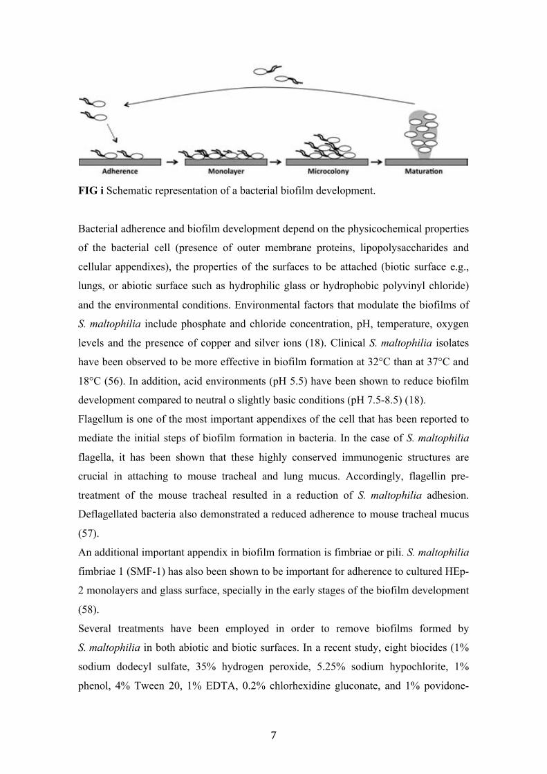
polymeric substance matrix (Fig. i). The first step of the biofilm formation is the
adhesion of the bacteria to the surface. Then, the non-motile adhered bacteria grow and
produce exopolysaccharide (EPS). Once the biofilm is matured, cells start to detach
reverting the planktonic cells and completing the cycle (Fig. i).
7
FIG i Schematic representation of a bacterial biofilm development.
Bacterial adherence and biofilm development depend on the physicochemical properties
of the bacterial cell (presence of outer membrane proteins, lipopolysaccharides and
cellular appendixes), the properties of the surfaces to be attached (biotic surface e.g.,
lungs, or abiotic surface such as hydrophilic glass or hydrophobic polyvinyl chloride)
and the environmental conditions. Environmental factors that modulate the biofilms of
S. maltophilia include phosphate and chloride concentration, pH, temperature, oxygen
levels and the presence of copper and silver ions (18). Clinical S. maltophilia isolates
have been observed to be more effective in biofilm formation at 32°C than at 37°C and
18°C (56). In addition, acid environments (pH 5.5) have been shown to reduce biofilm
development compared to neutral o slightly basic conditions (pH 7.5-8.5) (18).
Flagellum is one of the most important appendixes of the cell that has been reported to
mediate the initial steps of biofilm formation in bacteria. In the case of S. maltophilia
flagella, it has been shown that these highly conserved immunogenic structures are
crucial in attaching to mouse tracheal and lung mucus. Accordingly, flagellin pre-
treatment of the mouse tracheal resulted in a reduction of S. maltophilia adhesion.
Deflagellated bacteria also demonstrated a reduced adherence to mouse tracheal mucus
(57).
An additional important appendix in biofilm formation is fimbriae or pili. S. maltophilia
fimbriae 1 (SMF-1) has also been shown to be important for adherence to cultured HEp-
2 monolayers and glass surface, specially in the early stages of the biofilm development
(58).
Several treatments have been employed in order to remove biofilms formed by
S. maltophilia in both abiotic and biotic surfaces. In a recent study, eight biocides (1%
sodium dodecyl sulfate, 35% hydrogen peroxide, 5.25% sodium hypochlorite, 1%
phenol, 4% Tween 20, 1% EDTA, 0.2% chlorhexidine gluconate, and 1% povidone-

8
iodine) were tested on S. maltophilia biofilms present in dental unit water lines (59).
From these biocides, the most effective combination was the one composed for 5.25%
sodium hypochlorite and 1% phenol, which was shown to remove almost the entire
biofilm. The commonly used disinfecting agent peracetic acid (PAA) was found to
inhibit the growth of a S. maltophilia monoculture biofilm and a dual-culture biofilm
composed by S. maltophilia and Candida parapsilosis (60).
The use of antibiotics at their minimal inhibitory concentrations (MICs) and at
concentrations below the MICs on S. maltophilia cell adherence and the subsequent
biofilm formation have been studied (61, 62). It has been reported that some antibiotics
at suboptimal MICs (e.g., moxifloxacin) are effective in preventing biofilm formation of
S. maltophilia (62). On the other hand, It has been reported that the MDR isolates
showed a higher level of biofilm formation than the non-MDR isolates. In addition, the
biofilm ability have been correlated with resistance to ceftazidime, cefepime, ticarcillin-
clavulanic acid, piperacillin-tazobactam, aztreonam, and gentamicin (63).
1.3.4 Bacterial motility
Three kinds of bacterial motilities have been described for S. maltophilia, and all of
them have been related to pathogenesis. These include swimming, twitching and
swarming.
Swimming motility is a mode of bacterial movement powered by rotating flagella that
takes place as individual cells moving in liquid environments. It is usually tested into
semi-liquid agar plates containing 0.25% agar. In S. maltophilia, deletion of fliI resulted
in the suppression of swimming motility (64). It has been also reported that disruption
of rpfF resulted in the loss of swimming motility in S. maltophilia strain K279a (65).
Another study demonstrated that xanB gene -a lipopolysaccharide/exopolysaccharide
biosynthetic gene- is also required for swimming motility in S. maltophilia strain WR-C
(40). In this mentioned study, it is also reported that the other genes involved in the
lipopolysaccharide/exopolysaccharide biosynthetic pathway rmlA, rmlC, and xanB were
essential in twitching motility (40).
Twitching motility is a sort of surface motility powered by the extension and retraction
of type IV pili, which results in a slow cell movement compared to other types of
motility. Several clinical isolates have been studied to determine the possible correlation
between twitching motility and biofilm formation, concluding that there is no
correlation between these two phenotypes (37, 64).
9
Swarming motility is a rapid and coordinated translocation of a bacterial population
across solid or semi-solid surfaces, which require cellular differentiation
(hiperflagellation), self-production of biosurfactant and quorum sensing-mediated
synchronization. Swarming motility is perhaps the most complex motility described in
bacteria. It depends on several interconnected pathways that enable the whole
population to colonize rapidly new environments. Thousands of papers have been
recently published addressing this topic, the reader could easily address to extensive and
detailed reviews about swarming motility in bacteria (66–71). However, swarming
motility in S. maltophilia has been rarely reported. It is known that swarming motility
requires very specific conditions to take place in the laboratory and many times these
precise conditions are depending on the specie or even on the strain. In our laboratory,
we have spent much time trying to optimize the conditions that facilitate this motility
behaviour. Finally, although S. maltophilia do not display a fast swarming motility, we
have observed that modified M9-salts and BM2 mediums are the most optimum
conditions for the evaluation of swarming motility in this bacterium. Accordingly, we
recently have reported swarming motility in S. maltophilia (72).
One important molecular mechanism involved in bacterial pathogenesis is quorum
sensing (QS). During the last decasdes, QS has aroused much interest because it has
been demonstrated that these cell–cell communication systems control many
phenotypes, including virulence, in numerous bacterial pathogens.
Since QS is the main topic of this work, in the next section, I will address the issue of
QS, with special emphasis in the one described in the nosocomial pathogen
S. maltophilia, the DSF (Diffusible Signal Factor) quorum sensing system.
1.4 Quorum Sensing
1.4.1 Generalities
It is well established that bacterial cells can communicate with each other to facilitate
their rapid adaptation to fluctuations in the environment. This cell-cell communication
mechanism, known as quorum sensing (QS), relies on the production, detection, and
response to diffusible signal molecules (also called autoinducers) in a cell-density-
dependent manner (73–76). Throw this QS communication, numerous bacterial species
regulate a variety of functions such as biofilm formation, toxin production,
10
exopolysaccharide synthesis, extracellular enzyme production, motility, and plasmid
transfer (77–81). When the population density is low, each single cell produces basal
levels of signalling molecules, which are accumulated in the extracellular space.
Concentration of QS signals increase accordingly to the cell density and, once the
concentration reaches a critical threshold, the population synchronize the gene
expression and coordinate various biological activities.
The first QS system was described in the symbiotic gram-negative bacteria Vibrio
fishery, during the 80’s decade (82). In the present, it represents one of the best-
characterized quorum sensing systems. The signalling molecule responsible for this
communication is the AHL-signal (Acyl Homoserine Lactone), which is synthesized by
the enzyme LuxI. When the signal concentration is the optimum, AHL interacts with its
receptor LuxR and induces the transcriptional expression of the lux genes, which are
responsible for the production of bioluminescence. Since this discovery, AHL-based QS
have been described in numerous symbiotic and pathogenic bacteria (83). Hence,
several clinically relevant gram-negative pathogens use quorum sensing systems to
regulate processes associated with virulence (84, 85) (Fig. ii). Furthermore, QS systems
are not restricted to gram-negative bacteria, since several gram-positive bacteria use
peptide-based signalling molecules to regulate biological functions similarly to gram-
negative bacteria (Fig. ii). Some interesting reviews could be consulted to obtain
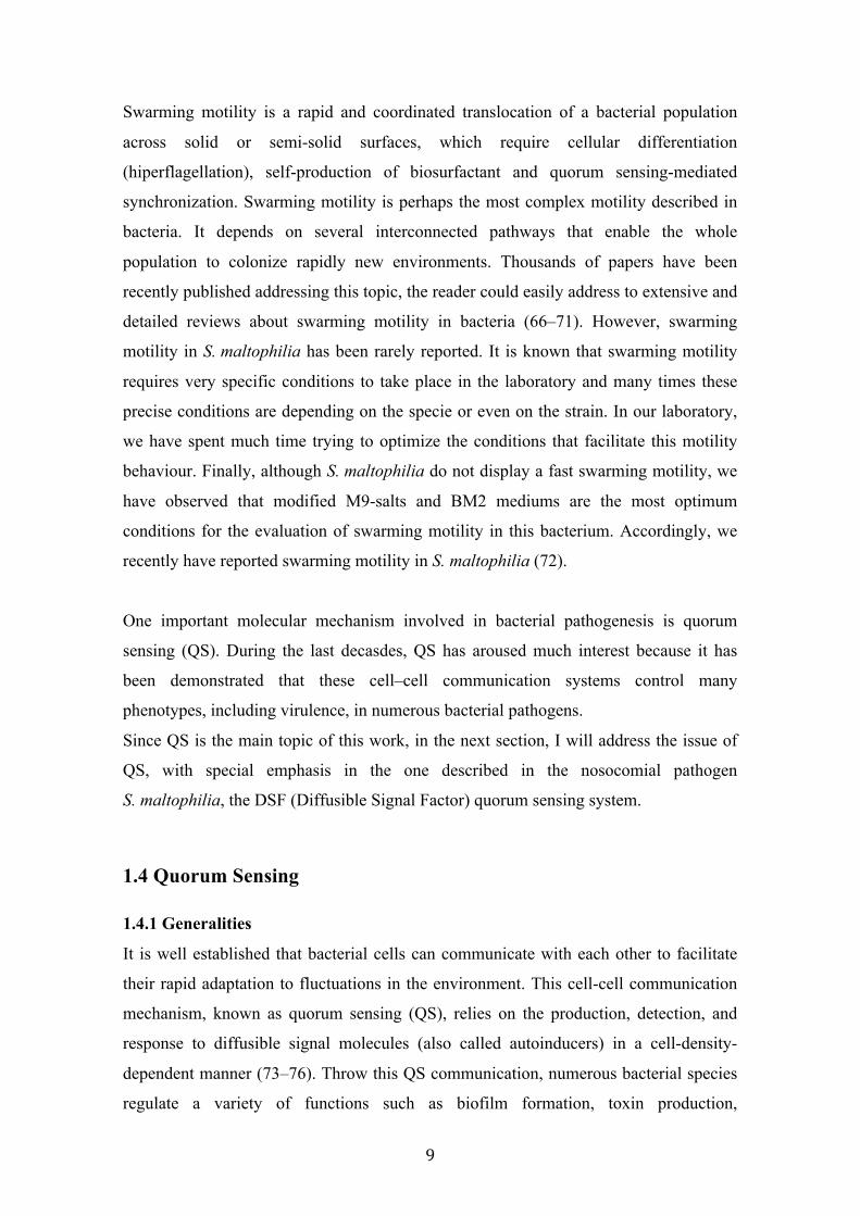
detailed information about quorum sensing in gram-positive bacteria (86–90).
FIG ii. QS signalling molecules used in gram-negative (91) and gram-positive bacteria
(89). Information has been extracted from cited reviews.

11
As observed in the figure ii, one important family of signalling molecules in gram-
negative bacteria is DSF-family. These molecules are simple medium-length fatty acids
that have been demonstrated to synchronize several biological processes in various
gram-negative bacterial pathogens. These interesting features will be detailed in the
following sections.
1.4.2 Generalities of DSF quorum sensing system
The diffusible signal factor (DSF) molecule represents an important type of QS signal
found in many gram-negative bacterial pathogens. This small fatty acid molecule (cis-
11-methyl-2-dodecenoic acid) was for the first time described in the phytopathogen
Xanthomonas campestris pv. campestris (92, 93). In the past few years, it has been
reported that DSF family signals are widespread (91). DSF-QS system has been
described in various gram-negative human and plant pathogens, most of them belonging
to the Xanthomonadales, including X. campestris, X. axonopodis (94, 95) and X. oryzae
(96, 97); Xylella fastidiosa (98, 99), Burkholderia cenocepacea (100, 101) and
S. maltophilia (65, 102). In addition, several DSF structural analogues have been
identified in unrelated microorganisms including the fatty acid trans-2-decenoic acid
(SDSF) produced by Streptocuccus mutans (103) and the farnesoic acid synthesized by
the yeast Candida albicans (100).
Bacterial DSF activity has been primarily detected using different bacterial bioassays.
The first-generation DSF detection methods are based on the protease and
endoglucanase-restoration bioassay. Since it was demonstrated that in Xanthomonas
campestris pv. campestris (Xcc) the production of exoproteases and endoglucanases
depends on the DSF communication (92), the Xcc ∆rpfF mutant (DSF minus) is used as
a reporter strain (92). Thus, the DSF-producer candidate strains are streaked onto plates
containing either skimmed milk (for proteolysis-based bioassay) or
carboxymethylcellulose (CMC) (for endoglucanase-based bioassay) closely to the Xcc
reporter strain (∆rpfF mutant) and the presence of a clear halo around the bacterial
reporter colony indicates DSF activity (92).
Second-generation DSF-activity detection methods are based on engineered DSF
biosensors strains. Classically, the promoter of the DSF-inducible operon engXCA
(which encodes for an endoglucanase) is fused with a promoterless reporter gusA gene
(which encodes for a ß-glucuronidase). The genic fusion is cloned into a replicative
12
vector and the resultant plasmid is mobilized to a DSF-minus Xcc mutant (Xcc ∆rpfF)
generating the DSF reporter strain (104, 105). Then, a monolayer of the biosensor strain
is seeded onto a nutrient broth plate containing the GusA substract X-Glu (5-Bromo-4-
chloro-3-indolyl β-D-glucopyranoside) and the candidate strains are pin inoculated onto
it. After an overnight incubation, the presence of a blue halo around the colony of
candidate strains indicates DSF activity. These engineered biosensor strains have been
widely used in detection of DSF-family signals in other bacterial species, including
members of the Burkholderia cepacia complex and Xanthomonas oryzae pv. oryzae
(100, 101). Another DSF biosensor, which was constructed using engXCA promoter
fused to a promoterless gfp gene, has been employed to detect the DSF-family signals
from Xylella fastidiosa and Xanthomonas axonopodis pv. glycines (95, 98, 106).
1.4.3 DSF-QS in Xanthomonas campestris pv. campestris
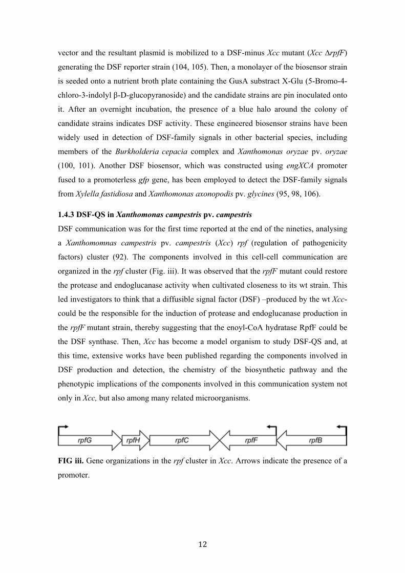
DSF communication was for the first time reported at the end of the nineties, analysing
a Xanthomomnas campestris pv. campestris (Xcc) rpf (regulation of pathogenicity
factors) cluster (92). The components involved in this cell-cell communication are
organized in the rpf cluster (Fig. iii). It was observed that the rpfF mutant could restore
the protease and endoglucanase activity when cultivated closeness to its wt strain. This
led investigators to think that a diffusible signal factor (DSF) –produced by the wt Xcc-
could be the responsible for the induction of protease and endoglucanase production in
the rpfF mutant strain, thereby suggesting that the enoyl-CoA hydratase RpfF could be
the DSF synthase. Then, Xcc has become a model organism to study DSF-QS and, at
this time, extensive works have been published regarding the components involved in
DSF production and detection, the chemistry of the biosynthetic pathway and the
phenotypic implications of the components involved in this communication system not
only in Xcc, but also among many related microorganisms.
FIG iii. Gene organizations in the rpf cluster in Xcc. Arrows indicate the presence of a
promoter.

13
1.4.3.1 DSF biosynthesis, RpfF and RpfB
It was found that mutation on rpfF or rpfB genes (Fig. iii) abolished DSF production,
suggesting that these two enzymes are required for DSF synthesis in Xcc (92, 105) (Fig.
iv). Further in silico analysis predicted that rpfF encode a putative enoyl-CoA hydratase
and rpfB encode a putative long-chain fatty acyl CoA ligase (92, 105). More recently, it
has been shown that RpfF belongs to the members of the crotonase superfamily (107).
Two putative catalytic glutamate residues (Glu141 and Glu161) have been detected in
RpfF, which are conserved in the enoyl-CoA hydratases. Accordingly, substitution of
these two residues in RpfF resulted in the suppression of DSF synthesis, demonstrating
their important role in DSF production (107). In addition, structural analysis of RpfF
also revealed a hydrophobic pocket, which is comprised by several hydrophobic
residues including Leu136, Gly137, Gly138, Gly85, Leu276, Met170, and Trp258
(107). It has been suggested that these amino acids compose the putative substrate-
binding pocket for DSF biosynthesis (107). Accordingly, a single-point mutation in any
of these residues resulted in a drastic fall or even abolition of DSF production (107).
1.4.3.2 DSF detection by RpfC
Another important component in the rpf cluster is the gene rpfC (Fig. iii), which
encodes for a two-component hybrid sensor kinase (Fig. iv). It is composed by five
transmembrane (TMR) domains, an histidine kinase (HisKA) domain, a receiver (REC)
domain and a histidine phosphotransfer (HPT) domain. It has been reported that the
exogenous addition of DSF could fully restore the rpfF but not rpfC mutant phenotype,
strongly suggesting that RpfC is responsible of DSF detection (105, 108). It has been
demonstrated that RpfC uses a conserved phosphorelay mechanism to transduce the
DSF signal (Fig. iv). However, it still remains undetermined how the RpfC could detect
DSF and trigger the phosphorelay process.
Curiously, it has been reported that –in addition to sense DSF- RpfC also acts
negatively blocking the DSF biosynthesis (105) (Fig. iv). Based on classic DSF
bioassay, it has been demonstrated that the rpfC mutant produced higher amount of DSF
compared to the Xcc wt strain (105). It has been reported that the REC domain of RpfC
directly interacts with the catalytic domain of the RpfF, blocking its DSF-synthesis
activity (107) (Fig. iv).
14
Although these results provide evidences about the interaction between RpfC and RpfF,
the mechanisms by which RpfC senses DSF and subsequently liberates free active-RpfF
remain uncertain.
1.4.3.3 Signal transduction by RpfG
Since the genes rpfG, rpfH, and rpfC are organized into an operon in the genome of
Xcc, it was suggested that these three genes could have related functions (105).
Specifically, RpfG is a cytoplasmic regulator element that contains a typical receiver
domain and a HD-GYP domain. It has been reported that mutation in the rpfG and rpfC
genes lead to similar phenotypic disorders, related to insensitivity to DSF (105, 109). It
has been demonstrated that the HD-GYP domain of RpfG plays a key role in DSF
signal transduction (110). In particular, it has been revealed that this HD-GYP domain
has a novel function related to cyclic diguanilate (c-diGMP) metabolism. It is known
that c-diGMP is a global second messenger with important implications in the
regulation of numerous biological functions in bacteria (111–113). It was postulated
that this novel HD-GYP domain might be key component in bacterial signalling
enabling adaptation to environmental changes by modulating the c-diGMP levels (114).
It has been confirmed that the HD-GYP domain degraded c-di-GMP to generate two
molecules of GMP (110) (Fig. iv). In concordance with the predicted function of this
new phosphodiesterase domain, in trans expression of the HD-GYP domain in the rpfG
mutant was sufficient to restore the mutant phenotypes (110).
Altogether, these findings reveal that the RpfG is a novel c-diGMP degradation enzyme,
which is activated by the DSF signalling and regulates biological functions by the
modulation of the intracellular concentration of this important second messenger
(Fig. iv).

15
FIG iv. Schematic representation of the DSF-QS system in Xcc. (A) At low cell
density, the DSF sensor RpfC blocks the DSF synthase RpfF through its receiver
domain (REC), which restricts DSF biosynthesis at a basal level. The cytoplasmic
regulator element RpfG remains inactive. (B) At high cell density, RpfC senses the
accumulation of DSF molecules and triggers a phosphorylation cascade that ends in the
activation of the phosphodiesterase RpfG. The increment of intracellular GMP triggers
the expression of virulence genes.
1.4.4 DSF-QS in Xylella fastidiosa
X. fastidiosa is the causal agent of Pierce's disease, responsible for large economic
losses in agriculture (115, 116). In the case of X. fastidiosa, the signalling
communication is mediated by a DSF derivative, identified as 12-methyl-tetradecanoic
acid and designated xfDSF (99). In silico analysis showed that this pathogen harbours a
conserved rpfGCF gene cluster, displaying a high identity to the Xcc rpf (91). As
expected, mutation of rpfF in X. fastidiosa also abolished xfDSF production and,
contrary to that observed in Xcc, it enhanced virulence, impaired insect transmission,
and disabled biofilm formation in host insects (106). Mutation of the rpfC also resulted
in the overproduction of xfDSF, a deficiency in the virulence ability of X. fastidiosa
(98). Interestingly, recent findings have evidenced that in the case of X. fastidiosa, the
xfDSF synthase RpfF is also essential for xfDSF perception. This new findings have
made reconsider the role of the RpfC not only in X. fastidiosa, but also in related
bacteria sharing the DSF-QS (117).

16
Nevertheless, much work is needed to understand the complexity of xfDSF-QS system
in this important bacterial pathogen.
1.4.5 DSF-QS in Burkholderia spp.
The Burkholderia cepacia complex is a group comprised of nine related species which
have recently emerged as problematic opportunistic pathogens in patients with CF and
immunocompromised individuals (118–121). A DSF derivative (cis-2-dodecenoic acid)
has been recently identified in B. cenocepacia and designated BDSF (Fig. ii) (100).
Although the synthase Bcam0581 shares near 40% of protein identity with the RpfF of
Xcc, it has been demonstrated that Bcam0581 is essential for BDSF production and can
functionally substitute the rpfF gene in Xcc ∆rpfF mutant (100). Nevertheless, contrary
to the genetic organization observed for the other related bacteria sharing the DSF-QS,
Bcam0581 is not encoded in the rpf gene cluster. Hence, genomes of B. cepacea strains
do not contain neither rpfC nor rpfG homologues flanking bcam0581 gene. This
suggests that BDSF-QS system in B. cenocepacia could have a distinct evolutionary
origin (100).
However, deletion of bcam0581 resulted into alteration of similar processes as observed
in the rpfF mutant in Xcc. These include delayed bacterial growth in minimal medium,
reduced swarming motility, alteration in biofilm development and reduced virulence
(101, 122–124). Interestingly, it has been suggested that certain biological functions
such as motility, biofilm formation, and virulence, are coregulated by the BDSF
signalling and the AHL-dependent QS system in B. cenocepacia (125). More recently, it
has been identified Bcam0227 as a potential BDSF sensor, which shares 35.6% of
identity with the RpfC of Xcc (124). This putative BDSF sensor contains four domains,
including a histidine kinase phosphoacceptor domain, a histidine kinase domain, a
CheY-like receiver domain, and a C-terminal histidine phosphotransfer domain (124).
Deletion of bcam0227 also resulted into attenuation of bacterial virulence (124).
Additionally, it has been shown that other members of the B. cepacia complex also
produced different derivative DSF-QS signals (101), which complicates the
understanding of the mechanism of the BDSF-QS in this important opportunistic
bacterium.
1.4.6 DSF-QS in Stenotrophomonas maltophila
S. maltophilia harbours a rpf gene cluster highly conserved (≈65-80% peptide
similarity) with the model strain Xcc (65). It has been demonstrated that disruption of

17
DSF signalling has a drastic effect on S. maltophilia K279a, since the rpfF mutant
shows reduced swimming motility, reduced exoprotease production, altered LPS,
reduced tolerance to a range of antibiotics and to heavy metals and reduced virulence in
a Caenorhabditis elegans infection model (65). In addition, FecA, a ferric citrate
receptor, has been shown to be positively regulated by the DSF-QS system (126). This
receptor contributes to the internalization of iron, an essential element for the expression
of virulence-related genes (126). In the S. maltophilia WR-C wild type strain and a
flagella-defective xanB mutant, flagella-independent translocation was stimulated not
only by the main DSF but also by its derivative 11-methyl-dodecanoic acid (127).
Regarding the interaction of S. maltophilia with plants, DSF seems to be involved in
oilseed germination, plant colonization and biofilm architecture in the environmental
strain R551-3 (17). In a recent S. maltophilia population study, the authors detected
rpfF+ genotypes in 61% of the 89 tested strains, suggesting that an important
population of S. maltophilia lacks the rpfF gene (128).
With the rapid increase in the number of S. maltophilia sequenced genomes it is now
possible to compare the rpf cluster among different strains. A preliminary analysis
showed that all sequenced genomes contain the rpfF gene. In addition, at least two rpf
cluster variants can be detected on the basis of sequence and genomic organization, with
main differences found in the rpfF and rpfC genes. However, while numerous and
extensive works have been published in other related bacteria sharing the DSF-QS, in
the case of S. maltophilia, more efforts are needed to understand how this cell-cell
communication system regulates virulence-related processes.
1.5 Pseudomonas aeruginosa, an overview
1.5.1 Microbiology of P. aeruginosa
P. aeruginosa is a gram-negative, aerobic, coccobacillus bacterium with unipolar
motility. It belongs to γ -proteobacteria class and frequently it is responsible of cause
disease in animals, including humans. P. aeruginosa contains a large genome, with one
of the largest regulatory networks of any bacteria sequenced (129). Part of the
significance of this large genome is that it provides the bacterium with a tremendous
diversity of metabolic pathways and physiological responses, which allows the bacteria
to acclimatize to many diverse environments.

18
Even though P. aeruginosa is most predominant in the environment, the organism is
mainly studied in the context of lung infections in CF patients. It is the most prevalent
cause of morbidity and mortality in CF patients, who are unable to clear the bacteria
from their lungs. P. aeruginosa has also been isolated in samples taken from chronic
sinusitis and burn patients, and has been implicated in urinary tract infections (130). The
bacterium is a frequent culprit of hospital-acquired infections, and once P. aeruginosa
infections have become established, they are characteristically difficult to treat.
1.5.2 Mechanisms involved in pathogenesis
P. aeruginosa displays three kinds of surface motility: swarming (131), twitching (132),
and sliding (133). Twitching and swarming motilities are strongly linked with biofilm
development and pathogenesis, and their mechanical components, the flagellum and
type IV pili (TFP), have been demonstrated to be virulence factors (133, 134). TFP are
essential for twitching motility, but they are generally not considered important for
swarming behaviour. Little is known about sliding motility, but it doesn’t seem to be
dependent on flagella and TFP (133).
It is known that bacterial motility and adhesion are two important properties directly
related to virulence. Biofilm development and swarming represent two contrasting
lifestyles that are mutually complimentary. One opinion is that surface motility,
including swarming, allows cells to colonize surfaces and then establish non-motile
biofilms. It has been reported that surface attachment proteins are up-regulated in
bacteria within the biofilm and predictably down-regulated in swarming cells (135). It
has become evident that biofilm formation is a population strategy to protect the
bacteria from desiccation, nutrient deficiency, antibiotics, and human immune response.
Cell motility is necessary for the expansion and maturation of the biofilm, and the
biofilm represents a ‘safe-haven’ community of sessile cells that have stopped moving.
One important factor implied in the regulation of several virulence-related traits,
including biofilm formation and bacterial motility, is quorum sensing.
1.5.3 Quorum sensing regulatory network in P. aeruginosa
The signalling network of P. aeruginosa is probably the most complex systems known
and, to date, the most studied among bacteria. It consists of numerous interconnected
signalling pathways that, together, regulate complex population behaviours. The best-
studied communication system in P. aeruginosa is undoubtedly the AHL-mediated QS
system, which is involved in the regulation of several virulence-related phenotypes, not

19
only in P. aeruginosa but also in most of the species sharing related QS systems (136–
138). Nevertheless, some other well-studied QS systems based on different signalling
molecules coexist in this bacterium. These include the Pseudomonas Quinolone System
(PQS) -which is mainly based on the molecule 4-quinolone and has been shown to be
essential for the binding of iron facilitating the work of the siderophores (139, 140) -,
the pigmented signalling system, which includes pyoverdine (PVD), phenazine and
pyocyanine which are responsible for the production of several virulence factors,
including exotoxin A (ToxA) and PrpL endoprotease (141) - and the recently reported
cis-DA system, based on the fatty acid molecule cis-decenoic (142, 143), which is
indeed the subject of this study. cis-DA system has aroused attention recently, since this
new small fatty acid produced by P. aeruginosa has been show to stimulate biofilm
dispersion in several gram-negative and gram-positive bacteria (142). Although P.
aeruginosa harbours 12 putative enoyl-CoA hydratases, it recently has been reported
that the enoyl-CoA hydratase DspI (PA0745) is the responsible for the cis-DA synthesis
(143). In addition, it has been recently reported that mutation of PA0745 resulted into
attenuation of virulence in the C. elegans model (144). Nevertheless, an alternative
ECH is also present in the PA0745 cluster, the ECH PA0744. Curiously, none of the
mentioned studies reported information about this alternative ECH, which could be
probably involved in the biosynthesis of the fatty acid signal cis-DA.
Interestingly, we have observed that in the S. maltophilia genome, there exists
homologues of the cis-DA synthase PA0745 and the alternative ECH PA0744, which
corresponded to Smlt0266 and Smlt0267, respectively. Since this fatty acid seems to
represent a new fatty acid-mediated QS system in gram-negative bacteria and could
present parallelisms to DSF-QS system, we aim to study this new communication
system not only in S. maltophilia but also in P. aeruginosa.

20

OBJECTIVES

22
2. OBJECTIVES The main objective of this thesis is to study the fatty acid-mediated quorum sensing systems that coexist in Stenotrophomonas maltophilia and the implications that they have in virulence regulation in this pathogen. More specifically, our aim is to get insights into the DSF-based and the recently reported cis-DA-based quorum sensing systems in S. maltophilia. Specific objectives have been:
To analyse the molecular aspects that governs the DSF synthesis and perception To investigate the environmental conditions that modulates DSF production To identify and characterise the DSF molecules produced by S. maltophilia. To evaluate the role of the DSF quorum sensing system in the regulation of biological functions To investigate if the new cis-DA quorum sensing system is present in S. maltophilia To evaluate the implication of the enoyl-CoA hydratases Smlt0266 and Smlt0267 in the regulation of cis-DA quorum sensing system in S. maltophilia. To investigate the implication of the orthologous enoyl-CoA hydratases PA0745 and PA0744 in the regulation of the cis-DA quorum sensing in Pseudomonas aeruginosa.

MATERIALS AND METHODS

24
3. MATERIALS AND METHODS
3.1 Bacterial strains and growth conditions.
3.1.1 S. maltophilia and other Xanthomonads
A panel of 78 S. maltophilia clinical isolates were collected from intensive care units
(ICUs) of different European Hospitals. Name, geographic origin, hospital and isolation
source per strain are given in Table 1. From this collection, E77 (RpfF-1-variant group)
and M30 (RpfF-2-variant group) (145) were used as model strains to characterize ∆rpfF
mutants (Sections 4.1 and 4.2). S. maltophilia strain M30 wt and the ∆ smlt0266,
∆smlt0267 and ∆ smlt0266-0267 mutants were also used to study the new cis-DA-QS
system (Section 4.3). S. maltophilia strains were routinely grown at 37ºC in Luria-
Bertani (LB) medium on a rotary shaker. When needed, LB was supplemented with the
following antibiotics: tetracycline (Tc) 17 µg/mL, chloramphenicol (Cm) 32 µg/mL and
erythromycin (Erm) 500 µg/mL. For phenotypic analysis in minimal media, strains
were grown in BM2 medium (62 mM potassium phosphate buffer, pH 7, 2 mM MgSO4,
10 µM FeSO4, supplemented with glucose 0.4 %) (146) or a modified M9-salts medium
(modified M9 salts [33.9g/L Na2HPO4 7H2O, 15g/L KH2PO4, 2,5g/L NaCl without
NH4Cl], 0.5% casamino acids, 2 mM MgSO4, 0.1 mM CaCl2 and supplemented with
0.2% glucose) (147).
Xanthomonas campestis pv. campestris (Xcc) 8523 pL6engGUS was obtained from the
authors (148). Xcc 8523 pL6engGUS was used as a reporter strain for the detection of
S. maltophilia DSF activity. Xcc was routinely grown at 28ºC in NYG medium (0.5%
peptone, 0.3% yeast extract and 2% glycerol) supplemented with Tc 10 µg/mL.
3.1.2 P. aeruginosa
P. aeruginosa strain MPAO1 wt and the ∆PA0745 (strain name PW2354) and ∆PA0744
(strain name PW2353) mutants were obtained from the transposon mutant library from
the University of Washington (149) and were used to study the new cis-DA-QS system
(Section 4.3) in this bacterium. Bacteria were routinely grown at 37ºC in LB medium on
a rotary shaker. When needed, LB was supplemented with Tc 17 µg/mL, Cm 32 µg/mL
and Gm 40 µg/mL. For phenotypic analysis in minimal media, strains were grown in
modified M9-salts medium (147).
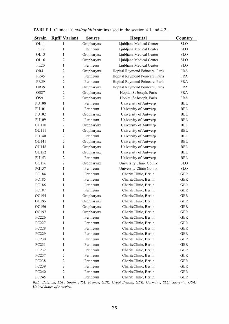
25
TABLE 1. Clinical S. maltophilia strains used in the section 4.1 and 4.2.
Strain RpfF Variant Source Hospital Country OL11 1 Oropharynx Ljubljana Medical Center SLO PL12 1 Perineum Ljubljana Medical Center SLO OL13 1 Oropharynx Ljubljana Medical Center SLO OL16 2 Oropharynx Ljubljana Medical Center SLO PL20 1 Perineum Ljubljana Medical Center SLO OR41 2 Oropharynx Hopital Raymond Poincare, Paris FRA PR45 2 Perineum Hopital Raymond Poincare, Paris FRA PR59 2 Perineum Hopital Raymond Poincare, Paris FRA OR79 1 Oropharynx Hopital Raymond Poincare, Paris FRA OS87 2 Oropharynx Hopital St Joseph, Paris FRA OS91 2 Oropharynx Hopital St Joseph, Paris FRA
PU100 1 Perineum University of Antwerp BEL PU101 1 Perineum University of Antwerp BEL PU102 1 Oropharynx University of Antwerp BEL PU109 2 Perineum University of Antwerp BEL OU110 2 Oropharynx University of Antwerp BEL OU111 1 Oropharynx University of Antwerp BEL PU140 2 Perineum University of Antwerp BEL OU141 2 Oropharynx University of Antwerp BEL OU148 1 Oropharynx University of Antwerp BEL OU152 1 Oropharynx University of Antwerp BEL PU153 2 Perineum University of Antwerp BEL OG156 2 Oropharynx University Clinic Golnik SLO PG157 1 Perineum University Clinic Golnik SLO PC184 1 Perineum ChariteClinic, Berlin GER PC185 1 Perineum ChariteClinic, Berlin GER PC186 1 Perineum ChariteClinic, Berlin GER PC187 1 Perineum ChariteClinic, Berlin GER OC194 1 Oropharynx ChariteClinic, Berlin GER OC195 1 Oropharynx ChariteClinic, Berlin GER OC196 1 Oropharynx ChariteClinic, Berlin GER OC197 1 Oropharynx ChariteClinic, Berlin GER PC226 1 Perineum ChariteClinic, Berlin GER PC227 1 Perineum ChariteClinic, Berlin GER PC228 1 Perineum ChariteClinic, Berlin GER PC229 1 Perineum ChariteClinic, Berlin GER PC230 1 Perineum ChariteClinic, Berlin GER PC231 1 Perineum ChariteClinic, Berlin GER PC232 1 Perineum ChariteClinic, Berlin GER PC237 2 Perineum ChariteClinic, Berlin GER PC238 2 Perineum ChariteClinic, Berlin GER PC239 2 Perineum ChariteClinic, Berlin GER PC240 2 Perineum ChariteClinic, Berlin GER PC245 1 Perineum ChariteClinic, Berlin GER
BEL: Belgium, ESP: Spain, FRA: France, GBR: Great Britain, GER: Germany, SLO: Slovenia, USA: United States of America.
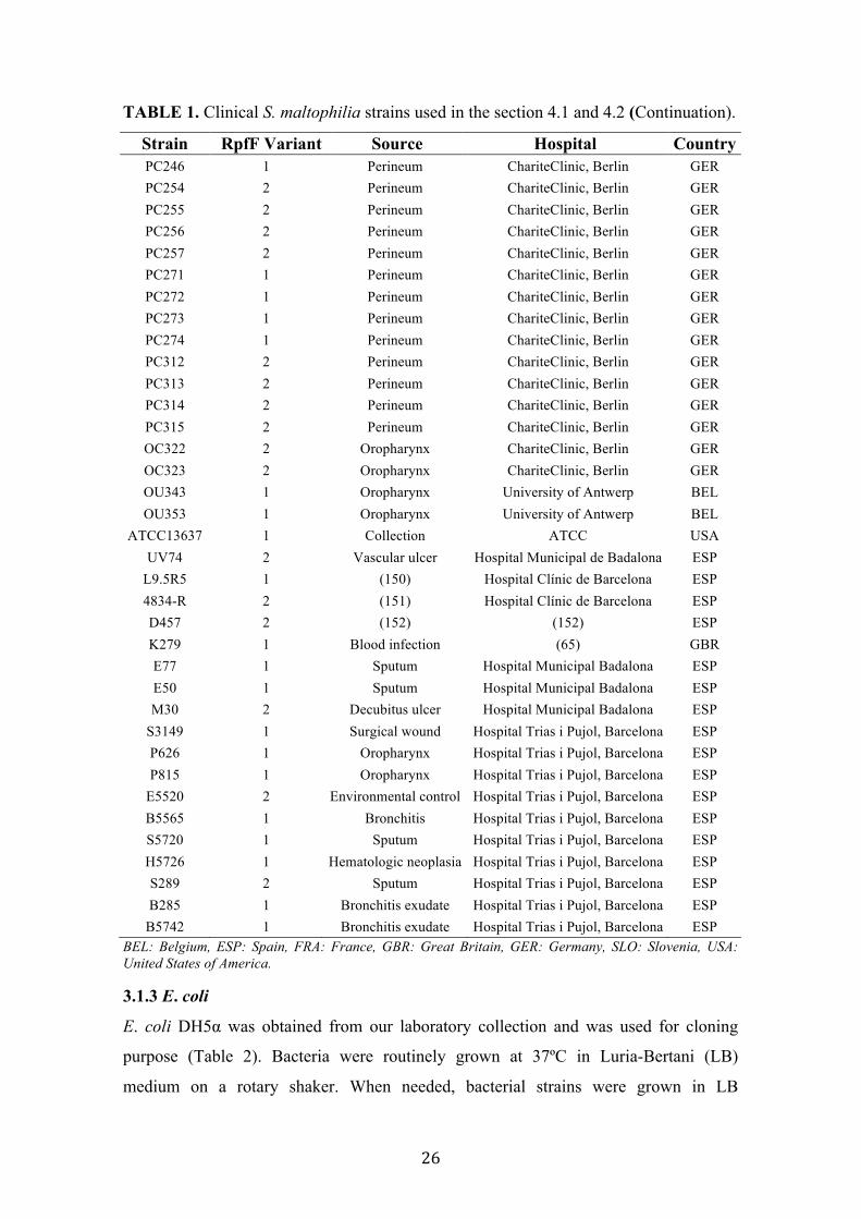
26
TABLE 1. Clinical S. maltophilia strains used in the section 4.1 and 4.2 (Continuation).
Strain RpfF Variant Source Hospital Country PC246 1 Perineum ChariteClinic, Berlin GER PC254 2 Perineum ChariteClinic, Berlin GER PC255 2 Perineum ChariteClinic, Berlin GER PC256 2 Perineum ChariteClinic, Berlin GER PC257 2 Perineum ChariteClinic, Berlin GER PC271 1 Perineum ChariteClinic, Berlin GER PC272 1 Perineum ChariteClinic, Berlin GER PC273 1 Perineum ChariteClinic, Berlin GER PC274 1 Perineum ChariteClinic, Berlin GER PC312 2 Perineum ChariteClinic, Berlin GER PC313 2 Perineum ChariteClinic, Berlin GER PC314 2 Perineum ChariteClinic, Berlin GER PC315 2 Perineum ChariteClinic, Berlin GER OC322 2 Oropharynx ChariteClinic, Berlin GER OC323 2 Oropharynx ChariteClinic, Berlin GER OU343 1 Oropharynx University of Antwerp BEL OU353 1 Oropharynx University of Antwerp BEL
ATCC13637 1 Collection ATCC USA UV74 2 Vascular ulcer Hospital Municipal de Badalona ESP
L9.5R5 1 (150) Hospital Clínic de Barcelona ESP 4834-R 2 (151) Hospital Clínic de Barcelona ESP D457 2 (152) (152) ESP K279 1 Blood infection (65) GBR E77 1 Sputum Hospital Municipal Badalona ESP E50 1 Sputum Hospital Municipal Badalona ESP M30 2 Decubitus ulcer Hospital Municipal Badalona ESP
S3149 1 Surgical wound Hospital Trias i Pujol, Barcelona ESP P626 1 Oropharynx Hospital Trias i Pujol, Barcelona ESP P815 1 Oropharynx Hospital Trias i Pujol, Barcelona ESP
E5520 2 Environmental control Hospital Trias i Pujol, Barcelona ESP B5565 1 Bronchitis Hospital Trias i Pujol, Barcelona ESP S5720 1 Sputum Hospital Trias i Pujol, Barcelona ESP H5726 1 Hematologic neoplasia Hospital Trias i Pujol, Barcelona ESP S289 2 Sputum Hospital Trias i Pujol, Barcelona ESP B285 1 Bronchitis exudate Hospital Trias i Pujol, Barcelona ESP
B5742 1 Bronchitis exudate Hospital Trias i Pujol, Barcelona ESP BEL: Belgium, ESP: Spain, FRA: France, GBR: Great Britain, GER: Germany, SLO: Slovenia, USA: United States of America.
3.1.3 E. coli
E. coli DH5α was obtained from our laboratory collection and was used for cloning
purpose (Table 2). Bacteria were routinely grown at 37ºC in Luria-Bertani (LB)
medium on a rotary shaker. When needed, bacterial strains were grown in LB

27
supplemented with ampicillin (Ap) 20 µg/mL, Tc 17 µg/mL, Cm 3.2 µg/mL, Gm 40
µg/mL and Erm 500 µg/mL. In order to avoid unnecessary information and tables, all E.
coli DH5α cloning-derived strains have been excluded from this manuscript.
E. coli strain OP50 (Table 2) was provided by the “Caenorhabditis Genetic Center”
(CGC) (University of Minnesota, USA) and used as food source for the maintenance of
Caenorhabditis elegans. E. coli strain OP50 was routinely grown at 37ºC in Luria-
Bertani (LB) medium on a rotary shaker.
TABLE 2. E. coli parental strains used in this study.
Strains Relevant characteristics Reference
DH5α fhuA2 lac(del)U169 phoA glnV44Φ80' lacZ(del)M15 gyrA96 recA1 relA1 endA1 thi-1 hsdR17
Lab. collection
OP50 Non-pathogenic strain, C. elegans maintenance CGC CGC: Caenorhabditis Genetics Center (www.cbs.umn.edu/research/resources/cgc).
3.1.4 Bacterial preservation
S. maltophilia, Xcc and E. coli bacterial strains were routinely conserved by
cryopreservation. 1.5 mL from an overnight bacterial culture was mixed with 0.75 mL
of 50% sterile glycerol (15% v/v final glycerol concentration), frozen in dry ice and
then stored at -80ºC. For bacterial recovery, frozen culture surfaces were scrapped and
seeded onto their respective medium plates.
P. aeruginosa strains were routinely preserved at DMSO 15% and then stored at -80ºC.
For a short-term storage, P. aeruginosa strains were preserved in water. One single
bacterial colony was suspended in 1.5 mL of sterile distilled water and stored at 4ºC.
For bacterial recovery, 100 µL of the samples were seeded onto their respective medium
plates.
3.2 Oligonucleotides and plasmid vectors Primers for PCR experiments were designed according the following standard. The
length of each primer was ranged from 18-25 nucleotides with a >50% of
guanine/cytosine (GC) content greater than 50% in order to result in a melting
temperature (Tm) from 50-65 ˚C. The software Oligos 7.0 was used to check and avoid
the secondary structure, hairpin, primer dimmer and cross dimmer caused by designed
primers. Common primers used in this study are listed in Table 3. All primer were
synthesized and purchased at Sigma-Aldrich.
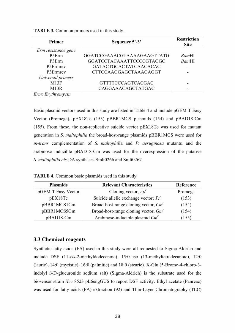
28
TABLE 3. Common primers used in this study.
Primer Sequence 5'-3' Restriction Site
Erm resistance gene P5Erm GGATCCGAAACGTAAAAGAAGTTATG BamHI P3Erm GGATCCTACAAATTCCCCGTAGGC BamHI
P5Ermrev GATACTGCACTATCAACACAC - P3Ermrev CTTCCAAGGAGCTAAAGAGGT -
Universal primers M13F GTTTTCCCAGTCACGAC - M13R CAGGAAACAGCTATGAC -
Erm: Erythromycin.
Basic plasmid vectors used in this study are listed in Table 4 and include pGEM-T Easy
Vector (Promega), pEX18Tc (153) pBBR1MCS plasmids (154) and pBAD18-Cm
(155). From these, the non-replicative suicide vector pEX18Tc was used for mutant
generation in S. maltophilia the broad-host-range plasmids pBBR1MCS were used for
in-trans complementation of S. maltophilia and P. aeruginosa mutants, and the
arabinose inducible pBAD18-Cm was used for the overexpression of the putative
S. maltophilia cis-DA synthases Smlt0266 and Smlt0267.
TABLE 4. Common basic plasmids used in this study.
Plasmids Relevant Characteristics Reference pGEM-T Easy Vector Cloning vector, Apr Promega
pEX18Tc Suicide allelic exchange vector; Tcr (153) pBBR1MCS1Cm Broad-host-range cloning vector, Cmr (154) pBBR1MCS5Gm Broad-host-range cloning vector, Gmr (154)
pBAD18-Cm Arabinose-inducible plasmid Cmr. (155)
3.3 Chemical reagents Synthetic fatty acids (FA) used in this study were all requested to Sigma-Aldrich and
include DSF (11-cis-2-methyldodecenoic), 15:0 iso (13-methyltetradecanoic), 12:0
(lauric), 14:0 (myristic), 16:0 (palmitic) and 18:0 (stearic). X-Glu (5-Bromo-4-chloro-3-
indolyl ß-D-glucuronide sodium salt) (Sigma-Aldrich) is the substrate used for the
biosensor strain Xcc 8523 pL6engGUS to report DSF activity. Ethyl acetate (Panreac)
was used for fatty acids (FA) extraction (92) and Thin-Layer Chromatography (TLC)
29
experiments, acetonitrile (Sigma-Aldrich) was used for FA separation using HPLC
technique and hexane (Sigma-Aldrich ) was also used in TLC experiments.
The antimicrobials tetracycline, minocycline, gentamicin, kanamycin, trimethoprim
were purchased to Apollo Scientific Ltd. Sulfamethoxazole, norfloxacin, ciprofloxacin,
erythromycin and levofloxacin were bought at Sigma-Aldrich and chloramphenicol was
purchased to Roche Diagnostics.
The anaesthesia MS-222 (Sigma-Aldrich) was used in zebrafish experiments.
Crystal violet (Sigma-Aldrich) was used for biofilm staining; Congo Red (Sigma-
Aldrich) was used for the evaluation of exopolysaccharide production and colony
morphology and Coomassie Brilliant Blue (Sigma-Aldrich) was used to visualize
protein spots in SDS-PAGE separation.
3.4 Molecular biology techniques
3.4.1 General guidelines
Molecular techniques have been performed following the standard guidelines according
to Sambrook et al., (1989) (156). Restriction enzymes, T4 DNA ligase and FastStart
DNA Polymerase have been purchased to Roche. In all cases, protocols have been
followed according to the manufacturer’s instructions. Specific molecular techniques
and protocols are detailed below, in its respective section.
3.4.2 Polymerase Chain Reaction (PCR) and quantitative reverse transcription-
PCR (qRT-PCR).
For routinely PCR amplification, FastStart DNA Polymerase (Roche) has been used,
following the manufacturer’s instructions.
Primers for qRT-PCR were designed to have a length near 20 nt and a Tm circa 60˚C
and were predicted to produce no secondary structure, hairpin, primer dimmer and cross
dimmer, to amplify a desired 50-150 bp in size amplicon (Table 5). Total RNA was
isolated from S. maltophilia cultures grown in the same conditions as for DSF
extraction using GeneJet RNA Purification Kit (Thermo Scientific) and DNA was
eliminated using TURBO DNase (Ambion, Life Technologies). 1 µg of RNA was used
to synthetize cDNA using iScript cDNA Synthesis Kit (Bio-Rad). Quantitative real time
PCR was performed using CFX96 real-time PCR system (Bio-Rad) and PCR
amplification was detected using SsoAdvanced SYBR Green Supermix (Bio-Rad). PCR
products between 80 and 110 bp were amplificated for rpfC, rpfF and gyrA, the latter

30
used as endogenous gene to normalize gene expression (157), using primers listed in
Table 5. Differences in the relative amounts of mRNA for the four genes rpfF-1, rpfC-
1, rpfF-2 and rpfC-2 were determined using the 2-∆∆Ct method (158). RNA samples
were extracted in three different experiments and results are given as mean values.
TABLE 5. Primers used in qRT-PCR analysis.
Primer Sequence 5'-3' Tm (ºC) P1rpfC-RT GTTCCGCACGCCGTTGAA 59 P2rpfC-RT CGAGGCCTGGATGGTGTT 57 P1rpfF-RT CTGAGCTGCCACACCATC 56 P2rpfF-RT GAACAGCACCTCCGGCAG 58 P1gyrA-RT GTCGACGGCCAGGGTAAC 58 P2gyrA-RT GCCTCGGTGTATCGCATT 55
3.5 DNA Sequencing All PCR amplifications and plasmid constructs have been routinely verified by
sequencing (Macrogen Inc., Korea).
3.5.1 rpfF sequence determination and analysis
PCR products of 682 to 721 bp, containing the rpfF promoter and the region encoding
the N-terminal fragment, were amplified from all 78 S. maltophilia strains using the
primers PrpfFtypeUp and PrpfFTypeDw (Table 6) and directly sequenced (Macrogen
Inc). Translation of partial ORFs to amino acids and sequence alignments were done
using MEGA V5.2 (159) and BioEdit, respectively. A phylogenetic tree using
neighbour-joining method (default parameters) was constructed with MEGA V5.2
based on a trimmed alignment to the 108 N-terminal residues of the RpfF from strain
K279a. RpfC-variant determination was based on the RpfC sequences from the publicly
available sequenced genomes (strains K279a, R551-3, D457 and JV3, with GenBank
accession number AM743169.1, CP001111-1, HE798556.1 and CP002986.1,
respectively) and our draft genome sequences (strains E77, M30 and UV74), using
SMART software (160) for the identification and annotation of protein domains. The
presence of two rpfC variants among the collection of S. maltophilia strains was
verified by PCR, without sequencing, using primers PrpfCTypeUp and PrpfCTypeDw
(Table 6). Lengths of the amplicons were used to discriminate rpfC-1 (ca. 1015 bp)
from rpfC-2 (ca. 450 bp) variant strains.

31
All rpfF-amplified sequences from this S. maltophilia strain collection were deposited
in GenBank under accession numbers KJ149475-KJ149552.
TABLE 6. Primers used in the determination of S. maltophilia rpfF and rpfC variants.
Primer Sequence 5'-3' Tm (ºC) PrpfFtypeUp GCAGAAGACCAACGTCGGCAAG 61 PrpfFtypeDw CTTCCTAGGCGACGATGGTGTG 60 PrpfCTypeUp TGTTCGGCTGGCTGCTGT 60 PrpfCTypeDw GGCCAGCACTTCGGTCAT 57
3.5.2 Genome sequencing
Genomes of S. maltophilia strains M30, E77 and UV74 were sequenced at “Servei de
Genòmica i Bioinformatica” (IBB, UAB, Barcelona, Spain) and a first draft constructed
in collaboration with the group of Bioinformatics at IBB. From these, only the genome
of the M30 was assembled and deposited in NCBI
[http://www.ncbi.nlm.nih.gov/genome], at this time. Genomic DNA was extracted from
the strain M30 using GenElute bacterial genomics DNA kit (Sigma-Aldrich), and
whole-genome sequencing was performed using Illumina MiSeq technology. The low-
quality reads were filtered, and the remaining reads were de novo assembled using
VelvetOptimiser version 2.2.5 (161) relaying on Velvet version 1.2.10 (162) and
improved with the IMAGE program from the PAGIT package version 1 (163). The
assembly resulted in 193 contigs (G-C content, 66.3%), with an N50 contig size of
46,399 nucleotides, covering a total of 4,902,008 bp. The average length of the contigs
was 25.4 kb and the biggest contig contained 142,025 bp. Genome annotation was
performed by the NCBI Prokaryotic Genome Annotation Pipeline version 2.5 (rev.
434060), and 4,515 genes were predicted, of which 4,392 are coding sequences (CDSs),
43 are pseudogenes, 11 are rRNAs (5S, 16S, and 23S), 68 are tRNAs, and 1 is a
noncoding RNA (ncRNA).
This whole-genome shotgun project has been deposited at DDBJ/EMBL/GenBank
under the accession no. JELS00000000. The version described here is JELS02000000.
32
3.6 Mutants generation and complementation
3.6.1 Generation and complementation of S. maltophilia ∆rpfF and ∆rpfC mutants
Primers, plasmids and strains used for the generation of S. maltophilia mutants are
listed in Tables 7, 8 and 9, respectively. S. maltophilia E77 ∆rpfF and M30 ∆rpfF and
∆rpfC mutants were obtained by allelic-exchange recombination using erythromycin as
antibiotic-resistance cassette. Briefly, rpfF upstream and downstream flanking regions
were amplified by PCR (Table 7) and inserted, flanking the erythromycin cassette into
pEX18Tc generating plasmids pEXE77rpfF and pEXM30rpfF for E77 and M30,
respectively (Table 8). The erythromycin cassette was previously amplified from
plasmid pGEM-Erm from our vector collection. Both strains were electroporated (164)
with their respective suicide vector and transformants were selected in LB plates
containing 500 µg/mL Erm and subsequently streaked onto LB plates containing
17 µg/mL Tc to discard single cross-over events. rpfF deletion was also verified by
PCR using primers combination P1DemRpfFE77-P5Ermrev (for E77 upstream region),
P2DemRpfFE77-P3Ermrev (for E77 downstream region), P1DemRpfFM30-P5Ermrev
(for M30 upstream region) and P2DemRpfFM30-P3Ermrev (for M30 downstream
region) (Table 7). The obtained fragments were subsequently sequenced.
To generate ∆ rpfC mutant in the strain M30, the same strategy was followed. The
upstream and downstream regions of rpfC from M30 were amplified using primers pairs
P1M30rpfCUp-P2M30rpfCUp and P3M30rpfCDw-P4M30rpfCDw (Table 7),
respectively. Both fragments were inserted, flanking the erythromycin cassette, into the
pEX18Tc, generating pEXM30rpfC (Table 8). It was electroporated into S. maltophilia
strain M30 and the mutant candidates were screened and verified as described above,
with primer pairs P1DemRpfCM30-P5Ermrev (for upstream region) and
P2DemRpfCM30-P3Ermrev (Table 7).
A fragment of ca. 1100 bp containing either the E77 or the M30 rpfF ORF and their
predicted promoter was amplified by PCR using primers combination ProrpfFE77FUp-
ProrpfFE77FDw (for E77) and ProrpfFM30FUp-ProrpfFM30FDw (for M30) (Table 7),
ligated to pBBR1MCS1-Cm generating prpfFE77 (for E77) and prpfFM30 (for M30)
(Table 8) and introduced into E77 and/or M30 for either homologous or heterologous
trans-complementation of ΔrpfF (Table 9).
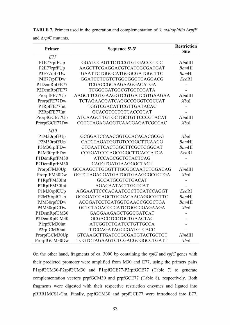
33
TABLE 7. Primers used in the generation and complementation of S. maltophilia ∆rpfF
and ∆rpfC mutants.
Primer Sequence 5'-3' Restriction Site
E77 P1E77rpfFUp GGATCCAGTTCTCCGTGTGACCGTCC HindIII P2E77rpfFUp AAGCTTCGAGGACGTCATCGCGATGAT BamHI P3E77rpfFDw GAATTCTGGGCATGGGCGATGGCTTC BamHI P4E77rpfFDw GGATCCTCGTCTGGCGGGTCAGGACG EcoRI
P1DemRpfFE77 TCGACCGCAAGAAGGACATGA - P2DemRpfFE77 TCGGCGATGGCGTGCTCGATA - ProrpfFE77Up AAGCTTCGTGAAGGTCGTGATCGTGAAGAA HindIII ProrpfFE77Dw TCTAGAACGATCAGGCCGGGTCGCCAT XbaI P1RpfFE77Int TGGTCGACATTCGTTGATACAC - P2RpfFE77Int GCACGTCCTGTCACCGCAT -
ProrpfGCE77Up ATCAAGCTTGTGCTGCTGTTCCCGTACAT HindIII ProrpfGCE77Dw CGTCTAGAGAGGTCAACGAGATCGCCAC XbaI
M30 P1M30rpfFUp GCGGATCCAACGGTCCACACACGCGG XbaI P2M30rpfFUp CATCTAGATGGTGTCCGGCTTCAACG BamHI P3M30rpfFDw CTGAATTCACTGGCTTCGCTGGGCAT BamHI P4M30rpfFDw CCGGATCCCAGCGCGCTTCACCATCA EcoRI
P1DemRpfFM30 ATCCAGCGCTGTACTCAG - P2DemRpfFM30 CAGGTGATGAAGGGCTACT - ProrpfFM30Up GCCAAGCTTGGGTTTGCGGCAATCTGGACAG HindIII ProrpfFM30Dw GGTCTAGACGATGATGGTGAAGCGCGCTGA XbaI P1RpfFM30Int GCCATGCGTCTGACAT - P2RpfFM30Int AGACAATACTTGCTCAT - P1M30rpfCUp AGGAATTCCCAGAATCGCTTCATCCAGGT EcoRI P2M30rpfCUp GCGGATCCAGCTGCGACAACAGGCGTTTC BamHI P3M30rpfCDw ACGGATCCTGATGGTGAAGCGCGCTGA BamHI P4M30rpfCDw GCTCTAGACCCCATCTGGCCGAGAAGA XbaI
P1DemRpfCM30 GAGGAAGAGCTGGCGATCAT - P2DemRpfCM30 GCGACCTCCTGCTGAACTAC -
P1rpfCM30int ATCGGTCTGATCCTGTTGCCA - P2rpfCM30int TTCCAGATAGCCGATGTCACC -
ProrpfGCM30Up GTCAAGCTTGATCCGCGATGTACTGCTGT HindIII ProrpfGCM30Dw TCGTCTAGAAGTCTCGACGCGGCCTGATT XbaI
On the other hand, fragments of ca. 3000 bp containing the rpfG and rpfC genes with
their predicted promoter were amplified from M30 and E77, using the primers pairs
P1rpfGCM30-P2rpfGCM30 and P1rpfGCE77-P2rpfGCE77 (Table 7) to generate
complementation vectors prpfGCM30 and prpfGCE77 (Table 8), respectively. Both
fragments were digested with their respective restriction enzymes and ligated into
pBBR1MCS1-Cm. Finally, prpfGCM30 and prpfGCE77 were introduced into E77,
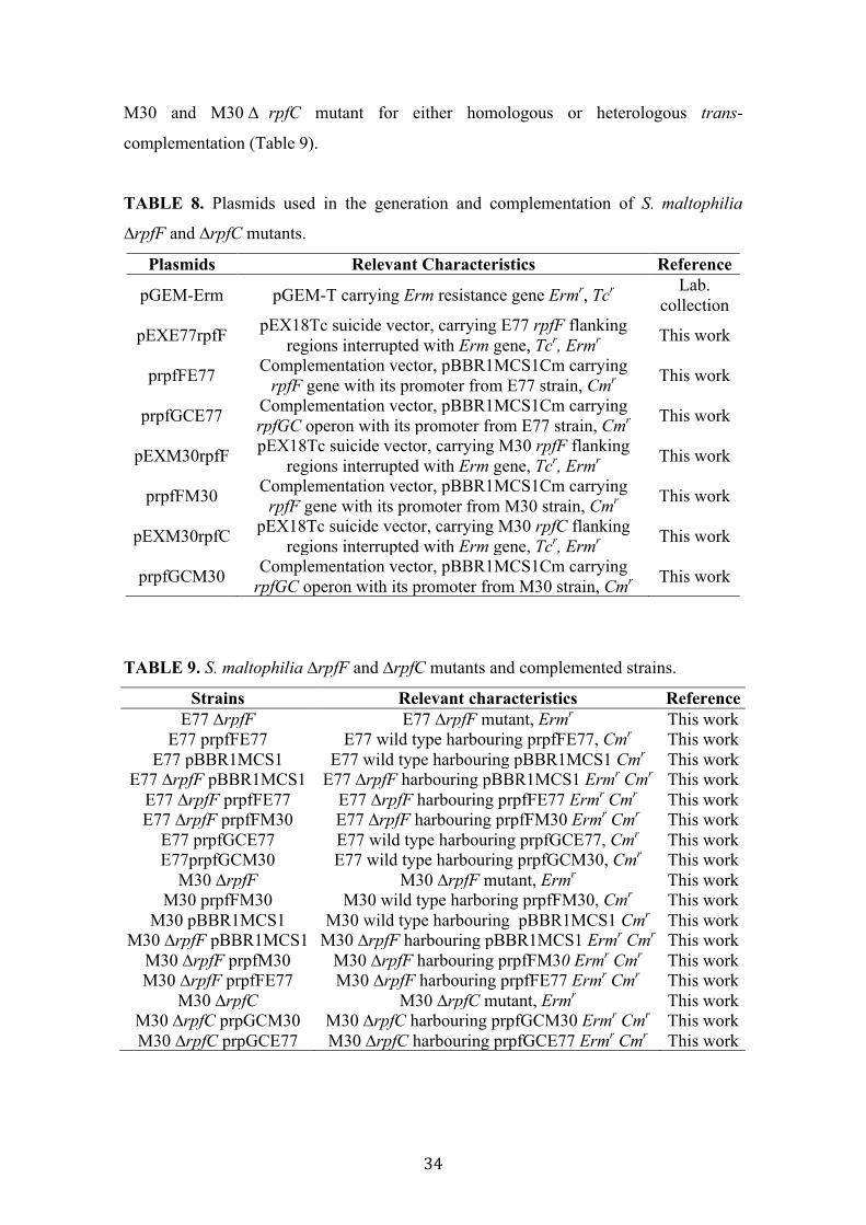
34
M30 and M30 ∆ rpfC mutant for either homologous or heterologous trans-
complementation (Table 9).
TABLE 8. Plasmids used in the generation and complementation of S. maltophilia
∆rpfF and ∆rpfC mutants.
Plasmids Relevant Characteristics Reference
pGEM-Erm pGEM-T carrying Erm resistance gene Ermr, Tcr Lab. collection
pEXE77rpfF pEX18Tc suicide vector, carrying E77 rpfF flanking regions interrupted with Erm gene, Tcr, Ermr This work
prpfFE77 Complementation vector, pBBR1MCS1Cm carrying rpfF gene with its promoter from E77 strain, Cmr This work
prpfGCE77 Complementation vector, pBBR1MCS1Cm carrying rpfGC operon with its promoter from E77 strain, Cmr This work
pEXM30rpfF pEX18Tc suicide vector, carrying M30 rpfF flanking regions interrupted with Erm gene, Tcr, Ermr This work
prpfFM30 Complementation vector, pBBR1MCS1Cm carrying rpfF gene with its promoter from M30 strain, Cmr This work
pEXM30rpfC pEX18Tc suicide vector, carrying M30 rpfC flanking regions interrupted with Erm gene, Tcr, Ermr This work
prpfGCM30 Complementation vector, pBBR1MCS1Cm carrying rpfGC operon with its promoter from M30 strain, Cmr This work
TABLE 9. S. maltophilia ∆rpfF and ∆rpfC mutants and complemented strains.
Strains Relevant characteristics Reference E77 ∆rpfF E77 ∆rpfF mutant, Ermr This work
E77 prpfFE77 E77 wild type harbouring prpfFE77, Cmr This work E77 pBBR1MCS1 E77 wild type harbouring pBBR1MCS1 Cmr This work
E77 ∆rpfF pBBR1MCS1 E77 ∆rpfF harbouring pBBR1MCS1 Ermr Cmr This work E77 ∆rpfF prpfFE77 E77 ∆rpfF harbouring prpfFE77 Ermr Cmr This work E77 ∆rpfF prpfFM30 E77 ∆rpfF harbouring prpfFM30 Ermr Cmr This work
E77 prpfGCE77 E77 wild type harbouring prpfGCE77, Cmr This work E77prpfGCM30 E77 wild type harbouring prpfGCM30, Cmr This work
M30 ∆rpfF M30 ∆rpfF mutant, Ermr This work M30 prpfFM30 M30 wild type harboring prpfFM30, Cmr This work
M30 pBBR1MCS1 M30 wild type harbouring pBBR1MCS1 Cmr This work M30 ∆rpfF pBBR1MCS1 M30 ∆rpfF harbouring pBBR1MCS1 Ermr Cmr This work
M30 ∆rpfF prpfM30 M30 ∆rpfF harbouring prpfFM30 Ermr Cmr This work M30 ∆rpfF prpfFE77 M30 ∆rpfF harbouring prpfFE77 Ermr Cmr This work
M30 ∆rpfC M30 ∆rpfC mutant, Ermr This work M30 ∆rpfC prpGCM30 M30 ∆rpfC harbouring prpfGCM30 Ermr Cmr This work M30 ∆rpfC prpGCE77 M30 ∆rpfC harbouring prpfGCE77 Ermr Cmr This work

35
3.6.2 Generation and complementation of S. maltophilia M30 ∆smlt0266, ∆smlt0267
and ∆smlt0266-0267 mutants
Primers and plasmids used for the cloning process are listed in Tables 10 and 11,
respectively. Strains and mutants object of this study are listed in Table 12.
S. maltophilia ∆smlt0266 and ∆smlt0266-0267 mutants were obtained by allelic-
exchange recombination using erythromycin as antibiotic-resistance cassette.
S. maltophilia M30 ∆sml0267 mutant was obtained by interrupting smlt0267 gene with
the whole suicide vector pEX0267 containing an intragenic region of smlt0267 for
homologous recombination.
Briefly, smlt0266 and smlt0266-0267 upstream and downstream flanking regions were
amplified by PCR using primer pairs P1BE0266-P2BE0266 and P3BD0266-P4BD0266
(for smlt0266) and P1BE0266-P2BE0266 and P3BD0267-P4BD0267 (for smlt0267)
(Table 10) and inserted, flanking the erythromycin cassette, into the pEX18Tc vector,
thus generating plasmids pEX0266 and pEX0266-0267 (Table 11). M30 was
electroporated with these suicide vectors and transformants were selected in LB plates
containing 500 µg/mL Erm and subsequently streaked onto LB plates containing
17 µg/mL Tc to discard single cross-over events. ∆smlt0266 deleted mutant was also
verified by PCR using primers pairs P1Dem0266-P3Ermrev (for upstream region) and
P2Dem0266-P5Ermrev (for downstream region) (Table 10) and the amplified fragments
were subsequently sequenced. The double mutant ∆smlt0266-0267 was also verified by
PCR using primers P1Dem02660267-P3Ermrev (for upstream region) and
P2Dem02660267-P5Ermrev (for downstream region) and the amplified fragments were
subsequently sequenced. To generate interrupted smlt0267 mutant in the M30 strain, an
intragenic fragment of ca. 500 bp, was amplified using primers P10267int-P20267int
(Table 10) and cloned into pEX18Cm, generating pEX0267 (Table 11). M30 strain was
electroporated and plated onto LB containing 3.2 µg/mL chloramphenicol. The mutant
candidates were screened and verified by PCR and sequencing, using primers
P1Dem0267-M13Fw (for upstream region) and P2Dem0267-M13Rv (for downstream
region) (Table 10). In S. maltophilia, the genomic organization showed the same
organization as dsp operon. However, in order to avoid interferences with the other
components of the operon, smlt0264, smlt0265 and smlt268 were excluded from the
complementation vector. Then, a fragment of ca. 2000 bp corresponding to smlt0266
(dspI homologue) and smlt0267 (dspII homologue) was amplified and cloned into the
non-replicative vector pBAD18-Cm, generating pBAD0266-0267 (Table 11). Then, a

36
fragment containing both genes plus the arabinose promoter was excised and subcloned
into the replicative vector pBBR1MCS1-Cm, generating pBADSMdsp (Table 11).
pBADSMdsp construction was confirmed by restriction and PCR using primers M13F
and M13R and electroporated into the M30 wt and the M30 ∆ smlt0266, ∆ smlt0266-
0267 and ∆sml0267 mutants for in-trans complementation.
TABLE 10. Primers used in the generation and complementation of S. maltophilia
∆smlt0266, ∆sml0267 and ∆smlt0266-0267 mutants.
Primer Sequence 5'-3' Restriction Site P1BE0266 GAATTCGCGGTCTGTACATGGA EcoRI P2BE0266 GGATCCCGCAGTTCCTCTTCG BamHI P1BD0266 AAGCTTCTGCAGCAGCGCATCGAACA HindIII P2BD0266 GGATCCATGAGCACCGACACCGTTGC BamHI P1BD0267 GGATCCAGCCGCATTGCATTC BamHI P2BD0267 AAGCTTCTGGTGATGAGCTGC HindIII P10267int CTAGAATTCTGCTGCTGAAGCAGCTGAAT EcoRI P20267int TCAGAATTCGTAGGTGTGGATGAGATGATC EcoRI P0266RBS TCTAGACCCGTCACCTGCT XbaI P0267END TCTAGATGCAATGCGGCTCAT XbaI
3.6.3 Arabinose-induction of pBADSMdsp
In order to corroborate that the induction was functional in the hybrid vector, M30 was
electroporated with this construct. A 25 mL culture of M30 transformed with
pBADSMdsp was grown for an overnight at 37ºC in LB supplemented with
0.2% arabinose and 30 µg/mL Cm. 10 mL were centrifuged and the cell pellet was
ressuspended twice in 10 mL of 250 mM sucrose. Finally, 1 mL aliquot was boiled for
10 minutes and total proteins were separated by SDS-PAGE and the gel was stained
with coomasie brilliant blue.
3.6.4 Complementation of P. aeruginosa ∆dspI and ∆dspII mutants
P. aeruginosa ∆dspI (∆PA0745, with strain name PW2354) and ∆dspII (∆PA0744, with
strain name PW2353) were obtained from the PAO1 transposon mutant library at the
University of Washington (149).
In P. aeruginosa, dspI and dspII are expected to be co-transcribed in the dsp operon
(PA0747, PA0746, PA0745, PA0744, PA0743). Consequently, a fragment of ca. 6750
bp containing the genes PA0747, PA0746, PA0745, PA0744 and its predicted promoter
was amplified by PCR using primers P1PAdsp (5’-
CTGAAGCTTCGGCAGCATGCTGATGACCA-3’) and P2PAdsp (5'-
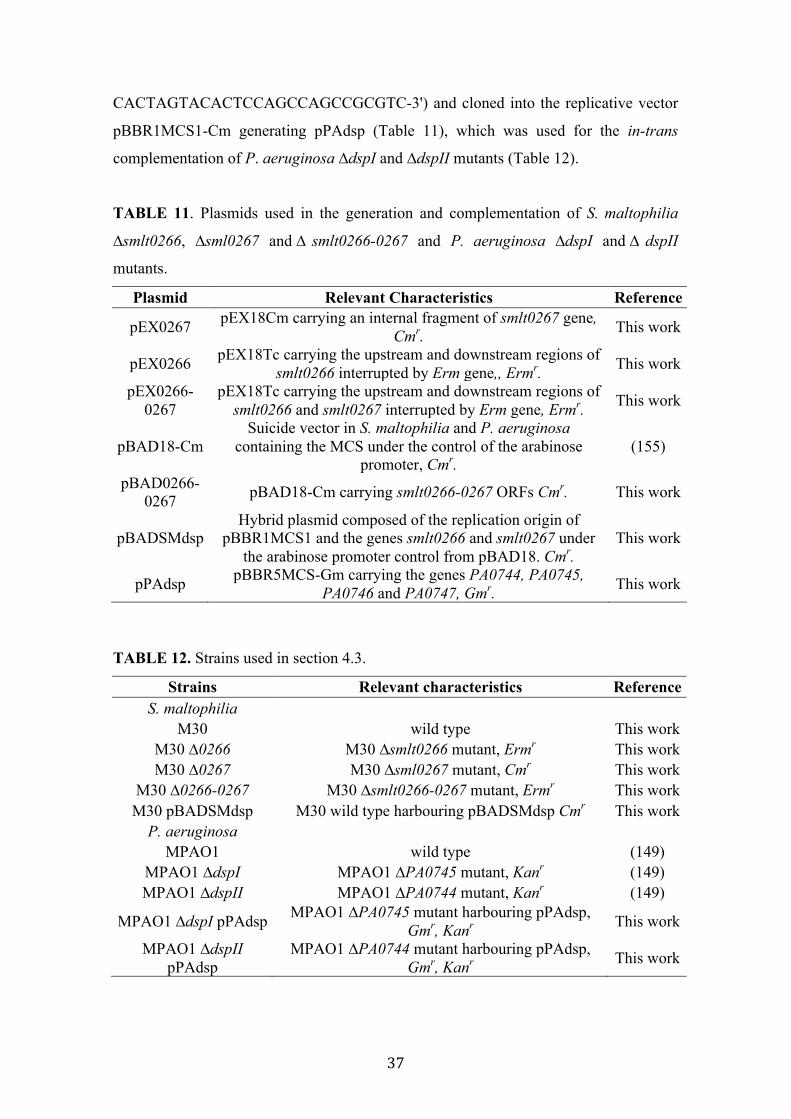
37
CACTAGTACACTCCAGCCAGCCGCGTC-3') and cloned into the replicative vector
pBBR1MCS1-Cm generating pPAdsp (Table 11), which was used for the in-trans
complementation of P. aeruginosa ∆dspI and ∆dspII mutants (Table 12).
TABLE 11. Plasmids used in the generation and complementation of S. maltophilia
∆smlt0266, ∆sml0267 and ∆ smlt0266-0267 and P. aeruginosa ∆dspI and ∆ dspII
mutants.
Plasmid Relevant Characteristics Reference
pEX0267 pEX18Cm carrying an internal fragment of smlt0267 gene, Cmr. This work
pEX0266 pEX18Tc carrying the upstream and downstream regions of smlt0266 interrupted by Erm gene,, Ermr. This work
pEX0266-0267
pEX18Tc carrying the upstream and downstream regions of smlt0266 and smlt0267 interrupted by Erm gene, Ermr. This work
pBAD18-Cm Suicide vector in S. maltophilia and P. aeruginosa
containing the MCS under the control of the arabinose promoter, Cmr.
(155)
pBAD0266-0267 pBAD18-Cm carrying smlt0266-0267 ORFs Cmr. This work
pBADSMdsp Hybrid plasmid composed of the replication origin of
pBBR1MCS1 and the genes smlt0266 and smlt0267 under the arabinose promoter control from pBAD18. Cmr.
This work
pPAdsp pBBR5MCS-Gm carrying the genes PA0744, PA0745, PA0746 and PA0747, Gmr. This work
TABLE 12. Strains used in section 4.3.
Strains Relevant characteristics Reference S. maltophilia
M30 wild type This work M30 ∆0266 M30 ∆smlt0266 mutant, Ermr This work M30 ∆0267 M30 ∆sml0267 mutant, Cmr This work
M30 ∆0266-0267 M30 ∆smlt0266-0267 mutant, Ermr This work M30 pBADSMdsp M30 wild type harbouring pBADSMdsp Cmr This work
P. aeruginosa MPAO1 wild type (149)
MPAO1 ∆dspI MPAO1 ∆PA0745 mutant, Kanr (149) MPAO1 ∆dspII MPAO1 ∆PA0744 mutant, Kanr (149)
MPAO1 ∆dspI pPAdsp MPAO1 ∆PA0745 mutant harbouring pPAdsp, Gmr, Kanr This work
MPAO1 ∆dspII pPAdsp
MPAO1 ∆PA0744 mutant harbouring pPAdsp, Gmr, Kanr This work

38
3.7 General analytical tools
3.7.1 SDS-PAGE
Sodium Dodecyl Sulfate-Polyacrylamide Gel Electrophoresis (SDS-PAGE) was
performed as previously described (165) with modification (166) using a
12% polyacrylamide gel in a vertical slab gel apparatus (Bio-Rad).
3.7.2 Protein Identification by Mass Spectrometry
Protein bands of interest were excised from the gel using a cut tip. In-gel trypsin
digestion was performed as described previously (167). MALDI-MS analysis of tryptic
peptides was performed on an Ultraflex time-of-flight instrument (Bruker Daltonics).
Samples were prepared using α -cyano-4-hydroxy-cinnamic acid. Calibration was
performed in the external mode using a peptide calibration standard kit (Bruker
Daltonics). The spectra were processed using Flex Analysis 2.2 software (Bruker
Daltonics). Peak lists were generated using the signals in the 800–4000 mass:charge
ratio (m/z) range, with a signal:noise threshold >3. The SNAP algorithm included in the
software was used to select the monoisotopic peaks from the isotopic distributions
observed. After removing m/z values corresponding to commonly observed matrix
cluster ions, an internal statistical calibration was applied. Peaks corresponding to
frequently seen keratin and trypsin autolysis peptides were then removed. The resulting
final peak was used for the identification of the protein by peptide mass fingerprint. The
Mascot 2.0 program (Matrix Science) was used to search the NCBI non-redundant
database (http://ncbi.nlm.nih.gov), with no limitation on taxonomy. Search parameters
were as follows: trypsin cleavages excluding N-terminal to P, one missed cleavage
permission, carbamidomethylation and methionine oxidation as variable modification,
mass tolerance <50 ppm, monoisotopic mass values.
3.7.3 Lipid extraction from culture supernatants
DSF extraction from culture supernatants was carried out following the ethyl acetate
method (92). Briefly, overnight bacterial cultures grown on indicated media were
harvested by centrifugation and the supernatant was extracted with the same volume of
ethyl acetate. The organic phase was evaporated to dryness using a rotary evaporator
and the residues were dissolved in 3 mL of methanol (for supernatant DSF bioassay and
TLC analysis) or dichloromethane (for GC/MS analysis).

39
3.7.4 Lipid separation by Thin-Layer Chromatography (TLC)
For supernatant fatty acid separation using TLC technique, 5 µL aliquots of the lipid
extractions dissolved in methanol were spotted onto a silica gel 60 TLC plate
(20x20 cm, Merck) and separated with ethyl acetate:hexane (20:80, vol/vol) as running
solvents.
3.7.5 Lipid identification from culture supernatants by GC/MS
This experiment was mainly performed at “Servei d’Ànalisi Química (SAQ)” in the
“Universitat Autònoma de Barcelona (UAB)”. Bacterial cultures were grown on 2 L of
LB for 48 h at 30 ºC with vigorous shaking (250 rpm). Cultures were centrifuged and
supernatants were extracted with the ethyl acetate method (92). Dry residues were
dissolved in 3 mL of dichloromethane. Total fatty acids were methylated –generating
Fatty Acids Methyl Esters (FAME)- and separated by Gas Chromatography (6890,
Agilent Technologies) using an Agilent 19091S-433 column coupled to a Mass
Spectrometry detector (Hewlett-Packard 5973).
3.7.6 Purification of fatty acids by High-Liquid Pressure Chromatography (HPLC)
Dry residues of concentrated supernatants were dissolved in 100 µL of 30% Methanol,
and injected into the HPLC (Waters Alliance) and separated using a se C18 reverse-pha
column (100 Å, pore size) (Phenomena) developed with a Methanol gradient 20-99% in
30 minutes at a flow rate of 1 ml/min and monitored at 210 nm. Fractions of interest
were collected and used for further analysis. After each run, column was cleaned with
100% acetonitrile at the same flow rate to avoid cross contamination and interferences.
3.7.7 Extraction and analysis of total cellular fatty acids
Analysis of total cellular FA was carried out by the “Leibniz Institute DSMZ-German
Collection of Microorganisms and Cell Cultures” (Braunschweig, Germany), as
follows. Fatty acid methyl esters were obtained from 40 mg cells scraped from Petri
dishes by saponification, methylation and extraction using minor modifications of the
method of Miller (1982) (168) and Kuykendall et al. (1988) (169). The fatty acid
methyl esters mixtures are separated using Sherlock Microbial Identification System
(MIS) (MIDI, Microbial ID, Newark, DE 19711 U.S.A.) which consisted of an Agilent
model 6890N gas chromatograph fitted with a 5% phenyl-methyl silicone capillary
column (0.2 mm x 25 m), a flame ionization detector, Agilent model 7683A automatic
sampler, and a HP-computer with MIDI data base (Hewlett-Packard Co., Palo Alto,

40
California, U.S.A.). Peaks are automatically integrated and fatty acid names and
percentages calculated by the MIS Standard Software (Microbial ID). The gas
chromatographic parameters are as follows: carrier gas, ultra-high-purity hydrogen;
column head pressure 60kPa; injection volume 2 µL; column split ratio, 100:1; septum
purge 5 ml/min; column temperature, 170 to 270°C at 5°C/min; injection port
temperature, 240°C; and detector temperature, 300°C.
3.8 DSF extraction, detection and quantification.
3.8.1 Colony and supernatant DSF bioassay
DSF determination was performed using strain Xcc 8523 pL6engGUS (DSF-reporter
strain) as previously described (105), with few modifications. Briefly, the DSF-reporter
strain was grown in 10 mLof NYG medium supplemented with Tc (10 µg/mL) to an
optical density of 0.7 at 600 nm. Cells were harvested and reconstituted with 1 mL of
fresh NYG and added to 100 mL of temperate NYG medium containing 1% of BD
Difco Agar Noble (NYGA) and supplemented with 80 µg/mL X-Glu and then plated
into petri plates upon solidification.
For colony-based DSF bioassay, candidate strains were pin inoculated onto NYGA
plates containing 80 µg/mL X-Glu and the the DSF-reporter strain and incubated for
24 h at 28ºC. Presence of a blue halo around the colony indicates DSF activity.
For supernatant-based DSF bioassay, bacterial cultures were grown in 250 mL of LB
for 48 h at 30ºC (OD600 nm about 4). Supernatants were extracted with the ethyl
acetate method and residues were dissolved in 200 µL of methanol. 3 µL of each
sample were deposited into hand-generated wells in 5.5-cm plates containing NYGA
supplemented with 80 µg/mL of X-Glu and seeded with the DSF-reporter strain
previously prepared to an estimated final optical density at 0.07 at 600 nm. Plates were
incubated for 24 h at 30ºC. DSF activity was determined by the presence of a blue halo
around the well.
3.8.2 TLC coupled to DSF bioassay
TLC analysis was used to separate DSF from total fatty acids present in the supernatant
(See section 3.7.4). After separation, TLC plates were air-dried for at least 1 h and
overlaid with 100 mL of cooled unsolidified NYGA containing 80 µg/mL of X-Glu and
the DSF-reporter strain at an optical density of 0.07 at 600 nm. TLC plates were

41
incubated overnight at 28°C and DSF activity was identified by the presence of blue
spots.
3.8.3 Liquid DSF bioassay
200 µL of DSF reporter solution consisting of Xcc 8523 pL6engGUS strain culture
adjusted to an optical density of 0.07 at 600 nm in NYG medium and supplemented
with X-Glu (80 µg/mL) were deposited into wells in a sterile 96 microtitter well plate.
Synthetic DSF was added to wells with an increasing concentration (from 0.05 to
1.5 mM) and incubated at 28ºC for 24 h. The presence of DSF molecules turned wells
to blue with intensity proportional to the concentration of DSF molecules. The
absorbance was read at 620 nm and a calibration curve was generated. Samples residues
dissolved in methanol were then tested with increasing concentrations into wells and the
lecture was done as described above. The quantification was measured using the
formula generated by the calibration curve.
3.8.4 DSF detection and quantification from infected animal tissues
Two fishes from each tank (E77, M30, E77:M30 and ∆ rpfF-1:∆rpfF-2 plus two
uninfected fishes) were randomly chosen and sacrificed by anaesthesia (MS-222,
220 ppm) after 48 hpi. Since in the mixed wild type inoculum all fishes died within the
first 48 h, it was the criterion for evaluating DSF production from each culture in in-
vivo infection. Sacrificed fishes were introduced into a falcon tube containing 15 mL of
PBS and homogenized using a Politron Homogenizer (MARK). The homogenized
solution was extracted with the same volume of ethyl acetate and subsequently dried.
Dry residues were dissolved in 100 µL of 30% methanol and DSF activity was detected
and quantified by liquid and solid supernatant DSF bioassay.
3.9 Phenotypic analysis of mutant, complemented and wild type
strains.
3.9.1 Biofilm formation
To analyze biofilm formation on polystyrene surface, 200 µL from an overnight
bacterial cultures adjusted to an optical density of 0.1 at 600 nm in modified M9-salts or
BM2 medium were inoculated into 96-well microtiter non-treated plates (BrandTech
781662) and incubated statically for 24 h at 30 ºC. Plates were then washed three times
with water, fixed at 60 °C for 1 h and stained during 15 min with 200 µL of 0.1%

42
crystal violet. The dye was discarded and the plates were rinsed in standing water and
allowed to dry for 30 min at 37°C. Crystal violet from attached bacteria was dissolved
in 250 µL 95% ethanol for 15 min, and the OD of the extracted dye was measured at
550 nm.
Biofilm formation on glass surface was assayed by inoculating 2 mL from the same
adjusted cultures as described above into glass tubes and incubated for 24 h at 30ºC
with agitation (250 rpm). Biofilm formation was measured by crystal-violet method by
staining the tubes with 2.5 mL of 0.1% crystal violet.
3.9.2 Swimming motility
The swimming motility was determined in TrA plates (1% tryptone, 0.5% NaCl,
0.25% agar) (40). Thus, 5 µL of adjusted overnight cultures were spotted on TrA plates
and incubated at 30ºC for 24 h. Quantification was done by measuring the diameter of
the colony.
3.9.3 Twitching motility
Twitching motility was assessed via subagar stab inoculations (stab assay) from
overnight fresh plates as previously described (170). Plates were incubated at 30ºC for
24 h and the agar medium was extracted from the plate. Then, twitching zones were
visualized by staining with 1% (wt/vol) crystal violet. The quantification was done by
measuring the diameter of the blue halos.
3.9.4 Swarming motility
Swarm agar was made based on modified M9-salts medium without NH4Cl, with
0.5% casamino acids, 2 mM MgSO4, 0.1 mM CaCl2 supplemented with 0.4% glucose
and solidified with 0.5% BD Difco Agar Noble (147). Plates containing 20 mL of fresh
swarm medium were dried under laminar flow for 20 minutes before inoculation. The
inoculation was performed using a sterile digralski spatula containing biomass from a
fresh LB plate and softly deposited on the top of a semi-solid modified M9-salts
medium plate. Inoculated swarm plates were sealed to keep the humidity and incubated
at 28 ºC for 3-5 days.
3.9.5 Surfactant imaging
Strains of interest were seeded on modified M9-salts medium plates solidified with
0.7% Noble Agar (Difco) (to avoid swarming motility) and incubated at 28ºC for
5 days. Wetting agent images from isolated colonies were visualized using stereoscopic
43
microscope Olympus SZX16. Plates were illuminated using diffuse reflected light, and
angled to capture the refractive quality of the layer.
3.9.6 TEM microscopy imaging
Samples for transmission electron microscopy (TEM) visualization were prepared as
follows. Cells were picked from the edge of the tendril (for swarmer strains) and from
the edge of the non-swarming colonies using a Kölle handle and ressuspended in 25 µL
of distilled water. 5 µL of each sample were deposited on copper grids and contrasted
with conventional 0.2% uranyl acetate solution. Observation was done using a Jeol
1400 (Jeol LTD, Tokyo, Japan) transmission electron microscope equipped with a CCD
GATAN ES1000W Erlangshen camera.
3.9.7 Determination of antibiotic susceptibility
The susceptibility of S. maltophilia wt and mutant strains to the following antimicrobial
agents was tested: tetracycline, minocycline, gentamicin, kanamycin, trimethoprim,
sulfamethoxazole, norfloxacin, ciprofloxacin, erythromycin, levofloxacin or
chloramphenicol. The MICs for selected antibiotics were determined by the resazurin
microtitre assay (171) using 96-well plates by serial two-fold dilutions of each drug in
100 µL of LB. 100 µL of a bacterial suspension (final OD550nm = 0,005) was added to
the antibiotic dilutions, mixed and incubated at 37°C for 16 h before developing with
resazurin (30 µL 0.01%) [44]. The MIC was defined as the lowest drug concentration
that prevented bacterial growth. The microdilution assay followed the Clinical and
Laboratory Standards Institute (CLSI, www.clsi.org) guidelines for antimicrobial
susceptibility testing.
3.9.8 Congo red binding assay
Congo red binding assays were performed as previously described (172). Briefly,
overnight bacterial cultures grown in LB were diluted to OD600nm of 0.1 and 5 µL
were spotted onto CR plates (10 g/l tryptone broth with 10 g/l agar, 40 µg/mL Congo
red, and 20 µg/mL Coomassie brilliant blue). The plates were incubated for 72 h at
room temperature to assess colony morphology.
3.9.9 Determination of virulence in the Caenorhabditis elegans model
Caenorhabditis elegans CF512 (fer-15(b26) II; fem-1(hc17) IV), a strain showing
temperature-dependent sterility, was provided by CGC. Nematodes were routinely
maintained in NGM plates (1.7% Agar, 50 mM NaCl, 0.25% peptone, 1 mM CaCl2,
44
5 µg/mL cholesterol, 25 mM KH2PO4 , 1 mM MgSO4) seeded with E. coli OP50, at
16 ºC.
Virulence determination of bacterial strains in the C. elegans CF512 infection model
was based on the “Slow Killing” method (173). Bacterial strains were grown in BHI
broth overnight at 30 °C and 100 µL of each strain culture were spread on a 5.5 cm
diameter NGM agar plate and incubated at 30°C for 24 h. Each plate was then seeded
with 15-20 adult hermaphrodite CF512 worms, incubated at 25°C (sterility conditions)
and scored for live worms every 24 h. E. coli OP50 was used as a negative control. A
worm was considered dead when it no longer responded to touch. Three replicates per
strain were performed.
3.9.10 Determination of virulence in the adult zebrafish infection model
Adult (9–12 months) wild-type zebrafish (Danio rerio) were kept in a 12 h light:12 h
dark cycle at 28°C and fed twice daily with dry food. All fish used in infection
experiments were transferred to an isolated system and acclimated for three days before
infection. Adult zebrafish (n=12 per condition) were infected by intraperitoneal
injection (174) with 20 µL of a 5×10⁸ cfu/mL suspension of each S. maltophilia strain.
The strains were previously grown at 28 °C in blood agar plates (BioMérieux) for 20 h
and collected directly from the plates with sterile phosphate buffered saline (PBS). Two
control groups were injected with PBS and showed no mortality. Fishes were observed
daily for signs of disease and mortality.
One fish from each tank was sacrificed at 72 h postinfection and divided into three
sections (anterior, abdominal, and posterior regions) with a sterile surgical blade. All
weights were annotated, and every section was homogenized in 3 ml of PBS. After
serial dilution, bacteria were plated onto LB medium containing 20 µg/mL Ap (for WT
E77), LB containing 500 µL Erm (for the E77 ∆rpfF mutant), or LB supplemented with
Cm (for the complemented E77 ∆rpfF mutant). Finally, CFU were counted and divided
per gram of tissue. All of the isolates obtained post-mortem from infected zebrafish
were identified as S. maltophilia on the basis of cell and colony morphology, the
analytical profile index, and the 16S rRNA gene sequence (data not shown).
3.10 Bioinformatic Tools General bioinformatics tools used in this study include protein and nucleotide BLAST
(175), ClustalW (176), Oligos 7.0 (177), CodonCode , Serial Cloner 5.2, MEGA 5.0

45
(159), Artemis and SMART (160). Software was run with default parameters unless
otherwise stated.
3.11 Statistical Analysis Statistical analyses were performed using the GraphPad Prism program version 5.00.
Comparison of strain phenotypic data was performed by one-way analysis of variance
(ANOVA) with a Bonferroni’s multiple comparison post-test or unpaired t-test with
Welch correction for unequal variances, as indicated in figure and table captions.
Survival curves of zebrafish infection experiments were analyzed using the Kaplan-
Meier method. Differences were evaluated using the log-rank test.
3.12 Ethics Statements Zebrafish were handled in compliance with Directive 2010/63/EU of the European
Parliament and of the Council on the protection of animals used for scientific purposes
and with Decree 214/1997 of the Government of Catalonia, which regulates the use of
animals for experimental and other scientific purposes. Experimental protocols have
been reviewed and approved by the Animal and Human Experimentation Ethics
Committee (CEEAH) of Universitat Autònoma de Barcelona (UAB), Spain
(ref #CEEAH-1968).

46

RESULTS AND DISCUSSION
48
4. RESULTS AND DISCUSSION
4.1 Molecular Basis of DSF-QS System in S. maltophilia It is known that S. maltophilia presents the DSF-QS system like other members of the
Xanthomonadales family (65, 127). Lot of literature is published regarding DSF-QS in
Xanthomonas (91, 92, 105, 107–110), Xylella (98, 99, 117) and Burkholderia (101,
123–125) species, however, in the case of S. maltophilia, little is known about the
mechanisms by which S. maltophilia synthesize and sense DSF molecules. In this
chapter, we have focused on the molecular aspects of DSF-QS in S. maltophilia, more
specifically, in the study of the molecular basis involved in DSF production.
4.1.1 Two variants of rpf cluster with main differences in rpfF and rpfC genes are
distributed among S. maltophilia population.
A preliminary analysis comparing the few publicly available genomes (strains K279a,
R551-3, D457, and JV3, with GenBank accession numbers AM743169.1, CP001111.1,
HE798556.1, and CP002986.1, respectively) showed that all genomes contain the rpfF
gene. Nevertheless, two different rpfF variants could be distinguished based on its N-
terminus (Fig. 1). Taking the two model S. maltophilia strains D457 and K279a and
comparing it to the well-studied DSF-producer strain Xcc, we could observe that two rpf
clusters could be distinguished based on the rpfF but also on the rpfC gene (Fig. 1).
Accordingly, we have named rpf-1 (K279a) to the cluster variant displaying high
similarity to the Xcc rpf cluster, and rpf-2 (D457) to that with significant differences.
Additionally, we have sequenced the genome of three more clinical isolates E77, UV74
and M30. From these, the genome of the strain M30 was submitted to the NCBI (with
accession number JELS02000000) (10) and a first draft was constructed for the E77 and
UV74 genomes. Analysis of the complete rpf cluster in these new genomes also led us
to identify two rpf cluster variants among these strains. In order to conclude that two
variants of the synthase RpfF and the sensor component RpfC were distributed among
the population of S. maltophilia, the rpfF and rpfC genes were amplified for a collection
of 78 clinical isolates (Table 1). Amplification of the corresponding DNA region
demonstrated that all S. maltophilia strains contain the rpfF and rpfC genes. For the
rpfF gene, slightly different fragment lengths were obtained due to the previously
observed N-terminal region’s variability. All of the amplified rpfF sequences were

49
deposited in the GenBank database and assigned accession numbers KJ149475 to
KJ149552. In addition, alignment of the translated N-terminal regions and subsequent
phylogenetic analysis confirmed that the RpfF of S. maltophilia might be distributed
into two distinct variants, which we have named RpfF-1 and RpfF-2 (Fig. 1, 2).
FIG 1 (A) Comparison of the rpf cluster in Xanthomonas campestris pv. campestris
(Xcc) and two Stenotrophomonas maltophilia strains (K279a and D457). The alignment
was performed using tblastx (percent identity cut-off = 45%) from the Blast suite and
visualized with the Artemis Comparison Tool. Conserved protein regions are paired by
shaded blocks where colour intensity is proportional to sequence identity. The scales are
relative positions in base pairs. (B) Alignment of RpfF proteins from Xcc and
S. maltophilia K279a (RpfF-1) and D457 (RpfF-2). Identical amino acid residues are
shaded black with similar residues shaded grey. The hypervariable region is marked
with “#”, binding pocket residues are marked with “*” and Glutamate catalytic residues
are marked with “+”.
Continuing with the analysis of the rpf cluster in the S. maltophilia available genomes
we observed that rpfC gene also differed significantly between the two S. maltophilia

50
variant groups defined by the rpfF gene (Fig. 1A). In order to demonstrate this
association, we amplified the rpfC gene from all S. maltophilia strains in our collection.
The obtained fragment lengths let us to discriminate and prove that two rpfC variants
were also distributed among S. maltophilia population. It also verified that each RpfF-
variant group carries an associated RpfC-variant. Hence, RpfF-1 variant strains have an
associated RpfC-1 variant and RpfF-2 variant strains harbour an associated RpfC-2
variant.
Altogether, from all S. maltophilia strains in our collection, the RpfC/F-1 variant was
present in 47 out of 78 strains (60.26%), whereas RpfC/F-2 was present in the
remaining 31 strains (39.74%) (Fig. 2). Of the additional four complete genome
sequences available at this time, K279a and R551-3 contain the RpfC/F-1 variant and
D457 and JV3 contain the RpfC/F-2 variant.
FIG 2 Phylogenetic analysis (Neighbourhood-Joining) of 82 S. maltophilia clinical
strains based on the first 108 aminoacids of RpfF. E77 (blue) and M30 (red) were taken
as model strains to study the implication of each variant rpf-1 and rpf-2 in DSF
production and virulence regulation.

51
4.1.2 RpfC-1 but not RpfC-2 contains a transmembrane sensor input domain
highly related to the Xcc RpfH-RpfC complex
RpfC-1 (belonging to RpfF-1 variant strains) and RpfC-2 (belonging to RpfF-2 variant
strains) differ in their N-terminal regions, corresponding to the transmembrane (TMR)
domain or sensor input domain (107) (Fig. 3). It has been postulated that in Xcc an
additional integral membrane protein, RpfH, participates in DSF sensing (105). In
S. maltophilia the RpfH protein appears to be fused to RpfC-1 generating a sensor input
domain with ten TMR, as would happen in a putative Xcc RpfH-RpfC complex.
However, the TMR domain of the RpfC-2 variant contains only five TMR.
Interestingly, tblastx analysis revealed that the five TMR present in the RpfC-2 variant
are highly related to Xcc RpfH, while the five regions that are absent would correspond
to the Xcc RpfC TM domain (Fig. 3). This indicates that both RpfC-variant groups carry
a putative RpfH but only the RpfC-1 variant contains its own five TMR in the sensor
input domain.
FIG 3 SMART-software analysis of RpfC and RpfH from Xcc and RpfC from
S. maltophilia K279a and D457, where HisKA is a Histidine Kinase domain,
HATPase_c a Histidine ATPase domain, REC a CheY-like receiver domain and HPT a
Histidine phosphotransferase domain.
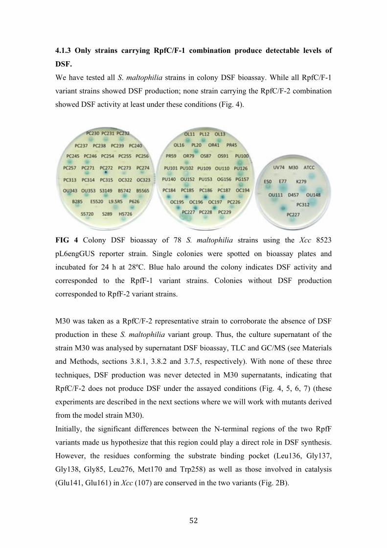
52
4.1.3 Only strains carrying RpfC/F-1 combination produce detectable levels of
DSF.
We have tested all S. maltophilia strains in colony DSF bioassay. While all RpfC/F-1
variant strains showed DSF production; none strain carrying the RpfC/F-2 combination
showed DSF activity at least under these conditions (Fig. 4).
FIG 4 Colony DSF bioassay of 78 S. maltophilia strains using the Xcc 8523
pL6engGUS reporter strain. Single colonies were spotted on bioassay plates and
incubated for 24 h at 28ºC. Blue halo around the colony indicates DSF activity and
corresponded to the RpfF-1 variant strains. Colonies without DSF production
corresponded to RpfF-2 variant strains.
M30 was taken as a RpfC/F-2 representative strain to corroborate the absence of DSF
production in these S. maltophilia variant group. Thus, the culture supernatant of the
strain M30 was analysed by supernatant DSF bioassay, TLC and GC/MS (see Materials
and Methods, sections 3.8.1, 3.8.2 and 3.7.5, respectively). With none of these three
techniques, DSF production was never detected in M30 supernatants, indicating that
RpfC/F-2 does not produce DSF under the assayed conditions (Fig. 4, 5, 6, 7) (these
experiments are described in the next sections where we will work with mutants derived
from the model strain M30).
Initially, the significant differences between the N-terminal regions of the two RpfF
variants made us hypothesize that this region could play a direct role in DSF synthesis.
However, the residues conforming the substrate binding pocket (Leu136, Gly137,
Gly138, Gly85, Leu276, Met170 and Trp258) as well as those involved in catalysis
(Glu141, Glu161) in Xcc (107) are conserved in the two variants (Fig. 2B).
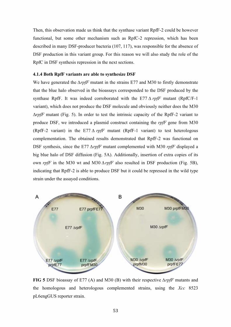
53
Then, this observation made us think that the synthase variant RpfF-2 could be however
functional, but some other mechanism such as RpfC-2 repression, which has been
described in many DSF-producer bacteria (107, 117), was responsible for the absence of
DSF production in this variant group. For this reason we will also study the role of the
RpfC in DSF synthesis repression in the next sections.
4.1.4 Both RpfF variants are able to synthesize DSF
We have generated the ∆rpfF mutant in the strains E77 and M30 to firstly demonstrate
that the blue halo observed in the bioassays corresponded to the DSF produced by the
synthase RpfF. It was indeed corroborated with the E77 ∆ rpfF mutant (RpfC/F-1
variant), which does not produce the DSF molecule and obviously neither does the M30
∆rpfF mutant (Fig. 5). In order to test the intrinsic capacity of the RpfF-2 variant to
produce DSF, we introduced a plasmid construct containing the rpfF gene from M30
(RpfF-2 variant) in the E77 ∆ rpfF mutant (RpfF-1 variant) to test heterologous
complementation. The obtained results demonstrated that RpfF-2 was functional on
DSF synthesis, since the E77 ∆rpfF mutant complemented with M30 rpfF displayed a
big blue halo of DSF diffusion (Fig. 5A). Additionally, insertion of extra copies of its
own rpfF in the M30 wt and M30 ∆ rpfF also resulted in DSF production (Fig. 5B),
indicating that RpfF-2 is able to produce DSF but it could be repressed in the wild type
strain under the assayed conditions.
FIG 5 DSF bioassay of E77 (A) and M30 (B) with their respective ∆rpfF mutants and
the homologous and heterologous complemented strains, using the Xcc 8523
pL6engGUS reporter strain.

54
Moreover, TLC-bioassay analysis of culture supernatants of E77 and M30, their ∆rpfF
mutants and the complemented strains suggested that DSF is the only fatty acid with
signalling activity that depends on the RpfF synthase function, since no other
differential blue spot was observed when comparing M30 with E77 and with their
respective ∆rpfF mutants and complemented strains (Fig. 6).
FIG 6 Scanned image of a TLC coupled to DSF bioassay of culture supernatants from
E77 and M30, their respective ∆rpfF mutants and complemented strains. 5 µl of each
concentrated supernatant and 2 µl of synthetic DSF were spotted onto TLC silica plate
and separated with ethyl acetate:hexane (20:80, vol/vol) as running solvents. Once the
TLC was run and dry, 100 ml of DSF reporter solution was added to the plate and
incubated for 24 h at 28ºC. Blue spots indicate the presence of DSF molecules.
All concentrated supernatants plus the synthetic DSF were dissolved in toluene and total
fatty acids were methylated prior to the Gas Chromatography/Mass Spectrometry
(GC/MS) identification. Each sample was separated by GC and compared to the
synthetic DSF, which showed a retention time of 17.8 minutes. Only the concentrated
supernatants from the strains displaying DSF production in the supernatant bioassay
showed a peak at the same retention time (Fig. 7). In addition, these peaks showed the
same mass spectra as the synthetic DSF (Fig. 8), concluding that both RpfF variants
produce DSF. MS analysis further supported that both S. maltophilia groups only
produce the canonical DSF 11-cis-2methyl-dodecenoic acid since no other differential
GC peak with a mass spectra fatty acid-compatible was observed comparing each other
the DSF-producer strains (Fig. 7, 8).
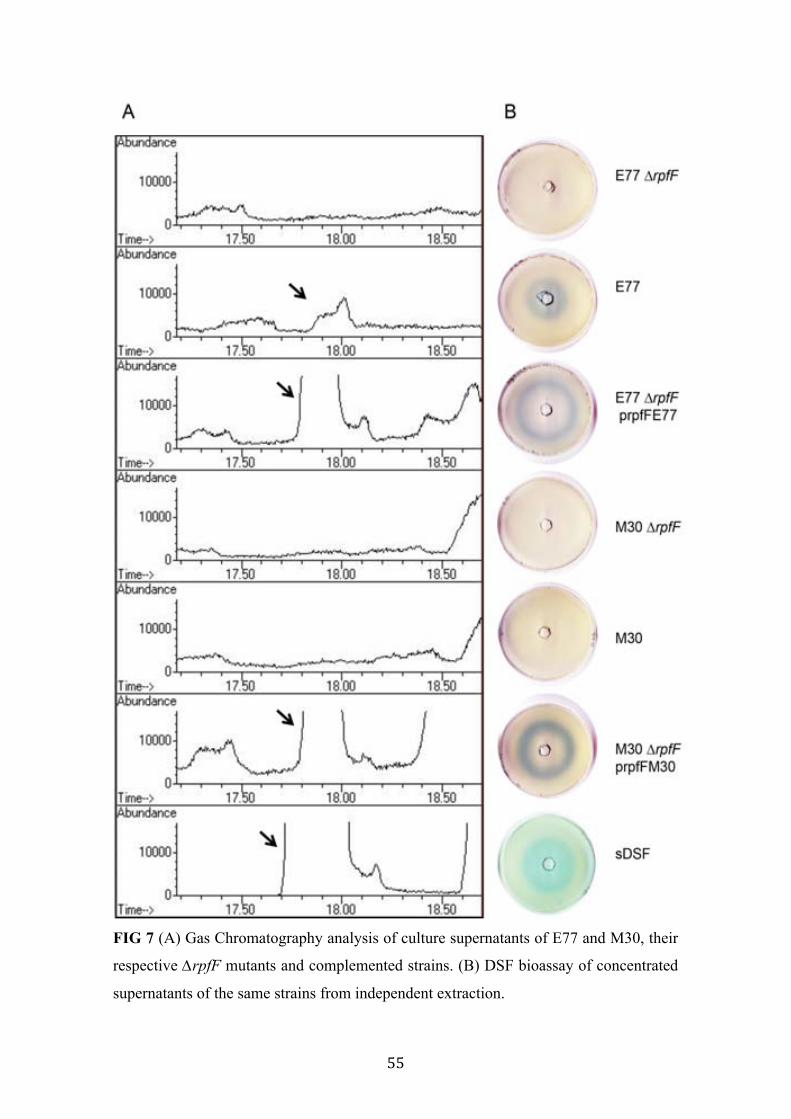
55
FIG 7 (A) Gas Chromatography analysis of culture supernatants of E77 and M30, their
respective ∆rpfF mutants and complemented strains. (B) DSF bioassay of concentrated
supernatants of the same strains from independent extraction.

56
FIG 8 Mass spectra of the Gas Chromatography peaks with DSF activity (17.8 min.)
(See also Fig. 7). (A) E77, (B) E77 ΔrpfF prpfFE77, (C) M30 ΔrpfF prpfFM30 and (D)
Synthetic DSF.

57
4.1.5 RpfF-2 is permanently repressed by RpfC-2
Since both RpfF variants were shown to be functional on DSF production, we wanted to
study the implication of each RpfC variant in DSF synthesis repression. To test the
hypothesis that RpfF-2 was permanently blocked by RpfC-2, the DSF-producer strain
E77 was separately transformed with plasmids expressing each of the RpfC variants.
Since rpfC is expected to be co-transcribed jointly with rpfG in both rpf-1 and rpf-2
clusters, we generated the vectors prpfGCE77 and prpfGCM30 harbouring the full
rpfGC operon. These vectors were effective repressors since in both cases resulted in a
reduction of DSF synthesis in E77 strain (Fig 9). However, while E77 harbouring the
prpfGCE77 vector showed only a small decrease in DSF synthesis, provision of
prpfGCM30 resulted in a strong inhibition of DSF production (Fig. 9A), suggesting that
RpfC-2 is a stronger repressor of the RpfF activity. In order to corroborate this
hypothesis, we generated the ∆ rpfC mutant in the M30 strain (RpfC-2). Consistently
with the previous result, M30 ∆rpfC became a DSF-producer strain (Fig. 9B). In trans
complementation of M30 ∆ rpfC with vectors prpfGCE77 and prpfGCM30 led to a
scenario similar to that observed for the E77 strain (Fig. 9B). While the plasmid
construct derived from E77 (rpfC-1) slightly reduce DSF production in the M30 ∆rpfC
mutant, homologous complementation of rpfC-2 abolished the DSF synthesis in the
M30 ∆rpfC mutant (Fig. 9B).
FIG 9 (A) DSF bioassay of E77 wt and E77 complemented with vectors prpfGCE77
and prpfGCM30. (B) DSF bioassay of M30 wt, M30 ∆ rpfC and M30 ∆ rpfC
complemented with vectors prpfGCE77 and prpfGCM30.
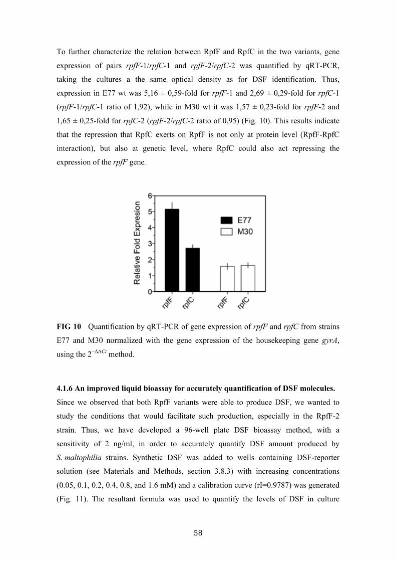
58
To further characterize the relation between RpfF and RpfC in the two variants, gene
expression of pairs rpfF-1/rpfC-1 and rpfF-2/rpfC-2 was quantified by qRT-PCR,
taking the cultures a the same optical density as for DSF identification. Thus,
expression in E77 wt was 5,16 ± 0,59-fold for rpfF-1 and 2,69 ± 0,29-fold for rpfC-1
(rpfF-1/rpfC-1 ratio of 1,92), while in M30 wt it was 1,57 ± 0,23-fold for rpfF-2 and
1,65 ± 0,25-fold for rpfC-2 (rpfF-2/rpfC-2 ratio of 0,95) (Fig. 10). This results indicate
that the repression that RpfC exerts on RpfF is not only at protein level (RpfF-RpfC
interaction), but also at genetic level, where RpfC could also act repressing the
expression of the rpfF gene.
FIG 10 Quantification by qRT-PCR of gene expression of rpfF and rpfC from strains
E77 and M30 normalized with the gene expression of the housekeeping gene gyrA,
using the 2−ΔΔCt method.
4.1.6 An improved liquid bioassay for accurately quantification of DSF molecules.
Since we observed that both RpfF variants were able to produce DSF, we wanted to
study the conditions that would facilitate such production, especially in the RpfF-2
strain. Thus, we have developed a 96-well plate DSF bioassay method, with a
sensitivity of 2 ng/ml, in order to accurately quantify DSF amount produced by
S. maltophilia strains. Synthetic DSF was added to wells containing DSF-reporter
solution (see Materials and Methods, section 3.8.3) with increasing concentrations
(0.05, 0.1, 0.2, 0.4, 0.8, and 1.6 mM) and a calibration curve (rI=0.9787) was generated
(Fig. 11). The resultant formula was used to quantify the levels of DSF in culture

59
supernatants of S. maltophilia strains. Concentrated S. maltophilia supernatants were
dissolved in 20 µl of 30% methanol and 0.25, 0.5, 1, 2, 4 and 8 µl were tested in this
new liquid bioassay. The 96 well-plate was incubated for an overnight at 28ºC and the
absorbance of the wells was read at 620 nm.
This new methodology not only allows us to test a large number of samples in a single
96 well plate but also reduce time and costs. Altogether, this new DSF bioassay method
becomes a useful tool in order to study the conditions that modulate DSF production in
S. maltophilia.
FIG 11 Liquid DSF bioassay. (A) Wells containing DSF-reporter solution (See
Materials and Methods, section 3.8.3) inoculated with increasing concentrations of
synthetic DSF. (B) Calibration curve generated by reading the absorbance of the wells
at 620 nm.
4.1.7 DSF production is temperature and media-dependent
Little is known about which conditions facilitate DSF production in S. maltophilia.
Hence, we have quantified the DSF levels from culture supernatants grown in LB
medium in different temperatures, 20ºC, 25ºC, 30ºC, 37ºC and 42ºC for 48 h. In E77
strain (RpfC/F-1 variant), the growth temperature is critical in such production. Then,
using the liquid DSF bioassay, we have observed that cultures grown in 30 ºC are the

60
most productive ones, since their supernatants showed a production of 1.4 +- 0.2 µg/ml
of DSF, followed by those grown in 25ºC (0.6 ± 0.07 µg/ml) and 37 ºC
(0.25 ± 0.03 µg/ml) (Table 13). On the other hand, DSF production is not detected when
E77 is grown at 20ºC or 42ºC (Table 13). We again observed that DSF is never detected
in M30 (RpfC/F-2 variant) supernatants (Table 13).
TABLE 13 Quantification of DSF production in culture supernatants of E77 and M30
grown in LB medium at different temperatures for 48 h.
DSF production (µg/mL)
20ºC 25ºC 30ºC 37ºC 42ºC E77 ND 0.6 ± 0.07 1.4 ± 0.2 0.25 ± 0.03 ND M30 ND ND ND ND ND ND: Not Detected
Since we observed that 30ºC was the most optimum temperature for DSF production in
the E77 strain, we then wanted to study the implication of the media in such production
at the most optimum temperature. We quantified the DSF production of E77 grown in
another rich medium (NYG) and two minimal mediums (BM2 and modified M9-salts).
The obtained results indicate that the DSF production is clearly higher in the rich media
supernatants followed by those grown in BM2 and modified M9-salts (Table 14).
Again, none of these media facilitated DSF production in the M30 strain (RpfC/F-2
variant) (Table 14).
TABLE 14 Quantification of DSF production in culture supernatants of E77 and M30
grown in different media conditions at 30ºC for 48 h.
DSF production (µg/mL)
M9* BM2 NYG LB E77 0.3 ± 0.03 0.4 ± 0.03 1.1 ± 0.15 1.4 ± 0.2 M30 ND ND ND ND ND: Not Detected M9*: Modified M9-salts medium
4.1.8 13-methyl-tetradecanoic acid (C:15 iso), a possible DSF precursor, is the most
abundant fatty acid in S. maltophilia
We have analysed the total cellular fatty acid for the representative strains
S. maltophilia E77 by gas chromatography (See Materials and Methods, section 3.7.7).
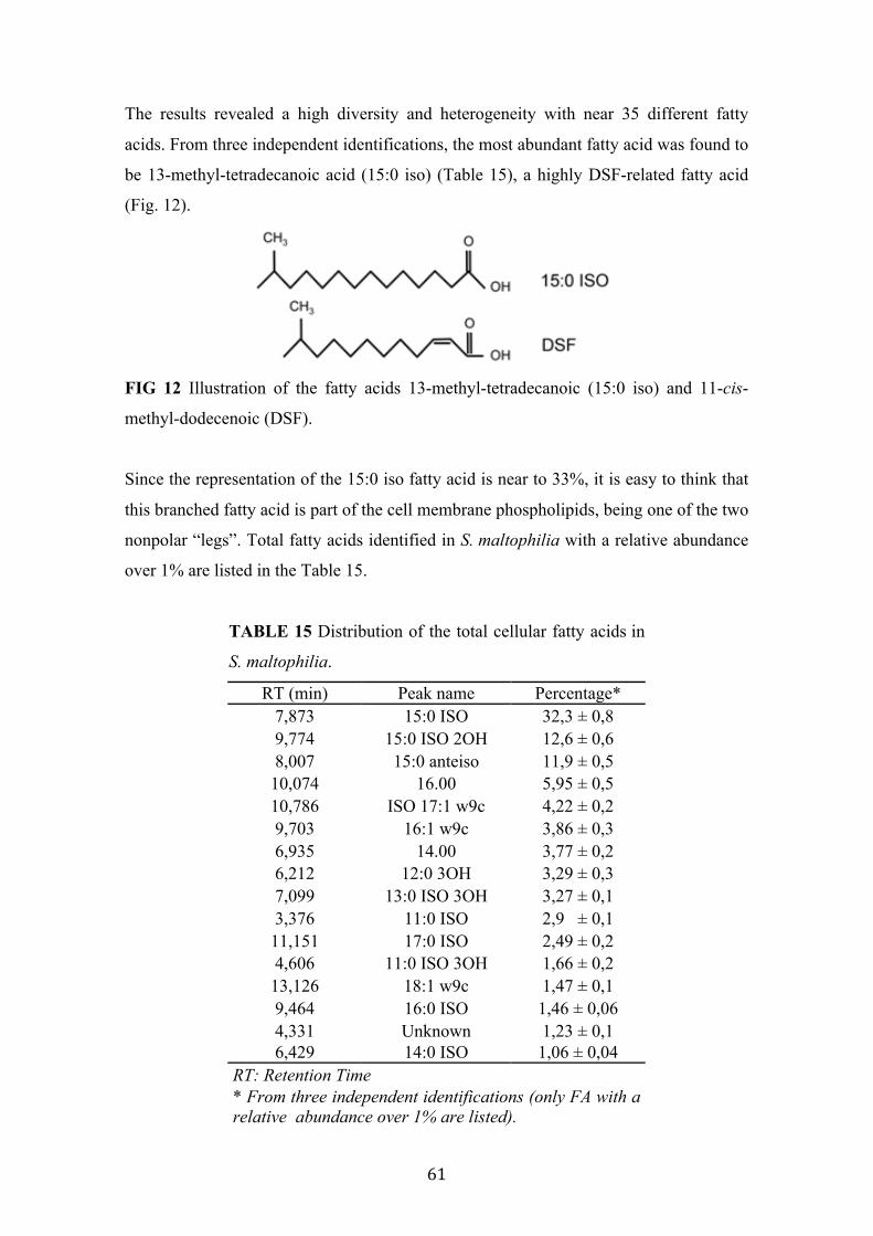
61
The results revealed a high diversity and heterogeneity with near 35 different fatty
acids. From three independent identifications, the most abundant fatty acid was found to
be 13-methyl-tetradecanoic acid (15:0 iso) (Table 15), a highly DSF-related fatty acid
(Fig. 12).
FIG 12 Illustration of the fatty acids 13-methyl-tetradecanoic (15:0 iso) and 11-cis-
methyl-dodecenoic (DSF).
Since the representation of the 15:0 iso fatty acid is near to 33%, it is easy to think that
this branched fatty acid is part of the cell membrane phospholipids, being one of the two
nonpolar “legs”. Total fatty acids identified in S. maltophilia with a relative abundance
over 1% are listed in the Table 15.
TABLE 15 Distribution of the total cellular fatty acids in
S. maltophilia.
RT (min) Peak name Percentage* 7,873 15:0 ISO 32,3 ± 0,8 9,774 15:0 ISO 2OH 12,6 ± 0,6 8,007 15:0 anteiso 11,9 ± 0,5 10,074 16.00 5,95 ± 0,5 10,786 ISO 17:1 w9c 4,22 ± 0,2 9,703 16:1 w9c 3,86 ± 0,3 6,935 14.00 3,77 ± 0,2 6,212 12:0 3OH 3,29 ± 0,3 7,099 13:0 ISO 3OH 3,27 ± 0,1 3,376 11:0 ISO 2,9 ± 0,1 11,151 17:0 ISO 2,49 ± 0,2 4,606 11:0 ISO 3OH 1,66 ± 0,2 13,126 18:1 w9c 1,47 ± 0,1 9,464 16:0 ISO 1,46 ± 0,06 4,331 Unknown 1,23 ± 0,1 6,429 14:0 ISO 1,06 ± 0,04
RT: Retention Time * From three independent identifications (only FA with a relative abundance over 1% are listed).
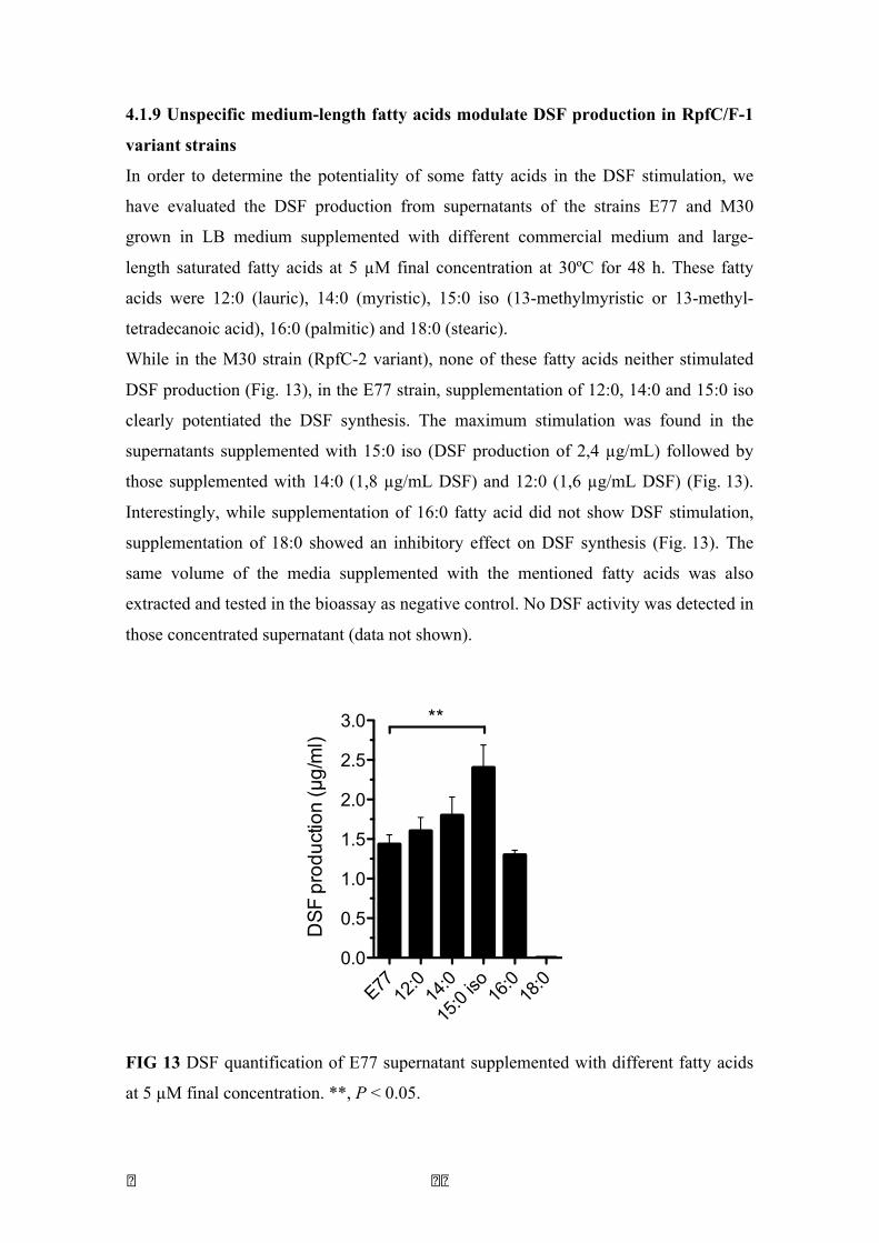
� � � �
4.1.9 Unspecific medium-length fatty acids modulate DSF production in RpfC/F-1
variant strains
In order to determine the potentiality of some fatty acids in the DSF stimulation, we
have evaluated the DSF production from supernatants of the strains E77 and M30
grown in LB medium supplemented with different commercial medium and large-
length saturated fatty acids at 5 µM final concentration at 30ºC for 48 h. These fatty
acids were 12:0 (lauric), 14:0 (myristic), 15:0 iso (13-methylmyristic or 13-methyl-
tetradecanoic acid), 16:0 (palmitic) and 18:0 (stearic).
While in the M30 strain (RpfC-2 variant), none of these fatty acids neither stimulated
DSF production (Fig. 13), in the E77 strain, supplementation of 12:0, 14:0 and 15:0 iso
clearly potentiated the DSF synthesis. The maximum stimulation was found in the
supernatants supplemented with 15:0 iso (DSF production of 2,4 µg/mL) followed by
those supplemented with 14:0 (1,8 µg/mL DSF) and 12:0 (1,6 µg/mL DSF) (Fig. 13).
Interestingly, while supplementation of 16:0 fatty acid did not show DSF stimulation,
supplementation of 18:0 showed an inhibitory effect on DSF synthesis (Fig. 13). The
same volume of the media supplemented with the mentioned fatty acids was also
extracted and tested in the bioassay as negative control. No DSF activity was detected in
those concentrated supernatant (data not shown).
FIG 13 DSF quantification of E77 supernatant supplemented with different fatty acids
at 5 µM final concentration. **, P < 0.05.
E77 12:0
14:0
15:0
iso 16:0
18:0
0.0
0.5
1.0
1.5
2.0
2.5
3.0 **
DS
F pr
oduc
tion
(µg/
ml)
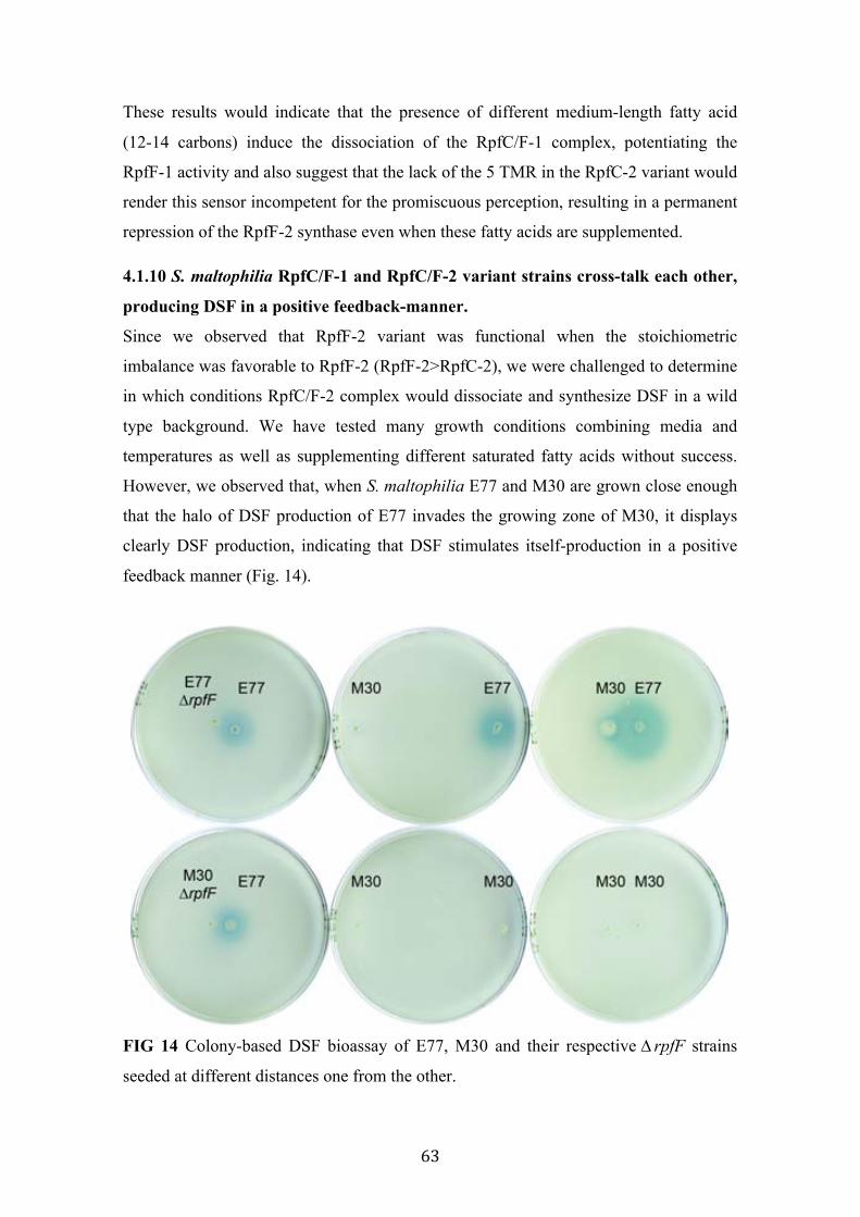
63
These results would indicate that the presence of different medium-length fatty acid
(12-14 carbons) induce the dissociation of the RpfC/F-1 complex, potentiating the
RpfF-1 activity and also suggest that the lack of the 5 TMR in the RpfC-2 variant would
render this sensor incompetent for the promiscuous perception, resulting in a permanent
repression of the RpfF-2 synthase even when these fatty acids are supplemented.
4.1.10 S. maltophilia RpfC/F-1 and RpfC/F-2 variant strains cross-talk each other,
producing DSF in a positive feedback-manner.
Since we observed that RpfF-2 variant was functional when the stoichiometric
imbalance was favorable to RpfF-2 (RpfF-2>RpfC-2), we were challenged to determine
in which conditions RpfC/F-2 complex would dissociate and synthesize DSF in a wild
type background. We have tested many growth conditions combining media and
temperatures as well as supplementing different saturated fatty acids without success.
However, we observed that, when S. maltophilia E77 and M30 are grown close enough
that the halo of DSF production of E77 invades the growing zone of M30, it displays
clearly DSF production, indicating that DSF stimulates itself-production in a positive
feedback manner (Fig. 14).
FIG 14 Colony-based DSF bioassay of E77, M30 and their respective ∆ rpfF strains
seeded at different distances one from the other.

� � �
In addition, mixed cultures of E77 and M30 grown in LB at 30ºC for 48 h displayed a
higher DSF production than E77 axenic cultures (Fig. 15). Finally, supplementation of
LB with 0,05 µM DSF triggers DSF production of M30 up to a maximum point of
0,6 µg/mL production after 48 h of growth (Fig. 15).
These results indicate, in one hand, that the sensor RpfC-2 is much more specific,
liberating active-RpfF-2 only upon detection of DSF molecules, in the other, that DSF
is produced in a positive feedback-manner in S. maltophilia.
FIG 15 DSF production in cultures of axenic E77, axenic M30, a mixed culture of E77
and M30 and a culture of M30 supplemented with 0.05 µM DSF. **, P < 0.05.
4.1.11 Discussion
In this chapter, we have proposed to study the molecular mechanisms that govern DSF
production in S. maltophilia.
We have characterized 78 S. maltophilia clinical strains, isolated from diverse sources
in different European hospitals, for the rpf cluster. We have first demonstrated that the
78 strains contain the rpfF gene but the RpfF product is distributed into two different
variants that we have named RpfF-1 and RpfF-2 (Fig. 1, 2). We have also shown that
the isolates present two RpfC variants, each associated to the RpfF variants (Fig. 1A).
The two RpfC variants are different in their N-terminal region, which corresponds to a
E77 M30
E77:M
30
M30 0,
05 µM
DSF
0,05 µ
M DSF
0.0
0.5
1.0
1.5
2.0
2.5**
DS
F pr
oduc
tion
(µg/
ml)
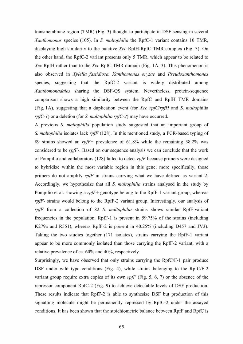
65
transmembrane region (TMR) (Fig. 3) thought to participate in DSF sensing in several
Xanthomonas species (105). In S. maltophilia the RpfC-1 variant contains 10 TMR,
displaying high similarity to the putative Xcc RpfH-RpfC TMR complex (Fig. 3). On
the other hand, the RpfC-2 variant presents only 5 TMR, which appear to be related to
Xcc RpfH rather than to the Xcc RpfC TMR domain (Fig. 1A, 3). This phenomenon is
also observed in Xylella fastidiosa, Xanthomonas oryzae and Pseudoxanthomonas
species, suggesting that the RpfC-2 variant is widely distributed among
Xanthomonadales sharing the DSF-QS system. Nevertheless, protein-sequence
comparison shows a high similarity between the RpfC and RpfH TMR domains
(Fig. 1A), suggesting that a duplication event (for Xcc rpfC/rpfH and S. maltophilia
rpfC-1) or a deletion (for S. maltophilia rpfC-2) may have occurred.
A previous S. maltophilia population study suggested that an important group of
S. maltophilia isolates lack rpfF (128). In this mentioned study, a PCR-based typing of
89 strains showed an rpfF+ prevalence of 61.8% while the remaining 38.2% was
considered to be rpfF-. Based on our sequence analysis we can conclude that the work
of Pompilio and collaborators (128) failed to detect rpfF because primers were designed
to hybridize within the most variable region in this gene; more specifically, those
primers do not amplify rpfF in strains carrying what we have defined as variant 2.
Accordingly, we hypothesize that all S. maltophilia strains analysed in the study by
Pompilio et al. showing a rpfF+ genotype belong to the RpfF-1 variant group, whereas
rpfF- strains would belong to the RpfF-2 variant group. Interestingly, our analysis of
rpfF from a collection of 82 S. maltophilia strains shows similar RpfF-variant
frequencies in the population. RpfF-1 is present in 59.75% of the strains (including
K279a and R551), whereas RpfF-2 is present in 40.25% (including D457 and JV3).
Taking the two studies together (171 isolates), strains carrying the RpfF-1 variant
appear to be more commonly isolated than those carrying the RpfF-2 variant, with a
relative prevalence of ca. 60% and 40%, respectively.
Surprisingly, we have observed that only strains carrying the RpfC/F-1 pair produce
DSF under wild type conditions (Fig. 4), while strains belonging to the RpfC/F-2
variant group require extra copies of its own rpfF (Fig. 5, 6, 7) or the absence of the
repressor component RpfC-2 (Fig. 9) to achieve detectable levels of DSF production.
These results indicate that RpfF-2 is able to synthesize DSF but production of this
signalling molecule might be permanently repressed by RpfC-2 under the assayed
conditions. It has been shown that the stoichiometric balance between RpfF and RpfC is

66
crucial for DSF production in many Xanthomonadales. In Xcc, RpfC physically
interacts with the RpfF active site, inhibiting the DSF-synthesis activity (104, 105, 107).
RpfC has been also shown to repress the RpfF activity in X. fastidiosa (117).
Furthermore, analysis of mRNA levels in the strains E77 and M30 by qRT-PCR shows
that the rpfF to rpfC expression ratio in the DSF-producer strain (variant 1) doubles that
found in the non-producer one (variant 2) (Fig. 10). Together with the observation that
complementation of extra copies of rpfF-2 and/or deletion of ∆rpfC-2 result in clearly
DSF production in RpfC/F-2 strains, it is evident that the stoichiometry balance
between these two components is also a crucial factor in DSF production in
S. maltophilia.
Although DSF-QS system has been deeply studied among Xanthomonadales species,
the biosynthetic pathway and catalytic mechanisms involved in DSF production as well
as how the sensor RpfC perceive DSF signals remain to be a mystery. We here have
generated an improved liquid DSF bioassay (Fig. 11), a useful tool to investigate the
implication of the temperature and the media as well as the potentiality of some fatty
acids in the modulation of DSF production in S. maltophilia. While these factors were
not involved in DSF production in the M30 strain, they were clearly involved in the
regulation of DSF production in the E77 strain (Table 13, 14). Moreover, while lauric
(12:0) and myristic (14:0) acid moderately stimulate DSF production, the most
abundant fatty acid in S. maltophilia 15:0 iso (Fig. 12) (Table 15) strongly stimulates
DSF production in RpfC/F-1 strain E77 (Fig. 13). Since RpfF has an enoyl-CoA
hydratase/isomerase function, and there is not a putative fatty acid methyl-transferase in
the rpf cluster, we hypothesize that the only fatty acid that would really represent a DSF
precursor is 15:0 iso, which already contains a branched methyl in the penultimate
carbon (C13) (Fig. 12). 15:0 iso would become 11-cis-2methyl-dodecenoic acid by the
hydratase activity of the RpfF -liberating one acetil-coA-, followed by the isomerase
activity -also done by the RpfF-, generating the typical unsaturation between the alpha-
beta carbons (Fig. 12). The analysis of the total cellular fatty acids further supported
that 15:0 iso could represent the principal DSF precursor in S. maltophilia since its
relative amount was 33% followed by 15:0 iso 2OH (13%) (Table 15), a hypothetical
intermediate in the DSF biosynthetic pathway, between 15:0 iso and DSF. The idea that
the principal DSF precursor comes from the membrane phospholipids would give sense
to the fact that the sensor component RpfC, which is expected to sense diffusible

67
signals, is anchored to the cell membrane, because it is also controlling somehow the
homeostasis of the cell membrane. Nevertheless, a lot of work remains to clarify this
hypothesis.
However, since the RpfC has also a RpfF-repression function, we hypothesize that the
DSF stimulation observed in the other supplemented supernatants (12:0 and 14:0) is due
to the presence of a competent sensor input domain (composed of 10 TMR) (Fig. 3),
which promiscuously sense DSF-related fatty acids, allowing free-active RpfF with the
subsequent synthesis of DSF. In addition, this promiscuous sensing only occurs upon
detection of fatty acids with 12-14 carbons length, since palmitic acid (16:0) didn’t
show DSF stimulation and stearic acid (18:0) showed a drastic inhibitory effect of DSF
production (Fig. 13). Nevertheless, more efforts are needed to clarify such phenomenon.
It has been suggested that, in Xcc, RpfC could play a positive-feedback role in DSF
synthesis, liberating active RpfF upon detection of DSF molecules (107). Accordingly
with this hypothesis and taking all results together, we hypothesize that DSF production
in RpfC/F-1 strains is due to the presence of a competent sensor input domain
composed of 10 TMR (Fig 3) capable of detect not only DSF molecules but also some
fatty acid derivatives, or DSF precursors, that would enable the liberation of active
RpfF-1 and the subsequent synthesis of DSF. On the other hand, the missing TMR in
RpfC-2 would render this factor incompetent for this promiscuous sensing leading to a
permanent inhibition of RpfF-2 by RpfC-2 until the presence of few DSF molecules
trigger the dissociation of this complex enabling free-active RpfF-2 to synthetize DSF.
This indicates that the complex RpfC/F-2 is much more specific than RpfC/F-1, sensing
only DSF molecules. Recent findings reported that, in X. fastidiosa (RpfC-2 situation, 5
TMR), RpfF is also required for DSF detection (117), something that might also be
occurring in S. maltophilia strains harbouring the RpfC/F-2 combination.
With all these observations, it is evident that some initial DSF production must occur in
order to triggers the DSF production in RpfC/F-2 strains. However, the specific
conditions that would facilitate this initial DSF production in axenic cultures have not
been yet elucidated and will require further studies. Nevertheless, it is well established
that, in their natural niches, there exist competence and communication between
populations of the same and different species (104), even between organisms from
different kingdoms (100), which could generate this initial levels of DSF and induce the
activation of the entire DSF system in the RpfC/F-2 variant population.
68
It is known that many Xanthomonadales produce more or even different signalling
molecules than the canonical DSF. Although DSF was found to be the unique signalling
molecule in Xanthomonas campestris pv. campestris (104) and Xanthomonas
axonopodis pv. citri (Xac) (94, 95), in Xanthomonas orizae pv orizae (Xoo), three
signalling fatty acids molecules have been described: DSF, BDSF (cis-2-dodecenoic
acid) and CDSF ((2Z, 5Z)-11-methyldodecadienoic acid) (96). Recently, 2(Z)-
tetradecenoic acid (xfDSF) was reported to be produced by Xylella fastidiosa strain
temecula1 (99). More complex is the DSF-like pattern produced by S. maltophilia strain
WR-C, which includes DSF and seven derivatives (127). Although their exact structure
remains unclear and were not probed to be synthesized by RpfF, GC/MS analysis of
pyrrolidide derivatives suggest that they could be: ∆2-tridecenoic acid, 10-methyl-
dodecanoic acid, ∆2-methyl-tridecanoic acid, ∆2-tetradecenoic acid, ∆2-12-methyl-
tetradecenoic acid and ∆2-13-methyl-tetradecenoic acid (127). But DSF-like production
is not only restricted to Xanthomonadales since cis-2-decenoic acid is produced by
P. aeruginosa (142, 143) and BDSF is produced by B. cenocepacea (100). Assuming a
high degree of heterogeneity in DSF-like synthesis among bacteria, it wouldn’t be
strange to hypothesize that some DSF-derivative molecules are strain-dependent
synthesized.
We have also been working for a long time under the premise that the RpfF-2 variant
could produce a different DSF signalling molecule. Using several approaches, I have
not been able to identify an alternative signal molecule. Although we cannot totally
reject such hypothesis, the results from the experiments of TLC coupled to DSF
bioassay (Fig. 6) and GC/MS (Fig. 7, 8) strongly suggest that the canonical DSF is the
unique fatty acid with signalling activity produced by both RpfF-variants and
consequently by S. maltophilia.
69
4.2 Phenotypic implications of DSF-QS in S. maltophilia
After demonstrate that two variants of the cluster rpf are distributed among the
population of S. maltophilia and characterize the molecular mechanisms involved in
DSF synthesis in each variant, we wanted to determine the role that each RpfF variant
have in the regulation of virulent-associated processes. To address this question, we
have studied some virulence-related phenotypes including swarming motility, biofilm
formation and virulence capacity in the C. elegans and zebrafish models for the two
mutant model strains E77 ∆rpfF (variant 1) and M30 ∆rpfF (variant 2).
4.2.1 Optimization of swarming motility assay for S. maltophilia
One important virulent factor that has been subject of extensive work in related bacteria
is swarming motility. In addition, in Burkholderia cenocepacea it has been
demonstrated that DSF-QS regulates this kind of motility (178). However, few articles
are published relating swarming motility and S. maltophilia. In fact, there is only one
paper reporting swarming motility in this bacterium, which was published by our group
last year (72). However, in this mentioned study, a slow and particular kind of motion is
reported. Therefore, we have tried to optimize the conditions that could facilitate this
motility in S. maltophilia, by studying various factors such as the media, temperature,
incubation time and inoculation process in the model strain E77. From four different
media (LB, YEB, modified-M9 and BM2) we have observed that swarming motility
takes place in minimal media (modified-M9 salts and BM2) (Fig. 16), whereas it is not
observed in rich media conditions (LB, YEB) (data not shown). In addition, we have
noted that it is facilitated in modified M9-salts medium more than in BM2, although no
drastic differences are observed (Fig. 16). We have also noted that swarming activation
is faster with streak inoculation than with pin inoculation, suggesting that a high-density
population facilitates the initiation of this type of motion (Fig. 16). Regarding the
incubation temperature, it seems that only 30ºC allows S. maltophilia to move in a
swarming-manner, since it was not observed when plates were incubated neither 25ºC
nor 37ºC (data not shown).

70
FIG 16 Optimization of swarming motility assay in S. maltophilia E77. (A) S.
maltophilia seeded in BM2 0.5% agar after inoculation with different techniques from
different media and growth conditions. (B) S. maltophilia seeded in modified M9-salts
0.5% agar after inoculation with different techniques from different media and growth
conditions. liq: overnight liquid culture. col: pin inoculation from a bacterial colony.
streak: “streak” inoculation from an overnight plate culture.
4.2.2 ∆rpfF-1 but not ∆rpfF-2 mutant shows alteration in swarming motility
We have investigated the implication of each RpfF variant in swarming motility.
Aforementioned optimization experiments supported the idea that QS could be involved
in swarming activation in S. maltophilia. To corroborate this hypothesis, we tested the
ability of E77 and M30 ΔrpfF mutants to swarm under previously defined conditions
(modified M9-salts medium with 0.5% agar concentration), relative to the wt strains.

71
As shown in the Figure 17, E77 wt displays tendril-like motility, whereas M30 wt
hardly swarms, likely due to its DSF deficiency. The E77 ΔrpfF mutant showed a clear
loss of motility and a phenotype restoration when rpfF was in-trans complemented. On
the contrary, the swarming motility of the M30 ∆ rpfF mutant was not significantly
different from that of the M30 wt strain, suggesting that RpfF does not intervene in
swarming control in M30. However, this behaviour does not seem to be strictly linked
to the RpfF variant because, in the one hand, E77 ∆ rpfF displayed an atypical non-
tendril swarming morphology when it was heterologous complemented with the M30
rpfF gene. On the other, heterologous complementation of M30 ∆ rpfF with the E77
rpfF gene resulted in motility similar to that of the M30 ∆rpfF mutant strain.
FIG 17 Swarming motility assay for E77 and M30, their ∆ rpfF mutants and the
homologous and heterologous complemented strains, on modified M9-salts medium
solidified with 0.5% noble agar and incubated at 30 ºC for 4 days.
Since RpfF-1 was shown to be essential in swarming motility in E77, we wanted to get
more insights into the physiological and molecular mechanisms involved in this kind of
motility. Thus, we have initially evaluated the surfactant production and the cell
morphology of active-bacteria taken from the edge of the swarming tendril after
motility experiments, for the E77 wt, the ∆ rpfF mutant and the complemented strain.
The obtained results demonstrated that RpfF-1 was directly involved in the surfactant-
like production, since the mutant strain showed a loss of the surfactant halo and it was
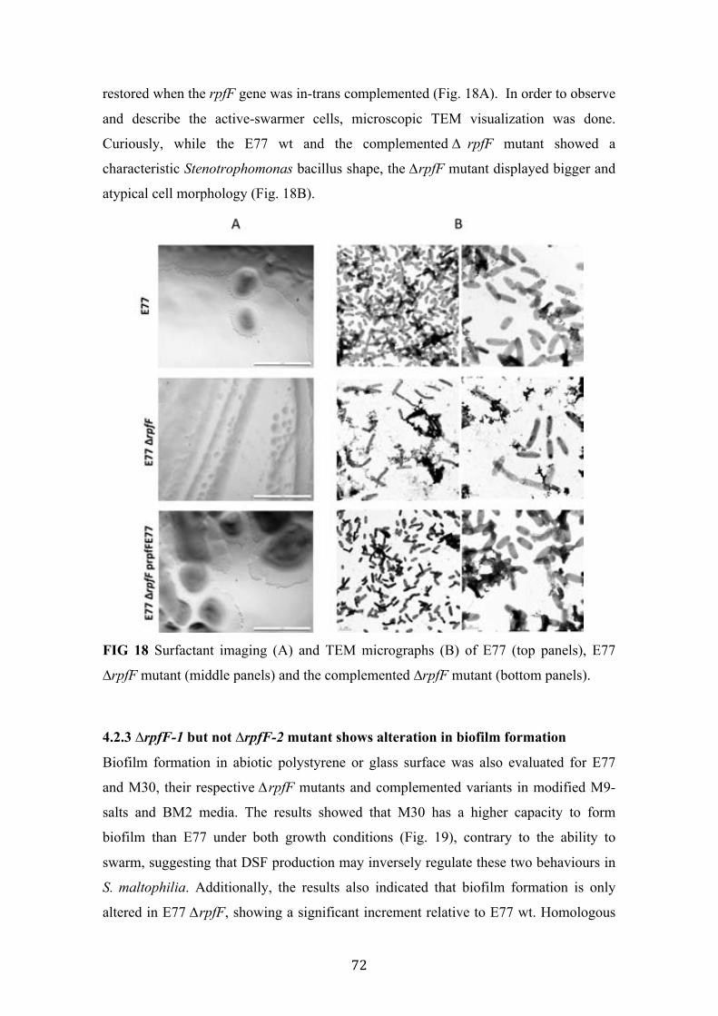
72
restored when the rpfF gene was in-trans complemented (Fig. 18A). In order to observe
and describe the active-swarmer cells, microscopic TEM visualization was done.
Curiously, while the E77 wt and the complemented ∆ rpfF mutant showed a
characteristic Stenotrophomonas bacillus shape, the ∆rpfF mutant displayed bigger and
atypical cell morphology (Fig. 18B).
FIG 18 Surfactant imaging (A) and TEM micrographs (B) of E77 (top panels), E77
∆rpfF mutant (middle panels) and the complemented ∆rpfF mutant (bottom panels).
4.2.3 ∆rpfF-1 but not ∆rpfF-2 mutant shows alteration in biofilm formation
Biofilm formation in abiotic polystyrene or glass surface was also evaluated for E77
and M30, their respective ∆rpfF mutants and complemented variants in modified M9-
salts and BM2 media. The results showed that M30 has a higher capacity to form
biofilm than E77 under both growth conditions (Fig. 19), contrary to the ability to
swarm, suggesting that DSF production may inversely regulate these two behaviours in
S. maltophilia. Additionally, the results also indicated that biofilm formation is only
altered in E77 ∆rpfF, showing a significant increment relative to E77 wt. Homologous
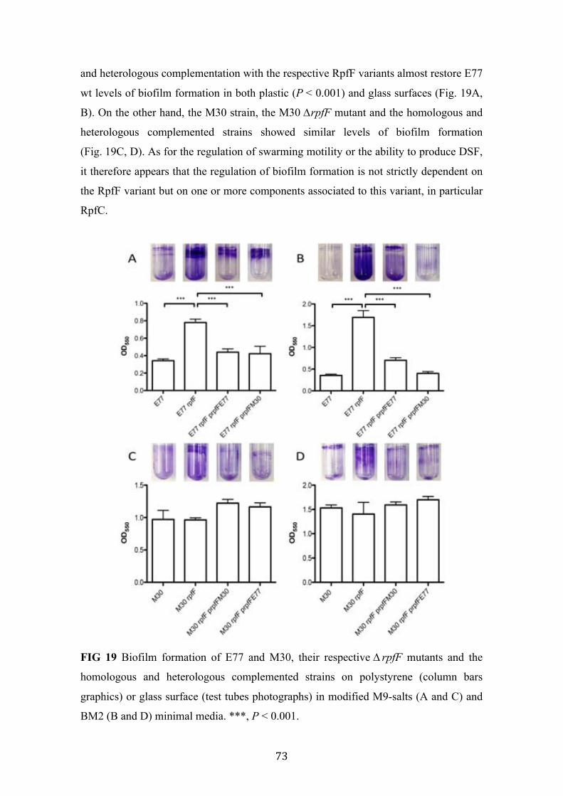
73
and heterologous complementation with the respective RpfF variants almost restore E77
wt levels of biofilm formation in both plastic (P < 0.001) and glass surfaces (Fig. 19A,
B). On the other hand, the M30 strain, the M30 ∆rpfF mutant and the homologous and
heterologous complemented strains showed similar levels of biofilm formation
(Fig. 19C, D). As for the regulation of swarming motility or the ability to produce DSF,
it therefore appears that the regulation of biofilm formation is not strictly dependent on
the RpfF variant but on one or more components associated to this variant, in particular
RpfC.
FIG 19 Biofilm formation of E77 and M30, their respective ∆ rpfF mutants and the
homologous and heterologous complemented strains on polystyrene (column bars
graphics) or glass surface (test tubes photographs) in modified M9-salts (A and C) and
BM2 (B and D) minimal media. ***, P < 0.001.

74
4.2.4 Only the ΔrpfF-1 mutant shows attenuation in C. elegans
To elucidate the direct implication of each RpfF variant in S. maltophilia virulence in
vivo, the killing ability of E77 and M30, their respective ∆rpfF mutants and
complemented strains were tested in C. elegans. Although both wild type E77 and M30
strains showed similar virulence capacity in the C. elegans model (LT50 or time of 50%
death of 6.04 and 4.99 days respectively), significant attenuation was observed here for
E77 ∆ rpfF (Fig. 20). In line with the observations made for the phenotypes analysed
previously, the virulence of E77 ∆rpfF is restored after complementation with either its
own rpfF (RpfF-1 variant) or the M30 rpfF (RpfF-2 variant), indicating once more that
E77 is able to respond to heterologous DSF production in a RpfC-1-variant background
(Fig. 20). Infection with the M30 ∆ rpfF mutant showed no significant differences
relative to M30 wt.
FIG 20 Virulence determination of E77 (A) and M30 (B), their respective ∆ rpfF
mutants and the homologous and heterologous complemented strains in the C. elegans
CF512 model of infection. E. coli OP50 was used as negative control and showed no
mortality within the firsts five days. **, P < 0.05.

75
4.2.5 Both ∆rpfF mutants show attenuation in the adult Zebrafish infection model
In order to corroborate that only ∆rpfF-1 mutant showed an attenuated phenotype in in-
vivo models, both E77 and M30 strains with their ∆ rpfF-1 and ∆ rpfF-2 mutants and
complemented strain were also evaluated in an adult zebrafish infection model.
12 fishes per sample were injected with a dose consisting of 5·108 cfu/mL and the
mortality was periodically scored during five days. Surprisingly, and contrary to the
previous results, in this animal model both ∆ rpfF-1 and ∆ rpfF-2 mutants showed
attenuation compared to the wild type strains. In addition, when E77 ∆ rpfF and M30
∆rpfF were in-trans complemented with their respective rpfF, their virulence capacity
was partially restored, indicating that in this animal model, the RpfF-2 is participating
in the regulation of the virulence capacity (Fig. 21).
FIG 21 Virulence determination of S. maltophilia strains E77 (A) and M30 (B) with
their respective ∆ rpfF mutants and the complemented strains in the adult zebrafish
model. ***, P < 0.001.
4.2.6 Attenuation of ∆rpfF-1 mutant is due to its incapacity to disseminate throw
the fish tissues
To get more insights into the dissemination capacity of the E77 strain during zebrafish
infection, two fishes from each tank were sacrificed after 72 h post-injection and
divided into three sections (anterior, abdominal and posterior) prior to CFU counting.
Interestingly, bacterial recovery showed the ability of E77 wt to disseminate through the
fish body from the abdominal to the anterior and posterior regions. On the contrary, the
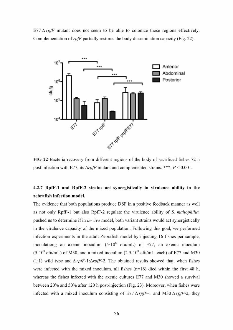
76
E77 ∆ rpfF mutant does not seem to be able to colonize those regions effectively.
Complementation of rpfF partially restores the body dissemination capacity (Fig. 22).
FIG 22 Bacteria recovery from different regions of the body of sacrificed fishes 72 h
post infection with E77, its ∆rpfF mutant and complemented strains. ***, P < 0.001.
4.2.7 RpfF-1 and RpfF-2 strains act synergistically in virulence ability in the
zebrafish infection model.
The evidence that both populations produce DSF in a positive feedback manner as well
as not only RpfF-1 but also RpfF-2 regulate the virulence ability of S. maltophilia,
pushed us to determine if in in-vivo model, both variant strains would act synergistically
in the virulence capacity of the mixed population. Following this goal, we performed
infection experiments in the adult Zebrafish model by injecting 16 fishes per sample,
inoculationg an axenic inoculum (5·108 cfu/mL) of E77, an axenic inoculum
(5·108 cfu/mL) of M30, and a mixed inoculum (2.5·108 cfu/mL, each) of E77 and M30
(1:1) wild type and ∆ rpfF-1:∆rpfF-2. The obtained results showed that, when fishes
were infected with the mixed inoculum, all fishes (n=16) died within the first 48 h,
whereas the fishes infected with the axenic cultures E77 and M30 showed a survival
between 20% and 50% after 120 h post-injection (Fig. 23). Moreover, when fishes were
infected with a mixed inoculum consisting of E77 ∆ rpfF-1 and M30 ∆ rpfF-2, they

77
showed a survival near to 40% after 120 hpi, demonstrating that RpfF is an essential
trait for the full virulence capacity of S. maltophilia population (Fig. 23). Symptoms of
disease as well as the fish behavior were also evaluated during the days of infection.
Interestingly, fishes infected with the mixed wild type inoculum showed slightly more
signs of disease compared to the other tanks, but showed a drastic change in their
behaviour (spasms, compromised swimming and sudden death), especially between 24
and 48 h (data not shown).
FIG 23 Virulence determination of E77, M30, a mixed inoculum of E77 and M30 (1:1)
and a mixed inoculum of E77 ∆ rpfF-1 and M30 ∆ rpfF-2 (1:1) in the adult zebrafish
model. ***, P < 0.001.
4.2.8 The full virulence capacity observed in the mixed inoculum correlates with
in-vivo DSF production
In order to corroborate that both RpfF were active during the in-vivo infection process
and also that they had an implication on the lethal virulence capacity observed in the
fishes infected with the mixed inoculum, in-situ DSF quantification was done. Two
fishes from each tank were sacrificed after 48 h post-injection and in-situ DSF
production was determined (see Materials and Methods, section 3.8.4). Interestingly, the
condition that showed the fulminant mortality (mixed inoculum of E77 and M30 wt)
was the only condition that showed clearly DSF production in the in-vivo infection

78
environment (Fig. 24). Accordingly, the DSF production correlated with the virulence
capacity observed in the fishes infected with the mixed inoculum.
FIG 24 (A) Regular bioassay of DSF extracted from sacrificed fishes after 48 hpi with
E77, M30 and the MIX inoculum (E77:M30, 1:1). (B) Quantification of DSF with the
liquid bioassay, from the same samples. ***, P < 0.001.
4.2.9 Discussion
It is well known that the DSF-QS system regulates certain virulence traits in many
bacteria (148, 179–185), including Stenotrophomonas (17, 65, 127). However, the
regulation that each RpfF variant exerts on the physiology and pathogenicity of this
bacterium remains uncertain. Specifically, in this chapter, we have focused into the
implications of each RpfF variant into the regulation of virulence factors.
To test that, we have generated a ∆rpfF mutant for a strain representative of each
variant group, i.e., E77 for the RpfF-1 variant group and M30 for the RpfF-2 one.
The E77 ∆rpfF mutant showed attenuation in both C. elegans (Fig. 20) and zebrafish
(Fig. 21) infection models, proving that DSF-mediated regulation affects the virulence
capacity of RpfC/F-1 strains. This is in line with the results reported for the K279a
∆rpfF (variant 1), which showed a loss of virulence in the C. elegans model (65).
Moreover, the recovery of bacteria from sacrificed fishes 72 h post-injection

79
(intraperitoneal injection) showed that E77 is able to disseminate to anterior and
posterior regions through the fish body, while E77 ∆ rpfF was impaired in crossing
intraperitoneal barriers (Fig. 22). This is in concordance with the results showing a loss
of swarming motility (Fig. 17) and a drastic increment in the biofilm formation capacity
(Fig. 19A, B) of the E77 ∆rpfF mutant, two important virulence-related traits that could
be related to the virulence attenuation observed in the animal models.
We have studied for the first time the environmental factors that could modulate
swarming motility in S. maltophilia. Although much work is needed to elucidate the
exact conditions that influence this motility behaviour, we here propose the modified
M9-salt medium 0.5% agar and 30ºC as a good approach to test swarming ability of
S. maltophilia strains (Fig. 16). Many evidences of the implication of RpfF and DSF-
like fatty acids in bacterial motility have been indeed reported (179, 181, 186, 187). Our
results reinforce previous evidence that one of the main functions of DSF-QS is to
regulate bacterial motility, especially in those carrying the RpfF-1 variant. Moreover,
we have showed the implication of the RpfF-1 variant in surfactant production and cell
morphology during swarming motility in S. maltophilia strain E77 (Fig. 18).
Many studies have also studied the implication of DSF-like molecules in biofilm
regulation. There is however some controversy on whether DSF-like molecules may act
stimulating or inhibiting the sessile or motile bacterial lifestyles. Thus, DSF molecules
have been shown to positively regulate biofilm formation in Xanthomonas oryzae pv.
oryzae (188), Burkholderia cenocepacea (181, 184) and Xylella fastidiosa (182, 189).
On the contrary, in Xanthomonas campestris pv. campestris (Xcc) the DSF-mediated
QS acts as a negative regulator of biofilm development (190–192). Additionally, fatty
acid-mediated biofilm dispersion is not restricted to species sharing the DSF-QS
system. For example, the fatty acid cis-2-decenoic produced by P. aeruginosa PAO1
stimulates biofilm dispersion in several gram-positive and gram-negative bacteria (193,
194). Our findings indicate that the DSF-QS system in S. maltophilia E77 has a similar
regulatory function than that described for Xcc, where DSF also plays an important role
in preventing biofilm formation and stimulating bacterial motility.
On the contrary, all virulence-related phenotypes evaluated on M30 were unaltered in
the ∆ rpfF mutant and in the corresponding complemented strain, suggesting that
RpfC/F-2 variant strains may not use the DSF-QS system to regulate these virulence
factors. However, while the M30 ∆ rpfF mutant didn’t show attenuation in the

80
C. elegans model, it certainly showed attenuation in the Zebrafish infection model
(Fig. 21). It is evident that C. elegans and zebrafish models are totally different. While
C. elegans is a very simple worm showing only innate immune response (195, 196),
zebrafish is a vertebrate with a more developed immune system, which presents innate
and adaptive immune responses (197). It is also known that the bacterial processes
required for the infection as well as the inoculation in these two models are completely
different. Together, it seems that under this environment, RpfC/F-2 variant strains could
use the DSF-QS to regulate, somehow, its virulence ability.
In the chapter 4.2 it is reported that both RpfC/F-1 and RpfC/F-2 variant strains act
synergistically in DSF production (Fig. 14). Here, we have evaluated the implication of
this DSF synergism in the virulence ability of the entire population. Accordingly, the
results strongly suggest that both populations act synergistically in the development of
infection processes (Fig. 23) and also that the fulminant virulence ability is DSF-
dependent (Fig. 24).
Together, our findings indicate, in one hand, that the RpfC/F-2 complex regulates
virulence processes but only in very specific conditions (i. e. in Zebrafish infection). On
the other hand, that both populations communicate each other, throw the DSF-QS,
potentiating the DSF production and the virulence capacity of the whole population.
Since bacterial species sharing DSF-QS are almost ubiquitous and frequently share
ecological niches, it is easy to think that RpfC/F-2 variant strains could often be in
contact with DSF-producer bacteria (i. e., Xcc or S. maltophilia RpfC/F-1 variant). In
this situation, a DSF-producer strain would act as a starter strain, triggering the
reciprocal DSF-communication by synthesising the initial DSF molecules. It seems that
RpfC/F-2 strains have evolved as a receptor group in this DSF communication, showing
a lethargic DSF-deficient phenotype in axenic conditions, something that would render
energetic saving as well as a more fine regulation of processes related to DSF-
communication.
Furthermore, interspecies communication is not restricted to Xanthomonadales.
Recently, It has been reported that the DSF molecule produced by S. maltophilia
influenced biofilm formation of P. aeruginosa (198) Another study showed that the
DSF-family signal C10:Δ2 (cis-DA) produced by P. aeruginosa not only dispersed its
own biofilm but also induced the biofilm dispersion of numerous bacterial species
including E. coli, Klebsiella pneumoniae, Proteus mirabilis, Streptococcus pyogenes,

81
Bacillus subtilis, and Staphylococcus aureus (142). In addition, BDSF from B.
cenocepacia was found to be a functional substitute of the canonical DSF signal, and
regulate biofilm formation and virulence factor production in Xcc (101). In this line, a
novel signal 11-Me-C12:Δ2,5 identified from B. multivorans could also substitute DSF
and C12:Δ2 and influence the biofilm development and virulence factor production in
Xcc and B. cenocepacia, respectively (101). Interestingly, beside interspecies
communication these mentioned DSF-derived signals (DSF, C12:Δ2 and 11-Me-
C12:Δ,2,5) have been shown to inhibit the morphological transition of Candida
albicans through interkingdom interference (100, 101, 104). Likewise, a fatty acid
produced by the gram-positive Streptococcus mutans -trans-2-decenoic, designated
SDSF- is also a potent inhibitor of morphological transition in C. albicans (103).
Moreover, the signal C10:Δ2 produced for P. aeruginosa was found to be able to induce
biofilm dispersion in C. albicans, as well as diverse bacteria (142).
Together with our results, it is evident that complex population processes, such as QS,
are not well addressed when they are studied in axenic conditions.
82
4.3 cis-DA-mediated Quorum Sensing System in S. maltophilia and P.
aeruginosa
New recent discoveries reported that the fatty acid cis-decenoic (cis-DA) is involved in
biofilm dispersion in the important pathogen P. aeruginosa (142, 143). It has been
shown that the putative enoyl-CoA hydratase (ECH) DspI (PA0745) is required for cis-
DA synthesis (143), but also for the full virulence ability of P. aeruginosa strain PA14
(144). Interestingly, a preliminary sequence analysis revealed that S. maltophilia
harbours a DspI orthologous, the putative ECH Smlt0266, displaying a significant
identity with PA0745. In addition, we have recently reported that Smlt0266 is
overexpressed in the S. maltophilia clinical-isolate M30 compared to the attenuated
strain ATCC13637 (72). For this reason, in this chapter we will try to determine if this
new cis-DA QS is also present and functional in S. maltophilia, taking M30 as a model
strain.
4.3.1 smlt0266 is the dspI orthologous in S. maltophilia
A preliminary genomic analysis using the Web-based comparative genomic tools
(http://www.microbesonline.org) (199) revealed that in the genome of MPAO1, the
gene encoding for the cis-DA synthase DspI (PA0745) (143) is organized into an
operon, conformed by genes encoding for typical beta-oxidation components. Thus,
dspI is flanked by upstream genes encoding for the putative aldehyde-dehydrogenase
PA0747 and the hypothetical acyl-CoA dehydrogenase PA0746, and flanked by a
downstream gene encoding for the putative enoyl-CoA hydratase/isomerase PA0744
(Fig. 25). Interestingly, a subsequent comparative genome analysis showed that the
genome of S. maltophilia strain M30 harbours an orthologous operon composed by the
genes smlt0264, smlt0265, smlt0266 and smlt0267 (Fig. 25). Protein blast analysis
revealed a high homology between Smlt0266 and the P. aeruginosa cis-DA synthase
DspI (68%) (Fig. 25). The elevated similarity observed between these components
suggested that this operon likely regulate similar functions in both bacterial species. As
mentioned, in P. aeruginosa the ECH DspI (PA0745) apart of being responsible for the
cis-DA synthesis (143), it is also involved in many virulence-related processes (144).
However, nothing is known about the alternative enoyl-CoA hydratase/isomerase
PA0744, which displays significant similarity with the ECH Smlt0267 in S. maltophilia.

83
Since several synthases of fatty acids signalling molecules (i.e., RpfF/DSF) often
present enoyl-CoA hydratase/isomerase activity, we wanted to study the role of these
two ECH in the synthesis and regulation of the new lipid-mediated cis-DA QS in S.
maltophilia and P. aeruginosa.
To do that, we have generated the S. maltophilia ∆smlt0266, ∆smlt0267 and ∆smlt0266-
smlt0267 mutants and we have compared their phenotype with the respective
P. aeruginosa orthologous mutants ∆dspI (∆PA0745) and ∆dspII (∆PA0744), obtained
from the PAO1 transposon mutant library at the University of Washington (149).
FIG 25 Comparison of the dsp operon between P. aeruginosa and S. maltophilia.
Protein identity is indicated in percentage in brackets and grey arrows indicate the genes
subject of this study.
4.3.2 Smlt0266 and Smlt0267 inversely regulate biofilm formation in S. maltophilia.
It is well known that DspI is involved in biofilm dispersion in P. aeruginosa, by
synthesizing the dispersal inducer fatty acid cis-DA (143). To test if the orthologous
gene smlt0266 similarly regulates this behaviour in S. maltophilia, we have analysed the
biofilm formation of the strains M30 wt, M30-derivative mutants ∆smlt0266, ∆smlt0267
and the double mutant ∆smlt0266-0267 and the M30 transformed with pBADSMdsp
(Table 5). The later strain is the wt strain overexpressing the putative cis-DA synthase
Smlt0266 under the control of the arabinose-inducible promoter (induced with
arabinose at 0.2% concentration) (See Materials and Methods, section 3.6.3). As for
P. aeruginosa, Smlt0266, but even more, Smlt0267, are directly involved in the biofilm
formation capacity in S. maltophila. As shown in the Figure 26, deletion of the
smlt0266 gene led to a significant increase of biofilm formation. On the other hand,
deletion of the smlt0267 resulted in a drastic reduction of biofilm formation (Fig. 26),
suggesting that these two components inversely regulate biofilm formation in
S. maltophilia. However, while the absence of smlt0266 resulted into a little increase of
biofilm formation, deletion of smlt0267 resulted into strongly reduced levels, almost
abolition, of biofilm formation (Fig. 26).

84
To corroborate that the product of smlt0266 was involved in biofilm dispersion, we
overexpressed the orthologous cis-DA synthase Smlt0266 under the control of the
arabinose-inducible promoter (M30 pBADSMdsp), resulting in a significant reduction
of biofilm formation compared to the M30 wt strain (Fig. 26). Altogether, the results
suggested that Smlt0267 could process the hypothetical dispersal fatty acid, preventing
its accumulation and consequently the biofilm dispersion. Thus, when the alternative
ECH Smlt0267 is absent (∆smlt0267), the dispersal fatty acid accumulates and induces
biofilm dispersion (Fig. 26).
FIG 26 (A) Representative images of biofilm formation of S. maltophilia strain M30
wt, and the M30-derived mutants on a 24-wells plate after incubation in LB at 37ºC for
72 h. Wells were directly observed after washing three times with distilled water. (B)
Quantification of the biofilm formation in 96-well plates under the same conditions,
revealed using the crystal violet method and reading the absorbance at 550nm. ***, P <
0,001.
4.3.3 Preliminary identification of cis-DA-like fatty acid produced by the DspI
orthologous Smlt0266
Once we observed that overexpression of Smlt0266 resulted also in biofilm dispersion
in S. maltophilia, we were challenged to identify the product of the mentioned ECH.
Thus, we initially extracted the M30 culture supernatant grown at 37ºC in LB for 48 h,

85
using the ethyl acetate method (92). The concentrated dry samples were fractionated
using HPLC and compared to fractions resulting from the synthetic cis-DA (Sigma)
subjected to the same purification process. It is known that, in P. aeruginosa, cis-DA
fatty acid is produced in very small amount. Accordingly, similar results were obtained
from the supernatant purification of the wild type strain M30 (Fig. 27A). However,
when the expression of Smlt0266 is induced (0,2% arabinose) an increased peak with
the same retention time that synthetic cis-DA (min. 19) is observed in their supernatant
chromatogram (Fig. 27B, C).
FIG 27 (A) HPLC chromatogram of purified M30 wt supernatant. (B) HPLC
chromatogram of purified M30 pBADSMdsp supernatant after arabinose induction
(0.2%) for 48 h at 37ºC. (C) HPLC chromatogram of purified synthetic cis-DA.
Fractionation was performed using a C18 reverse-phase column (100 Å, pore size)
(Phenomena) developed with a Methanol gradient 20-99 % in 30 minutes at a flow rate
of 1 mL/min and monitored at 210 nm.

86
Together, the results indicate that, in one hand, the fatty acid cis-DA-like produced by
S. maltophilia is structurally related, if not identical, to the synthetic cis-DA, since they
showed the same retention time. In the other, that the production level of this signalling
molecule is very low but seems to be functional, under the tested conditions.
4.3.4 Smlt0267 is involved in antibiotic resistance and virulence regulation.
Since fatty acid-mediated QS systems have emerged as a virulence regulation processes
in many bacteria, we have also studied the implication of the both ECH Smlt0266 and
Smlt0267 into the regulation of several virulence-related phenotypes. Thus,
S. maltophilia ∆smlt0266 and ∆smlt0267 mutants have been evaluated for their ability
in bacterial motility, antibiotic resistance and exopolysaccharides production. As
mentioned before, the cis-DA synthase DspI has been shown to be involved in biofilm
regulation and virulence (142–144), however, little is known about what phenotypic
features makes ∆ dspI (∆PA0745, smlt0266 orthologous) an attenuated strain in
P. aeruginosa (144) and nothing is known about the implication of the alternative ECH
dspII (PA0744, smlt0267 orthologous) in such behaviour. Curiously, while deletion of
smlt0267 resulted into an alteration of all these phenotypes, none of them were
significantly altered in the ∆smlt0266 mutant. Hence, the ∆smlt0267 mutant showed an
alteration of exopolysaccharides production (observed as red spots on the top of the
bacterial colony) and colony morphology (Fig. 28), an increased susceptibility to
antibiotics, specially sulfamethoxazole and trimethoprim, (Table 16) as well as a
reduction of swimming (Fig. 29) and twitching motility (Fig. 30). Swarming motility
was not evaluated for the M30 strain, since it doesn’t seem to be a swarmer strain (102).
FIG 28 Representative images of the analysis of exopolysaccharide production and
colony morphology of S. maltophilia strains M30 (A), ∆ smlt0266 (B) and ∆ smlt0267
(C) mutant, seeded in Congo Red plates and incubated at 25ºC for 72 h.

87
FIG 29 Representative images of swimming motility assay of S. maltophilia strains
M30 wt (A) and the mutants ∆smlt0266 (B) and ∆smlt0267 (C), seeded in Tra medium
solidified with 0.25% agar.
FIG 30 Representative images of twitching motility assay of S. maltophilia strains M30
wt (A) and the mutants ∆ smlt0266 (B) and ∆ smlt0267 (C) seeded in LB medium
solidified with 1% agar, incubated at 30ºC for 24 h. After removing the agar medium,
plates were revealed with crystal violet 0.1%.
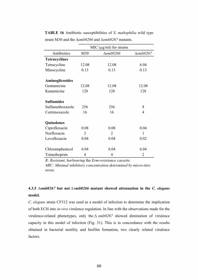
88
TABLE 16 Antibiotic susceptibilities of S. maltophilia wild type
strain M30 and the ∆smlt0266 and ∆smlt0267 mutants.
MIC (µg/ml) for strains Antibiotics M30 ∆smlt0266 ∆smlt0267
Tetracyclines Tetracycline 12.08 12.08 6.04 Minocycline 0.13 0.13 0.13
Aminoglicosides Gentamicine 12.08 12.08 12.08 Kanamicine 128 128 128
Sulfamides Sulfamethoxazole 256 256 8 Cotrimoxazole 16 16 4
Quinolones Ciprofloxacin 0.08 0.08 0.04 Norfloxacin 2 2 1 Levofloxacin 0.04 0.04 0.02
Chloramphenicol 6.04 6.04 6.04 Trimethoprim 4 4 2 R: Resistant, harbouring the Erm-resistance cassette. MIC: Minimal inhibitory concentration determined by micro-titre assay.
4.3.5 ∆smlt0267 but not ∆ smlt0266 mutant showed attenuation in the C. elegans
model.
C. elegans strain CF512 was used as a model of infection to determine the implication
of both ECH into in-vivo virulence regulation. In line with the observations made for the
virulence-related phenotypes, only the ∆ smlt0267 showed diminution of virulence
capacity in this model of infection (Fig. 31). This is in concordance with the results
obtained in bacterial motility and biofilm formation, two clearly related virulence
factors.
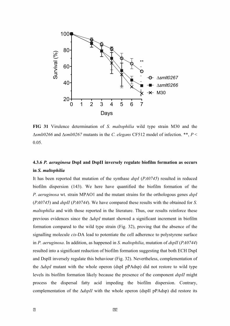
� � �
FIG 31 Virulence determination of S. maltophilia wild type strain M30 and the
∆smlt0266 and ∆smlt0267 mutants in the C. elegans CF512 model of infection. **, P <
0.05.
4.3.6 P. aeruginosa DspI and DspII inversely regulate biofilm formation as occurs
in S. maltophilia
It has been reported that mutation of the synthase dspI (PA0745) resulted in reduced
biofilm dispersion (143). We here have quantified the biofilm formation of the
P. aeruginosa wt. strain MPAO1 and the mutant strains for the orthologous genes dspI
(PA0745) and dspII (PA0744). We have compared these results with the obtained for S.
maltophilia and with those reported in the literature. Thus, our results reinforce these
previous evidences since the ∆dspI mutant showed a significant increment in biofilm
formation compared to the wild type strain (Fig. 32), proving that the absence of the
signalling molecule cis-DA lead to potentiate the cell adherence to polystyrene surface
in P. aeruginosa. In addition, as happened in S. maltophilia, mutation of dspII (PA0744)
resulted into a significant reduction of biofilm formation suggesting that both ECH DspI
and DspII inversely regulate this behaviour (Fig. 32). Nevertheless, complementation of
the ∆dspI mutant with the whole operon (dspI pPAdsp) did not restore to wild type
levels its biofilm formation likely because the presence of the component dspII might
process the dispersal fatty acid impeding the biofilm dispersion. Contrary,
complementation of the ∆dspII with the whole operon (dspII pPAdsp) did restore its
0 1 2 3 4 5 6 720
40
60
80
100
M30∆smlt0266∆smlt0267
Days
Surv
ival
(%)
**

90
biofilm formation to the wild type levels, perhaps due to the same phenomenon. This
suggests that the alternative ECH DspII could play an important role in the processing
of the cis-DA signal and consequently in the prevention of biofilm dispersion.
FIG 32 Biofilm formation of P. aeruginosa MPAO1 wt, the ∆dspI and ∆dspII mutants
and the complemented strains on polystyrene surface, grown in LB medium at 37ºC for
24 h. ***, P < 0,001.
4.3.7 P. aeruginosa DspI and DspII regulate virulence-related phenotypes similarly
to S. maltophilia Smlt0266 and Smlt0267
We have evaluated the same virulence-related phenotypes that were altered in the
∆smlt0266 and ∆smlt0267 mutants in S. maltophilia. These included, bacterial motility
(swimming, swarming and twitching), exopolysaccharide production and colony
morphology as well as the susceptibility to the same antibiotics that were tested for the
S. maltophilia mutants. Interestingly, the ∆ dspI and ∆dspII mutants showed an
alteration of its motility as well (Fig. 33, 34), indicating that cis-DA system is also
involved in the control of the population motility. Particularly, the ∆dspII was shown to
have an alteration of swarming (Fig. 33) and twitching motility (Fig. 34), whereas the
∆dspI did not show alteration in these motilities compared to the parental strain
MPAO1 (Fig. 33, 34). The alteration in the swarming motility observed for the ∆dspII
mutant consisted in a drastic change of its morphology and the absence of pigmentation.
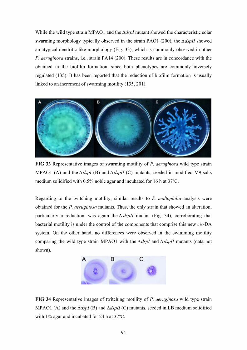
91
While the wild type strain MPAO1 and the ∆dspI mutant showed the characteristic solar
swarming morphology typically observed in the strain PAO1 (200), the ∆dspII showed
an atypical dendritic-like morphology (Fig. 33), which is commonly observed in other
P. aeruginosa strains, i.e., strain PA14 (200). These results are in concordance with the
obtained in the biofilm formation, since both phenotypes are commonly inversely
regulated (135). It has been reported that the reduction of biofilm formation is usually
linked to an increment of swarming motility (135, 201).
FIG 33 Representative images of swarming motility of P. aeruginosa wild type strain
MPAO1 (A) and the ∆dspI (B) and ∆dspII (C) mutants, seeded in modified M9-salts
medium solidified with 0.5% noble agar and incubated for 16 h at 37ºC.
Regarding to the twitching motility, similar results to S. maltophilia analysis were
obtained for the P. aeruginosa mutants. Thus, the only strain that showed an alteration,
particularly a reduction, was again the ∆ dspII mutant (Fig. 34), corroborating that
bacterial motility is under the control of the components that comprise this new cis-DA
system. On the other hand, no differences were observed in the swimming motility
comparing the wild type strain MPAO1 with the ∆dspI and ∆dspII mutants (data not
shown).
FIG 34 Representative images of twitching motility of P. aeruginosa wild type strain
MPAO1 (A) and the ∆dspI (B) and ∆dspII (C) mutants, seeded in LB medium solidified
with 1% agar and incubated for 24 h at 37ºC.
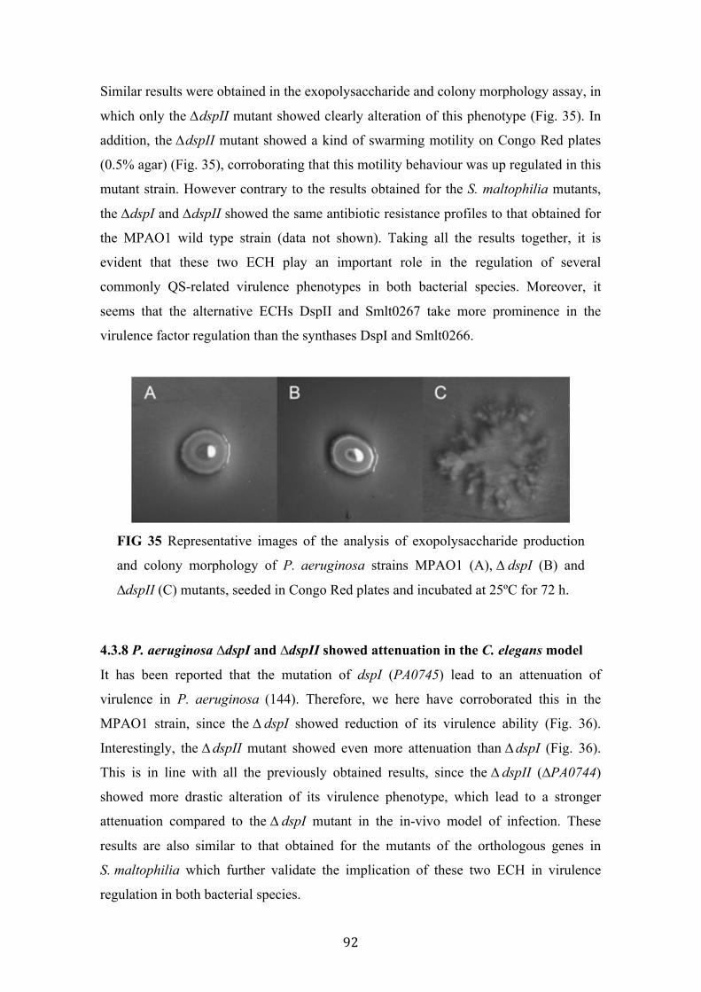
92
Similar results were obtained in the exopolysaccharide and colony morphology assay, in
which only the ∆dspII mutant showed clearly alteration of this phenotype (Fig. 35). In
addition, the ∆dspII mutant showed a kind of swarming motility on Congo Red plates
(0.5% agar) (Fig. 35), corroborating that this motility behaviour was up regulated in this
mutant strain. However contrary to the results obtained for the S. maltophilia mutants,
the ∆dspI and ∆dspII showed the same antibiotic resistance profiles to that obtained for
the MPAO1 wild type strain (data not shown). Taking all the results together, it is
evident that these two ECH play an important role in the regulation of several
commonly QS-related virulence phenotypes in both bacterial species. Moreover, it
seems that the alternative ECHs DspII and Smlt0267 take more prominence in the
virulence factor regulation than the synthases DspI and Smlt0266.
FIG 35 Representative images of the analysis of exopolysaccharide production
and colony morphology of P. aeruginosa strains MPAO1 (A), ∆ dspI (B) and
∆dspII (C) mutants, seeded in Congo Red plates and incubated at 25ºC for 72 h.
4.3.8 P. aeruginosa ∆dspI and ∆dspII showed attenuation in the C. elegans model
It has been reported that the mutation of dspI (PA0745) lead to an attenuation of
virulence in P. aeruginosa (144). Therefore, we here have corroborated this in the
MPAO1 strain, since the ∆ dspI showed reduction of its virulence ability (Fig. 36).
Interestingly, the ∆ dspII mutant showed even more attenuation than ∆ dspI (Fig. 36).
This is in line with all the previously obtained results, since the ∆ dspII (∆PA0744)
showed more drastic alteration of its virulence phenotype, which lead to a stronger
attenuation compared to the ∆ dspI mutant in the in-vivo model of infection. These
results are also similar to that obtained for the mutants of the orthologous genes in
S. maltophilia which further validate the implication of these two ECH in virulence
regulation in both bacterial species.
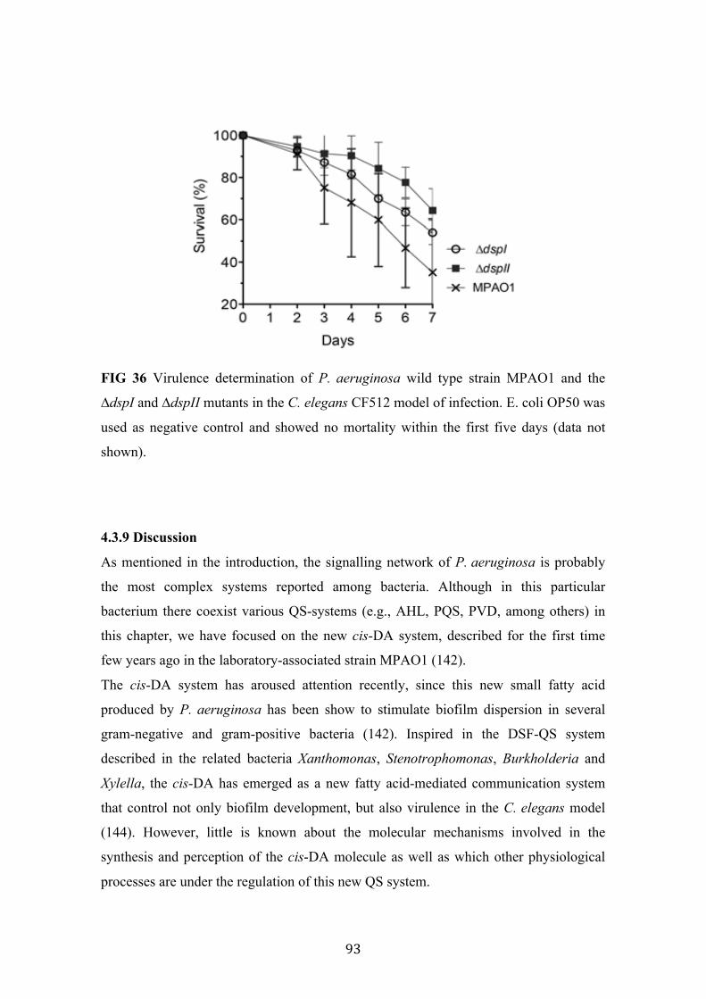
93
FIG 36 Virulence determination of P. aeruginosa wild type strain MPAO1 and the
∆dspI and ∆dspII mutants in the C. elegans CF512 model of infection. E. coli OP50 was
used as negative control and showed no mortality within the first five days (data not
shown).
4.3.9 Discussion
As mentioned in the introduction, the signalling network of P. aeruginosa is probably
the most complex systems reported among bacteria. Although in this particular
bacterium there coexist various QS-systems (e.g., AHL, PQS, PVD, among others) in
this chapter, we have focused on the new cis-DA system, described for the first time
few years ago in the laboratory-associated strain MPAO1 (142).
The cis-DA system has aroused attention recently, since this new small fatty acid
produced by P. aeruginosa has been show to stimulate biofilm dispersion in several
gram-negative and gram-positive bacteria (142). Inspired in the DSF-QS system
described in the related bacteria Xanthomonas, Stenotrophomonas, Burkholderia and
Xylella, the cis-DA has emerged as a new fatty acid-mediated communication system
that control not only biofilm development, but also virulence in the C. elegans model
(144). However, little is known about the molecular mechanisms involved in the
synthesis and perception of the cis-DA molecule as well as which other physiological
processes are under the regulation of this new QS system.
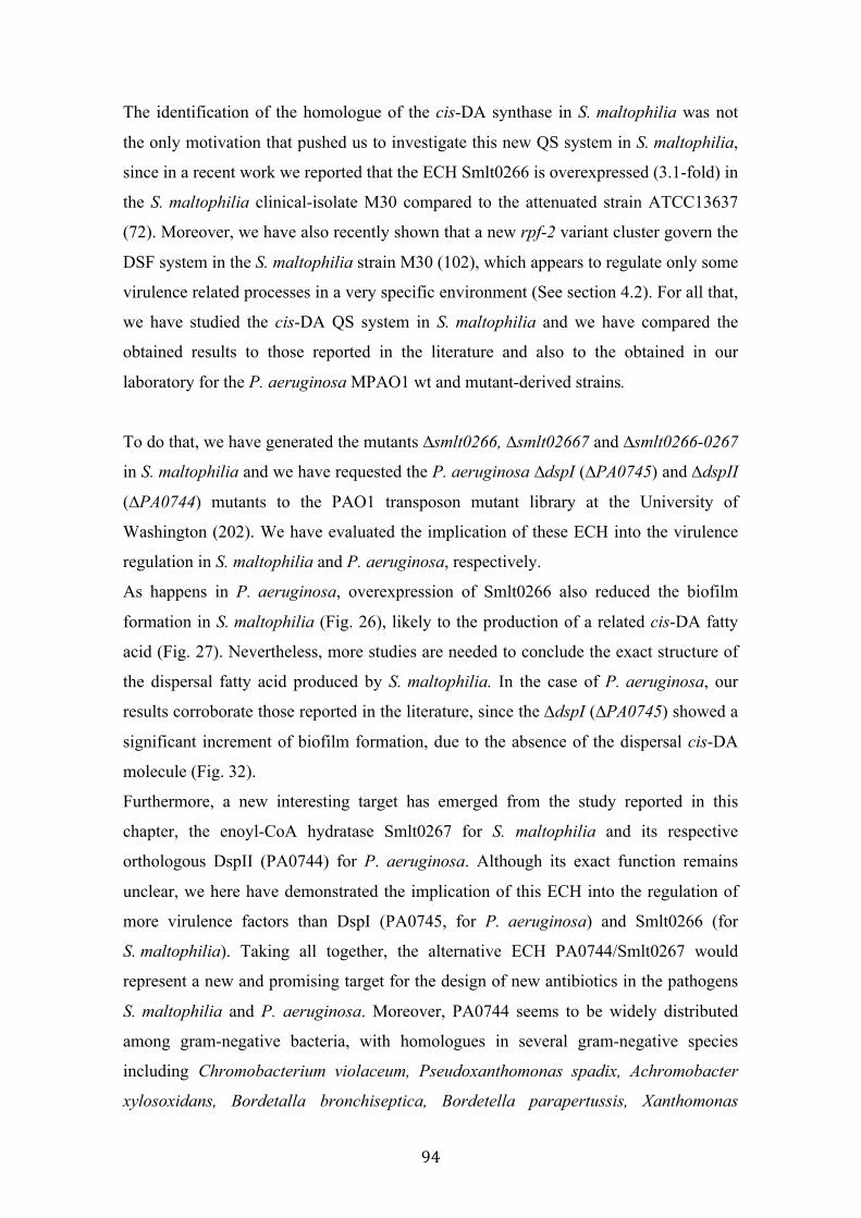
94
The identification of the homologue of the cis-DA synthase in S. maltophilia was not
the only motivation that pushed us to investigate this new QS system in S. maltophilia,
since in a recent work we reported that the ECH Smlt0266 is overexpressed (3.1-fold) in
the S. maltophilia clinical-isolate M30 compared to the attenuated strain ATCC13637
(72). Moreover, we have also recently shown that a new rpf-2 variant cluster govern the
DSF system in the S. maltophilia strain M30 (102), which appears to regulate only some
virulence related processes in a very specific environment (See section 4.2). For all that,
we have studied the cis-DA QS system in S. maltophilia and we have compared the
obtained results to those reported in the literature and also to the obtained in our
laboratory for the P. aeruginosa MPAO1 wt and mutant-derived strains.
To do that, we have generated the mutants ∆smlt0266, ∆smlt02667 and ∆smlt0266-0267
in S. maltophilia and we have requested the P. aeruginosa ∆dspI (∆PA0745) and ∆dspII
(∆PA0744) mutants to the PAO1 transposon mutant library at the University of
Washington (202). We have evaluated the implication of these ECH into the virulence
regulation in S. maltophilia and P. aeruginosa, respectively.
As happens in P. aeruginosa, overexpression of Smlt0266 also reduced the biofilm
formation in S. maltophilia (Fig. 26), likely to the production of a related cis-DA fatty
acid (Fig. 27). Nevertheless, more studies are needed to conclude the exact structure of
the dispersal fatty acid produced by S. maltophilia. In the case of P. aeruginosa, our
results corroborate those reported in the literature, since the ∆dspI (∆PA0745) showed a
significant increment of biofilm formation, due to the absence of the dispersal cis-DA
molecule (Fig. 32).
Furthermore, a new interesting target has emerged from the study reported in this
chapter, the enoyl-CoA hydratase Smlt0267 for S. maltophilia and its respective
orthologous DspII (PA0744) for P. aeruginosa. Although its exact function remains
unclear, we here have demonstrated the implication of this ECH into the regulation of
more virulence factors than DspI (PA0745, for P. aeruginosa) and Smlt0266 (for
S. maltophilia). Taking all together, the alternative ECH PA0744/Smlt0267 would
represent a new and promising target for the design of new antibiotics in the pathogens
S. maltophilia and P. aeruginosa. Moreover, PA0744 seems to be widely distributed
among gram-negative bacteria, with homologues in several gram-negative species
including Chromobacterium violaceum, Pseudoxanthomonas spadix, Achromobacter
xylosoxidans, Bordetalla bronchiseptica, Bordetella parapertussis, Xanthomonas
95
campestris, Xanthomonas axonopodis and Ralstonia eutropha, among much other, as
predicted by nucleotide BLAST tool (203), which makes this target even more
interesting.
It is well known that S. maltophilia and P. aeruginosa are capable of forming biofilm
easily, which complicates the treatment of colonized patients. Thus, the study of the
molecular aspects that potentiates the biofilm formation, and more interestingly the
biofilm dispersion, is mandatory to develop new treatments against infections caused by
these two important pathogens.
P. aeruginosa is perhaps one of the most studied gram-negative bacteria and many
virulence-traits that this bacterium exploits during infection process have been
elucidated. Discovery of new genes and processes involved in the virulence regulation
of this bacterium is therefore an arduous work. In a published high-throughput study for
the identification of all the P. aeruginosa genes involved in virulence in the C. elegans
model, the authors reported that the mutant ∆PA0745 was one of the most attenuated
strains (144). In a recent work, designed to identify the cis-DA synthase, all the
annotated putative ECH were evaluated for the synthesis of the cis-DA signalling
molecule with the subsequent biofilm dispersion, finally identifying PA0745 (DspI) as
the synthase (143). In any of these reports, the alternative ECH PA0744 was included in
the study. Both mentioned works were performed using the other well-studied
P. aeruginosa strain PA14. Although in both cases authors also requested the required
mutants in the transposon mutant library of University of Washington (202), in this
strain, the mutant ∆ PA0744 is not available. It is evident that, since this mutant is
available for the transposon library of MPAO1, this particular gene doesn’t have vital
function is P. aeruginosa.
Accordingly, we here report for the first time the role of the ECHs PA0744/Smlt0267 in
the virulence regulation of P. aeruginosa and S. maltophilia, and propose a new and
promising target to develop new strategies against bacterial infections caused by these
two important human pathogens.

96

GENERAL DISCUSSION AND FUTURE PERSPECTIVES

98
5. GENERAL DISCUSSION AND FUTURE
PERSPECTIVES In this work, we have investigated two quorum-sensing systems mediated by fatty acid
molecules in S. maltophilia and P. aeruginosa species.
A fatty acid (FA) is a carboxylic acid with an aliphatic tail (chain), which could be
either saturated or unsaturated -those that present carbon–carbon double bonds-. In
nature, FAs generally have an even number of carbon atoms, from 4 to 28. FAs are
usually conforming triglycerides or phospholipids, however, when they are not attached
to these molecules, they are termed "free" fatty acids. Usually, FAs are categorized
depending on the chain length, as follows. Short-chain fatty acids (SCFA) are fatty
acids with aliphatic tails of fewer than six carbons. Medium-chain fatty acids (MCFA)
are fatty acids with aliphatic tails of 6–12 carbons. Long-chain fatty acids (LCFA) are
fatty acids with aliphatic tails 13 to 21 carbons. Very long chain fatty acids (VLCFA)
are fatty acids with aliphatic tails longer than 22 carbons.
FAs play three typical roles within biological systems. These include energy storage,
phospholipid membrane formation, and signalling pathways.
Catabolism of FAs occurs by a pathway called β-oxidation and yields more adenosine
triphosphate (ATP) per gram than carbohydrates or proteins. The oxidation process of
fatty acids to obtain energy typically occurs in four steps:
1- Dehydrogenation by acyl-CoA dehydrogenase, yielding 1 FADH2.
2- Hydration by enoyl-CoA hydratase.
3- Dehydrogenation by 3-hydroxyacyl-CoA dehydrogenase, yielding 1 NADH.
4- Cleavage by thiolase, yielding 1 acetyl-CoA and a fatty acid that has now
been shortened by 2 carbons (acyl-CoA).
This cycle repeats until the fatty acid has been completely reduced to acetyl-CoA or, in
the case of fatty acids with odd numbers of carbon atoms, acetyl-CoA and 1 molecule of
propionyl-CoA per molecule of fatty acid.
Fatty acids have also an important structural function in the cell, since they are essential
in the formation of the membrane phospholipids. The organization of a gram-negative
bacterial cell wall consist of an outer membrane and an inner membrane, separated by a
region known as the periplasmic space, which has very important functions in the
99
survival and operation of the cell. Bacterial membranes are mainly composed of
different phospholipids, which consist of a hydrophilic head and a hydrophobic tail. The
major phospholipids in E. coli include the zwitterionic phosphatidylethanolamine (PE,
70% of the membrane), the anionic phosphatidylglycerol (PG, 20%), and cardiolipin
(CL, 10%) (204).
Polyunsaturated fatty acids (e.g., 18:3 ω3c) are found in plants, algae and cyanobacteria,
but are often not present in bacteria. Monounsaturated fatty acids (particularly at the
omega-7 position), odd-chain saturated fatty acids (e.g., 15:0), branched-chain fatty
acids (mainly iso/anetiso and 10-methyl) and cyclopropane fatty acids (e.g., 19:0 cyclo
ω7c) are mostly synthesized by bacteria.
The third function described for fatty acid is cellular signalling. Prostaglandins were the
first fatty acid-derived molecules to be discovered as signalling molecules in eukaryotic
organisms (205). In bacteria, cellular fatty acid-mediated communication has been
deeply studied in the last years. As mentioned along this work, quorum-sensing systems
mediated by fatty acid molecules have been shown to regulate several virulence-related
biological functions especially in Xhantomonadales species (206). This is the case of
the DSF quorum sensing, which is indeed the main research line that I have been
developing during my thesis.
During this PhD thesis period we have been able to elucidate that, in S. maltophilia
there exist two populations that differentially control the DSF-QS (Sections 4.1 and
4.2). These two S. maltophilia groups -here designated rpf-1 and rpf-2 variant groups-
differentiate each other basically in the molecular mechanisms responsible of synthesize
and sense DSF molecules. Taking one model strain from each group, we have also
observed that the DSF-QS could also differentially regulate virulent processes between
the two populations, as a consequence of the variance in the molecular regulation.
Since this is the first time that two different mechanisms govern the DSF-QS in same
specie, many questions arise. It is important to know the frequencies found for each
DSF-QS variant in the population of clinical S. maltophilia isolates. Taking two recent
S. malotphilia population studies (102, 128) from a total of 171 isolates (mainly clinical
strains), rpf-1 variant have been more commonly isolated with ca. 60% of
representation. In concordance with the results published for the model organism Xcc,
we here have observed similar traits in terms of DSF production and detection, as well
as a comparable biological regulation between S. maltophilia rpf-1 variant strains and

100
Xcc. It has been reported that in Xcc, the DSF-QS regulates several virulence-related
behaviours such as bacterial motility, biofilm development and virulence (91),
something that we have also observed in S. maltophilia rpf-1 variant strains (Section 4.1
and 4.2). Moreover, we have studied the conditions that modulate DSF production in
S. maltophilia.
Regarding the rpf-1 variant strains, we have observed that the temperature and the
media composition are important factors involved in the DSF synthesis. In addition, we
have observed that some medium and long-chain fatty acids are capable of stimulate
DSF production, suggesting that the sensor RpfC-1 (which contains ten TMR in its
input domain) is more promiscuous enabling free-active RpfF-1 to synthesize DSF upon
detection not only DSF molecules but also 12 to 14-carbon length fatty acids (Section
4.1). Additionally, we have analysed the total cellular fatty acid in S. maltophilia. As
shown in the Table 15 (Section 4.1), the most abundant fatty acid in S. maltophilia
(≈30%) is 13-methyl-tetradecanoic (15:0 iso) a highly related DSF fatty acid. Although
it needs to be verified, we postulate that 15:0 iso takes part in the conformation of the
cell membrane phospholipids, being one of the two nonpolar “legs”. Interestingly, it has
been reported that in several Xanthomonas species (including Xcc, Xoo and Xac, among
many others), the most abundant fatty acid is also 15:0 iso (207). Curiously, iso-
branched fatty acids have been found to be common in gram-positive but unusual in
gram-negative bacteria. Thereby, I think it is remarkable that 15:0 iso is present in these
well-established DSF producer bacteria such as Xanthomonas sp. and S. maltophilia,
whereas it is not present in non DSF-producer bacteria such as P. aeruginosa and E. coli
(Data not shown, unpublished results).
There are three aspects that make me think that DSF communication appears as a
consequence of the membrane homeostasis regulation. The first one is that 15:0 iso was
the most stimulating fatty acid tested in DSF production in S. maltophilia.
The second one is that the upstream gene of rpfF the long-chain fatty acyl CoA ligase
rpfB, is indeed annotated as fadD. Hence, RpfB displays 60% identity with the E. coli
FadD. Interestingly, a recent report shows that, in Sinorhizobium meliloti, FadD plays a
major role in the activation of endogenous fatty acids released from membrane lipids
during stationary phase of growth (208). In this line, we hypothesize that RpfB could be
responsible of conferring the precursor15:0 iso to RpfF to synthesize DSF.
101
The third one relies in that the sensor RpfC -which is expected to sense diffusible
molecules- is anchored in the membrane. It doesn’t make sense that a molecule able to
diffuse the cell membrane needs a membrane receptor to be detected.
Thus, I think that the sensor RpfC senses possible alterations on the cell membrane
stability and liberate RpfF that -jointly with RpfB- synthesize DSF from the membrane
precursor 15:0 iso fatty acid. However, it is evident that extensive work is needed to
validate such hypothesis.
On the other hand, the DSF-QS regulation is uncertain and seems to be particular in the
S. maltophilia rpf-2 population. DSF production was never detected in the rpf-2 axenic
cultures. However, we have showed that rpf-2 variant strains are able to synthesize DSF
only upon detection of DSF-itself, which indicates that DSF is produced in a positive
feedback-manner in S. maltophilia rpf-2 strains. This also suggests that the sensor
RpfC-2 (containing only five TMR) is much more specific only allowing the
dissociation of RpfF-2 when sensing DSF. It has been reported that in the related
bacterium Xylella fastidiosa (which harbours a similar rpf-2 cluster), the synthase RpfF
is also participating in DSF perception, something that might also be occurring in these
S. maltophilia variant strains.
Regarding the biological regulation, from all the phenotypes evaluated for the ∆rpfF-2
mutant, only virulence attenuation in the zebrafish model was observed. Furthermore,
we have also showed that there exist a synergism in DSF production and virulence
ability between both S. maltophilia groups. Considering that rpf-2 variant strains
usually cohabit with DSF-producer bacteria (e.g., Xanthomonadales) it is easy to think
that rpf-2 triggers the DSF regulation upon detection of DSF molecules produced by
neighbour bacteria.
Consequently, these results have revoked our initial hypothesis, which was that the
DSF-QS was permanently switched off in this variant group. Although we have not
been able to elucidate how, it is evident that DSF-QS regulate biological functions in
this important S. maltophilia group. A hypothetic model of DSF regulation for each
S. maltophilia variant group is proposed in the figure v.
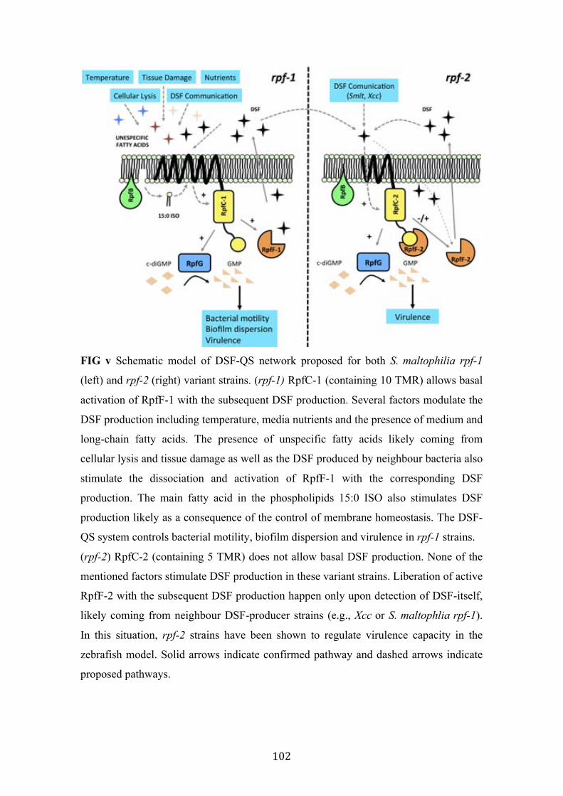
102
FIG v Schematic model of DSF-QS network proposed for both S. maltophilia rpf-1
(left) and rpf-2 (right) variant strains. (rpf-1) RpfC-1 (containing 10 TMR) allows basal
activation of RpfF-1 with the subsequent DSF production. Several factors modulate the
DSF production including temperature, media nutrients and the presence of medium and
long-chain fatty acids. The presence of unspecific fatty acids likely coming from
cellular lysis and tissue damage as well as the DSF produced by neighbour bacteria also
stimulate the dissociation and activation of RpfF-1 with the corresponding DSF
production. The main fatty acid in the phospholipids 15:0 ISO also stimulates DSF
production likely as a consequence of the control of membrane homeostasis. The DSF-
QS system controls bacterial motility, biofilm dispersion and virulence in rpf-1 strains.
(rpf-2) RpfC-2 (containing 5 TMR) does not allow basal DSF production. None of the
mentioned factors stimulate DSF production in these variant strains. Liberation of active
RpfF-2 with the subsequent DSF production happen only upon detection of DSF-itself,
likely coming from neighbour DSF-producer strains (e.g., Xcc or S. maltophlia rpf-1).
In this situation, rpf-2 strains have been shown to regulate virulence capacity in the
zebrafish model. Solid arrows indicate confirmed pathway and dashed arrows indicate
proposed pathways.

103
What does each rpf variant confers to S. maltophilia pathogenic strains during human
infections is a crucial and complicated question that must be addressed in the future.
As mentioned, to the date, S. maltophilia rpf-1 strains are more commonly isolated, but
it will be interesting to follow the evolution of S. maltophilia infections in a long term
in order to establish a presumed correlation between rpf variants and infection success.
Regarding to the rpf/DSF-QS evolution, “the chicken or the egg” dilemma is also here
present. It would be interesting to define which rpf variant appeared first in S.
maltophilia. This would additionally help us to get insights into the evolution processes
that have occurred in Xanthomnadales. In my opinion, I opted to think that rpf-1 variant
strains are more evolved, since it seems that their promiscuous DSF production and
perception allow them to activate the entire virulence-related machinery under a high
variety of environments (Fig. v). Nevertheless it is evident that these hypotheses will
require further study.
In the third block of this thesis, I have been working with a new fatty acid-mediated QS
system in S. maltophilia that has been recently identified in the important pathogen
P. aeruginosa (Section 4.3). In this case, the cellular communication is mediated by a
smaller 10-carbon FA cis-decenoic (cis-DA). It has been reported that this new FA
stimulates biofilm dispersion not only in P. aeruginosa, but also in many gram-negative
and gram-positive bacteria (142). Although much work is needed to complete the study,
I have been able to preliminary characterize two important components in the cluster
that control cis-DA synthesis in S. maltophilia. In P. aeruginosa, it has been reported
that the enoyl-CoA hydratase (ECH) DspI (PA0745) is responsible for the synthesis of
this new signalling FA (143). Sequence genome analysis has revealed that
S. maltophilia harbours the homologous synthase Smlt0266. Mutation of smlt0266 lead
to a similar alteration that is observed in P. aeruginosa ∆dspI. Additionally, a new
interesting target for both S. maltophilia and P. aeruginosa has emerged from this work,
the alternative ECH DspII (PA0744) in P. aeruginosa and the corresponding Smlt0267
in S. maltophilia. While mutation of the dspI (in P. aeruginosa) and smlt0266 (in
S. maltophilia) lead to alteration mainly in the biofilm development, mutation of the
alternative ECH dspII (in P. aeruginosa) and smlt0267 (in S. maltophilia) lead to a
drastic effect in the virulent phenotype of both pathogens. These common alterations
include exopolysaccharide production, biofilm development, bacterial motility and

104
virulence in the C. elegans model (Section 4.3). These results are as interesting as
complex to understand, by the moment. Initially, I was reluctant to think that the
synthase of a new QS system was organized in a clear β -oxidation operon. The gene
that encodes for the synthase dspI/smlt0266 is upstream flanked by components
involved in the first steps of classic fatty acid oxidation pathway such as a putative
aldehyde-dehydrogenase and a hypothetical acyl-CoA dehydrogenase, and downstream
flanked by a putative enoyl-CoA hydratase/isomerase (Fig. 25). However, the evidence
that the mutation of dspI/smlt0266 but specially dspII/smlt0267 lead to an alteration of
several typically QS-regulated processes, led me reconsider my initial doubts. Although
I still have some concerns, it is evident that the two ECH are involved in the regulation
of virulence processes in both P. aeruginosa and S. maltophilia, which make them a
promising targets to design new antibacterial strategies.
The fatty acid signalling function in bacteria communication has been studied for no
more than two decades. Nevertheless, it has aroused rapidly the attention of the
scientific community since it has been demonstrated that this kind of signalling systems
regulate several virulent processes in many important pathogens. Proof of this is DSF-
QS described in several human and plant pathogens such as Xanthomonas sp,
X. fasidiosa, B. cepacea and S. maltophilia. In addition, fatty acid-mediated signalling
molecules have been also described in the gram-positive Streptococcus mutans, in the
yeast Candida albicans and more recently in P. aeruginosa. Taking all together, it is
evident that FA-mediated communication systems are a phenomenon widely distributed
among microorganisms and will yield new important discoveries in a recent future.
Along this work we have tried to participate in increase the knowledge of the FA-
mediated QS system in the important nosocomial pathogen S. maltophilia by studying
in detail the DSF-QS and providing the first strokes of the new cis-DA QS system.
However, more efforts will be required to completely elucidate the complexity of these
cellular communication systems, especially in the well-established human pathogens.

CONCLUSIONS

106
6. CONCLUSIONS
1. Two variants of the cluster rpf (here named rpf-1 and rpf-2) with significant
differences in the genes encoding the DSF synthase RpfF and the sensor
component RpfC are distributed among the population of Stenotrophomonas
maltophilia.
2. Only strains harbouring the cluster variant rpf-1 display detectable levels of DSF
under wild type conditions, and they represent 60% of total isolates.
3. In rpf-1 variant strains, RpfC-1 contains ten transmembrane regions (TMR)
highly related to the sensor input domain present in Xanthomonas campestris pv.
campestris composed by RpfH and RpfC. In rpf-2 variant strains, the sensor
input domain of RpfC-2 only contains five TMR.
4. The absence on DSF production observed in rpf-2 variant strains is likely due to
the permanent repression that RpfC-2 exerts on RpfF-2, although both RpfF
variants are functional and are able to synthesize the canonical DSF molecule.
5. RpfC-1 variant -containing ten TMR- liberates free active-RpfF-1 with the
subsequent DSF synthesis upon detection not only DSF, but also saturated
medium length fatty acids. These include lauric (12:0), myristic (14:0) and 13-
methylmyristic (15:0 iso), which is indeed the most abundant fatty acid in S.
maltophilia.
6. RpfC-2 only allows activation of RpfF-2 upon detection of DSF-itself,
indicating that this sensor component is much more specific and, at the same
time, that DSF is produced in a positive feedback manner in S. maltophilia.
7. DSF quorum sensing system seems to regulate more biological functions
including biofilm formation, swarming motility, and virulence in C. elegans and
zebrafish models in S. maltophilia strains harbouring the cluster variant rpf-1,
than in those harbouring the rpf-2 variant, which seems to regulates only
virulence in an infection model.
8. DSF inversely regulate biofilm formation and swarming motility in the rpf-1
variant strain E77.
9. Both S. maltophilia rpf-variant groups act synergistically in DSF production
both in-vitro and in-vivo and in their virulence capacity in the zebrafish infection
model.

107
10. S. maltophilia also uses the cis-DA quorum sensing system to regulate numerous
virulence-related processes. While mutation of the gene encoding the putative
cis-DA synthase Smlt0266 results in an increment of biofilm formation,
mutation of the gene encoding the alternative enoyl-coA hydratase Smlt0267
lead to reduction of several virulence factors including biofilm formation,
twitching and swimming motility, antibiotic resistance and virulence.
11. The smlt0267 orthologous gene PA0744 also regulates similar functions in
P. aeruginosa, demonstrating that Smlt0267/PA0744 represent a novel potential
target for the development of new antimicrobial therapies.

108

REFERENCES

110
7. REFERENCES 1. Hugh R, Leifson E. 1963. A description of the type strain of Pseudomonas maltophilia. Int. Bull. Bacteriol. Nomencl. Taxon. 13:133–138. 2. Palleroni NJ, Bradbury JF. 1993. Stenotrophomonas, a new bacterial genus for Xanthomonas maltophilia (Hugh 1980) Swings et al. 1983. Int. J. Syst. Bacteriol. 43:606–609. 3. Palleroni NJ, Kunisawa R, Contopoulou R, Doudoroff M. 1973. Nucleic Acid Homologies in the Genus Pseudomonas. Int. J. Syst. Bacteriol. 23:333–339. 4. Swings J, Vos PD, Mooter MV den, Ley JD. 1983. Transfer of Pseudomonas maltophilia Hugh 1981 to the Genus Xanthomonas as Xanthomonas maltophilia (Hugh 1981) comb. nov. Int. J. Syst. Bacteriol. 33:409–413. 5. Denton M, Kerr KG. 1998. Microbiological and Clinical Aspects of Infection Associated with Stenotrophomonas maltophilia. Clin. Microbiol. Rev. 11:57–80. 6. Maes M. 1993. Fast classification of plant-associated bacteria in the Xanthomonas genus. FEMS Microbiol. Lett. 113:161–165. 7. Nesme X, Vaneechoutte M, Orso S, Hoste B, Swings J. 1995. Diversity and Genetic Relatedness within Genera Xanthomonas and Stenotrophomonas Using Restriction Endonuclease Site Differences of PCR-amplified 16S rRNA Gene. Syst. Appl. Microbiol. 18:127–135. 8. Crossman LC, Gould VC, Dow JM, Vernikos GS, Okazaki A, Sebaihia M, Saunders D, Arrowsmith C, Carver T, Peters N, Adlem E, Kerhornou A, Lord A, Murphy L, Seeger K, Squares R, Rutter S, Quail MA, Rajandream M-A, Harris D, Churcher C, Bentley SD, Parkhill J, Thomson NR, Avison MB. 2008. The complete genome, comparative and functional analysis of Stenotrophomonas maltophilia reveals an organism heavily shielded by drug resistance determinants. Genome Biol. 9:R74. 9. Schoch PE, Cunha BA. 1987. Pseudomonas maltophilia. Infect. Control IC 8:169–172. 10. Huedo P, Conchillo-Solé O, Yero D, Martínez-Servat S, Daura X, Gibert I. 2014. Draft Genome Sequence of Stenotrophomonas maltophilia Strain M30, Isolated from a Chronic Pressure Ulcer in an Elderly Patient. Genome Announc. 2. 11. Berg G, Knaape C, Ballin G, Seidel D. 1994. Biological control of Verticillium dahliae kleb. by natural occurring rhizosphere bacteria. Arch. Phytopathol. Plant Prot. 29:249–262. 12. J D, R B. 1980. [Presence of Pseudomonas maltophilia in the rhizosphere of several cultivated plants]. Can. J. Microbiol. 26:460–463. 13. Heuer H, Smalla K. 1999. Bacterial phyllosphere communities of Solanum tuberosum L. and T4-lysozyme-producing transgenic variants. FEMS Microbiol. Ecol. 28:357–371. 14. Lambert B, Joos H. 1989. Fundamental aspects of rhizobacterial plant growth promotion research. Trends Biotechnol. 7:215–219. 15. Berg G, Eberl L, Hartmann A. 2005. The rhizosphere as a reservoir for opportunistic human pathogenic bacteria. Environ. Microbiol. 7:1673–1685. 16. Ryan RP, Monchy S, Cardinale M, Taghavi S, Crossman L, Avison MB, Berg G, van der Lelie D, Dow JM. 2009. The versatility and adaptation of bacteria from the genus Stenotrophomonas. Nat. Rev. Microbiol. 7:514–525. 17. Alavi P, Müller H, Cardinale M, Zachow C, Sánchez MB, Martínez JL, Berg G. 2013. The DSF quorum sensing system controls the positive influence of Stenotrophomonas maltophilia on plants. PloS One 8:e67103. 18. Brooke JS. 2012. Stenotrophomonas maltophilia: an emerging global

111
opportunistic pathogen. Clin. Microbiol. Rev. 25:2–41. 19. Lai C-H, Wong W-W, Chin C, Huang C-K, Lin H-H, Chen W-F, Yu K-W, Liu C-Y. 2006. Central venous catheter-related Stenotrophomonas maltophilia bacteraemia and associated relapsing bacteraemia in haematology and oncology patients. Clin. Microbiol. Infect. Off. Publ. Eur. Soc. Clin. Microbiol. Infect. Dis. 12:986–991. 20. O’Donnell MJ, Tuttlebee CM, Falkiner FR, Coleman DC. 2005. Bacterial contamination of dental chair units in a modern dental hospital caused by leakage from suction system hoses containing extensive biofilm. J. Hosp. Infect. 59:348–360. 21. Hutchinson GR, Parker S, Pryor JA, Duncan-Skingle F, Hoffman PN, Hodson ME, Kaufmann ME, Pitt TL. 1996. Home-use nebulizers: a potential primary source of Burkholderia cepacia and other colistin-resistant, gram-negative bacteria in patients with cystic fibrosis. J. Clin. Microbiol. 34:584–587. 22. Schable B, Villarino ME, Favero MS, Miller JM. 1991. Application of multilocus enzyme electrophoresis to epidemiologic investigations of Xanthomonas maltophilia. Infect. Control Hosp. Epidemiol. Off. J. Soc. Hosp. Epidemiol. Am. 12:163–167. 23. Wainwright CE, France MW, O’Rourke P, Anuj S, Kidd TJ, Nissen MD, Sloots TP, Coulter C, Ristovski Z, Hargreaves M, Rose BR, Harbour C, Bell SC, Fennelly KP. 2009. Cough-generated aerosols of Pseudomonas aeruginosa and other Gram-negative bacteria from patients with cystic fibrosis. Thorax 64:926–931. 24. Safdar A, Rolston KV. 2007. Stenotrophomonas maltophilia: changing spectrum of a serious bacterial pathogen in patients with cancer. Clin. Infect. Dis. Off. Publ. Infect. Dis. Soc. Am. 45:1602–1609. 25. Vento S, Cainelli F, Temesgen Z. 2008. Lung infections after cancer chemotherapy. Lancet Oncol. 9:982–992. 26. Falagas ME, Kastoris AC, Vouloumanou EK, Rafailidis PI, Kapaskelis AM, Dimopoulos G. 2009. Attributable mortality of Stenotrophomonas maltophilia infections: a systematic review of the literature. Future Microbiol. 4:1103–1109. 27. Rommens JM, Iannuzzi MC, Kerem B, Drumm ML, Melmer G, Dean M, Rozmahel R, Cole JL, Kennedy D, Hidaka N. 1989. Identification of the cystic fibrosis gene: chromosome walking and jumping. Science 245:1059–1065. 28. Heijerman H. 2005. Infection and inflammation in cystic fibrosis: a short review. J. Cyst. Fibros. Off. J. Eur. Cyst. Fibros. Soc. 4 Suppl 2:3–5. 29. Nadesalingam K, Conway SP, Denton M. 2005. Risk factors for acquisition of methicillin-resistant Staphylococcus aureus (MRSA) by patients with cystic fibrosis. J. Cyst. Fibros. Off. J. Eur. Cyst. Fibros. Soc. 4:49–52. 30. Bjarnsholt T, Jensen PØ, Fiandaca MJ, Pedersen J, Hansen CR, Andersen CB, Pressler T, Givskov M, Høiby N. 2009. Pseudomonas aeruginosa biofilms in the respiratory tract of cystic fibrosis patients. Pediatr. Pulmonol. 44:547–558. 31. Costerton JW, Stewart PS, Greenberg EP. 1999. Bacterial Biofilms: A Common Cause of Persistent Infections. Science 284:1318–1322. 32. Nicodemo AC, Paez JIG. 2007. Antimicrobial therapy for Stenotrophomonas maltophilia infections. Eur. J. Clin. Microbiol. Infect. Dis. Off. Publ. Eur. Soc. Clin. Microbiol. 26:229–237. 33. Demko CA, Stern RC, Doershuk CF. 1998. Stenotrophomonas maltophilia in cystic fibrosis: incidence and prevalence. Pediatr. Pulmonol. 25:304–308. 34. Goss CH, Otto K, Aitken ML, Rubenfeld GD. 2002. Detecting Stenotrophomonas maltophilia does not reduce survival of patients with cystic fibrosis. Am. J. Respir. Crit. Care Med. 166:356–361.

112
35. Figueirêdo PMS, Furumura MT, Santos AM, Sousa ACT, Kota DJ, Levy CE, Yano T. 2006. Cytotoxic activity of clinical Stenotrophomonas maltophilia. Lett. Appl. Microbiol. 43:443–449. 36. Windhorst S, Frank E, Georgieva DN, Genov N, Buck F, Borowski P, Weber W. 2002. The major extracellular protease of the nosocomial pathogen Stenotrophomonas maltophilia: characterization of the protein and molecular cloning of the gene. J. Biol. Chem. 277:11042–11049. 37. Di Bonaventura G, Prosseda G, Del Chierico F, Cannavacciuolo S, Cipriani P, Petrucca A, Superti F, Ammendolia MG, Concato C, Fiscarelli E, Casalino M, Piccolomini R, Nicoletti M, Colonna B. 2007. Molecular characterization of virulence determinants of Stenotrophomonas maltophilia strains isolated from patients affected by cystic fibrosis. Int. J. Immunopathol. Pharmacol. 20:529–537. 38. Neal DJ, Wilkinson SG. 1982. Lipopolysaccharides from Pseudomonas maltophilia. Structural studies of the side-chain, core, and lipid-A regions of the lipopolysaccharide from strain NCTC 10257. Eur. J. Biochem. FEBS 128:143–149. 39. Brooke JS, Vo A, Watts P, Davis NA. 2008. Mutation of a lipopolysaccharide synthesis gene results in increased biofilm of Stenotrophomonas maltophilia on plastic and glass surfaces. Ann. Microbiol. 58:35–40. 40. Huang T-P, Somers EB, Wong ACL. 2006. Differential Biofilm Formation and Motility Associated with Lipopolysaccharide/Exopolysaccharide-Coupled Biosynthetic Genes in Stenotrophomonas maltophilia. J. Bacteriol. 188:3116–3120. 41. McKay GA, Woods DE, MacDonald KL, Poole K. 2003. Role of phosphoglucomutase of Stenotrophomonas maltophilia in lipopolysaccharide biosynthesis, virulence, and antibiotic resistance. Infect. Immun. 71:3068–3075. 42. Cullmann W. 1991. Antibiotic susceptibility and outer membrane proteins of clinical Xanthomonas maltophilia isolates. Chemotherapy 37:246–250. 43. Zhang L, Li X-Z, Poole K. 2000. Multiple Antibiotic Resistance in Stenotrophomonas maltophilia: Involvement of a Multidrug Efflux System. Antimicrob. Agents Chemother. 44:287–293. 44. Sanchez MB, Hernandez A, Martinez JL. 2009. Stenotrophomonas maltophilia drug resistance. Future Microbiol. 4:655–660. 45. Akova M, Bonfiglio G, Livermore DM. 1991. Susceptibility to β-lactam antibiotics of mutant strains of Xanthomonas maltophilia with high- and low-level constitutive expression of L1 and L2 β-lactamases. J. Med. Microbiol. 35:208–213. 46. Alonso A, Martínez JL. 2000. Cloning and Characterization of SmeDEF, a Novel Multidrug Efflux Pump from Stenotrophomonas maltophilia. Antimicrob. Agents Chemother. 44:3079–3086. 47. Chang L-L, Chen H-F, Chang C-Y, Lee T-M, Wu W-J. 2004. Contribution of integrons, and SmeABC and SmeDEF efflux pumps to multidrug resistance in clinical isolates of Stenotrophomonas maltophilia. J. Antimicrob. Chemother. 53:518–521. 48. Avison MB, Higgins CS, Heldreich CJ von, Bennett PM, Walsh TR. 2001. Plasmid Location and Molecular Heterogeneity of the L1 and L2 β-Lactamase Genes of Stenotrophomonas maltophilia. Antimicrob. Agents Chemother. 45:413–419. 49. Naiemi N al, Duim B, Bart A. 2006. A CTX-M extended-spectrum β-lactamase in Pseudomonas aeruginosa and Stenotrophomonas maltophilia. J. Med. Microbiol. 55:1607–1608. 50. Avison MB, Heldreich CJ von, Higgins CS, Bennett PM, Walsh TR. 2000. A TEM-2 β-lactamase encoded on an active Tn1-like transposon in the genome of a clinical isolate of Stenotrophomonas maltophilia. J. Antimicrob. Chemother. 46:879–
113
884. 51. Martínez JL. 2008. Antibiotics and Antibiotic Resistance Genes in Natural Environments. Science 321:365–367. 52. Gales AC, Jones RN, Forward KR, Liñares J, Sader HS, Verhoef J. 2001. Emerging importance of multidrug-resistant Acinetobacter species and Stenotrophomonas maltophilia as pathogens in seriously ill patients: geographic patterns, epidemiological features, and trends in the SENTRY Antimicrobial Surveillance Program (1997-1999). Clin. Infect. Dis. Off. Publ. Infect. Dis. Soc. Am. 32 Suppl 2:S104–113. 53. Toleman MA, Bennett PM, Bennett DMC, Jones RN, Walsh TR. 2007. Global emergence of trimethoprim/sulfamethoxazole resistance in Stenotrophomonas maltophilia mediated by acquisition of sul genes. Emerg. Infect. Dis. 13:559–565. 54. Hu L-F, Chang X, Ye Y, Wang Z-X, Shao Y-B, Shi W, Li X, Li J-B. 2011. Stenotrophomonas maltophilia resistance to trimethoprim/sulfamethoxazole mediated by acquisition of sul and dfrA genes in a plasmid-mediated class 1 integron. Int. J. Antimicrob. Agents 37:230–234. 55. Falagas ME, Rafailidis PI, Ioannidou E, Alexiou VG, Matthaiou DK, Karageorgopoulos DE, Kapaskelis A, Nikita D, Michalopoulos A. 2010. Colistin therapy for microbiologically documented multidrug-resistant Gram-negative bacterial infections: a retrospective cohort study of 258 patients. Int. J. Antimicrob. Agents 35:194–199. 56. Di Bonaventura G, Stepanović S, Picciani C, Pompilio A, Piccolomini R. 2007. Effect of environmental factors on biofilm formation by clinical Stenotrophomonas maltophilia isolates. Folia Microbiol. (Praha) 52:86–90. 57. Zgair AK, Chhibber S. 2011. Adhesion of Stenotrophomonas maltophilia to mouse tracheal mucus is mediated through flagella. J. Med. Microbiol. 60:1032–1037. 58. De Oliveira-Garcia D, Dall’Agnol M, Rosales M, Azzuz ACGS, Alcántara N, Martinez MB, Girón JA. 2003. Fimbriae and adherence of Stenotrophomonas maltophilia to epithelial cells and to abiotic surfaces. Cell. Microbiol. 5:625–636. 59. Liaqat I, Sabri AN. 2008. Effect of biocides on biofilm bacteria from dental unit water lines. Curr. Microbiol. 56:619–624. 60. Kovaleva J, Degener JE, van der Mei HC. 2010. Mimicking disinfection and drying of biofilms in contaminated endoscopes. J. Hosp. Infect. 76:345–350. 61. Bonaventura GD, Spedicato I, D’Antonio D, Robuffo I, Piccolomini R. 2004. Biofilm Formation by Stenotrophomonas maltophilia: Modulation by Quinolones, Trimethoprim-Sulfamethoxazole, and Ceftazidime. Antimicrob. Agents Chemother. 48:151–160. 62. Pompilio A, Catavitello C, Picciani C, Confalone P, Piccolomini R, Savini V, Fiscarelli E, D’Antonio D, Bonaventura GD. 2010. Subinhibitory concentrations of moxifloxacin decrease adhesion and biofilm formation of Stenotrophomonas maltophilia from cystic fibrosis. J. Med. Microbiol. 59:76–81. 63. Liaw S-J, Lee Y-L, Hsueh P-R. 2010. Multidrug resistance in clinical isolates of Stenotrophomonas maltophilia: roles of integrons, efflux pumps, phosphoglucomutase (SpgM), and melanin and biofilm formation. Int. J. Antimicrob. Agents 35:126–130. 64. Pompilio A, Crocetta V, Confalone P, Nicoletti M, Petrucca A, Guarnieri S, Fiscarelli E, Savini V, Piccolomini R, Di Bonaventura G. 2010. Adhesion to and biofilm formation on IB3-1 bronchial cells by Stenotrophomonas maltophilia isolates from cystic fibrosis patients. BMC Microbiol. 10:102.
114
65. Fouhy Y, Scanlon K, Schouest K, Spillane C, Crossman L, Avison MB, Ryan RP, Dow JM. 2007. Diffusible Signal Factor-Dependent Cell-Cell Signaling and Virulence in the Nosocomial Pathogen Stenotrophomonas maltophilia. J. Bacteriol. 189:4964–4968. 66. Kearns DB. 2010. A field guide to bacterial swarming motility. Nat. Rev. Microbiol. 8:634–644. 67. Kaiser D. 2007. Bacterial swarming: a re-examination of cell-movement patterns. Curr. Biol. CB 17:R561–570. 68. Terashima H, Kojima S, Homma M. 2008. Flagellar motility in bacteria structure and function of flagellar motor. Int. Rev. Cell Mol. Biol. 270:39–85. 69. Patrick JE, Kearns DB. 2012. Swarming motility and the control of master regulators of flagellar biosynthesis. Mol. Microbiol. 83:14–23. 70. Partridge JD, Harshey RM. 2013. Swarming: flexible roaming plans. J. Bacteriol. 195:909–918. 71. Jarrell KF, McBride MJ. 2008. The surprisingly diverse ways that prokaryotes move. Nat. Rev. Microbiol. 6:466–476. 72. Ferrer-Navarro M, Planell R, Yero D, Mongiardini E, Torrent G, Huedo P, Martínez P, Roher N, Mackenzie S, Gibert I, Daura X. 2013. Abundance of the Quorum-Sensing Factor Ax21 in Four Strains of Stenotrophomonas maltophilia Correlates with Mortality Rate in a New Zebrafish Model of Infection. PLoS ONE 8:e67207. 73. Federle MJ, Bassler BL. 2003. Interspecies communication in bacteria. J. Clin. Invest. 112:1291–1299. 74. Fuqua C, Greenberg EP. 2002. Listening in on bacteria: acyl-homoserine lactone signalling. Nat. Rev. Mol. Cell Biol. 3:685–695. 75. Fuqua WC, Winans SC, Greenberg EP. 1994. Quorum sensing in bacteria: the LuxR-LuxI family of cell density-responsive transcriptional regulators. J. Bacteriol. 176:269–275. 76. Whitehead NA, Barnard AM, Slater H, Simpson NJ, Salmond GP. 2001. Quorum-sensing in Gram-negative bacteria. FEMS Microbiol. Rev. 25:365–404. 77. Fuqua C, Winans SC, Greenberg EP. 1996. Census and consensus in bacterial ecosystems: the LuxR-LuxI family of quorum-sensing transcriptional regulators. Annu. Rev. Microbiol. 50:727–751. 78. Ohtani K, Hayashi H, Shimizu T. 2002. The luxS gene is involved in cell-cell signalling for toxin production in Clostridium perfringens. Mol. Microbiol. 44:171–179. 79. Marketon MM, Glenn SA, Eberhard A, González JE. 2003. Quorum sensing controls exopolysaccharide production in Sinorhizobium meliloti. J. Bacteriol. 185:325–331. 80. Quiñones B, Dulla G, Lindow SE. 2005. Quorum sensing regulates exopolysaccharide production, motility, and virulence in Pseudomonas syringae. Mol. Plant-Microbe Interact. MPMI 18:682–693. 81. Rice SA, Koh KS, Queck SY, Labbate M, Lam KW, Kjelleberg S. 2005. Biofilm formation and sloughing in Serratia marcescens are controlled by quorum sensing and nutrient cues. J. Bacteriol. 187:3477–3485. 82. Nealson KH, Platt T, Hastings JW. 1970. Cellular Control of the Synthesis and Activity of the Bacterial Luminescent System1. J. Bacteriol. 104:313–322. 83. Atkinson S, Williams P. 2009. Quorum sensing and social networking in the microbial world. J. R. Soc. Interface 6:959–978.

115
84. Geske GD, O’Neill JC, Miller DM, Mattmann ME, Blackwell HE. 2007. Modulation of bacterial quorum sensing with synthetic ligands: systematic evaluation of N-acylated homoserine lactones in multiple species and new insights into their mechanisms of action. J. Am. Chem. Soc. 129:13613–13625. 85. Raina S, De Vizio D, Odell M, Clements M, Vanhulle S, Keshavarz T. 2009. Microbial quorum sensing: a tool or a target for antimicrobial therapy? Biotechnol. Appl. Biochem. 54:65–84. 86. Rutherford ST, Bassler BL. 2012. Bacterial Quorum Sensing: Its Role in Virulence and Possibilities for Its Control. Cold Spring Harb. Perspect. Med. 2:a012427. 87. Kleerebezem M, Quadri LEN, Kuipers OP, De Vos WM. 1997. Quorum sensing by peptide pheromones and two-component signal-transduction systems in Gram-positive bacteria. Mol. Microbiol. 24:895–904. 88. Miller MB, Bassler BL. 2001. Quorum Sensing in Bacteria. Annu. Rev. Microbiol. 55:165–199. 89. Waters CM, Bassler BL. 2005. Quorum sensing: cell-to-cell communication in bacteria. Annu. Rev. Cell Dev. Biol. 21:319–346. 90. Bassler BL. 1999. How bacteria talk to each other: regulation of gene expression by quorum sensing. Curr. Opin. Microbiol. 2:582–587. 91. He Y-W, Zhang L-H. 2008. Quorum sensing and virulence regulation in Xanthomonas campestris. FEMS Microbiol. Rev. 32:842–857. 92. Barber CE, Tang JL, Feng JX, Pan MQ, Wilson TJ, Slater H, Dow JM, Williams P, Daniels MJ. 1997. A novel regulatory system required for pathogenicity of Xanthomonas campestris is mediated by a small diffusible signal molecule. Mol. Microbiol. 24:555–566. 93. Wang L-H, He Y, Gao Y, Wu JE, Dong Y-H, He C, Wang SX, Weng L-X, Xu J-L, Tay L, Fang RX, Zhang L-H. 2004. A bacterial cell-cell communication signal with cross-kingdom structural analogues. Mol. Microbiol. 51:903–912. 94. Andrade MO, Alegria MC, Guzzo CR, Docena C, Rosa MCP, Ramos CHI, Farah CS. 2006. The HD-GYP domain of RpfG mediates a direct linkage between the Rpf quorum-sensing pathway and a subset of diguanylate cyclase proteins in the phytopathogen Xanthomonas axonopodis pv citri. Mol. Microbiol. 62:537–551. 95. Thowthampitak J, Shaffer BT, Prathuangwong S, Loper JE. 2008. Role of rpfF in virulence and exoenzyme production of Xanthomonas axonopodis pv. glycines, the causal agent of bacterial pustule of soybean. Phytopathology 98:1252–1260. 96. He Y-W, Wu J, Cha J-S, Zhang L-H. 2010. Rice bacterial blight pathogen Xanthomonas oryzae pv. oryzae produces multiple DSF-family signals in regulation of virulence factor production. BMC Microbiol. 10:187. 97. Rai R, Ranjan M, Pradhan BB, Chatterjee S. 2012. Atypical regulation of virulence-associated functions by a diffusible signal factor in Xanthomonas oryzae pv. oryzae. Mol. Plant-Microbe Interact. MPMI 25:789–801. 98. Chatterjee S, Wistrom C, Lindow SE. 2008. A cell-cell signaling sensor is required for virulence and insect transmission of Xylella fastidiosa. Proc. Natl. Acad. Sci. U. S. A. 105:2670–2675. 99. Beaulieu ED, Ionescu M, Chatterjee S, Yokota K, Trauner D, Lindow S. 2013. Characterization of a diffusible signaling factor from Xylella fastidiosa. mBio 4:e00539–00512. 100. Boon C, Deng Y, Wang L-H, He Y, Xu J-L, Fan Y, Pan SQ, Zhang L-H. 2008. A novel DSF-like signal from Burkholderia cenocepacia interferes with Candida

116
albicans morphological transition. ISME J. 2:27–36. 101. Deng Y, Wu J, Eberl L, Zhang L-H. 2010. Structural and functional characterization of diffusible signal factor family quorum-sensing signals produced by members of the Burkholderia cepacia complex. Appl. Environ. Microbiol. 76:4675–4683. 102. Huedo P, Yero D, Martínez-Servat S, Estibariz I, Planell R, Martínez P, Ruyra A, Roher N, Roca I, Vila J, Daura X, Gibert I. 2014. Two Different rpf Clusters Distributed among a Population of Stenotrophomonas maltophilia Clinical Strains Display Differential Diffusible Signal Factor Production and Virulence Regulation. J. Bacteriol. 196:2431–2442. 103. Vílchez R, Lemme A, Ballhausen B, Thiel V, Schulz S, Jansen R, Sztajer H, Wagner-Döbler I. 2010. Streptococcus mutans inhibits Candida albicans hyphal formation by the fatty acid signaling molecule trans-2-decenoic acid (SDSF). Chembiochem Eur. J. Chem. Biol. 11:1552–1562. 104. Wang L-H, He Y, Gao Y, Wu JE, Dong Y-H, He C, Wang SX, Weng L-X, Xu J-L, Tay L, Fang RX, Zhang L-H. 2004. A bacterial cell-cell communication signal with cross-kingdom structural analogues. Mol. Microbiol. 51:903–912. 105. Slater H, Alvarez-Morales A, Barber CE, Daniels MJ, Dow JM. 2000. A two-component system involving an HD-GYP domain protein links cell-cell signalling to pathogenicity gene expression in Xanthomonas campestris. Mol. Microbiol. 38:986–1003. 106. Newman KL, Almeida RPP, Purcell AH, Lindow SE. 2004. Cell-cell signaling controls Xylella fastidiosa interactions with both insects and plants. Proc. Natl. Acad. Sci. U. S. A. 101:1737–1742. 107. Cheng Z, He Y-W, Lim SC, Qamra R, Walsh MA, Zhang L-H, Song H. 2010. Structural basis of the sensor-synthase interaction in autoinduction of the quorum sensing signal DSF biosynthesis. Struct. Lond. Engl. 1993 18:1199–1209. 108. He Y-W, Wang C, Zhou L, Song H, Dow JM, Zhang L-H. 2006. Dual signaling functions of the hybrid sensor kinase RpfC of Xanthomonas campestris involve either phosphorelay or receiver domain-protein interaction. J. Biol. Chem. 281:33414–33421. 109. Dow JM, Crossman L, Findlay K, He Y-Q, Feng J-X, Tang J-L. 2003. Biofilm dispersal in Xanthomonas campestris is controlled by cell-cell signaling and is required for full virulence to plants. Proc. Natl. Acad. Sci. U. S. A. 100:10995–11000. 110. Ryan RP, Fouhy Y, Lucey JF, Crossman LC, Spiro S, He Y-W, Zhang L-H, Heeb S, Cámara M, Williams P, Dow JM. 2006. Cell-cell signaling in Xanthomonas campestris involves an HD-GYP domain protein that functions in cyclic di-GMP turnover. Proc. Natl. Acad. Sci. U. S. A. 103:6712–6717. 111. Ross P, Weinhouse H, Aloni Y, Michaeli D, Weinberger-Ohana P, Mayer R, Braun S, de Vroom E, van der Marel GA, van Boom JH, Benziman M. 1987. Regulation of cellulose synthesis in Acetobacter xylinum by cyclic diguanylic acid. Nature 325:279–281. 112. Hickman JW, Tifrea DF, Harwood CS. 2005. A chemosensory system that regulates biofilm formation through modulation of cyclic diguanylate levels. Proc. Natl. Acad. Sci. U. S. A. 102:14422–14427. 113. Tischler AD, Camilli A. 2005. Cyclic diguanylate regulates Vibrio cholerae virulence gene expression. Infect. Immun. 73:5873–5882. 114. Galperin MY, Nikolskaya AN, Koonin EV. 2001. Novel domains of the prokaryotic two-component signal transduction systems. FEMS Microbiol. Lett. 203:11–21.

117
115. Mollenhauer HH, Hopkins DL. 1974. Ultrastructural Study of Pierce’s Disease Bacterium in Grape Xylem Tissue. J. Bacteriol. 119:612–618. 116. Shi XY, Dumenyo CK, Hernandez-Martinez R, Azad H, Cooksey DA. 2009. Characterization of Regulatory Pathways in Xylella fastidiosa: Genes and Phenotypes Controlled by gacA. Appl. Environ. Microbiol. 75:2275–2283. 117. Ionescu M, Baccari C, Da Silva AM, Garcia A, Yokota K, Lindow SE. 2013. Diffusible signal factor (DSF) synthase RpfF of Xylella fastidiosa is a multifunction protein also required for response to DSF. J. Bacteriol. 195:5273–5284. 118. Mahenthiralingam E, Urban TA, Goldberg JB. 2005. The multifarious, multireplicon Burkholderia cepacia complex. Nat. Rev. Microbiol. 3:144–156. 119. Frickmann H, Neubauer H, Loderstaedt U, Derschum H, Hagen RM. 2014. rpsU-based discrimination within the genus Burkholderia. Eur. J. Microbiol. Immunol. 4:106–116. 120. Vanlaere E, Baldwin A, Gevers D, Henry D, De Brandt E, LiPuma JJ, Mahenthiralingam E, Speert DP, Dowson C, Vandamme P. 2009. Taxon K, a complex within the Burkholderia cepacia complex, comprises at least two novel species, Burkholderia contaminans sp. nov. and Burkholderia lata sp. nov. Int. J. Syst. Evol. Microbiol. 59:102–111. 121. Vanlaere E, Lipuma JJ, Baldwin A, Henry D, De Brandt E, Mahenthiralingam E, Speert D, Dowson C, Vandamme P. 2008. Burkholderia latens sp. nov., Burkholderia diffusa sp. nov., Burkholderia arboris sp. nov., Burkholderia seminalis sp. nov. and Burkholderia metallica sp. nov., novel species within the Burkholderia cepacia complex. Int. J. Syst. Evol. Microbiol. 58:1580–1590. 122. Ryan RP, McCarthy Y, Watt SA, Niehaus K, Dow JM. 2009. Intraspecies signaling involving the diffusible signal factor BDSF (cis-2-dodecenoic acid) influences virulence in Burkholderia cenocepacia. J. Bacteriol. 191:5013–5019. 123. Deng Y, Boon C, Eberl L, Zhang L-H. 2009. Differential modulation of Burkholderia cenocepacia virulence and energy metabolism by the quorum-sensing signal BDSF and its synthase. J. Bacteriol. 191:7270–7278. 124. McCarthy Y, Yang L, Twomey KB, Sass A, Tolker-Nielsen T, Mahenthiralingam E, Dow JM, Ryan RP. 2010. A sensor kinase recognizing the cell-cell signal BDSF (cis-2-dodecenoic acid) regulates virulence in Burkholderia cenocepacia. Mol. Microbiol. 77:1220–1236. 125. Deng Y, Lim A, Wang J, Zhou T, Chen S, Lee J, Dong Y-H, Zhang L-H. 2013. Cis-2-dodecenoic acid quorum sensing system modulates N-acyl homoserine lactone production through RpfR and cyclic di-GMP turnover in Burkholderia cenocepacia. BMC Microbiol. 13:148. 126. Huang T-P, Wong ACL. 2007. A cyclic AMP receptor protein-regulated cell-cell communication system mediates expression of a FecA homologue in Stenotrophomonas maltophilia. Appl. Environ. Microbiol. 73:5034–5040. 127. Huang T-P, Lee Wong AC. 2007. Extracellular fatty acids facilitate flagella-independent translocation by Stenotrophomonas maltophilia. Res. Microbiol. 158:702–711. 128. Pompilio A, Pomponio S, Crocetta V, Gherardi G, Verginelli F, Fiscarelli E, Dicuonzo G, Savini V, D’Antonio D, Bonaventura GD. 2011. Phenotypic and genotypic characterization of Stenotrophomonas maltophilia isolates from patients with cystic fibrosis: Genome diversity, biofilm formation, and virulence. BMC Microbiol. 11:159. 129. Stover CK, Pham XQ, Erwin AL, Mizoguchi SD, Warrener P, Hickey MJ,

118
Brinkman FSL, Hufnagle WO, Kowalik DJ, Lagrou M, Garber RL, Goltry L, Tolentino E, Westbrock-Wadman S, Yuan Y, Brody LL, Coulter SN, Folger KR, Kas A, Larbig K, Lim R, Smith K, Spencer D, Wong GK-S, Wu Z, Paulsen IT, Reizer J, Saier MH, Hancock REW, Lory S, Olson MV. 2000. Complete genome sequence of Pseudomonas aeruginosa PAO1, an opportunistic pathogen. Nature 406:959–964. 130. Bodey GP, Bolivar R, Fainstein V, Jadeja L. 1983. Infections caused by Pseudomonas aeruginosa. Rev. Infect. Dis. 5:279–313. 131. Köhler T, Curty LK, Barja F, Delden C van, Pechère J-C. 2000. Swarming of Pseudomonas aeruginosa Is Dependent on Cell-to-Cell Signaling and Requires Flagella and Pili. J. Bacteriol. 182:5990–5996. 132. Berg HC. 2003. The rotary motor of bacterial flagella. Annu. Rev. Biochem. 72:19–54. 133. Murray TS, Kazmierczak BI. 2008. Pseudomonas aeruginosa exhibits sliding motility in the absence of type IV pili and flagella. J. Bacteriol. 190:2700–2708. 134. Overhage J, Bains M, Brazas MD, Hancock REW. 2008. Swarming of Pseudomonas aeruginosa is a complex adaptation leading to increased production of virulence factors and antibiotic resistance. J. Bacteriol. 190:2671–2679. 135. Caiazza NC, Merritt JH, Brothers KM, O’Toole GA. 2007. Inverse regulation of biofilm formation and swarming motility by Pseudomonas aeruginosa PA14. J. Bacteriol. 189:3603–3612. 136. Daniels R, Vanderleyden J, Michiels J. 2004. Quorum sensing and swarming migration in bacteria. FEMS Microbiol. Rev. 28:261–289. 137. Juhas M, Eberl L, Tümmler B. 2005. Quorum sensing: the power of cooperation in the world of Pseudomonas. Environ. Microbiol. 7:459–471. 138. Schuster M, Greenberg EP. 2006. A network of networks: quorum-sensing gene regulation in Pseudomonas aeruginosa. Int. J. Med. Microbiol. IJMM 296:73–81. 139. Diggle SP, Cornelis P, Williams P, Cámara M. 2006. 4-quinolone signalling in Pseudomonas aeruginosa: old molecules, new perspectives. Int. J. Med. Microbiol. IJMM 296:83–91. 140. Dubern J-F, Diggle SP. 2008. Quorum sensing by 2-alkyl-4-quinolones in Pseudomonas aeruginosa and other bacterial species. Mol. Biosyst. 4:882–888. 141. Rada B, Leto TL. 2013. Pyocyanin effects on respiratory epithelium: relevance in Pseudomonas aeruginosa airway infections. Trends Microbiol. 21:73–81. 142. Davies DG, Marques CNH. 2009. A fatty acid messenger is responsible for inducing dispersion in microbial biofilms. J. Bacteriol. 191:1393–1403. 143. Amari DT, Marques CNH, Davies DG. 2013. The putative enoyl-coenzyme A hydratase DspI is required for production of the Pseudomonas aeruginosa biofilm dispersion autoinducer cis-2-decenoic acid. J. Bacteriol. 195:4600–4610. 144. Feinbaum RL, Urbach JM, Liberati NT, Djonovic S, Adonizio A, Carvunis A-R, Ausubel FM. 2012. Genome-wide identification of Pseudomonas aeruginosa virulence-related genes using a Caenorhabditis elegans infection model. PLoS Pathog. 8:e1002813. 145. Ferrer-Navarro M, Planell R, Yero D, Mongiardini E, Torrent G, Huedo P, Martínez P, Roher N, Mackenzie S, Gibert I, Daura X. 2013. Abundance of the Quorum-Sensing Factor Ax21 in Four Strains of Stenotrophomonas maltophilia Correlates with Mortality Rate in a New Zebrafish Model of Infection. PloS One 8:e67207. 146. Overhage J, Lewenza S, Marr AK, Hancock REW. 2007. Identification of Genes Involved in Swarming Motility Using a Pseudomonas aeruginosa PAO1 Mini-
119
Tn5-lux Mutant Library. J. Bacteriol. 189:2164–2169. 147. Tremblay J, Déziel E. 2008. Improving the reproducibility of Pseudomonas aeruginosa swarming motility assays. J. Basic Microbiol. 48:509–515. 148. Slater H, Alvarez-Morales A, Barber CE, Daniels MJ, Dow JM. 2000. A two-component system involving an HD-GYP domain protein links cell-cell signalling to pathogenicity gene expression in Xanthomonas campestris. Mol. Microbiol. 38:986–1003. 149. Jacobs MA, Alwood A, Thaipisuttikul I, Spencer D, Haugen E, Ernst S, Will O, Kaul R, Raymond C, Levy R, Chun-Rong L, Guenthner D, Bovee D, Olson MV, Manoil C. 2003. Comprehensive transposon mutant library of Pseudomonas aeruginosa. Proc. Natl. Acad. Sci. 100:14339–14344. 150. Ribera A, Ruiz J, Jiminez de Anta MT, Vila J. 2002. Effect of an efflux pump inhibitor on the MIC of nalidixic acid for Acinetobacter baumannii and Stenotrophomonas maltophilia clinical isolates. J. Antimicrob. Chemother. 49:697–698. 151. Ribera A, Jurado A, Ruiz J, Marco F, Del Valle O, Mensa J, Chaves J, Hernández G, Jiménez de Anta MT, Vila J. 2002. In vitro activity of clinafloxacin in comparison with other quinolones against Stenotrophomonas maltophilia clinical isolates in the presence and absence of reserpine. Diagn. Microbiol. Infect. Dis. 42:123–128. 152. Alonso A, Martínez JL. 1997. Multiple antibiotic resistance in Stenotrophomonas maltophilia. Antimicrob. Agents Chemother. 41:1140–1142. 153. Hoang TT, Karkhoff-Schweizer RR, Kutchma AJ, Schweizer HP. 1998. A broad-host-range Flp-FRT recombination system for site-specific excision of chromosomally-located DNA sequences: application for isolation of unmarked Pseudomonas aeruginosa mutants. Gene 212:77–86. 154. Kovach ME, Phillips RW, Elzer PH, Roop RM, Peterson KM. 1994. pBBR1MCS: a broad-host-range cloning vector. BioTechniques 16:800–802. 155. Guzman LM, Belin D, Carson MJ, Beckwith J. 1995. Tight regulation, modulation, and high-level expression by vectors containing the arabinose PBAD promoter. J. Bacteriol. 177:4121–4130. 156. Sambrook, J., Fritsch, E. F., and Maniatis, T.. 1989. Molecular Cloning: A Laboratory Manual. Cold Spring Harbor Laboratory Press, Cold Spring Harbor, NY. 157. Gould VC, Avison MB. 2006. SmeDEF-mediated antimicrobial drug resistance in Stenotrophomonas maltophilia clinical isolates having defined phylogenetic relationships. J. Antimicrob. Chemother. 57:1070–1076. 158. Livak KJ, Schmittgen TD. 2001. Analysis of Relative Gene Expression Data Using Real-Time Quantitative PCR and the 2−ΔΔCT Method. Methods 25:402–408. 159. Tamura K, Peterson D, Peterson N, Stecher G, Nei M, Kumar S. 2011. MEGA5: molecular evolutionary genetics analysis using maximum likelihood, evolutionary distance, and maximum parsimony methods. Mol. Biol. Evol. 28:2731–2739. 160. Schultz J, Milpetz F, Bork P, Ponting CP. 1998. SMART, a simple modular architecture research tool: identification of signaling domains. Proc. Natl. Acad. Sci. U. S. A. 95:5857–5864. 161. Zerbino DR. 2010. Using the Velvet de novo assembler for short-read sequencing technologies. Curr. Protoc. Bioinforma. Ed. Board Andreas Baxevanis Al Chapter 11:Unit 11.5. 162. Zerbino DR, Birney E. 2008. Velvet: Algorithms for de novo short read assembly using de Bruijn graphs. Genome Res. 18:821–829.
120
163. Swain MT, Tsai IJ, Assefa SA, Newbold C, Berriman M, Otto TD. 2012. A post-assembly genome-improvement toolkit (PAGIT) to obtain annotated genomes from contigs. Nat. Protoc. 7:1260–1284. 164. Choi K-H, Kumar A, Schweizer HP. 2006. A 10-min method for preparation of highly electrocompetent Pseudomonas aeruginosa cells: Application for DNA fragment transfer between chromosomes and plasmid transformation. J. Microbiol. Methods 64:391–397. 165. Laemmli UK. 1970. Cleavage of structural proteins during the assembly of the head of bacteriophage T4. Nature 227:680–685. 166. Gallagher SR. 2006. One-dimensional SDS gel electrophoresis of proteins. Curr. Protoc. Mol. Biol. Ed. Frederick M Ausubel Al Chapter 10:Unit 10.2A. 167. Shevchenko A, Wilm M, Vorm O, Mann M. 1996. Mass spectrometric sequencing of proteins silver-stained polyacrylamide gels. Anal. Chem. 68:850–858. 168. Miller LT. 1982. Single derivatization method for routine analysis of bacterial whole-cell fatty acid methyl esters, including hydroxy acids. J. Clin. Microbiol. 16:584–586. 169. Kuykendall LD, Roy MA, O’neill JJ, Devine TE. 1988. Fatty Acids, Antibiotic Resistance, and Deoxyribonucleic Acid Homology Groups of Bradyrhizobium japonicum. Int. J. Syst. Bacteriol. 38:358–361. 170. Rashid MH, Kornberg A. 2000. Inorganic polyphosphate is needed for swimming, swarming, and twitching motilities of Pseudomonas aeruginosa. Proc. Natl. Acad. Sci. U. S. A. 97:4885–4890. 171. Nateche F, Martin A, Baraka S, Palomino JC, Khaled S, Portaels F. 2006. Application of the resazurin microtitre assay for detection of multidrug resistance in Mycobacterium tuberculosis in Algiers. J. Med. Microbiol. 55:857–860. 172. Friedman L, Kolter R. 2004. Genes involved in matrix formation in Pseudomonas aeruginosa PA14 biofilms. Mol. Microbiol. 51:675–690. 173. Tan MW, Mahajan-Miklos S, Ausubel FM. 1999. Killing of Caenorhabditis elegans by Pseudomonas aeruginosa used to model mammalian bacterial pathogenesis. Proc. Natl. Acad. Sci. U. S. A. 96:715–720. 174. Kinkel MD, Eames SC, Philipson LH, Prince VE. 2010. Intraperitoneal injection into adult zebrafish. J. Vis. Exp. JoVE. 175. Altschul SF, Gish W, Miller W, Myers EW, Lipman DJ. 1990. Basic local alignment search tool. J. Mol. Biol. 215:403–410. 176. Thompson JD, Higgins DG, Gibson TJ. 1994. CLUSTAL W: improving the sensitivity of progressive multiple sequence alignment through sequence weighting, position-specific gap penalties and weight matrix choice. Nucleic Acids Res. 22:4673–4680. 177. SantaLucia J. 2007. Physical principles and visual-OMP software for optimal PCR design. Methods Mol. Biol. Clifton NJ 402:3–34. 178. Deng Y, Schmid N, Wang C, Wang J, Pessi G, Wu D, Lee J, Aguilar C, Ahrens CH, Chang C, Song H, Eberl L, Zhang L-H. 2012. Cis-2-dodecenoic acid receptor RpfR links quorum-sensing signal perception with regulation of virulence through cyclic dimeric guanosine monophosphate turnover. Proc. Natl. Acad. Sci. U. S. A. 109:15479–15484. 179. Fouhy Y, Scanlon K, Schouest K, Spillane C, Crossman L, Avison MB, Ryan RP, Dow JM. 2007. Diffusible Signal Factor-Dependent Cell-Cell Signaling and Virulence in the Nosocomial Pathogen Stenotrophomonas maltophilia. J. Bacteriol. 189:4964–4968.
121
180. Barber CE, Tang JL, Feng JX, Pan MQ, Wilson TJ, Slater H, Dow JM, Williams P, Daniels MJ. 1997. A novel regulatory system required for pathogenicity of Xanthomonas campestris is mediated by a small diffusible signal molecule. Mol. Microbiol. 24:555–566. 181. Deng Y, Schmid N, Wang C, Wang J, Pessi G, Wu D, Lee J, Aguilar C, Ahrens CH, Chang C, Song H, Eberl L, Zhang L-H. 2012. Cis-2-dodecenoic acid receptor RpfR links quorum-sensing signal perception with regulation of virulence through cyclic dimeric guanosine monophosphate turnover. Proc. Natl. Acad. Sci. U. S. A. 109:15479–15484. 182. Chatterjee S, Wistrom C, Lindow SE. 2008. A cell-cell signaling sensor is required for virulence and insect transmission of Xylella fastidiosa. Proc. Natl. Acad. Sci. U. S. A. 105:2670–2675. 183. Deng Y, Boon C, Eberl L, Zhang L-H. 2009. Differential modulation of Burkholderia cenocepacia virulence and energy metabolism by the quorum-sensing signal BDSF and its synthase. J. Bacteriol. 191:7270–7278. 184. McCarthy Y, Yang L, Twomey KB, Sass A, Tolker-Nielsen T, Mahenthiralingam E, Dow JM, Ryan RP. 2010. A sensor kinase recognizing the cell-cell signal BDSF (cis-2-dodecenoic acid) regulates virulence in Burkholderia cenocepacia. Mol. Microbiol. 77:1220–1236. 185. Twomey KB, O’Connell OJ, McCarthy Y, Dow JM, O’Toole GA, Plant BJ, Ryan RP. 2012. Bacterial cis-2-unsaturated fatty acids found in the cystic fibrosis airway modulate virulence and persistence of Pseudomonas aeruginosa. ISME J. 6:939–950. 186. Huang T-P, Lee Wong AC. 2007. Extracellular fatty acids facilitate flagella-independent translocation by Stenotrophomonas maltophilia. Res. Microbiol. 158:702–711. 187. Ryan RP, McCarthy Y, Kiely PA, O’Connor R, Farah CS, Armitage JP, Dow JM. 2012. Dynamic complex formation between HD-GYP, GGDEF and PilZ domain proteins regulates motility in Xanthomonas campestris. Mol. Microbiol. 86:557–567. 188. Rai R, Ranjan M, Pradhan BB, Chatterjee S. 2012. Atypical regulation of virulence-associated functions by a diffusible signal factor in Xanthomonas oryzae pv. oryzae. Mol. Plant-Microbe Interact. MPMI 25:789–801. 189. Beaulieu ED, Ionescu M, Chatterjee S, Yokota K, Trauner D, Lindow S. 2013. Characterization of a diffusible signaling factor from Xylella fastidiosa. mBio 4:e00539–00512. 190. Dow JM, Crossman L, Findlay K, He Y-Q, Feng J-X, Tang J-L. 2003. Biofilm dispersal in Xanthomonas campestris is controlled by cell-cell signaling and is required for full virulence to plants. Proc. Natl. Acad. Sci. U. S. A. 100:10995–11000. 191. He Y-W, Xu M, Lin K, Ng Y-JA, Wen C-M, Wang L-H, Liu Z-D, Zhang H-B, Dong Y-H, Dow JM, Zhang L-H. 2006. Genome scale analysis of diffusible signal factor regulon in Xanthomonas campestris pv. campestris: identification of novel cell-cell communication-dependent genes and functions. Mol. Microbiol. 59:610–622. 192. Tao F, Swarup S, Zhang L-H. 2010. Quorum sensing modulation of a putative glycosyltransferase gene cluster essential for Xanthomonas campestris biofilm formation. Environ. Microbiol. 12:3159–3170. 193. Davies DG, Marques CNH. 2009. A fatty acid messenger is responsible for inducing dispersion in microbial biofilms. J. Bacteriol. 191:1393–1403.

122
194. Amari DT, Marques CNH, Davies DG. 2013. The putative enoyl-coenzyme A hydratase DspI is required for production of the Pseudomonas aeruginosa biofilm dispersion autoinducer cis-2-decenoic acid. J. Bacteriol. 195:4600–4610. 195. Engelmann I, Pujol N. 2010. Innate immunity in C. elegans. Adv. Exp. Med. Biol. 708:105–121. 196. Marsh EK, May RC. 2012. Caenorhabditis elegans, a Model Organism for Investigating Immunity. Appl. Environ. Microbiol. 78:2075–2081. 197. Meeker ND, Trede NS. 2008. Immunology and zebrafish: spawning new models of human disease. Dev. Comp. Immunol. 32:745–757. 198. Ryan RP, Fouhy Y, Garcia BF, Watt SA, Niehaus K, Yang L, Tolker-Nielsen T, Dow JM. 2008. Interspecies signalling via the Stenotrophomonas maltophilia diffusible signal factor influences biofilm formation and polymyxin tolerance in Pseudomonas aeruginosa. Mol. Microbiol. 68:75–86. 199. Dehal PS, Joachimiak MP, Price MN, Bates JT, Baumohl JK, Chivian D, Friedland GD, Huang KH, Keller K, Novichkov PS, Dubchak IL, Alm EJ, Arkin AP. 2010. MicrobesOnline: an integrated portal for comparative and functional genomics. Nucleic Acids Res. 38:D396–400. 200. Yeung ATY, Parayno A, Hancock REW. 2012. Mucin Promotes Rapid Surface Motility in Pseudomonas aeruginosa. mBio 3:e00073–12. 201. Verstraeten N, Braeken K, Debkumari B, Fauvart M, Fransaer J, Vermant J, Michiels J. 2008. Living on a surface: swarming and biofilm formation. Trends Microbiol. 16:496–506. 202. Held K, Ramage E, Jacobs M, Gallagher L, Manoil C. 2012. Sequence-verified two-allele transposon mutant library for Pseudomonas aeruginosa PAO1. J. Bacteriol. 194:6387–6389. 203. Altschul SF, Gish W, Miller W, Myers EW, Lipman DJ. 1990. Basic local alignment search tool. J. Mol. Biol. 215:403–410. 204. Barák I, Muchová K. 2013. The role of lipid domains in bacterial cell processes. Int. J. Mol. Sci. 14:4050–4065. 205. Moncada S, Ferreira SH, Vane JR. 1975. Inhibition of prostaglandin biosynthesis as the mechanism of analgesia of aspirin-like drugs in the dog knee joint. Eur. J. Pharmacol. 31:250–260. 206. Deng Y, Wu J, Tao F, Zhang L-H. 2011. Listening to a new language: DSF-based quorum sensing in Gram-negative bacteria. Chem. Rev. 111:160–173. 207. Vauterin L, Yang P, Swings J. 1996. Utilization of Fatty Acid Methyl Esters for the Differentiation of New Xanthomonas Species. Int. J. Syst. Bacteriol. 46:298–304. 208. Pech-Canul A, Nogales J, Miranda-Molina A, Alvarez L, Geiger O, Soto MJ, Lopez-Lara IM. 2011. FadD Is Required for Utilization of Endogenous Fatty Acids Released from Membrane Lipids. J. Bacteriol. 193:6295–6304.

ANNEX

124
8. ANNEX
8.1 PUBLICATIONS
Published Ahead of Print 25 April 2014. 2014, 196(13):2431. DOI: 10.1128/JB.01540-14. J. Bacteriol.
Isidre GibertNerea Roher, Ignasi Roca, Jordi Vila, Xavier Daura andEstibariz, Raquel Planell, Paula Martínez, Àngels Ruyra, Pol Huedo, Daniel Yero, Sònia Martínez-Servat, Iratxe Production and Virulence RegulationDifferential Diffusible Signal Factor maltophilia Clinical Strains Displayamong a Population of Stenotrophomonas
Clusters DistributedrpfTwo Different
http://jb.asm.org/content/196/13/2431Updated information and services can be found at:
These include:
SUPPLEMENTAL MATERIAL Supplemental material
REFERENCEShttp://jb.asm.org/content/196/13/2431#ref-list-1at:
This article cites 36 articles, 13 of which can be accessed free
CONTENT ALERTS more»articles cite this article),
Receive: RSS Feeds, eTOCs, free email alerts (when new
http://journals.asm.org/site/misc/reprints.xhtmlInformation about commercial reprint orders: http://journals.asm.org/site/subscriptions/To subscribe to to another ASM Journal go to:
on July 7, 2014 by UN
IVE
RS
ITA
T A
UT
ON
OM
A D
E B
AR
CE
LON
Ahttp://jb.asm
.org/D
ownloaded from
on July 7, 2014 by U
NIV
ER
SIT
AT
AU
TO
NO
MA
DE
BA
RC
ELO
NA
http://jb.asm.org/
Dow
nloaded from

Two Different rpf Clusters Distributed among a Population ofStenotrophomonas maltophilia Clinical Strains Display DifferentialDiffusible Signal Factor Production and Virulence Regulation
Pol Huedo,a,b Daniel Yero,a,b Sònia Martínez-Servat,a,b Iratxe Estibariz,a,b Raquel Planell,a Paula Martínez,a,b Àngels Ruyra,a
Nerea Roher,a Ignasi Roca,c Jordi Vila,c Xavier Daura,a,d Isidre Giberta,b
Institut de Biotecnologia i de Biomedicina (IBB), Universitat Autònoma de Barcelona (UAB), Cerdanyola del Vallès (Barcelona), Spaina; Departament de Genètica i deMicrobiologia, Universitat Autònoma de Barcelona (UAB), Cerdanyola del Vallès (Barcelona), Spainb; Department of Clinical Microbiology, School of Medicine, IDIBAPS andBarcelona Centre for International Health Research (CRESIB, Hospital Clínic-Universitat de Barcelona), Barcelona, Spainc; Catalan Institution for Research and AdvancedStudies (ICREA), Barcelona, Spaind
The quorum-sensing (QS) system present in the emerging nosocomial pathogen Stenotrophomonas maltophilia is based on thesignaling molecule diffusible signal factor (DSF). Production and detection of DSF are governed by the rpf cluster, which en-codes the synthase RpfF and the sensor RpfC, among other components. Despite a well-studied system, little is known about itsimplication in virulence regulation in S. maltophilia. Here, we have analyzed the rpfF gene from 82 S. maltophilia clinical iso-lates. Although rpfF was found to be present in all of the strains, it showed substantial variation, with two populations (rpfF-1and rpfF-2) clearly distinguishable by the N-terminal region of the protein. Analysis of rpfC in seven complete genome sequencesrevealed a corresponding variability in the N-terminal transmembrane domain of its product, suggesting that each RpfF varianthas an associated RpfC variant. We show that only RpfC–RpfF-1 variant strains display detectable DSF production. Heterolo-gous rpfF complementation of �rpfF mutants of a representative strain of each variant suggests that RpfF-2 is, however, func-tional and that the observed DSF-deficient phenotype of RpfC–RpfF-2 variant strains is due to permanent repression of RpfF-2by RpfC-2. This is corroborated by the �rpfC mutant of the RpfC–RpfF-2 representative strain. In line with this observations,deletion of rpfF from the RpfC–RpfF-1 strain leads to an increase in biofilm formation, a decrease in swarming motility, and rel-ative attenuation in the Caenorhabditis elegans and zebrafish infection models, whereas deletion of the same gene from the rep-resentative RpfC–RpfF-2 strain has no significant effect on these virulence-related phenotypes.
Quorum sensing (QS) is a bacterial cell-cell communicationprocess that allows bacteria to synchronize particular behav-
iors on a population-wide scale. Within current knowledge, QS inStenotrophomonas maltophilia depends on the diffusible signalfactor QS (DSF-QS) system, which is based mainly on the fattyacid DSF (cis-11-methyl-2-dodecenoic acid) (1, 2). DSF syn-thesis is fully dependent on RpfF, an enoyl coenzyme A hydra-tase encoded by the rpf (regulation of pathogenicity factors)cluster, a set of genes that includes all of the components nec-essary for the synthesis and detection of DSF molecules. Inaddition to RpfF, rpf encodes the aconitase RpfA, the fatty acidligase RpfB, the two-component sensor-effector hybrid systemRpfC, and the cytoplasmic regulator element RpfG (1, 2). TheDSF-QS system was first described in the phytopathogen Xan-thomonas campestris pv. campestris, where it plays an importantrole in virulence regulation (3). Since then, this system has beendescribed in several members of the order Xanthomonadales, in-cluding the genera Xanthomonas, Xylella, and Stenotrophomonas,as well as in members of the order Burkholderiales (1, 3–5). Thespecific functions regulated by the DSF-QS system are dependenton the species, but it has been suggested that it controls severalvirulence-related phenotypes (6). In the case of S. maltophilia,little is known about the mechanisms implicated in DSF-QS reg-ulation. It has been demonstrated that disruption of DSF signalinghas a drastic effect on S. maltophilia K279a, since the rpfF mutantshows reduced swimming motility, reduced exoprotease produc-tion, altered lipopolysaccharide, reduced tolerance to a range ofantibiotics and to heavy metals, and reduced virulence in a Cae-
norhabditis elegans infection model (1). In addition, FecA, a ferriccitrate receptor, has been shown to be positively regulated by theDSF-QS system. This receptor contributes to the internalizationof iron, an essential element for the expression of virulence-re-lated genes (7). In the S. maltophilia WR-C wild-type (WT) strainand a flagellum-defective xanB mutant, flagellum-independenttranslocation was stimulated not only by the main DSF but also byits derivative 11-methyl-dodecanoic acid (2). Regarding the inter-action of S. maltophilia with plants, DSF seems to be involved inoilseed germination, plant colonization, and biofilm architecturein the environmental strain R551-3 (8). Recently, the BDSF sys-tem (a DSF variant in Burkholderia species) has also been shown tocontribute to the swarming motility phenotype of Burkholderiacenocepacia (9).
In a recent S. maltophilia population study, the authors de-tected rpfF� genotypes in 61% of the 89 strains tested, suggestingthat an important population of S. maltophilia lacks the rpfF gene
Received 6 February 2014 Accepted 16 April 2014
Published ahead of print 25 April 2014
Address correspondence to Xavier Daura, [email protected], or Isidre Gibert,[email protected].
Supplemental material for this article may be found at http://dx.doi.org/10.1128/JB.01540-14.
Copyright © 2014, American Society for Microbiology. All Rights Reserved.
doi:10.1128/JB.01540-14
July 2014 Volume 196 Number 13 Journal of Bacteriology p. 2431–2442 jb.asm.org 2431
on July 7, 2014 by UN
IVERSITAT AU
TON
OM
A DE BAR
CELO
NA
http://jb.asm.org/
Dow
nloaded from

(10). With the rapid increase in the number of S. maltophilia se-quenced genomes, it is now possible to compare the rpf clusters ofdifferent strains. A preliminary analysis showed that all of thegenomes sequenced contain the rpfF gene. In addition, at least tworpf cluster variants can be detected on the basis of sequence andgenomic organization, with main differences found in the rpfFand rpfC genes. The genetic variation observed in the rpfF genetranslates into two distinct protein variants, here named RpfF-1and RpfF-2. Furthermore, we can associate each of these RpfFvariants with a corresponding RpfC variant, i.e., RpfC-1 andRpfC-2, respectively. We have also investigated the DSF produc-tion of representative strains from each variant group, revealingthat only the strains carrying the RpfF–RpfC-1 variants show de-tectable DSF production under the conditions assayed. Moreover,characterization of the �rpfF mutant of a strain from each RpfFvariant group indicates that the virulence-related phenotypes aredifferently regulated in the two populations.
MATERIALS AND METHODSStrains and growth conditions. A panel of 78 S. maltophilia clinical iso-lates were collected from point prevalence studies in the intensive careunits of different European hospitals. For the name, geographic origin,hospital, and isolation source of each strain, see Table S1 in the supple-mental material. From this collection, E77 (RpfF-1 variant group) andM30 (RpfF-2 variant group) (11) were used as model strains to character-ize �rpfF mutants (see Table S2). Escherichia coli OP50 was provided bythe Caenorhabditis Genetics Center (CGC). X. campestris pv. campestris8523/pL6engGUS was obtained from the authors of reference 12.
Bacteria were routinely grown at 37°C in Luria-Bertani (LB) mediumon a rotary shaker. When needed, LB was supplemented with tetracycline(Tc) at 17 �g/ml, chloramphenicol (Cm) at 3.2 �g/ml, erythromycin(Erm) at 500 �g/ml, and ampicillin (Ap) at 20 �g/ml. For phenotypicanalysis in minimal medium, strains were grown in BM2 medium (62 mMpotassium phosphate buffer, pH 7, 2 mM MgSO4, 10 �M FeSO4, supple-mented with glucose 0.4%) or a modified M9-salts medium withoutNH4Cl (0.5% Casamino Acids, 2 mM MgSO4, 0.1 mM CaCl2) and sup-plemented with 0.2% glucose.
Sequence determination and analysis. PCR products of 682 to 721 bpcontaining the rpfF promoter and the region encoding the N-terminalfragment were amplified from all 78 S. maltophilia strains with primersPrpfFtypeUp and PrpfFTypeDw (see Table S3 in the supplemental mate-rial) and directly sequenced (Macrogen Inc.). Translation of partial openreading frames (ORFs) to amino acids and sequence alignments weredone with MEGA V5.2 (13) and BioEdit, respectively. A phylogenetic treewas constructed with MEGA V5.2 on the basis of a trimmed alignmentwith the 108 N-terminal residues of RpfF from strain K279a. In parallel,the genomes of strains E77, M30, and UV74 were sequenced and a firstdraft was constructed (to be reported upon completion). RpfC variantdetermination was then based on the RpfC sequences from the publiclyavailable sequenced genomes (strains K279a, R551-3, D457, and JV3, withGenBank accession numbers AM743169.1, CP001111.1, HE798556.1,and CP002986.1, respectively) and our draft genome sequences (strainsE77, M30, and UV74), by using SMART (14) for the identification andannotation of protein domains.
Generation and complementation of �rpfF and �rpfC mutants. Forthe primers and plasmids used for cloning, see Tables S3 and S4 in thesupplemental material, respectively. S. maltophilia E77 �rpfF and M30�rpfF and �rpfC mutants were obtained by allelic-exchange recombina-tion with an Erm resistance cassette. Briefly, rpfF upstream and down-stream flanking regions were amplified by PCR (see Table S3 in the sup-plemental material) and inserted, flanking the Erm resistance cassette,into the pEX18Tc vector (15), thus generating plasmids pEXE77rpfF andpEXM30rpfF for E77 and M30, respectively. Both strains were electropo-rated (16) with the respective suicide vectors, and transformants were
selected on LB plates containing 500 �g/ml Erm and subsequentlystreaked onto LB plates containing 17 �g/ml Tc to discard single-cross-over events. rpfF deletion was also verified by PCR and DNA sequencing.To generate a �rpfC mutant of the M30 strain, the same strategy was used.For the primers used to amplify upstream and downstream regions of rpfCfrom M30, see Table S3 in the supplemental material. Both fragmentswere inserted, flanking the Erm resistance cassette, into pEX18Tc, gener-ating pEXM30rpfC. Strain M30 was electroporated, and the mutant can-didates were screened and verified with the corresponding primers (seeTable S3) as described above.
A fragment of ca. 1,100 bp containing either the E77 or the M30 rpfFORF and the predicted promoter was amplified by PCR, ligated topBBR1MCS-Cm (17), and introduced into E77 and/or M30 for eitherhomologous or heterologous trans complementation of �rpfF. On theother hand, a fragment of ca. 3,000 bp was amplified from M30 and E77 togenerate complementation vectors prpfGCM30 and prpfGCE77 (see Ta-ble S4), respectively. These fragments contained the rpfG and rpfC operonwith its own promoters. Both fragments were digested with the respectiverestriction enzymes and ligated into pBBR1MCS1-Cm. Finally, prpf-GCM30 and prpfGCE77 were introduced into E77, M30, and the M30�rpfC mutant for either homologous or heterologous trans complemen-tation.
Supernatant DSF extraction. DSF extraction from culture superna-tants was carried out by the ethyl acetate method (3). Briefly, overnightbacterial cultures grown on LB medium were harvested by centrifugationand the supernatant was extracted with the same volume of ethyl acetate.The organic phase was evaporated to dryness with a rotary evaporator,and the residues were dissolved in an appropriate volume of methanol(for supernatant DSF bioassay and analysis by thin-layer chromatography[TLC]) or dichloromethane (for analysis by gas chromatography-massspectrometry [GC-MS]).
DSF bioassay and TLC analysis. DSF determination was performedwith X. campestris pv. campestris 8523/pL6engGUS (DSF reporter strain)as previously described (12), with a few modifications. Briefly, the DSFreporter strain was grown in 10 ml of NYG medium (0.3% yeast extract,0.5% peptone, 2% glycerol) supplemented with Tc (10 �g/ml) to an op-tical density at 600 nm (OD600) of 0.7. Cells were harvested, reconstitutedwith 1 ml of fresh NYG, added to 100 ml of cold NYG medium containing1% BD Difco Noble agar (NYGA) supplemented with 80 �g/ml X-Glu(5-bromo-4-chloro-3-indolyl �-D-glucuronide sodium salt; Sigma), andplated into petri plates upon solidification.
For colony-based DSF bioassays, candidate strains were pin inocu-lated onto plates of NYGA containing X-Glu (80 �g/ml) seeded with theDSF reporter strain and incubated for 24 h at 28°C. The presence of a bluehalo around the colony indicates DSF activity.
For supernatant-based DSF bioassays, bacterial cultures were grownin 250 ml of LB for 48 h at 30°C (OD600 of about 4). Supernatants wereextracted by the ethyl acetate method, and residues were dissolved in 200�l of methanol. A 3-�l volume of each sample was deposited into a hand-generated well in a 5.5-cm plate containing NYGA supplemented with 80�g/ml X-Glu and seeded with the DSF reporter strain to a final OD600 of0.07. Plates were incubated for 24 h at 30°C. DSF activity was determinedby the presence of a blue halo around the well.
For supernatant TLC analysis, 3-�l aliquots of dissolved methanolresidues were spotted onto a silica gel 60 TLC plate (20 by 20 cm; Merck)and separated with ethyl acetate-hexane (20:80, vol/vol) as running sol-vents. TLC plates were subsequently air dried for at least 1 h and overlaidwith 100 ml of unsolidified NYGA containing 80 �g/ml X-Glu and theDSF reporter strain at an OD600 of 0.07. TLC plates were incubated over-night at 28°C, and DSF activity was identified by the presence of bluespots.
Identification of DSF molecules from culture supernatants by GC-MS. Bacterial cultures were grown in 2 liters of LB for 48 h at 30°C withvigorous shaking (250 rpm). Cultures were centrifuged, and supernatantswere extracted by the ethyl acetate method. Dry residues were dissolved in
Huedo et al.
2432 jb.asm.org Journal of Bacteriology
on July 7, 2014 by UN
IVERSITAT AU
TON
OM
A DE BAR
CELO
NA
http://jb.asm.org/
Dow
nloaded from

3 ml of dichloromethane. DSF molecules were identified by GC (AgilentTechnologies 6890) with an Agilent 19091S-433 column coupled to an MSdetector (Hewlett-Packard 5973).
Determination of virulence in a C. elegans model. C. elegans CF512[fer-15(b26)II; fem-1(hc17)IV], a strain showing temperature-dependentsterility, was provided by CGC. Nematodes were routinely maintained onNGM plates (1.7% agar, 50 mM NaCl, 0.25% peptone, 1 mM CaCl2, 5�g/ml cholesterol, 25 mM KH2PO4, 1 mM MgSO4) seeded with E. coliOP50 at 16°C.
Determination of the virulence of S. maltophilia strains in the C.elegans CF512 infection model was based on the “slow killing” method(18). Strains were grown in brain heart infusion broth overnight at 30°C,and 100 �l of each strain culture was spread onto a 5.5-cm-diameter NGMagar plate and incubated at 30°C for 24 h. Each plate was then seeded with15 to 20 adult hermaphrodite CF512 worms, incubated at 25°C (sterilityconditions), and scored for live worms every 24 h. E. coli OP50 was used asa negative control. A worm was considered dead when it no longer re-sponded to touch. Three replicates per strain were prepared.
Determination of virulence in a zebrafish model. Adult (9- to 12-month-old) WT zebrafish (Danio rerio) were subjected to a 12-h light-dark cycle at 28°C and fed twice daily with dry food. All of the fish usedin infection experiments were transferred to an isolated system andacclimated for 3 days before infection. Adult zebrafish (n � 12 percondition) were infected by intraperitoneal injection (19) with 20 �l ofa 5 � 108-CFU/ml suspension of each S. maltophilia strain. The strainswere previously grown at 28°C on blood agar plates (bioMérieux) for 20 hand collected directly from the plates with sterile phosphate-buffered sa-line (PBS). Two control groups were injected with PBS, and there were nodeaths. Fish were observed daily for signs of disease and death.
One fish from each tank was sacrificed at 72 h postinfection and di-vided into three sections (anterior, abdominal, and posterior regions)with a sterile surgical blade. All weights were annotated, and every sectionwas homogenized in 3 ml of PBS. After serial dilution, bacteria were platedonto LB medium containing 20 �g/ml Ap (for WT E77), LB containing500 �g/ml Erm (for the E77 �rpfF mutant), or LB supplemented with Cm(for the complemented E77 �rpfF mutant). Finally, CFU were countedand divided per gram of tissue. All of the isolates obtained postmortemfrom infected zebrafish were identified as S. maltophilia on the basis of celland colony morphology, the analytical profile index, and the 16S rRNAgene sequence (data not shown).
Biofilm formation. To analyze biofilm formation on a polystyrenesurface, 200-�l volumes of bacterial cultures grown to an OD600 of 0.1in modified M9 or BM2 medium were inoculated into the wells ofuntreated 96-well microtiter plates (BrandTech 781662) and incu-bated for 24 h at 30°C. The plates were then washed three times withwater, fixed at 60°C for 1 h, and stained for 15 min with 200 �l of 0.1%crystal violet. The dye was discarded, and the plates were rinsed instanding water and allowed to dry for 30 min at 37°C. Crystal violetwas dissolved in 250 �l of 95% ethanol for 15 min, and the OD550 of theextracted dye was measured.
Biofilm formation on a glass surface was assayed by inoculating 2 ml ofthe same medium and adjusted OD as described above into glass tubesand incubating them for 24 h at 30°C with agitation (250 rpm). Biofilmformation was measured by crystal violet staining as described above.
Swarming assay. Swarm agar was made on the basis of modified M9salts medium without NH4Cl (0.5% Casamino Acids, 2 mM MgSO4, 0.1mM CaCl2) supplemented with 0.4% glucose and solidified with 0.5% BDDifco Noble agar. Plates containing 20 ml of fresh swarm medium weredried under a laminar-flow hood for 20 min before inoculation. Inocula-tion was performed with a sterile Drigalski spatula containing biomassfrom a fresh LB plate by softly depositing it on top of a semisolid modifiedM9 plate. Inoculated swarm plates were sealed to maintain the humidityand incubated at 28°C for 3 to 5 days.
Quantitative reverse transcription (qRT)-PCR. Gene expressionanalysis was performed to determine the ratios of rpfF to rpfC mRNAs in
S. maltophilia E77 and M30. Total RNA was isolated from cultures grownunder the same conditions as for DSF extraction with a GeneJet RNApurification kit (Thermo Scientific), and DNA was eliminated withTURBO DNase (Ambion, Life Technologies). One microgram of RNAwas used to synthesize cDNA with an iScript cDNA synthesis kit (Bio-Rad). Quantitative real-time PCR was performed with the CFX96 real-time PCR system (Bio-Rad), and PCR amplification was detected withSsoAdvanced SYBR green Supermix (Bio-Rad). PCR products of 80 to110 bp were amplified for rpfC, rpfF, and gyrA; the latter was used as anendogenous gene to normalize gene expression (20). For the primersused, see Table S3 in the supplemental material. Differences in therelative amounts of mRNA for the rpfF-1, rpfC-1, rpfF-2, and rpfC-2genes were determined by the 2���CT method (21). RNA samples wereextracted in three different experiments, and results are given as meanvalues.
Ethics statement. Zebrafish were handled in compliance with Direc-tive 2010/63/EU of the European Parliament and of the Council on theProtection of Animals Used for Scientific Purposes and with decree 214/1997 of the Government of Catalonia, which regulates the use of animalsfor experimental and other scientific purposes. Experimental protocolshave been reviewed and approved by the Animal and Human Experimen-tation Ethics Committee of the Universitat Autònoma de Barcelona,Spain (reference number CEEAH-1968).
Nucleotide sequence accession numbers. All of the amplified rpfFsequences from this S. maltophilia strain collection have been deposited inthe GenBank database and assigned accession numbers KJ149475 toKJ149552.
RESULTSS. maltophilia harbors two RpfF variants that apparently differin DSF production. Amplification and sequencing of the corre-sponding DNA region demonstrated that all of the S. maltophiliastrains in this study (see Table S1 in the supplemental material)contain the rpfF gene. However, slightly different rpfF fragmentlengths were obtained because of the region’s variability. In addi-tion, alignment of the translated N-terminal regions and subse-quent phylogenetic analysis revealed that RpfF of S. maltophiliamay be distributed into two distinct variants, which we havenamed RpfF-1 and RpfF-2 (Fig. 1 and 2A and B). The RpfF-1variant is present in 47 (60.26%) of the 78 strains, whereas RpfF-2is present in the remaining 31 strains (39.74%) (Fig. 1). Of theadditional four complete genome sequences available, K279a andR551-3 contain the RpfF-1 variant and D457 and JV3 contain theRpfF-2 variant.
Interestingly, no strain carrying the RpfF-2 variant showedDSF activity when tested with the X. campestris pv. campestris8523/pL6engGUS bioassay (Fig. 1B; see Fig. S1 in the supplemen-tal material). To corroborate the absence of DSF production inthese strains, culture supernatants were analyzed with the DSFreporter bioassay, as well as by TLC and GC-MS (see Materials andMethods) with M30 as a representative strain. DSF productionwas never detected in M30 supernatants by any of these threetechniques, indicating that RpfF-2 does not produce DSF underthe conditions assayed (Fig. 3B and 4; see Fig. S2 in the supple-mental material).
Initially, the significant differences between the N-terminal re-gions of the two RpfF variants made us hypothesize that this re-gion could play a direct role in DSF synthesis. However, the resi-dues that form the substrate binding pocket (Leu136, Gly137,Gly138, Gly85, Leu276, Met170, and Trp258), as well as thoseinvolved in catalysis (Glu141, Glu161), in X. campestris pv. camp-estris (22) are conserved in the two variants (Fig. 2B). In order to
Two rpf Clusters in S. maltophilia Population
July 2014 Volume 196 Number 13 jb.asm.org 2433
on July 7, 2014 by UN
IVERSITAT AU
TON
OM
A DE BAR
CELO
NA
http://jb.asm.org/
Dow
nloaded from
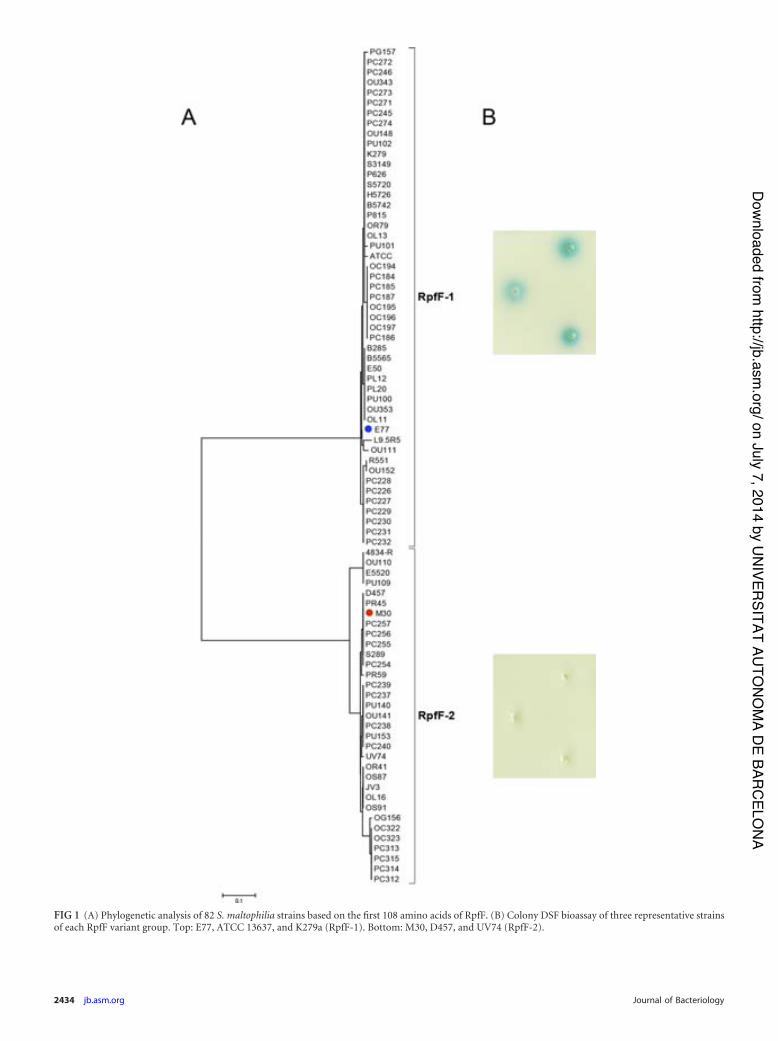
FIG 1 (A) Phylogenetic analysis of 82 S. maltophilia strains based on the first 108 amino acids of RpfF. (B) Colony DSF bioassay of three representative strainsof each RpfF variant group. Top: E77, ATCC 13637, and K279a (RpfF-1). Bottom: M30, D457, and UV74 (RpfF-2).
2434 jb.asm.org Journal of Bacteriology
on July 7, 2014 by UN
IVERSITAT AU
TON
OM
A DE BAR
CELO
NA
http://jb.asm.org/
Dow
nloaded from
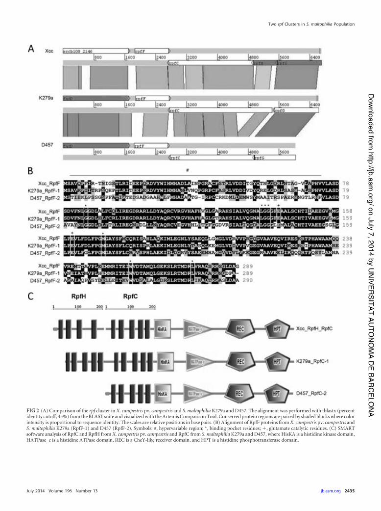
FIG 2 (A) Comparison of the rpf cluster in X. campestris pv. campestris and S. maltophilia K279a and D457. The alignment was performed with tblastx (percentidentity cutoff, 45%) from the BLAST suite and visualized with the Artemis Comparison Tool. Conserved protein regions are paired by shaded blocks where colorintensity is proportional to sequence identity. The scales are relative positions in base pairs. (B) Alignment of RpfF proteins from X. campestris pv. campestris andS. maltophilia K279a (RpfF-1) and D457 (RpfF-2). Symbols: #, hypervariable region; *, binding pocket residues; �, glutamate catalytic residues. (C) SMARTsoftware analysis of RpfC and RpfH from X. campestris pv. campestris and RpfC from S. maltophilia K279a and D457, where HisKA is a histidine kinase domain,HATPase_c is a histidine ATPase domain, REC is a CheY-like receiver domain, and HPT is a histidine phosphotransferase domain.
Two rpf Clusters in S. maltophilia Population
July 2014 Volume 196 Number 13 jb.asm.org 2435
on July 7, 2014 by UN
IVERSITAT AU
TON
OM
A DE BAR
CELO
NA
http://jb.asm.org/
Dow
nloaded from
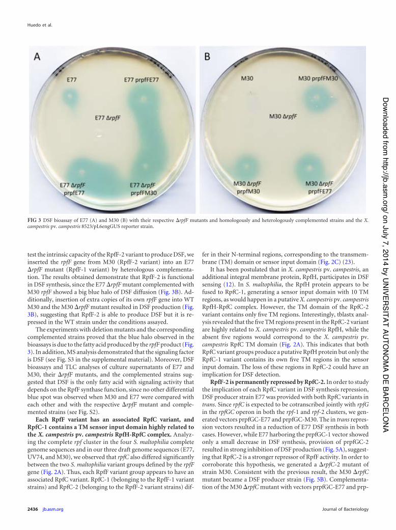
test the intrinsic capacity of the RpfF-2 variant to produce DSF, weinserted the rpfF gene from M30 (RpfF-2 variant) into an E77�rpfF mutant (RpfF-1 variant) by heterologous complementa-tion. The results obtained demonstrate that RpfF-2 is functionalin DSF synthesis, since the E77 �rpfF mutant complemented withM30 rpfF showed a big blue halo of DSF diffusion (Fig. 3B). Ad-ditionally, insertion of extra copies of its own rpfF gene into WTM30 and the M30 �rpfF mutant resulted in DSF production (Fig.3B), suggesting that RpfF-2 is able to produce DSF but it is re-pressed in the WT strain under the conditions assayed.
The experiments with deletion mutants and the correspondingcomplemented strains proved that the blue halo observed in thebioassays is due to the fatty acid produced by the rpfF product (Fig.3). In addition, MS analysis demonstrated that the signaling factoris DSF (see Fig. S3 in the supplemental material). Moreover, DSFbioassays and TLC analyses of culture supernatants of E77 andM30, their �rpfF mutants, and the complemented strains sug-gested that DSF is the only fatty acid with signaling activity thatdepends on the RpfF synthase function, since no other differentialblue spot was observed when M30 and E77 were compared witheach other and with the respective �rpfF mutant and comple-mented strains (see Fig. S2).
Each RpfF variant has an associated RpfC variant, andRpfC-1 contains a TM sensor input domain highly related tothe X. campestris pv. campestris RpfH-RpfC complex. Analyz-ing the complete rpf cluster in the four S. maltophilia completegenome sequences and in our three draft genome sequences (E77,UV74, and M30), we observed that rpfC also differed significantlybetween the two S. maltophilia variant groups defined by the rpfFgene (Fig. 2A). Thus, each RpfF variant group appears to have anassociated RpfC variant. RpfC-1 (belonging to the RpfF-1 variantstrains) and RpfC-2 (belonging to the RpfF-2 variant strains) dif-
fer in their N-terminal regions, corresponding to the transmem-brane (TM) domain or sensor input domain (Fig. 2C) (23).
It has been postulated that in X. campestris pv. campestris, anadditional integral membrane protein, RpfH, participates in DSFsensing (12). In S. maltophilia, the RpfH protein appears to befused to RpfC-1, generating a sensor input domain with 10 TMregions, as would happen in a putative X. campestris pv. campestrisRpfH-RpfC complex. However, the TM domain of the RpfC-2variant contains only five TM regions. Interestingly, tblastx anal-ysis revealed that the five TM regions present in the RpfC-2 variantare highly related to X. campestris pv. campestris RpfH, while theabsent five regions would correspond to the X. campestris pv.campestris RpfC TM domain (Fig. 2A). This indicates that bothRpfC variant groups produce a putative RpfH protein but only theRpfC-1 variant contains its own five TM regions in the sensorinput domain. The loss of these regions in RpfC-2 could have animplication for DSF detection.
RpfF-2 is permanently repressed by RpfC-2. In order to studythe implication of each RpfC variant in DSF synthesis repression,DSF producer strain E77 was provided with both RpfC variants intrans. Since rpfC is expected to be cotranscribed jointly with rpfGin the rpfGC operon in both the rpf-1 and rpf-2 clusters, we gen-erated vectors prpfGC-E77 and prpfGC-M30. The in trans repres-sion vectors resulted in a reduction of E77 DSF synthesis in bothcases. However, while E77 harboring the prpfGC-1 vector showedonly a small decrease in DSF synthesis, provision of prpfGC-2resulted in strong inhibition of DSF production (Fig. 5A), suggest-ing that RpfC-2 is a stronger repressor of RpfF activity. In order tocorroborate this hypothesis, we generated a �rpfC-2 mutant ofstrain M30. Consistent with the previous result, the M30 �rpfCmutant became a DSF producer strain (Fig. 5B). Complementa-tion of the M30 �rpfC mutant with vectors prpfGC-E77 and prp-
FIG 3 DSF bioassay of E77 (A) and M30 (B) with their respective �rpfF mutants and homologously and heterologously complemented strains and the X.campestris pv. campestris 8523/pL6engGUS reporter strain.
Huedo et al.
2436 jb.asm.org Journal of Bacteriology
on July 7, 2014 by UN
IVERSITAT AU
TON
OM
A DE BAR
CELO
NA
http://jb.asm.org/
Dow
nloaded from

fGC-M30 in trans led to a scenario similar to that obtained withthe E77 strain (Fig. 5B). To further characterize the relationshipbetween RpfF and RpfC in the two variants, the expression of thegene pairs rpfF-1–rpfC-1 and rpfF-2–rpfC-2 was quantified byqRT-PCR using the 2���CT method with gyrA as an endogenouscontrol. Thus, expression in WT E77 was 5.16-fold 0.59-fold forrpfF-1 and 2.69-fold 0.29-fold for rpfC-1 (rpfF-1/rpfC-1 ratio of1.92), while in WT M30 it was 1.57-fold 0.23-fold for rpfF-2 and1.65-fold 0.25-fold for rpfC-2 (rpfF-2/rpfC-2 ratio of 0.95) (seeFig. S4 in the supplemental material).
�rpfF mutants display different virulence-associated pheno-types as a function of the native RpfF variant. We have investi-gated the implication of the two RpfF variants for virulence-asso-ciated phenotypes such as biofilm formation and swarmingmotility. We had previously observed that swarming activation ofS. maltophilia is faster with streak inoculation than with pin inoc-ulation, suggesting that a high-density population facilitates the
initiation of this type of motion (unpublished results). This sup-ports the idea that QS could be involved in swarming activation inS. maltophilia. To corroborate this hypothesis, we tested the abilityof E77 and M30 �rpfF mutants to swarm on modified M9 me-dium with a 0.5% agar concentration, relative to that of the WTstrains. WT E77 displays tendril-like motility, whereas WT M30hardly swarms, likely because of its DSF deficiency (Fig. 6). TheE77 �rpfF mutant shows a clear motility loss and phenotyperestoration when rpfF is complemented in trans. On the contrary,the swarming motility of the M30 �rpfF mutant is not signifi-cantly different from that of the WT M30 strain, suggesting thatRpfF does not intervene in swarming control in M30. However,this behavior does not seem to be strictly linked to the RpfF vari-ant. Thus, on the one hand the E77 �rpfF mutant displayed anatypical nontendril swarming morphology when heterologouslycomplemented with the M30 rpfF gene. On the other, heterolo-gous complementation of the M30 �rpfF mutant with the E77 rpfF
FIG 4 (A) GC analysis of culture supernatants of E77, M30, and their respective �rpfF mutants and complemented strains. (B) DSF bioassay of concentratedsupernatants of the same strains from independent extractions.
Two rpf Clusters in S. maltophilia Population
July 2014 Volume 196 Number 13 jb.asm.org 2437
on July 7, 2014 by UN
IVERSITAT AU
TON
OM
A DE BAR
CELO
NA
http://jb.asm.org/
Dow
nloaded from

gene resulted in motility similar to that of the M30 �rpfF mutantstrain.
Biofilm formation by E77, M30, and the respective �rpfF mu-tants and complemented variants on a polystyrene or glass surfacewas also evaluated under two different medium conditions (M9and BM2). The results show that M30 has a higher capacity thanE77 to form biofilm under both growth conditions (Fig. 7), con-trary to the ability to swarm, suggesting that DSF production mayinversely regulate these two behaviors in S. maltophilia. Addition-ally, the results also indicate that biofilm formation is altered onlyin the E77 �rpfF mutant, showing a significant increase relative tothat of WT E77. Homologous and heterologous complementationwith the respective RpfF variants restores almost WT E77 levels ofbiofilm formation (P 0.0005) on both glass and plastic surfaces(Fig. 7A and B). On the other hand, the M30 strain, the M30 �rpfFmutant, and the homologously and heterologously comple-mented strains show similar levels of biofilm formation (Fig. 7Cand D). As for the regulation of swarming motility or the ability toproduce DSF, it therefore appears that the regulation of biofilm
formation is not strictly dependent on the RpfF variant but on oneor more components associated with this variant, in particular,RpfC. Specifically, the results suggest that RpfF is involved in theregulation of biofilm formation and swarming motility only instrains that natively carry RpfF-1 (even when this is replaced withRpfF-2). This is likely connected to the ability of these strains toproduce DSF.
The �rpfF-1 mutant, but not the �rpfF-2 mutant, shows at-tenuation in C. elegans. To elucidate the direct implication ofeach RpfF variant in S. maltophilia virulence in vivo, the killingability of E77, M30, and the respective �rpfF mutants and com-plemented strains was tested in C. elegans. Although the WT E77and M30 strains showed similar virulence capacities in the C. el-egans model (with times required to kill 50% of the nematodes,6.04 and 4.99 days, respectively), significant attenuation was ob-served here for the E77 �rpfF mutant (Fig. 8). In line with theobservations made for the phenotypes analyzed previously, thevirulence of the E77 �rpfF mutant is restored after complementa-tion with either its own rpfF gene (RpfF-1 variant) or the M30 rpfF
FIG 5 (A) DSF bioassay of WT E77 and E77 complemented with vectors prpfGCE77 and prpfGCM30. (B) DSF bioassay of WT M30, the M30 �rpfC mutant,and the M30 �rpfC mutant complemented with vectors prpfGCE77 and prpfGCM30.
FIG 6 Swarming motility assay of E77, M30, and their �rpfF mutants and homologously and heterologously complemented strains on modified M9 mediumsolidified with 0.5% Noble agar and incubated at 30°C for 4 days.
Huedo et al.
2438 jb.asm.org Journal of Bacteriology
on July 7, 2014 by UN
IVERSITAT AU
TON
OM
A DE BAR
CELO
NA
http://jb.asm.org/
Dow
nloaded from

gene (RpfF-2 variant), indicating once more that E77 is able torespond to heterologous DSF production in an RpfC-1 variantbackground (Fig. 8). Infection with the M30 �rpfF mutant showsno significant differences from WT M30. These results suggestagain that the RpfF-RpfC pair may regulate virulence only in thosestrains carrying the variant 1 combination.
The �rpfF-1 mutant shows attenuation in zebrafish due to itsinability to disseminate through fish tissues. E77, its �rpfF mu-tant, and the complemented strain were evaluated in zebrafish as avertebrate model. Similar results were obtained, corroboratingthat RpfF-1 is involved in virulence regulation (Fig. 9A). Interest-ingly, recovery of bacteria from sacrificed fish from each tank at 72h postinjection showed the ability of WT E77 to disseminatethrough the fish body from the abdominal region to the anterior
and posterior regions. On the contrary, the E77 �rpfF mutant doesnot seem to be able to colonize those regions effectively. Comple-mentation of rpfF partially restores its body dissemination capac-ity (Fig. 9B).
DISCUSSION
We have characterized 78 S. maltophilia clinical strains isolatedfrom diverse sources in different European hospitals for the rpfFgene. We have first demonstrated that the 78 strains contain therpfF gene but the RpfF product is distributed into two differentvariants that we have named RpfF-1 and RpfF-2 (Fig. 1 and 2A).We also show that the isolates produce two RpfC variants, eachassociated with one of the RpfF variants (Fig. 2A). The two RpfCvariants are different in the N-terminal region, which corresponds
FIG 7 Biofilm formation by E77, M30, and their respective �rpfF mutants and homologously and heterologously complemented strains on polystyrene (plots)and glass (tubes) surfaces in M9 (A and C) and BM2 (B and D) minimal media. ***, P 0.0005.
Two rpf Clusters in S. maltophilia Population
July 2014 Volume 196 Number 13 jb.asm.org 2439
on July 7, 2014 by UN
IVERSITAT AU
TON
OM
A DE BAR
CELO
NA
http://jb.asm.org/
Dow
nloaded from

to a TM domain (Fig. 2C) thought to participate in DSF sensing inseveral Xanthomonas species (12). In S. maltophilia, the RpfC-1variant contains 10 TM regions that display high similarity to theputative X. campestris pv. campestris RpfH-RpfC TM complex(Fig. 2A and C). On the other hand, the RpfC-2 variant has onlyfive TM regions, which appear to be related to the X. campestris pv.campestris RpfH TM domain rather than that of X. campestris pv.campestris RpfC (Fig. 2A). This phenomenon is also observed inXylella fastidiosa, Xanthomonas oryzae, and Pseudoxanthomonasspecies, suggesting that the RpfC-2 variant is widely distributedamong the members of the order Xanthomonadales that share theDSF-QS system. Nevertheless, protein sequence comparisonshows a high similarity between the RpfC and RpfH TM domains,suggesting that a duplication event (for X. campestris pv. campes-tris rpfC to rpfH and S. maltophilia rpfC-1) or a deletion (for S.maltophilia rpfC-2) may have occurred.
A previous S. maltophilia population study suggested that animportant group of S. maltophilia isolates lack rpfF (10). PCR-based typing of 89 strains showed an rpfF� prevalence of 61.8%,while the remaining 38.2% were considered to be rpfF mutants.On the basis of our sequence analysis, we can conclude that thework of Pompilio and collaborators (10) failed to detect rpfF be-cause the primers they used were designed to hybridize within themost variable region of this gene; more specifically, those primersdo not amplify rpfF in strains carrying what we have defined as
FIG 8 Determination of virulence of E77 (A), M30 (B), and their respective�rpfF mutants and homologously and heterologously complemented strainsin a C. elegans CF512 model of infection.
FIG 9 (A) Virulence of E77, the �rpfF mutant, and the complemented strain in 9-month-old zebrafish. (B) Bacterial recovery from different regions of the bodiesof sacrificed fish at 72 h postinfection with E77, the �rpfF mutant, or the complemented strain. ***, P 0.0005.
Huedo et al.
2440 jb.asm.org Journal of Bacteriology
on July 7, 2014 by UN
IVERSITAT AU
TON
OM
A DE BAR
CELO
NA
http://jb.asm.org/
Dow
nloaded from
variant 2. Accordingly, we hypothesize that all of the S. maltophiliastrains analyzed in the study by Pompilio et al. and showing anrpfF� genotype belong to the RpfF-1 variant group, whereas therpfF mutant strains would belong to the RpfF-2 variant group.Interestingly, our analysis of rpfF from a collection of 82 S. malto-philia strains shows similar RpfF variant frequencies in the popu-lation. RpfF-1 is present in 59.75% of the strains (including K279aand R551), whereas RpfF-2 is present in 40.25% (including D457and JV3). Taking the two studies together (171 strains), strainscarrying the RpfF-1 variant appear to be more commonly isolatedthan those carrying the RpfF-2 variant, with relative prevalences ofca. 60 and 40%, respectively.
Surprisingly, we have observed that only strains carrying theRpfC–RpfF-1 pair produce DSF under WT conditions, whilestrains belonging to the RpfC–RpfF-2 variant group require extracopies of their own rpfF gene (Fig. 3 and 4) or the absence of therepressor component RpfC-2 (Fig. 5) to achieve detectable DSFproduction levels. These results indicate that RpfF-2 is able tosynthesize DSF but the production of this signaling molecule ispermanently repressed by RpfC-2 under the conditions assayed. Ithas been shown that the stoichiometric balance between RpfF andRpfC is crucial for DSF production in many members of the orderXanthomonadales. In X. campestris pv. campestris, RpfC physicallyinteracts with the RpfF active site, inhibiting DSF synthesis activity(12, 22, 24). RpfC has also been shown to repress the RpfF activityof X. fastidiosa (25). Analysis of mRNA levels in E77 and M30 byqRT-PCR shows that the rpfF/rpfC expression ratio in the DSFproducer strain (variant 1) is double that found in the nonpro-ducer one (variant 2), suggesting, together with the observationthat variant 2 strains complemented with extra rpfF copies pro-duce DSF, that the different phenotypes of the two variants may bepartly due to the different regulation of the stoichiometry of thesetwo components. It has also been suggested that in X. campestrispv. campestris, RpfC could play a positive-feedback role in DSFsynthesis, liberating active RpfF upon the detection of DSF mole-cules (22). Assuming similar mechanisms in S. maltophilia, wehypothesize that DSF production in RpfC–RpfF-1 strains is due tothe presence of a competent sensor input domain, i.e., composedof 10 TM regions, in RpfC-1, which would enable the liberation ofactive RpfF-1 upon DSF detection and the subsequent synthesis ofDSF. On the other hand, the missing TM regions in RpfC-2 wouldrender this factor incompetent for DSF sensing, leading to perma-nent inhibition of RpfF-2 by RpfC-2 in a situation of equal num-bers of copies. Demonstrating that RpfC-1 liberates free activeRpfF after DSF detection and understanding its mechanism orunveiling why the S. maltophilia population produces two RpfCvariants and what implications it may have for DSF-mediated reg-ulation are questions that require further studies. The possibilitythat RpfC–RpfF-2 variant strains may produce DSF under specificenvironmental conditions or that RpfF-2 may produce a differentyet undetected DSF derivative cannot be ruled out. Comparison ofGC-MS spectra from M30, its �rpfF mutant, and the comple-mented strain did not, however, reveal any peak compatible withthe mass of a DSF derivative.
It is well known that the DSF-QS system regulates certain vir-ulence traits in many bacteria (1, 3, 9, 12, 26–29). To determinethe possible implication of each RpfF variant for virulence regu-lation, we generated an rpfF deletion mutant for a strain represen-tative of each variant group, i.e., E77 for the RpfF-1 variant groupand M30 for the RpfF-2 group. All of the phenotypes evaluated in
M30 were unaltered in the �rpfF mutant and in the correspondingcomplemented strain, suggesting that RpfC–RpfF-2 variantstrains may not use the DSF-QS system to regulate these virulencefactors, likely because of their inability to produce and sense DSFmolecules under the conditions assayed. On the contrary, the E77�rpfF mutant showed attenuation in both the C. elegans (Fig. 8A)and zebrafish (Fig. 9A) infection models, proving that DSF-medi-ated regulation affects the virulence of RpfC–RpfF-1 strains.Moreover, the recovery of bacteria from sacrificed fishes at 72 hpostinjection showed that E77 is able to disseminate to the ante-rior and posterior regions through the fish body, while the E77�rpfF mutant had serious problems in crossing intraperitonealbarriers (Fig. 9B). This is in concordance with the results showinga loss of swarming motility (Fig. 6) and a drastic increase in bio-film formation capacity (Fig. 7A and B) by the E77 �rpfF mutant,two important virulence-related traits that would explain attenu-ation in the animal models and especially in the zebrafish experi-ments. Much evidence of the implication of RpfF and DSF-likefatty acids in bacterial motility has indeed been reported (1, 2, 9,30). Our results thus reinforce previous evidence that one of themain functions of DSF-QS is to regulate bacterial motility. Manystudies have also demonstrated the implication of DSF-like mol-ecules in biofilm regulation. There is, however, some controversyabout whether DSF-like molecules may act by stimulating or in-hibiting the sessile or motile bacterial lifestyle. Thus, DSF mole-cules have been shown to positively regulate biofilm formation inX. oryzae pv. oryzae (31), B. cenocepacia (9, 28), and X. fastidiosa(4, 26). On the contrary, in X. campestris pv. campestris, the DSF-mediated QS acts as a negative regulator of biofilm development(32–34). Additionally, fatty acid-mediated biofilm dispersion isnot restricted to species with the DSF-QS system. For example, thefatty acid cis-2-decenoic acid produced by Pseudomonas aerugi-nosa PAO1 stimulates biofilm dispersion in several Gram-positiveand Gram-negative bacteria (35, 36). Our findings indicate thatthe DSF-QS system in S. maltophilia E77 has a regulatory functionsimilar to that described for X. campestris pv. campestris, whereDSF also plays an important role in preventing biofilm formationand stimulating bacterial motility.
ACKNOWLEDGMENTS
This work was supported by funding under the Seventh Research Frame-work Programme of the European Union (HEALTH-F3-2009-223101)and the Spanish MICINN (BFU2010-17199). I.G. acknowledges supportfrom the Catalan AGAUR (2009SGR-00108). I.R. and J.V. acknowledgesupport from the SATURN project (European Community FP7,HEALTH-F3-2009-241796).
We thank C. Prat and J. Domínguez (Servei de Microbiologia, Hospi-tal Universitari Germans Trias i Pujol, Institut d’Investigació GermansTrias i Pujol, Universitat Autònoma de Barcelona, Badalona, Spain) andJ. L. Martínez (Centro Nacional de Biotecnología, CSIC, Cantoblanco,Madrid, Spain) for providing some of the clinical strains used in thisstudy.
REFERENCES1. Fouhy Y, Scanlon K, Schouest K, Spillane C, Crossman L, Avison MB,
Ryan RP, Dow JM. 2007. Diffusible signal factor-dependent cell-cellsignaling and virulence in the nosocomial pathogen Stenotrophomonasmaltophilia. J. Bacteriol. 189:4964 – 4968. http://dx.doi.org/10.1128/JB.00310-07.
2. Huang T-P, Lee Wong AC. 2007. Extracellular fatty acids facilitate fla-gella-independent translocation by Stenotrophomonas maltophilia. Res.Microbiol. 158:702–711. http://dx.doi.org/10.1016/j.resmic.2007.09.002.
Two rpf Clusters in S. maltophilia Population
July 2014 Volume 196 Number 13 jb.asm.org 2441
on July 7, 2014 by UN
IVERSITAT AU
TON
OM
A DE BAR
CELO
NA
http://jb.asm.org/
Dow
nloaded from

3. Barber CE, Tang JL, Feng JX, Pan MQ, Wilson TJ, Slater H, Dow JM,Williams P, Daniels MJ. 1997. A novel regulatory system required forpathogenicity of Xanthomonas campestris is mediated by a small diffusiblesignal molecule. Mol. Microbiol. 24:555–566. http://dx.doi.org/10.1046/j.1365-2958.1997.3721736.x.
4. Beaulieu ED, Ionescu M, Chatterjee S, Yokota K, Trauner D, Lindow S.2013. Characterization of a diffusible signaling factor from Xylella fastid-iosa. mBio 4:e00539 – 00512. http://dx.doi.org/10.1128/mBio.00539-12.
5. Boon C, Deng Y, Wang L-H, He Y, Xu J-L, Fan Y, Pan SQ, Zhang L-H.2008. A novel DSF-like signal from Burkholderia cenocepacia interfereswith Candida albicans morphological transition. ISME J. 2:27–36. http://dx.doi.org/10.1038/ismej.2007.76.
6. Deng Y, Wu J, Tao F, Zhang L-H. 2011. Listening to a new language:DSF-based quorum sensing in Gram-negative bacteria. Chem. Rev. 111:160 –173. http://dx.doi.org/10.1021/cr100354f.
7. Huang T-P, Wong ACL. 2007. A cyclic AMP receptor protein-regulatedcell-cell communication system mediates expression of a FecA homologuein Stenotrophomonas maltophilia. Appl. Environ. Microbiol. 73:5034 –5040. http://dx.doi.org/10.1128/AEM.00366-07.
8. Alavi P, Müller H, Cardinale M, Zachow C, Sánchez MB, Martínez JL,Berg G. 2013. The DSF quorum sensing system controls the positiveinfluence of Stenotrophomonas maltophilia on plants. PLoS One 8:e67103.http://dx.doi.org/10.1371/journal.pone.0067103.
9. Deng Y, Schmid N, Wang C, Wang J, Pessi G, Wu D, Lee J, Aguilar C,Ahrens CH, Chang C, Song H, Eberl L, Zhang L-H. 2012. Cis-2-dodecenoic acid receptor RpfR links quorum-sensing signal perceptionwith regulation of virulence through cyclic dimeric guanosine monophos-phate turnover. Proc. Natl. Acad. Sci. U. S. A. 109:15479 –15484. http://dx.doi.org/10.1073/pnas.1205037109.
10. Pompilio A, Pomponio S, Crocetta V, Gherardi G, Verginelli F, Fis-carelli E, Dicuonzo G, Savini V, D’Antonio D, Di Bonaventura G. 2011.Phenotypic and genotypic characterization of Stenotrophomonas malto-philia isolates from patients with cystic fibrosis: genome diversity, biofilmformation, and virulence. BMC Microbiol. 11:159. http://dx.doi.org/10.1186/1471-2180-11-159.
11. Ferrer-Navarro M, Planell R, Yero D, Mongiardini E, Torrent G, HuedoP, Martínez P, Roher N, Mackenzie S, Gibert I, Daura X. 2013. Abun-dance of the quorum-sensing factor Ax21 in four strains of Stenotroph-omonas maltophilia correlates with mortality rate in a new zebrafish modelof infection. PLoS One 8:e67207. http://dx.doi.org/10.1371/journal.pone.0067207.
12. Slater H, Alvarez-Morales A, Barber CE, Daniels MJ, Dow JM. 2000. Atwo-component system involving an HD-GYP domain protein links cell-cell signalling to pathogenicity gene expression in Xanthomonas campes-tris. Mol. Microbiol. 38:986 –1003. http://dx.doi.org/10.1046/j.1365-2958.2000.02196.x.
13. Tamura K, Peterson D, Peterson N, Stecher G, Nei M, Kumar S. 2011.MEGA5: molecular evolutionary genetics analysis using maximum likeli-hood, evolutionary distance, and maximum parsimony methods. Mol.Biol. Evol. 28:2731–2739. http://dx.doi.org/10.1093/molbev/msr121.
14. Schultz J, Milpetz F, Bork P, Ponting CP. 1998. SMART, a simplemodular architecture research tool: identification of signaling domains.Proc. Natl. Acad. Sci. U. S. A. 95:5857–5864. http://dx.doi.org/10.1073/pnas.95.11.5857.
15. Hoang TT, Karkhoff-Schweizer RR, Kutchma AJ, Schweizer HP. 1998.A broad-host-range Flp-FRT recombination system for site-specific exci-sion of chromosomally-located DNA sequences: application for isolationof unmarked Pseudomonas aeruginosa mutants. Gene 212:77– 86. http://dx.doi.org/10.1016/S0378-1119(98)00130-9.
16. Choi K-H, Kumar A, Schweizer HP. 2006. A 10-min method for prep-aration of highly electrocompetent Pseudomonas aeruginosa cells: applica-tion for DNA fragment transfer between chromosomes and plasmidtransformation. J. Microbiol. Methods 64:391–397. http://dx.doi.org/10.1016/j.mimet.2005.06.001.
17. Kovach ME, Phillips RW, Elzer PH, Roop RM, II, Peterson KM. 1994.pBBR1MCS: a broad-host-range cloning vector. Biotechniques 16:800–802.
18. Tan MW, Mahajan-Miklos S, Ausubel FM. 1999. Killing of Caenorhab-ditis elegans by Pseudomonas aeruginosa used to model mammalian bacte-rial pathogenesis. Proc. Natl. Acad. Sci. U. S. A. 96:715–720. http://dx.doi.org/10.1073/pnas.96.2.715.
19. Kinkel MD, Eames SC, Philipson LH, Prince VE. 2010. Intraperitonealinjection into adult zebrafish. J. Vis. Exp. 42:2126. http://dx.doi.org/10.3791/2126.
20. Gould VC, Avison MB. 2006. SmeDEF-mediated antimicrobial drugresistance in Stenotrophomonas maltophilia clinical isolates having definedphylogenetic relationships. J. Antimicrob. Chemother. 57:1070 –1076.http://dx.doi.org/10.1093/jac/dkl106.
21. Livak KJ, Schmittgen TD. 2001. Analysis of relative gene expression datausing real-time quantitative PCR and the 2(-Delta Delta C(T)) Method.Methods 25:402– 408. http://dx.doi.org/10.1006/meth.2001.1262.
22. Cheng Z, He Y-W, Lim SC, Qamra R, Walsh MA, Zhang L-H, Song H.2010. Structural basis of the sensor-synthase interaction in autoinductionof the quorum sensing signal DSF biosynthesis. Structure 18:1199 –1209.http://dx.doi.org/10.1016/j.str.2010.06.011.
23. Ryan RP, Fouhy Y, Garcia BF, Watt SA, Niehaus K, Yang L, Tolker-Nielsen T, Dow JM. 2008. Interspecies signalling via the Stenotrophomo-nas maltophilia diffusible signal factor influences biofilm formation andpolymyxin tolerance in Pseudomonas aeruginosa. Mol. Microbiol. 68:75–86. http://dx.doi.org/10.1111/j.1365-2958.2008.06132.x.
24. Wang L-H, He Y, Gao Y, Wu JE, Dong Y-H, He C, Wang SX, WengL-X, Xu J-L, Tay L, Fang RX, Zhang L-H. 2004. A bacterial cell-cellcommunication signal with cross-kingdom structural analogues.Mol. Microbiol. 51:903–912. http://dx.doi.org/10.1046/j.1365-2958.2003.03883.x.
25. Ionescu M, Baccari C, Da Silva AM, Garcia A, Yokota K, Lindow SE.2013. Diffusible signal factor (DSF) synthase RpfF of Xylella fastidiosa is amultifunction protein also required for response to DSF. J. Bacteriol. 195:5273–5284. http://dx.doi.org/10.1128/JB.00713-13.
26. Chatterjee S, Wistrom C, Lindow SE. 2008. A cell-cell signaling sensor isrequired for virulence and insect transmission of Xylella fastidiosa. Proc.Natl. Acad. Sci. U. S. A. 105:2670 –2675. http://dx.doi.org/10.1073/pnas.0712236105.
27. Deng Y, Boon C, Eberl L, Zhang L-H. 2009. Differential modulation ofBurkholderia cenocepacia virulence and energy metabolism by the quo-rum-sensing signal BDSF and its synthase. J. Bacteriol. 191:7270 –7278.http://dx.doi.org/10.1128/JB.00681-09.
28. McCarthy Y, Yang L, Twomey KB, Sass A, Tolker-Nielsen T, Mahen-thiralingam E, Dow JM, Ryan RP. 2010. A sensor kinase recognizing thecell-cell signal BDSF (cis-2-dodecenoic acid) regulates virulence in Burk-holderia cenocepacia. Mol. Microbiol. 77:1220 –1236. http://dx.doi.org/10.1111/j.1365-2958.2010.07285.x.
29. Twomey KB, O’Connell OJ, McCarthy Y, Dow JM, O’Toole GA, PlantBJ, Ryan RP. 2012. Bacterial cis-2-unsaturated fatty acids found in thecystic fibrosis airway modulate virulence and persistence of Pseudomonasaeruginosa. ISME J. 6:939 –950. http://dx.doi.org/10.1038/ismej.2011.167.
30. Ryan RP, McCarthy Y, Kiely PA, O’Connor R, Farah CS, Armitage JP,Dow JM. 2012. Dynamic complex formation between HD-GYP, GGDEFand PilZ domain proteins regulates motility in Xanthomonas campestris.Mol. Microbiol. 86:557–567. http://dx.doi.org/10.1111/mmi.12000.
31. Rai R, Ranjan M, Pradhan BB, Chatterjee S. 2012. Atypical regulation ofvirulence-associated functions by a diffusible signal factor in Xanthomo-nas oryzae pv. oryzae. Mol. Plant Microbe Interact. 25:789 – 801. http://dx.doi.org/10.1094/MPMI-11-11-0285-R.
32. Dow JM, Crossman L, Findlay K, He Y-Q, Feng J-X, Tang J-L. 2003.Biofilm dispersal in Xanthomonas campestris is controlled by cell-cell sig-naling and is required for full virulence to plants. Proc. Natl. Acad. Sci.U. S. A. 100:10995–11000. http://dx.doi.org/10.1073/pnas.1833360100.
33. He Y-W, Xu M, Lin K, Ng Y-JA, Wen C-M, Wang L-H, Liu Z-D, ZhangH-B, Dong Y-H, Dow JM, Zhang L-H. 2006. Genome scale analysis ofdiffusible signal factor regulon in Xanthomonas campestris pv. campestris:identification of novel cell-cell communication-dependent genes andfunctions. Mol. Microbiol. 59:610 – 622. http://dx.doi.org/10.1111/j.1365-2958.2005.04961.x.
34. Tao F, Swarup S, Zhang L-H. 2010. Quorum sensing modulation of aputative glycosyltransferase gene cluster essential for Xanthomonas camp-estris biofilm formation. Environ. Microbiol. 12:3159 –3170. http://dx.doi.org/10.1111/j.1462-2920.2010.02288.x.
35. Davies DG, Marques CNH. 2009. A fatty acid messenger is responsiblefor inducing dispersion in microbial biofilms. J. Bacteriol. 191:1393–1403.http://dx.doi.org/10.1128/JB.01214-08.
36. Amari DT, Marques CNH, Davies DG. 2013. The putative enoyl-coenzyme A hydratase DspI is required for production of the Pseudomonasaeruginosa biofilm dispersion autoinducer cis-2-decenoic acid. J. Bacte-riol. 195:4600 – 4610. http://dx.doi.org/10.1128/JB.00707-13.
Huedo et al.
2442 jb.asm.org Journal of Bacteriology
on July 7, 2014 by UN
IVERSITAT AU
TON
OM
A DE BAR
CELO
NA
http://jb.asm.org/
Dow
nloaded from
Draft Genome Sequence of Stenotrophomonas maltophilia Strain M30,Isolated from a Chronic Pressure Ulcer in an Elderly Patient
Pol Huedo,a,b Óscar Conchillo-Solé,a Daniel Yero,a,b Sònia Martínez-Servat,a,b Xavier Daura,a,c Isidre Giberta,b
Institut de Biotecnologia i de Biomedicina, Universitat Autònoma de Barcelona, Barcelona, Spaina; Departament de Genètica i de Microbiologia, Universitat Autònoma deBarcelona, Barcelona, Spainb; Catalan Institution for Research and Advanced Studies, Barcelona, Spainc
P.H. and O.C.-S. contributed equally to this article.
Stenotrophomonas maltophilia is an emerging opportunistic pathogen with an increasing prevalence of multidrug-resistantstrains. Here, we report the draft genome sequence of S. maltophilia strain M30, isolated from a pressure ulcer in an elderly pa-tient.
Received 21 May 2014 Accepted 27 May 2014 Published 12 June 2014
Citation Huedo P, Conchillo-Solé Ó, Yero D, Martínez-Servat S, Daura X, Gibert I. 2014. Draft genome sequence of Stenotrophomonas maltophilia strain M30, isolated from achronic pressure ulcer in an elderly patient. Genome Announc. 2(3):e00576-14. doi:10.1128/genomeA.00576-14.
Copyright © 2014 Huedo et al. This is an open-access article distributed under the terms of the Creative Commons Attribution 3.0 Unported license.
Address correspondence to Xavier Daura, [email protected], or Isidre Gibert, [email protected].
Stenotrophomonas maltophilia is an aerobic ubiquitous Gram-negative bacillus commonly isolated from hospital environ-
ments (1). Although S. maltophilia displays limited invasivenessand pathogenic capacity, it is capable of infecting a wide rangeof tissues and organs, especially in immunocompromised patients(2). Its disease patterns include bacteremia, catheter-relatedinfections, pneumonia, biliary and urinary tract infections, andskin infections. The therapeutic agent of choice is typicallytrimethoprim-sulfamethoxazole (3, 4), but resistance to this drugis increasingly being reported (5). New insights into the mecha-nisms of drug resistance are needed in order to identify new effec-tive drug targets.
S. maltophilia strain M30 was isolated from a pressure ulcer ofan elderly patient in the Hospital Municipal de Badalona (Barce-lona, Spain) in 2009. The M30 strain is a multidrug-resistant(MDR) organism, showing resistance not only to tetracycline,kanamycin, sulfamethoxazole, and erythromycin (6), but also tothe complement-mediated bactericidal action of serum (6). Mul-tilocus sequence type analysis (7) revealed that M30 belongs to anew sequence type (sequence type 76 [ST-76]) (6) clusteringwithin a new genetic group (genogroup C) previously describedby Kaiser et al. (7). This genetic group comprises clinical isolatesfrom different geographic regions and includes the model MDRstrain D457 (8). Recently, it was demonstrated that in strain M30,the diffusible signal factor (DSF)-mediated quorum-sensing sys-tem is regulated by a new rpf cluster variant (9).
Genomic DNA was extracted with GenElute bacterial genom-ics DNA kit (Sigma-Aldrich), and whole-genome sequencing wasperformed using Illumina MiSeq technology at the UniversitatAutònoma de Barcelona Genomics core facility. The low-qualityreads were filtered, and the remaining reads were de novo assem-bled using VelvetOptimiser version 2.2.5 (10) relaying on Velvetversion 1.2.10 (11) and improved with the IMAGE program fromthe PAGIT package version 1 (12). The assembly resulted in 193contigs (G�C content, 66.3%), with an N50 contig size of 46,399
nucleotides, covering a total of 4,902,008 bp. The average length ofthe contigs is 25.4 kb,nd a the biggest contig contains 142,025 bp.
Genome annotation was performed by the NCBI ProkaryoticGenome Annotation Pipeline version 2.5 (rev. 434060), and 4,515genes were predicted, of which 4,392 are coding sequences(CDSs), 43 are pseudogenes, 11 are rRNAs (5S, 16S, and 23S), 68are tRNAs, and 1 is a noncoding RNA (ncRNA). Among the pre-dicted CDSs of M30, we found that 357 genes are not shared withthe six strains of S. maltophilia (K279a, D457, JV3, R551-3, EPM1,and Ab55555) for which complete genome sequences were avail-able at NCBI (http://www.ncbi.nlm.nih.gov/genome) at the timeof analysis. Most of these unique genes encode hypothetical pro-teins, enzymes related to DNA metabolism and repair, and trans-posases and integrases, indicating that horizontal gene transfermay be an important source of genomic diversity in S. maltophilia.Notably, the set of genes exclusive to M30 also encoded proteinsinvolved in host-microbe interactions, including ankyrin repeat(ANK)-containing proteins (13), two predicted hemolysins, oneprotease, and one amidohydrolase.
This draft genome will help improve our understanding ofgenome-associated resistance mechanisms in S. maltophilia andthe virulence factors the bacterium exploits in order to become apathogen.
Nucleotide sequence accession numbers. This whole-genomeshotgun project has been deposited at DDBJ/EMBL/GenBank un-der the accession no. JELS00000000. The version described in thispaper is JELS02000000.
ACKNOWLEDGMENTS
This work has been supported by funding under the Seventh ResearchFramework Programme of the European Union (HEALTH-F3-2009-223101) and the Spanish MICINN (BFU2010-17199). I.G. acknowledgessupport from the Catalan AGAUR (2009SGR-00108).
We thank A. Calderón and T. Falgueras from Badalona Serveis Assis-tencials (Hospital Municipal de Badalona) for providing strain M30.
Genome AnnouncementsMay/June 2014 Volume 2 Issue 3 e00576-14 genomea.asm.org 1
REFERENCES1. Brooke JS. 2012. Stenotrophomonas maltophilia: an emerging global op-
portunistic pathogen. Clin. Microbiol. Rev. 25:2– 41. http://dx.doi.org/10.1128/CMR.00019-11.
2. Denton M, Kerr KG. 1998. Microbiological and clinical aspects of infec-tion associated with Stenotrophomonas maltophilia. Clin. Microbiol. Rev.11:57– 80.
3. Gales AC, Jones RN, Forward KR, Liñares J, Sader HS, Verhoef J. 2001.Emerging importance of multidrug-resistant Acinetobacter species andStenotrophomonas maltophilia as pathogens in seriously ill patients: geo-graphic patterns, epidemiological features, and trends in the SENTRYAntimicrobial Surveillance Program (1997–1999). Clin. Infect. Dis. 32:S104 –S113. http://dx.doi.org/10.1086/320183.
4. Hu L-F, Chang X, Ye Y, Wang Z-X, Shao Y-B, Shi W, Li X, Li J-B. 2011.Stenotrophomonas maltophi l ia resistance to trimethoprim/sulfamethoxazole mediated by acquisition of sul and dfrA genes in aplasmid-mediated class 1 integron. Int. J. Antimicrob. Agents 37:230 –234. http://dx.doi.org/10.1016/j.ijantimicag.2010.10.025.
5. Toleman MA, Bennett PM, Bennett DMC, Jones RN, Walsh TR. 2007.Global emergence of trimethoprim/sulfamethoxazole resistance inStenotrophomonas maltophilia mediated by acquisition of sul genes.Emerg. Infect . Dis. 13:559 –565. http://dx.doi.org/10.3201/eid1304.061378.
6. Ferrer-Navarro M, Planell R, Yero D, Mongiardini E, Torrent G, HuedoP, Martínez P, Roher N, Mackenzie S, Gibert I, Daura X. 2013. Abun-dance of the quorum-sensing factor Ax21 in four strains of Stenotroph-omonas maltophilia correlates with mortality rate in a new zebrafish model
of infection. PLoS One 8:e67207. http://dx.doi.org/10.1371/journal.pone.0067207.
7. Kaiser S, Biehler K, Jonas D. 2009. A Stenotrophomonas maltophiliamultilocus sequence typing scheme for inferring population structure. J.Bacteriol. 191:2934 –2943. http://dx.doi.org/10.1128/JB.00892-08.
8. Lira F, Hernández A, Belda E, Sánchez MB, Moya A, Silva FJ, MartínezJL. 2012. Whole-genome sequence of Stenotrophomonas maltophiliaD457, a clinical isolate and a model strain. J. Bacteriol. 194:3563–3564.http://dx.doi.org/10.1128/JB.00602-12.
9. Huedo P, Yero D, Martínez-Servat S, Estibariz I, Planell R, Martínez P,Ruyra A, Roher N, Roca I, Vila J, Daura X, Gibert I. 2014. Two differentrpf clusters distributed among a population of Stenotrophomonas malto-philia clinical strains display differential DSF production and virulenceregulation. J. Bacteriol., in press. http://dx.doi.org/10.1128/JB.01540-14.
10. Zerbino DR. 2010. Using the Velvet de novo assembler for short-readsequencing technologies. Curr. Protoc. Bioinformatics Chapter 11:Unit11.5. http://dx.doi.org/10.1002/0471250953.bi1105s31.
11. Zerbino DR, Birney E. 2008. Velvet: algorithms for de novo short readassembly using de Bruijn graphs. Genome Res. 18:821– 829. http://dx.doi.org/10.1101/gr.074492.107.
12. Swain MT, Tsai IJ, Assefa SA, Newbold C, Berriman M, Otto TD. 2012.A post-assembly genome-improvement toolkit (PAGIT) to obtain anno-tated genomes from contigs. Nat. Protoc. 7:1260 –1284. http://dx.doi.org/10.1038/nprot.2012.068.
13. Al-Khodor S, Price CT, Kalia A, Kwaik YA. 2010. Functional diversity ofankyrin repeats in microbial proteins. Trends Microbiol. 18:132–139.http://dx.doi.org/10.1016/j.tim.2009.11.004.
Huedo et al.
Genome Announcements2 genomea.asm.org May/June 2014 Volume 2 Issue 3 e00576-14

Abundance of the Quorum-Sensing Factor Ax21 in FourStrains of Stenotrophomonas maltophilia Correlates withMortality Rate in a New Zebrafish Model of InfectionMario Ferrer-Navarro1., Raquel Planell1., Daniel Yero1, Elıas Mongiardini1, Gerard Torrent1,
Pol Huedo1,2, Paula Martınez1,2, Nerea Roher1, Simon Mackenzie1,3, Isidre Gibert1,2*, Xavier Daura1,4*
1 Institut de Biotecnologia i de Biomedicina (IBB), Universitat Autonoma de Barcelona (UAB), Cerdanyola del Valles, Barcelona, Spain, 2 Departament de Genetica i de
Microbiologia, Universitat Autonoma de Barcelona (UAB), Cerdanyola del Valles, Barcelona, Spain, 3 Institute of Aquaculture, University of Stirling, Stirling, Scotland, United
Kingdom, 4 Catalan Institution for Research and Advanced Studies (ICREA), Barcelona, Spain
Abstract
Stenotrophomonas maltophilia is a Gram-negative pathogen with emerging nosocomial incidence. Little is known about itspathogenesis and the genomic diversity exhibited by clinical isolates complicates the study of pathogenicity and virulencefactors. Here, we present a strategy to identify such factors in new clinical isolates of S. maltophilia, incorporating an adult-zebrafish model of S. maltophilia infection to evaluate relative virulence coupled to 2D difference gel electrophoresis toexplore underlying differences in protein expression. In this study we report upon three recent clinical isolates and use thecollection strain ATCC13637 as a reference. The adult-zebrafish model shows discrimination capacity, i.e. from very low tovery high mortality rates, with clinical symptoms very similar to those observed in natural S. maltophilia infections in fish.Strain virulence correlates with resistance to human serum, in agreement with previous studies in mouse and rat andtherefore supporting zebrafish as a replacement model. Despite its clinical origin, the collection strain ATCC13637 showedobvious signs of attenuation in zebrafish, with null mortality. Multilocus-sequence-typing analysis revealed that the mostvirulent strains, UV74 and M30, exhibit the strongest genetic similitude. Differential proteomic analysis led to theidentification of 38 proteins with significantly different abundance in the three clinical strains relative to the reference strain.Orthologs of several of these proteins have been already reported to have a role in pathogenesis, virulence or resistancemechanisms thus supporting our strategy. Proof of concept is further provided by protein Ax21, whose abundance is shownhere to be directly proportional to mortality in the zebrafish infection model. Indeed, recent studies have demonstrated thatthis protein is a quorum-sensing-related virulence factor.
Citation: Ferrer-Navarro M, Planell R, Yero D, Mongiardini E, Torrent G, et al. (2013) Abundance of the Quorum-Sensing Factor Ax21 in Four Strains ofStenotrophomonas maltophilia Correlates with Mortality Rate in a New Zebrafish Model of Infection. PLoS ONE 8(6): e67207. doi:10.1371/journal.pone.0067207
Editor: Jamunarani Vadivelu, University of Malaya, Malaysia
Received March 3, 2013; Accepted May 15, 2013; Published June 26, 2013
Copyright: ! 2013 Ferrer-Navarro et al. This is an open-access article distributed under the terms of the Creative Commons Attribution License, which permitsunrestricted use, distribution, and reproduction in any medium, provided the original author and source are credited.
Funding: This work has been supported by funding under the Seventh Research Framework Programme of the European Union (ref. HEALTH-F3-2009-223101)and the Spanish MICINN (ref. BFU2010-17199). I.G. acknowledges support from the Catalan AGAUR (ref. 2009SGR-00108). P.M. and P.H. are recipients of afellowship from Universitat Autonoma de Barcelona. The funders had no role in study design, data collection and analysis, decision to publish, or preparation ofthe manuscript.
Competing Interests: The authors have declared that no competing interests exist.
* E-mail: [email protected] (XD); [email protected] (IG)
. These authors contributed equally to this work.
Introduction
Stenotrophomonas maltophilia is a non-fermentative Gram-negativebacterium with increasing incidence in hospital environments[1,2]. This obligate aerobic bacterium can be found in almost anyaquatic or humid environment, including drinking-water supplies[3] and is now recognized as an emerging nosocomial pathogen. S.maltophilia has been associated with respiratory infections, septice-mia, biliary sepsis, endocarditis, conjunctivitis, meningitis, urinarytract infections and various wound infections in immunocompro-mised patients as well as in cystic fibrosis (CF) patients [2,4,5].Currently, S. maltophilia has been isolated from the lungs ofapproximately 10% of the CF patients in USA and up to 25% ofthose in Europe [1] and displays significant morbidity andmortality rates among debilitated patients [2,5,6,7,8].
S. maltophilia exhibits high-level intrinsic resistance to a variety ofstructurally unrelated antibiotics, including b-lactams, quinolones,
aminoglycosides, tetracycline, disinfectants and heavy metals[9,10]. Intrinsic resistance may be due to reduced outer-membrane permeability, changes in LPS structure, the productionof multidrug efflux pumps and the presence of integrons for site-specific insertion of resistance gene cassettes [11,12]. Theproduction of melanin-like pigments and biofilms have also beenlinked to antimicrobial resistance [12]. Thus, the adhesion of S.maltophilia to medical implants, catheters and epithelial cells,leading to the formation of biofilms, confers natural protectionagainst different antimicrobial agents and host immune defenses.In this regard, the development of therapies against S. maltophiliainfection represents a significant challenge for both clinicians andmicrobiologists. In addition, knowledge of virulence factors isscarce and limited to homology relationships.
Recently, four S. maltophilia genomes have been fully sequencedand assembled (strains K279a, R551–3, JV3 and D457), and
PLOS ONE | www.plosone.org 1 June 2013 | Volume 8 | Issue 6 | e67207

putative virulence factors have been identified by homologyrelationships [11,13,14]. These factors include type I, II, IV, andV protein-secretion systems, various pili, fimbriae, putativeadhesins, tissue-degradative exoenzymes, siderophores, quorum-sensing factors and proteins involved in polysaccharide synthesisand intracellular signaling. Some fimbrial structures have beenidentified and characterized and their role in adhesion to epithelialcells and abiotic surfaces has been demonstrated [15]. However,the level of understanding of this bacterium’s pathogenicity andvirulence is still limited and the number of S. maltophilia strainsphenotypically and genotypically analyzed is minor. Furthermore,there is considerable uncertainty about the route(s) of infection ofS. maltophilia. Additionally, the remarkable diversity of sourcesfrom which S. maltophilia strains have been isolated indicates thatthese bacteria exhibit a high level of genomic plasticity andmetabolic heterogeneity, possibly allowing them to expand theirpathogenic potential. Heterogeneity is also illustrated among S.maltophilia isolates recovered from a single patient, showingphenotypic variation over time as a consequence of horizontalgene transfer or high mutation rates [16].
In order to provide answers to some of the above thedevelopment of an appropriate infection models is essential.Previous studies suggest a limited invasiveness of S. maltophilia inmice, as indicated by a transient and minimal presence of themicroorganism in animal organs. For example, S. maltophilia CFstrains were shown to cause no mortality in a neonatal mousemodel of respiratory tract infection [17]. Despite this lack of robustinvasiveness, mouse models of S. maltophilia infection have providedinformation on the type of host immune response induced by thisopportunistic pathogen [2]. More recently, a model of acuterespiratory infection in DBA/2 mice following a single exposure toaerosolized bacteria enabled the investigation of bacterial clear-ance, histological damage, and inflammatory response in the lungsof infected mice [18]. However, while bacterial colonization andmortality were achieved in that model, infection disseminated at avery low rate even using high doses of a virulent strain and most ofthe animals were able to resolve S. maltophilia lung colonization in arelatively short time period. For that reason, animal-weight loss isoften taken as the best criterion for the comparison of pathogenesisand virulence of tested strains [18,19]. In addition, lung infectionmodels tend to be time-consuming, labor intensive and haveassociated welfare issues. Therefore, alternative, simple models ofS. maltophilia infection are still needed to test the virulence ofphenotypically and genotypically diverse strains.
In recent years the zebrafish (Danio rerio) has emerged as animportant model of vertebrate development, human disease andmicrobial infection [20]. Nevertheless, to our knowledge, zebrafishhas not yet been reported as a model of S. maltophilia infection. Ourchoice of adult zebrafish as a plausible model was motivated by aseries of observations. The existing literature in relation to otherbacterial pathogen models successfully developed in the zebrafish[20,21,22]. S. maltophilia as a natural pathogen of fish causinginfectious intussusception syndrome in adult channel catfish [23].The inclusion of a wild-type phenotypic population diversity asopposed to inbred mouse lines. The adaptive and innate immunesystem of zebrafish has significant similarities to mammaliansystems [24,25]. The zebrafish is recognized as an importantvertebrate model with genomic enablement and last, but not leastrelevant, ethical, economic and process-simplicity considerations.
In the study presented here, we have tested a combinedapproach that uses an adult-zebrafish model of S. maltophiliainfection for the evaluation of relative virulence and the successiveanalysis by fluorescence-based two-dimensional Difference in-GelElectrophoresis (DIGE) [26] of the underlying differences in
protein expression, in a quest for virulence factors. This analysiswas applied to three recent clinical isolates and the collection strainATCC13637, as a reference. Although a colloidal Coomassie-stained 2DE proteomic analysis to find heat-induced changes in S.maltophilia protein abundance has been reported [27], to ourknowledge no quantitative proteomic comparison between S.maltophilia strains with distinct virulence phenotypes has beenpreviously performed.
Results
Zebrafish Infection Model Confirms Attenuation ofCollection Strain and Points at Varying Virulence of theClinical Isolates
Adult zebrafish were used as an infection model to determinethe virulence of the individual strains. An intraperitoneal injectionof 108 cfu resulted in strain-dependent mortality rates (Figure 1),where UV74 was the most aggressive, with 84% mortality 48 hpost-injection (p.i.), followed by strains M30 (55%), E77 (5%) andATCC13637 (0%). Although the experimental period was during7 days, in all cases mortality occurred during the first 48 h.Clinical symptoms of dead fish included cutaneous hemorrhageunder the lower jaw, on the belly, and around the anus; all deadfish had a distended abdomen containing bloody or clear fluid andsevere enteritis with intussusception in the lower intestine. Allisolates obtained from post-mortem UV74 infected zebrafish wereidentified as S. maltophilia based on morphology, API (analyticalprofile index) and 16S rDNA sequence (data not shown). Nozebrafish died when injected with 20 ml of sterile PBS (data notshown). Interestingly, injection of 108 heat-inactivated UV74bacteria did not produce mortality in zebrafish. This suggests thatviable bacteria producing thermo-labile proteins are required forinfection and that mortality is not caused by non-specificactivation due to bacterial components such as lipopolysaccha-rides.
Strain-genotyping Analysis Reveals a Distant GeneticRelationship between the Strains
The genetic relationship of the four S. maltophilia strains wasassessed using multilocus sequence typing (MLST). The isolateswere compared and organized based on similarities among sevenhousekeeping genes (atpD, gapA, guaA, mutM, nuoD, ppsA and recA).
Figure 1. Survival curves of adult zebrafish injected with S.maltophilia ATCC13637, E77, M30, UV74 (108 cfu in 20 ml).Control groups injected with heat-inactivated UV74 (108 cfu in 20 ml)and sterile PBS (20 ml) presented no mortality (not shown). Zebrafishmortalities were recorded for 168 h post-infection (hpi) and survivalcurves analyzed using the Kaplan-Meier method (log-rank test: ***,p#0.001; **, p#0.01).doi:10.1371/journal.pone.0067207.g001
Ax21 Correlates with S. maltophilia Virulence
PLOS ONE | www.plosone.org 2 June 2013 | Volume 8 | Issue 6 | e67207

This analysis resulted in the three clinical strains being classifiedinto new, different sequence types (M30 as ST-76, UV74 as ST-77and E77 as ST-81), indicating they are not clonally related despitebeing isolated from a common hospital setting. Nevertheless,strains M30 and UV74 share the same allele for genes atpD andmutM. The collection strain ATCC13637 had been alreadycharacterized by MLST and assigned the ST-14 [28]. In addition,thirty-five concatenate sequences from different S. maltophiliastrains, isolated from human clinical cases, were obtained frompublic databases and used to provide the context for the genotypicclassification of the three clinical isolates and the collection strain.Strains were chosen such that they cover as far as possible the fullgenetic breadth of the species [28]. The phylogenetic analysisrevealed distant genetic relationships between the three clinicalstrains and the reference that according to a previous classificationbased on AFLP fingerprinting [29] would belong to a differentgenomic group (Figure 2). Strains M30 and UV74 show the closestgenetic relationship, clustering within genomic group C aspreviously described by Kaiser et al. [28]. The MLST resultsimply that the three pair wise (collection:clinical) proteomicanalyses described below are non-redundant.
The Most Virulent Strain, UV74, also shows the MostResistant Phenotype
MIC determination included the following antibiotic families:tetracyclines, aminoglycosides, macrolides, sulfonamides, chlor-amphenicol and fluoroquinolones (Table 1). UV74 presented themost resistant phenotype to the antibiotics tested. The mostdistinguishing feature of this strain is its significantly higher MICfor ciprofloxacin (16 to 64-fold). In addition, the new clinicalisolates were significantly more resistant to kanamycin than theATCC13637 strain (8 to 32-fold).
The Collection Strain shows Reduced Biofilm-formationand Swimming Capacities
The strains presented similar growth curves except for E77,whose doubling time during the exponential phase was 1.3 h asopposed to 1 h for the other three strains (Figure S1). The capacityto form biofilms on polystyrene plates was tested (Table 2) andstatistical analysis revealed that the collection strain had a reducedability to form biofilms in comparison to the clinical strains. M30presented the greatest biofilm-formation capacity.
The analysis of different types of bacterial motility (swimming,twitching, swarming) showed that ATCC13637 displays signifi-cantly reduced swimming compared to the clinical strains
Figure 2. Neighbor-joining radiation tree based on the concatenated sequences of the MLST loci, showing relationships among thethree recent clinical isolates (solid circles), the ATCC13637 strain and 35 previously characterized S. maltophilia pathogenic strains[28]. Previously defined genomic groups [28,29] are indicated (#1 to #7, C and D).doi:10.1371/journal.pone.0067207.g002
Ax21 Correlates with S. maltophilia Virulence
PLOS ONE | www.plosone.org 3 June 2013 | Volume 8 | Issue 6 | e67207

(Table 2). Twitching migration capabilities however were similaramong the four strains. Finally, all strains presented swarmingmotility consisting of branches or tentacles radiating from theinoculation point. Although E77 presented swarming under theconditions described in materials and methods, observation of thistype of motility in the other strains required 12 days of incubationat 30uC in the medium described by Kholer et al. [30]. To ourknowledge, this is the first time swarming has been described for S.maltophilia.
Serum Sensitivity Correlates with Virulence in ZebrafishResistance of the four strains to antibody/complement-mediat-
ed bactericidal action of serum was also tested (Figure 3). TheATCC13637 and E77 strains clearly showed a higher sensitivity tothe bactericidal action of serum (0.0044% and 0.082% survival,respectively) compared to the M30 and UV74 (5% and 16%survival, respectively). As a control, when the incubation wasperformed with Hank’s balanced salt solution (HBSS) orinactivated serum no mortality was observed. Notably, theseresults correlate with zebrafish mortality (p = 0.059).
Adhesion to HeLa Cells Correlates with Serum SensitivityTo test the relative adhesion capacities of the S. maltophilia
strains to eukaryotic cells, adhesion experiments with HeLa cellswere performed (Figure 4). For ATCC13637, the number ofadhered bacteria to HeLa cells after 2 h of incubation was 1.7% ofthe initial bacterial load. This was significantly lower than thepercentages found for M30 (12%), E77 (5%) and UV74 (45%).This results correlate with serum sensitivity (p = 0.008) and alsofollow the strain-virulence order.
Differential Proteomics Points at Specific Processes andHighlights a Protein that Correlates with Virulence
To analyze potential differences in the protein profiles of thecollection and clinical strains, independent samples were takenfrom each strain in the exponential-growth phase (OD = 1) andwere Cy-dye labeled and pairwise separated by 2DE. An exampleof the resulting fluorescence images is shown in Figure S2, with theATCC13637 sample in red and the clinical sample in green. Atotal of 1807, 1776 and 1677 protein spots were detected in theATCC13637-M30, ATCC13637-E77 and ATCC13637-UV74gels, respectively. Using a 1.5-fold threshold for clinical:collectionabundance ratio, ca. 100 spots displayed significant differentialcontent of protein from collection and clinical sources (p,0.05,ANOVA test). These spots were excised and in-gel digested withtrypsin. Following MALDI-MS time-of-flight analysis, 38 proteinswere identified for which the absolute value of the ratio was$1.5 for at least two clinical isolates. The low identification rate(38 out of 100 differential spots) stems partly from the fact that theidentification is performed on the analytical gels, to avoiduncertainties with mismatching spots on the preparative silver-stained gels. The protein amount loaded on the analytical gels islow (25 mg per sample), making the identification of weak-intensityspots impossible. Some of the proteins were identified in severalspots, indicating the presence of different post-translationallymodified forms. A detailed list of the identified proteins andquantified abundance differences is given in Table S1, with thecorresponding statistical analysis provided in Table S2. Examplefluorescence images and expression profiles of some of theidentified proteins are shown in Figures S2 and S3, respectively.In addition, a survey on orthologs of the 38 proteins that havebeen linked to pathogenesis, virulence or resistance has beenperformed and summarized in Table S3.
Table 1. MICs for several antibiotics against the S. maltophilia collection and clinical strains.
MIC (mg/ml)
Strain Tc Mino Gent Kan Trim Sul Cipro Nor Levo Ery Cm
ATCC13637 12.8 0.2 3.2 16.0 16.0 512.0 0.2 12.8 0.8 6.4 6.4
M30 12.8 0.1 3.2 128.0 4.0 256.0 0.8 2.0 0.4 12.8 6.4
UV74 25.6 0.8 3.2 512.0 32.0 512.0 12.8 12.8 0.8 12.8 12.8
E77 6.4 0.2 3,2 256.0 8.0 64.0 0.4 12.8 0.2 12.8 6.4
Tc, tetracycline; Mino, minocycline; Gent, gentamicin; Kan, kanamycin; Trim, trimethoprim; Sul, sulphamethoxazole; Cipro, ciprofloxacin; Nor, norfloxacine; Levo,levofloxacin; Ery, erythromycin; Cm, chloramphenicol.doi:10.1371/journal.pone.0067207.t001
Table 2. Swimming, twitching and biofilm formation of the S. maltophilia collection and clinical strains.
Swimminga (cm) Twitchinga (cm) Biofilm formationa (OD620nm)
Strain 24 h 24 h 24 h
ATCC13637 2.160.5 1.560.2 0.1660.02
M30 3.160.5b 1.460.2 0.3360.02b
UV74 3.860.5b 1.460.2 0.3160.02b
E77 3.460.5b 1.660.2 0.1960.02b
aValues represent the mean and standard deviation.bp#0.001 significance of difference with ATCC13637 by one-way analysis of variance (ANOVA) with a Bonferroni’s multiple comparison post-test.doi:10.1371/journal.pone.0067207.t002
Ax21 Correlates with S. maltophilia Virulence
PLOS ONE | www.plosone.org 4 June 2013 | Volume 8 | Issue 6 | e67207
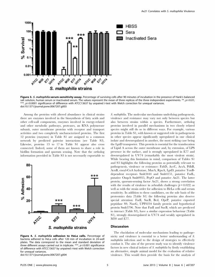
Among the proteins with altered abundance in clinical strainsthere are enzymes involved in the biosynthesis of fatty acids andother cell-wall components, enzymes involved in energy-relatedand other metabolic pathways, proteases, an RNA polymerasesubunit, outer membrane proteins with receptor and transportactivities and two completely uncharacterized proteins. The first12 proteins (enzymes) in Table S1 are assigned to a commonnetwork by predicted pairwise interactions (see Table S3).Likewise, proteins 13 to 17 in Table S1 appear also crossconnected. Indeed, some of them are known to share a role inbiofilm formation and quorum sensing. Note that the orthologinformation provided in Table S3 is not necessarily exportable to
S. maltophilia. The molecular mechanisms underlying pathogenesis,virulence and resistance may vary not only between species butalso between strains within a species. Furthermore, orthologproteins involved in parallel mechanisms in two closely relatedspecies might still do so in different ways. For example, variousproteins in Table S1, with known or suggested role in pathogenesisin other species appear significantly upregulated in one clinicalisolate and downregulated in another, the most striking case beingthe LptD transporter. This protein is essential for the translocationof Lipid A across the outer membrane and, by extension, of LPSpresence in the surface, and is strongly upregulated in E77 anddownregulated in UV74 (remarkably the most virulent strain).While bearing this limitation in mind, comparison of Tables S1and S3 highlights the following proteins as potentially relevant topathogenesis, virulence or resistance: FabD, AccC, AcsA, PdhB,AcnB, enoyl-CoA hydratase, MurA, RpoA, LptD, putative TonB-dependent receptors Smlt3444 and Smlt4151, putative FadL,putative OmpA Smlt0955, PepO and putative Ax21. The latterprotein, quorum-sensing factor Ax21, shows a strong correlationwith the results of virulence in zebrafish challenges (p = 0.022) aswell as with the strain order for adhesion to HeLa cells and serumsensitivity. In addition to these candidates, on the sole basis of theproteomics data (Table S1) the following proteins also deservespecial attention: FadI, SucB, Bcd, OprP, putative exportedpeptidase S9, NuoG, UPF0234 family protein and hypotheticalprotein Smlt3796. Note that FadI and SucB, which are predictedto interact (Table S3), have a similar expression behaviour (TableS1), strongly downregulated in UV74 and weakly upregulated inM30 and E77.
Discussion
The elucidation of molecular mechanisms leading to pathoge-nicity and virulence is essential to a better understanding of S.maltophilia infection and to the design of strategies to effectivelycombat it. The aim of the present study was to identify virulencefactors in new clinical isolates of S. maltophilia by firstly establishingan alternative, simple animal model for the evaluation of relativevirulence. This would then provide the basis for the analysis of
Figure 3. S. maltophilia serum-sensitivity assay. Percentage of surviving cells after 90 minutes of incubation in the presence of Hank’s balancedsalt solution, human serum or inactivated serum. The values represent the mean of three replicas of the three independent experiments. **, p#0.01;***, p#0.0001 significance of difference with ATCC13637 by unpaired t-test with Welch correction for unequal variances.doi:10.1371/journal.pone.0067207.g003
Figure 4. S. maltophilia adhesion to HeLa cells. Percentage ofbacteria adhered to HeLa cells after 120 min of incubation in 24-wellplates. The data correspond to the mean and standard deviation ofthree different assays carried out in triplicate. ***, p#0.001 significanceof difference with ATCC13637 by unpaired t-test with Welch correctionfor unequal variances.doi:10.1371/journal.pone.0067207.g004
Ax21 Correlates with S. maltophilia Virulence
PLOS ONE | www.plosone.org 5 June 2013 | Volume 8 | Issue 6 | e67207

underlying differences in protein expression, in a quest to identifyvirulence factors. To our knowledge this is the first time thezebrafish has been used as a model of S. maltophilia infection.
The present study shows that the adult-zebrafish model has ahigh discrimination capacity, i.e. from very low to very highmortality rates for different strains of S. maltophilia, and correlateswith serum sensitivity. This is also known to correlate withvirulence in mouse and rat models [17,31]. Taken together, theseproperties point toward the zebrafish as a promising animal modelfor the study of S. maltophilia pathogenicity and virulence inhumans.
Furthermore, the symptoms observed in the adult-zebrafishmodel after infection are very similar to those reported in a recentstudy on aquacultured adult channel catfish [23]. This includesascites and enteritis, prolapse in the rectum and intussusception inthe lower intestine, cutaneous hemorrhage in some cases. Affectedfish were also listless, sought the water surface, became morelethargic as the disease progressed, and most died within a fewdays. These common symptoms suggest that the process followingintraperitoneal infection is parallel to natural infection.
The collection strain ATCC13637 showed obvious signs ofattenuation, with null zebrafish mortality in our experiments. Thisstrain was isolated in 1960 from the oropharyngeal region of apatient with mouth cancer [32]. Since then, it has likely undergonea large number of in vitro serial passages. It is well known that therepeated in vitro sub-culturing of microorganisms during extendedperiods of time results in adaptation to the laboratory environmentleading to changes in physiology, including pathogenic capacityand production of virulence factors. As a matter of fact, thisproperty has been used in vaccine development to obtain live-attenuated strains [33]. This result points once more at theshortcomings of using standard laboratory strains in studies relatedto pathogenesis and virulence, even when their origin is clinical.
The three new S. maltophilia clinical isolates, M30, UV74 andE77, were chosen on the basis of their different clinical origin(decubitus ulcer, vascular ulcer and sputum, respectively). Despitebeing collected from patients of the same hospital and during thesame year, the MLST analysis reported here demonstrates thatthey are not clonally related. Interestingly, the most virulent S.maltophilia strains (UV74 and M30) are the genetically most similarones by means of MLST (Figure 2). Thus, M30 and UV74 clusterwithin the previously described genomic group C [28], whichincludes worldwide clinical strains isolated from tracheal secretionand blood cultures. Genetic and mutation-frequency analysisbecomes particularly relevant when dealing with S. maltophiliaclinical isolates, as the proportion of isolates showing highmutation frequencies (hypermutators) appears to be significantlyhigher for S. maltophilia than for other organisms [34]. However,whereas for different bacterial species the strains isolated from CFpatients with chronic lung infections often show high mutationfrequencies [35,36], hypermutators have rarely been found amongisolates from other types of infections [35,37]. None of the strainstested in the present study were obtained from CF patients, forwhich reason mutation rates were not determined.
Phenotypic analysis of the four strains revealed that UV74presents also the most resistant pattern to the set of antibacterialstested. In addition, the correlation found between zebrafishmortality, serum sensitivity and HeLa-cell adhesion points at thepotential use of animal-free models for virulence-screeningpurposes. The correlation is particularly significant between serumsensitivity and HeLa-cell adhesion (p = 0.008), but also notablebetween zebrafish mortality and serum sensitivity (p = 0.059). Asmentioned, a direct relationship between S. maltophilia virulenceand serum sensitivity has been already described in a mouse model
of respiratory tract infection [17] and a rat model of lung infection[31].
The demonstration of the attenuated character of ATCC13637in the zebrafish infection model motivated the comparison of theproteomes of the three clinical isolates to the collection referencewith the purpose of identifying potential virulence factors withinthe set of proteins showing a higher abundance in the clinicalstrains. Clearly, S. maltophilia will not necessarily show the sameprotein expression profiles under in vitro exponential-growth andin vivo infection conditions. It is very likely that a number ofproteins relevant to pathogenicity and virulence will only beupregulated (or suppressed) in the latter case and will be thereforemissed in this study. Nevertheless, there remains a certainpossibility that proteins differentially expressed in the clinicaland collection strains under in vitro exponential-growth conditionsmay also be related to pathogenicity and virulence, e.g. virulencefactors that acquire a more constitutive character as result ofadaptation to the clinical/infective environment.
Of the 38 proteins identified by differential proteomics, 24 arealready documented for their potential implication in pathogen-esis, virulence or resistance mechanisms in other bacteria (TableS3), validating the strategy presented here. Further proof ofconcept is provided by the identification of the putative quorum-sensing factor Ax21 (Smlt0387), whose abundance is shown tocorrelate with mortality in the zebrafish infection model (p = 0.02).It should be noted that this protein has been found in more thanone spot in the gels, suggesting a different composition of isoformsbetween the collection and clinical isolates, being the more basicisoform the predominant in the latter. McCarthy et al. [38] haverecently published a study in which deletion of smlt0387 affects thetranscription of genes encoding proteins involved in transcriptionalregulation, antibiotic resistance and pilus assembly, and influencesvarious phenotypes leading to reduced motility, reduced toleranceto some antibiotics, reduced biofilm formation and reducedvirulence in the larval Galleria mellonella infection model. Inaddition, it has been shown that the homologous protein inXanthomonas oryzae (60% identity), a microorganism closely relatedto S. maltophilia, triggers innate immunity in both plants andanimals [39].
Ax21 has two conserved domains. One of them (pfam13505) isfound in a wide range of outer-membrane-bound beta-barrelproteins. The other one (TIGR04273) is present in the sulfation-dependent quorum-sensing factor Ax21 protein family. Thisfamily consists of proteins closely related to Ax21 (Activator ofXA21-mediated immunity), a 194-residue protein present in plantand animal pathogens [40] that is secreted by a type I secretionsystem (RaxABC) and appears to be sulfated at a tyrosine found ina motif LSYN in the N-terminal region. In X. oryzae the smallprotein Ax21 serves as a quorum-sensing factor, inducing density-dependent gene expression and thus regulating biofilm formation,bacterial motility and virulence. In addition, Ax21 is critical for X.oryzae virulence at low densities in rice leaves [40]. Contrary to theXanthomonas case, Smlt0387 would apparently perform its intra-species signalling activity in a non-sulfation-dependent manner[38]. Our results further suggest that in S. maltophilia the relativeabundance of this protein is directly proportional to virulence.Further studies (in progress) will be needed to address the potentialrole of the other proteins identified in the pathogenesis, virulenceor resistance of S. maltophilia.
Ax21 Correlates with S. maltophilia Virulence
PLOS ONE | www.plosone.org 6 June 2013 | Volume 8 | Issue 6 | e67207
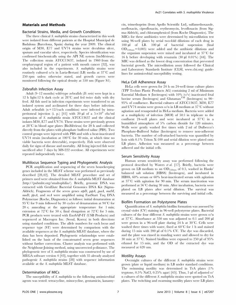
Materials and Methods
Bacterial Strains, Media, and Growth ConditionsThe three clinical S. maltophilia strains characterized in this work
were isolated from different patients at the Hospital Municipal deBadalona (Barcelona, Spain) during the year 2009. The clinicalorigin of M30, E77 and UV74 strains were decubitus ulcer,sputum and vascular ulcer, respectively. Species identification wasconfirmed biochemically using the API NE system (bioMerieux).The collection strain ATCC13637, isolated in 1960 from theoropharyngeal region of a patient with mouth cancer [32], wasalso included in the experiments. S. maltophilia strains wereroutinely cultured o/n in Luria-Bertani (LB) media at 37uC and250 rpm unless otherwise stated, and growth curves weremonitored following the optical density at 550 nm (Figure S1).
Zebrafish Infection AssayAdult (9–12 months) wild-type zebrafish (D. rerio) were kept in a
12 h light:12 h dark cycle at 28uC and fed twice daily with dryfeed. All fish used in infection experiments were transferred to anisolated system and acclimated for three days before infection.Adult zebrafish (n = 12/each condition) were infected by intra-peritoneal injection (i. p.) [41] with 20 ml of a 56109 cfu/mlsuspension of S. maltophilia strain ATCC13637 and the clinicalisolates M30, E77 and UV74. These strains were previously grownat 28uC in blood agar plates (BioMerieux) for 20 h and collecteddirectly from the plates with phosphate buffered saline (PBS). Twocontrol groups were injected with PBS and with a heat-inactivatedUV74 strain (incubation at 100uC for 30 min, at which time noviable bacteria were detectable), respectively. Fish were observeddaily for signs of disease and mortality. All living injected fish weresacrificed after 7 days by MS-222 overdose. All experiments wererepeated independently twice.
Multilocus Sequence Typing and Phylogenetic AnalysisPCR amplification and sequencing of the seven housekeeping
genes included in the MLST scheme was performed as previouslydescribed [28,42]. The detailed MLST procedure and set ofprimers used were obtained from the S. maltophilia MLST database(http://pubmlst.org/smaltophilia/). Briefly, genomic DNA wasextracted with GenElute Bacterial Genomics DNA Kit (Sigma-Aldrich). Fragments of the seven genes atpD, gapA, guaA, mutM,nuoD, ppsA, and recA were amplified using FastStart Taq DNAPolymerase (Roche, Diagnostics) as follows: initial denaturation at95uC for 9 min followed by 30 cycles of denaturation at 94uC for20 s; annealing at the appropriate temperature for 1 min;extension at 72uC for 50 s; final elongation at 72uC for 5 min.PCR products were treated with ExoSAP-IT (USB Products) andsequenced at Macrogen Inc. (Seoul, Korea) in both directionsusing standard conditions. The allele numbers for each locus andsequence type (ST) were determined by comparison with theavailable sequences at the S. maltophilia MLST database, where thedata has been deposited. Phylogenetic relationships were estab-lished on the basis of the concatenated seven gene sequenceswithout further corrections. Cluster analysis was performed withthe Neighbour-Joining method, using uncorrected p-distance. Thephylogenetic tree of S. maltophilia strains was constructed by use ofMEGA software version 4 [43], together with 35 already analyzedpathogenic S. maltophilia strains [28] with sequence informationavailable at the S. maltophilia MLST database.
Determination of MICsThe susceptibility of S. maltophilia to the following antimicrobial
agents was tested: tetracycline, minocycline, gentamicin, kanamy-
cin, trimethoprim (from Apollo Scientific Ltd), sulfamethoxazole,norfloxacin, ciprofloxacin, erythromycin, levofloxacin (from Sig-ma-Aldrich), and chloramphenicol (from Roche Diagnostics). TheMICs for these antibiotics were determined by microdilution testusing 96-well plates by serial two-fold dilutions of each drug in100 ml of LB. 100 ml of bacterial suspension (finalOD550nm = 0,005) were added and the antibiotic dilutions andthe organism suspension were mixed and incubated at 37uC for16 h before developing with resazurin (30 ml 0.01%) [44]. TheMIC was defined as the lowest drug concentration that preventedbacterial growth. The microdilution assay followed the Clinicaland Laboratory Standards Institute (CLSI, www.clsi.org) guide-lines for antimicrobial susceptibility testing.
HeLa Cell Adherence AssayHeLa cells were grown for 24 h on 24-well tissue culture plates
(TPP Techno Plastic Products AG) containing 2 ml of MinimumEssential Medium a (Invitrogen) with 10% (v/v) inactivated fetalbovine serum (Invitrogen) and GlutamaxTM (Invitrogen) to 90–95% of confluence. Bacterial cultures of ATCC13637, M30, E77and UV74 strains were grown o/n in LB medium at 37uC withoutagitation and resuspended in HeLa medium. Bacteria were addedat a multiplicity of infection (MOI) of 50:1 in triplicate to theconfluent 24-well plates and were incubated at 37uC in ahumidified atmosphere of 5% carbon dioxide for 120 minutes.Wells were gently washed five times with 2 ml of Dulbecco’sPhosphate-Buffered Saline (Invitrogen) to remove non-adherentbacteria. The number of cell-attached bacteria was quantified bylysis with 0.1% Triton X-100 and serial dilution were plated ontoLB plates. Adhesion was measured as a percentage betweenadhered and the initial cells.
Serum Sensitivity AssayHuman serum sensitivity assay was performed following the
protocol described by Waters et al. [17]. Briefly, bacteria weregrown on LB medium to an OD550nm of 0.5, washed in Hanks’balanced salt solution (HBSS) (Invitrogen), and incubated inHBSS, 60% serum or 60% heat-inactivated serum with agitationat 37uC with agitation for 90 min. The serum inactivation wasperformed at 56uC during 30 min. After incubation, bacteria wereplated on LB plates after serial dilution. The survival wasmeasured as a percentage between the surviving and initial cells.
Biofilm Formation on Polystyrene PlatesQuantification of S. maltophilia biofilm formation was assessed by
crystal violet (CV) staining in 96-well polystyrene plates. Bacterialcultures of the four different S. maltophilia strains were grown o/nat 37uC. Absorbance at 550 nm was adjusted to 0.1 and 200 mlwere grown in a 96-well plate during 24 h at 30uC. Cells werewashed three times with water, fixed at 60uC for 1 h and stainedduring 15 min with 200 ml of 0.1% CV. The dye was discarded,and the plate was rinsed in standing water and allowed to dry for30 min at 37uC. Stained biofilms were exposed to 250 ml of 95%ethanol for 15 min, and the OD of the extracted dye wasmeasured at 620 nm.
Motility AssaysOvernight cultures of the different S. maltophilia strains were
grown (plate or liquid medium) in LB under standard conditions.The swimming motility was determined in TrA plates (1%tryptone, 0.5% NaCl, 0.25% agar) [45]. Thus, 5 ml of adjusted o/n cultures of the different S. maltophilia strains were spotted on TrAplates. The twitching and swarming motility plates were LB plates
Ax21 Correlates with S. maltophilia Virulence
PLOS ONE | www.plosone.org 7 June 2013 | Volume 8 | Issue 6 | e67207
at 1% agar and BM2 at 0.5% agar [46], respectively. Twitchingwas assessed via subagar stab inoculations (stab assay) from o/nfresh plates as previously described [47]. The twitching zones werethen visualized by staining with 1% (wt/vol) crystal violet and theirdiameters measured. Noble agar (DifcoTM) was used in thepreparation of the three motility assay plates. The growth haloswere measured in cm after 24 h of incubation at 30uC for theswimming and twitching motilities and after 7 days at 30uC forswarming.
StatisticsStatistical analyses were performed using the GraphPad Prism
program version 5.00. Comparison of strain phenotypic data wasperformed by one-way analysis of variance (ANOVA) with aBonferroni’s multiple comparison post-test or unpaired t-test withWelch correction for unequal variances, as indicated in figure andtable captions. Survival curves of zebrafish infection experimentswere analyzed using the Kaplan-Meier method. Differences wereevaluated using the log-rank test. The relationship betweenrelative protein abundance in the different strains and pathoge-nicity variables, such as zebrafish mortality rates and adhesion tohuman cells, was evaluated with Pearson’s chi-squared test.p#0.05 was considered significant.
Sample Preparation for Two-dimensional GelElectrophoresis
20 ml of exponential culture (OD = 1) were washed with PBS1X three times and resuspended in lysis solution (8 M urea, 2 Mthiourea, 2.5% 3-[(3-cholamidopropyl) dimethylammonio]-1 pro-panesulfonate (CHAPS), 2% ASB-14, 40 mM Tris-HCl, pH 8.8).ASB-14 was used to increase the presence of membrane proteinsin the 2DE [48]. Then, samples were disrupted by sonication andcentrifuged in order to discard any insoluble cellular debris. Inorder to remove salts and other contaminants, samples werecleaned with 2D Clean-Up Kit (GE Healthcare). Resulting pelletswere resuspended in the above-mentioned lysis solution. Proteinconcentration was determined with 2D-Quant Kit (GE Health-care) and adjusted to 2 mg/ml by the addition of a DIGE labelingbuffer (7 M urea, 2 M thiourea, 2.5% w/v CHAPS, 40 mM Tris,pH 8.8). A pool consisting of equal amounts of each of the twosamples analyzed in the experiment was prepared as an internalstandard for quantitative comparisons [49]. The clinical isolateswere labeled with Cy3 and the collection strain ATCC13637 waslabeled with Cy5. A third fluorescent dye, Cy2, was used to labelthe internal standard sample. Labeling was carried out by theaddition of 400 pmol of the required Cydye in 1 ml of anhydrousN,N-dimethylformamide per 50 mg of protein. After 30 min ofincubation on ice in the dark, the reaction was quenched with10 mM lysine and the samples incubated for a further 10 min.Samples were combined according to the experimental design,using 50 mg of protein per Cy dye per gel, and diluted two-foldwith isoelectric focusing (IEF) sample buffer (7 M urea, 2 Mthiourea, 4% w/v CHAPS, 2% dithiothreitol [DTT], 2%pharmalytes, pH 3–10). One clinical strain and the ATCC13637strain sample, together with an aliquot of the internal standardpool, were then separated by two-dimensional electrophoresis (2-DE) in each of the gels. This experimental design allows theaccurate quantification and statistical assessment of the differencesin protein abundances observed between the two sample groups.
2D Difference Gel ElectrophoresisThe 2-DE was performed using GE Healthcare reagents and
equipment. First-dimension isoelectric focusing was performed on
immobilized pH gradient strips (24 cm, pH 3–10) using an EttanIPGphor System (GE Healthcare). Samples were applied near thebasic end of the strips by cup-loading, after being incubated o/n in450 ml of rehydration buffer (7 M urea, 2 M thiourea, 2.5% w/vCHAPS, 2% ASB-14 w/v, 0.5% pharmalytes, pH 3–10, 100 mMDeStreak reagent). After focusing at 70 kVh, strips were equil-ibrated, first for 15 min in 10 ml of reducing solution (6 M urea,100 mM Tris-HCl, pH 8, 30% v/v glycerol, 2% w/v SDS, 5 mg/ml dithiothreitol [DTT]) and then in 10 ml of alkylating solution(6 M urea, 100 mM Tris-HCl, pH 8, 30% v/v glycerol, 2% w/vSDS, 22.5 mg/ml iodoacetamide) for 15 min on a rockingplatform. Second dimension SDS-PAGE was performed by layingthe strips on 12.5% isocratic Laemmli gels (24620 cm), cast in lowfluorescence glass plates, on an Ettan DALT Six system. Gels wererun at 20uC at a constant power of 2.5 W per gel for 60 minfollowed by 17 W per gel until the bromophenol blue trackingfront had run off the end of the gel. Triplicate gels were run foreach sample using independent biological replicates. Fluorescenceimages of the gels were obtained on a Typhoon 9400 scanner (GEHealthcare). Cy2, Cy3 and Cy5 images were scanned atexcitation/emission wavelengths of 488/520 nm, 532/580 nmand 633/670 nm, respectively, at a resolution of 100 mm. Bothimage analysis and statistical quantification of relative proteinlevels were performed using Progenesis SameSpots V.4 (NonlinearDynamics) (See Table S1 for detailed statistics of each spot). Thedata were analyzed as pairwise comparisons.
Protein Identification by Mass SpectrometryIn order to excise the spots of interest, gels were silver stained as
described elsewhere [50]. Protein spots of interest were excisedfrom the gel using a cut tip. The selected spots are those that aredifferentially expressed in at least two clinical isolates. In-geltrypsin digestion was performed as described previously [51].MALDI-MS analysis of tryptic peptides was performed on anUltraflex time-of-flight instrument (Bruker Daltonics). Sampleswere prepared using a-cyano-4-hydroxy-cinnamic acid. Calibra-tion was performed in the external mode using a peptidecalibration standard kit (Bruker Daltonics). The spectra wereprocessed using Flex Analysis 2.2 software (Bruker Daltonics). Peaklists were generated using the signals in the 800–4000 mass:chargeratio (m/z) range, with a signal:noise threshold .3. The SNAPalgorithm included in the software was used to select themonoisotopic peaks from the isotopic distributions observed. Afterremoving m/z values corresponding to commonly observed matrixcluster ions, an internal statistical calibration was applied. Peakscorresponding to frequently seen keratin and trypsin autolysispeptides were then removed. The resulting final peak list was usedfor the identification of the proteins by peptide mass fingerprint.The Mascot 2.0 program (Matrix Science) was used to search theNCBI non-redundant database (http://ncbi.nlm.nih.gov, March2010), with no limitation on taxonomy. Search parameters were asfollows: trypsin cleavages excluding N-terminal to P, one missedcleavage permission, carbamidomethylation and methionineoxidation as variable modification, mass tolerance ,50 ppm,monoisotopic mass values. Criteria for positive identification werea significant Mascot probability score (p,0.05), and at least fivematching peptide masses. A minimum score of 83, and a .50-point difference between this score and the score of the secondranked non-homologous match was obtained for all the identifieddifferentially expressed proteins.
Ax21 Correlates with S. maltophilia Virulence
PLOS ONE | www.plosone.org 8 June 2013 | Volume 8 | Issue 6 | e67207
Ethics StatementStenotrophomonas maltophilia strains were obtained from the
internal collection of Hospital Municipal de Badalona (Barcelona,Spain) and have no link with patient data.
Zebrafish were handled in compliance with Directive 2010/63/EU of the European Parliament and of the Council on theprotection of animals used for scientific purposes and with Decree214/1997 of the Government of Catalonia, which regulates theuse of animals for experimental and other scientific purposes.Experimental protocols have been reviewed and approved by theAnimal and Human Experimentation Ethics Committee(CEEAH) of the Universitat Autonoma de Barcelona (UAB),Spain (ref # CEEAH-1968).
Supporting Information
Figure S1 Growth curves of the four S. maltophilia strains inLuria-Bertani (LB) media at 37uC and 250 rpm.(TIF)
Figure S2 Representative image of the gels obtained for M30 vs.ATCC13637. Superimposed images in pseudo-color from Cy3(green, clinical isolate) and Cy5 (red, ATCC13637 strain) labeledsamples run on a two-dimensional DIGE gel. The horizontaldimension corresponds to isoelectric point (pI) and ranges from 3(left) to 10 (right). The vertical dimension corresponds to mass andranges from <15 kDa (bottom) to <200 kDa (top).(TIF)
Figure S3 DIGE image analysis from the M30/ATCC13637comparison. (a) Superimposed images in pseudo-color from Cy3(green, clinical isolate) and Cy5 (red, ATCC13637 strain) labeledsamples run on the two-dimensional gel. The horizontaldimension corresponds to isoelectric point (pI) and ranges from 3(left) to 10 (right). The vertical dimension corresponds to mass andranges from <15 kDa (bottom) to <200 kDa (top). The six spotswith largest difference in protein abundance between the twosamples are marked. Three-dimensional images representing the
intensity of these spots, corresponding to the Cy3 image (M30strain, left-hand panel for each protein) and the Cy5 image(collection strain, right-hand panel for each protein), are shown. (b)Standardized abundance plots for the six proteins. Each graphdisplays the abundance observed for the spot in each of the threegel images corresponding to the clinical isolate sample (pink) andthe ATCC13637 strain sample (blue), after standardizing thevalues using the internal standard pool images (Cy2) of each of thethree gels. The line links the average abundance values for eachgroup of samples. ANOVA’s test for the difference in abundancebetween each two groups results in p values ,0.05 in all casesshown.(TIF)
Table S1 S. maltophilia proteins presenting significant abundancedifference in the clinical and ATCC13637 strains.(DOCX)
Table S2 Statistical report for each identified DIGE spot.(DOCX)
Table S3 Orthologs of the differentially abundant proteinsreported to be involved in pathogenesis, virulence or resistancemechanisms.(DOCX)
Acknowledgments
The authors thank A. Calderon and T. Falgueras, from Badalona ServeisAssistencials (Hospital Municipal de Badalona), for providing clinicalisolates and F. Canals, from Hospital Vall d’Hebron, for assistance inimage acquisition.
Author Contributions
Conceived and designed the experiments: MFN RP DY NR SM IG XD.Performed the experiments: MFN RP DY EM GT PH PM NR. Analyzedthe data: MFN RP DY NR SM IG XD. Wrote the paper: MFN RP DYNR SM IG XD.
References
1. Looney WJ, Narita M, Muhlemann K (2009) Stenotrophomonas maltophilia: anemerging opportunist human pathogen. Lancet Infect Dis 9: 312–323.
2. Brooke JS (2012) Stenotrophomonas maltophilia: an emerging global opportunisticpathogen. Clin Microbiol Rev 25: 2–41.
3. Cervia J, Ortolano G, Canonica F (2008) Hospital tap water as a source ofStenotrophomonas maltophilia infection. Clin Infect Dis 46: 1485–1487.
4. Marshall WF, Keating MR, Anhalt JP, Steckelberg JM (1989) Xanthomonasmaltophilia: an emerging nosocomial pathogen. Mayo Clin Proc 64: 1097–1104.
5. Denton M, Kerr KG (1998) Microbiological and clinical aspects of infectionassociated with Stenotrophomonas maltophilia. Clin Microbiol Rev 11: 57–80.
6. Jang TN, Wang FD, Wang LS, Liu CY, Liu IM (1992) Xanthomonas maltophiliabacteremia: an analysis of 32 cases. J Formos Med Assoc 91: 1170–1176.
7. Khardori N, Elting L, Wong E, Schable B, Bodey GP (1990) Nosocomialinfections due to Xanthomonas maltophilia (Pseudomonas maltophilia) in patients withcancer. Rev Infect Dis 12: 997–1003.
8. Vartivarian SE, Papadakis KA, Palacios JA, Manning JT, Anaissie EJ (1994)Mucocutaneous and soft tissue infections caused by Xanthomonas maltophilia: a newspectrum. Ann Intern Med 121: 969–973.
9. Toleman MA, Bennett PM, Bennett DM, Jones RN, Walsh TR (2007) Globalemergence of trimethoprim/sulfamethoxazole resistance in Stenotrophomonasmaltophilia mediated by acquisition of sul genes. Emerg Infect Dis 13: 559–565.
10. Zhang L, Li XZ, Poole K (2000) Multiple antibiotic resistance in Stenotrophomonasmaltophilia: involvement of a multidrug efflux system. Antimicrob AgentsChemother 44: 287–293.
11. Crossman LC, Gould VC, Dow JM, Vernikos GS, Okazaki A, et al. (2008) Thecomplete genome, comparative and functional analysis of Stenotrophomonasmaltophilia reveals an organism heavily shielded by drug resistance determinants.Genome Biol 9: R74.
12. Liaw SJ, Lee YL, Hsueh PR (2010) Multidrug resistance in clinical isolates ofStenotrophomonas maltophilia: roles of integrons, efflux pumps, phosphoglucomutase(SpgM), and melanin and biofilm formation. Int J Antimicrob Agents 35: 126–130.
13. Lira F, Hernandez A, Belda E, Sanchez MB, Moya A, et al. (2012) Whole-genome sequence of Stenotrophomonas maltophilia D457, a clinical isolate and amodel strain. J Bacteriol 194: 3563–3564.
14. Rocco F, De Gregorio E, Colonna B, Di Nocera PP (2009) Stenotrophomonasmaltophilia genomes: a start-up comparison. Int J Med Microbiol 299: 535–546.
15. de Oliveira-Garcia D, Dall’Agnol M, Rosales M, Azzuz AC, Alcantara N, et al.(2003) Fimbriae and adherence of Stenotrophomonas maltophilia to epithelial cellsand to abiotic surfaces. Cell Microbiol 5: 625–636.
16. Valdezate S, Vindel A, Martın-Davila P, Del Saz BS, Baquero F, et al. (2004)High genetic diversity among Stenotrophomonas maltophilia strains despite theiroriginating at a single hospital. J Clin Microbiol 42: 693–699.
17. Waters VJ, Gomez MI, Soong G, Amin S, Ernst RK, et al. (2007)Immunostimulatory properties of the emerging pathogen Stenotrophomonasmaltophilia. Infect Immun 75: 1698–1703.
18. Di Bonaventura G, Pompilio A, Zappacosta R, Petrucci F, Fiscarelli E, et al.(2010) Role of excessive inflammatory response to Stenotrophomonas maltophilia lunginfection in DBA/2 mice and implications for cystic fibrosis. Infect Immun 78:2466–2476.
19. Pompilio A, Pomponio S, Crocetta V, Gherardi G, Verginelli F, et al. (2011)Phenotypic and genotypic characterization of Stenotrophomonas maltophilia isolatesfrom patients with cystic fibrosis: genome diversity, biofilm formation, andvirulence. BMC Microbiol 11: 159.
20. Meijer AH, Spaink HP (2011) Host-pathogen interactions made transparentwith the zebrafish model. Curr Drug Targets 12: 1000–1017.
21. Patterson H, Saralahti A, Parikka M, Dramsi S, Trieu-Cuot P, et al. (2012) Adultzebrafish model of bacterial meningitis in Streptococcus agalactiae infection. DevComp Immunol 38: 447–455.
22. Vojtech LN, Sanders GE, Conway C, Ostland V, Hansen JD (2009) Hostimmune response and acute disease in a zebrafish model of Francisellapathogenesis. Infect Immun 77: 914–925.
Ax21 Correlates with S. maltophilia Virulence
PLOS ONE | www.plosone.org 9 June 2013 | Volume 8 | Issue 6 | e67207

23. Geng Y, Wang K, Chen D, Huang X, He M, et al. (2010) Stenotrophomonasmaltophilia, an emerging opportunist pathogen for cultured channel catfish,Ictalurus punctatus, in China. Aquaculture 308: 132–135.
24. Postlethwait JH, Yan YL, Gates MA, Horne S, Amores A, et al. (1998)Vertebrate genome evolution and the zebrafish gene map. Nat Genet 18: 345–349.
25. Trede NS, Zapata A, Zon LI (2001) Fishing for lymphoid genes. TrendsImmunol 22: 302–307.
26. Gorg A, Walter W, Michael JD (2004) Current two-dimensional electrophoresistechnology for proteomics. Proteomics 4: 3665–3685.
27. De Carolis E, Posteraro B, Florio AR, Colonna B, Prosseda G, et al. (2011)Analysis of heat-induced changes in protein expression of Stenotrophomonasmaltophilia K279a reveals a role for GroEL in the host-temperature adaptation.Int J Med Microbiol 301: 273–281.
28. Kaiser S, Biehler K, Jonas D (2009) A Stenotrophomonas maltophilia multilocussequence typing scheme for inferring population structure. J Bacteriol 191:2934–2943.
29. Hauben L, Vauterin L, Moore ERB, Hoste B, Swings J (1999) Genomicdiversity of the genus Stenotrophomonas. Int J Syst Evol Microbiol 49: 1749–1760.
30. Kohler T, Curty LK, Barja F, van Delden C, Pechere JC (2000) Swarming ofPseudomonas aeruginosa is dependent on cell-to-cell signaling and requires flagellaand pili. J Bacteriol 182: 5990–5996.
31. McKay GA, Woods DE, MacDonald KL, Poole K (2003) Role ofphosphoglucomutase of Stenotrophomonas maltophilia in lipopolysaccharide biosyn-thesis, virulence, and antibiotic resistance. Infect Immun 71: 3068–3075.
32. Hugh R, Ryschenkow E (1961) Pseudomonas maltophilia, an alcaligenes-like species.J Gen Microbiol 26: 123–132.
33. Ellis RW, Brodeur, Bernard R. (2003) New bacterial vaccines: Eurekah.com andLuwer academic/Plenum Publishers.
34. Turrientes MC, Baquero MR, Sanchez MB, Valdezate S, Escudero E, et al.(2010) Polymorphic mutation frequencies of clinical and environmentalStenotrophomonas maltophilia populations. Appl Environ Microbiol 76: 1746–58.
35. Oliver A, Canton R, Campo P, Baquero F, Blazquez J (2000) High Frequency ofHypermutable Pseudomonas aeruginosa in Cystic Fibrosis Lung Infection. Science288: 1251–1253.
36. Prunier AL, Malbruny B, Laurans M, Brouard J, Duhamel JF, et al. (2003) Highrate of macrolide resistance in Staphylococcus aureus strains from patients withcystic fibrosis reveals high proportions of hypermutable strains. J Infect Dis 187:1709–1716.
37. Gutierrez O, Juan C, Perez JL, Oliver A (2004) Lack of association betweenhypermutation and antibiotic resistance development in Pseudomonas aeruginosa
isolates from intensive care unit patients. Antimicrob Agents Chemother 48:3573–3575.
38. McCarthy Y, Dow JM, Ryan RP (2011) The Ax21 protein is a cell-cell signalregulating virulence in the nosocomial pathogen Stenotrophomonas maltophilia.J Bacteriol 193: 6375–6378.
39. Lee S-W, Han S-W, Sririyanum M, Park C-J, Seo Y-S, et al. (2009) A type I–secreted, sulfated peptide triggers XA21-mediated innate immunity. Science326: 850–853.
40. Ronald PC (2011) Small protein-mediated quorum sensing in a gram-negativebacterium: novel targets for control of infectious disease. Discov Med 12: 461–470.
41. Kinkel MD, Eames SC, Philipson LH, Prince VE (2010) Intraperitonealinjection into adult zebrafish. J Vis Exp 42: 2126.
42. Vasileuskaya-Schulz Z, Kaiser S, Maier T, Kostrzewa M, Jonas D (2011)Delineation of Stenotrophomonas spp. by multi-locus sequence analysis andMALDI-TOF mass spectrometry. Syst Appl Microbiol 34: 35–39.
43. Tamura K, Dudley J, Nei M, Kumar S (2007) MEGA4: Molecular EvolutionaryGenetics Analysis (MEGA) software version 4.0. Mol Biol Evol 24: 1596–1599.
44. Nateche F, Martin A, Baraka S, Palomino JC, Khaled S, et al. (2006)Application of the resazurin microtitre assay for detection of multidrug resistancein Mycobacterium tuberculosis in Algiers. J Med Microbiol 55: 857–860.
45. Huang TP, Somers EB, Wong AC (2006) Differential biofilm formation andmotility associated with lipopolysaccharide/exopolysaccharide-coupled biosyn-thetic genes in Stenotrophomonas maltophilia. J Bacteriol 2006 188: 3116–3120.
46. Overhage J, Lewenza S, Marr AK, Hancock REW (2007) Identification of genesinvolved in swarming motility using a Pseudomonas aeruginosa PAO1 mini-Tn5-luxmutant library. J Bacteriol 189: 2164–2169.
47. Rashid MH, Kornberg A (2000) Inorganic polyphosphate is needed forswimming, swarming, and twitching motilities of Pseudomonas aeruginosa. Proc NatlAcad Sci USA 97: 4885–4890.
48. Mark PM, Ben RH, Martin BS, Thierry R, Amanda SN, et al. (2000) Proteomicanalysis of the Escherichia coli outer membrane. Eur J Biochem 267: 2871–2881.
49. Andrew A, Stephen Olu D, Lennart B, Christian A, Erik S, et al. (2003) A novelexperimental design for comparative two-dimensional gel analysis: two-dimensional difference gel electrophoresis incorporating a pooled internalstandard. Proteomics 3: 36–44.
50. Parraga-Nino N, Colome-Calls N, Canals F, Querol E, Ferrer-Navarro M(2012) A comprehensive proteome of Mycoplasma genitalium. J Proteome Res 11:3305–3316.
51. Shevchenko A, Wilm M, Vorm O, Mann M (1996) Mass spectrometricsequencing of proteins silver-stained polyacrylamide gels. Anal Chem 68: 850–858.
Ax21 Correlates with S. maltophilia Virulence
PLOS ONE | www.plosone.org 10 June 2013 | Volume 8 | Issue 6 | e67207